Embed Size (px)
Citation preview
Urinary Notes.notebook
1
April 01, 2019
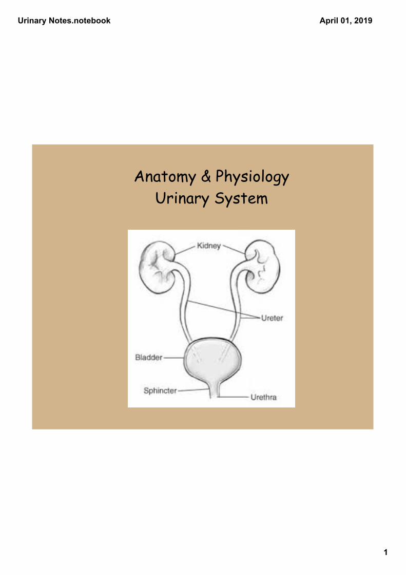
Anatomy & PhysiologyUrinary System
Urinary Notes.notebook
2
April 01, 2019
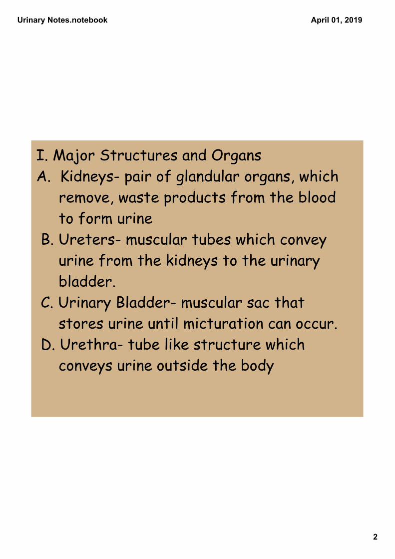
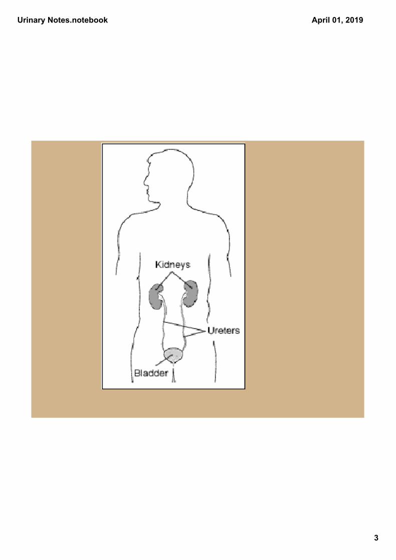
I. Major Structures and Organs A. Kidneys- pair of glandular organs, which remove, waste products from the blood to form urine B. Ureters- muscular tubes which convey urine from the kidneys to the urinary bladder. C. Urinary Bladder- muscular sac that stores urine until micturation can occur. D. Urethra- tube like structure which conveys urine outside the body
Urinary Notes.notebook
3
April 01, 2019
Urinary Notes.notebook
4
April 01, 2019
II. Kidneys A. Location
1.Reddish brown bean shaped organ located retroperitoneally. (Behind the parietal peritoneum of the abdominal cavity against the muscles of the back.)
2. Located between the 12th thoracic rib and the 3rd lumbar vertebra.
3.The left kidney is usually 1-2 cm higher than the right.
Urinary Notes.notebook
5
April 01, 2019
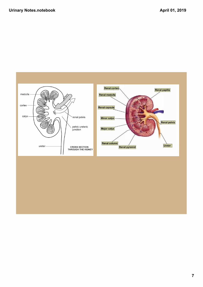
B. Structure of the Kidney 1. Lateral surface is convex in shape,
medial surface is concave 2. Medial depression is a hollow chamber
called the renal sinus a. The entrance to the sinus is called
the hilus, and through it pass the renal artery & vein, nerves, lymphatic vessels, and the ureter.
b. The superior end of the ureter is expanded to form a funnel shaped part called the renal pelvis.
c. The renal pelvis is divided into major calyces and are further divided into minor calyces (calyx)
d. There are several projections of kidney tissue into the renal sinus, which form the kidney wall. These projections are called the renal papilla and each are pierced by tiny openings into a minor calyx.
Urinary Notes.notebook
6
April 01, 2019
C. Kidney is divided into 2 primary regions 1. Medulla- the inner portion composed of
collecting ducts and loops of Henle and has a striated appearance.
a. composed of conical masses of tissue called renal pyramids.
2. Cortex- outer portion composed of nephrons, which give it a grainy appearance. Some of the cortex dips into the medulla to form renal columns.
Urinary Notes.notebook
7
April 01, 2019
Urinary Notes.notebook
8
April 01, 2019
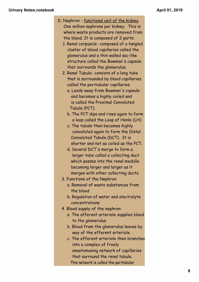
. D. Nephron - functional unit of the kidney. One million nephrons per kidney. This is where waste products are removed from the blood. It is composed of 2 parts:
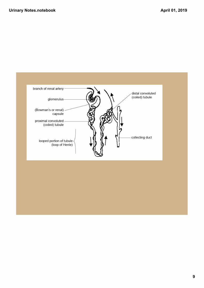
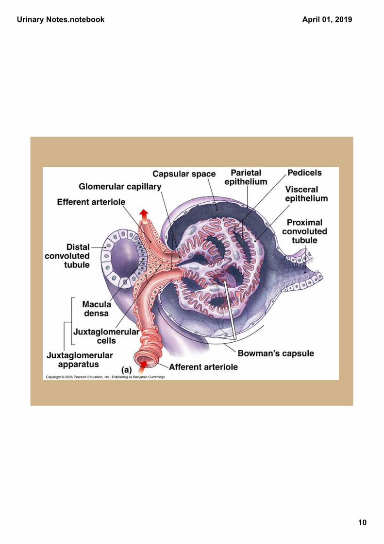
1. Renal corpuscle- composed of a tangled cluster of blood capillaries called the glomerulus and a thin walled sac-like structure called the Bowman's capsule that surrounds the glomerulus. 2. Renal Tubule- consists of a long tube that is surrounded by blood capillaries called the peritubular capillaries.
a. Leads away from Bowman's capsule and becomes a highly coiled and is called the Proximal Convoluted Tubule (PCT).
b. The PCT dips and rises again to form a loop called the Loop of Henle (LH)
c. The tubule then becomes highly convoluted again to form the Distal Convoluted Tubule (DCT). It is shorter and not as coiled as the PCT.
d. Several DCT's merge to form a larger tube called a collecting duct which passes into the renal medulla becoming larger and larger as it merges with other collecting ducts.
3. Functions of the Nephron a. Removal of waste substances from
the blood b. Regulation of water and electrolyte
concentrations4. Blood supply of the nephron
a. The afferent arteriole supplies blood to the glomerulus
b. Blood from the glomerulus leaves by way of the efferent arteriole.
c. The efferent arteriole then branches into a complex of freely anastomosing network of capillaries
that surround the renal tubule. This network is called the peritubular
capillary system. system.
Urinary Notes.notebook
9
April 01, 2019
Urinary Notes.notebook
10
April 01, 2019
Urinary Notes.notebook
11
April 01, 2019
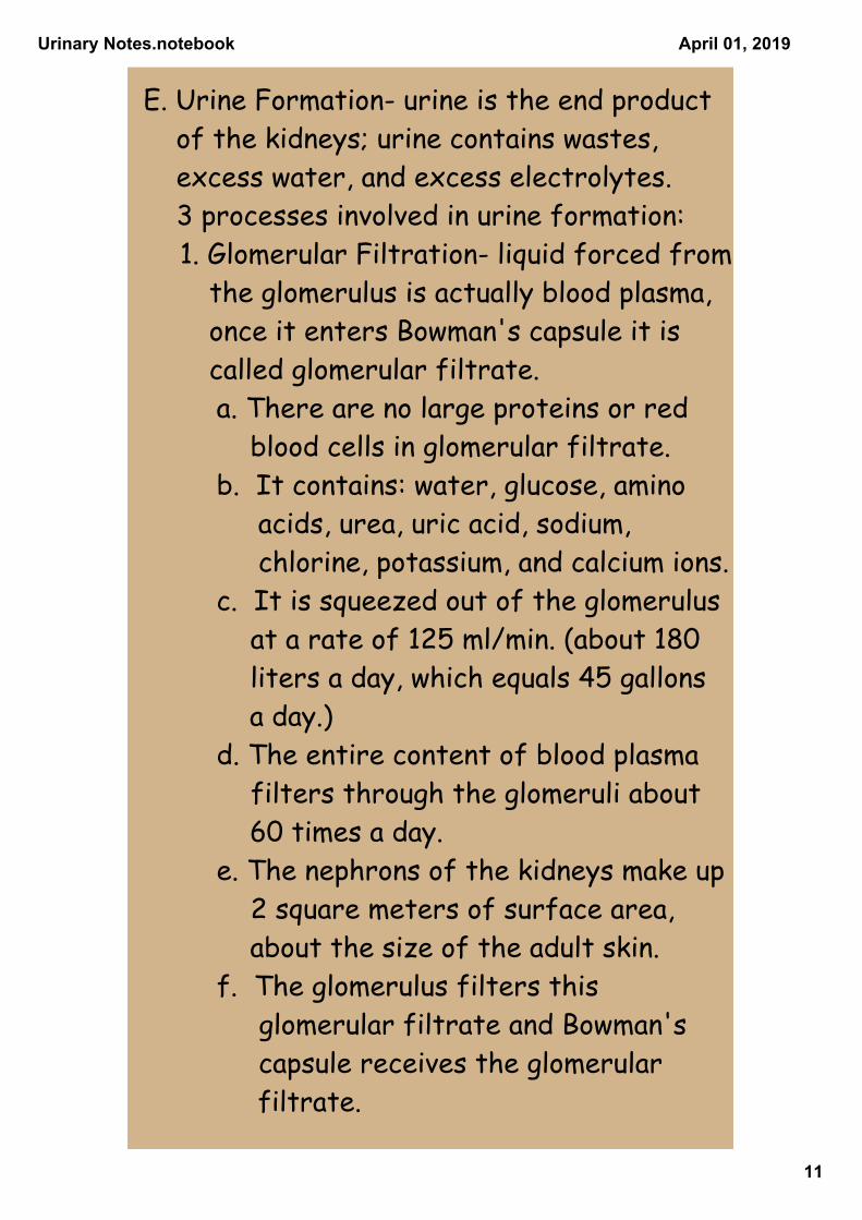
E. Urine Formation- urine is the end product of the kidneys; urine contains wastes, excess water, and excess electrolytes. 3 processes involved in urine formation:
1. Glomerular Filtration- liquid forced from the glomerulus is actually blood plasma, once it enters Bowman's capsule it is called glomerular filtrate.
a. There are no large proteins or red blood cells in glomerular filtrate.
b. It contains: water, glucose, amino acids, urea, uric acid, sodium, chlorine, potassium, and calcium ions.
c. It is squeezed out of the glomerulus at a rate of 125 ml/min. (about 180 liters a day, which equals 45 gallons a day.)
d. The entire content of blood plasma filters through the glomeruli about 60 times a day.
e. The nephrons of the kidneys make up 2 square meters of surface area, about the size of the adult skin.
f. The glomerulus filters this glomerular filtrate and Bowman's capsule receives the glomerular filtrate.
Urinary Notes.notebook
12
April 01, 2019
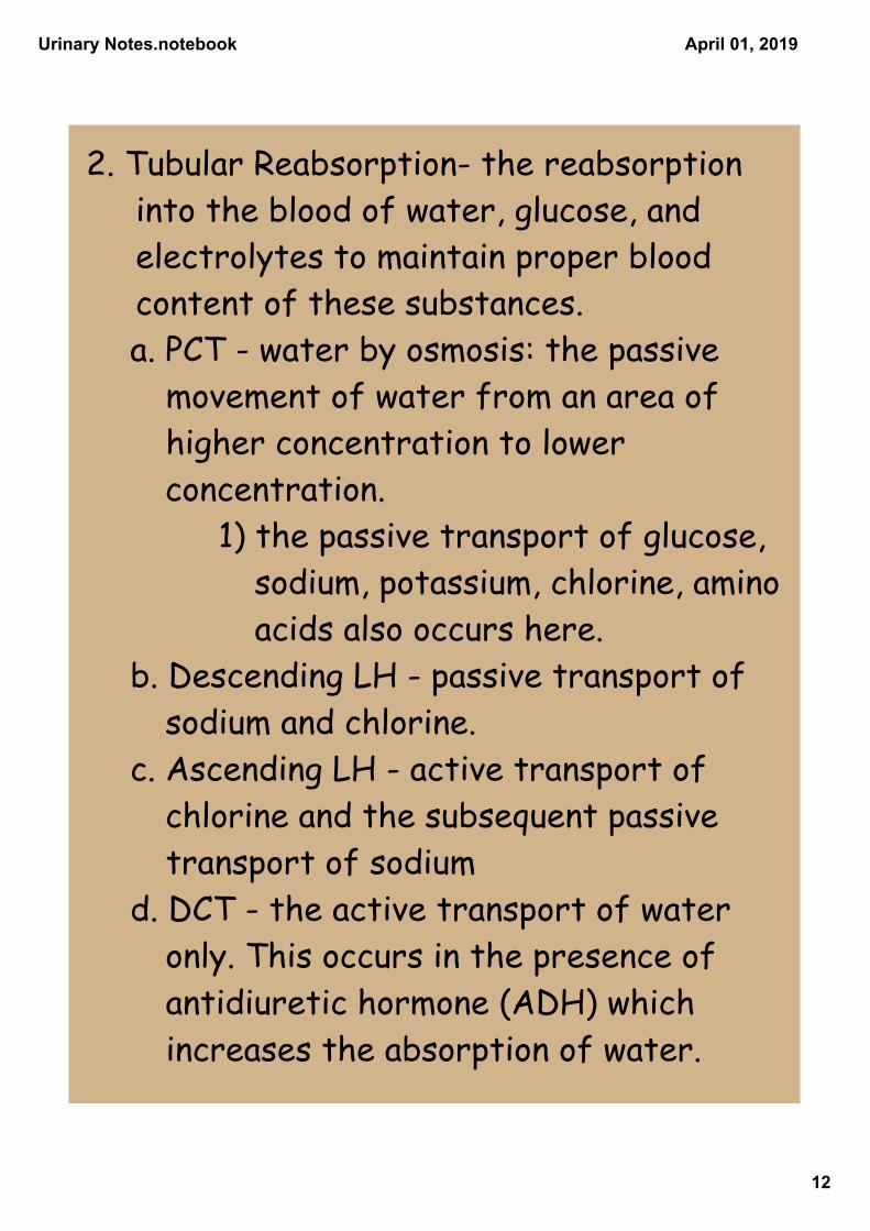
2. Tubular Reabsorption- the reabsorption into the blood of water, glucose, and electrolytes to maintain proper blood content of these substances.
a. PCT - water by osmosis: the passive movement of water from an area of higher concentration to lower concentration.
1) the passive transport of glucose, sodium, potassium, chlorine, amino acids also occurs here.
b. Descending LH - passive transport of sodium and chlorine.
c. Ascending LH - active transport of chlorine and the subsequent passive transport of sodium
d. DCT - the active transport of water only. This occurs in the presence of antidiuretic hormone (ADH) which increases the absorption of water.
Urinary Notes.notebook
13
April 01, 2019
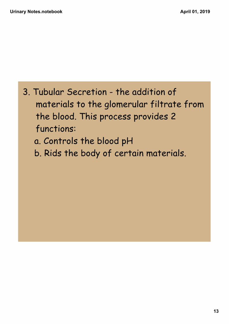
3. Tubular Secretion - the addition of materials to the glomerular filtrate from the blood. This process provides 2 functions:
a. Controls the blood pH b. Rids the body of certain materials.
Urinary Notes.notebook
14
April 01, 2019
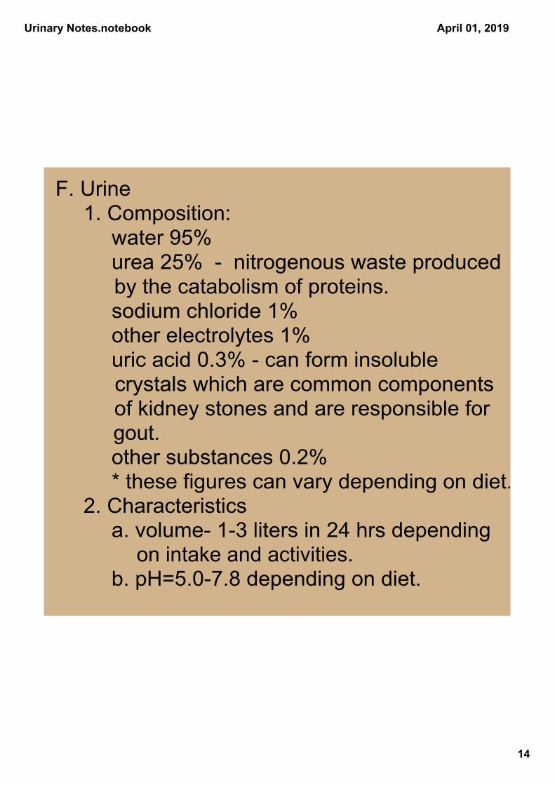
F. Urine 1. Composition:
water 95% urea 25% nitrogenous waste produced
by the catabolism of proteins. sodium chloride 1% other electrolytes 1% uric acid 0.3% can form insoluble
crystals which are common components of kidney stones and are responsible for gout.
other substances 0.2% * these figures can vary depending on diet.
2. Characteristics a. volume 13 liters in 24 hrs depending
on intake and activities. b. pH=5.07.8 depending on diet.
Urinary Notes.notebook
15
April 01, 2019
III. Elimination of Urine A. After being formed, urine passes
from the collecting ducts through openings into the renal papillae and enters the major calyces of the renal sinus.
1. From there, it passes through the renal pelvis and is conveyed by a ureter to the urinary bladder.
2. It is excreted outside the body by way of the urethra.
Urinary Notes.notebook
16
April 01, 2019
B. The Ureters 1. They are paired tubular organs approx. 10” in length. 2. The superior end is expanded and
funnel shaped and is called the renal pelvis
3. The inferior end joins the urinary bladder from underneath.
4. It conveys urine from the kidney to the urinary bladder.
5. The ureter is composed of 3 layers: a. Mucous coat- inner layer, composed
of cuboidal epithelium continuous with the linings of the renal tubules and urinary bladder
b. Muscular coat- middle layer, smooth muscle in circular and longitudinal planes. Its contraction helps move urine.
c. Fibrous coat- outer layer, composed of the connective tissue.
Urinary Notes.notebook
17
April 01, 2019
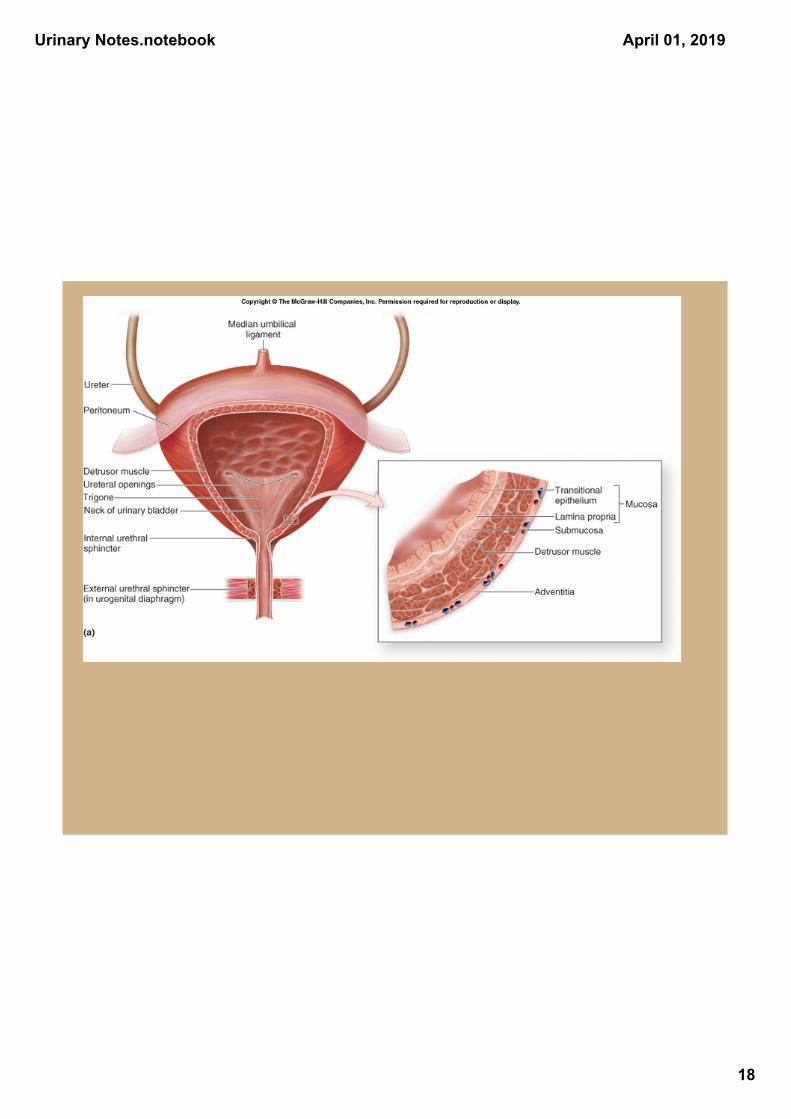
C. The Urinary Bladder 1. A hollow, distensible, muscular organ 2. located within the pelvic cavity, behind
the pubic symphysis. 3. The bladder is somewhat spherical,
when empty it is highly folded. a. as it fills with urine, the walls become smoother and it swells and extends upward into the- dome.
4. The floor of the bladder internally is called the trigone. It has an opening at each of its angles. The upper corners are the ureters and the lower corner is the urethra
5. The bladder wall consists of 4 layers- a. Mucous coat - epithelium adapted for
stretching and shrinking. b. Submucous coat - composed of
connective tissue with lots of elastic fibers.
c. Muscular coat - coarse bundles of smooth muscle in all planes.
1) Detrusor muscle-contraction of this muscle forces urine from the bladder d. Serous coat - parietal peritoneum and fibrous connective tissue.
Urinary Notes.notebook
18
April 01, 2019
Urinary Notes.notebook
19
April 01, 2019
D. The Urethra 1. Function- conveys urine from the urinary
bladder to outside the body. 2. lined with mucous membrane and large
amounts of smooth muscle and contains many mucous glands called urethral glands.
a. female 1.6 inches in length b. male- 7 inches in length- functions as both a urinary canal and as a passageway for cells and secretions of the reproductive system c. in the male it is divided into 3 segments-
1) prostatic urethra - through the prostate gland
2) membranous urethra- site of the external urethral sphincter. It must relax before micturation can occur.
3) penile urethra- through the penis, exits the body through the external urethral orifice.
Urinary Notes.notebook
20
April 01, 2019
E. Micturation 1. Process by which urine is expelled from
the urinary bladder. a. It involves the contraction of the
detrusor muscle, and is aided by the contraction of the abdominal muscles. b. Relaxation of the external urethral sphincter is voluntary.
c. The need to urinate is stimulated by the distention of the bladder as it fills with urine.
2. The micturation reflex center is located in the sacral segments of the spinal cord. This reflex can be overridden by parts of the cerebral cortex and midbrain at approx. 2-4 years of age. a. The hypothalamus and midbrain assist in micturation. b. Bladder may hold up to 800 ml.
1) desire to urinate at about 150ml. 2) feeling uncomfortable at 300ml.
Urinary Notes.notebook
21
April 01, 2019
IV. Disorders and Diseases of the Urinary System
A. Nephritis - inflammation of the nephron and or kidney.
1. Glomerulonephritis- occurs in the glomerulus
a. Can lead to kidney failure and is sign of kidney disease.
b. Can cause: proteinuria- amino acids or protein in the urine glycosuria- glucose present in the urine hematuria- blood cells in the urine.
B. Gout- uric acid crystals form in the joints. causes an arthritis like disease of swelling and pain in the lower extremities.
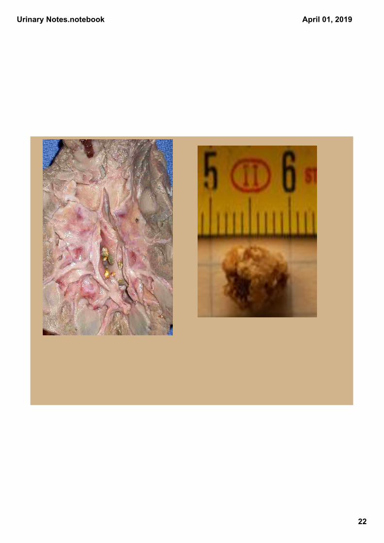
C. Renal Caliculi (kidney stones)- a. Caused by uric acid combining with
calcium oxalates, calcium phosphate, or magnesium phosphate, which form crystals within collecting ducts, renal pelvis, or ureters.
b. These blocks the flow of urine, backs up the system, & decreases urine production.
c. If the stone passes into the ureter it may stimulate severe pain which usually begins in the region of the kidney and tends to radiate to the abdomen, pelvis, and legs.
D. Urinary Tract Infection (UTI) more common in females due to the shortness of the urethra. Bacteria are the usual cause.
Urinary Notes.notebook
22
April 01, 2019