Embed Size (px)
Citation preview

891
Anatomy of the Citrus Leaf Petiole: Healthy vs. Huanglongbing Ed Etxeberria* and Cody Narciso University of Florida, IFAS, Citrus Research and Education Center, Lake Alfred, FL, USA *Corresponding author: [email protected] Abstract
Phloem cells from HLB-affected trees become obstructed with callose and P-protein plugs. The presence of these plugs is believed to hinder the translocation of photoassimilates (nitrogenous and reduced carbon compounds) to the root system. However, even with seemingly collapsed phloem tissue, citrus trees remain viable and produce fruit for some time, suggesting the incomplete plugging of phloem elements or the existence of alternative routes for photoassimilate transport. In this study, we examined the basic structure of phloem tissue from HLB-free and HLB-affected trees under light and scanning electron microscopy. To avoid any possible interference with callose induced by injury during sampling, we employed a freeze substitution technique. Sieve elements from HLB-free trees show sizable lateral pores to phloem and ray parenchyma. Early stage HLB-affected phloem cells contain dark electron dense material and jagged appearing cell walls. Eventually, these cells totally collapse into almost a solid cell-wall barrier. The large number of wall perforations, along the cortex, ray and vascular parenchyma with abundant pit fields, could be essential anatomical features necessary to support symplastic transport of photoassimlates in an HLB-compromised phloem system.
Keywords: phloem starch, pit fields, sieve elements, sieve pores, symplastic transport INTRODUCTION
Huanglongbing (HLB, formerly citrus greening) is a highly destructive disease of citrus presumably caused by the fastidious, gram-negative, obligate parasitic, phloem-limited α-proteobacterium Candidatus Liberibacter ssp. (CLssp; Jagoueix et al., 1994; Bové, 2006). Phloem cells from HLB-affected trees become obstructed with callose and P-protein plugs (Achor et al., 2010) and eventually collapse (Folimonova and Achor, 2010). These plugs are deposited in both lateral pit fields (Koh et al., 2011) as well as in and around sieve plates (Folimonova and Achor, 2010; Koh et al., 2011). In addition, starch grains are commonly observed within the sieve elements (Folimonova and Achor, 2010). The presence of these plugs and of starch-containing amyloplasts in sieve elements is believed to hinder the transport of photoassimilates from photosynthetic source leaves to the remaining heterotrophic sink tissues (Schneider, 1968). The backlog of reduced carbon compounds in the phloem cells promotes sugar conversion to starch in virtually all living cells in HLB-affected leaves (Schneider, 1968; Etxeberria et al., 2009), a distinctive characteristic of HLB-affected trees (Achor et al., 2010).
However, two observations place doubts on the soundness of this hypothesis. First, parenchyma cells from woody stems and bark tissue downstream from the leaf canopy also accumulate copious amounts of starch. Second, HLB-affected branches with evident HLB symptoms and seemingly obstructed phloem are capable of producing vegetative tissue and fruit for some time (Ikpechukwu et al., 2011). If photassimilate transport were blocked at the leaf/petiole level, there would be insufficient carbohydrate to sustain the synthesis of starch downstream from the leaves and to support the growth of developing fruits. As part of our continuous efforts to understand the changes in carbohydrate metabolism brought about by HLB, we theorize the existence of an ancillary (albeit less efficient) route for photoassimilate transport. To answer this question, we examined the basic structure of phloem and cortex tissues from HLB-free and HLB-affected leaves under light and scanning electron microscopy. Our observations
Proc. XIIth Intl. Citrus Congress Eds.: B. Sabater-Muñoz et al. Acta Hort. 1065, ISHS 2015
892
demonstrate abundant symplastic connections between photosynthetic, vascular and cortex parenchyma cells capable of supporting symplastic transport of photoassimilates. MATERIALS AND METHODS Plant Material
Leaves from HLB-affected trees were collected from 5-year-old ‘Valencia’ orange (Citrus sinensis (L.) Osbeck) trees grown at the Citrus Research and Education Center in Lake Alfred, FL. The trees had been previously determined to be HLB-affected by PCR. Control HLB-free samples were obtained from ‘Valencia’ trees grown in an HLB-free greenhouse.
Leaf petioles were cut off immediately after leaf sampling and frozen in liquid nitrogen. Once in the laboratory, the petioles were transferred to microfuge tubes containing 1.5 ml of cold 100% ethanol. The tubes were placed in the freezer at -20°C and the ethanol solution was changed every 24 h for 5 d.
Tissue Preparation for SEM
Samples were further dehydrated using a Ladd critical point dryer (Ladd Research Industries, Burlington, VT). The dried tissue was then re-cut with a razor blade, mounted on stubs, and coated with gold/palladium (Ladd Research Industries). Coated samples were viewed on a Hitachi S530 scanning electron microscope (Tokyo, Japan).
Tissue Preparation for Light Microscopy
After alcohol dehydration, samples were fixed in 3% glutaraldehyde in 0.1 M potassium phosphate buffer, pH 7.2, at room temperature for 4 h, and then overnight in the refrigerator. Samples were post-fixed for 4 h at room temperature in 2% osmium tetroxide in the above buffer. The samples were then dehydrated in an acetone series and embedded in Spurr’s resin. For light microscopy, 1-µm sections were cut with glass knives, stained with methylene blue/azure A, and post-stained in basic fuchsin. Light micrographs were taken on a Leitz Laborlux S compound microscope (Wetzlar, Germany).
RESULTS HLB-Free Petiole
Phloem tissue from HLB-free petioles appeared as an orderly ring of compact cells delimited by the larger xylem vessels to the interior and by thick walled tracheids to the exterior (Fig. 1). The phloem elements have thinner primary walls than all other tissues in the petiole. Large phloem parenchyma cells are interspersed within the tissue as part of the vascular rays and bordering the inner face of the tracheary ring. Although difficult to visualize in light micrographs, sieve plates become clear at higher magnification under SEM (Fig. 2). Sieve plates were never transversal but always oblique. Sometimes, the plates were arranged at such steep angles that they appeared as part of the lateral walls as previously reported by Schneider (1952) and Koh et al. (2012). It is noteworthy that the sizes of individual sieve pores were comparable to or smaller than the diameter of the smallest morphological form expressed by CLssp (Garnier et al., 1984; Bové, 2006; Shokrollah et al., 2010; Mann et al., 2011).

893
Fig. 1. Cross-section light micrograph of a petiole from a HLB-free tree. Phloem cells (ph) have smooth edges and lack starch grains.
Fig. 2. Scanning electron micrograph of a sieve plate from a petiole mid-rib. The leaf was collected from an HLB-free tree, and the tissue was incubated in β-glucanase and α-amylase to remove all interfering polysaccharides.
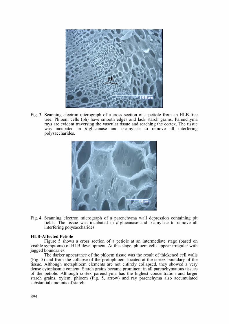
Another anatomical characteristic prominent in SEM micrographs was the numerous wall depressions, commonplace to all parenchyma cells (Fig. 3). These areas were of approximately 2-5 μm in diameter with distinctly thinner walls than the rest of the cell (Fig. 3). These pit fields are abundant and sometimes parted by wall ridges (Fig. 4). Abundant pit fields were also observed in cortex parenchymal (Fig 4) and epidermal photosynthetic cells (not shown).
894
Fig. 3. Scanning electron micrograph of a cross section of a petiole from an HLB-free tree. Phloem cells (ph) have smooth edges and lack starch grains. Parenchyma rays are evident traversing the vascular tissue and reaching the cortex. The tissue was incubated in β-glucanase and α-amylase to remove all interfering polysaccharides.
Fig. 4. Scanning electron micrograph of a parenchyma wall depression containing pit fields. The tissue was incubated in β-glucanase and α-amylase to remove all interfering polysaccharides.
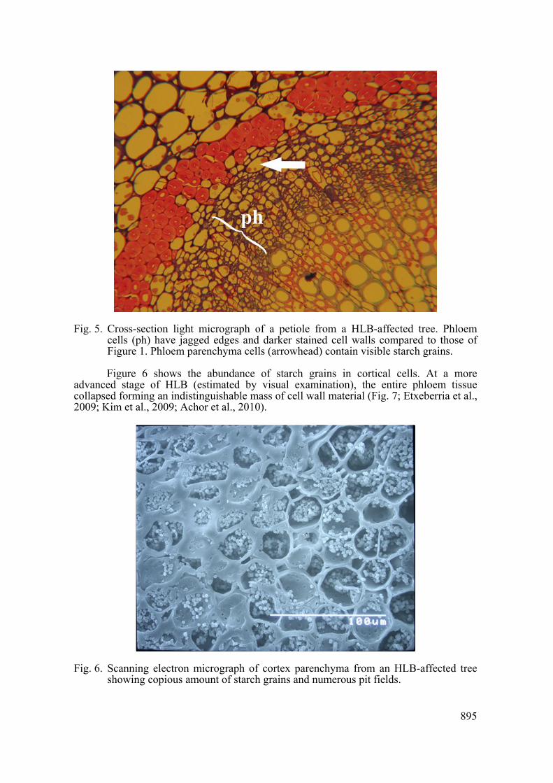
HLB-Affected Petiole Figure 5 shows a cross section of a petiole at an intermediate stage (based on
visible symptoms) of HLB development. At this stage, phloem cells appear irregular with jagged boundaries.
The darker appearance of the phloem tissue was the result of thickened cell walls (Fig. 5) and from the collapse of the protophloem located at the cortex boundary of the tissue. Although metaphloem elements are not entirely collapsed, they showed a very dense cytoplasmic content. Starch grains became prominent in all parenchymatous tissues of the petiole. Although cortex parenchyma has the highest concentration and larger starch grains, xylem, phloem (Fig. 5, arrow) and ray parenchyma also accumulated substantial amounts of starch.
895
Fig. 5. Cross-section light micrograph of a petiole from a HLB-affected tree. Phloem cells (ph) have jagged edges and darker stained cell walls compared to those of Figure 1. Phloem parenchyma cells (arrowhead) contain visible starch grains.
Figure 6 shows the abundance of starch grains in cortical cells. At a more
advanced stage of HLB (estimated by visual examination), the entire phloem tissue collapsed forming an indistinguishable mass of cell wall material (Fig. 7; Etxeberria et al., 2009; Kim et al., 2009; Achor et al., 2010).
Fig. 6. Scanning electron micrograph of cortex parenchyma from an HLB-affected tree showing copious amount of starch grains and numerous pit fields.

896
Fig. 7. Cross-section scanning electron micrograph of an HLB-affected petiole at an advanced disease stage. In this figure, metaphloem tissue has entirely collapsed forming a solid mass of cell wall. The external protophloem and inner part of the cortex are being crushed by the expanding xylem elements.
DISCUSSION
A well-described metabolic change in HLB-affected citrus tissue is the deposition of callose plugs throughout the phloem elements, followed by the hyper-accumulation of starch in all aerial parts and its disappearance from the roots (Etxeberria et al., 2009). This preferential sequestration of photoassimilates, mostly in the form of starch in leaves and stem parenchyma cells, denotes a carbohydrate imbalance that can culminate in tree death.
In his investigation into HLB-affected tissues, Schneider (1968) concluded that starch accumulation in leaves resulted from necrotic phloem pockets scattered throughout the vascular system. These blockages impede phloem transport of photoassimilates creating a backlog of sugars in the leaves that in turn trigger accelerated rates of starch synthesis. Phloem collapse and blockage have been further detailed by Achor et al. (2010) and Folimonova et al. (2010). Nevertheless, the continuous development of vegetative tissue and fruit in HLB-affected branches is an indication that photoassimilates are being somehow transported from source leaves despite a severely compromised phloem system. The anatomical analyses presented in this communication support the likely existence of an alternate non-vascular temporary route for photoassimilates to heterotrophic organs that bypasses the phloem. This conclusion is supported by three observations. First, the complete obliteration of the phloem tissue as the disease advances (Figs. 5 and 7) is likely to obstruct all transport through sieve elements. Second, the lack of microscopically visible “wound phloem” that characteristically develops along impaired phloem elements (Jacobsen and Eschrich, 1990). Third, the abundant starch reserves in the vascular pith indicate transport through vascular rays.
Our observations, based on static images, do not allow us to determine the type of transport route to heterotrophic cells. However, the copious plasmodesmata pit fields (Fig. 4) throughout the cortex (Figs. 3 and 6) and ray parenchyma cells (Fig. 3) revealed

897
the presence of the essential anatomical components necessary to support a symplastic continuum. Whether transport occurs exclusively through a symplastic route or whether it is accompanied by apoplastic movement is a matter that needs further physiological analyses.
Literature Cited Achor, D., Etxeberria, E., Wang, N., Folimonova, S.Y., Chung, K.R. and Albrigo, L.G.
2010. Sequence of anatomical symptoms in citrus affected wit HLB disease. Plant Pathol. J. 9:56-64.
Albrecht, U. and Bowman, K. 2009. Candidatus Liberibacter asiaticus and Huanglongbing effects on citrus seeds and seedlings. HortScience 44:1967-1973
Bové, J.M. 2006. Huanglongbing: A destructive, newly-emerging, century-old disease of citrus. J. Plant Pathol. 88:3-37.
Etxeberria, E., Gonzalez, P., Achor, D. and Albrigo, G. 2009. Anatomical distribution of abnormally high levels of starch in HLB-affected Valencia orange trees. Physiol. Mol. Plant Pathol. 74:76-83.
Folimonova, S.Y. and Achor, D. 2010. Early events of citrus greening (Huanglongbing) disease development at the ultrastructural level. Phytopathology 100:949-958.
Garnier, M., Danel, N. and Bové, J.M. 1984. The greening organism is a Gram negative bacterium. p.115-124. In: S.M. Garnsey, L.W. Timmer, and J.A. Dodds (eds.), Proc. 9th Conf. Int. Organ. Citrus Virol. IOCV, Riverside, CA, USA.
Ikpechukwu, C.O., Sims, C.A., Danyluk, M.D., Spann, T.M. and Goodrich, R.M. 2011. Effect of fruit size and huanglongbing disease on orange juice attributes. Proc. Fla. State Hort. Soc. 124:202-206.
Jagoueix, S., Bové, J.M. and Garnier, M. 1994. The phloem-limited bacterium of greening disease is a member of the alpha subdivision of the Proteobacteria. Int. J. Syst. Bacteriol. 44:379-386.
Kim, J.S., Sagaram, U.S., Burns, J.K., Li, J.L. and Wang, N. 2009. Response of sweet orange (Citrus sinensis) to ‘Candidatus Liberibacter asiaticus’ infection: microscopy and microarray analyses. Phytopathology 99:50-57.
Koh, E.J., Zhou, L., Williams, D.S., Park, J., Ding, N., Duan, Y.P. and Kang, B.H. 2012. Callose deposition in the phloem plasmodesmata and inhibition of phloem transport in citrus leaves infected with “Candidatus Liberibacter asiaticus”. Protoplasma 249:687-697.
Mann, R.S., Pelz-Stelinski, K., Herman, S.L., Tiwari, S. and Stelinski, L. 2011. Sexual transmission of a plant pathogenic bacterium, Candidatus Liberibacter asiaticus, between conspecific insect vectors during mating. PLoS One 6:e29197.
Schneider, H. 1968. Anatomy of greening-disease sweet orange shots. Phytopathology 58:1155-1160.
Schneider, H. 1952. The phloem of sweet orange tree trunk and the seasonal production of xylem and phloem. Hilgardia 12:331-365.
Shokrollah, H., Abdullah, T.L., Sijam, K. and Abdullah, S.N.A. 2010. Ultrastructures of Candidatus Liberibacter asiaticus and its damage in huanglongbing (HLB) infected citrus. African J. Biotechnol. 9:5897-5901.

898