Embed Size (px)
Citation preview

Anatomy & Embryology - GIS
Done By
Dana Tarawneh
Corrected By
Dana Tarawneh

The last part of Video #7
3rd & Final Part of lecture #5
To be Continued in Lecture #7
Some info were taken from 018 Sheet.Thanks to Rama Al-Abbady ♥

The small intestine
The small intestine is made up of 3 parts :
• The duodenum which is 25cm/10 inches and is retroperitoneal except the first and last
inches.
• The jejunum and ileum which are 6 meters long and are intraperitoneal (they have a
mesentery).
We can sometimes say that the small intestine is 6 meters long ( this includes all 3 parts )
but with taking into consideration the differences between these 3 parts

DOUDENUM

duodenum• The duodenum is ac-shaped
• Concave tube.
• About 10” in length.
• It has four parts, the 1st is 2 inches, the 2nd is 3 inches (vertical) ,the
3rd is 4 inches (horizontal) & the 4th is 1 inch.
• It joins the stomachto the jejunum.
• It curves around the head of the pancreas to the left andbackwards. Notice the location of the uncinate process of the head,
left to the 4th part of duodenum.The superior mesenteric artery and
veins pass in front of this process.
• It is importantbecause it (2nd part) receives the opening of the common bile and pancreatic ducts. they secrete substances to
help in the digestion of fat which occurs in the duodenum.
body of
pancreas
neck of
pancreas
head of
pancreas
Small
uncinate
process

duodenum….cont
• Most of the duodenum is retroperitoneal except the 1st & last inches (they are intraperitoneal ,(1st inch) because the greater & lesser omentum areattached to the 1st part of the duodenum | (last inch) completes as jejunum).
• This short segment (1st inch) has the lesser omentum on its upper border, the greateromentum on its lower border & the lesser sac posterior toit.
• The duodenum extends from the pylorus to the jejunum (it begins at the pyloric sphincter (stomach) and ends at the ligament of Treitz which continues as the jejunum) .
• It is divided into 4parts.

Site ofduodenum
continues to the liver.All absorptive material of the
• GI tract reaches the liver through these portal veins.
L-3
• The duodenum is situated in the epigastric & umbilical regions
• For purposes of description, is divided into fourparts
Gallbladder
Right lobe of liver
Falciform ligament
Pancreas
Duodenum
• The portal vein forms behind the neck of pancreas from
the splenic and superior mesenteric veins and

Parts of the duodenum & Theirrelations
• The common bile & the pancreatic ducts have
the same opening in the duodenum. So, this
means that their meeting forms a bulge in the
duodenal wall called ampulla of Vater.
• Around this ampulla is the sphincter of Oddi
(a smooth muscle).
• This opening on the inside is known as themajor duodenal papilla .Sometimes there is
another opening 1 inch above the major
papilla for accessory pancreatic ducts known
as the minor duodenal papilla.

Parts of the duodenum&Theirrelations
• The liver has left and right
lobes .
• The left and right hepatic
ducts form the common
hepatic duct
• It meets with the cystic
duct of the gallbladder to
form the common bile
duct .

You’re about to read a bit of histology ,so bare with me
• The GI track has 4 layers (mucosa, submucosa, muscular layer and adventitia or serosa).
• The folds on the inside of the duodenum are called plicae circulares.
• The lining epithelium of the duodenum (mucosa) is simple columnar epithelium with goblet
cells.
• In the submucosa of duodenum there are glands known as Brunner’s glands which produce
the alkaline secretion that neutralizes the acidity of the chyme when it comes from the
stomach.
• Note: in the entirety of the GI track only 2 organs have glands in their submucosa, and they
are the oesophagus and duodenum.
• In the mucosa of the duodenum there is lamina propria which also contains glands called
crypts ofLieberkühn.
• The lamina propria is a thin layer of loose (areolar) connective tissue, which lies beneath the
epithelium, and together with the epithelium and basement membrane constitutes the
mucosa,from the web.

A New Technique ERCP (Endoscopic retrograde cholangiopancreatography)
• This is a new technique where an endoscope is placed through the mouth and proceeds
retrogradely through the pharynx, oesophagus, stomach and duodenum .then you find the
major duodenal papilla and you cut the sphincter of Oddi entering either the pancreatic or
common bile ducts based on what you want to do.
• This technique is used in the treatment of stones that form in the common bile duct and block it
thus causing jaundice (yellow sclera, yellow and itchy skin) which used to be treated with
surgery in the past. Nowadays ,when you find the stone with the endoscope you use a basket
and remove the stone leaving it in the duodenum & it gets out with the stool.
• Sometimes stasis of the secretion of pancreas happens and it becomes like mud or forms
stones and closes the pancreatic duct ,this may cause pancreatitis which is very dangerous,
and it is treated with ERCP, by entering the duct with the endoscope and adding saline which
will dissolve thestones.

1st part of Duodenum- The first part is 2 inches long & it’s divided into 2
parts :
• The 1st inch (intraperitoneal)
- it’s a common site for peptic or duodenal ulcers.
- Duodenal ulcers are more common than gastric ulcers and
happen because the chyme that comes from the stomach is
acidic (the duodenal secretions are alkaline and work to
neutralize that acidity, but ulcers could still happen if the
acidity was too high) . The posterior wall of duodenum is
affected the most.
• The 2nd inch (retroperitoneal)
- It begins from the pyloduodenal junction- At the level of the transpyloric line- Runs upward and backward at the levelof the
1st lumbar vertebra 1 inch to the right.- It reaches the neck of the gall bladder .
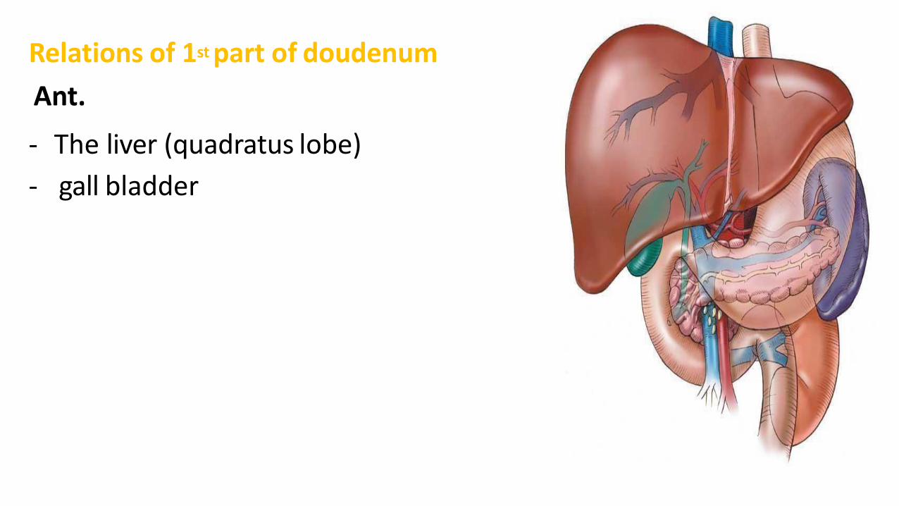
Relations of 1st part of doudenum
Ant.
- The liver (quadratus lobe)
- gall bladder

Relations of 1st part of duodenum……cont
Sup.
- the epiploic foramen (anterior tothis
foramen is a free edge of lesser omentum
containing three structures :
1. the common pyloric duct
2. the hepatic artery
3. portal vein

Relations of 1st part duodenum……cont
post.
- The lesser sac (behind the stomach and lesser
omentum)
- gastroduodenal Artery (if there is a peptic ulcer on
the posterior wall of the 1st inch perforation and
infiltration may occur along with bleeding from
this artery)
- the Bile duct
- portal vein
- I.V.C
Inf.
• The head of the pancreas

2nd part of duodenum
• It is 3” long• runs downward vertically on the right side• In front of the Rt.kidney & right ureter .
• It ends next to the 3rd and 4th lumbarvertebrae.
• halfway of it, The bile duct and the main pancreatic duct pierce the medial wall, and then form the ampulla that opens in the major duodenal papilla
• The accessory pancreatic duct (if present) opens in the minor duodenal papilla more superiorly.
• Importance of the 2nd part lies in that it receives the common bile andpancreatic ducts’secretions.

Note :anatomical sphincter means it’s surrounded by circular smooth muscles .Absence of these circular
smooth muscles means it’s a physiological sphincter

Hepaticopancreatic ampulla (Ampullaof Vater)


Relations of2nd partofduodenumAnt.• The gallbladder(fundus)• Right lobe of the liver• Transverse colon• coiled of small intestine.Post.• Hilum of Rt. Kidney• Rt. Ureter.Lateral.• Right colic flexure• Ascending colon• Right lobe of the liver.Medial.-Head of pancreas-Bile and pancreaticducts.

3rd part of duodenum
• 4” long Runs horizontally to the left
• Runs in front ofthe vertebralcolumn
• On the subcostal plane.
• Under the lowermargin of the head ofpancreas
• Above the coils ofthe jejunum.

Relations of 3rd part of duodenumAnteriorly:
- The root of mesentery of the smallintestine
-the superior mesenteric vessels contained within the
mesentery
- coils of jejunum
Posteriorly:
-The right ureter
-The right psoas muscle
-The inferior vena cava
-The aorta
Superiorly:
- The head of the pancreas
Inferiorly:
- Coils of jejunum

4th part ofduodenum…..cont
• 1” long
• Runs upwardto the left
• End in the duodejejunal junction at the level of the 2nd lumbar vertebrae 1” to theleft.
• The junction (flexure) is held in position by the ligament ofTreitz, which is attached to the right crus of the diaphragm (duodenal recess).

Relationaof 4othf
Ant.
- The beginning of the root of themesentery
- coils of the jejunum.
Post.
- Lt. psoas major
-the sympathetic chain on the left margin of the aorta.
Sup.
- Uncinate process of thepancreas.

You’re about to read a bit of embryology ,so bare with me
• The GI track is divided to three parts: 1. Foregut (oesophagus(lower part), stomach and upper half of duodenum).
• Blood supply of foregut→ Celiac trunk of abdominal aorta2. Midgut (from lower half of duodenum to lateral\distal thirdof transverse colon)
• Blood supply of midgut → Superior mesenteric artery 3. Hindgut(from distal third of transverse colon to rectum)
• Blood supply of hindgut → Inferior mesenteric artery

Blood supply of duodenum
• Arteries
1 upper half (1st part + upper1/2 of 2nd part) , it follows the foregut and is
supplied by the superior pancreaticoduodenal artery, a branch of the gastroduodenal artery (from celiac trunk)
2The lower half (lower ½of 2nd part +3rd+4th part) ,it follows the midgut and is
supplied by the inferior pancreaticoduodenal artery, a branch of the superior mesenteric artery
• The upper & lower halves are separated by the major duodenal papilla and
sphincter of Oddi.

Arterial supply and venous drainage of theduodenum

Blood supply forduodenum

Veins ofduodenum
• The superior pancreaticoduodenal vein drains into the portal vein
• The inferior vein joins the superior mesenteric vein .


Lymphaticdrainage
• The lymph vessels followthe arteries
• drainage upward → via pancreaticoduodenal nodes →
• the gastroduodenal nodes → the celiac nodes .
• drainage downward→ via pancreaticoduodenal nodes →the superior mesenteric nodes around the origin of thesuperior mesenteric artery.

Nervesupply
• Sympathetic nerve
• parasympathetic nerves from:
1 The celiac plexus
2 Superior mesentericplexus.