Embed Size (px)
Citation preview

BioMed CentralBMC Bioinformatics
ss
Open AcceResearch articleAnalyses of domains and domain fusions in human proto-oncogenesQi Liu1,2, Jinling Huang3, Huiqing Liu1, Ping Wan1,5, Xiuzi Ye*4 and Ying Xu*1Address: 1Computational Systems Biology Laboratory, Department of Biochemistry and Molecular Biology, Institute of Bioinformatics, University of Georgia, Athens, GA 30602, USA, 2Zhejiang California International Nanosystems Institute, Zhejiang University, Hangzhou, 310029, PR China, 3Department of Biology, East Carolina University, Greenville, NC 27858, USA, 4College of Computer Science, Zhejiang University, Hangzhou, 310027, PR China and 5College of Life Science, Capital Normal University, Beijing, 100037, PR China
Email: Qi Liu - [email protected]; Jinling Huang - [email protected]; Huiqing Liu - [email protected]; Ping Wan - [email protected]; Xiuzi Ye* - [email protected]; Ying Xu* - [email protected]
* Corresponding authors
AbstractBackground: Understanding the constituent domains of oncogenes, their origins and their fusionsmay shed new light about the initiation and the development of cancers.
Results: We have developed a computational pipeline for identification of functional domains ofhuman genes, prediction of the origins of these domains and their major fusion events duringevolution through integration of existing and new tools of our own. An application of the pipelineto 124 well-characterized human oncogenes has led to the identification of a collection of domainsand domain pairs that occur substantially more frequently in oncogenes than in human genes onaverage. Most of these enriched domains and domain pairs are related to tyrosine kinase activities.In addition, our analyses indicate that a substantial portion of the domain-fusion events ofoncogenes took place in metazoans during evolution.
Conclusion: We expect that the computational pipeline for domain identification, domain originand domain fusion prediction will prove to be useful for studying other groups of genes.
BackgroundAn oncogene is a modified gene that promotes unregulatedproliferation of cells, increasing the chance that a normalcell develops into a tumor cell, possibly resulting in cancer[1]. The normal copy of such a gene is called a proto-onco-gene. The first oncogene, SRC, was discovered in a chickenretrovirus in 1970 [2]. Since then, numerous oncogeneshave been identified and classified into different groupsbased on their cellular functions. As of now, oncogeneshave been identified at all levels of signal transductioncascades that control cell growth, proliferation and differ-entiation [1-3].
Protein domains are compact and semi-independentunits of a protein, each of which may consist of one ormore contiguous segments of a peptide chain and have itsown biological function [3]. They are generally viewed asthe basic unit of protein function and evolution. Varioussequence- and structure-based methods have been devel-oped for the identification of protein domains [4-6], andseveral domain databases, such as DALI [7], PFAM [8],SMART [9] and Prodom [10], have been established.
Recent studies on oncogenes and cancer pathology havepointed to the importance of individual domains and
Published: 17 March 2009
BMC Bioinformatics 2009, 10:88 doi:10.1186/1471-2105-10-88
Received: 3 July 2008Accepted: 17 March 2009
This article is available from: http://www.biomedcentral.com/1471-2105/10/88
© 2009 Liu et al; licensee BioMed Central Ltd. This is an Open Access article distributed under the terms of the Creative Commons Attribution License (http://creativecommons.org/licenses/by/2.0), which permits unrestricted use, distribution, and reproduction in any medium, provided the original work is properly cited.
Page 1 of 16(page number not for citation purposes)
BMC Bioinformatics 2009, 10:88 http://www.biomedcentral.com/1471-2105/10/88
domain fusions in oncogenesis. It has been reported thatgenes containing domains from specific domain familiesmay have particular relevance to human cancer [11-13].For example, the tyrosine kinase domain is known to playsignificant roles in the development of numerous diseasessuch as cancer [11]. Another example is the ATM-relateddomain that is required for histone acetyltransferaserecruitment and Myc-dependent oncogenesis [12]. Addi-tionally, CML, a form of leukaemia, is associated with thefusion of Bcr and Abl genes or their constituent domains[13]. Therefore, understanding the constituent domainsof oncogenes as well as their origins may shed new lightabout the initiation and development of cancers.
In this study, we have developed an integrated computa-tional pipeline for studying the domain composition,domain fusion and domain origin. Specifically, our com-putational pipeline includes the following key compo-nents: (1) identification of the origin of each componentdomain of known oncogenes and the relevant fusionevents; (2) co-occurrence analysis of oncogene domains;(3) identification of the domains and domain pairs thatappear more frequently in oncogenes than in the back-ground, namely the collection of all human genes; and (4)functional analyses of the identified frequent domainsand domain pairs. We then applied this pipeline to allwell characterized human oncogenes, and had a numberof new and interesting observations. To the best of ourknowledge, this is the first comprehensive analysis specif-ically addressing the domain composition, origin andfusion of oncogenes.
Results and discussionUsing the computational procedures outlined in Materialand Methods, we have carried out a detailed analysis ofoncogene domains and co-occurring domains for theirorigins and functional analysis.
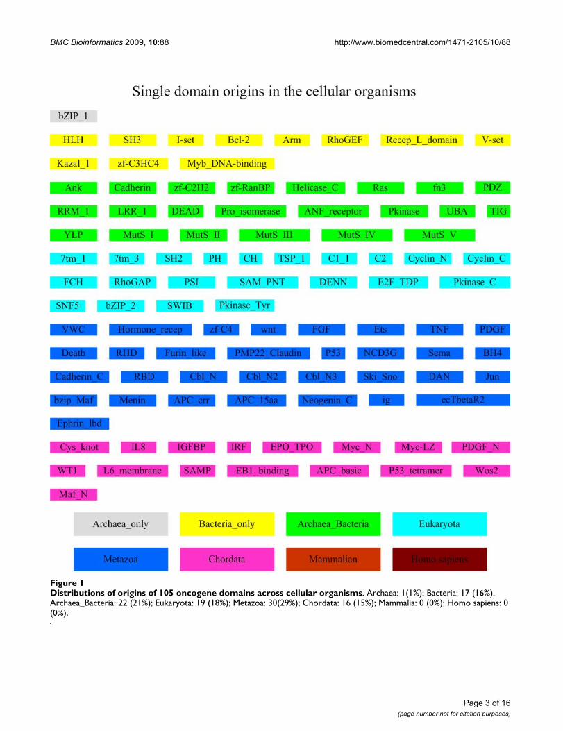
A. Origin of oncogene domainsOrigin of distinct domains in cellular organisms103 distinct domains [see Additional file 1] have beenidentified from 124 oncogenes, based on Pfam domainassignments. We have considered the subtype scenariosfor specific domains, i.e., the different alignments for aspecific domain in one clan and using one domain ID todenote the corresponding subtypes. In our dataset, thereexist two alignments SH3_2 and SH3_1 for the SH3domain. The same holds for the SAM domain, whereSAM_PNT is the entry for the SAM domain and two differ-ent alignments, SAM_1 and SAM_2, exist for this entry,respectively. Although they have different accession num-bers in Pfam, we just use SH3 and SAM_PNT to denotethese two types of domains, respectively. The distributionof these domains' origins across different cellular organ-isms is given in Figure 1. About 50% (55/103) of onco-
gene domains have their origins in the early stages oforganismal evolution prior to the emergence of the meta-zoans, and no domains are found to arise from mammals.It should be noted that these results have been furtherrefined by our literature survey from the original subtrac-tive searching results (see Material and Methods), to takepotential HGT into consideration. Based on the literaturesearch, we found that domain SWIB and non-enzymaticdomains ig and SAM are likely to have arisen in eukaryote.Their homologs are identified in prokaryotes, likelyresulted from HGTs from eukaryotes [14]. Also the originof tyrosine kinases (Pkinase_Tyr) is probably in eukaryoteand their presence in bacteria may also be explained mostparsimoniously by HGT events [14].
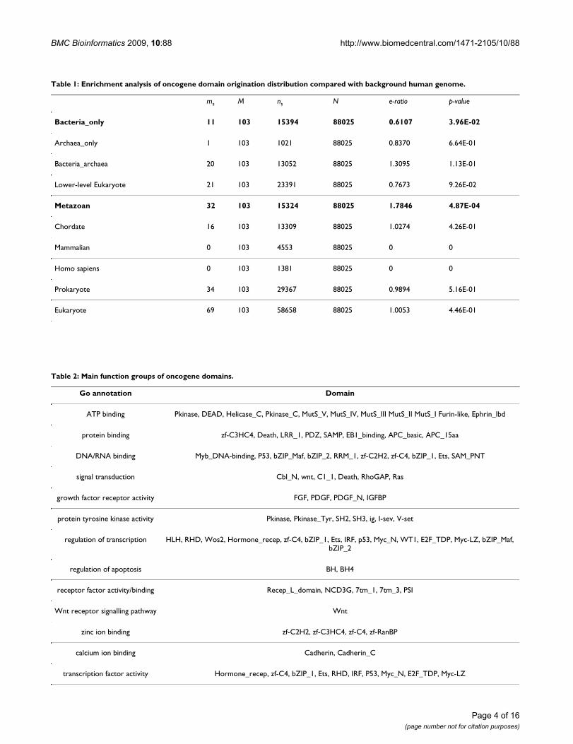
In order to further analyze the statistical differencebetween the domain origin distribution of oncogenes ver-sus that of the other genes, we have compared our resultswith Lipika et al. [15], which presented an analysis on theorigins of the conserved domains in the whole humanproteome. Table 1 presents a thorough calculation of theenrichment ratios of oncogene domains that originatedfrom 8 categories (i.e., Bacteria_only, Archaea_only,Bacteria_archaea, low level Eukaryotes, Metazoan, Chor-date, Mammalian, Homo sapiens) compared with thewhole domain dataset in the human genes and their p-val-ues. Our results indicate that the origin distribution ofoncogene domains is largely consistent with that reportedby Lipika et al. [15] for the whole human proteome,EXCEPT FOR those of bacterial or metozoan origins.
Domain functionsWe divided the oncogene domains into groups based ontheir GO annotation (Table 2). These oncogene domainsshow diverse functions, including regulation of transcrip-tion and apoptosis, protein kinase activity and DNA/RNA/protein binding activity.
Further analyses suggest that domains with different func-tions might have come from different origins (Figure 1;Table 2). For example, domains related to immunoglobu-lin and tyrosine kinase (e.g., SH2, SH3, I-sev, and V-set)are found in archaea, bacteria or in both. These domainsare known to be closely related to oncogenesis [16] (Notethat another two important oncogenesis-related domains:Pkinase_Tyr and ig, originated in eukaryotes, but werehorizontally transferred to prokaryotes [14]). Otherdomains such as rhodopsin domains (7tm_1, 7tm_3),cyclin dependent kinases (CDKs) domains (Cyclin_N,Cyclin_C) and the intracellular signalling domains (PH,CH) seem to have originated in eukaryotes. Severaldomains related to the development of the nervous sys-tem such as wnt, ephrin_lbd and Sema seem to have orig-inated in metazoans. In addition, function domainsrequired by vertebrates such as hormones involved in
Page 2 of 16(page number not for citation purposes)
BMC Bioinformatics 2009, 10:88 http://www.biomedcentral.com/1471-2105/10/88
Page 3 of 16(page number not for citation purposes)
Distributions of origins of 105 oncogene domains across cellular organismsFigure 1Distributions of origins of 105 oncogene domains across cellular organisms. Archaea: 1(1%); Bacteria: 17 (16%), Archaea_Bacteria: 22 (21%); Eukaryota: 19 (18%); Metazoa: 30(29%); Chordata: 16 (15%); Mammalia: 0 (0%); Homo sapiens: 0 (0%).
BMC Bioinformatics 2009, 10:88 http://www.biomedcentral.com/1471-2105/10/88
Page 4 of 16(page number not for citation purposes)
Table 1: Enrichment analysis of oncogene domain origination distribution compared with background human genome.
ms M ns N e-ratio p-value
Bacteria_only 11 103 15394 88025 0.6107 3.96E-02
Archaea_only 1 103 1021 88025 0.8370 6.64E-01
Bacteria_archaea 20 103 13052 88025 1.3095 1.13E-01
Lower-level Eukaryote 21 103 23391 88025 0.7673 9.26E-02
Metazoan 32 103 15324 88025 1.7846 4.87E-04
Chordate 16 103 13309 88025 1.0274 4.26E-01
Mammalian 0 103 4553 88025 0 0
Homo sapiens 0 103 1381 88025 0 0
Prokaryote 34 103 29367 88025 0.9894 5.16E-01
Eukaryote 69 103 58658 88025 1.0053 4.46E-01
Table 2: Main function groups of oncogene domains.
Go annotation Domain
ATP binding Pkinase, DEAD, Helicase_C, Pkinase_C, MutS_V, MutS_IV, MutS_III MutS_II MutS_I Furin-like, Ephrin_lbd
protein binding zf-C3HC4, Death, LRR_1, PDZ, SAMP, EB1_binding, APC_basic, APC_15aa
DNA/RNA binding Myb_DNA-binding, P53, bZIP_Maf, bZIP_2, RRM_1, zf-C2H2, zf-C4, bZIP_1, Ets, SAM_PNT
signal transduction Cbl_N, wnt, C1_1, Death, RhoGAP, Ras
growth factor receptor activity FGF, PDGF, PDGF_N, IGFBP
protein tyrosine kinase activity Pkinase, Pkinase_Tyr, SH2, SH3, ig, I-sev, V-set
regulation of transcription HLH, RHD, Wos2, Hormone_recep, zf-C4, bZIP_1, Ets, IRF, p53, Myc_N, WT1, E2F_TDP, Myc-LZ, bZIP_Maf, bZIP_2
regulation of apoptosis BH, BH4
receptor factor activity/binding Recep_L_domain, NCD3G, 7tm_1, 7tm_3, PSI
Wnt receptor signalling pathway Wnt
zinc ion binding zf-C2H2, zf-C3HC4, zf-C4, zf-RanBP
calcium ion binding Cadherin, Cadherin_C
transcription factor activity Hormone_recep, zf-C4, bZIP_1, Ets, RHD, IRF, P53, Myc_N, E2F_TDP, Myc-LZ
BMC Bioinformatics 2009, 10:88 http://www.biomedcentral.com/1471-2105/10/88
mitogenic and inflammatory activity (Myc_N, Myc_LZ,Maf_N, Cys_knot) seem to have originated in chordates.
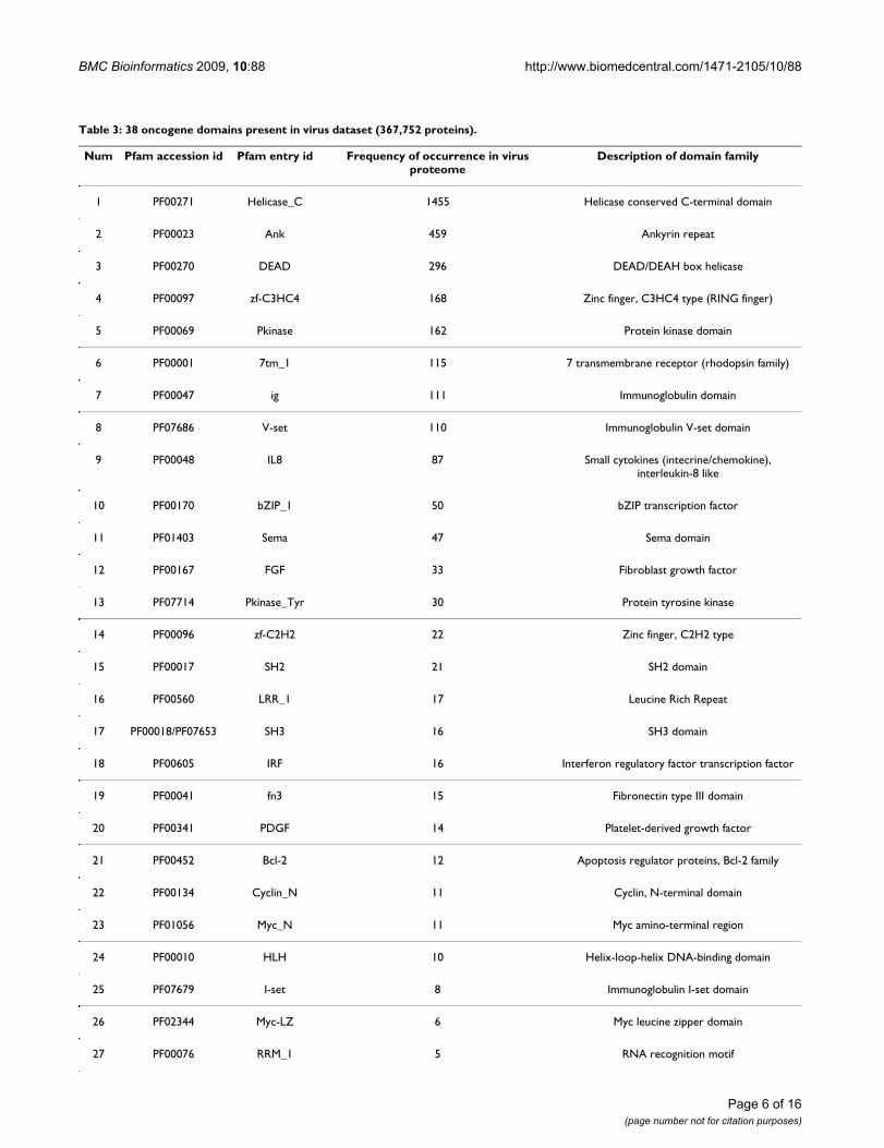
Domains originated from virusesAmong the 103 identified oncogene domains, 38 arefound to be present in viruses (Table 3). The three mostfrequently occurring domains in virus proteins areHelicase_C, Ank and DEAD. Ank has been reported indiverse groups of proteins such as enzymes, toxins andtranscription factors. The existence of Ank in bothprokaryotes and viruses may have resulted from horizon-tal gene transfers [17]. The Helicase domain family(including Helicase_C and DEAD) is reportedly related tohepatitis virus-associated hepatocellular carcinoma andinvolved in cell growth control [18]. In addition, someother families such as Zinc finger domains (zf-C3HC4, zf-C2H2, zf-C4), Immunoglobulin-related domains (ig, V-set, I-set) and protein-tyrosine kinase related domains(Pkinase_Tyr, SH2, SH3) also have remote homologs inviruses and all these three domain families are closelyrelated to oncogenesis. Overall, 20 of the 38 virus-origi-nated domains are known to be related to oncogenesis.
B. Oncogene domain fusionDomain fusion in cellular organismsWe have identified 50 whole domain fusion events in the124 oncogenes. Among them, 21 contain two distinctdomains (domain pairs) and the others contain at leastthree different domains. Their initial appearance in cellu-lar organisms and their presence/absence in viruses aregiven [see Additional file 2].
Fused domains in virusesAmong the 50 fused domains, 7 events have been identi-fied in viruses. These 7 fused domains can be divided into4 categories according to their functions: pkinase-relateddomain fusion ({SH2, SH3, Pkinase_Tyr}, {SH2, FCH,Pkinase_Tyr}); platelet-derived growth factor domainfusion ({PDGF, PDGF_N}); helicases-related domainfusion ({DEAD, Helicase_C}) and DNA/ligand-bindingdomain fusion ({Hormone_recep, zf-C4}; {HLH,Myc_N, Myc-LZ}; {HLH, Myc_N}). Interestingly, ~90%of the virus proteins harbouring these fused domainscome from the Potyviridae family and the remainingalmost all come from the Orthoretrovirinae family. Poty-viridae is one of the largest and most important familiesof plant viruses. Although the relationship between retro-viruses and cancer has been widely established [19-21],the possible link between Potyviridae and oncogenesis isunknown.
C. Proteome-wide patterns of origins of oncogenesWe have also examined the origins of all the oncogenes asa whole. Our goal is to find out at what stage in evolutionall component domains of an oncogene are fused together
for the first time, considered as the origin of the oncogene[see Additional file 3].
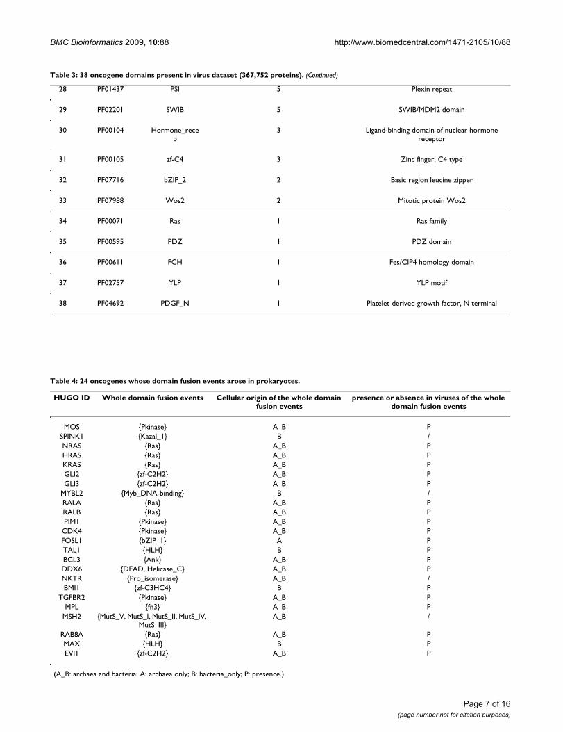
Among the 24 oncogenes whose initial domain fusionsoccurred in prokaryotes, 20 have the same domainfusions in viruses (Table 4). It seems that domains withprokaryotic origins tend to present in viruses.
We have divided the oncogenes into six categories accord-ing to their functions: signal transducers, no-receptorkinases, growth factors, growth factor receptors, transcrip-tion factors and others. Based on our examination of theoncogene origins, we have observed some general rela-tionships between the origins and the functional catego-ries of the oncogenes (Table 5).
Signal transducersIn our dataset, most of the oncogenes acting as signaltransducers originated from prokaryotes. We haveobserved that a large number of such genes contain theRas and Pkinase domains, and are involved in signaltransduction, protein binding and kinase activities. It isbelieved that most ras proteins exist in an inactive state inthe resting cell where they bind GDP [22], and their onco-genesis is closely related to their interactions with otherreceptors.
No-receptor kinasesNon-receptor kinases oncogenes are mostly tyrosinekinases discovered through retroviral transduction and/orthrough DNA transfection that do not have a receptor-liketransmembrane domain. These proteins are partly associ-ated with the inner surface of the plasma membrane, andmore related to cell differentiation than to proliferation.Another group of serine/threonine kinases such as RAF1also belongs to this category. Our analysis shows that allthe oncogenes of this group originated from metazoans.
Growth factorsOnly one oncogene PDGFB (sis) is known to be a growthfactor. This gene encodes one of the two polypeptidechains that together constitute PDGF, a platelet-derivedgrowth factor domain. Our analysis shows that the PDGFdomain generally originated in metazoans or chordates,and the corresponding oncogene first came into being inchordates.
Growth factor receptorsThe ERBB oncogene family was originally isolated fromchicken erthroleukemia, encoding an epidermal growthfactor (EGF) receptor [1]. Several other oncogenes alsoencode proteins with a receptor-like domain, includingKIT and ROS [1]. These oncogenes consist of an extracel-lular ligand-binding domain, a transmembrane domainand an intracellular domain. Our analysis results show
Page 5 of 16(page number not for citation purposes)
BMC Bioinformatics 2009, 10:88 http://www.biomedcentral.com/1471-2105/10/88
Table 3: 38 oncogene domains present in virus dataset (367,752 proteins).
Num Pfam accession id Pfam entry id Frequency of occurrence in virus proteome
Description of domain family
1 PF00271 Helicase_C 1455 Helicase conserved C-terminal domain
2 PF00023 Ank 459 Ankyrin repeat
3 PF00270 DEAD 296 DEAD/DEAH box helicase
4 PF00097 zf-C3HC4 168 Zinc finger, C3HC4 type (RING finger)
5 PF00069 Pkinase 162 Protein kinase domain
6 PF00001 7tm_1 115 7 transmembrane receptor (rhodopsin family)
7 PF00047 ig 111 Immunoglobulin domain
8 PF07686 V-set 110 Immunoglobulin V-set domain
9 PF00048 IL8 87 Small cytokines (intecrine/chemokine), interleukin-8 like
10 PF00170 bZIP_1 50 bZIP transcription factor
11 PF01403 Sema 47 Sema domain
12 PF00167 FGF 33 Fibroblast growth factor
13 PF07714 Pkinase_Tyr 30 Protein tyrosine kinase
14 PF00096 zf-C2H2 22 Zinc finger, C2H2 type
15 PF00017 SH2 21 SH2 domain
16 PF00560 LRR_1 17 Leucine Rich Repeat
17 PF00018/PF07653 SH3 16 SH3 domain
18 PF00605 IRF 16 Interferon regulatory factor transcription factor
19 PF00041 fn3 15 Fibronectin type III domain
20 PF00341 PDGF 14 Platelet-derived growth factor
21 PF00452 Bcl-2 12 Apoptosis regulator proteins, Bcl-2 family
22 PF00134 Cyclin_N 11 Cyclin, N-terminal domain
23 PF01056 Myc_N 11 Myc amino-terminal region
24 PF00010 HLH 10 Helix-loop-helix DNA-binding domain
25 PF07679 I-set 8 Immunoglobulin I-set domain
26 PF02344 Myc-LZ 6 Myc leucine zipper domain
27 PF00076 RRM_1 5 RNA recognition motif
Page 6 of 16(page number not for citation purposes)
BMC Bioinformatics 2009, 10:88 http://www.biomedcentral.com/1471-2105/10/88
28 PF01437 PSI 5 Plexin repeat
29 PF02201 SWIB 5 SWIB/MDM2 domain
30 PF00104 Hormone_recep
3 Ligand-binding domain of nuclear hormone receptor
31 PF00105 zf-C4 3 Zinc finger, C4 type
32 PF07716 bZIP_2 2 Basic region leucine zipper
33 PF07988 Wos2 2 Mitotic protein Wos2
34 PF00071 Ras 1 Ras family
35 PF00595 PDZ 1 PDZ domain
36 PF00611 FCH 1 Fes/CIP4 homology domain
37 PF02757 YLP 1 YLP motif
38 PF04692 PDGF_N 1 Platelet-derived growth factor, N terminal
Table 3: 38 oncogene domains present in virus dataset (367,752 proteins). (Continued)
Page 7 of 16(page number not for citation purposes)
Table 4: 24 oncogenes whose domain fusion events arose in prokaryotes.
HUGO ID Whole domain fusion events Cellular origin of the whole domain fusion events
presence or absence in viruses of the whole domain fusion events
MOS {Pkinase} A_B PSPINK1 {Kazal_1} B /NRAS {Ras} A_B PHRAS {Ras} A_B PKRAS {Ras} A_B PGLI2 {zf-C2H2} A_B PGLI3 {zf-C2H2} A_B P
MYBL2 {Myb_DNA-binding} B /RALA {Ras} A_B PRALB {Ras} A_B PPIM1 {Pkinase} A_B P
CDK4 {Pkinase} A_B PFOSL1 {bZIP_1} A PTAL1 {HLH} B PBCL3 {Ank} A_B PDDX6 {DEAD, Helicase_C} A_B PNKTR {Pro_isomerase} A_B /BMI1 {zf-C3HC4} B P
TGFBR2 {Pkinase} A_B PMPL {fn3} A_B P
MSH2 {MutS_V, MutS_I, MutS_II, MutS_IV, MutS_III}
A_B /
RAB8A {Ras} A_B PMAX {HLH} B PEVI1 {zf-C2H2} A_B P
(A_B: archaea and bacteria; A: archaea only; B: bacteria_only; P: presence.)
BMC Bioinformatics 2009, 10:88 http://www.biomedcentral.com/1471-2105/10/88
that these genes generally originated in metazoans orchordates, representing important regulatory proteinsinvolved in phosphorylation [23].
Transcription factorsTranscription factors are nuclear proteins that regulate theexpression of their target genes. They typically belong tomulti-gene families that share common DNA-bindingdomains such as zinc fingers. Our data shows that onco-genes acting as transcription factors mostly originated inchordates, and a few of them (25%) came from metazo-ans. It has been speculated that the pathologically acti-vated form of these transcription factors no longer fulfilstheir physiological regulating functions but acts as a car-cinogen [1,24].
Many oncogenes of this category have been identified inour dataset. One representative is JUN, which can bindtightly to other nuclear onco-proteins. In addition, a sub-stantial portion of oncogenes in this category belongs tothe myc gene family that is related to nuclear transcriptionand myeloblastosis. It has been reported that the Mycgenes have been found in a wide variety of vertebrates,including mammals, birds, amphibians, and fish [25,26].The myeloblastosis function in these oncogenes may haveevolved in response to some specific needs by chordates.
Programmed cell death regulatorsThe first oncogene shown to regulate programmed celldeath is BCL2 [27]. Several other oncogenes related toapoptosis have also been identified in our dataset. Wefound that these oncogenes often originated in metazo-ans. The mechanisms of apoptosis have not been fully elu-cidated, but previous studies indicate that the process ofapoptosis is controlled by a diverse range of cell signalswhich may originate either extracellularly (extrinsicinducers) or intracellularly (intrinsic inducers) [1,27].This type of complex cell signal network may be moreactive and required by metazoans.
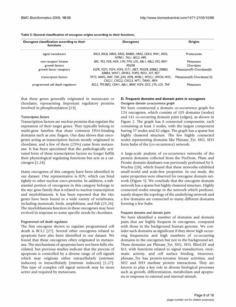
D. Frequent domains and domain pairs in oncogenesOncogene domain co-occurrence graphWe have constructed a domain co-occurrence graph for124 oncogenes, which consists of 105 domains (nodes)and 141 co-occurring domain pairs (edges), as shown inFigure 2. The graph has 8 connected components, eachcontaining at least 3 nodes, with the largest componenthaving 37 nodes and 82 edges. The graph has a sparse buthighly clustered structure. The few highly connectednodes representing domains like Pkinase_Tyr, SH2, SH3form hubs of the (co-occurrence) network.
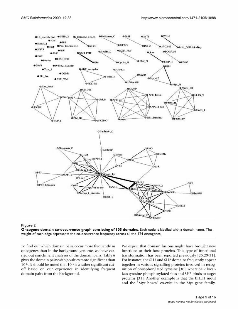
A large-scale analysis of co-occurrence networks of theprotein domains collected from the ProDom, Pfam andProsite domain databases was previously performed by S.Wuchty [28], which found that these networks exhibitedsmall-world and scale-free properties. In our study, thesame properties were observed for oncogene domain net-work (Figure 3). We conclude that the oncogene domainnetwork has a sparse but highly clustered structure. Highlyconnected nodes emerge in the network which predomi-nantly shapes the topology of the underlying network anda few domains are connected to many different domainsforming a few hubs.
Frequent domains and domain pairsWe have identified a number of domains and domainpairs that are highly frequent in oncogenes, comparedwith those in the background human genome. We con-sider such domains as significant if they show high occur-ring frequencies and high numbers of co-occurringdomains in the oncogenes but not in the background set.These domains are Pkinase_Tyr, SH2, SH3, RhoGEF andfn3, with functions related to signal transduction, enzy-matic activity, and cell surface binding. Moreover,pkinase_Tyr has protein-tyrosine kinase activities, andSH2 and SH3 mediate protein interactions. They areknown to play a key role in diverse biological processessuch as growth, differentiation, metabolism and apopto-sis in response to external and internal stimuli.
Table 5: General classification of oncogene origins according to their functions.
Oncogene classification according to their functions
Oncogenes Origins
signal transducers RALA, RALB, NRAS, KRAS, RAB8A, HRAS, CDK4, PIM1, MOS, NTRK1, TAL1, BCL3, MPL
Prokaryotes
non-receptor kinases SRC, YES, FGR, HCK, LYN, FYN, LCK, ABL1, ABL2, FES, RAF1 Metazoansgrowth factors PDGFB Chordates
growth factor receptors EGFR, FGF3, FGF4, FGF6, FLT1, MET, PDGFB, ERBB2, ERBB3, ERBB4, WNT1, EPHA3, THPO, ROS1, KIT, RET
Metazoans(9) Chordates(6)
transcription factors TP73, MAFG, MAF, TNF, JUN, MYB, MYBL1, MYCL1, MYCN, MYC, CXCL1, CXCL2, CXCL3, WT1, TIAM1, IRF4
Metazoans(4) Chordates(12)
programmed cell death regulators BCL2, TP53BP2, CDH1, ABL1, BRAF, FGF4, DCC, LTA, LCK, TNF Metazoans
Page 8 of 16(page number not for citation purposes)
BMC Bioinformatics 2009, 10:88 http://www.biomedcentral.com/1471-2105/10/88
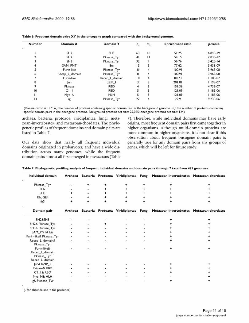
To find out which domain pairs occur more frequently inoncogenes than in the background genome, we have car-ried out enrichment analyses of the domain pairs. Table 6gives the domain pairs with p-values more significant than10-6. It should be noted that 10-6 is a rather significant cut-off based on our experience in identifying frequentdomain pairs from the background.
We expect that domain fusions might have brought newfunctions to their host proteins. This type of functionaltransformation has been reported previously [25,29-31].For instance, the SH3 and SH2 domains frequently appeartogether in various signalling proteins involved in recog-nition of phosphorylated tyrosine [30], where SH2 local-izes tyrosine-phosphorylated sites and SH3 binds to targetproteins [31]. Another example is that the bHLH motifand the "Myc boxes" co-exist in the Myc gene family.
Oncogene domain co-occurrence graph consisting of 105 domainsFigure 2Oncogene domain co-occurrence graph consisting of 105 domains. Each node is labelled with a domain name. The weight of each edge represents the co-occurrence frequency across all the 124 oncogenes.
Page 9 of 16(page number not for citation purposes)
BMC Bioinformatics 2009, 10:88 http://www.biomedcentral.com/1471-2105/10/88
bHLH uses a common mechanism for DNA binding anddimerization while the Myc boxes, on the another hand,appear to be unique to the Myc family and are involved intranscription activation and neoplastic transformation[25]. While the individual functions of these two domainsare generally understood, their synergistic effects in theirbounded protein complex are not known [25].
Two significant triad domain fusions, {SH2, SH3,Pkinase_Tyr} and {Furin-like, Recep_L_domain,Pkinase_Tyr}, are found (Figure 2) and they form sixfused domain pairs (shown in Table 6). Pkinase_Tyr areknown to be related to protein tyrosine kinase activitiesand amino acid phosphorylation. The other two domains,Furin-like and Recep_L_domain, are involved in signaltransduction by receptor tyrosine kinases [32]. It is alsonoteworthy that domains corresponding to the tyrosine
kinase family are among the most frequent families inoncogenes. These domains may carry essential functionsas standalone domains and may also extend their func-tionality to accomplish complex tasks in combinationwith other domains.
E. Phylogenetic profiling diversities of frequent domains and domain pairsDiverse origins of frequent domains and domain pairs arefound in cellular organisms through our phylogeneticprofile analyses, which provide complementary informa-tion to our earlier analysis of domains and domain pairs.Phylogenetic profiling is a computational technique forfunctional analyses of domains and their fusions [33]. Wehave calculated the phylogenetic profiles of all oncogenedomains and domain pairs to find their taxonomic distri-bution across 495 cellular genomes, grouped into 7 taxa:
Frequency distribution of node degrees in oncogene domain networkFigure 3Frequency distribution of node degrees in oncogene domain network. The distribution follows a generalized power law:. Parameter values of the fit (solid curve) are a = 1.125; b = -0.887, and r = 0.101.
Page 10 of 16(page number not for citation purposes)
BMC Bioinformatics 2009, 10:88 http://www.biomedcentral.com/1471-2105/10/88
archaea, bacteria, protozoa, viridiplantae, fungi, meta-zoan-invertebrates, and metazoan-chordates. The phylo-genetic profiles of frequent domains and domain pairs arelisted in Table 7.
Our data show that nearly all frequent individualdomains originated in prokaryotes, and have a wide dis-tribution across many genomes, while the frequentdomain pairs almost all first emerged in metazoans (Table
7). Therefore, while individual domains may have earlyorigins, most frequent domain pairs first came together inhigher organisms. Although multi-domain proteins aremore common in higher organisms, it is not clear if thisobservation about frequent oncogene domain pairs isgenerally true for any domain pairs from any groups ofgenes, which will be left for future study.
Table 6: Frequent domain pairs XY in the oncogene graph compared with the background genome.
Number Domain X Domain Y ns ms Enrichment ratio p-value
1 SH2 SH3 63 16 51.25 6.84E-192 SH2 Pkinase_Tyr 41 11 54.15 7.83E-173 SH3 Pkinase_Tyr 32 9 56.76 3.42E-144 SAM_PNT Ets 13 5 77.62 3.43E-095 Furin-like Pkinase_Tyr 8 4 100.91 3.96E-086 Recep_L_domain Pkinase_Tyr 8 4 100.91 3.96E-087 Furin-like Recep_L_domain 10 4 80.73 1.18E-078 Jun bZIP_1 3 3 201.81 1.19E-079 Pkinase RBD 4 3 151.36 4.73E-0710 C1_1 RBD 5 3 121.09 1.18E-0611 Myc_N HLH 5 3 121.09 1.18E-0613 ig Pkinase_Tyr 27 4 29.9 9.23E-06
(P-value cutoff is 10-6. ns: the number of proteins containing specific domain pair in the background genome. ms: the number of proteins containing specific domain pairs in the oncogene proteins. Background proteins set size: 25,025; oncogene proteins set size: 124)
Table 7: Phylogenetic profiling analysis of frequent individual domains and domain pairs through 7 taxa from 495 genomes.
Individual domain Archaea Bacteria Protozoa Viridiplantae Fungi Metazoan-invertebrates Metazoan-chordates
Phinase_Tyr - + + + + + +SH2 - - + + + + +SH3 + - + + + + +
RhoGEF - + + + + + +fn3 + + + + + + +
Domain pair Archaea Bacteria Protozoa Viridiplantae Fungi Metazoan-invertebrates Metazoan-chordates
SH2&SH3 - - - - - + +SH2& Pkinase_Tyr - - + - - + +SH3& Pkinase_Tyr - - - - - + +SAM_PNT& Ets - - - - - + +
Furin-like& Pkinase_Tyr - - - - - + +Recep_L_domain&
Pkinase_Tyr- - - - - + +
Furin-like& Recep_L_domain
Pkinase_Tyr Recep_L_domain
- - - - - + +
Jun& bZIP_1 - - - - - + +Pkinase& RBD - - - - - + +C1_1& RBD - - - - - + +
Myc_N& HLH - - - - - - +ig& Pkinase_Tyr - - - - - + +
(- for absence and + for presence)
Page 11 of 16(page number not for citation purposes)
BMC Bioinformatics 2009, 10:88 http://www.biomedcentral.com/1471-2105/10/88
ConclusionWe have analyzed the origins of component domains anddomain fusions of oncogenes, and studied the uniquecharacteristics of the oncogene domain pairs in compari-son with those in the background human genome. Mostof these domains and domain pairs are functionallyrelated to protein tyrosine kinase activities, which areclosely related to cancer pathophysiology. Our phyloge-netic profile analysis provides additional evidence to sup-port our observation that frequent domain pairs inoncogenes tend to originate in higher organisms. Theknowledge gained from this computational study mayprovide useful insights about the complex processes ofoncongenesis.
MethodsA. Data sources124 proto-oncogenes of Homo sapiens were collected fromCNIO OncoChip project website http://nciarray.nci.nih.gov/gi_acc_ug_title.shtml and the CancerGenome Anatomy Project database http://cgap.nci.nih.gov/Info/CGAPDownload [see Additionalfile 4], and their protein sequences were obtained fromthe Uniprot database [34] (only the primary protein formwas used). The pre-calculated domain structures of theseproteins were retrieved from the Pfam-A database (version21.0) [8], using HMMER [8] and RPS-BLAST [8] (E-valuecutoff 0.001; sequences were masked for coiled-coils andlow complexity regions). Our list includes all the impor-tant proto-oncogenes previously reported in the literature[2,35-37]. All these proto-oncogenes were manuallycurated based on the published literature.
A proto-oncogene only becomes an oncogene whenmutations or over-expressions take place [37]. Note that"oncogenes" are different from "cancer genes". Com-monly we consider oncogenes as those involved in uncon-trollable cell growth while cancer genes are generallyreferred to genes that are identified with somatic or germ-line mutations in cancer tissues. Futreal et al. recently con-ducted a census of human cancer genes on the basis ofgenetic evidence [16], whose cancer-gene list partly over-laps our oncogene list. Throughout the rest of the paper,we use oncogenes to refer proto-oncogenes for the termi-nology simplicity.
Two sets of genomes and their encoded proteins wereused in our study, one including the whole set of proteinsencoded in 495 sequenced genomes (with 34 archaea,422 bacteria and 39 eukaryotes) from the Integr8 database(release 58) [38] and the other including 367,752 proteinsequences with Pfam annotations from 6,774 sequencedvirus genomes. The second data set was downloaded fromthe Uniprot database at the FTP site [39].
The complete set of proteins of Homo sapiens with Pfamdomain annotation was downloaded from the Integr8database, which contains 25,025 protein sequences with-out splicing isoforms. This dataset set served as the back-ground for our statistical analyses.
It should be noted that currently there is no well-acceptedbenchmark dataset for oncogenes. Since our data weremainly selected from CNIO OncoChip project and CancerGenome Anatomy Project database, a likely bias may existwhen compared with other datasets. One future plan ofour work is to investigate several other cancer gene data-sets, including those identified by exon sequencing stud-ies such as TCGA [40] dataset from the group at JohnHopkins [41] and the cancer gene lists compiled by Fut-real et al.[16], to derive a more comparative dataset ofoncogenes.
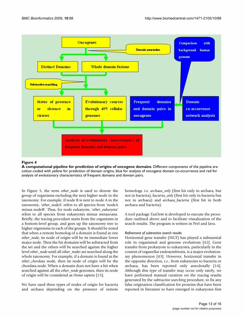
B. A computational pipeline for domain analyses of oncogenesOur computational pipeline for identification of the ori-gins of oncogene domains and domain fusion events con-sists of three main steps (Figure 4). The first step is topredict the origins of domains and domain fusion eventsin oncogenes, which is done through application of a sub-tractive search procedure [15], in conjunction with identi-fication and analyses of horizontal gene transfers to avoidpitfalls, which could potentially lead to misclassificationof domain origins in prokaryotes. The second step is toperform comparative analyses on domains between onco-genes and the background, namely the whole collectionof human proteins. Domains and domain pairs withhigher occurrence frequencies in oncogenes than in thebackground are identified, through an analysis of adomain co-occurrence graph. Detailed analyses on thesedomains and domain pairs are carried out in the third stepof the pipeline, through a combination of a domain/domain pair enrichment analysis and a phylogenetic pro-file analysis (see following sections for details).
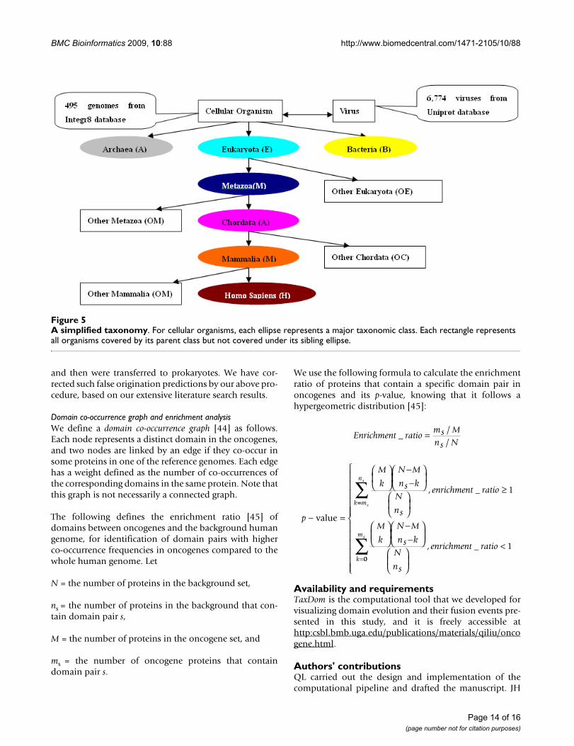
Subtractive searchFirst we generate the domain list of all the 124 oncogenes,and search them against all the sequenced genomes,which are organized into a simplified taxonomy tree,including viruses, archaea, bacteria, eukaryotes, plus a fewincreasingly finer subclasses of eukaryotes leading toHomo sapiens, namely metazoans, chordates and mamma-lian (Figure 5). The questions we ask here are (a) for eachdomain, where did it occur for the first time going from asimplest class of organisms to the most complex one? (b)for each pair of co-occurring domains, where did the co-occurrence take place for the first time in the aforemen-tioned taxonomy?
Page 12 of 16(page number not for citation purposes)
BMC Bioinformatics 2009, 10:88 http://www.biomedcentral.com/1471-2105/10/88
In Figure 5, the term other_node is used to denote thegroup of organisms excluding the next higher node in thetaxonomy. For example, if node B is next to node A in thetaxonomy, 'other_nodeA' refers to all species from 'nodeAminus nodeB'. Thus, for node eukaryote, 'other_eukaryota'refers to all species from eukaryotes minus metazoans.Briefly, the tracing procedure starts from the organisms ina bottom-level group, and goes up the taxonomy tree tohigher organisms in each of the groups. It should be notedthat when a remote homolog of a domain is found at oneother_node, its node of origin will be its immediate lowermajor node. Then the hit domains will be subtracted fromthe set and the others will be searched against the higherlevel other_node until all other_nodes are searched along thewhole taxonomy. For example, if a domain is found at theother_chordata node, then its node of origin will be thechordata node. When a domain does not have a hit whensearched against all the other_node genomes, then its nodeof origin will be considered as Homo sapiens [15].
We have used three types of nodes of origin for bacteriaand archaea depending on the presence of remote
homologs, i.e. archaea_only (first hit only in archaea, butnot in bacteria), bacteria_only (first hit only in bacteria butnot in archaea) and archaea_bacteria (first hit in botharchaea and bacteria).
A tool package TaxDom is developed to execute the proce-dure outlined above and to facilitate visualization of thesearch results. The program is written in Perl and Java.
Refinement of subtractive search resultsHorizontal gene transfer (HGT) has played a substantialrole in organismal and genome evolutions [42]. Genetransfer from prokaryote to eukaryotes, particularly in thecontext of organellar endosymbiosis, is a major evolution-ary phenomenon [43]. However, horizontal transfer inthe opposite direction, i.e., from eukaryotes to bacteria orarchaea, has been reported only anecdotally [14].Although this type of transfer may occur only rarely, wehave performed manual curation on the tracing resultsgenerated by the subtractive searching procedure, to fix anyfalse origination classification for proteins that have beenreported in literature to have emerged in eukaryotes first
A computational pipeline for prediction of origins of oncogene domainsFigure 4A computational pipeline for prediction of origins of oncogene domains. Different components of the pipeline are colour-coded with yellow for prediction of domain origins, blue for analysis of oncogene domain co-occurrence and red for analysis of evolutionary characteristics of frequent domains and domain pairs.
Page 13 of 16(page number not for citation purposes)
BMC Bioinformatics 2009, 10:88 http://www.biomedcentral.com/1471-2105/10/88
and then were transferred to prokaryotes. We have cor-rected such false origination predictions by our above pro-cedure, based on our extensive literature search results.
Domain co-occurrence graph and enrichment analysisWe define a domain co-occurrence graph [44] as follows.Each node represents a distinct domain in the oncogenes,and two nodes are linked by an edge if they co-occur insome proteins in one of the reference genomes. Each edgehas a weight defined as the number of co-occurrences ofthe corresponding domains in the same protein. Note thatthis graph is not necessarily a connected graph.
The following defines the enrichment ratio [45] ofdomains between oncogenes and the background humangenome, for identification of domain pairs with higherco-occurrence frequencies in oncogenes compared to thewhole human genome. Let
N = the number of proteins in the background set,
ns = the number of proteins in the background that con-tain domain pair s,
M = the number of proteins in the oncogene set, and
ms = the number of oncogene proteins that containdomain pair s.
We use the following formula to calculate the enrichmentratio of proteins that contain a specific domain pair inoncogenes and its p-value, knowing that it follows ahypergeometric distribution [45]:
Availability and requirementsTaxDom is the computational tool that we developed forvisualizing domain evolution and their fusion events pre-sented in this study, and it is freely accessible athttp:csbl.bmb.uga.edu/publications/materials/qiliu/oncogene.html.
Authors' contributionsQL carried out the design and implementation of thecomputational pipeline and drafted the manuscript. JH
Enrichment ratioms Mns N
_//
=
p
M
k
N M
ns kN
ns
enrichment ratiok
− =
⎛
⎝⎜
⎞
⎠⎟
−−
⎛
⎝⎜
⎞
⎠⎟
⎛
⎝⎜
⎞
⎠⎟
≥
value
, _ 1==
=
∑
⎛
⎝⎜
⎞
⎠⎟
−−
⎛
⎝⎜
⎞
⎠⎟
⎛
⎝⎜
⎞
⎠⎟
<
m
n
k
s
s
M
k
N M
ns kN
ns
enrichment ratio, _ 100
ms
∑
⎧
⎨
⎪⎪⎪⎪⎪⎪
⎩
⎪⎪⎪⎪⎪⎪
A simplified taxonomyFigure 5A simplified taxonomy. For cellular organisms, each ellipse represents a major taxonomic class. Each rectangle represents all organisms covered by its parent class but not covered under its sibling ellipse.
Page 14 of 16(page number not for citation purposes)
BMC Bioinformatics 2009, 10:88 http://www.biomedcentral.com/1471-2105/10/88
was responsible for the evolutionary analysis of the onco-genes. HL and PW participated in the preparation andanalysis of oncogene data. YX and XY conceived the studyand coordinated the involved data analyses as well as writ-ing the manuscript. All authors read and approved thefinal manuscript.
Additional material
AcknowledgementsThis work was supported in part by National Science Foundation (DBI-0354771, ITR-IIS-0407204, CCF-0621700, DBI-0542119), the National Institutes of Health (R01GM075331) and the Distinguished Cancer Clini-cians and Scientists Program from Georgia Cancer Coalition. JH acknowl-edges the support by a Research and Creativity Award from the East Carolina University. The authors would also like to thank other members of the Computational Systems Biology Laboratory for their helpful discus-sions.
References1. Pierotti Micro A, Frattini Milo, Sozzi Gabriella: Oncogenes. In Can-
cer Medicine 7th edition. Edited by: James F. Holland et al.Lea&Febiger, London; 2007.
2. Steven Martin G: The road to Src. Oncogene 2004, 23:7910-7917.3. Vogel C, Bashton M, Kerrison ND, Chothia C, Teichmann SA: Struc-
ture, function and evolution of multidomain proteins. CurrOpin Struct Biol 2004, 14:208-216.
4. Holm L, Sander C: The FSSP database of structurally alignedprotein fold families. Nucleic Acids Res 1994, 22:3600-3609.
5. Siddiqui AS, Barton GJ: Continuous and discontinuous domains:An algorithm for the automatic generation of reliable pro-tein domain definitions. Protein Sci 1995, 4:872-884.
6. Swindells MB: A procedure for detecting structural domains inproteins. Protein Sci 1995, 4:103-112.
7. Holm L, Sander C: Dictionary of recurrent domains in proteinstructures. Proteins 1998, 33:88-96.
8. Finn Robert D, Mistry Jaina, Schuster-Böckler Benjamin, Griffiths-Jones Sam, Hollich1 Volker, Lassmann1 Timo, Moxon Simon, MarshallMhairi, Khanna2 Ajay, Durbin Richard, Eddy2 Sean R, Sonnhammer1Erik LL, Bateman Alex: Pfam: clans, web tools and services.Nucleic Acids Research Database Issue 2006, 34:D247-D251.
9. Schultz J, Milpetz F, Bork P, Ponting CP: SMART, a simple modu-lar architecture research tool: identification of signallingdomains. Proc Natl Acad Sci 1998, 95:5857-5864.
10. Servant F, Bru C, Carrere S, Courcelle E, Gouzy J, Peyruc D, Kahn D:ProDom: automated clustering of homologous domains.Brief Bioinform 2002, 3:246-251.
11. Robinson Dan R, Wu1 Yi-Mi, Lin Su-Fang: The protein tyrosinekinase family of the human genome. Oncogene 2000,19:5548-5557.
12. Park Jeonghyeon, Kunjibettu Sudeesha, McMahon Steven B, ColeMichael D: The ATM-related domain of TRRAP is required forhistone acetyltransferase recruitment and Myc-dependentoncogenesis. Genes Dev 2001, 15:1619-1624.
13. Westbrook CA, Hooberman AL, Spino C, Dodge RK, Larson RA,Davey F, Wurster-Hill DH, Sobol RE, Schiffer C, Bloomfield CD:Clinical Significance of the BCR-ABL Fusion Gene in AdultAcute Lymphoblastic Leukemia: A Cancer and LeukemiaGroup B Study. Blood 1992, 80(12):2983-2990.
14. Ponting CP, Aravind L, Schultz J, Bork P, Koonin EV: Eukaryotic sig-nalling domain homologues in archaea and bacteria. Ancientancestry and horizontal gene transfer. J Mol Biol 1999,289(4):729-745.
15. Lipika R Pal, Chittibabu Guda: Tracing the origin of functionaland conserved domains in the human proteome: implica-tions for protein evolution at the modular level. BMC Evolu-tionary Biology 2006, 6:91.
16. Futreal PA, Coin L, Marshall M, Down T, Hubbard T, Wooster R,Rahman N, Stratton MR: A census of human cancer genes. NatRev Cancer 2004, 4(3):177-83.
17. Bork P: Hundreds of ankyrin-like repeats in functionally-diverse proteins: mobile modules that cross phyla horizon-tally? Proteins: Structure, Function, and Genetics 1993, 17(4):363-74.
18. Chang PC, Chi CW, Chau GY, Li FY, Tsai YH, Wu JC: DDX3, aDEAD box RNA helicase, is deregulated in hepatitis virus-associated hepatocellular carcinoma and is involved in cellgrowth control. Oncogene 2006, 25:1991-2003.
19. Robinson HL: Retroviruses and cancer. Rev Infect Dis 1982,4(5):1015-25.
20. Beral V, Newton R, Weiss RA, eds: Infection and Human Cancer.Cancer Surveys 1998, 33:1-396.
21. Coffin J, Hughes SH, Varmus HE, eds: Retroviruses Cold Spring HarborLaboratory Press, New York; 1997.
22. Hurley JB, Simon MI, Teplow DB: Homologies between signaltransducing G proteins and gene products. Science 1984,226(4676):860-862.
23. Klein G: Cellular Oncogene Activation Marcel Dekker Inc, NY; 1988. 24. Banerjee R, Caruccio L, Zhang YJ, Mckercher S, Santelia RM: Effects
of carcinogen-induced transcription factors on the activationof hepatitis B virus expression in human hepatoblastomaHepG2 cells and its implication on hepatocellular carcino-mas. Hepatology 2000, 32(2):367-74.
25. Atchley WR, Fitch WM: Myc and Max: Molecular Evolution of aFamily of Proto-Oncogene Products and Their DimerizationPartner. Proc Natl Acad Sci 1995, 92:10217-10221.
26. Walker CW, Boom JD, Marsh AG: First non-vertebrate memberof the myc gene family is seasonally expressed in an inverte-brate testis. Oncogene 1992, 7(10):2007-2012.
27. Korsmeyer SJ: Bcl-2 initiates a new category of oncogenes:regulators of cell death. Blood 1992, 80:879-886.
28. Wuchty Stefan: Scale-free behavior in protein domain net-works. Mol Biol Evol 2001, 18:1694-1702.
29. Bashton Matthew, Chothia Cyrus: The Generation of New Pro-tein Functions. Structure 2007, 15:85-99.
30. Hegyi Hedi, Gerstein Mark: Annotation Transfer for Genomics:Measuring Functional Divergence in Multi-Domain Proteins.Genome Res 2001, 11:1632-1640.
31. Vogel Christine, Berzuini Carlo, Bashton Matthew: Supra-domains:Evolutionary Units Larger than Single Protein Domains. JMol Biol 2004, 336:809-823.
32. Raz E, Schejter ED, Shilo BZ: Interallelic complementationamong DER/flb alleles: implications for the mechanism of
Additional file 1Supplementary S2. 103 domains encoded from oncogene proteins.Click here for file[http://www.biomedcentral.com/content/supplementary/1471-2105-10-88-S1.xls]
Additional file 2Supplementary S3. 50 whole domain fusions of oncogenes.Click here for file[http://www.biomedcentral.com/content/supplementary/1471-2105-10-88-S2.xls]
Additional file 3Supplementary S4. Proteome-wide patterns of origin nodes in oncogene proteins.Click here for file[http://www.biomedcentral.com/content/supplementary/1471-2105-10-88-S3.xls]
Additional file 4Supplementary S1. oncogene list.Click here for file[http://www.biomedcentral.com/content/supplementary/1471-2105-10-88-S4.xls]
Page 15 of 16(page number not for citation purposes)
BMC Bioinformatics 2009, 10:88 http://www.biomedcentral.com/1471-2105/10/88
Publish with BioMed Central and every scientist can read your work free of charge
"BioMed Central will be the most significant development for disseminating the results of biomedical research in our lifetime."
Sir Paul Nurse, Cancer Research UK
Your research papers will be:
available free of charge to the entire biomedical community
peer reviewed and published immediately upon acceptance
cited in PubMed and archived on PubMed Central
yours — you keep the copyright
Submit your manuscript here:http://www.biomedcentral.com/info/publishing_adv.asp
BioMedcentral
signal transduction by receptor-tyrosine kinases. Genetics1991, 129(1):191-201.
33. Pellegrini Matteo, Marcotte Edward M, Thompson Michael J, Eisen-berg David, Grothe Robert, Yeates Todd O: Assigning proteinfunctions by comparative genome analysis: Protein phyloge-netic profiles. Proc Natl Acad Sci 1999, 96:4285-4288.
34. The UniProt Consortium: The Universal Protein Resource(UniProt). Nucleic Acids Res 2007, 35:D193-197.
35. Darmoul Dalila, Gratio Valérie, Devaud Hélène, Peiretti Franck,Laburthe Marc: Activation of proteinase-activated receptor 1promotes human colon cancer cell proliferation through epi-dermal growth factor receptor transactivation. Mol CancerRes 2004, 2(9):514-522.
36. Espinosa AV, Porchia L, Ringel MD: Targeting BRAF in thyroidcancer. Br J Cancer 2007, 96(1):16-20.
37. Robert AW: The biology of Cancer 1st edition. Garland Science; Lon-don; 2006.
38. Paul K, Bower L, Morris L, Horne A, Petryszak R, Kanz C, Kanapin A,Das U, Michoud K, Phan I, Gattiker A, Kulikova T, Faruque N, DugganK, Mclaren P, Reimholz B, Duret L, Penel S, Reuter I, Apweiler R:Integr8 and genome reviews: integrated views of completegenomes and proteomes. Nucleic Acids Res 2005, 33:D297-D302.
39. The Uniprot virus data [ftp://ftp.ebi.ac.uk/pub/databases/uniprot/current_release/knowledgebase/]
40. The Cancer Genome Atlas Research Network: Comprehensivegenomic characterization defines human glioblastoma genesand core pathways. Nature 2008, 455(7216):1061-1068.
41. Sjoblom T, Jones S, Wood LD, Parsons DW, Lin J, Barber TD, Man-delker D, Leary RJ, Ptak J, Silliman N: The Consensus CodingSequences of Human Breast and Colorectal Cancers. Science2006, 314:268-274.
42. Salzberg Steven L, White Owen, Peterson Jeremy, Eisen Jonathan A:Microbial Genes in the Human Genome: Lateral Transfer orGene Loss? Science 2001, 292:1903-1906.
43. Doolittle WF: You are what you eat: a gene transfer ratchetcould account for bacterial genes in eukaryotic nucleargenomes. Trends Genet 1998, 14(8):307-311.
44. Ye Y, Godzik Z: A Comparative analysis of protein domainorganization. Genome Res 2004, 14:343-353.
45. Xing Yi, Xu Qiang, Lee Christopher: Widespread production ofnovel soluble protein isoforms by alternative splicingremoval of transmembrane anchoring domains. FEBS Letters2003, 555:572-578.
Page 16 of 16(page number not for citation purposes)