Embed Size (px)
Citation preview

DOI: 10.1126/science.1063830, 357 (2001);294 Science
et al.Yuanqing WangOrigin of the Mammalian Middle EarAn Ossified Meckel's Cartilage in Two Cretaceous Mammals and
This copy is for your personal, non-commercial use only.
clicking here.colleagues, clients, or customers by , you can order high-quality copies for yourIf you wish to distribute this article to others
here.following the guidelines
can be obtained byPermission to republish or repurpose articles or portions of articles
): January 8, 2014 www.sciencemag.org (this information is current as of
The following resources related to this article are available online at
http://www.sciencemag.org/content/294/5541/357.full.htmlversion of this article at:
including high-resolution figures, can be found in the onlineUpdated information and services,
http://www.sciencemag.org/content/suppl/2001/10/11/294.5541.357.DC1.html can be found at: Supporting Online Material
http://www.sciencemag.org/content/294/5541/357.full.html#ref-list-1, 1 of which can be accessed free:cites 24 articlesThis article
38 article(s) on the ISI Web of Sciencecited by This article has been
http://www.sciencemag.org/content/294/5541/357.full.html#related-urls2 articles hosted by HighWire Press; see:cited by This article has been
http://www.sciencemag.org/cgi/collection/paleoPaleontology
subject collections:This article appears in the following
registered trademark of AAAS. is aScience2001 by the American Association for the Advancement of Science; all rights reserved. The title
CopyrightAmerican Association for the Advancement of Science, 1200 New York Avenue NW, Washington, DC 20005. (print ISSN 0036-8075; online ISSN 1095-9203) is published weekly, except the last week in December, by theScience
on
Janu
ary
8, 2
014
ww
w.s
cien
cem
ag.o
rgD
ownl
oade
d fr
om
on
Janu
ary
8, 2
014
ww
w.s
cien
cem
ag.o
rgD
ownl
oade
d fr
om
on
Janu
ary
8, 2
014
ww
w.s
cien
cem
ag.o
rgD
ownl
oade
d fr
om
on
Janu
ary
8, 2
014
ww
w.s
cien
cem
ag.o
rgD
ownl
oade
d fr
om
on
Janu
ary
8, 2
014
ww
w.s
cien
cem
ag.o
rgD
ownl
oade
d fr
om
on
Janu
ary
8, 2
014
ww
w.s
cien
cem
ag.o
rgD
ownl
oade
d fr
om

13. P. R. Cummins, B. L. N. Kennet, J. R. Bowman, M. G.Bostock, Bull. Seismol. Soc. Am. 82, 323 (1992).
14. H. M. Benz, J. E. Vidale, Nature 365, 147 (1993).15. We included 1530 events with a depth from 0 to 75
km, a magnitude of 6.0 # Mw # 7.0, and fromstations in the epicentral distance range 100° # D #160° for the period 1 January 1980 to 29 March1998. The SS phases in the individual traces arehand-picked, and the data is deconvolved for receivereffects and bandpass-filtered between 15 and 75 s.
16. The size of the caps corresponds to the Fresnel zoneof the SS rays. Neighboring caps overlap partly toensure smoothing.
17. B. Efron, R. Tibshirani, Science 253, 390 (1991). Thedata set was also divided into two subsets withepicentral distances from 100° to 130° and 130° to160°; splitting was still present in stacks of thesubsets.
18. Supplementary material is available at www.sciencemag.org/cgi/content/full/294/5541/354/DC1
19. The stacks are cross-correlated with the SS pulse;splitting is determined by two cross-correlationmaxima (instead of one) in the depth range of 480to 600 km.
20. We find single reflections from 520 km for stacks
corresponding to different regional types (shields,tectonically active regions, stable continents, andoceans). The stacks exhibit somewhat different am-plitudes and, in particular, the shield stack shows asmaller amplitude, confirming an earlier study (6).
21. N. A. Simmons, H. Gurrola, Nature 405, 559 (2000).22. D. Canil, Phys. Earth Planet. Inter. 86, 25 (1994).23. S. Koito, M. Akaogi, O. Kubuta, T. Suzuki, Phys. Earth
Planet. Inter. 120, 1 (2000).24. D. J. Weidner, Y. Wang, in Earth’s Deep Interior:
Mineral Physics and Tomography from the Atomic tothe Global Scale (Geophysical Monograph 117,American Geophysical Union, Washington, DC,2000), pp. 215–235.
25. J. Ritsema, H. J. van Heijst, J. H. Woodhouse, Science286, 1925 (1999).
26. Y. Fei, C. Bertka, in Mantle Petrology: Field Observa-tions and High-Pressure Experimentation, Special Pub-lication in honor of Francis R. Boyd, Geochem. Soc.Spec. Pub. (Geochemical Society, Washington Uni-versity, St. Louis, MO, 1999), vol. 6, pp. 189–207.
27. T. Inoue, D. J. Weidner, P. A. Northrup, J. B. Parise,Earth Planet. Sci. Lett. 160, 107 (1998).
28. H. Yusa, T. Inoue, Y. Ohishi, Geophys. Res. Lett. 27,413 (2000).
29. A. Dziewonski, D. Anderson, Phys. Earth Planet. Inter.25, 297 (1981).
30. Previous SS-precursor studies suggest a shear waveimpedance contrast of 6 to 10% for the 410-kmdiscontinuity and 10% for the 660-km discontinuity(9). In the mid–transition zone region, computersimulations for a pyrolite mantle composition give ajump of 1.66% in shear wave impedance for thegarnet transition and 3.13% for the olivine transitionof the b-phase to g-phase (24).
31. W. D. Mooney, G. Laske, G. Masters, Eos (Fall Suppl.)76, F421 (1995).
32. C. H. Chapman, Geophys. Res. Lett. 3, 153 (1976).The synthetics are basically delta pulses computedusing WKBJ ray tracing for PREM (including attenua-tion from PREM) and then filtered in the same way asthe data.
33. We thank A. Jephcoat and A. Kleppe for useful dis-cussions on mineralogical phase transitions. A.D. wasfunded by a Scatcherd Scholarship from Oxford Uni-versity. We also acknowledge support under U.K.Natural Environment Research Council grantGR11534.
18 June 2001; accepted 24 August 2001
An Ossified Meckel’s Cartilagein Two Cretaceous Mammalsand Origin of the Mammalian
Middle EarYuanqing Wang,1* Yaoming Hu,1,2,3 Jin Meng,2* Chuankui Li1
An ossified Meckel’s cartilage has been recovered from two early Cretaceousmammals from China. This element is similar to Meckel’s cartilage in prenataland some postnatal extant mammals and indicates the relationship of Meckel’scartilage with the middle ear in early mammals. The evidence shows that brainexpansion may not be the initial factor that caused the separation of post-dentary bones from the dentary as middle ear ossicles during mammalianevolution. The failure of the dentary to seize reduced postdentary elementsduring ontogeny of early mammals is postulated as an alternative mechanismfor the separation. Modifications of both feeding and hearing apparatuses inearly mammals may have led to the development of the definitive mammalianmiddle ear.
In nonmammalian vertebrates with jaws, thecraniomandibular joint is between the quad-rate region of the palatoquadrate and the ar-ticular region of Meckel’s cartilage (or itsreplacement). In unequivocal mammals (1,2), the joint is between the squamosal and thedentary. The definitive mammalian middleear (DMME) is formed by transference ofaccessory jaw elements, including the angu-
lar, articular plus prearticular, and quadrate,to the cranium of mammals as strictly audi-tory ossicles (renamed as the tympanic, mal-leus, and incus) (3). This transference is oneof the central topics of comparative anatomyand evolutionary biology of vertebrates (3–8). Although developmental studies of extantmammals have long demonstrated homolo-gies of these elements among jawed verte-brates (9, 10), the only fossil evidence on thiscritical transference is the presence of persis-tent grooves on the medial surface of thedentary bone, which may have lodged theanterior end of the postdentary unit (PDU,consisting of the endochondral articular anddermal prearticular, angular, and surangular)in some early mammals (3).
Four nearly complete Repenomamus adultskulls with articulated lower jaws (11) andone with articulated lower jaws of an un-named Gobiconodon species (Figs. 1 and 2)
were discovered from the Yixian Formationof the lower Cretaceous in Liaoning, China(12). Of the two taxa, Repenomamus (11)represents one of the largest Mesozoic mam-mals, and is most closely related to gobicon-odontids (13–15) in sharing basic structuresof jaws, teeth, occlusal pattern, and somecranial features (Figs. 1 to 3). Gobiconodon-tids are related to triconodontids within tri-conodonts, a diverse grade of basal mamma-liaform groups with uncertain relationships(2, 15–18) (Fig. 3). Among these specimens,a structure that we recognize as an ossifiedMeckel’s cartilage (OMC) was preserved intwo skulls of Repenomamus (IVPP speci-mens V12549 and V12728) and one skull ofGobiconodon (IVPP V12585). Of the twoOMCs in Repenomamus, the one in V12549is in its original location (Figs. 1, A to C, and2, A and C), whereas the other in V12728 isdisplaced and lies between the mandible andthe skull (Figs. 1D and 2B). The OMC isrod-like, with a pointed anterior tip and aflared posterior end. It measures 33 mm longin V12549 and 40 mm in V12728. The ante-rior portion of the OMC in V12549 is lodgedin a depression that appears to be an expand-ed posterior portion of the meckelian groove.The OMC-dentary contact may have hadsome mobility. During preparation, the OMCwas separated from the dentary. In all lowerjaws of Repenomamus, the anterior tip of themeckelian groove is below m3 (the thirdlower molariform tooth) and continues ante-riorly as a slit that parallels the course of themandibular canal within the dentary. Themandibular canal, as revealed by radiograph-ic imaging, is low in position, ventral to thelong roots of the check teeth, and extendsanteriorly to the symphysis. The radiographshows that in lateral view, the mandibularcanal turns slightly dorsally at the positionwhere the anterior tip of the OMC is situated,and extends posteriorly to the mandibular
1Institute of Vertebrate Paleontology and Paleoan-thropology (IVPP), Chinese Academy of Sciences, PostOffice Box 643, Beijing, 100044, China. 2Division ofPaleontology, American Museum of Natural History(AMNH), Central Park West at 79th Street, New York,NY 10024, USA. 3Biology Program (Ecology, Evolu-tionary Biology, and Behavior), Graduate School andCity College, City University of New York, NY 10016–4309, USA.
*To whom correspondence should be addressed. E-mail: [email protected] (Y.W.); [email protected] ( J.M.)
R E P O R T S
www.sciencemag.org SCIENCE VOL 294 12 OCTOBER 2001 357
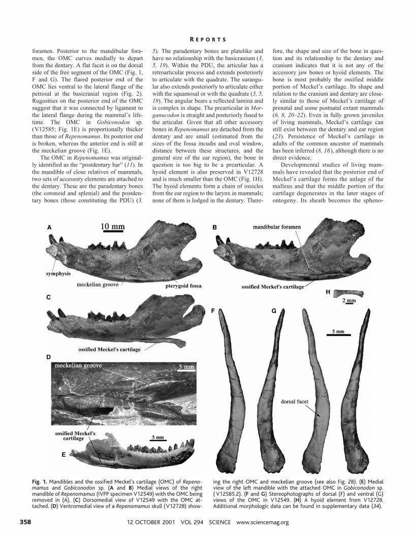
foramen. Posterior to the mandibular fora-men, the OMC curves medially to departfrom the dentary. A flat facet is on the dorsalside of the free segment of the OMC (Fig. 1,F and G). The flared posterior end of theOMC lies ventral to the lateral flange of thepetrosal at the basicranial region (Fig. 2).Rugosities on the posterior end of the OMCsuggest that it was connected by ligament tothe lateral flange during the mammal’s life-time. The OMC in Gobiconodon sp.(V12585; Fig. 1E) is proportionally thickerthan those of Repenomamus. Its posterior endis broken, whereas the anterior end is still atthe meckelian groove (Fig. 1E).
The OMC in Repenomamus was original-ly identified as the “postdentary bar” (11). Inthe mandible of close relatives of mammals,two sets of accessory elements are attached tothe dentary. These are the paradentary bones(the coronoid and splenial) and the postden-tary bones (those constituting the PDU) (3,
5). The paradentary bones are platelike andhave no relationship with the basicranium (3,5, 19). Within the PDU, the articular has aretroarticular process and extends posteriorlyto articulate with the quadrate. The surangu-lar also extends posteriorly to articulate eitherwith the squamosal or with the quadrate (3, 5,19). The angular bears a reflected lamina andis complex in shape. The prearticular in Mor-ganucodon is straight and posteriorly fused tothe articular. Given that all other accessorybones in Repenomamus are detached from thedentary and are small (estimated from thesizes of the fossa incudis and oval window,distance between these structures, and thegeneral size of the ear region), the bone inquestion is too big to be a prearticular. Ahyoid element is also preserved in V12728and is much smaller than the OMC (Fig. 1H).The hyoid elements form a chain of ossiclesfrom the ear region to the larynx in mammals;none of them is lodged in the dentary. There-
fore, the shape and size of the bone in ques-tion and its relationship to the dentary andcranium indicates that it is not any of theaccessory jaw bones or hyoid elements. Thebone is most probably the ossified middleportion of Meckel’s cartilage. Its shape andrelation to the cranium and dentary are close-ly similar to those of Meckel’s cartilage ofprenatal and some postnatal extant mammals(6, 8, 20–22). Even in fully grown juvenilesof living mammals, Meckel’s cartilage canstill exist between the dentary and ear region(23). Persistence of Meckel’s cartilage inadults of the common ancestor of mammalshas been inferred (8, 16), although there is nodirect evidence.
Developmental studies of living mam-mals have revealed that the posterior end ofMeckel’s cartilage forms the anlage of themalleus and that the middle portion of thecartilage degenerates in the later stages ofontogeny. Its sheath becomes the spheno-
Fig. 1. Mandibles and the ossified Meckel’s cartilage (OMC) of Repeno-mamus and Gobiconodon sp. (A and B) Medial views of the rightmandible of Repenomamus (IVPP specimen V12549) with the OMC beingremoved in (A). (C) Dorsomedial view of V12549 with the OMC at-tached. (D) Ventromedial view of a Repenomamus skull (V12728) show-
ing the right OMC and meckelian groove (see also Fig. 2B). (E) Medialview of the left mandible with the attached OMC in Gobiconodon sp.( V12585.2). (F and G) Stereophotographs of dorsal (F) and ventral (G)views of the OMC in V12549. (H) A hyoid element from V12728.Additional morphologic data can be found in supplementary data (34).
R E P O R T S
12 OCTOBER 2001 VOL 294 SCIENCE www.sciencemag.org358

mandibular ligament ( pterygomandibularin monotremes) and the anterior ligamentof the malleus (3, 9, 10). The OMC inRepenomamus and Gobiconodon provides
evidence for the relationship of Meckel’scartilage with the DMME in early mam-mals, which is otherwise inferred only fromembryological evidence of living mammals.
It also shows that, while the anlage of themalleus is reduced, or posteriorly shifted, toform the malleus, a significant middle seg-ment of Meckel’s cartilage is persisted andossified in adults, probably remaining in itsearly ontogenetic position. A similar condi-tion is probably present in other early mam-mals, such as triconodontids and symmetro-dontids. The function of the OMC in adults ofthese mammals is unclear. A dorsal facet onthe OMC of Repenomamus (Fig. 1F) suggeststhat muscle was attached. If so, the OMCmay have functioned as the inflected angu-lar process in marsupials (24 ), or the ptery-goid shelf in multituberculates (25), forpartial insertion of the medial pterygoidmuscle that originates on the pterygoid re-gion of the skull. When opening and clos-ing the lower jaw, the attached OMC couldrotate with the jaw, with its contact at thelateral flange serving as the fulcrum. Mas-tication may not have interfered with hear-ing in Repenomamus.
Identification of the OMC leads to theconclusion that Repenomamus and Gobicon-odon have a DMME, because the dentary ofthe two taxa lacks other scars for the PDU.The fossa incudis immediately medial to thesecondary craniomandibular joint (SCMJ,dentary-squamosal) in Repenomamus indi-cates an intermediate condition between themandibular ear of nonmammalian synapsids,such as Morganucodon, and the DMME ofmore advanced mammals in which the earossicles are widely separated from the SCMJand lie behind intervening secondary auditorystructures (7). This relationship shows that inearly mammals the SCMJ is lateral, not an-terior [as shown in ontogenesis of livingmammals (8, 9)], to the primary joint (mal-leo-incudal 5 quadrate-articular); the ear os-sicles are medial, not posterior [as shown inextant mammals (7)], to the SCMJ. Otherfeatures of Repenomamus, such as an elon-gated promontorium and a distinct externalauditory meatus, can be attributed to moreefficient hearing of airborne sound, while theexpanded glenoid fossa and mandibular con-dyle, enlarged pterygoid, and broad masseter-ic fossa (Figs. 1, A to E, and 2) are related tomore powerful mastication.
Discovery of the OMC helps to interpretgrooves present near the mandibular foramenof the dentary in many early mammals andtheir relatives. These puzzling grooves wereknown at least since Owen (26) and havebeen considered either as holding dentalnerves and arteries (13, 27) or as facets forthe PDU in Peramus and Amphitherium (3).Because Peramus and Amphitherium are inthe Trechnotheria (1) (Fig. 3), the latter in-terpretation of the PDU in these taxa arguesfor multiple origins of the DMME (3). Ourevidence demonstrates that in some earlymammals, an OMC is probably the primary
Fig. 2. Skulls and basicranial region of Repenomamus. (A) Ventral view of V12549, showingrelationship of the ossified Meckel’s cartilage (indicated by arrow) with the dentary and ear region.(B) Ventromedial view of V12728 showing the displaced OMC. (C) Close ventral view of thebasicranial region (V12549). The maximum skull dimensions of V12549 are 108 mm by 71 mm.Using the method of (18), the width of the “brain vault” is 28 mm, which is the maximum distancebetween the squamosal-parietal sutures. The width between the temporomandibular joints is 60mm (between the midpoints of the glenoid fossae). The actual brain vault of Repenomamus isnarrower, as reviewed by radiographic imaging and by direct observation from a broken skull(V12613) in which the wall of the braincase measures 3.7 mm thick.
R E P O R T S
www.sciencemag.org SCIENCE VOL 294 12 OCTOBER 2001 359

occupant for the grooves in question. Theevidence, viewed within the phylogeny,weakens the hypothesis of multiple origins ofthe DMME (3).
The phylogeny based on 112 craniodentalcharacters from 20 taxa (Fig. 3) is largely inkeeping with other recent phylogenetic hy-potheses of mammals and their relatives (2,15–18). Within the phylogeny, acquisition ofthe DMME in Repenomamus and Gobicon-odon is consistent with the prediction thattriconodontids have ear ossicles (3). Whetherthe DMME is a synapomorphy for Mamma-lia, which probably occurred in the middleJurassic, or it is shared by Mammalia andHadrocodium and thus evolved in the earlyJurassic (18) (Fig. 3), depends on the inter-pretation of Hadrocodium. The type speci-men of Hadrocodium (IVPP V8275) wasoriginally regarded as a juvenile Morganuc-odon (28), but is now considered an adult, orsubadult, of a distinctive taxon in which thePDU is detached from the dentary (18). In our
view, however, many features, such as itssmall size, erupting first upper postcaninetooth (28), only two molars, slender mandi-ble, large space between m2 and the coronoidprocess, large promontorium, and large brainvault (18), suggest that V8275 is a postsuck-ling juvenile. Whether the common ancestorof Hadrocodium and mammals evolved theDMME in the early Jurassic requires furthertesting (Fig. 3).
The most uncertain issue in the evolutionof the DMME is how the PDU became de-tached from the dentary and translocated tothe basicranium as ear ossicles (3–8). Onemodel suggests that brain expansion in-creased the distance between the middle earand the mandible during ontogeny and evo-lution of mammals and thus tore off the earossicles from the mandible (7, 18). This mod-el is not consistent with the narrow braincasesof our specimens of Repenomamus and Go-biconodon. The maximum ratio of the esti-mated brain vault to the skull width in
V12549 of Repenomamus is 49% (see Fig. 2caption), smaller than those of Sinoconodonand Morganucodon, in which the PDU is stillattached to the dentary (18, 29, 30). Thisindicates that detachment of the ear ossiclesis not necessarily associated with expansionof the brain during mammalian evolution. InRepenomamus, the fossa incudis, which re-flects the position of the incus and malleus, isimmediately medial to the SCMJ. The dis-tance between the SCMJ and the fossa incu-dis is proportionally similar to, or even small-er than, the space between the quadrate recessand the SCMJ in Morganucodon (18, 30, 31).This shows that separation of the PDU fromthe dentary does not require greater distancebetween the ear and the mandible.
Our specimens permit an alternative hy-pothesis for the origin of the DMME. Duringevolution of synapsids, the PDU is reduced insize and loosened to enhance hearing of high-frequency airborne sounds (5), whereas thedentary was enlarged for attachment of moremuscle to facilitate efficient mastication (3, 32,33). The position of the OMC in Repenomamussuggests that the common ancestor of mammalsprobably had a developmental pattern in whichMeckel’s cartilage extended from dentary to theear region. Because of its close relationshipwith the cartilage, the dentary was probablytilted in position. Reduction of the PDU in-creasingly weakened its tie to the dentary untila critical point was reached where the dentary,while erecting to a more vertical position duringontogeny, no longer seized the PDU, which wasmoored at the basicranium by connective tissue.This hypothesis is similar to the detachingmechanism of the ear ossicles in marsupials(6), without requiring brain expansion as theinitial trigger. Modifications in both feedingand hearing apparatuses toward efficient func-tions have led to the decoupling of the PDU anddentary. Expansion of the brain, along withchanges in the otic capsule, may have causeddisplacements of the ear ossicles to a positioneither more vertical (6), horizontal (8), or pos-teriorly distant from the SCMJ (7) in moreadvanced mammals.
References and Notes1. M. C. McKenna, S. K. Bell, Classification of Mammals
Above the Species Level (Columbia Univ. Press, NewYork, 1997).
2. T. Rowe, J. Vertebr. Paleontol. 8, 241 (1988).3. E. F. Allin, J. A. Hopson, in The Evolutionary Biology of
Hearing, D. B. Webster, R. R. Fay, A. N. Popper, Eds.(Springer-Verlag, New York, 1992), pp. 587–614.
4. J. A. Hopson, Am. Zool. 6, 437 (1966).5. E. F. Allin, J. Morphol. 147, 403 (1975).6. W. Maier, Neth. J. Zool. 40, 55(1990).7. T. Rowe, Mem. Cal. Acad. Sci. 20, 71 (1996).8. U. Zeller, in Mesozoic Differentiation, Multitubercu-
lates, Monotremes, Early Therians, and Marsupials,vol. 1, Mammal Phylogeny, F. S. Szalay, M. J. Novacek,M. C. McKenna, Eds. (Springer-Verlag, New York,1993), pp. 95–107.
9. E. Gaupp, Die Reichertsche Theorie (Archiv fur Anato-mie und Entwicklungsgeschichte, 1912, Hammer-,Amboss-, und Kieferfrage, 1913).
Fig. 3. Phylogenetic relationships and distributions of main mammaliaform groups. The cladogramis the consensus (tree length 5 275; CI 5 0.589; RI 5 0.706) of four equally most-parsimonioustrees that are obtained by branch-and-bound searches using PAUP* 4.0 b8 based on 112craniodental characters across 20 terminal taxa. Search options include: all characters unordered,equally weighted, accelerated transformation, multistate as uncertainty, rooting at tritylodontids,and monophyletic in-group. Numbers at each node represent assigned branch length, bootstrap-ping value, and Bremer supporting index. Bootstrapping value is obtained by 1000 replications ofheuristic searches. Red dots represent occurrences of genera (1) used in the phylogenetic analyses.Pink bars are distributions of higher taxa (1) represented by the genera. Character list, sources ofdata, and detailed tree descriptions can be found in supplementary data (34).
R E P O R T S
12 OCTOBER 2001 VOL 294 SCIENCE www.sciencemag.org360
10. E. S. Goodrich, Studies on the Structure and Develop-ment of Vertebrates (Macmillan, London, 1930).
11. J.-l. Li, Y. Wang, Y.-q. Wang, C-k. Li, Chin. Sci. Bull. 45,2545 (2000).
12. X.-l. Wang et al., Vert. PalAsiat. 36, 81 (1998).13. F. A. Jr. Jenkins, C. R. Schaff, J. Vertebr. Paleontol. 8,
1 (1988).14. Z. Kielan-Joworoska, D. Dashzeveg, Acta Palaeontol.
Pol. 43, 413 (1998); H.-J. Kuhn, Abh. Senckenb.Naturforsch. Ges. 28, 1 (1971).
15. Q. Ji, Z.-x. Luo, S.-a. Ji, Nature 398, 326 (1999).16. G. W. Rougier, J. R. Wible, M. J. Novacek, Am. Mus.
Novit. 3187, 1 (1966).17. Y.-m. Hu, Y.-q. Wang, Z.-x. Luo, C.-k. Li, Nature 390,
137 (1997).18. Z.-x. Luo, A. W. Crompton, A.-l. Sun, Science 292,
1535 (2001).19. K. A. Kermack, F. Mussett, H. W. Rigney, Zool. J. Linn.
Soc. 53, 87 (1973).20. H.-J. Kuhn, Abh. Senckenb. Naturforsch. Ges. 28, 1
(1971).21. U. Zeller, in Morphogenesis of the Mammalian Skull,
H.-J. Kuhn, U. Zeller, Eds. (Verlag Paul Parey, Ham-burg and Berlin, 1987), pp. 17–50.
22. C. T. Clark, K. K. Smith, J. Morphol. 215, 119 (1993).23. H.-J. Kuhn, in (21), pp. 1–12.24. M. R. Sanchez-Villagra, K. K. Smith, J. Mamm. Evol. 4,
119 (1997).25. P. P. Gambaryan, Z. Kielan-Jaworowska, Acta Palae-
ontol. Pol. 40, 45 (1995).26. R. Owen, Monograph of the Fossil Mammalia of the
Mesozoic Formations (Paleontographical Society,London, 1871).
27. G. G. Simpson, A Catalogue of the Mesozoic Mam-malia in the Geological Department of the BritishMuseum (Clowes & Sons, London and Beccles, 1928).
28. A. W. Crompton, Z.-x. Luo, in (8), pp. 30–44.29. A. W. Crompton, A.-l. Sun, Zool. J. Linn. Soc. 85, 99
(1985).30. K. A. Kermack, F. Mussett, H. W. Rigney, Zool. J. Linn.
Soc. 71, 1 (1981).31. Z.-x. Luo, A. W. Crompton, J. Vertebr. Paleontol. 14,
341 (1994).
32. A. W. Crompton, Proc. Zool. Soc. London 140, 697(1963).
33. H. R. Barghusen, J. A. Hopson, Science N.Y. 168, 573(1970).
34. Supplementary material is available at www.sciencemag.org/cgi/content/full/294/5541/357/DC1
35. We thank J. Li and Y. Wang for valuable discussion; M.Zhang, Z. Zhou, X. Xu, X. Wang, F. Zhang, F. Jin, andJ. Zhang for help in coordinating the research andfieldwork; S. Xie, H. Wang (IVPP), and A. Davidson(AMNH) for preparation of the specimens; and fouranonymous reviewers for instructive comments. Thiswork is supported by the Ministry of Science andTechnology, P. R. China (Major Basic Research Project,G2000077700), National Natural Science Foundationof China (49832002), and Chinese Academy of Sci-ences (Grand Research Projects, KZCX3-J-03, KZ951-B1-410).
27 June 2001; accepted 30 August 2001
Genetic Basis for ActivityDifferences Between
Vancomycin and GlycolipidDerivatives of VancomycinUlrike S. Eggert,1 Natividad Ruiz,2 Brian V. Falcone,1
Arthur A. Branstrom,3 Robert C. Goldman,3 Thomas J. Silhavy,2
Daniel Kahne1*
Small molecules that affect specific protein functions can be valuable tools fordissecting complex cellular processes. Peptidoglycan synthesis and degradationis a process in bacteria that involves multiple enzymes under strict temporaland spatial regulation. We used a set of small molecules that inhibit thetransglycosylation step of peptidoglycan synthesis to discover genes that helpto regulate this process. We identified a gene responsible for the susceptibilityof Escherichia coli cells to killing by glycolipid derivatives of vancomycin, thusestablishing a genetic basis for activity differences between these compoundsand vancomycin.
Vancomycin (Fig. 1A) is the drug of lastresort for treating resistant Gram-positivebacterial infections, and the emergence ofvancomycin resistance presents a seriousthreat to public health. Vancomycin inhibitsthe maturation of the peptidoglycan layer sur-rounding bacterial cells by binding to D-Ala-D-Ala, a dipeptide found in peptidoglycanprecursors (Fig. 1B) (1). Resistance to van-comycin arises when microorganisms acquiregenes that lead to the substitution of D-Ala-D-Ala by D-Ala-D-Lac (2), which vancomy-cin does not bind. Remarkably, vancomycinderivatives with a hydrophobic substituent onthe carbohydrate moiety are active against
vancomycin-resistant strains (3) even thoughthey contain the same peptide binding pocketas vancomycin. The mechanism of action ofthese derivatives may be fundamentally dif-ferent from that of vancomycin (4). Unlikevancomycin, they retain activity against bothvancomycin-sensitive and vancomycin-resis-tant strains even when the peptide bindingpocket is damaged (5). In vitro, they block adifferent step of peptidoglycan synthesis thandoes vancomycin (5). In addition, they killbacteria very rapidly, whereas vancomycinonly stops growth (6).
Because vancomycin and its derivativesaffect cells differently (i.e., produce differentphenotypes), it might be possible, using achemical genetics approach, to identify genesinvolved in the cellular response to thesecompounds. The synthesis of peptidoglycanfrom its disaccharide precursor involves nu-merous enzymes with overlapping functionsthat are subject to tight temporal and spatial
regulation (7). Most of the major enzymes inpeptidoglycan synthesis—the transglycosy-lases and transpeptidases—have been identi-fied, but how these enzymes are regulatedremains poorly understood. In developing anexperimental approach to probe the cellularresponse to glycolipid derivatives of vanco-mycin, we focused on the following facts:Vancomycin blocks the transpeptidation stepof peptidoglycan synthesis and kills cellsslowly; glycolipid derivatives of vancomycinblock the transglycosylation step of pepti-doglycan synthesis (Fig. 1B) and provoke arapid lethal response in cells (Fig. 1C).Moenomycin, another transglycosylase in-hibitor, also induces a rapid lethal response incells (8). Thus, inhibiting the transglycosyla-tion step of peptidoglycan synthesis may ac-tivate a pathway that triggers rapid cell death.If so, it should be possible to identify com-ponents of this pathway by selecting for mu-tants that are resistant to small molecules thatinhibit transglycosylation.
We initiated a search for mutants resis-tant to three different transglycosylase in-hibitors: chlorobiphenyl vancomycin, des-leucyl chlorobiphenyl vancomycin, andmoenomycin (Fig. 1A). Moenomycin bindsdirectly to key bacterial transglycosylases(9). Chlorobiphenyl vancomycin inhibitstransglycosylation by binding to the D-Ala-D-Ala terminus of the peptidoglycan pre-cursor lipid II and also by binding to com-ponents of the transglycosylation complex(5). Desleucyl chlorobiphenyl vancomycincannot bind D-Ala-D-Ala and is proposed toinhibit transglycosylation primarily by thelatter mechanism (5).
Mutants resistant to transglycosylase in-hibitors were obtained by growing E. coliimp (10) on plates impregnated with chlo-robiphenyl vancomycin, desleucyl chloro-biphenyl vancomycin, or moenomycin.Three mutants were isolated that were re-sistant to each of these antibiotics (BE101,BE102, and BE103; Table 1). The muta-
1Department of Chemistry, 2Department of MolecularBiology, Princeton University, Princeton, NJ 08544,USA. 3Advanced Medicine Inc., 901 Gateway Boule-vard, South San Francisco, CA 94080, USA.
*To whom correspondence should be addressed. E-mail: [email protected]
R E P O R T S
www.sciencemag.org SCIENCE VOL 294 12 OCTOBER 2001 361