Embed Size (px)
Citation preview

An investigation of the phytochemistry and biological activity of Asparagus laricinus
Sandile Lawrence Fuku
A thesis submitted to the department of Health Sciences, Central University of
Technology, Free State, in fulfillment of the requirements for the degree of
Doctor Technologiae: Biomedical Technology
Promoter : Prof SS Mashele, PhD
Co-promoter : Dr. I. Manduna, Ph.D
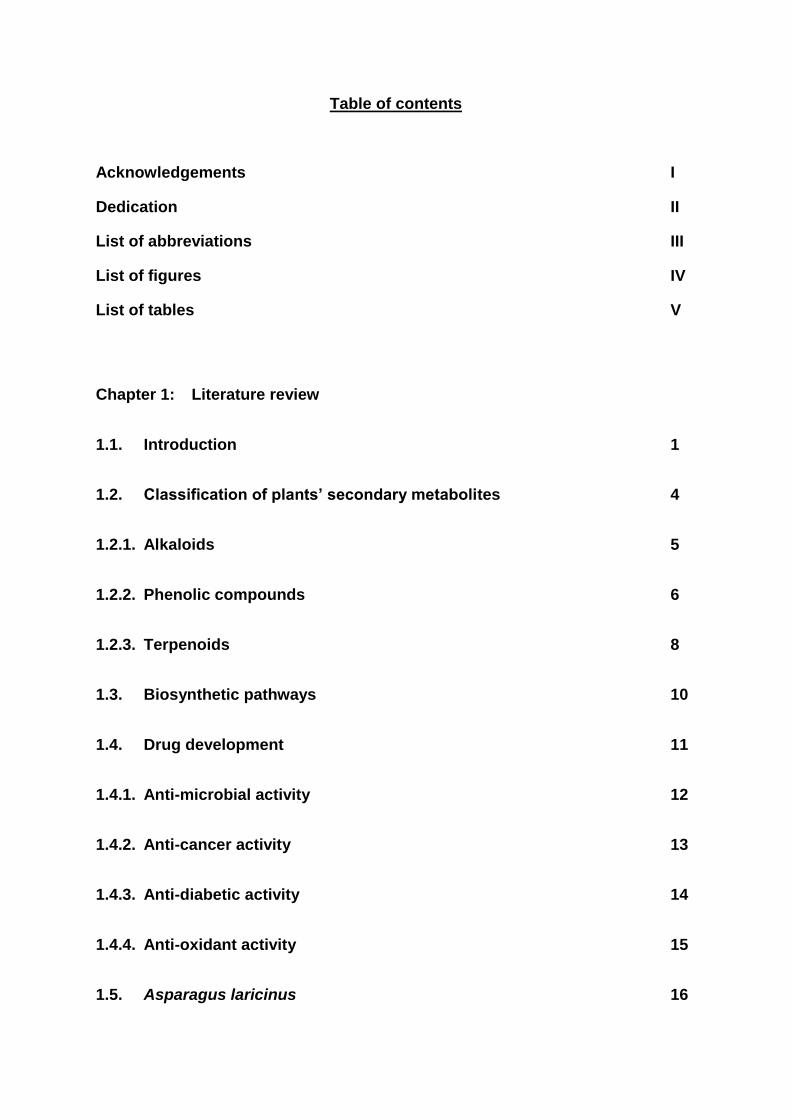
Table of contents
Acknowledgements I
Dedication II
List of abbreviations III
List of figures IV
List of tables V
Chapter 1: Literature review
1.1. Introduction 1
1.2. Classification of plants’ secondary metabolites 4
1.2.1. Alkaloids 5
1.2.2. Phenolic compounds 6
1.2.3. Terpenoids 8
1.3. Biosynthetic pathways 10
1.4. Drug development 11
1.4.1. Anti-microbial activity 12
1.4.2. Anti-cancer activity 13
1.4.3. Anti-diabetic activity 14
1.4.4. Anti-oxidant activity 15
1.5. Asparagus laricinus 16

1.6. References 18
Chapter 2: In vitro anticancer screening of Asparagus laricinus extracts 30
2.1. Introduction 30
2.2. Materials and methods 32
2.2.1. Plant material 32
2.2.2. In vitro anti-cancer screening 32
2.2.3. Analysis of results 33
2.3. Results 34
2.4. Discussion 35
2.5. Conclusion 37
2.6. References 38
Chapter 3: Evaluation of the mutagenicity and cytotoxicity effect of
Asparagus laricinus 41
3.1. Introduction 41
3.2. Materials and methods 44
3.2.1. Plant material 44
3.2.2. The Ames test 44
3.2.3. Determination of total phenolic content 46
3.2.4. Cytotoxicity 47
3.3. Results 49

3.3.1. Mutagenicity and anti-mutagenicity 49
3.3.2 Cytotoxicity 53
3.4. Discussion 53
3.5. Conclusion 55
3.6 References 57
Chapter 4: Evaluation of the antimicrobial, antiradical and antioxidant
activities of Asparagus laricinus aqueous extract 62
4.1. Introduction 62
4.2. Materials and methods 64
4.2.1. Antimicrobial activity 64
4.2.2. Thin layer chromatography analysis and antioxidant activity of extract’s
constituents 65
4.2.3. Free radical scavenging activity 66
4.2.4. Oxidative stress 67
4.3. Results 68
4.3.1 Minimum inhibitory concentration 68
4.3.2. Thin layer chromatography analysis and antioxidant activity of extract’s
constituents 70
4.3.3. Free radical scavenging activity 71
4.3.3. Oxidative stress 72

4.4. Discussion 72
4.5. Conclusion 75
4.6 References 76
Chapter 5: Chemical composition of both aqueous and methanol extracts of
Asparagus laricinus 81
5.1. Introduction 81
5.2. Materials and methods 83
5.2.1. Phytochemical screening 83
5.2.2. Alkaloid determination using Harborne (1973) method 83
5.2.3. Test for tannins 83
5.2.4. Test for saponin 84
5.2.5 Test for steroids 84
5.2.6. Test for terpenoids (Salkowski test) 84
5.3. Gas chromatography-mass spectrometry 85
5.4. Liquid chromatography/mass spectometry 85
5.5. Results 87
5.5.1 Phytochemical screening 87
5.5.2 Gas chromatography-mass spectrometry 88
5.5.3 Liquid chromatography/mass spectometry 88
5.6. Discussion 95

5.7. Conclusion 96
5.8. References 97
Summary 102

I
Acknowledgements
I am highly indebted to Professor Samson Mashele for his patience and kindness in
supervising this study. Sir, your contributions and guidance made it possible for me to
make this modest contribution in science.
I would like to acknowledge the humility and assistance I received from Mr. Dan
Mokgawa in making it possible for me to settle at the Central University of Technology
(CUT). Let me express my gratitude to Dr. I. Manduna for her assistance in the course
of this study.
To colleagues and staff members in the Biomedical Technology division, I cannot
express my gratitude for your co-operation and thoughtfulness whenever I needed you.
Paks, I know you had to abandon your family duties in order to get me to Pretoria on
several occasions and for that I am thankful.
To my family, Ndiyabulela maMpondomise ngo kundixhasa, ndithi makudede
ubumnyama kuvele ukukhaya! I would be naïve if I did not acknowledge the
contribution of my lovely partner (Nobuhle), maXaba enkosi ngokuma nam.
I would like to thank my friends (Mabaki Mohlomi, Lemphane Mohokare, Mohau
Mpakana and Godfrey Tlou) whose support renewed my hope and purpose in pursuing
a dream that almost faded. In the same breath, let me acknowledge all comrades in the
Mass Democratic Movement for their efforts in creating a family away from home during
difficult times in my studies.
I would also like to acknowledge the financial contributions received from the National
Research Fund (NRF).

II
Dedication
I dedicate this thesis to my beloved son Lwazi and my late grandfather Mlulu Fuku . It is
the birth of the former and the passing away of the latter that has reaffirmed the unity of
opposites- that death stole the one person who rooted my life in necessity, and birth
restored the prestige by granting me a life to root in necessity


IV
List figures
Figure 1.1: Alkaloids (Taken from Kennedy and Whightman, 2011).
(5)
Figure 1.2: Phenols (Aggarwal and Shishodia, 2006; Dewick, 2002)
(7)
Figure 1.3: Basic building unit and various classes of terpenes (Taken
from Hao et al., 2013, ). (9)
Figure 1.4: Principal biosynthetic pathways leading to synthesis of
secondary metabolites. Adapted from Rawamat et al., 2009.
(10)
Figure 2.1: Growth inhibitory effect of ethanol extracts of Asparagus
laricinus on three human cancer cell lines: MCF7(▲),
TK10(▄) and UACC62(□). (35)
Figure 2.2: Growth inhibitory effect of aqueous extracts of Asparagus
laricinus on three human cancer cell lines: MCF-7(▲), TK-
10(▄) and UACC-62 (□) (35)
Figure 3.1: Standard curve of absorbance against gallic acid
concentration Y = 0.003604*X + 0.1098, R2 = 0.9909.
(47)
Figure 3.2: Cytotoxicity of aqueous extracts of A. laricinus on the growth
of Vero cells were examined my MTT assay.
(52)

V
Figure 4.1: A digital photograph of the Streptococcus pneumonia
ATCCC 6301 treated with increasing concentration of
Asparagus laricinus aqueous extract. (52)
Figure 4.2: Chromatogram of Chromatogram of gallic acid (1) and A.
laricinus aqueous extract (2 and 3), separated with
methanol:chloroform: hexane (70:20:10%) and sprayed with
DPPPH. The yellow spots indicated antioxidant activity (RF
values: a = 0.87; b = 0.38; c = 0.35) (70)
Figure 4.3(a-d): Scavenging activity of A. laricinus aqueous extract on the
free radical DPPH, (a) A. laricinus extract, (b) Trolox®, (c)
and ascorbic acid, (d) EC50 shift graph of A. laricinus extract
with Trolox® as the control. (71)
Figure 4.4: Yeast cell (BY4742) sensitivity to hydrogen peroxide.
Exponentially growing yeast cells (30 oC) were treated with
10mM (H2O2) for 90 minutes. Cell treated with H2O2 only
(▲), untreated cells (●) and Cells treated with H2O2 and
aqueous extract (■). (72)
Figure 5.1: GC/MS chromatogram of the aqueous extract of A. laricinus
roots. (88)
Figure 5.2: Representative LC-MS chromatograms for ethanol extract (a) and aqueous extract (b) in negative ion electrospray.(89)

VI
Figure 5.3: Representative LC-MS chromatograms for ethanol extract (a) and aqueous extract (b) in positive ion electrospray. (90)

VII
List of tables
Table 2.1. Cytotoxic activity (GI50 values) of plant extracts and etoposide that are used in the treatment of cancer cell lines
(35)
Table 3.1: List of Salmonella typhimurium strains used in Ames test
(46)
Table 3.2: Summary of mutagenic/anti-mutagenic properties of
Asparagus laricinus extract. (49)
Table 3.3: Mutagenic activities of Asparagus laricinus extract with
metabolic activation (+S9). (51)
Table 4.1 Antimicrobial activity of the aqueous extract of Asparagus laricinus against selected clinical strains of bacteria . (68)
Table 5.1: Phytochemical screening of aqueous and ethanol extracts of
the roots of A. laricinus (87)
Table 5.2: Analysis of LC/MS chromatograms of Aspagagus laricinus
ethanol extract. (91)
Table 5.3: Analysis of LC/MS chromatograms of Aspagagus laricinus
aqueous extract. (93)

1
Chapter 1
Literature review
1.1. Introduction
Medicinal plants are part of indigenous people‟s cultural heritage, thus since ancient
times treatment of various diseases using medicinal plants has been part of human
culture. The value of medicinal plants to mankind has been very well proven. It is
estimated that 70% to 80% of people worldwide rely mainly on traditional health care
systems, especially on herbal medicines (Stanley and Luz, 2003).
In many societies the medicinal properties of plants were discovered mostly through
trial and error, but use was also influenced by the belief systems of the people
involved and often became entangled with religious and mythical practices (Mathias
et al., 1996). Besides that, medicinal plants are proving to be rich resources of
constituents that can be used in drug development and synthesis.
Medicinal plants have been a source of a wide variety of biologically active
compounds for many centuries and have been used extensively as crude material or
as pure compounds for treating various disease conditions. Between 1% and 10% of
plants out of an estimated 250 000 to 500 000 species of plants on earth are used by
humans (Boris, 1996).

2
Plants used for medicinal purposes contribute significantly to the development of
major medical drugs that are used today. Most common medicines have compounds
extracted from plants as their primary active ingredients and many have provided
blueprints for synthetic or partially synthesized drugs (Simpson and Ogorzaly, 2001).
There has been a major resurgence of interest in traditionally used medicinal plants,
with a number of international and local initiatives actively exploring the botanical
resources of southern Africa with the intention to screen indigenous plants for
pharmacologically active compounds (Gurib-Fakim et al., 2010; Rybicki et al., 2012).
South Africa is considered a “hot spot” for biodiversity and more than 22 000 plant
species occur within its boundaries. This represents 10% of the world‟s species,
although the land surface of South Africa is less than 1% of the earth‟s surface
(Coetzee et al., 1999).
Plants have also been used by man for various purposes, among others as arrow
and dart poisons for hunting, poisons for murder, hallucinogens used for ritualistic
purposes, stimulants for endurance and hunger suppression, as well as medicine
(Duke et al., 2008; Cragg and Newman, 2005).
A derivative of the polyhydroxy diterpenoid ingenol isolated from the sap of
Euphorbia peplus (known as “petty spurge” in England or “radium weed” in
Australia), which is a potential chemotherapeutic agent for skin cancer, is currently
under clinical development by Peplin Biotech for the topical treatment of certain skin
cancers (Kedei et al., 2004; Ogbourne et al., 2004). Combretastatin A-4 phosphate,

3
a stilbene derivative from the South African bush willow, Combretum caffrum, acts as
an anti-angiogenic agent causing vascular shutdowns in tumors (Newman et al.,
2005; Holwell et al., 2002).
Further reliance on plants for drug development is demonstrated by the use of
galantamine hydrobromide, an alkaloid obtained from the plant Galanthus nivalis
used traditionally in Turkey and Bulgaria for the treatment of Alzheimer‟s disease
(Howes et al., 2003; Heinrich and Teoh, 2004).
The plant chemicals used for the above-mentioned purposes are secondary
metabolites, which are derived biosynthetically from plant primary metabolites (e.g.
carbohydrates, amino acids and lipids). Secondary metabolites are organic
compounds that are exclusively produced by plants and that are not directly involved
in the normal growth, development and reproduction of a plant (Firn and Jones,
2003). Yet, they have many functions that are important for the plant‟s long-term
health and appearance.
Plants, being stationary, have to cope with a number of challenges, including
engineering their own pollination and seed dispersal, local variation in the supply of
the simple nutrients that they require to synthesize their food and the coexistence of
herbivores and pathogens in their immediate environment. Plants have therefore
evolved secondary biochemical pathways that allow them to synthesize a spectrum
of organic molecules, often in response to specific environmental stimuli, such as
herbivore-induced damage, pathogen attacks, or nutrient deprivation (Reymond et
al., 2000; Hermsmeier et al., 2001).

4
The biosynthesis of secondary metabolites is derived from the fundamental
processes of photosynthesis, glycolysis and the Krebs cycle to afford biosynthetic
intermediates which, ultimately, result in the formation of secondary metabolites also
known as natural products (Dewick, 2002).
It is hypothesized that secondary metabolism utilizes amino acids and the acetate
and shikimate pathways to produce “shunt metabolites” (intermediates) that have
adopted an alternate biosynthetic route, leading to the biosynthesis of secondary
metabolites (Sarker et al., 2006).
Modifications in the biosynthetic pathways that produce secondary metabolites are
probably due to natural causes (e.g. viruses or environmental changes) or unnatural
causes (e.g. chemical or radiation processes) in an effort to adapt or provide
longevity for the plant (Sarker et al., 2006). Plants‟ secondary metabolites can be
classified into several groups according to their chemical classes, such alkaloids,
terpenoids and phenolics (Harbone, 1984; Wink, 2003).
1.2. Classification of plants’ secondary metabolites
The palette of secondary metabolites can be divided into a number of distinct groups
on the basis of their chemical structure and synthetic pathways. These groups can,
in turn, be broadly differentiated in terms of the nature of their ecological roles and
therefore their ultimate effects and comparative toxicity in the consuming animal
(Kennedy and Whightman, 2011). The three main groups of secondary metabolites

5
in plants that are of interest in this study are alkaloids, phenolic compounds and
terpenoids (Firn and Jones, 2003; Wink, 2003).
1.2.1. Alkaloids
Alkaloids are complex N-containing heterocyclic organic compounds and are among
the most important plant materials for the development and production of drugs
(Facchini, 2001). Although no single classification exists, Kennedy and Whightman
(2011) argued that alkaloids are often distinguished on the basis of a structural
similarity (e.g. indole alkaloids) or a common precursor (e.g. benzylisoquinoline,
tropane, pyrrolizidine, or purine alkaloids).
Figure 1.1. Alkaloids (taken from Kennedy and Whightman, 2011).

6
The recorded use of alkaloids for medicinal purposes stretches back some 5 000
years (Goldman, 2001) and this class of molecules has contributed to the majority of
poisons, neurotoxins, traditional psychedelics (e.g. atropine, scopolamine and
hyoscyamine, from the plant Atropa belladonna) and social drugs consumed by
humans, e.g. nicotine, caffeine, methamphetamine (ephedrine), cocaine and opiates,
(Zenk and Juenger, 2007).
1.2.2. Phenolic compounds
Phenolic compounds (Harborne and Williams, 2000) are based on phenol (an
oxygen linked to a fully saturated C6 ring), the simplest member of this class of plant
substances. Phenolic compounds include, among others, flavonoids and tannins
(Harborne and Williams, 2000). Phenols are the compounds containing a hydroxyl
group (—OH) directly attached to an aromatic ring.

7
Figure 1.2. Phenols (Aggarwal and Shishodia, 2006; Dewick, 2002)
Phenolic compounds, in particular flavonoids, are generally involved in the protection
of plants from attack by microbes and insects (Cushnie and Lamb, 2005; Friedman,
2007).
Special classes of plant phenolics are the tannins, characteristically astringent, bitter
plant polyphenols that are toxic to herbivores owing to their capacity to either bind
and precipitate, or shrink proteins and other macromolecules (Cushnie and Lamb,
2005). Some phenols are used as chemopreventive agents, e.g. resveratrol (3, 5,
4-trihydroxystilbene) is an oligomeric polyphenol found as dimer, trimer and tetramer.
This molecule is implicated in the prevention of cancer and cardiovascular diseases

8
in vasoprotection and neuroprotection (Ates et al., 2007; Delmas et al., 2006; Vitrac
et al., 2004).
Acetylsalicylic acid (aspirin) a time-honored analgesic and antipyretic drug, was
derived from salicylic acid, which occurs in willow trees. Some other important
salicylic acid derivatives are methyl salicylate, a common ingredient of liniments
(Dewick, 2002). Eugenols extracted from cloves are used as an anesthetic and
antiseptic in pharmaceutical and dental preparations (Daniel et al., 2009).
1.2.3. Terpenoids
Terpenoids are dimers or combinations of isoprene, a common organic compound
that is highly volatile because of its low boiling point (Zwenger and Basu, 2008).
This class of molecules is diverse and consists of groups of more than 30 000 lipid-
soluble compounds. Their structure includes one or more five-carbon isoprene units,
which are ubiquitously synthesized by all organisms through two potential pathways,
the mevalonate and deoxy-d-xylulose pathways (Rohmer, 1999).
The general chemical structure of terpens is C10H16, and they occur as diterpenes,
triterpenes, and tetraterpenes (C20, C30, and C40), as well as hemiterpenes (C5) and
sesquiterpenes (C15) (Arif et al., 2011).

9
Figure 1.3. Basic building unit and various classes of terpenes (taken from Hao et al., 2013).
Geraniol, an acyclic dietary monoterpene, represents the only monoterpene that has
been studied in vitro against liver cancer cells. Geraniol was shown to inhibit the
growth of HepG2 human hepatic carcinoma cells by decreasing 3-

10
hydroxymethylglutaryl coenzyme A (HMG-CoA) reductase, the major rate-limiting
enzyme in cholesterol biosynthesis in mammals (Polo and Bravo, 2006).
Ardisiacrispin (A+B), a triterpenoid saponin mixture in the fixed proportion 2:1 of
ardisiacrispin A and ardisiacrispin B, is derived from Ardisia crenata. This mixture
exerted cytotoxic activity against Bel-7402 liver cancer cells through pro-apoptotic,
anti-proliferative and microtubule disruptive activities (Li et al., 2008).
1.3. Biosynthetic pathways
The synthesis of different classes of secondary metabolites from primary metabolites
is presented in schematic form in Fig. 1.4.
Figure 1.4. Principal biosynthetic pathways leading to synthesis of secondary metabolites. Adapted from Ramawat et al. (2009).

11
Secondary metabolites are predominantly synthesized via two principal biosynthetic
pathways. The first one, the shikimic acid pathway, produces a pool of aromatic
amino acids, which in turn are converted into diverse compounds such as phenolics
(lignins, tannins, quinones) and alkaloids (Mustafa and Verpoorte, 2007). The
second one is the acetyl-CoenzymeA mevalonic acid pathway, which produces a
vast array of terpenoids (Eisenreich et al., 2004).
1.4. Drug development
Though enormous progress has been made in medicinal chemistry, the development
of a novel drug has become more and more difficult. The reasons are manifold,
including the fact that good drugs are available for major diseases, and developing a
better drug that is active on the same target without being more expensive becomes
increasingly difficult in view of the sophistication in industrial production methods
(Wess et al., 2001).
Despite the recent interest in molecular modeling, combinatorial chemistry, and other
synthetic chemistry techniques by pharmaceutical companies and funding
organizations, natural products, particularly medicinal plants, remain an important
source of new drugs, new drug leads and new chemical entities (Newman et al.,
2000; Newman et al 2003; Butler, 2004).
Interestingly, of the 877 novel medicines that were developed in the period 1981-
2002, 6% were natural products, 27% were derivatives of natural products and 16%
were synthetics developed on the model of a natural product (Newman et al., 2003).

12
This demonstrates that nature is an important source for developing novel leads for
medicines. Even when new chemical structures are not found during drug discovery
from medicinal plants, known compounds with new biological activity can provide
important drug leads. Since the sequencing of the human genome, thousands of
new molecular targets have been identified as important in various diseases (Kramer
and Cohen, 2004). The history of drug discovery and even drug chemistry that is
inexorably bound to the plant kingdom confirms that the process of deriving drugs
from plant sources is certainly not a new phenomenon.
Several known compounds isolated from traditionally used medicinal plants have
already been shown to act on newly validated molecular targets, as exemplified by
indirubin, which selectively inhibits cyclin-dependent kinases (Hoessel et al., 1999;
Eisenbrand et al., 2004) and kamebakaurin, which has been shown to inhibit NF- kB
(Hwang et al., 2001; Lee et al., 2002).
1.4.1. Anti-microbial activity
It is estimated that plant materials are present in or have provided the models for
50% of Western drugs developed today (Robbers et al., 1996). Many commercial
drugs used in modern medicine today were initially used in crude form in traditional
or folk healing practices, or for other purposes that confirmed their potential useful
biological activity. Avancini et al. (2000) demonstrated the antimicrobial actions of
“carqueja” (Baccharis trimera Less.) extracts on Gram-positive (Staphylococcus
aureus and Streptococcus uberis) and Gram-negative (Salmonella gallinarum and
Escherichia coli) bacterial strains.

13
High antimicrobial activity of Thymus, Origanum and Eugenia caryophillus species
has been attributed to their phenolic components such as thymol and carvacrol
(Lambert et al., 2001; Hazzit et al., 2009). Similarly, the antimicrobial activity of
Cinnamomum zeylanicum has been related to its cinnamaldehyde content (Juliani et
al., 2009).
1.4.2. Anti-cancer activity
The search for anti-cancer agents from plant sources started in the 1950s with the
discovery and development of the vinca alkaloids, vinblastine and vincristine, and the
isolation of the cytotoxic podophyllotoxins (Cragg and Newman, 2005). The first
agents to advance into clinical use were the so-called vinca alkaloids, vinblastine
and vincristine, isolated from the Madagascar periwinkle, Catharanthusroseus
(Gueritte and Fahy, 2005). These agents are primarily used in combination with
other cancer chemotherapeutic drugs for the treatment of a variety of cancers,
including leukemia, lymphoma, advanced testicular cancer, breast and lung cancer
and Kaposi‟s sarcoma.
Combretastatins were isolated from an indigenous South African plant, Combretum
caffrum (Pinney et al., 2005). These molecules have served as a model for the
treatment of tumors and as a result many synthetic analogues of Combretastatin-4
have been created in order to improve its cytoxicity and inhibition of tubulin
polymerization (Nam, 2003; Ohsumi et al., 1998). That has provided an impressive
display of the power of a relatively simple natural product structure that can be

14
altered to produce superior products through medicinal and combinatorial chemistry
(Li and Sham, 2002).
1.4.3. Anti-diabetic activity
The aqueous leaf extract of A. squamosa has been reported to ameliorate
hyperthyroidism (Sunanda and Anand, 2003), which is considered a causative factor
for diabetes mellitus. However, the mechanism involved in the extract‟s inhibition of
hyperthyroidism is not known. More than 1000 plant species are being used for the
treatment of Type II diabetes mellitus worldwide (Trojan-Rodrigues et al., 2011).
Similar to the A. squamosa extract, very little is known about the mechanism of
action of these anti-diabetic plants, thus limiting their use in standard diabetes care.
Many plants have shown anti-diabetic action through the release of insulin and some
extra pancreatic mechanisms (Jung et al., 2006). However, more investigations must
be carried out to evaluate the exact mechanism of action of medicinal plants with
anti-diabetic and insulin-mimetic activity (Patel et al., 2012).
Phytoconstituents such as alkaloids inhibit alpha-glucosidase and decrease glucose
transport through the intestinal epithelium and imidazoline compounds stimulate
insulin secretion in a glucose-dependent manner (Patel et al., 2012). This shows
most researchers‟ preference for pure or semi-pure molecules in evaluating
mechanisms of action. This leads to poor validation of the health benefits and
biosafety of many plants that are traditionally used.

15
1.4.4. Anti-oxidant activity
Antioxidant-based drugs/formulations for the prevention and treatment of complex
diseases such as atherosclerosis, stroke, diabetes, Alzheimer‟s disease and cancer
have emerged in the last three decades (Devasagayam et al., 2004). This coincides
with the upsurge of interest in the therapeutic potentials of plants as antioxidants in
reducing free radical induced tissue injury. Although many synthetic anti-oxidants,
such as butylated hydroxyanisole (BHA) and butylated hydroxytoluene
(BHT) are commercially available, their toxicity is a growing concern. Both
carcinogenic and anticarcinogenic properties have been reported for the synthetic
antioxidants BHA and BHT (Botterweek et al., 2000).
In dealing with the toxicity of synthetic anti-oxidants, a worldwide trend towards the
use of natural phytochemicals present in berries, tea, herbs, oilseeds, beans, fruit
and vegetables has increased (Lee and Shibamoto, 2000; Wang and Jiao, 2000).
Natural antioxidants, especially phenolics and flavonoids from tea, wine, vegetables
and spices, are already exploited commercially either as antioxidant additives or
nutritional supplements (Schuler, 1990; Chu et al., 2000).
Many other plant species have also been investigated in the search for novel
antioxidants, but there is generally still a demand to find more information concerning
the antioxidant potential of plant species, as they are safe and bioactive.

16
1.5. Asparagus laricinus
Knowledge about the medicinal value of many plants that form part of the rich
biodiversity in South Africa is largely contained in the oral traditions of various ethnic
groups that constitute the indigenous people of South Africa (van der Merwe et al.,
2001). Loss of indigenous culture, in favor of western European-derived culture, is
an accelerating process among indigenous people around the world (Prance, 1994).
Consequently, the traditional knowledge that forms the basis of the use of medicinal
plants is in danger of being lost and warrants rigorous scientific investigation.
The current study was prompted by case reports describing unexpected
improvement in patients who had been terminally ill with advanced prostate cancer.
Clinicians had no explanation for the improvement. When questioned by the
clinicians, these patients reported that they had been treated with an extract from the
root of a medicinal plant. This plant material was offered to researchers for initial
analysis for anti-cancer activity. After characterization by a botanist, the plant was
found to be Asparagus laricinus (A. laricinus), belonging to a monogeneric family
called Asparagaceae, under the subfamily Asparagoidiae (Brummit, 1992). This
plant is commonly known as lesitwane among the Batswana clans in South Africa.
Traditionally tubers of this plant are used to treat sores, redwater and uterine
infection. However, informants in the study attributed the action of this medicinal
plant to a physical mechanism that they could not explain owing to lack of knowledge
(van der Merwe et al., 2001). These claims, the efficacy of the use of the A. laricinus
plant and its therapeutic potential have not been investigated scientifically.

17
Objectives of the study: To prepare a crude plant extract of A. laricinus, broadly
establish the bioactivity and describe the phytochemical properties of A. laricinus
water extract.
Specific aims:
To conduct in vitro anti-cancer screening of A. laricinus extract
To evaluate the mutagenic and cytotoxic effect of the plant
To evaluate the radical scavenger and anti-oxidative stress
activities
To evaluate the anti-microbial activity of A. laricinus aqueous
extract
To examine the chemical composition of both aqueous and
methanol extracts

18
1.6. References
Aggarwal B.B., and Shishodia S. 2006. Molecular targets of dietary agents for
prevention and therapy of cancer. Biochemical Pharmacology, (71): 1397–1421.
Arif T., Mandal T.K., and Dabur R. 2011. Natural products: Anti-fungal agents
derived from plants. Opportunity, Challenge and Scope of Natural Products in
Medicinal Chemistry, 283–311. ISBN: 978-81-308-0448-4.
Ates O., Cayli S, Altinoz E., Gurses I., Yucel N., Sener M., Kocak A., and
Yologlu S. 2007. Neuroprotection by resveratrol against traumatic brain injury in
rats. Molecular Cell Biochemistry, (294): 137–144.
Avancini C.A.M., Wiest J.M., and Mundstock E. 2000. Bacteriostatic and
bactericidal activity of the Baccharis trimera (Less.) D.C. - Compositae decocto, as
disinfectant or antiseptic. Brazilian Journal of Veterinary and Animal Science, 3(52):
230–234.
Borris R.P. 1996. Natural products research: Perspectives from a major
pharmaceutical company. Ethnopharmacoogy, (51): 29.
Botterweek A.A., Verhagen H., Goldbohm R.A., Kleinjans J., and van den
Brandt P.A. 2000. Intake of butylated hydroxyanisole and butylated hydroxytoluene
and stomach cancer risk: Results from the analyses in the Nederland cohort study.
Food and Chemical Toxicology, 38 (7): 599–605.

19
Brummitt R.K. 1992. Vascular plant families and genera. Royal Botanical
Gardens, Kew, p 804.
Butler M.S. 2004. The role of natural product chemistry in drug discovery. Journal
of Natural Products, 67 (12): 2141–2153.
Chu, Y.H., Chang, C.L., and Hsu, H.F. 2000. Flavonoid content of several
vegetables and their antioxidant activity. Journal of the Science of Food and
Agriculture, (80): 561–566.
Coetzee C., Jefthas E., and Reinten E. 1999. Indigenous plant genetic resources
of South Africa. J. Janick (ed.), ASHS Press, Alexandria, VA.
Cragg G.M. and Newman D.J. 2005. Biodiversity: A continuing source of novel
drug leads. Pure Applied Chemistry, (77): 7–24.
Cushnie T.P., and Lamb A.J. 2005. Antimicrobial activity of flavonoids.
International Journal of Antimicrobial Agents, (26): 343–356.
Daniel A.N., Sartoretto S.M., Schmidt G., Caparroz-Assef S.M., Bersani-Amado
C.A., and Cuman R.K.N. 2009. Anti-inflammatory and antinociceptive activities of
eugenol essential oil in experimental animal models. Revista Brasileira de
Farmacognosia, (19): 212- 217.
20
Delmas D., Jannin B., and Latruffe N. 2006. Resveratrol: Preventing properties
against vascular alterations and ageing. Molecular Nutrition and Food Research, 49
(5): 377-395.
Devasagayam T.P., Tilak J.C., Boloor K.K., Sane K.S., Ghaskadbi S.S., and Lele
R.D. 2004. Free radicals and antioxidants in human health: Current status and
future prospects. The Journal of Association of Physicians of India, (52): 794-804.
Dewick P.M. 2002. Medicinal Natural Products: A Biosynthentic Approach, 2nd ed.;
John Wiley and Son: West Sussex, UK, p. 520.
Duke J.A., Duke P.A.K., and du Cellier J.L. 2008. Duke's Handbook of Medicinal
Plants of the Bible; CRC Press Taylor and Francis Group: Boca Raton, FL, USA, p.
552.
Eisenbrand G., Hippe F., Jakobs S., and Muehlbeyer S. 2004. Molecular
mechanisms of indirubin and its derivatives: Novel anticancer molecules with their
origin in traditional Chinese phytomedicine. Journal of Cancer Research and Clinical
Oncology, 130(11): 627–635.
Facchini P.J. 2001. Alkaloid biosynthesis in plants: Biochemistry, cell biology,
molecular regulation, and metabolic engineering applications. Annual Review of
Plant Physiology and Plant Molecular Biology, (52): 29–66.

21
Firn R.D., and Jones CG. 2003. Natural products - a simple model to explain
chemical diversity. Natural Products Reports, (20): 382-391.
Friedman M. 2007. Overview of antibacterial, antitoxin, antiviral, and antifungal
activities of tea flavonoids and teas. Molecular Nutrition and Food Research, (51):
116-134.
Goldman P. 2001. Herbal medicines today and the roots of modern pharmacology.
Annals of Internal Medicine, (135): 594–600.
Gueritte F., and Fahy J. 2005. The vinca alkaloids. In: Cragg, G.M., Kingston,
D.G.I., Newman D.J. (Eds.), Anticancer Agents from Natural Products. Boca Raton,
FL: Brunner-Routledge Psychology Press, Taylor & Francis Group, pp. 123–136.
Gurib-Fakim A,. Brendler T., Philips L.D., and Eloff J.N. 2010. Green Gold
Success Stories Using Southern African Medicinal Plant Species, AAMPS Publishing
Hao D., Gu X., Xiao P., Liang Z., Xu L., and Peng Y. 2013. Research progress in
the phytochemistry and biology of Ilex pharmaceutical resources. Acta
Pharmaceutica Sinica B, 3(1): 8–19.
Harborne J.B. 1984. Phytochemical Methods; A guide to modern techniques of
plant Analysis.2nd Edition, London New York.

22
Harborne J.B, and Williams C.A. 2000. Advances in flavonoid research since
1992. Phytochemistry, (55): 481–504.
Hazzit M., Baaliouamer A., Verissimo A.R., Faleiro M.L., and Miguel M.G. 2009.
Chemical composition and biological activities of Algerian Thymus oils. Food
Chemisty, (116): 714–721.
Heinrich M., and Teoh H.L. 2004. Galanthamine from snowdrop-the development
of a modern drug against Alzheimer's disease from local Caucasian knowledge.
Journal of Ethnopharmacology, (92): 147–162.
Hermsmeier D., Schittko U., and Baldwin I.T. 2001. Molecular interactions
between the specialist herbivore Manduca sexta (Lepidoptera, Sphingidae) and its
natural host Nicotiana attenuata. I. Large-scale changes in the accumulation of
growth- and defense-related plant mRNAs. Plant Physiology, (125): 683–700.
Hoessel R., Leclerc S., Endicott J.A., Nobel M.E., Lawrie A., Tunnah P., Leost
M., Damiens E., Marie D., Marko D., Niederberger E., Tang W., Eisenbrand G.,
and Meijer L. 1999. Indirubin, the active constituent of a Chinese antileukaemia
medicine, inhibits cyclin-dependent kinases. Nature Cell Biology, 1 (1): 60–67.
Holwell S.E., Cooper P.A., Grosios J.W., Lippert J.W., III; Pettit, G.R.; Snyder,
S.D.; Bibby, M.C. 2002. Combretastatin A-1 phosphate, a novel tubulin-binding
agent with in-vivo anti-vascular effects in experimental tumors. Anticancer
Research, (22): 707–712.

23
Howes M.-J.R.; Perry N.S.L, and Houghton P.J. 2003. Plants with traditional
uses and activities, relevant to the management of Alzheimer's disease and other
cognitive disorders. Phytotherapy Research, (17): 1–18.
Hwang B.Y., Lee J.H., Koo T.H., Kim H.S., Hong Y.S., Ro J.S., Lee K.S., and Lee
J.J. 2001. Kaurane diterpenes from Isodon japonicus inhibit nitric oxide and
prostaglandin E2 production and NF-kB activation in LPS stimulated macrophage
RAW 264.7 cells. Planta Medica, 67 (5): 406–410.
Juliani H.R., Koroch A.R., and Simon, J.E. 2009. Chemical diversity of essential
oils of Ocimum species and their associated antioxidant and antimicrobial activity. In
Chemat, F., Varshney, V.K., and Allaf, K. (Eds.) Essential Oils and Aromas: Green
Extractions and Applications, Dehradun, India:.Har Krishan Bhalla & Sons:
Jung M., Park M., Lee H.C., Kang Y.H., Kang E.S., and Kim S.K. 2006.
Antidiabetic agents from medicinal plants. Current Medicinal Chemistry, 13(10):
1203–1218.
Kedei N., Lundberg D.J., Toth A., Welburn, P., Garfield, S.H., and Blumberg
P.M. 2004. Characterization of the interaction of ingenol 3-angelate with protein
kinase C. Cancer Research, (64): 3243–3255.

24
Kennedy D., and Whightman E. 2011. Herbal extracts and phytochemicals: Plant
secondary metabolites and enhancement of human brain function. Advances in
Nutrition, (2): 32–50.
Kramer R., and Cohen D. 2004. Functional genomics to new drug targets. Nature
Reviews Drug Discovery, 3(11): 965– 972.
Lambert R.J.W., Skandamis P.N., Coote P., and Nychas G.J.E. 2001. A study of
the minimum inhibitory concentration and mode of action of oregano essential oil,
thymol and carvacrol. Journal of Applied Microbioogy. (91): 453–462.
Lee J.H., Koo T.H., Hwang B.Y., and Lee J.J. 2002. Kaurane diterpene,
kamebakaurin, inhibits NF-kappa B by directly targeting the DNA-binding activity of
p50 and blocks the expression of antiapoptotic NF-kappa B target genes. Journal of
Biological Chemistry, 277(21): 18411–18420.
Lee K.G., and Shibamoto T. 2000. Antioxidant properties of the aroma
compounds isolated from soyabean and mung beans. Journal of Agriculture and
Food Chemistry, (48): 4290–4293.
Li M., Wei S.Y., Xu B., Guo W., Liu D.L., Cui J.R., and Yao X.S. 2008.
Proapoptotic and microtubule-disassembly effects of ardisiacrispin (A+B),
triterpenoid saponins from Ardisia crenata on human hepatoma Bel-7402 cells.
Journal of Asian Natural Product Research, (10): 739–746.
25
Li Q., and Sham H.L. 2002. Discovery and development of antimitotic agents that
inhibit tubulin polymerisation for the treatment of cancer. Expert Opinion on
Therapeutic Patents, (12): 1663–1701.
Mathias E., McCorkle C. M., and Schillhorn Van Veen T. W . 1996. Introduction:
Ethnoveterinary research and development. In McCorkle C.M, Mathias E. and
Schillhorn van Veen T.W. (eds). Ethnoveterinary Research and Development .
Intermediate Technology Publications.
Mustafa N.R., and Verpoorte R. 2007. Phenolic compounds in Catharanthus
roseus. Phytochemistry Reviews, (6): 243–258.
Nam N.H. 2003. Combretastatin A-4 analogues as antimitotic antitumor agents.
Current Medicinal Chemistry, (10): 1697–1722.
Newman D.J., Cragg G.M., Snader K.M. 2000. The influence of natural products
upon drug discovery. Natural Product Reports, 17(3): 215–234.
Newman D.J., Cragg G.M., and Snader K.M. 2003. Natural products as sources
of new drugs over the period 1981–2002. Journal of Natural Products, 66 (7): 1022–
1037.

26
Newman, D.J and Cragg, G.M. 2005. In Zhang, L., Fleming, A., and Demain, A.L.,
(Eds.) Drug Discovery, Therapeutics, and Preventive Medicine, Totowa, NJ, USA:
Humana Press, p. 74.
Ogbourne S.M., Suhrbier A., and Jones B. 2004. Antitumour activity of ingenol 3-
angelate: Plasma membrane and mitochondrial disruption and necrotic cell death.
Cancer Research, (64): 2833–2839.
Ohsumi K., Nakagawa R., Fukuda Y., Hatanaka T., Morinaga Y., Nihei Y., Ohishi
K., Suga Y., Akiyama Y., and Tsuji T. 1998. Novel combretastatin analogues
effective against murine solid tumors: Design and structure-activity relationships,
Journal of Medicinal Chemistry, (41): 3022-3032.
Patel D.K., Prasad S.K., Kumar R., and Hemalatha S. 2012. An overview on
antidiabetic medicinal plants having insulin mimetic property. Asian Pacific Journal
of Tropical Biomedicine, 2(4): 320–330.
Pinney, K.G., Jelinek, C., Edvardsen, K., Chaplin, D.J., and Pettit, G.R. 2005.
The discovery and development of the combretastatins. In: Cragg, G.M., Kingston,
D.G.I., Newman, D.J. (eds.), Anticancer Agents from Natural Products. Brunner-
Routledge Psychology Press, Taylor & Francis Group, Boca Raton, FL, pp. 23–46.

27
Polo M.P. and de Bravo M.G. 2006. Effect of geraniol on fatty-acid and
mevalonate metabolism in the human hepatoma cell line Hep G2. Biochemical Cell
Biology, (84): 102-111.
Prance G.T. 1994. Ethnobotany and the Search for New Drugs. In Chadwick D J,
Marsh J (Eds). Chichester ; New York : J. Wiley, Chichester (Ciba Foundation
Symposium 185): 1–3.
Ramawat K.G., Dass S., and Mathur M. 2009. The chemical diversity of bioactive
molecules and therapeutic potential of medicinal plants. In: Ramawat K.G (ed.),
Herbal Drugs: Ethnomedicine to Modern Medicine, Berlin Heidelberg: Springer-
Verlag, p 7-32.
Reymond P., Weber H, Damond M, Farmer EE. 2000. Differential gene
expression in response to mechanical wounding and insect feeding in Arabidopsis.
Plant Cell. (12): 707–19.
Robbers J., Speedie M., and Tyler V. 1996. Pharmacognosy and
Pharmacobiotechnology. In: Williams and Wilkins (Eds), Baltimore.
Rohmer M. 1999. The discovery of a mevalonate-independent pathway for
isoprenoid biosynthesis in bacteria, algae and higher plants. Natural Product
Reports, (16): 565–74.

28
Rybicki E.P, Chikwamba R., Koch M., Rhodes J. I., and Groenewald J.H. 2012.
Plant-made therapeutics: An emerging platform in South Africa. Biotechnology
Advances, 2(30): 449–459.
Sarker S.D., Latif Z., and Gray A.I. 2006. Methods in Biotechnology: Natural
Product Isolation. In: Satyajit D.( Ed). Supercritical fluid extraction. Totowa, NJ,
USA: Human Press Inc, p. 528.
Schuler P. 1990. Natural antioxidants exploited commercially, In: Hudson B.J.F.
(Ed.) Food Antioxidants, London: Elsevier, pp: 99–170.
Shanley P., and Luz L. 2003. The impacts of forest degradation on medicinal plant
use and implication for health care in Eastern Amazonia. BioScience; 53(6): 573–
584.
Simpson B.B. and Ogorzaly, M.C. 2001. Economic Botany: Plants in our World.
3rd edition. Boston: McGraw Hill.
Sunanda P., and Anand K. 2003. Possible amelioration of hyperthyroidism by the
leaf extract of Annona squamosa. Current Science, (84): 1402–1404.
Trojan-Rodrigues M., Alves T.L.S., Soares GLG, and Ritter M.R. 2011. Plants
used as antidiabetics in popular medicine in Rio Grande do Sul, southern Brazil.
Journal of Ethnopharmacology, 139(1): 155–63.

29
Van der Merwe D, Swan GE, and Botha CJ. 2001. Use of ethnoveterinary
medical plants in cattle by Setswana-speaking people in the Madikwe area of the
North West Province of South Africa. Journal of South African Veterinary
Association, (72): 189-96.
Vitrac X, Krissa S, Decendit A, Deffieux G, and Merillon JM. 2004. Grapevine
polyphenols and their biological effects. In: KG Ramawat (Ed.) Biotechnology of
Medicinal Plants. Enfield, CT: Science Publishers, p 33.
Wang S.Y and Jiao H. 2000. Correlation of antioxidant capacities to oxygen radical
scavenging enzyme activities in blackberry. Journal of Agriculture and Food
Chemistry, (48): 5672–5676.
Wess G., Urmann M., and Sickenberger B. 2001. Medicinal chemistry:
Challenges and opportunities. Angewandte Chemie International Edition, 18(40):
3341–3350.
Wink M. 2003. Evolution of secondary metabolites from an ecological and
molecular phylogenetic perspective. Phytochemistry, (64): 3–19.
Zenk M.H., and Juenger M. 2007. Evolution and current status of the
phytochemistry of nitrogenous compounds. Phytochemistry, (68): 2757–72.
Zwenger S., and Basu C. 2008. Plant terpenoids: Applications and future
potentials. Biotechnology and Molecular Biology. Reviews, (3): 001–007.
30
Chapter 2
In vitro anti-cancer screening of Asparagus laricinus extracts
2.1. Introduction
Cancer is one of the most prominent diseases in humans and currently there is
considerable scientific and commercial interest in the continuing discovery of new
anti-cancer agents from natural product sources (Kinghorn et al., 2003). Historical
experiences with plants as therapeutic tools have helped to introduce single
chemical entities in modern medicine. Plants, especially those with
ethnopharmacological uses, have been the primary sources of medicines for early
drug discovery. Current drug discovery from terrestrial plants has relied mainly on
bioactivity-guided isolation methods, which, for example, have led to discoveries of
important anti-cancer agents, paclitaxel from Taxus brevifolia and camptothecin from
Camptotheca acuminate (Kinghorn, 1994).
The search for natural products as potential anti-cancer agents dates back at least to
the Ebers papyrus in 1550 BC. However, the current study begins with
investigations similar to those of Hartwell and co-workers in the late 1960s on the
application of podophyllotoxin and its derivatives as anti-cancer agents (Hartwell,
1967).
Podophyllotoxin, a bioactive lignan, was first isolated by Podwyssotzki in 1880 from
the North American plant Podophyllum peltatum Linnaeus (American podophyllum).

31
Two semisynthetic derivatives of podophyllotoxin, etoposide and teniposide, are
currently used in frontline cancer chemotherapy against various cancers (O‟Dweyer
et al., 1985). Since the publication of Hartwell‟s findings on posophyllotoxin
(Hartwell, 1967), natural products and their derived components as therapeutic
agents, especially those from plant sources, have increased as a component of
modern westernised medicine. Similarly, there has been an increase in publications
related to natural products, their chemistry, biological activities and uses.
Currently, over 85 or 48.6% of drugs used in clinical trials for anti-cancer activity are
actually derived from natural products or are natural products (Newman and Cragg,
2012). This demonstrates the rationale for the search for novel drug molecules in
medicinal plants, especially when literature shows that plant-derived compounds
have provided attractive possibilities for treatment strategies (Jain and Jain, 2011).
The aim of the present study was to identify the anti-cancer activity of A. laricinus
against three human cell lines, namely breast MCF-7, renal TK-10 and melanoma
UACC-62. These cell lines were selected because of their high sensitivity to detect
anti-cancer activity. The researcher demonstrated here that these extracts exhibit
anti-cancer activity against the three human cell lines.

32
2.2. Materials and methods
2.2.1. Plant material
The plant material (A. laricinus) was authenticated by scientists at the National
Botanical Gardens in Pretoria, South Africa. The collected root materials were dried
at room temperature, pulverised by a Macsalab mill (Model 200, LAB) and weighed.
The powder was then stored at room temperature until analysis. Plant material (10 g
of the dried roots) was soaked in a volume of 500 ml of ethanol or purified water for
72 hours under shaking conditions (120 rpms). The supernatant was filtered
passively through a Whatman® filter paper, 11 cm in diameter. The solvent (ethanol)
was removed completely under vacuum by using a speed evaporator (Univapo
100H) at 50°C. The aqueous sample was lyophilised for 72 hours in the VIRTIS 5 L
freeze drier (VIRTIS New York, USA) to obtain a dried powdered plant extract. The
dried samples were then reconstituted in either water or ethanol.
2.2.2. In vitro anti-cancer screening
The human cell lines TK-10, UACC-62 and MCF-7 were obtained from the National
Cancer Institute (NCI) in the framework of a collaborative research program between
the Council for Scientific and Industrial Research (CSIR) and the NCI. The extracts
and compounds were assayed in the three-cell line panel consisting of TK-10 (renal),
MCF-7 (breast), and UACC-62 (melanoma) cells. Cell lines were routinely
maintained as monolayer cell cultures at 37°C, 5% CO2 and 100% relative humidity

33
in Roswell Park Memorial Institute (RPMI) media containing 5% fetal bovine serum,
2 mM L-glutamate and 50 μg/ml gentamicin. The primary anti-cancer assay was
performed at the CSIR in accordance with the protocol of the Drug Evaluation
Branch, NCI (Leteurtre et al., 1994; Kuo et al., 1993; Monks et al., 1991). The
extracts or compounds were tested at a single concentration (100 ppm) and the
culture was incubated for 48 hours. End-point determinations were made with a
protein-binding dye, Sulforhodamine B (SRB).
The growth percentage of the treated cells was evaluated spectrophotometrically
versus controls not treated with test agents. All the extracts that reduced the growth
of two of the cell lines by 75% or more were tested further at 1/2 log serial dilutions
of concentrations ranging from 6.25-100 ppm. The blanks contained complete
medium without cells and etoposide was used as a standard. Results of five dose
screenings were reported as total growth inhibition (TGI). The biological activities
were separated into four categories: inactive (TGI >50 ppm), weak activity (15 ppm<
TGI <50 ppm), moderate activity (6.25 ppm< TGI <15 ppm) and potent activity (TGI
<6.25 ppm), according to NCI guidelines.
2.2.3. Analysis of results
For each tested extract, one additional parameter was calculated: GI50 (50% growth
inhibition, as opposed to TGI, which indicates 100% growth inhibition, indicates the
cytostatic activity of the test agent).

34
2.3. Results
The results obtained show significant growth inhibition of three human cancer cell
lines by both ethanol and aqueous extracts (Figure 2.1 and 2.2). However, the
ethanol extract exhibited more potent anti-cancer activity, where 6.25 µg/ml of the
extract inhibits 40% of both TK-10 and MCF-7. In contrast, it required 25 µg/ml of the
aqueous extract to achieve the same results. The UACC-62 cell line was the most
susceptible to inhibition by both extracts of A. laricinus.
Figure 2.1. Growth inhibitory effect of ethanol extracts of A. laricinus on three human cancer cell lines: MCF-7 (▲), TK-10 (▄) and UACC-62 (□).
Figure 2.2. Growth inhibitory effect of aqueous extracts of A. laricinus on three human cancer cell lines: MCF-7(▲), TK-10(▄) and UACC-62 (□)

35
The ethanol extract exhibited superior activity to etoposide and aqueous extract on
inhibition of the UACC-62. The aqueous extract showed the lowest activity
compared to both ethanol extract and etoposide; on all cell lines the activity status
was moderate (Table 2.1). The breast cancer cell line (MCF-7) was found to be
resistant to etoposide, yet susceptible to both aqueous and ethanol extracts.
Interestingly, the ethanol extract showed potent activity against the UACC-62 cell
line, while maintaining moderate activity against TK-10 and MCF-7.
Table 2.1. Cytotoxic activity (GI50 values) of plant extracts and etoposide that are used in the treatment of cancer cell lines
Treatment MCF7 cell line TK-10 cell line UACC-62 cell line
GI50 TGI Status GI50 TGI Status GI50 TGI Status
Ethanol extract <6.25 8.04 Moderate
activity
<6.25 8.34 Moderate
activity
<6.25 <6.25 Potent
activity
Aqueous extract 27.25 52.83 Weak
activity
29.70 49.98 Weak
activity
19.59 27.99 Weak
activity
Etoposide <6.25 >100 Inactive 9.72 25.19 Weak
activity
<6.25 38.54 Weak
activity
2.4. Discussion
The observed anti-cancer activity of water and ethanol extracts of A. laricinus at 50
µg/ml concentrations is meaningful and profiles this plant as a potential source of
therapeutic compounds. The first report of the anti-cancer activity of A. laricinus was
published by our research group (Mashele and Kalishnikov, 2010). It is of interest

36
that the extracts of the plant showed cytotoxicity against the cancer cell line, and if
this also occurs in vivo, the use of this plant by traditional healers for the treatment of
cancer patients would have some scientific basis.
The aqueous extract exhibits low anti-cancer activity compared to the ethanol
extract. The latter showed moderate activity while that of the former was weak.
Organic solvents have the disadvantage of dissolving molecules that have poor
bioavailability, the aqueous extract will be chosen for further experiments due to its
suitability in mimic the extraction conditions of most traditional users of the medicinal
plants. Fabricant and Farnsworth (2001) argued that proper action needs to be
taken to assure that potentially active constituents are not lost, distorted or destroyed
during the preparation of the extract from plant samples, especially when the plant is
selected on the basis of traditional usage.
Pytochemicals isolated from medicinal plants are found to act as potent antioxidants
and free radical scavengers. These natural products are supposed to minimize
deoxyribonucleic acid (DNA) damage by reacting with free radicals and in this way
they could prevent cancer (Rao et al., 2008). However, this does not suffice in this
study, where the plant extract was found to be cytotoxic to cancer cell lines. Thus,
the next chapter will evaluate the mutagenic and cytotoxic effects of A. laricinus
aqueous extract.
A more plausible proposal of the anti-cancer mechanism of plant extracts is that the
anti-cancer action of plants occurs via direct cytotoxic effects and/or indirectly

37
through immunological modulatory action (Kitakgishi et al., 2012). However, this
explanation offers no clarity in the cascade of reactions that could be involved.
2.5. Conclusion
It has been proven that both aqueous and ethanolic extracts of A. laricinus have anti-
cancer activity against TK-10 (renal), MCF-7 (breast), and UACC-62 (melanoma) cell
lines. Maximum activity was found in the ethanol extract of the plant. However, the
aqueous was chosen for further studies in order to be consistent with the traditional
usage of the plant. The aqueous extract of this plant has the potential to be used in
dealing with the side effects of cancer.

38
2.6. References
Fabricant D.S., and Farnsworth N.R. 2001. The value of plants used in traditional
medicine for drug discovery. Environmental Health Perspectives, (109): 69–75.
Hartwell J.L. 1967. Plants used against cancer. A survey. Lloydia, ( 30): 379–436.
Jain R., and Jain SK. 2011. Screening of in vitro cytotoxic activity of some
medicinal plants used traditionally to treat cancer in Chhattisgarh state, India. Asian
Journal of Tropical Medicine, S147-S150.
Kinghorn A.D. 1994. The discovery of drugs from higher plants. In: Gullo V.P.(Ed.)
The Discovery of Natural Products with Therapeutic Potential. Boston, MA:
Butterworth-Heinemann, pp 81–108.
Kinghorn A.D., Fransworth N.R., Soejarto D.D., and Codell G.A. 2003. Novel
strategies for discovery of plant-derived anti-cancer agent. Pharmaceutical Biology,
(41):53–67.
Kitagishi Y., Kobayashi M., and Matsuda S. 2012. Protection against cancer with
medicinal herbs via activation of tumor suppressor. Journal of Oncology. Article ID
236530, 7 pages, 2012. doi:10.1155/2012/236530.

39
Kuo SC, Lee HZ., Juang JP, Juang J.P., Lin Y.T., Wu T.S., Chang J.J., Lednicer
D., Paull K.D., and Lin CM. 1993. Synthesis and cytotoxicity of 1,6,7,8-substituted
2-(4'-substituted phenyl)-4-quinolones and related compounds: Identification as
antimitotic agents interacting with tubulin. Journal of Medicinal Chemistry, (36):
1146–1156.
Leteurtre F., Kohlhagen G., Paull K.D, and Pommier Y. 1994. Topoisomerase II
inhibition and cytotoxicity of the anthrapyrazoles DuP 937 and DuP 941
(Losoxantrone) in the National Cancer Institute preclinical antitumor drug discovery
screen. Journal of the National Cancer Institute, (86): 1239–1244.
Mashele S.S, and Kolesnikova N. 2010. In vitro anticancer screening of
Asparagus laricinus extracts. Pharmacologyonline, (2):246-52.
Monks A, Scudiero D, Skehan P, Sheomaker R., Paull K., Vistica K., Hose C.,
Langely J., Cronise P., Vaigro-Wolff A., Gray-Goodrich M., Campbell H., Mayo
J., and Boyd M. 1991. Feasibility of a high-flux anticancer drug screen using a
diverse panel of cultured human tumor cell lines. Journal of the National Cancer
Institute, (83): 757–766.
Newman D.J. and Cragg G.M. 2012. Natural products as sources of new drugs
over the last 30 years. Journal of Natural Products, 75(3): 311–335.

40
O'Dwyer P.J., Leyland-Jones B., Alonso M.T., Marsoni S., and Wittes R.E.
1985. Etoposide (VP-16-213) current status of an active anticancer drug. New
England Journal of Medicine, 312(11): 692–700.
Podwyssotzki V. 1880. Pharmakologische Studien Ãϋber Podophyllum peltatum.
Achieves of Experimental Pathology and Pharmacology., (13): 29–52.
Rao G.V., Kumar S., Islam M., and Mansour S.E. 2008. Folk medicines for
anticancer therapy - a current status. Cancer Therapy, (6): 913-922.
Van der Merwe D., Swan G.E., and Botha C.J. 2001. Use of ethnoveterinary
medical plants in cattle by Setswana-speaking people in the Madikwe area of the
North West Province of South Africa. Journal of South African Veterinary
Association, (72): 189-96.

41
Chapter 3
Evaluation of the mutagenicity and cytotoxicity effect of Asparagus laricinus
3.1. Introduction
One of the most prominent diseases in humans today is cancer. It is projected that
deaths from cancer are continuing to escalate, with an estimated increase from nine
million deaths from cancer in 2015 to 11.4 million deaths per year by 2030 (WHO
report, 2005). Generally, cancer begins after a mutational episode in a single cell
and then it progressively transforms to malignancy in multiple stages through
sequential acquisition of additional mutations (Khan and Pelengaris, 2006).
Because mutation is an important factor in carcinogenesis, the incidence of cancer
may be reduced by decreasing the rate of mutation. The best way for humans to
decrease the rate of mutation is to avoid exposure to or ingestion of mutagens and
carcinogens (Kim et al., 2006). Currently, there is marked scientific and commercial
interest in the continuing discovery of new anti-cancer agents from natural product
sources (Kinghorn et al., 2003). More than 50% of drugs used in clinical trials for
anti-cancer activity were isolated from natural sources or are related to them (Cragg
and Newman, 2005; Newman and Cragg, 2007). Hence, the search for natural
products to be used in cancer therapy represents an area of great interest in which
the plant kingdom is the most important source, providing many anti-tumor agents
with novel structures and unique mechanisms of action (Chang et al., 1999).

42
Present chemotherapy cancer treatments have proved to be ineffective as a result of
their toxicity and cells developing resistance (McWhirter et al., 1996; Balducci and
Extermann, 2005). Many conventional drugs also induce genetic damage that itself
can be carcinogenic. A segment of the research community is thus focusing on
identifying novel chemotherapeutic agents in plants that do not induce the
destructive effects of conventional cytotoxic therapeutic agents.
Several medicinal plants are traditionally used in the treatment of a variety of
ailments, including cancer, in many communities in South Africa and neighboring
countries. Since ancient times, herbal medicine has always been one of the main
components of the health care system. Plant compounds were initially used as a
source of anti-cancer agents by Hartwell in 1967, he used podophyllotoxin and its
derivatives as anti-cancer agents (Shams et al., 2012).
Many of the plants in phytotherapy have not yet been scientifically assessed for their
efficacy or safety to the tissue or organs of recipients. Thus, investigation of
traditionally used medicinal plants is valuable as a source of potential
chemotherapeutic agents and also to assess the safety of the continuous use of
medicinal plants. As a result, many studies are being directed at popular medicine,
with the aim of identifying natural products that exhibit therapeutic properties
(Hamburger and Hostettmann, 1991; Weisburger, 1996). Phenolic compounds and
flavonoids are also widely distributed in plants that have been reported to exert
multiple biological effects, including antioxidant and free radical scavenging abilities,
as well as anti-inflammatory and anti-carcinogenic effects etc. (Patel et al., 2010;

43
Chu et al., 2000). In this regard, this study will determine the phenolic content of A.
laricinus aqueous extract.
The Ames test (Maron and Ames, 1983; Mortelmans and Zeiger, 2000) was used in
this study, as it is widely used in the determination of possible gene mutations
caused by extracts. A positive response in any single bacterial strain, either with or
without metabolic activation, is sufficient to designate a substance as a mutagen
(Zeinger, 2001). The study being reported here evaluated the mutagenic and anti-
mutagenic activity of the water extract of A. laricinus roots, which are known to be
used in the treatment of cancer.
44
3.2. Materials and methods
3.2.1. Plant material
The plant material (A. laricinus) was authenticated by scientists at the National
Botanical Gardens in Pretoria, South Africa. The collected root materials were dried
at room temperature, pulverised by a mechanical mill and weighed. The powder was
then stored at room temperature until analysis. Plant material (10 g of the dried
roots) was weighed, pulverised using a Macsalab mill (Model 200, LAB) and soaked
in a volume of 500 ml of ethanol or purified water for 72 hours under shaking
conditions (120 rpms). The supernatant was filtered passively through a Whatman®
filter paper, 11 cm in diameter. The aqueous sample was lyophilised for 72 h in the
VIRTIS 5 L freeze drier (VIRTIS New York, USA) to obtain a dried powdered plant
extract. The dried samples were then reconstituted in water or ethanol.
3.2.2. The Ames test
The Ames test is a well-known bacterial mutagenicity test (Weisburger, 1996;
Mortelmans and Zeiger, 2000). In this test, reverse His−→His+ mutations are
visualized by plating Salmonella typhimurium bacteria in a histidine-poor growth
medium. In this medium only, His+ mutants are able to form visible colonies.
Different bacterial strains are available to identify different types of mutations. The
researcher used the strains TA97, TA98 TA100 and TA102. The strains TA98 and
TA100 are actually those that are most often used, as they detect the great majority

45
of mutagens. Strain TA98 gives an indication of frame-shift mutations, while a
positive response from strain TA100 indicates base-pair substitution. Table 3.1 lists
important characteristics of these mutant strains. For a substance to be considered
genotoxic in the Ames test, the number of revertant colonies on the plates containing
the test compounds should be more than twice the number of colonies produced on
the solvent control plates (i.e. a ratio above 2.0). In addition, a dose response
should be evident for the various concentrations of the mutagen tested. Toxicity can
be checked by investigation of the background layer of bacteria. Anti-mutagenicity
was expressed as percentage inhibition of mutagenicity according to the following
formula given for the Ames test (Ong et al., 1986):
% inhibition = [1 – T/M] x 100
where T is the number of revertants per plate in the presence of mutagen and the
test sample and M is the number of revertants per plate in the positive control
(mutagen alone). The same formula was also adopted for other tests. An extract
was considered to have no or only weak anti-mutagenic properties when the
percentage inhibition of mutagenicity (e.g. revertants in the Ames assay) was less
than 25%. When the percentage inhibition was between 25% and 40% it was
considered that the extract had a moderate anti-mutagenic effect and it was
concluded that an extract had a strong anti-mutagenic effect when the percentage
inhibition was higher than 40%. Dimethyl sulfoxide was used as a negative control,
while daunomycin (10 μl/plate) was used as a positive control. All experiments were
performed in triplicate. The metabolic activation (S9) was donated by the MRC Liver
Research Centre, Tygerberg, Cape Town.

46
Table 3.1. List of Salmonella typhimurium strains used in Ames test
Strain Amino Acid Marker
Histidine
mutation
Type of
mutation
Main DNA target
Salmonella
typhimuriumTA97
hisD6610 Frameshift rfa
Salmonella
typhimuriumTA98
hisD3052 Frameshift rfa
Salmonella
typhimuriumTA100
hisG46 Base pair
substitution
rfa
Salmonella
typhimuriumTA102
hisG428 Base pair
substitution
rfa
3.2.3. Determination of total phenolic content
The total phenolic content in A. laricinus extract was determined employing Folin-
Ciocalteu reagent (Singleton et al., 1999), with minor adaptations. Gallic acid was
used as a standard phenolic compound. In a test tube, 0.5 ml of plant extract (500
µg/ml) or standard solution was added, followed by 35 ml of H2O and 2.5 ml of Folin-
Ciocalteu reagent. The content was mixed thoroughly. After eight minutes, 7.5 ml
NaCO3 (10% w/v) was added. The mixture was allowed to stand in the dark for one
hour. Absorbance was measured at 760 nm using a spectrophotometer (Biowave II,
WPA). The concentration of total phenolic compounds in plant extracts was
determined from the Gallic acid standard curve. Data points were recorded in
triplicate and the regression line was drawn with the aid of graphpad prism 5.0.

47
Figure 3.1. Standard curve of absorbance against Gallic acid concentration Y = 0.003604*X + 0.1098, R2 = 0.9909.
3.2.4. Cytotoxicity
For the cytotoxicity assay, the cells were cultured in 96-well culture plates (Cellstar,
Greiner Bio-One, Germany). After incubation for 24 h at 37ºC, cells were exposed to
increasing concentrations of the extracts. Assays were carried out in triplicate.
Monolayers incubated only with Eagle's Minimum Essential Medium (EMEM) were
used as cellular controls. The concentration of the extracts that reduced the viable
cell number to 50% (IC50). This colorimetric assay is based on the capacity of
mitochondria succinate dehydrogenase enzymes in living cells to reduce the yellow
water soluble substrate 3-(4,5-dimethylthiazol-2-yl)-2,5-diphenyl tetrazolium bromide
(MTT) into an insoluble, colored formazan product, which is measured
spectrophotometrically. Since reduction of MTT can only occur in metabolically
active cells, the level of activity is a measure of the viability of the cells. The IC50
was measured by the MTT method (Mosmann, 1983). Briefly, monolayers treated
48
with extracts for 48 h at 37ºC were incubated with MTT solution for four hours at
37ºC. Subsequently, the supernatant was removed and acid-isopropanol (0.04N HCl
in isopropanol) was added. After gently shaking for 15 min, the absorbance was
read on a multiwall spectrophotometer at 570 nm. The optical density (OD) was
measured at 560 nm using a microplate ELISA reader (Labsystems Multiskan MS,
Finland). The survival fraction percentage (SF %) in the treated cultures was
calculated from the OD. In relation to culture controls, that represents 100% viability:
OD treated cells
SF % = ----------------------------- x 100
OD control

49
3.3. Results
3.3.1. Mutagenicity and anti-mutagenicity
Incubation of Salmonella typhimurium standard tester strains TA97, TA98, TA100
and TA102 with three serial dilutions without S9 metabolic activation showed
percentage inhibition below 25% for both TA 97 and TA 102; with an increase in
dilution the inhibition decreased (Table 3.2). This trend continued even when the
tester strains were incubated with both A. laricinus and daunomycin. However, this
decrease was not concentration-dependent. The results revealed that A. laricinus
extracts were non-mutagenic towards the Salmonella typhimurium strains TA97,
TA98, TA100 and TA102 for the assay without metabolic activation.
Table 3.2. Summary of mutagenic/antimutagenic properties of Asparagus laricinus aqueous extract.
Mutagen/anti-mutagen Mutagen + anti-mutagen
Strain TA 97 TA 98 TA 100 TA 102 TA 97 TA 98 TA 100 TA 102
Dilution Percentage inhibition Percentage inhibition
50x 21 49 28.6 22.7 20.2 23.4 31.6 37.5
100x 10.4 35.1 12.1 23.5 14.5 19.5 28.4 24
200x 9.1 21.9 19 21.5 7.6 17.1 15.5 21
The Ames test without S9 metabolic activation can only detect direct mutagens,
while S9 metabolic activation allows the detection of indirect mutagens, often caused
by conjugation reactions of metabolic oxidation systems.

50
The Ames test is carried out by adding the S9 mix to detect an indirect mutagenic
effect caused by metabolites of the test A. laricinus extract (Table 3.3). The strain
TA 102 showed higher colony plate count in A. laricinus extract treated cells
compared to the negative control. This suggests mutagenic activity from the extract.
However, other tester strains (TA97, TA98, and TA100) showed lower counts in cells
treated with extract than in the negative controls.
The S9 fraction contains a mixture of xenobiotic metabolizing enzymes, such as the
cytochrome P450s and sulfotransferase. Asparagus laricinus extract showed an
indirect mutagenic effect toward the tester strain TA102 after metabolic activation,

51
Table 3.3. Mutagenic activities of Asparagus laricinus aqueous extract with
metabolic activation (+S9). S. typhymurium
strain
Dilution Plate count (Mean +/-
SD)
TA 97 50X 154 +/-7
TA 97 100x 154+/-10
TA 97 200x 163+/-12
TA 97 control (+) n/a 351
TA 97 control (-) n/a 351
\TA 98 50x 40+/-6
TA 98 100x 32+/-8
TA 98 200x 45+/-6
TA 98 control (+) n/a 830
TA 98 control (-) n/a 53
TA 100 50x 123+/-4
TA 100 100x 131+/-3
TA 100 200x 132+/-8
TA 100 Control (+) n/a 1395
TA 100 Control (-) n/a 183
TA 102 50x 451+/-47
TA 102 100x 406+/-51
TA 102 200x 405+/-50
TA 102 control (+) n/a 1395
TA 102 control (-) n/a 391
*SD is standard deviation of three repetitions in the experiment

52
3.3.2 Cytotoxicity
The cytotoxic effect of the aqueous extract from A. laricinus was tested against Vero
cells using colorimetric method MTT assay. All the cells were exposed to various
concentrations: 0.976, 1.935, 3.906; 7.8125,15.625, 31.25, 62.50, 125.00, 250 and
500 μg/ml (Figure 3. 2).
Figure 3. 2. The cytotoxicity of different concentrations of A. laricinus aqueous extracts on the growth of Vero cells was examined using MTT assay.
The IC50 was determined based on a concentration that induced 50% inhibition on
the growth of the treated cells compared to the untreated cells in triplicate; 500 μg/ml
was found to induce 50% inhibition of Vero cells. The latter observation suggests
some cytotoxic effect of the A. laricinus extract on the Vero cells.

53
3.4. Discussion
The average His+ revertants observed for all the tester strains caused by the plant
extract at all the concentrations without metabolic activation did not satisfy the
criteria for mutagenicity. There was no notable dose-dependent increase in the
number of revertants and the numbers of revertants were all either not equal to or
more than twice that of the negative control (Bulmer et al., 2008).
There was also no decrease in the number of revertant colonies to levels far below
the negative control (spontaneous reversion), which could also be classified as toxic.
The extract had moderate (28%-34%) inhibitory effects using Salmonella
typhimurium TA100 strain. The upper limit was observed beginning with mutagen
and followed by the anti-mutagen (aqueous extract).
The lower limit was observed beginning with an anti-mutagen followed by a
mutagen. When both mutagen and anti-mutagen were operative on commencement
of a possible disease, 31% inhibition was observed. Similarly, using Salmonella
typhimurium TA102 strain, the extract showed a low to moderate inhibitory effect
(24%-38%) at 500 µg/ml of aqueous extract. The lower limit represented mutagen
followed by anti-mutagen and the upper limit was observed when both mutagen and
anti-mutagen were operative on commencement of a possible disease. However,
using Salmonella typhimurium TA97 strain, the extract had a low inhibitory effect
(20%-22%).

54
The degree of inhibition was not affected by the mode of interaction of the mutagen
and anti-mutagen. Finally, the inhibitory effect was observed over a wide spectrum
using Salmonella typhimurium TA98 strain (23%-49%). The upper limit was
observed beginning with a mutagen and followed by an anti-mutagen. The lower
limit was observed when there was simultaneous interaction between a mutagen and
the extract. When both mutagen and anti-mutagen were operative on
commencement of a possible disease, 35% inhibition was observed. The significant
anti-mutagenic activity of the plant extract against direct-acting mutagen suggests
that the extract may directly mitigate events that lead to DNA damage from
mutagens (Mussarat et al., 2006; Edenharder and Grunhage, 2003).
The cytotoxicity tests indicated no cytotoxic effect below 500 μg/ml concentration of
the A. laricinus aqueous extract. This is promising, if the therapeutic benefit would
have increased substantially before the 500 μg/ml dose is reached in in vivo studies.
The Folin-Ciocalteu method confirmed the total phenolic content in the extracts.
These are presented as Gallic acid equivalents (GAE) with reference to the Gallic
acid standard curve, which was plotted using the average absorbance values of two
independent sets of data against concentrations of Gallic acid in g/l. The A. laricinus
aqueous extract contained 4.2 g/l GAE. Polyphenols are the most abundant dietary
antioxidants and research on their role in the prevention of degenerative diseases
and cancer has developed quickly over the last few years (Yang et al., 1998). With a
transgenic adenocarcinoma of the mouse prostate model of prostate carcinogenesis,
it was recently demonstrated that oral consumption of 10 g/l of green tea
polyphenols decreased tumor incidence by 65% (Gupta et al., 2001). This
55
polyphenol preparation was enriched with epigallocatechin-3-gallate (62%),
epicatechin-3-gallate (24%), epigallocatechin (5%), epicatechin (6%) and caffeine
(„1%). It is worth mentioning that the ratio of individual constituent polyphenols may
vary from plant to plant. In a previous chapter the anti-cancer activity of A. laricinus
aqueous and ethanol extracts on cancer cell lines was demonstrated.
Many researchers have inferred the anti-mutagenic activity of plant extract to the
antioxidant activities of the polyphenols. However, there has now been a paradigm
shift as a result of a study that showed Asparagus racemosus extracts induced
excessive production of TNF-alpha compared to controls (Katiyar et al., 1997).
Naturally, white blood cells produce TNF-alpha protein to stimulate and activate the
immune system in response to infection or cancer. Literature suggests that the mode
of action of many plant extracts ranges between scavenging for free radicals and
modulation of the immune system. The evidence obtained in studies of a tea
polyphenol, i.e. (-)-epigallocatechin gallate [(-)-EGCG], indicates that plant
polyphenols target many enzymes directly associated with cancer, e.g. proteosome,
telomerase, growth factors, MAPKs, etc (Nasaani et al., 1998; Hofmann and
Sonenshein, 2003; Mukhtar and Ahmad, 1999; Chen et al., 2003).
3.5. Conclusion
A. laricinus polyphenol extract exhibited dose-dependent anti-mutagenic activity.
The plant extract showed no mutagenic effect on most of the tested strains.
However, mutagenic activity was observed with metabolic activation for the TA 102
tester strain only, to the exclusion of TA97, TA98 and TA100. These results indicate

56
the potential for use of this plant against cancer, despite the moderate cytotoxicity at
500 μg/ml concentration. The results obtained may aid in the development of anti-
cancer drugs from A. laricinus aqeous extract.

57
3.6 References
Balducci L., and Extermann M. 2005. Management of cancer in the older person:
A practical approach. Oncologist, (5): 224–237.
Bulmer A.C., Ried K, Blanchfield J.T, and Wagner K.H. 2008. The anti-
mutagenic properties of bile pigments. Mutation Research, 658(1-2): 28–41.
Chang C., Ashendel C., Chan T.C.K., Geahlen R.L., McLaughlin J., and Waters
D.J. 1999. Oncogene signal transduction inhibitors from Chinese medicinal plants.
Pure Applied Chemistry, (71): 1101–1104.
Chen C., G. Shen Hebbar V., Owuor R., Hu E.D., and Kong A.N. 2003.
Epigallocatechin-3-gallate-induced stress signals in HT-29 human colon
denocarcinoma cells. Carcinogenesis., (24): 1369–1378.
Chu Y.H., Chang C.L., and Hsu H.F. 2000. Flavonoid content of several
vegetables and their antioxidant activity. Journal of the Science of Food and
Agriculture, (80): 561–566.
Cragg G.M., and Newman D.J. 2005. Plants as a source of anti-cancer agents.
Journal of Ethnopharmacology, (100): 72–79.
Edenharder R., and Grunhage D. 2003. Free radical scavenging ability of
flavonoids as mechanism of protection against mutagenicity induced by test-butyl

58
hydroxide or cumene hydroperoxide in Salmonella typhimurium TA 102. Mutation
Research, (540): 1–8.
Gupta S., Hastak K., Ahmad N., Lewin J.S., and Mukhtar H. 2001. Inhibition of
prostate carcinogenesis in TRAMP by oral infusion of green tea polyphenols.
Proceedings of the National Academy of Sciences of the United States of America.
38(18): 10350–10355.
Hamburger M., and Hostettmann K. 1991. Bioactivity in plants: The link between
phytochemistry and medicine. Phytochemistry, 30(12): 3864–3874.
Hartwell J.L. 1967. Plants used against cancer. A survey. Lloydia, ( 30): 379–436.
Hofmann C.S., and Sonenshein G.E. 2003. Green tea polyphenol
epigallocatechin-3 gallate induces apoptosis of proliferating vascular smooth muscle
cells via activation of p53. Federation of American Societies for Experimental Biology
Journal, 2003, (17): 702–704.
Katiyar C.K., Brindavanam N.B., Tiwari P., and Narayana D.B.A. 1997.
Immunomodulator products from Ayurveda: Current status and future prospectives.
In: SN Upadhyay (Ed.) Immunomodulation, New Delhi: Narosa Publishing House.
Khan M., and Pelengaris S. 2006. The Molecular Biology of Cancer, First Edition.
Massachusetts: Blackwell Publishing

59
Kim S. Y., Shon Y. H., Lee J. S., Kim C. H., and Nam K. S. 2006. Antimutagenic
activity of soybeans fermented with basidiomycetes in Ames/Salmonella test.
Biotechnology Letters, (22): 1197–1202.
Kinghorn, A.D., Farnsworth, N.R., Soejarto, D.D., and Codell, G.A. 2003. Novel
strategies for the discovery of plant-derived anti-cancer agents. Pharmaceutical
Biology. (41): 53–67.
Maron D.R. and Ames B.N. 1983. Revised methods for the Salmonella
mutagenicity test. Mutation Research, (113): 173–215.
McWhirter A., Oakes D., and Steyn A. 1996. Reader‟s Digest: Illustrated
Encyclopedia of Essential Knowledge. Cape Town: Reader‟s Digest Association
(Pty) Limited.
Mortelmans K., and Zeiger E. 2000. The Ames Salmonella/microsome
mutagenicity Assay. Mutation Research, (455): 29–60.
Mosmann T. 1983. Rapid colorimetric assay for cellular growth and survival:
application to proliferation and cytotoxicity assay. Journal of Immunological
Methods, (65): 55-63.
Mukhtar H., and Ahmad N. 1999. Mechanism of cancer chemopreventive activity
of green Tea. Proceedings of the Society of Experimental Biology Medicine. (220):
234–238.

60
Musarrat J., Aqil F., and Ahmand I. 2006. Mutagenicity and antimutagenicity of
medicinal plants, In: Ahmand I., Aqil F., and Owais M. (Eds) Modern Phytomedicine,
Wiley-VCH, Germany.
Naasani I., Seimiya H., and Tsuruo T. 1998. Telomerase inhibition, telomere
shortening, and senescence of cancer cells by tea catechins. Biochemical and
Biophysical Research Communication, (249): 391–396.
Newman D.J., and Cragg G.M. 2007. Natural products as source of new drugs
over the last 25 years. Journal of Natural Products, (70): 461–477.
Ong T., Whong W.Z., Stewart J.D., and Brockman H.E. 1986. Chlorophyllin: A
potent antimutagen against environmental and dietary complex mixtures. Mutation
Research Letters, 173(2): 111–115.
Patel V.R., Patel P.R., and Kajal S.S. 2010. Antioxidant activity of some selected
medicinal plants in western region of India. Advances in Biological Research, (4):
23–26.
Shams A., Mehrabian S., and Irian S. 2012. Assessing the antioxidant and
anticarcinogenic activities of virgin olive oil and purified olive oil samples treated with
light and heat using the Ames test. International Journal of Microbiology Research,
(4): 173–177.

61
Singleton, V.L., Orthofer, R., and Lamuela-Raventos, R.M. 1999. Analysis of total
phenols and other oxidation substrates and antioxidants by means of Folin-
Ciocalteau reagent. Methods in Enzymology. (299): 152–178.
Weisburger J.H., Hara Y., Dolan L, Luo F., Pittmana B., and Zang E. 1996. Tea
polyphenols as inhibitors of mutagenicity of major classes of carcinogens. Mutation
Research: Genetic Toxicology. 371(1-2): 57–63.
World Health Organisation. 2005. URL: http://www.who.int/
mediacentre/factsheets/fs297/en/index.html
Yang G., Liao J., Kim K., Yurkow E.J., and Yang C.S. 1998. Inhibition of growth
and induction of apoptosis in human cancer cell lines by tea polyphenols.
Carcinogenesis, 4(19): 611–616.
Zeinger E. 2001. Mutagens that are not carcinogens: Faulty theory or faulty tests?
Mutation Research. (492): 29–38.

62
Chapter 4
Evaluation of the antimicrobial, antiradical and antioxidant activities of
Asparagus laricinus aqueous extract
4.1. Introduction
Plant-derived phytochemicals with dual functionalities in preventing lipid oxidation
and microbial growths have tremendous potential use in phytotheraphy and/or the
food industry. Several bioactive flavonoids, such as furocoumarins and
furanocoumarins, have been isolated from parsley leaf and are known to exhibit
antibacterial activities against both Gram-positive Listeria and Micrococcus species
and Gram-negative Escherichia and Erwinia species (Manderfield et al., 1997; Ulate-
Rodriguez et al., 1997). Some natural phenolic antioxidants such as caffeic acid, p-
coumaric acid, chlorogenic acid and ferulic acid exhibit antimicrobial activity (Garrote
et al., 2004). Thus, in investigating the biological activities of medicinal plants, it
becomes important to determine both the antimicrobial and antioxidant activities.
The validity of the term “antioxidant” depends on the environment of its action, i.e.
whether one considers an in vitro or in vivo action. In this context a precise definition
of conditions and processes in which antioxidant action is studied becomes crucial.
Outside this context, a statement that some compound is an antioxidant may not
yield any biologically meaningful information (Tirzitis and Bartosz, 2010). Thus, this
study evaluated anti-oxidative stress, as opposed to antioxidant activity.

63
Free radicals and reactive oxygen species in general are no longer seen only as
destructive factors but also (and perhaps first of all) as messengers involved in
intracellular and intercellular signaling (Bartosz, 2005; Halliwell, 2006). The revision
of the ideas on the role of free radical reactions in the functioning of cells and
organisms has led to a new concept of redox equilibrium.
According to this hypothesis, oxidative stress is a modulation of thiol redox reactions,
involved mainly in signaling pathways. Therefore, non-radical oxidants
(enzymatically generated hydrogen peroxide, other peroxides, quinones, etc.) play a
basic role in the oxidation of thiols for the sake of signaling, without the necessity of
formation of free radical intermediates (Ghezzi et al., 2005; Jones, 2006; 2008).
Thus, this study will evaluate both the ability to scavenge free radicals (antiradical
activity) and the ability to protect the cell from oxidative stress (antioxidant activity)
as mutually exclusive activities.
The aim of this chapter is to evaluate the antimicrobial, antiradical and antioxidant
activities of the A. laricinus aqueous extract.

64
4.2. Materials and methods
4.2.1. Antimicrobial activity
Prior to the test, bacterial and fungal cultures were prepared as described below.
The bacterial cultures were sub-cultured in nutrient broth (Muller-Hutton) at 37oC for
24 hours. The turbidity of the broth culture was then equilibrated with 0.5 McFarland
standard solution.
The minimum inhibitory concentration (MIC) of the aqueous extract was determined
by a colorimetric assay. In a 96-well plate, five columns were used, designated
“experimental”, and five wells in each column were filled with the same concentration
of extract. Three rows in a column were used for negative control (sterile water),
another three rows in a column were used for positive control (gentamicin), and two
wells were used for sterility control (Figure 4.1).
Bacteria were prepared as described above (approx. 1 × 10⁸ CFU/ml) to provide
enough stock to fill all plates for all extracts tested (i.e. all of the tests involving a
single bacterium species were performed on the same day). Then, 100 μl of broth
culture was placed into each well, except the sterility control wells, and 100 μl of the
plant extract at concentrations of 100, 50, 25, 12.5 and 6.3 were added in all wells
designated experimental, 100 μl positive control (10 mg/ml of gentamicin) and
negative control (sterile water) were also added in their respective wells. The
positive control cell (gentimicin) should have no bacterial activity and remain clear,
whereas the negative control cell (water) should have bacterial activity and change
65
to red, providing reference points for the visual analysis of the inhibitory effects of the
extract dilutions. The control column only contained 100 μl of the positive control
(gentamicin), negative control (water), and extraction dilution solutions (i.e. identical
to the experimental column except that no bacteria were present). Plates were
prepared in triplicate such that each extract-bacteria combination was replicated
across 96-well plates. Plates were covered and incubated at 37°C for 24 hours.
After the 24-hour incubation, 40 μl of a 0.2 mg/ml iodonitrotetrazolium violet (INT)
indicator solution was simultaneously added to every well and the plate was
incubated at 37°C for an additional 30 minutes. The INT indicator solution changes
from clear to red in the presence of bacterial activity and the degree of redness is a
good measure of inhibitory effects (Eloff, 1998). Following the 30-minute incubation,
plates were digitally photographed.
4.2.2. Thin layer chromatography analysis and antioxidant activity of extract
constituents
Qualitative screening for antioxidant activity was done using 2, 2-diphenyl-1-
picrylhydrazyl (DPPH), according to Tomohiro et al. (1994). Thin layer
chromatography (TLC) of extracts was developed with the mobile phase that was
made of three solvents, namely methanol:chloroform:hexane (70:20:10%). The
stationary phase was made of silica gel GF254 (Fluka, 20x20 cm2). The plates were
air-dried and sprayed with 0.05% DPPH in methanol. Antioxidant activity was
detected on the chromatogram by the appearance of yellow spots produced by

66
bleaching of DPPH (Bors et al., 1992). All detected active antioxidant constituents
were noted according to their retention factor (Rf) values. Gallic acid was used as
positive control.
4.2.3. Free radical scavenging activity
The free radical scavenging activity of the A. laricinus extracts was analyzed by
using DPPH assay (Von Gadow et al., 1997; Fuhrman et al., 2001). Different
concentrations (between 10-1x10-04 mg ml-1) of the extract were prepared. Aliquots
(100 μl) of the extract or standard solution (Trolox® and ascorbic acid) were mixed
with 2 ml of 0.1 mmol l-1 methanolic solution of DPPH radical. The tubes were mixed
and allowed to stand for 60 minutes in the dark. Absorbance was read against a
blank at 517 nm using a spectrophotometer (Biowave II, WPA). Methanol was used
as a blank, while 6-hydroxy-2,5,7,8- tetramethylchroman-2-carboxylic acid (Trolox®)
and ascorbic acid were used as positive controls. All determinations were performed
in triplicate. The percentage inhibition of the DPPH radical by the extracts was
calculated according to the equation used in Turner et al. (2011):
Inhibition (%) = [1- (As/A0)]x100
where As is the absorbance of the sample (i.e., extracts or standard) and A0 is the
absorbance of the DPPH solution. The EC50, defined as the concentration of the
sample leading to 50% of the reduction of the initial DPPH concentration, was

67
determined at 95% confidence interval, using GraphPad Prism® software. The data
on regression graphs represents Mean+/-SEM (standard error of means). The
experiment was done in triplicate.
4.2.4. Oxidative stress
Saccharomyces cerevisiae BY4742 strain was grown in YPD (2% glucose, 2%
pepetone and 1% yeast extract) agar plate. A single colony was picked and
inoculated in 5ml YPD broth and incubated at 30oC under shaking conditions (120
rpms) overnight. This served as the pre-inoculum.
From the overnight culture 100 μl of the pre-inoculum was transferred into a fresh
medium. The cells were grown to the exponential phase and harvested when the
OD of the culture at 610 nm (O.D610 nm) reached 0.1 absorbance units. At an OD of
0.1 (representing approximately 1.8 x 10⁶ viable cells ml-'), cells were harvested by
centrifugation at 25 "C (4000 x g for 5 minutes) and resuspended in (5 ml) fresh YPD
broth. Three samples were treated with 10 mM concentration of H2O2 and 100 μg/ml
of aqueous extract; three other samples were treated with H2O2 (10 mM) and three
untreated samples served as negative control. Aliquots (100 μl) were taken at 30-
minute intervals over a period of 1 hour and 30 minutes, diluted in YPD media and
plated on YPD plates to obtain viable cell counts. The method was adapted from
Izawa et al. (1995).
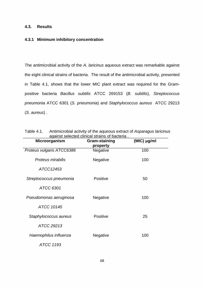
68
4.3. Results
4.3.1 Minimum inhibitory concentration
The antimicrobial activity of the A. laricinus aqueous extract was remarkable against
the eight clinical strains of bacteria. The result of the antimicrobial activity, presented
in Table 4.1, shows that the lower MIC plant extract was required for the Gram-
positive bacteria Bacillus subtilis ATCC 269153 (B. subtilis), Streptococcus
pneumonia ATCC 6301 (S. pneumonia) and Staphylococcus aureus ATCC 29213
(S. aureus) .
Table 4.1. Antimicrobial activity of the aqueous extract of Asparagus laricinus against selected clinical strains of bacteria .
Microorganism Gram-staining property
(MIC) μg/ml
Proteus vulgaris ATCC6386 Negative 100
Proteus mirabilis
ATCC12453
Negative 100
Streptococcus pneumonia
ATCC 6301
Positive 50
Pseudomonas aeruginosa
ATCC 10145
Negative 100
Staphylococcus aureus
ATCC 29213
Positive 25
Haemophilus influenza
ATCC 1193
Negative 100

69
Escherichia coli ATCC
133762
Negative 100
Bacillus subtilis ATCC
269153
Positive 50
*American type culture collection (ATCC) *Gentamicin was used as a positive control
S. aureus was the only tested strain that was inhibited by 25 μg/ml of the plant
extract. This was contrary to the 100 μg/ml that was required for all Gram-negative
strains (Pseudomonas aeruginosa ATCC10145 (P. aeruginosa), Proteus vulgaris
ATCC 6386 (P. vulgaris), Escherichia coli ATCC 133762 (E. coli) and Proteus
vulgaris ATCC 6386 (P. vulgaris)) and the 50 μg/ml in Gram-positive strains such as
Streptococcus pneumonia (Figure 4.1).
Figure 4.1. A digital photograph of the Streptococcus pneumonia ATCC 6301 treated with an increasing concentration of A. laricinus aqueous extract.

70
4.3.2. Thin layer chromatography analysis and antioxidant activity of extract’s
constituents
Figure 4.2 shows the TLC chromatogram for the qualitative screening for
antioxidants. Three spots with Rf of 0.87, 0.38 and 0.35 exhibited antioxidant activity
when sprayed with DPPH. This indicated differences in the polarity of the separated
antioxidant molecules.
Figure 4.2. Chromatogram of Gallic acid (1) and A. laricinus aqueous extract (2 and 3, lane 2 is a twofold dilution of lane 3), separated with methanol:chloroform:hexane (70:20:10%) and sprayed with DPPPH. The yellow spots indicate antioxidant activity (Rf values: a = 0.87; b = 0.38; c = 0.35).

71
4.3.3. Free radical scavenging activity
The addition of the A. laricinus extract to the DPPH solution induced a rapid
decrease in absorbance at 517 nm, indicating free radical scavenging activity of the
extract (Fig. 4.3). The EC50 value of the extract was recorded to be between 0.9205
and 1.188 mgml-1 and was higher than that of Trolox® (Figure 4.3d). The radical
scavenging activity of the extract was concentration-dependent and similar to that of
known antioxidants (Figure. 4.3a, b and c). A. laricinus showed concentration-
dependent free radical scavenging activity at concentrations above 1 mg ml-1, while
ascorbic acid no longer showed increasing activity above the same concentration.
Figure 4.3 (a-d). Scavenging activity of A. laricinus aqueous extract on the free radical DPPH. (a) A. laricinus, (b) Trolox®, (c) ascorbic acid, and (d) EC50 shift graph of A. laricinus extract with Trolox® as the control.

72
4.3.3. Oxidative stress
Figure 4.4, illustrates the response of Saccharomyces cerevisiae (BY4742) cells to
hydrogen peroxide treatment.
Figure 4.4. Yeast cell (BY4742) sensitivity to hydrogen peroxide. Exponentially growing yeast cells (30oC) were treated with 10 mM (H2O2) for 90 minutes. Cells treated with H2O2 only (▲), untreated cells (●) and cells treated with H2O2 and aqueous extract (■).
Cells that were incubated in hydrogen peroxide only showed a significant decrease
in colony-forming units per milliliter (CFU/ml) after 30 minutes. This is contrary to the
slight increase in colony-forming units in the cells that were incubated in hydrogen
peroxide and aqueous extract. As expected, the untreated cells grew exponentially
from the same incubation period.
4.4. Discussion
MICs are considered the gold standard for determining the susceptibility of
organisms to antimicrobials and are therefore used to judge the performance of all

73
other methods of susceptibility testing. MICs are used in diagnostic laboratories to
confirm unusual resistance, to give a definite answer when a borderline result is
obtained by other methods of testing or when diffusion methods are not appropriate
(Andrews, 2001).
Inhibition of Staphylococcus aureus ATCC 29213 by the A. laricinus plant extract
demonstrates huge potential for the use of the plant in the treatment of microbial
infections, especially in light of the growing antibiotic resistance in microoganisms.
The best known example of an antibiotic resistance mechanism is probably the
alternative penicillin-binding protein (PBP2a), which is produced in addition to the
“normal” penicillin-binding proteins by methicillin-resistant Staphylococcus aureus
(Hawkey, 1998). The observed antibacterial activity of the extract suggests a
possible antibiotic against such resistant strains of S. aureus.
The growth inhibition of Pseudomonas aeruginosa ATCC 10145 and Escherichia coli
ATCC 133762 is important, since it has been reported that patients with neutropenia
are found to suffer from infection by the two bacteria mentioned (Schimpff et al.,
1971; Cometta et al., 1996). These results show huge potential in alleviating
infection by both Gram-negative and Gram-positive clinical strains.
The antioxidant capacity of crude drugs is widely used as a parameter for evaluating
medicinal bioactive components. The present study has demonstrated the
antiradical activity of A. laricinus. The researcher observed three spots that
exhibited antioxidant properties on the TLC chromatograms. The characteristics of
the three different antioxidants observed in the A. laricinus extracts are yet to be

74
elucidated. The antioxidant in the plant extract may largely be due to polyphenols
(Thabrew et al., 1998). Phenolics are the largest group of phytochemicals and most
antioxidant activity of plants or plant products is attributed to them. Many studies
have shown that natural antioxidants are able to reduce DNA damage, mutagenesis
and carcinogenesis. These events are often associated with the termination of free
radical propagation in biological systems (Covacci et al., 2001; Zhu et al., 2002).
The effect of antioxidants on DPPH radical scavenging is thought to be due to their
hydrogen-donating ability or radical scavenging ability (Baumann et al., 1979).
Considering the fact that the A. laricinus extract used was crude and the comparison
was not on the basis of molar concentration, the above results are promising, despite
having a higher EC50 value than Trolox®.
H2O2 and superoxide anions (O2-) are often referred to as reactive oxygen species
(ROS) as they can lead to production of more reactive species, particularly in the
presence of metal ions. ROS are generated endogenously in many cells as a
consequence of metabolic processes. ROS can also be formed by exposure of cells
to ionizing radiation, redox-cycling chemicals present in the environment or by
exposure to heavy metals (Ames, 1983; Brennan and Schiestl, 1996). Through such
mechanisms, all aerobically growing organisms are continuously exposed to reactive
oxidants and oxidative stress occurs when the concentration of these oxidants
increases beyond the antioxidant buffering capacity of the cell. A side effect of
respiration in organisms is the production of ROS that can damage proteins, lipids
and DNA (Hererro et al., 2008).

75
The ability to protect Saccharomyces cerevisiae (S. cerevisiae) from oxidative stress
is seen as valuable to understanding the potential effect of the plant extract to
humans, since yeasts are eukaryotic cells and are equipped with several elaborate
defense mechanisms against different types of stress imposed by the environment,
and many of these are similar to mechanisms in higher organisms.
4.5. Conclusion
The aqueous extract of A. laricinus showed significant activity as an antioxidant. It
showed free radical scavenging activity comparable to Trolox and ascorbic acid.
The antimicrobial activity of the aqueous extract indicated potential use of the plant
in the treatment of microbial infections. A. laricinus aqueous extract has the ability to
protect cells from oxidative stress.
76
4.6 References
Ames, B. N. 1983. Dietary carcinogens and anticarcinogens. Science, (221):
1256–1263.
Andrews, J. M. 2001. Determination of minimum inhibitory concentration. Journal
of Antimicrobial Chemotherapy, (48): 5–16.
Bartosz G. 2003. Total antioxidant capacity. Advances in Clinical Chemistry, ( 37):
219–292.
Bartosz G. 2005. The other face of oxygen. Free radicals in nature. Polish
Scientific Publishers, Warsaw (in Polish).
Baumann J., Wurn G., and. Bruchlausen F.V. 1979. Prostaglandin synthetase
inhibiting O2 radical scavenging properties of some flavonoids and related phenolic
compounds. Naunyn-Schmiedeberg's Archives of Pharmacology, (30): 27–32.
Bors W., Saran M., and Elstner E.F. 1992. Screening for plant anti-oxidants. In:
Linskens, H.F. and Jackson J.F. (Eds.). Modern Methods of Plant Analysis - Plant
Toxin Analysis, Vol. 13. Berlin: Springer, pp: 277–295.
Brennan R.J., and Schiestl R.H. 1996. Cadmium is an inducer of oxidative stress
in yeast. Mutation Research, (356), 171–178.
77
Cometta A., Calandra T., Gaya H., Zinner S.H., de Bock R., Del Favero A.,
Bucaneve G., Crokaert F., Kern W.V, Klastersky J., Langenaeken I., Micozzi A.,
Padmos A., Paesmans M., Viscoli C., Glauser M.P. 1996. Monotherapy with
meropenem versus combination therapy with ceftazidime plus amikacin as empiric
therapy for fever in granulocytopenic patients with cancer. Antimicrobial Agents and
Chemotherapy, 40(5): 1108–1115
Covacci V., Torsello A., Palozza P., Sgambato A., Romano G., Boninsegna A.,
Cittadini A., and Wolf F.I. 2001. DNA oxidative damage during differentiation of
HL-60 human promyelocytic leukemia cells. Chemical Research in Toxicology, (14):
1492–1497.
Eloff J.N. 1998. A sensitive and quick microplate method to determine the minimal
inhibitory concentration of plant extracts for bacteria. Planta Medica, (64): 711–713.
Fuhrman B., Volkova N., Suraski A., and Aviram M. 2001. White wine with red
wine-like properties: Increased extraction of grape skin polyphenols improves the
antioxidant capacity of the derived white wine. Journal Agriculture and Food
Chemistry, (49): 3164–3168.
Garrote G., Cruz J.M., Moure A., Dominguez H., and Parajo J.C. 2004.
Antioxidant activity of byproducts from the hydrolytic processing of selected
lignocellulosic materials. Trends in Food Science and Technology, (15): 191–200.

78
Ghezzi P., Bonetto V., and Fratelli M. 2005. Thiol-disulfide balance: From the
concept of oxidative stress to that of redox regulation. Antioxidants and Redox
Signaling, (7): 964–972.
Halliwell B. 2006. Reactive species and antioxidants. Redox biology is a
fundamental theme for aerobic life. Plant Physiology, (141): 312–322.
Hawkey P.M. 1998. The origins and molecular basis of antibiotic resistance. British
Medical Journal, (317): 657-659.
Herrero, E., Ros J., Belli G., and Cabiscol E. 2008. Redox control and oxidative
stress in yeast cells. Biochimica et Biophysica Acta, (1780): 1217–35.
Izawa S., Inoue Y., and Kimura A. 1995. Oxidative stress response in yeast:
Effect of gluthathione on adaptation to hydrogen peroxide stress in Saccharomyces
cerevisiae. Federation of European Biochemical Societies Letters, (368): 73–76.
Jones D.P. 2006. Redefining oxidative stress. Antiox Redox Signal, (8): 1865–
1879.
Jones D.P. 2008. Radical-free biology of oxidative stress. American Journal of
Physiology - Cell Physiology, (295): C849–C868.

79
Manderfield M.M., Schafer H.W, P.M. Davidson P.M., and Zottola E.A. 1997.
Isolation and identification of antimicrobial furocoumarins from parsley. Journal of
Food Protection, (60): 72–77.
Schimpff S.C., Satterlee W., Young V.M., and Serpick A. 1971. Emperic therapy
with carbenicillin and gentamicin for febrile patients with cancer and ulocytopenia.
New England Journal of Medicine, (284): 1061-1065.
Thabrew M.I., Hughes R.D., and McFarlane I.G. 1998. Antioxidant activity of
Osbeckia aspera. Phytotherapy Research, (12): 288-290.
Tirzitis G., and Bartosz G. 2010. Determination of antiradical and antioxidant
activity: Basic principles and new insights. Acta Biochimica Polonica, 1(57): 139–
142.
Tomohiro T., Futoshi K., Naoharu W., Akihito Y., and Kanzo S. 1994. A simple
screening method for antioxidants and isolation of several antioxidants produced by
marine bacteria from fish and shellfish. Bioscience, Biotechnology and Biochemistry,
(58): 1780-1783.

80
Turner S., Davicino R., Alonso R., Feraro G., Filip R., and Anesini C. 2011.
Potential use of low-NDGA Larrea divaricata extracts as antioxidants in foods.
Revista Peruana de Biologia., (18): 159-164.
Ulate-Rodriguez J., Schafer H.W., Zottola E.A., and Davidson P.M. 1997.
Inhibition of Listeria monocytogenes, Escherichia coli O157:H7 and Micrococcus
luteus by linear uranocoumarins in a model food system. Journal of Food Protection,
(60):1050–1054.
Von Gadow A., Joubert E., and Hansmann C.F. 1997. Comparison of the
antioxidant activity of aspalthin with that of other plant phenols of rooibos tea
(Aspalathus linearis), α-tocopherol and BHT. Journal of Agriculture and Food
Chemistry, (45): 632–638.
Zhu Q.Y., Hackman R.M., Ensunsa J.L, Holt R.R., and Keen C.L. 2002.
Antioxidative activities of oolong tea. Journal of Agriculture and Food Chemistry,
(50): 6929–6934.

81
Chapter 5
Chemical composition of both aqueous and methanol extracts of Asparagus
laricinus
5.1. Introduction
Most medicinal plants are harvested from the wild and the production of medicinal
metabolites in plants is affected by plant genotype, cultivation, harvesting,
processing and distribution (Crozier, 2006). It has also been noted that the age of the
plant, the presence of flowers or pods, different geographical origin, soil and climate
play roles in plant adaptation mechanisms (Green, 1997). Because of that, the level
of metabolites represents integrative information of the cellular function and hence
defines the phenotype plant in response to genetic or environmental changes (Silas
et al., 2004).
Since individual plant species have been known to manufacture over 100 000
secondary metabolites (Yuliana et al., 2011), robust techniques that are able to
analyze these large numbers of metabolites in the shortest time possible are useful
in profiling. There is growing interest in correlating the phytochemical constituents of
a medicinal plant with its pharmacological activity (Prachayasittikul et al., 2008;
Turker and Usta, 2008; Nogueira et al., 2008). Many biologically active compounds
isolated from plants lead to the discovery of new medicinal drugs, which play efficient
protection and treatment roles against various diseases, including cancer (Sheeja

82
and Kuttan, 20O7; Mukherjee, 2007). Thus, screening of plant extracts for important
biological activities remain crucial in the search for novel molecules with relevant
biological activities.
The widespread use of liquid chromatometry – mass spectrometry (LC-MS) for
global metabolic profiling is relatively new and the literature indicates that MS-based
techniques, particularly in combination with chromatographic technologies, are most
popular, as these combine very high analytical precision with equally high detection
sensitivity (Lenz and Wilson, 2007; Wilson et al., 2005). Electro-spray ionisation
(ESl) is one of the most frequently used soft ionisation techniques in MS. ESI
eliminates the need for neutral molecule volatilization prior to ionization. In the
negative mode, ESI spectra of the phenolics show a predominant molecular ion [M-
H] with minimum fragmentation. It is known that the negative ion mode generally
provides improved detection limits (Ryan et aI., 1999). It is argued that HPLC-MS
offers some advantages over the gas chromatography- (GC) MS techniques
currently used in metabolomics. In particular, the ability to run samples with minimal
sample preparation by HPLC-MS is in contrast to the more extensive sample pre-
treatment techniques that are sometimes required to obtain comprehensive
metabolite fingerprints by GC-based techniques (Wilson et al., 2005).
Based on the literature search, no report is available on the detailed analyses of the
A. laricinus plant extract. Therefore, the aim of this chapter is to examine the
chemical composition of both aqueous and methanol extracts.

83
5.2. Materials and methods
5.2.1. Phytochemical screening
Chemical tests were carried out on the aqueous and methanol extracts of A.
laricinus. The samples were first powdered as explained in chapter 2 and subjected
to the chemical tests described below (Edoaga et al., 2005).
5.2.2. Alkaloid determination using Harborne (1973) method
Five grams (5 g) of the sample was weighed into a 250 ml beaker and 200 ml of 10%
acetic acid in ethanol was added, covered and allowed to stand for four hours. This
was filtered passively through a Whatman® filterpaper, 11 cm diameter. The extract
was concentrated in a water bath to one-quarter of the original volume. Concentrated
ammonium hydroxide was added dropwise to the extract until the precipitation was
complete. The whole solution was allowed to settle and the precipitate was collected
and washed with dilute ammonium hydroxide and then filtered. The residue was the
alkaloid, which was air-dried and weighed.
5.2.3. Test for tannins
About 0.5 g of the dried powdered samples was boiled in 20 ml of water in a test
tube and then filtered through a Whatman® filter, 11 cm diameter. A few drops of

84
0.1% ferric chloride was added and observed for brownish green or a blue-black
coloration.
5.2.4. Test for saponin
About 2 g of the powdered sample was boiled in 20 ml of distilled water in a water
bath (set at 100oC) and filtered though a. Whatman®, 11 cm diameter. Then 10 ml
of the filtrate was mixed with 5 ml of distilled water and shaken vigorously for a
stable persistent froth. The frothing was mixed with three drops of olive oil and
shaken vigorously, then observed for the formation of emulsion.
5.2.5 Test for steroids
Two milliliters of acetic anhydride was added to 0.5 g ethanolic extract of each
sample with 2 ml H2S04. The color changed from violet to blue or green in some
samples, indicating the presence of steroids.
5.2.6. Test for terpenoids (Salkowski test)
After mixing 5 ml of each extract in 2 ml of chloroform, concentrated H2S04 (3 ml)
was carefully added to form a layer. A reddish brown coloration of the interface was
formed to show positive results for the presence of terpenoids.

85
5.3. Gas chromatography-mass spectrometry
GC/MS analysis was carried out on a Shimadzu 2010 QB gas chromatograph with a
MSD detector equipped with an HP-5 fused silica capillary column (30 m x 0.25 mm
x 25 m film thickness). The aqueous plant extract was injected via an all-glass
injector working in split mode with helium as the carrier gas at a flow rate of 1 mL
min-1. The oven temperature was programmed as follows: Injector temperature
2000C, ion source 2000C, interphase 2000C. The column temperature was raised to
45°C (3 minutes hold at 45°C, 4 minutes), then gradually increased to 150°C (3
minutes hold at 150°C, 4 minutes) then raised to 250°C and a 15-minute hold. A split
ratio of 1:5 was used (Ajayi et al., 2011). Compound identification was accomplished
by comparing the retention times with those of authentic compounds or the spectral
data obtained from the Wiley Library and National Institute of Standards and
Technologies Library, as well as with data published in the literature.
5.4. Liquid chromatography/mass spectometry
The plant extracts were resuspended in 1 ml of a 50% (v/v) mixture of acetonitrile
and H2O containing 0.1% (v/v) formic acid. The suspensions were vortexed for 1
minute then sonicated for 5 minutes, vortexed again for 1 minute prior to
centrifugation at 10 000 rpm for 10 minutes. The supernatant (3 μl) was injected into
the LC-MS instrument. Metabolites were separated using a gradient of H2O with

86
0.1% formic acid (solvent A) and acetonitrile (solvent B), using a Waters UPLC at a
flow rate of 0.4 ml min-1 on a Waters BEH C18, 2.1x50 mm column. MS was
obtained on a Waters SYNAPT™ G2 MS (Manchester, England) using ESI running
in positive mode with a cone voltage of 15 V. The injections were repeated once to
ensure repeatability.

87
5.5. Results
5.5.1 Phytochemical screening
Phytochemical screening of the aqueous extract was positive for the presence of
alkaloids, saponins, tannins and terpenoids. However, the ethanolic extract only
tested positive for tannins and negative for the rest of the tests (Table 5.1).
Table 5. 1. Phytochemical screening of aqueous and ethanol extracts of the roots of A. laricinus for different molecular constituents
Key: -: Negative (absent): +: Positive (present)

88
5.5.2 Gas chromatography-mass spectrometry
The GC-MS chromatogram gave rise to three peaks, suggesting the existence of
three secondary metabolites and percentage values of composition of these
phytochemicals present in the aqueous root extract of A. laricinus. The results
suggested the presence of indole-3-carbinol, α-sitosterol and ferulic acid, as shown
in Figure 5.1.
Figure 5.1. GC/MS chromatogram of the aqueous extract of A. laricinus roots
5.5.3 Liquid chromatography/mass spectometry
Typical positive and negative ion LC-MS chromatograms for a both aqueous and
ethanol extracts of A. laricinus (Figures 5.2 and 5.3). The major peaks in each
solvent extract differed significantly, thus showing the effluence of solvents on
excluding other of molecules during extraction.

89
Figure 5.2. Representative LC-MS chromatograms for ethanol extract (a) and aqueous extract (b) in negative ion electrospray.
The aqueous constituents eluted in the predominantly polar regions of the solvent
gradient, while ethanol extract constituents appeared in the non-polar region.
(a)
(b)

90
Figure 5.3. Representative LC-MS chromatograms for ethanol extract (a) and aqueous extract (b) in positive ion electrospray.
Various compounds were identified based on their respective molecular formulae
(Tables 5.2 and 5.3). The tentative names and putative structures were obtained by
searching for structures and names corroborating the molecular formula and the
possible double bonds/carbon rings as reflected by the reported (double bond
equivalent) DBE.
(b)
(a)

91
Table 5.2. Analysis of LC/MS chromatograms of A. laricinus ethanol extract
Retention
time (min)
Fragmentation
ion (M/Z)
Molecular
formula
Tentative
identification
Putative structure
5.8 263.1275 C15H19O4
5-(1-Hydroxy-2,6,6-
triméthyl-4-oxo-2-
cyclohexén-1-yl)-3-
méthyl-2,4-
pentadiénoate
http://www.chemspider.c
om/Chemical-
Structure
http://www.chemspider.co
m/Chemical-Structure
6.3 277.1435 C16H21O4
4-(2-Methoxyphenyl)-
2,2-
dimethyltetrahydro-
2H-pyran-4-
yl]acetate
http://www.chemspider.c
om/Chemical-
Structure
http://www.chemspider.co
m/Chemical-Structure

92
7.8 265.1431 C15H21 O4 No inference No inference
8.4 279. 1594 C16H23O4 4-(4-
(Hexyloxy)phenyl]-
4-hydroxybutanoat
http://www.chemspider.c
om/Chemical-
Structure
http://www.chemspider.co
m/Chemical-Structure
Table 5.3. Analysis of LC/MS chromatograms of A. laricinus aqueous extract
Retention
time (min)
Fragmenta-
tion ion (M/Z)
Molecular
formula
Tentative
identification
Putative structure
2.0 380.2073 C20H30O6 1,4-bis(2-
hydroxymethyl-5,5-
dimethyl-1,3-
dioxan-2-
yl)benzene
http://www.chemspid
er.com/Chemical-
http://www.chemspider.co

93
Structure
m/Chemical-Structure
2.8 305.1148 C15H17 N2O5 1-Amino-2-(3,4-
dimethoxyphenyl)-
N-(2-
furoyloxy)ethanimin
ium
http://www.chemspid
er.com/Chemical-
Structure
http://www.chemspider.co
m/Chemical-Structure
3.8 556.2529 C34H38 N4O4S 1,4,6,7-tétrahydro-
8H-pyrazolo[3,4-
e][1,4]thiazépin-8-
yl]-N-
isobutylacétamide
http://www.chemspid
er.com/Chemical-
Structure
http://www.chemspider.co
m/Chemical-Structure
4.1 616.2509 C39H38 NO4S 5-(3,4,5-
Trimethoxyphenyl
)-3-(4-
dimethylaminophe
Not assigned

94
nyl)-6-(4-
phenoxyphenyl)-2-
phenyl-3,3a,5,6-
tetrahydro-
2H-pyrazolo[3,4-
d]thiazole (Tugart
et al., 2007)

95
5.6. Discussion
The phyto-chemical test that was done for initial screening of the extract was
complemented with detailed results obtained in GC/MS and LC/MS analysis.
However, the MS results were more effective, allowing samples to be analysed with
the minimum of quantities and analysis and covering as wide a range of classes of
metabolite as possible in a short period.
Phytochemical screening of indigenous plants is essential to evaluate their medical
value and their potential use in the treatment of various diseases. In the current
study of A. laricinus extract, three secondary metabolites were found in the aqueous
extract. The extract showed the presence of ferulic acid, a phenolic acid derivative
that has antioxidant activity and several therapeutic benefits in the treatment of
cancer. Ferulic acid is a plant constituent that arises from the metabolism of
phenylalanine and tyrosine. It occurs primarily in seeds and leaves, both in its free
form and covalently linked to lignin and other biopolymers. Because of its phenolic
nucleus and an extended side chain conjugation it readily forms a resonance
stabilized phenoxy radical, which accounts for its potent antioxidant effect (Imaida et
al., 1990; Srinivasan et al., 2007). β-Sitosterol has a number of therapeutic and
chemo-preventive uses in the medical field (Zak et al., 1990; Baskar et al., 2010).
Indole-3-carbinol is used in prostate cancer (Garikapaty et al., 2005). The
phytoconstituents detected in the plant materials could be responsible for the
cytotoxic activity, though their exact mode of action is poorly understood at present.
The LC-MS chromatograms showed distinct profiles of the extracts. This is
important in investigating the health benefit claims in the traditional use of medicinal

96
plants. It is therefore important to adapt isolation techniques to mimic the extraction
methods used in traditional usage of medicinal plants. Consistent with the chemical
tests used for initial screening of different classes of metabolites, LC/MS results
proved the presence of alkaloids in the aqueous extract, while no alkaloids could be
found in the ethanol extract.
It is important to note that the C39H38 NO4S in the aqueous extract was similar to a
chemically synthesized derivative of the potential biologically active 4-thiazolidinones
(Tugut et al., 2007). Predominantly, the aqueous extract had small ring heterocycles
containing nitrogen, sulfur and oxygen. Such molecules have been under
investigation for a long time because of their important medicinal properties. These
types of molecules have been shown to have various important biological activities
such as anti-microbial, anti-viral, diuretic, anti-histaminic, anti-cancer, anti-
convulsant, anti-inflammatory and analgesic properties (Vigorita et al., 2001; Kavitha
et al., 2006; Kucukguzel et al., 2006).
5.7. Conclusion
This analysis of the aqueous and ethanol extracts of A. laricinus has helped to
predict the compounds present in the respective solvents. The identified molecules
supported the evidence of important biological activities that had been observed in A.
laricinus. The use of chemical formulae in this study limited the identification of the
plant extracts‟ metabolites to a narrow potential search field.

97
5.8. References
Ajayi G.O., Olagunju J.A., Ademuyiwa O., and Martins O.C. 2011. Gas
chromatography-mass spectrometry analysis and phytochemical screening of
ethanolic root extract of Plumbago zeylanica, Linn. Journal of Medicinal Plant
Research, (5): 1756-1761.
Baskar A.A., Ignacimuthu S., Paulraj M.G., and Al Numair K.S. 2010.
Chemopreventive potential of β-sitosterol in experimental colon cancer model - An in
vitro and in vivo study. BMC Complementary Alternative Medicine. Vol., 10.
10.1186/1472-6882-10-24.
Crozier A. 2006. Phenols, polyphenols and tannins: An overview. In: Crozier A.,
Mike N. Clifford M.N., and Ashihara H. (Eds.) Plant Secondary Metabolites:
Occurrence, Structure and Role in the Human Diet. Wiley-Blackwell
Garikapaty V.P., Ashok B.T., Chen Y.G., Mittelman A., Latropoulos M., and
Tiwari R.K. 2005. Anti-carcinogenic and anti-metastatic properties of indole-3-
carbinol in prostate cancer. Oncology Reports, (13): 89-93.

98
Green C.P. 1997. Comparison of Tettnanger, Saaz, Hallertau and Fuggle hops
grown in the USA, Australia and Europe. Journal of the Institute of Brewing, 103:
239–243.
Imaida, K., M. Hirose, Yamaguchi S., Takahashi S., and Ito N. 1990. Effects of
naturally occurring antioxidants on combined 1,2-dimethylhy,drazine- and 1-methyl-
1-nitrosourea-initiated carcinogenesis in F344 male rats. Cancer Letters, (55): 53–
59.
Kavitha C. V., Basappa S., Nanjunda S., Mantelingu K., Doreswamy S., Sridhar
M. A., Prasad J.S., and Rangappa K.S. 2006. Synthesis of new bioactive
venlafaxine analogs: Novel thiazolidin-4-ones as antimicrobials. Bioorganic and
Medicinal Chemistry, (14): 2290–2299.
Kucukguzel G., Kocatepe A., De Clercq E., Sahin F., and Gulluce, M. 2006.
Synthesis and biological activity of 4-thiazolidinones, thiosemicarbazides derived
from diflunisal hydrazide. European Journal of Medical Chemistry, (41): 353–359.
Lenz E.M., and Wilson D. 2007. Analytical strategies in metabonomics. Journal of
Proteome Research, 6(2): 443-458.
Mukherjee P.K. 2007. Screening of Indian medicinal plants for acetyl
cholinesterase inhibitory activity. Phytotherapy Research, ( 21):1142–45.

99
Nogueira J.C., Diniz Mde F., and Lima E.O. 2008. In vitro antimicrobial activity of
plants of acute otitis externa. Brazilian Journal of Otorhinolaryngology, (74):118–24.
Prachayasittikul S., Buraparuangsang P., Worachartcheewan A., Isarankura-
Na-Ayudhya C., Ruchirawat S., and Prachayasittikul V. 2008. Antimicrobial and
antioxidant activity of bioreactive constituents from Hydnophytum formicarum Jack.
Molecules. (13): 904–21.
Ryan D., Robards K., Prenzler P.D., Jardine D., Herlt A., and Antolovich M.J.
1999. Liquid chromatography with electrospray ionisation mass spectrometric
detection of phenolic compounds from Olea europaea. Journal of Chromatography
A, (855): 529–537.
Sheeja K., and Kuttan G. 2007. Activation of cytotoxic T-lymphocyte responses
and attenuation of tumor growth in vivo by Andrographis paniculata extract and
andrographolide. Immunopharmacology and Immunotoxicology, (29):81–93.
Silas G., Villas-B., Sandrine M., Mats A., Jørn S., and Jens N. 2004. Mass
spectrometry in metabolome analysis. Mass Spectrometry Reviews, (24): 613–646.
South African herbal tea. Journal of Sustainable Agriculture 12, 67–84.
Srinivasan, M., Sudheer A.R., and Menon, V.P. 2007. Ferulic acid: Therapeutic
potential through its antioxidant property. Journal of Clinical Biochemistry and
Nutrition, (40): 92–100.

100
Turgut Z., Yolacan C., Aydogan F., Bagdatli E., and Ocal N. 2007. Synthesis of
new pyrazolothiazole derivatives from 4-thiazolidinones. Molecules, (12): 2151–
2159.
Turker A.U., and Usta C. 2008. Biological screening of some Turkish medicinal
plants for antimicrobial and toxicity studies. Natural Product, ( 22): 136–46.
Vigorita M.G., Ottana R., Monforte F., Maccari R., Trovato, A., Monforte M.T.,
and Taviano M. F. 2001. Synthesis and antiinflamatory, analgesic activity of 3,3‟-
(1,2-ethandiyl)-bis[2-aryl-4- thiazolidinone] chiral compounds. Bioorganic and
Medicinal Chemistry Letters, 11(21): 2791–2794.
Wilson I.D., Plumb R., Granger J., Major H., Williams R., and Lenz E.M. 2005.
HPLC-MS-based methods for the study of metabonomics. Journal of
Chromatography B, 1(817): 67–76.
Yuliana N.D, Khatib A., Verpoorte R., and Choi Y.H. 2011. Comprehensive
extraction method integrated with NMR metabolomics: A new bioactivity screening
method for plants, adenosine a1 receptor binding compounds in Orthosiphon
stamineus Benth. Analytical Chemistry, (830): 6902–6906.

101
Zak A., Zeman M., Vitkova D., Hrabak P., and Tvrzicka E. 1990. β-sitosterol in
the treatment of hypercholesterolemia. Casopis Lékaru Ceských , (129): 1320–
1323.
http://www.chemspider.com/Chemical-Structure (accessed 15:54, February 28,
2014)

102
Summary
This study reports on the anti-microbial, anti-oxidative stress, phytochemistry and
free radical radical scavenging activity of Asparagus laricinus Burch (Asparagaceae)
in order to explain the anti-cancer properties observed in extracts of this plant. After
demonstrating the anti-cancer properties of the A. laricinus, the study tested the
biosafety of this plant by evaluating the antimutagenic and mutagenic water extract.
The root extracts of Asparagus laricinus were qualitatively screened for antioxidant
properties using 2, 2-diphenyl-1-picrylhydrazyl (DPPH), on silica gel GF254 TLC
plates. Quantitative Radical scavenging activity using DPPH was also evaluated.
The extracts were tested for the presence of alkaloids, saponins, tannins, terpenoids
and steroids using standard phytochemical screening methods. Gas
chromatography/mass spectrometry (GC/MS) was used to further confirm the
phytoconstituents, in addition, LC/MS was used to profile the the phytoconstituents
of the plant extract S. Three spots that exhibited antioxidant activity were resolved
on the TLC plates. The results on the anti-bacterial activity of Asparagus laricinus
showed that 50 μg/ml of the plant extract was the (MIC) for the gram-positive
bacteria (Bacillus subtilis, Streptococcus pneumonia and Staphylococcus aureus).
While 100 μg/ml was required for other gram-negative strains (Pseudomonas
aeruginosa, Proteus vulgaris, Escherichia coli and Proteus vulgaris), Staphylococcus
aureus was inhibited by 25 μg/ml of the plant extract. Evaluation of the anti-oxidative
stress activity of A. laricinus aqueous extract conclusively demonstrated the ability of
the extract to protect Saccharomyces cerevisiae (BY4742) cells from oxidative stress
when the cells were challenged with 10 Mm (H2O2) for 1.5 hours. The results of this

103
study confirmed the anti-cancer and other biologically significant activities from the
aqueous extract of A. laricinus. The free radical scavenging activity of A. laricinus
extract was comparable with that of Trolox and was dependent on the concentration
of the extract. This has shown that the effectiveness of the extract can be compared
to that of commercial drugs. The aqueous extract of A. laricinus contained alkaloids,
saponins, tannins and terpenoids. However, the chemical tests on e ethanol extract
produced negative results. The phytoconstituents in the aqueous extract were
confirmed through the use of GC/MS and indole-3-carbinol, α-sitosterol and ferulic
acid were identified. These identified compounds have known biological activities,
and that is found to be consistent with the anticancer, anti-microbial and anti
oxidative stress activities reported in the current study. The phytochemicals present
in the water extracts of A. laricinus and the in vitro anti-cancer properties indicate the
potential use of this plant extract for development of novel drugs. This study has
also established a scientific basis for the continued use of A. laricinus in traditional
medicine for the treatment of cancer and other ailments.


ISSN 1011 5528 | www.smltsa.org.za 33
Volume 25 No. 2 | December 2011Medical Technology SA
eVALuAtion oF the AntimutAgenic And mutAgenic propertieS oF AspArAgus lAricinusS Mashele (PhD) | SL Fuku (M.Sc)
school of Health Technology, Biomedical Technology Division, central university of Technology, Free state, south Africa
corresponding author: s Mashele | tel: +27 51 507 3369 | fax: +27 51 507 3354 | email: [email protected]
Peer reviewed ORIGINAL ARTICLE
Abstract
several medicinal plants are traditionally used in the treatment of a variety of ailments including cancer in the rural areas of south Africa and our neighbouring countries where herbal medicine is one of the main components of the healthcare system. many of these herbs, including Asparagus laricinus, have not been scientifically assessed for efficacy or safety. the study on which this article is based evaluated the antimutagenic and mutagenic water extract of Asparagus laricinus roots used in the treatment of cancer. the Ames test recommended for mutagenicity screening was employed. the mutagenic activity was evaluated in salmonella typhimu-rium strains tA97, tA98, tA100 and tA102. the mutagenicity tests of aqueous extracts were negative throughout (1:10, 1:25, 1:50. 1:100, and 1:200). the highest antimutagenic effect in the order of 49% was observed using tA 98 (5µg daunomycin/plate), a strong mutagen. these results indicate the potential for use of this plant against cancer and oxidative stress-related diseases.
Keywords
medicinal plants, Asparagus laricinus, Ames test, antimutagenicity
INTRODUCTION
one of the most prominent diseases in humans today is can-cer. it is projected that deaths from cancer are continuing to escalate, with an estimate of nine million deaths from cancer in the year 2015 and 11.4 million deaths by 2030[1]. gener-ally, cancer begins after a mutational episode in a single cell and then it progressively transforms to malignancy in multiple stages through sequential acquisition of additional mutations[2]. in view of the fact that these initial events are the underlying causes of the whole progression of carcinogenesis, their inhi-bition would therefore be an efficient preventive measure[3]. currently, there is marked scientific and commercial interest in the continuing discovery of new anti-cancer agents from natural product sources[4]. more than 50% of drugs used in clinical tri-als for anti-cancer activity were isolated from natural sources or are related to them[5,6]. hence, the search for natural products to be used in cancer therapy represents an area of great interest in which the plant kingdom is the most important source, provid-ing many anti-tumour agents with novel structures and unique mechanisms of action[7].
present chemotherapy cancer treatments have proved to be in-effective as a result of their toxicity and cells developing resist-ance[8]. many conventional drugs also induce genetic damage that itself can be carcinogenic. A segment of the research com-munity is thus focusing on identifying novel chemotherapeutic agents in plants that do not induce the destructive effects of conventional cytotoxic therapeutic agents.
several medicinal plants are traditionally used in the treatment of a variety of ailments, including cancer in many communi-ties in south Africa and neighbouring countries. since ancient times, herbal medicine has always been one of the main components of the healthcare system. in this study Asparagus laricinus belonging to Asparagaceae family was investigated. this is a monogeneric family, which was previously included within the Liliaceae family[9]. the genus Asparagus comprises approximately 100 species and consists of herbs, shrubs and
vines. many of these herbs, including Asparagus laricinus, have not yet been scientifically assessed for their efficacy or safety to tissue or organs of recipients. thus, investigation of tradition-ally used medicinal plants is valuable as a source of potential chemotherapeutic agents and also to assess the safety for the continuous use of medicinal plants. As a result, most studies are being directed at popular medicine, with the aim of identifying natural products which exhibit therapeutic properties[10,11].
the Ames test[12] was used in this study as it is widely used in the determination of possible gene mutations caused by extracts. A positive response in any single bacterial strain either with or without metabolic activation is sufficient to designate a sub-stance as a mutagen[13]. the study being reported here evaluated the mutagenic and antimutagenic activity of the water extract of Asparagus laricinus roots which are known to be used in the treatment of cancer.
MATERIALS AND METHODS
Plantmaterial
the plant material (Asparagus laricinus) was authenticated by scientists at the national botanical gardens in pretoria, south Africa. the collected root materials were dried at room tem-perature and pulverised by a mechanical mill and weighed. it was then stored in a cool place until analysis. plant material (10 g of the dried roots) was weighed, pulverised and soaked in a volume of 500 ml of purified water for 72 hours with occasional stirring. the extracts were filtered and the solvent was removed completely by employing a rotatory evaporator.
TheAmestest
the Ames test is a well-known bacterial mutagenicity test[11,14]. in this test, reverse his−→his+ mutations are visualised by plat-ing salmonella typhimurium bacteria in a histidine-poor growth medium. in this medium only, his+ mutants are able to form vis-ible colonies. different bacterial strains are available to identify different types of mutations. We used the strains tA97, tA98,

34 www.smltsa.org.za | ISSN 1011 5528
Volume 25 No. 2 | December 2011Medical Technology SA
tA100 and tA102. the strains tA98 and tA100 are actually those that are most often used as they detect the great major-ity of mutagens. strain tA98 gives an indication of frame-shift mutations, while a positive response from strain tA100 indi-cates base-pair substitution. for a substance to be considered genotoxic in the Ames test, the number of revertant colonies on the plates containing the test compounds should be more than twice the number of colonies produced on the solvent control plates (i.e. a ratio above 2.0). in addition, a dose-response should be evident for the various concentrations of the mutagen tested. toxicity can be checked by investigation of the back-ground layer of bacteria.
Antimutagenicity was expressed as percentage inhibition of mutagenicity according to the following formula given for the Ames test[15]:
% inhibition = [1 – t/m] x 100
where T is the number of revertants per plate in the presence of mutagen and the test sample and M is the number of revertants per plate in the positive control (mutagen alone). the same for-mula was also adopted for other tests. An extract was considered to have no or only weak antimutagenic properties when the per-centage inhibition of mutagenicity (e.g. revertants in the Ames assay) was less than 25%. When the percentage inhibition was between 25% and 40% it was considered that the extract had a moderate antimutagenic effect and it was concluded that an extract had a strong antimutagenic effect when the percentage inhibition was higher than 40%. dimethyl sulfoxide (dmso) was used as a negative control, while daunomycin (10µl/plate) was used as a positive control. All experiments were performed in triplicate. the metabolic activation (s9) was donated by the mrc Liver research centre, tygerberg, cape town.
Determinationoftotalphenoliccontent
total phenolic content in Asparagus laricinus extract was deter-mined employing folin-ciocalteu reagent[16], with minor adap-tations. gallic acid was used as a standard phenolic compound. in a test tube, 0.5ml of plant extract or standard solution was added, followed by 35 ml of h2o and 2.5ml of folin-ciocalteu reagent. the content was mixed thoroughly. After 8 minutes, 7.5ml naco3 (10% w/v) was added. the mixture was allowed to stand in the dark for 1 hour. Absorbance was measured at 760nm using a spectrophotometer. the concentration of total phenolic compounds in plant extracts was determined from the gallic acid standard curve.
RESULTS AND DISCUSSION
the standard plate incorporation methods for the Ames test us-ing salmonella typhimurium standard tester strains tA97, tA98, tA100 and tA102 exposed to five dilutions with and without s9 metabolic activation of the herbal mixtures were performed. the Ames test without s9 metabolic activation can only detect direct mutagens while with s9 metabolic activation allows the detection of indirect mutagens, often caused by conjugation reactions of metabolic oxidation systems. table 1 and 2 present
the spontaneous reversion response of the salmonella typh-imurium tester strain to the different dilutions of the Asparagus laricinus extract. the results revealed that Asparagus laricinus extracts were non-mutagenic towards the salmonella typhimu-rium strains tA97, tA98, tA100 and tA102 for the assay without metabolic activation. the average his+ revertants observed for all the tester strains caused by the extracts at all the concentra-tions without metabolic activation did not satisfy the criteria for mutagenicity. there was no notable dose-dependent increase in the number of revertants and the numbers of revertants were all either not equal to or greater than twice that of the negative con-trol[17]. there was also no decrease in the number of revertant colonies to levels far below the negative control (spontaneous reversion) which could also be classified as toxic. the extract had moderate (28%-34%) inhibitory effects using salmonella typhimurium tA100 strain. the upper limit was observed begin-ning with mutagen and followed by the anti-mutagen (extract). the lower limit was observed beginning with an anti-mutagen followed by a mutagen. When both mutagen and anti-mutagen were operative on commencement of a possible disease, 31% inhibition was observed. similarly, using salmonella typhimu-rium tA102 strain, the extract showed a low to moderate in-hibitory effect (24%-38%). the lower limit represented mutagen followed by anti-mutagen and the upper limit was observed when both mutagen and anti-mutagen were operative on com-mencement of a possible disease. however, using salmonella
Table 2: Mutagenic activities of Asparagus laricinus extract with metabolic activation (+S9).
S. typhymurium strain Dilution Mean +/- SD
TA 97 50X 154 +/-7TA 97 100X 154+/-10TA 97 200X 163+/-12TA 97 control (+) n/a 351TA 97 control (-) n/a 351TA 98 50X 40+/-6TA 98 100X 32+/-8TA 98 200X 45+/-6TA 98 control (+) n/a 830TA 98 control (-) n/a 53TA 100 50X 123+/-4TA 100 100X 131+/-3TA 100 200X 132+/-8TA 100 Control (+) n/a 1395TA 100 Control (-) n/a 183TA 102 50X 451+/-47TA 102 100X 406+/-51TA 102 200X 405+/-50TA 102 control (+) n/a 1395TA 102 control (-) n/a 391SD is standard deviation of three repeats in the experiment
Table 1: Summary of mutagenic/anti-mutagenic properties of Asparagus laricinus extract.
Mutagen/anti-mutagen Mutagen + anti-mutagenTA 97 TA 98 TA 100 TA 102 TA 97 TA 98 TA 100 TA 102
Dilutions Percentage inhibition Percentage inhibition
50X 21 49 28.6 22.7 20.2 23.4 31.6 37.5100X 10.4 35.1 12.1 23.5 14.5 19.5 28.4 24200X 9.1 21.9 19 21.5 7.6 17.1 15.5 21
ISSN 1011 5528 | www.smltsa.org.za 35
Volume 25 No. 2 | December 2011Medical Technology SA
typhimurium tA97 strain, the extract had a low inhibitory effect (20%-22%). the degree of inhibition was not affected by the mode of interaction of the mutagen and anti-mutagen. finally, the inhibitory effect was observed over a wide spectrum using salmonella typhimurium tA98 strain (23%-49%). the upper limit was observed beginning with a mutagen and followed by the anti-mutagen. the lower limit was observed when there was simultaneous interaction between a mutagen and the extract. When both mutagen and anti-mutagen were operative on com-mencement of a possible disease, 35% inhibition was observed.
the Ames test with metabolic activation was carried out by ad-dition of the s9 mix so as to detect an indirect mutagenic effect caused by metabolites of the test Asparagus laricinus extract. the s9 fraction contains a mixture of xenobiotic metabolising enzymes, such as the cytochrome p450s and sulfotransferase. Asparagus laricinus extract showed an indirect mutagenic effect toward the tester strain tA102 after metabolic activation but not in the other tester strains (tA97, tA98, and tA100).
the rationale for screening for antimutagenicity was that anti-mutagenic compounds can possibly also be anticarcinogens. these results have stimulated us to carry out ongoing work to determine the active ingredients since these may prove valu-able lead compounds in the light of their antimutagenic ability. no data relative to cytotoxic components of Asparagus larici-nus had been reported and further phytochemical work on the isolation and identification of active structures of this plant is presently in progress.
As assessed by the folin-ciocalteu method, the total phenolic content in the extracts are presented as gallic acid equivalents (gAe) with reference to the gallic acid standard curve which was plotted using the average absorbance values of two inde-pendent sets of data against concentrations of gallic acid in g/l. the Asparagus laricinus aqueous extract contained 4.2g/l gAe. polyphenols are the most abundant dietary antioxidants and research on their role in the prevention of degenerative diseases and cancer has developed quickly over these last few years. With a transgenic adenocarcinoma of the mouse prostate model of prostate carcinogenesis (trAmp), it was recently dem-
onstrated that oral consumption of 10g/l green tea polyphenols decreased tumour incidence by 65%[18], the latter polyphenol preparation was enriched with epigallocatechin-3-gallate (62%), epicatechin-3-gallate (24%), epigallocatechin (5%), epi-catechin (6%), and caffeine (‘1%). it is worth mentioning that the ratio of individual constituent polyphenols may vary from plant to plant. evaluation of the Asparagus laricinus aqueous extract on human melanoma cell lines is presently in progress.
many researchers have inferred the antimutagenic activity of plant extract to the antioxidant activities of the polyphenols. however, there is now a paradigm shift. owing to a study that showed Asparagus racemosus extracts induced excessive pro-duction of tnf-alpha as compared to controls[19], similar reports are increasing. naturally, white blood cells produce tnf-alpha protein to stimulate and activate the immune system in response to infection or cancer. the current state of knowledge suggests that the mode of action of many plant extracts is between scav-enging for free radicals and modulation of the immune system. the evidence obtained in studies of a tea polyphenol i.e. (-)-epi-gallocatechin gallate [(-)-egcg], indicate that plant polyphe-nols target many enzymes directly associated with cancer (e.g. proteosome, telomerase, growth factors, mApKs, etc.)[20,21,22,23]
CONCLUSION
Asparagus laricinus polyphenol extract exhibited a dose de-pendent antimutagenic ability. the plant extract showed no mu-tagenic effect on all tested strains. however, mutagenic activity was observed with metabolic activation for the tA 102 tester strain only, to the exclusion of (tA97, tA98, and tA100). these results indicate the potential for use of this plant against cancer and oxidative stress-related diseases. Although these results support the use of Asparagus laricinus for treatment of cancer, further investigation is required, for this promising plant.
ACKNOWLEDGEMENT
the authors would like to thank central university of technol-ogy’s innovation fund and national research foundation for their support.
REFERENCES1. World health organisation, 2005. urL: http://www.who.int/
mediacentre/factsheets/fs297/en/index.html2. Khan, m., pelengaris, s. the molecular biology of cancer, first
edition. Blackwell publishing, Massachusetts 2006.3. bertram, J.s. the molecular biology of cancer. Molecular Aspects
of Medicine 2001, 21: 167-223.4. Kinghorn, A.d., farnsworth, n.r., soejarto, d.d., codell, g.A.
novel strategies for the discovery of plant-derived anti-cancer agents. pharmaceutical Biology. 2003, 41: 53-67
5. cragg, g.m., newman, d.J. plants as a source of anti-cancer agents. Journal of Ethnopharmacology 2005, 100: 72-79.
6. newman, d.J., cragg, g.m. natural products as source of new drugs over the last 25 years. Journal of natural products. 2007, 70: 461-477.
7. chang, c., Ashendel, c., chan, t.c.K., geahlen, r.L., mcLaugh-lin, J., and Waters, d.J. oncogene signal transduction inhibitors from chinese medicinal plants. pure Applied chemistry. 1999, 71: 1101-1104.
8. mcWhirter, A., oakes, d., and steyn, A. (1996). reader’s digest: illustrated encyclopedia of essential Knowledge (cape town, reader’s digest Association (pty) Limited).
9. brummitt, r.K. Vascular plant families and genera. royal botani-cal gardens, Kew. 1992: 804.
10. hamburger m., and hostettmann K. (1991). 7. bioactivity in plants: the link between phytochemistry and medicine. phytochemistry, 30(12): 3864-3874.
11. Weisburger J.h, hara Y, dolan L, Luo f, pittmana b,and Zang e. tea polyphenols as inhibitors of mutagenicity of major classes of carcinogens. Mutation research: genetic Toxicology. 1996, 371(1-2): 57-63.
12. maron, d.m., and Ames, b.n. revised methods for the salmo-nella mutagenicity test. Mutation research. 1983, 113: 173-215.
13. Zeinger e. mutagens that are not carcinogens: faulty theory or faulty tests? Mutation research. 2001, 492: 29-38.
14. mortelmans K., and Zeinger e. the Ames salmonella/microsome mutagenicity assay. Mutation research. 2000, 455: 29-60.
15. ong t, Whong W.Z, stewart J.d, and brockman h.e. “chloro-phyllin: a potent antimutagen against environmental and dietary complex mixtures. Mutation research letters. 1986, 173(2): 111-115.
16. singleton, V.L., orthofer, r., and Lamuela-raventos, r.m. Analysis of total phenols and other oxidation substrates and antioxidants by means of folin-ciocalteau reagent. Methods in Enzymology. 1999, 299:152-178.
17. bulmer A.c., ried K, blanchfield J.t, and Wagner K.h. the anti-mutagenic properties of bile pigments. Mutation research. 2008, 658(1-2): 28-41.

Volume 25 No. 2 | December 2011Medical Technology SA
d
18. gupta s., hastak K., Ahmad n., Lewin J.s., and mukhtar h. inhibition of prostate carcinogenesis in trAmp by oral infusion of green tea polyphenols. proceedings of the national Academy of sciences of the united states of America. 2001, 38(18): 10350-10355.
19. Katiyar cK, brindavanam nb, tiwari p, narayana dbA. immu-nomodulator products from Ayurveda: current status and future prospectives. in: sn upadhyay ed. immunomodulation, narosa publishing house, new delhi 1997: 163-87.
20. naasani, i., h. seimiya and tsuruo t. telomerase inhibition, telomere shortening, and senescence of cancer cells by tea catechins. Biochemical and Biophysical research communica-tion. 1998, 249: 391-396.
21. hofmann, c.s and sonenshein g.e. green tea polyphenol epigallocatechin-3 gallate induces apoptosis of proliferating vascular smooth muscle cells via activation of p53. Federation of American societies for Experimental Bilogy Journal. 2003, 17: 702-704.
22. mukhtar, h and Ahmad n. mechanism of cancer chemopre-ventive activity of green tea. proceedings of the society of Experimental Biology Medicine. 1999, 220: 234-238.
23. chen, c., g. shen, hebbar V., owuor r., hu e.d. and Kong A.n. epigallocatechin-3-gallate-induced stress signals in ht-29 human colon adenocarcinoma cells. carcinogenesis. 2003, 24: 1369-1378.


a
b
c
1 2 3