Embed Size (px)
Citation preview
An evaluation of echo in life support (ELS): is itfeasible? What does it add?
C Hayhurst,1 C Lebus,1 P R Atkinson,1,2 R Kendall,1 R Madan,1 J Talbot,2 P Ross,2
D Lewis3
ABSTRACTBackground Emergency physicians were trained toperform echo in life support (ELS)dthat is, limitedtransthoracic echocardiography during advanced lifesupport (ALS) management of cardiac arrest.Methods Data were collected on the adequacy of viewsobtained and timing of the scan, as well as the clinicalfindings of pericardial effusion and ventricular wallmotion. Any intervention performed as a result of thescan was also noted. ELS was performed on 50 patientsduring cardiac arrest.Results Adequate views were obtained in 47 (94%)scans, and 45 (90%) were obtained within the 10 srhythm check. Twenty patients (40%) had ventricularwall motion (VWM), three (6%) had pericardial effusionsand six patients (12%) had an intervention performed asa direct result of the scan. These includedpericardiocentesis, thrombolysis and insertion of a chestdrain. The presence of VWM had a positive predictivevalue of 55%. The absence of VWM resulted ina negative predictive value of 97% for predicting return ofspontaneous circulation (ROSC).Conclusion It is concluded that ELS is feasible and thatthe scan findings may guide further interventions.
INTRODUCTIONUltrasound has become a vital diagnostic tool inthe emergency department (ED) for managingpatients with trauma and hypotension. Limitedtransthoracic echo during a cardiac arrest or peri-arrest setting is a Level 1 skill recognised by theRoyal College of Radiologists and the College ofEmergency Medicine.1 2 The use of ELS (echo in lifesupport) has become integrated in the everydaypractice of many EDs. Managing pulseless electricalactivity (PEA) in cardiac arrest involves looking forunderlying causes (the ‘H’s and ‘T’s).3 In particularcardiac tamponade and thromboembolic events aredifficult to diagnose during resuscitation. It hasbeen shown in many small studies that trans-thoracic echocardiography can identify pericardialeffusions4 and right ventricular (RV) dilatation,5 6
suggesting a pulmonary embolus (PE) in arrest andperiarrest states. It is also possible to see the pres-ence or absence of ventricular wall motion (VWM)in PEA arrests.4 6 7 We assessed the ability ofemergency physicians to perform echocardiographyduring advance life support (ALS) management ofcardiac arrest.
METHODSWe collected data from a convenience sample of 56adult patients in cardiac arrest over 2 years and
5 months in two centres as part of a serviceevaluation.Normal ALS procedures were followed, and
scanning was only permitted during the 10 srhythm check by either the team leader or a doctornot directly involved in delivery of ALS. ELS did notcontribute to any decisions regarding cessation ofALS.Data collection forms were completed by scan-
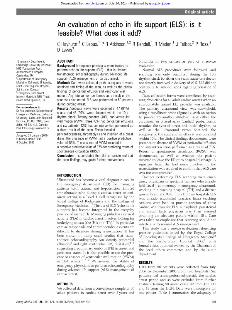
ning physicians for all adult cardiac arrests when anappropriately trained ELS provider was available.The primary ultrasound view was subxiphoid,using a curvilinear probe (figure 1), with an optionto proceed to another window using either thecurvilinear or phased array (cardiac) probe. Formsrecorded the type of arrest and initial rhythm, aswell as the ultrasound views obtained, theadequacy of the scan and whether it was obtainedwithin 10 s. The clinical findings documented werepresence or absence of VWM or pericardial effusionand any intervention performed as a result of ELS.Return of spontaneous circulation (ROSC) wasdocumented, as well as whether the patientsurvived to leave the ED or to hospital discharge. Asignature from the lead nurse involved in theresuscitation was required to confirm that ALS carewas not compromised.Doctors performing ELS scanning were emer-
gency physicians or specialist trainees who alreadyheld Level 1 competency in emergency ultrasound,working in a teaching hospital (TH) and a districtgeneral hospital (DGH). In both these hospitals ELSwas already established practice. Extra teachingsessions were held to provide revision of threecardiac windows for ELS: subxiphoid, parasternaland apical. Each physician was then assessedobtaining an adequate picture within 10 s. Carewas taken to emphasise that scanning should notinterfere with normal ALS management.This study was a service evaluation referencing
practice guidelines issued by the Royal Collegeof Radiologists,1 College of Emergency Medicine2
and the Resuscitation Council (UK),3 withformal ethics approval waived by the Chairman ofthe local ethics committee and by the auditdepartment.
RESULTSData from 56 patients were collected from July2006 to December 2008 from two hospitals. Sixpatients had scans performed outside the cardiacarrest period and so were excluded from furtheranalysis, leaving 50 arrest cases; 32 from the THand 18 from the DGH. Data were incomplete forone patient. Table 1 summarises the adequacy of
1Emergency Department,Cambridge University HospitalsNHS Foundation Trust,Addenbrooke’s Hospital,Cambridge, UK2Department of EmergencyMedicine, Dalhousie University,Saint John Regional Hospital,Saint John, Canada3Emergency Department,Ipswich Hospitals NHS Trust,Heath Road, Ipswich, UK
Correspondence toDr Paul Atkinson, Department ofEmergency Medicine, DalhousieUniversity, Saint John RegionalHospital, PO Box 2100, SaintJohn, NB E2L 4L2, Canada;[email protected]
Accepted 31 January 2010Published Online First4 October 2010
Emerg Med J 2011;28:119e121. doi:10.1136/emj.2009.084202 119
Original article
group.bmj.com on July 14, 2014 - Published by emj.bmj.comDownloaded from
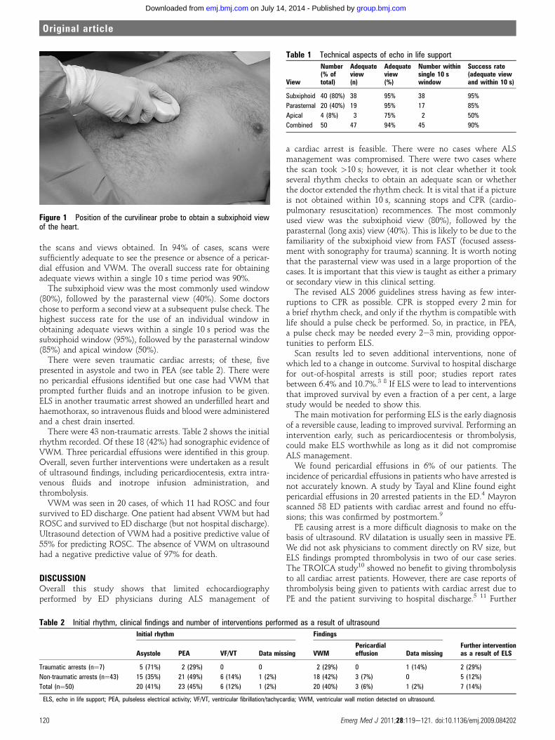
the scans and views obtained. In 94% of cases, scans weresufficiently adequate to see the presence or absence of a pericar-dial effusion and VWM. The overall success rate for obtainingadequate views within a single 10 s time period was 90%.
The subxiphoid view was the most commonly used window(80%), followed by the parasternal view (40%). Some doctorschose to perform a second view at a subsequent pulse check. Thehighest success rate for the use of an individual window inobtaining adequate views within a single 10 s period was thesubxiphoid window (95%), followed by the parasternal window(85%) and apical window (50%).
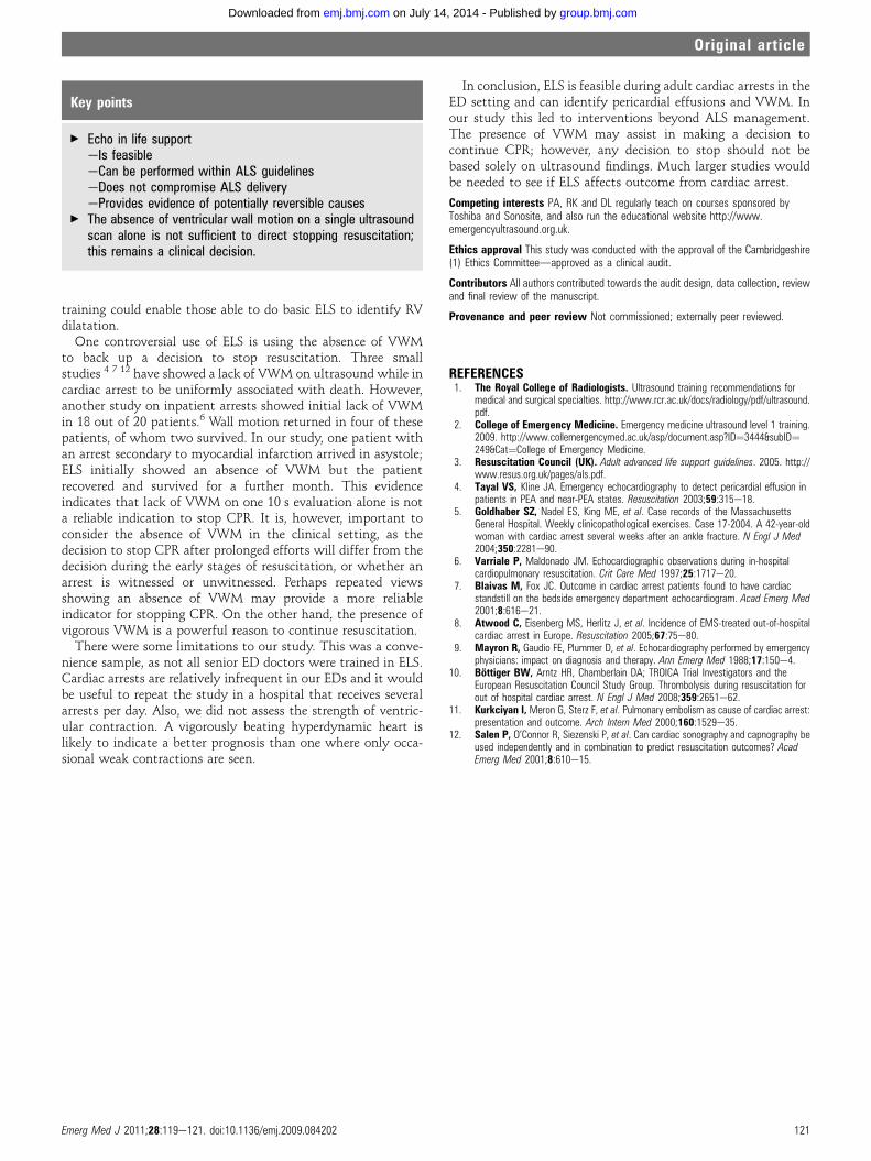
There were seven traumatic cardiac arrests; of these, fivepresented in asystole and two in PEA (see table 2). There wereno pericardial effusions identified but one case had VWM thatprompted further fluids and an inotrope infusion to be given.ELS in another traumatic arrest showed an underfilled heart andhaemothorax, so intravenous fluids and blood were administeredand a chest drain inserted.
There were 43 non-traumatic arrests. Table 2 shows the initialrhythm recorded. Of these 18 (42%) had sonographic evidence ofVWM. Three pericardial effusions were identified in this group.Overall, seven further interventions were undertaken as a resultof ultrasound findings, including pericardiocentesis, extra intra-venous fluids and inotrope infusion administration, andthrombolysis.
VWM was seen in 20 cases, of which 11 had ROSC and foursurvived to ED discharge. One patient had absent VWM but hadROSC and survived to ED discharge (but not hospital discharge).Ultrasound detection of VWM had a positive predictive value of55% for predicting ROSC. The absence of VWM on ultrasoundhad a negative predictive value of 97% for death.
DISCUSSIONOverall this study shows that limited echocardiographyperformed by ED physicians during ALS management of
a cardiac arrest is feasible. There were no cases where ALSmanagement was compromised. There were two cases wherethe scan took >10 s; however, it is not clear whether it tookseveral rhythm checks to obtain an adequate scan or whetherthe doctor extended the rhythm check. It is vital that if a pictureis not obtained within 10 s, scanning stops and CPR (cardio-pulmonary resuscitation) recommences. The most commonlyused view was the subxiphoid view (80%), followed by theparasternal (long axis) view (40%). This is likely to be due to thefamiliarity of the subxiphoid view from FAST (focused assess-ment with sonography for trauma) scanning. It is worth notingthat the parasternal view was used in a large proportion of thecases. It is important that this view is taught as either a primaryor secondary view in this clinical setting.The revised ALS 2006 guidelines stress having as few inter-
ruptions to CPR as possible. CPR is stopped every 2 min fora brief rhythm check, and only if the rhythm is compatible withlife should a pulse check be performed. So, in practice, in PEA,a pulse check may be needed every 2e3 min, providing oppor-tunities to perform ELS.Scan results led to seven additional interventions, none of
which led to a change in outcome. Survival to hospital dischargefor out-of-hospital arrests is still poor; studies report ratesbetween 6.4% and 10.7%.3 8 If ELS were to lead to interventionsthat improved survival by even a fraction of a per cent, a largestudy would be needed to show this.The main motivation for performing ELS is the early diagnosis
of a reversible cause, leading to improved survival. Performing anintervention early, such as pericardiocentesis or thrombolysis,could make ELS worthwhile as long as it did not compromiseALS management.We found pericardial effusions in 6% of our patients. The
incidence of pericardial effusions in patients who have arrested isnot accurately known. A study by Tayal and Kline found eightpericardial effusions in 20 arrested patients in the ED.4 Mayronscanned 58 ED patients with cardiac arrest and found no effu-sions; this was confirmed by postmortem.9
PE causing arrest is a more difficult diagnosis to make on thebasis of ultrasound. RV dilatation is usually seen in massive PE.We did not ask physicians to comment directly on RV size, butELS findings prompted thrombolysis in two of our case series.The TROICA study10 showed no benefit to giving thrombolysisto all cardiac arrest patients. However, there are case reports ofthrombolysis being given to patients with cardiac arrest due toPE and the patient surviving to hospital discharge.5 11 Further
Figure 1 Position of the curvilinear probe to obtain a subxiphoid viewof the heart.
Table 1 Technical aspects of echo in life support
View
Number(% oftotal)
Adequateview(n)
Adequateview(%)
Number withinsingle 10 swindow
Success rate(adequate viewand within 10 s)
Subxiphoid 40 (80%) 38 95% 38 95%
Parasternal 20 (40%) 19 95% 17 85%
Apical 4 (8%) 3 75% 2 50%
Combined 50 47 94% 45 90%
Table 2 Initial rhythm, clinical findings and number of interventions performed as a result of ultrasound
Initial rhythm Findings
Further interventionas a result of ELSAsystole PEA VF/VT Data missing VWM
Pericardialeffusion Data missing
Traumatic arrests (n¼7) 5 (71%) 2 (29%) 0 0 2 (29%) 0 1 (14%) 2 (29%)
Non-traumatic arrests (n¼43) 15 (35%) 21 (49%) 6 (14%) 1 (2%) 18 (42%) 3 (7%) 0 5 (12%)
Total (n¼50) 20 (41%) 23 (45%) 6 (12%) 1 (2%) 20 (40%) 3 (6%) 1 (2%) 7 (14%)
ELS, echo in life support; PEA, pulseless electrical activity; VF/VT, ventricular fibrillation/tachycardia; VWM, ventricular wall motion detected on ultrasound.
120 Emerg Med J 2011;28:119e121. doi:10.1136/emj.2009.084202
Original article
group.bmj.com on July 14, 2014 - Published by emj.bmj.comDownloaded from
training could enable those able to do basic ELS to identify RVdilatation.
One controversial use of ELS is using the absence of VWMto back up a decision to stop resuscitation. Three smallstudies 4 7 12 have showed a lack of VWM on ultrasound while incardiac arrest to be uniformly associated with death. However,another study on inpatient arrests showed initial lack of VWMin 18 out of 20 patients.6 Wall motion returned in four of thesepatients, of whom two survived. In our study, one patient withan arrest secondary to myocardial infarction arrived in asystole;ELS initially showed an absence of VWM but the patientrecovered and survived for a further month. This evidenceindicates that lack of VWM on one 10 s evaluation alone is nota reliable indication to stop CPR. It is, however, important toconsider the absence of VWM in the clinical setting, as thedecision to stop CPR after prolonged efforts will differ from thedecision during the early stages of resuscitation, or whether anarrest is witnessed or unwitnessed. Perhaps repeated viewsshowing an absence of VWM may provide a more reliableindicator for stopping CPR. On the other hand, the presence ofvigorous VWM is a powerful reason to continue resuscitation.
There were some limitations to our study. This was a conve-nience sample, as not all senior ED doctors were trained in ELS.Cardiac arrests are relatively infrequent in our EDs and it wouldbe useful to repeat the study in a hospital that receives severalarrests per day. Also, we did not assess the strength of ventric-ular contraction. A vigorously beating hyperdynamic heart islikely to indicate a better prognosis than one where only occa-sional weak contractions are seen.
In conclusion, ELS is feasible during adult cardiac arrests in theED setting and can identify pericardial effusions and VWM. Inour study this led to interventions beyond ALS management.The presence of VWM may assist in making a decision tocontinue CPR; however, any decision to stop should not bebased solely on ultrasound findings. Much larger studies wouldbe needed to see if ELS affects outcome from cardiac arrest.
Competing interests PA, RK and DL regularly teach on courses sponsored byToshiba and Sonosite, and also run the educational website http://www.emergencyultrasound.org.uk.
Ethics approval This study was conducted with the approval of the Cambridgeshire(1) Ethics Committeedapproved as a clinical audit.
Contributors All authors contributed towards the audit design, data collection, reviewand final review of the manuscript.
Provenance and peer review Not commissioned; externally peer reviewed.
REFERENCES1. The Royal College of Radiologists. Ultrasound training recommendations for
medical and surgical specialties. http://www.rcr.ac.uk/docs/radiology/pdf/ultrasound.pdf.
2. College of Emergency Medicine. Emergency medicine ultrasound level 1 training.2009. http://www.collemergencymed.ac.uk/asp/document.asp?ID¼3444&subID¼249&Cat¼College of Emergency Medicine.
3. Resuscitation Council (UK). Adult advanced life support guidelines. 2005. http://www.resus.org.uk/pages/als.pdf.
4. Tayal VS, Kline JA. Emergency echocardiography to detect pericardial effusion inpatients in PEA and near-PEA states. Resuscitation 2003;59:315e18.
5. Goldhaber SZ, Nadel ES, King ME, et al. Case records of the MassachusettsGeneral Hospital. Weekly clinicopathological exercises. Case 17-2004. A 42-year-oldwoman with cardiac arrest several weeks after an ankle fracture. N Engl J Med2004;350:2281e90.
6. Varriale P, Maldonado JM. Echocardiographic observations during in-hospitalcardiopulmonary resuscitation. Crit Care Med 1997;25:1717e20.
7. Blaivas M, Fox JC. Outcome in cardiac arrest patients found to have cardiacstandstill on the bedside emergency department echocardiogram. Acad Emerg Med2001;8:616e21.
8. Atwood C, Eisenberg MS, Herlitz J, et al. Incidence of EMS-treated out-of-hospitalcardiac arrest in Europe. Resuscitation 2005;67:75e80.
9. Mayron R, Gaudio FE, Plummer D, et al. Echocardiography performed by emergencyphysicians: impact on diagnosis and therapy. Ann Emerg Med 1988;17:150e4.
10. Bottiger BW, Arntz HR, Chamberlain DA; TROICA Trial Investigators and theEuropean Resuscitation Council Study Group. Thrombolysis during resuscitation forout of hospital cardiac arrest. N Engl J Med 2008;359:2651e62.
11. Kurkciyan I, Meron G, Sterz F, et al. Pulmonary embolism as cause of cardiac arrest:presentation and outcome. Arch Intern Med 2000;160:1529e35.
12. Salen P, O’Connor R, Siezenski P, et al. Can cardiac sonography and capnography beused independently and in combination to predict resuscitation outcomes? AcadEmerg Med 2001;8:610e15.
Key points
< Echo in life supporteIs feasibleeCan be performed within ALS guidelineseDoes not compromise ALS deliveryeProvides evidence of potentially reversible causes
< The absence of ventricular wall motion on a single ultrasoundscan alone is not sufficient to direct stopping resuscitation;this remains a clinical decision.
Emerg Med J 2011;28:119e121. doi:10.1136/emj.2009.084202 121
Original article
group.bmj.com on July 14, 2014 - Published by emj.bmj.comDownloaded from
doi: 10.1136/emj.2009.0842022010
2011 28: 119-121 originally published online October 4,Emerg Med J C Hayhurst, C Lebus, P R Atkinson, et al. is it feasible? What does it add?An evaluation of echo in life support (ELS):
http://emj.bmj.com/content/28/2/119.full.htmlUpdated information and services can be found at:
These include:
References
http://emj.bmj.com/content/28/2/119.full.html#related-urlsArticle cited in:
http://emj.bmj.com/content/28/2/119.full.html#ref-list-1This article cites 9 articles
serviceEmail alerting
the box at the top right corner of the online article.Receive free email alerts when new articles cite this article. Sign up in
CollectionsTopic
(831 articles)Radiology (diagnostics) � (922 articles)Radiology �
(58 articles)Echocardiography � (971 articles)Clinical diagnostic tests �
Articles on similar topics can be found in the following collections
Notes
http://group.bmj.com/group/rights-licensing/permissionsTo request permissions go to:
http://journals.bmj.com/cgi/reprintformTo order reprints go to:
http://group.bmj.com/subscribe/To subscribe to BMJ go to:
group.bmj.com on July 14, 2014 - Published by emj.bmj.comDownloaded from