Embed Size (px)
Citation preview
European Journal of Scientific Research
ISSN 1450-216X Vol.48 No.2 (2010), pp.315-325
© EuroJournals Publishing, Inc. 2010
http://www.eurojournals.com/ejsr.htm
An Efficient Denoising Technique for CT Images using Window-
based Multi-Wavelet Transformation and Thresholding
Syed Amjad Ali
Professor and Head of ECE Department, Lords Institute of Engineering and Technology
Himayathsagar, Hyderabad – 8
E-mail: [email protected]
Tel: +919490944104
Srinivasan Vathsal Professor and Director, R&D, Bhaskar Engineering College
Yenkapally, Moinabad, Ranga Reddy District.
E-mail:[email protected]
Tel: +919676863811
K. Lal kishore Rector, Jawahar Lal Nehru Technological University, Kukatpally, Hyderabad
E-mail:[email protected]
Tel: +919396438312
Abstract
Image denoising is one of the most significant tasks in image processing, analysis
and image processing applications. Medical Imaging is one among the emerging
application areas where the image denoising plays a vital role. In medical imaging, the
acquisition techniques and systems introduce noises and artifacts in the medical image that
leads to poor quality image. In this occasion, image denoising is an essential pre-requisite,
especially in Computed Tomography, which is an important and most common modality in
medical imaging. The significance of the denoising is mainly due to that the effectiveness
of clinical diagnosis using CT image depends upon the quality of the image. In this paper,
we propose an efficient noise reduction technique for CT images using window-based
Multi-wavelet transformation and thresholding. The technique removes Additive white
Gaussian noise from the CT images as well as it enhances the quality of the images. The
proposed technique consists of three different stages of processing, namely, window-based
multi-wavelet transformation and thresholding, reconstruction and quality enhancement. In
the first two processes, the AWGN is effectively removed from CT images and the images
are reconstructed. In the third process, the quality of the images is enhanced by means of
filtering techniques. Hence, denoised and quality enhanced CT images can be obtained
using the proposed multi-wavelet based denoising technique.
Keywords: Denoising, Multi-wavelet, Window, Thresholding, Computed Tomography
(CT) image, Additive White Gaussian Noise (AWGN)
An Efficient Denoising Technique for CT Images using Window-based
Multi-Wavelet Transformation and Thresholding 316
1. Introduction igital images play an important role both in day to-day applications, such as, satellite television,
magnetic resonance imaging, computer tomography as well as in areas of research and technology such
as geographical information systems and astronomy. In the diverse fields, mentioned above, scientists
are faced with the problem of recovering original images from incomplete, indirect and noisy images
[1]. Noises are added in the image during acquisition by camera sensors and transmission in the
channel [5]. The presence of noise gives an image a mottled, grainy, textured or snowy appearance [2].
Therefore, the problem of recovering an original image from noisy image has received an ever
increasing attention in recent years [4]. The recovering can be accomplished by image denoising, a
process of estimating the original image from an image that has been contaminated by noise
degradation [6].
In general, image denoising is the operation of removing unwanted noise from a noise-
corrupted image, restoring the image to its undegraded ideal [21]. The Image denoising methods can be
categorized as either transform domain methods or spatial domain methods. Transform domain
methods first transform an image from the spatial domain into a different domain (e.g., frequency
domain, wavelet domain) and suppress noise in the transform domain. Spatial domain methods
suppress noise directly in the spatial domain [14]. Yet, the image denoising using multi-wavelet
techniques becomes more effective because of the capability of capturing the signal energy in a very
few transformation energy values. The multi-wavelet transformation provides betters spatial and
spectral localization of image when compared with other multi-scale representations. Hence, it gains
more attraction in image denoising.
The image denoising plays a significant role in modern applications in various fields, including
medical imaging and preprocessing for computer vision [9]. Medical imaging acquisition technologies
and systems introduce noise and artifacts in the images that should be attenuated by denoising
algorithms. The denoising process, however, should not destroy anatomical details relevant from a
clinical point of view [3]. So, it is very difficult to suggest a robust method for noise removal which
works equally well for different modalities of medical images [7]. CT is one of the most important and
common modality in Medical Imaging, used for clinical diagnosis and computer-aided surgery [10].
Recently, several methods for de-noising have been developed and reported in the literature [8]. While
many other image denoising algorithms have been proposed over the years, excellent noise suppression
remains an open challenge, particularly in situations characterized by low signal-to-noise ratios [11].
In this paper, we propose an efficient noise reduction technique for CT images using window-
based Multi-wavelet transformation and thresholding. The multi-wavelet is chosen here, as it
outperforms single wavelets by its characteristics namely, orthogonality, short support, symmetry, and
high degree of vanishing moments. The technique denoises the CT images corrupted by AWGN as
well as improves the quality of the image. The proposed technique consists of three different stages of
processing, namely, window-based multi-wavelet transformation and thresholding, reconstruction and
quality enhancement. In the first two processes, the AWGN is effectively removed from CT images
and the images are reconstructed. In the third process, the quality of the images is enhanced by means
of filtering techniques. The rest of the paper is organized as follows: Section 2 gives a brief review of
the recent related works and Section 3 details the proposed denoising technique with required
mathematical formulations and pictorial representations. Section 4 discusses the implementation results
and Section 5 concludes the paper.
2. Related Works H.Rabbani et al. [12] have proposed noise reduction algorithms that could be used to enhance image
quality in various medical imaging modalities such as magnetic resonance and multidetector CT. The
noisy captured 3-D data were first transformed by discrete complex wavelet transform. Using a
D
317 Syed Amjad Ali, Srinivasan Vathsal and K. Lal kishore
nonlinear function, they have modeled the data as sum of the clean data plus additive Gaussian or
Rayleigh noise. They used a mixture of bivariate Laplacian probability density functions for the clean
data in the transformed domain. The MAP and minimum mean-squared error (MMSE) estimators
allowed them to efficiently reduce the noise. Furthermore, they have estimated the parameters of the
model using local information. Their experiments on CT images showed that among their derived
shrinkage functions usually BiLapGausMAP produced images with higher peak SNR.
Tang Jingtian et al. [13] have put forward an application of independence composition analysis
(ICA) method to remove the noise from a CT image. Experimental results indicated that their method
had a better performance in eliminating the Gaussian noise and salt and pepper noise, which were
superimposed on CT images, when compared with various methods.
Jin Li et al. [14] have proposed an anisotropic diffusion method for industrial CT image based
on the types of gradient directions. In their work, a parameter, termed as K was computed first by the
histogram of the gradient. Also, they have calculated the directions of gradient using Sobel operator
and then they have classified them. Experimental results have shown that their algorithm could remove
noise and artifacts from industrial CT volume data sets, which are better than the Gaussian filter and
other traditional algorithms. In the future, they would engage in reducing the time-cost of their
diffusion algorithm in 3D filtering.
S. Skiadopoulos et al. [15] have performed a comparative study between a multi-scale platelet
denoising method and the well-established Butterworth filter, which was applied as a pre- and post-
processing step on image reconstruction. The comparison was done with and/or without attenuation
correction. Quantitative evaluation was carried out by employing 1) a cardiac phantom containing two
different size cold defects, utilized in two experiments conducted to simulate conditions with and
without photon attenuation from myocardial surrounding tissue and 2) a pilot-verified clinical dataset
of 15 patients with ischemic defects. In addition, an observer preference study was carried out for the
clinical dataset, based on rankings from two nuclear medicine clinicians. Without photon attenuation
conditions, denoising by platelet and Butterworth post-processing methods outperformed Butterworth
pre-processing for large size defects. On the other hand, for the small size defects and with photon
attenuation conditions, all the methods have demonstrated similar denoising performance.
Guangming Zhang et al. [16] have proposed an extended model for CT medical image de-
noising, which was using independent component analysis and dynamic fuzzy theory. Firstly, a random
matrix was produced to separate the CT image for estimation. Then, dynamic fuzzy theory was applied
to construct a series of adaptive membership functions to generate the weights degree of truth. At last,
the weights degree was applied to optimize the value of matrix for image reconstruction. By applying
their model, the selection of matrix could be optimized scientifically and self-adaptively.
A. Borsdorf et al. [17] have presented a wavelet based structure-preserving method for noise
reduction in CT images that could be used in combination with different reconstruction methods. Their
approach was based on the assumption that the data could be decomposed into information and
temporally uncorrelated noise. The analysis of correlations between the wavelet representations of the
input images allowed separating information from noise down to a certain signal-to-noise level.
Wavelet coefficients with small correlation were suppressed, while those with high correlations were
assumed to represent structures and are preserved. The final noise-suppressed image was reconstructed
from the averaged and weighted wavelet coefficients of the input images. The quantitative and
qualitative evaluation based on phantom as well as real clinical data showed that high noise reduction
rates of around 40% could be achieved without noticeable loss of image resolution.
H. Rabbani [18] has presented an image denoising algorithm based on the modeling of
coefficients in each subband of steerable pyramid with a Laplacian probability density function (PDF)
with local variance. Within his framework, he described a method for image denoising based on
designing a maximum a posteriori (MAP) estimator, which relied on the zero-mean Laplacian random
variables with high local correlation. Despite the simplicity of his spatially adaptive denoising method,
both in its concern and implementation, his denoising results achieved better performance than several
An Efficient Denoising Technique for CT Images using Window-based
Multi-Wavelet Transformation and Thresholding 318
published methods such as Bayes least squared Gaussian scale mixture (BLS-GSM) technique that was
a state-of-the-art denoising technique.
3. Proposed Denoising Technique for CT Images The proposed CT image denoising technique is comprised of three different stages of processing,
namely, window-based multi-wavelet transformation and thresholding, reconstruction and quality
enhancement. By subjecting the noisy image to the three processing stages sequentially, noise is
effectively removed and the quality of the image is enhanced. Let, ),( yxI be the original CT image and
),( yxIAWGN
be the image affected by AWGN, where, 10 −≤≤ Mx , 10 −≤≤ Ny . The AWGN
I is
subjected to the first stage of the proposed technique, window-based thresholding.
3.1. Window-Based Multi-Wavelet Transformation and Thresholding
In the window-based thresholding, a duplicate of theAWGN
I , termed as 'AWGNI , is generated.
FromAWGN
I and 'AWGNI , a window of pixels are taken and subjected for multi-wavelet transformation.
The process of extracting the windows from the image AWGNI is given in the Figure. 1.
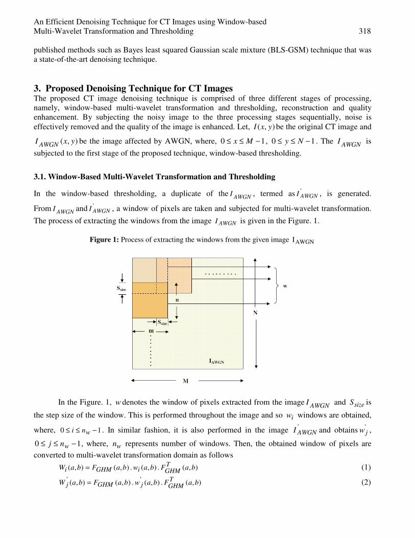
Figure 1: Process of extracting the windows from the given image AWGNI
In the Figure. 1, w denotes the window of pixels extracted from the imageAWGN
I and sizeS is
the step size of the window. This is performed throughout the image and so iw windows are obtained,
where, 10 −≤≤ wni . In similar fashion, it is also performed in the image 'AWGNI and obtains '
jw ,
10 −≤≤ wnj , where, wn represents number of windows. Then, the obtained window of pixels are
converted to multi-wavelet transformation domain as follows
),(.),(.),(),( baF baw baFbaWTGHMiGHMi = (1)
),(),(),(),( ''baF . baw . baFbaW
TGHMjGHMj = (2)
319 Syed Amjad Ali, Srinivasan Vathsal and K. Lal kishore
where, 10 −≤≤ ma , 10 −≤≤ nb and nm × represents the window size. In Eq. (1) and (2) GHMF is the
concatenated filter coefficient of GHM multi-wavelet transformation, iW and 'jW are iw and '
jw in
multi-wavelet domain, respectively. For every iW , 'jW that are closer to iW are selected based on L2
norm distance ( ijL2 ), which can be computed using Eq. (3),
∑ ∑−
=
−
=
−=1
0
1
0
2' |)),(),((|2
m
a
n
b
jiij baWbaWL (3)
Using the ijL2 , the 'jW windows that are closer to the iW , '
2ijLW can be identified
as φ−=ijij LL WW 2
'2 , where,
ijLW 2 is given as
≤
=else
LLifWW Tijj
L ij;
22;'
2φ
(4)
Every thi window sets in
ijLW 2 are sorted in ascending order based on their
corresponding ijL2 . From the sorted window set, cn number of windows are selected (for every iW )
and the remaining are skipped out, which leads to obtain '2ikLW , where, 10 −≤≤ cnk . From every th
k
window that corresponds to the thi window, the elements of similar position are subjected for CL
multi-wavelet transformation that can be given as
−−
−
=
−−
−
)(.)1,1(.)(
)(.)1,(.)(
)(.)2,0(.)(
)(.)1,0(.)(
)(.)0,0(.)(
'2
'2
'2
'2
'2
1,1
1,0
2,0
1,0
0,0
kF nmW kF
kF noW kF
kF W kF
kF W kF
kF W kF
V
V
V
V
V
TCLLCL
TCLikLCL
TCLLCL
TCLLCL
TCLLCL
ik
ik
ik
ik
ik
ik
ik
ik
ik
nm
n
M
M
M
M (5)
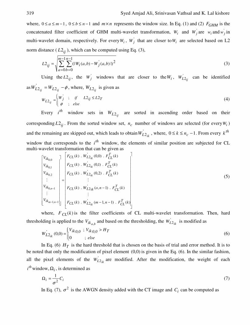
where, )(kF CL is the filter coefficients of CL multi-wavelet transformation. Then, hard
thresholding is applied to the baikV
,and based on the thresholding, the '
2ikLW is modified as
>
=else
HV VW
T0,0ik0,0ikL ik ;0
;)0,0('
2 (6)
In Eq. (6) TH is the hard threshold that is chosen on the basis of trial and error method. It is to
be noted that only the modification of pixel element )0,0( is given in the Eq. (6). In the similar fashion,
all the pixel elements of the '2ikLW are modified. After the modification, the weight of each
thi window, iΩ , is determined as
ii C2
1
σ=Ω (7)
In Eq. (7), 2σ is the AWGN density added with the CT image and iC can be computed as
An Efficient Denoising Technique for CT Images using Window-based
Multi-Wavelet Transformation and Thresholding 320
∑−
=
=
1
0
c
k
n
k
NZi nC (8)
where, kNZn is number of non-zero elements in the '
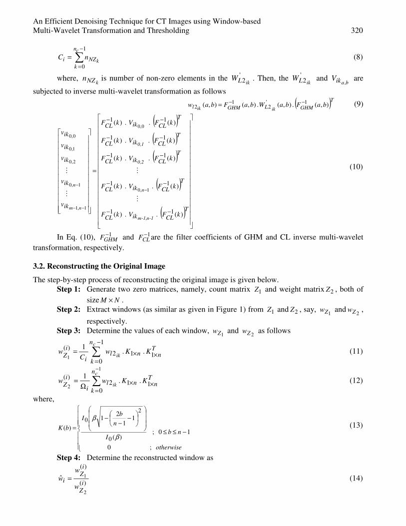
2ikLW . Then, the '2ikLW and
baikV,
are
subjected to inverse multi-wavelet transformation as follows
( )TGHMLGHMl baF baW baFbaw
ikik),(.),(.),(),( 1'
21
2−−= (9)
( )
( )( )
( )
( )
=
−−
−−
−−
−−
−−
−
−−
−
T
CLikCL
T
CLikCL
T
CLikCL
T
CLikCL
T
CLikCL
ik
ik
ik
ik
ik
kF V kF
kF V kF
kF V kF
kF V kF
kF V kF
v
v
v
v
v
-1n-1,m
n
0,2
0,1
nm
n
)(..)(
)(..)(
)(..)(
)(..)(
)(..)(
11
11
11
11
11
1,0
0,0
1,1
1,0
2,0
1,0
0,0
M
M
M
M (10)
In Eq. (10), 1−GHMF and 1−
CLF are the filter coefficients of GHM and CL inverse multi-wavelet
transformation, respectively.
3.2. Reconstructing the Original Image
The step-by-step process of reconstructing the original image is given below.
Step 1: Generate two zero matrices, namely, count matrix 1Z and weight matrix 2Z , both of
size NM × .
Step 2: Extract windows (as similar as given in Figure 1) from 1Z and 2Z , say, 1Zw and
2Zw ,
respectively.
Step 3: Determine the values of each window, 1Zw and
2Zw as follows
Tn n
n
k
li
i
ZK K w
Cw
c
ik ××
−
=∑=
11
1
0
2)(
..1
1 (11)
Tn
n
k
nli
i
ZK K ww
c
ik ×=
×∑−
Ω=
10
12)(
..1
1
2 (12)
where,
−≤≤
−
−−
=
otherwise
nb I
n
bI
bK
;0
10;)(
11
21
)(
0
2
0
β
β (13)
Step 4: Determine the reconstructed window as
)(
)(
2
1ˆi
Z
i
Zi
w
w
w = (14)
321 Syed Amjad Ali, Srinivasan Vathsal and K. Lal kishore
Step 5: Replace the obtained wn windows in their corresponding positions of the image
AWGNI so as to obtain the reconstructed image I .
Hence, the original image is reconstructed with the aid of Kaiser Window. However, for
clinical diagnosis or for any other medical imaging applications, the quality of the reconstructed CT
image has to be enhanced (as stated earlier). This can be accomplished in the subsequent process of
enhancing the quality of the original image.
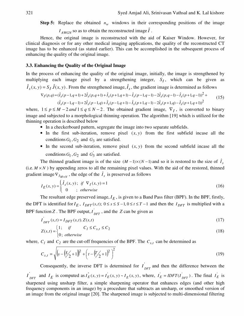
3.3. Enhancing the Quality of the Original Image
In the process of enhancing the quality of the original image, initially, the image is strengthened by
multiplying each image pixel by a strengthening integer, IS , which can be given as
),(ˆ.),(ˆ yxISyxI Is = . From the strengthened image, sI , the gradient image is determined as follows
2
2
))1,1(ˆ),1(ˆ2)1,1(ˆ)1,1(ˆ),1(ˆ2)1,1(ˆ(
))1,1(ˆ)1,(ˆ2)1,1(ˆ)1,1(ˆ)1,(ˆ2)1,1(ˆ(),(
++−+−−+−+−+−+−−
+−+−−−−−−++++++−=∇
qpIqpIqpIqpIqpIqpI
qpIqpIqpIqpIqpIqpIqp
ssssss
ssssssI (15)
where, 2121 −≤≤−≤≤ Nq nda Mp . The obtained gradient image, I∇ , is converted to binary
image and subjected to a morphological thinning operation. The algorithm [19] which is utilized for the
thinning operation is described below
• In a checkerboard pattern, segregate the image into two separate subfields.
• In the first sub-iteration, remove pixel ),( yx from the first subfield incase all the
conditions 1G , 2G and 3G are satisfied.
• In the second sub-iteration, remove pixel ),( yx from the second subfield incase all the
conditions 1G , 2G and '3G are satisfied.
The thinned gradient image is of the size )1()1( −×− NM and so it is restored to the size of sI
(i.e. NM × ) by appending zeros to all the remaining pixel values. With the aid of the restored, thinned
gradient imageNMI ×
∇ , the edge of the sI is preserved as follows
=∇
=otherwise
yx if yxIyxI Is
E;0
1),(;),(ˆ),( (16)
The resultant edge preserved image, EI , is given to a Band Pass filter (BPF). In the BPF, firstly,
the DFT is identified for EI , 10,10);,( −≤≤−≤≤ Tt Ss tsI DFT and then the DFTI is multiplied with a
BPF function Z . The BPF output, 'DFT
I , and the Z can be given as
),().,(),('tsZ tsItsI DFTDFT
= (17)
≤≤
= otherwise
CCC if tsZ
ts1
;0
;1),(
2, (18)
where, 1C and 2C are the cut-off frequencies of the BPF. The tsC , can be determined as
( )( ) ( )2
22
, 12
12
+−++−= TtSsC ts (19)
Consequently, the inverse DFT is determined for 'DFT
I and then the difference between the
'DFT
I and EI is computed as ),(~),(),( '''yxI yxIyxI EEE = , where, )( ''
DFTIIDFTI E = . The final '
EI is
sharpened using unsharp filter, a simple sharpening operator that enhances edges (and other high
frequency components in an image) by a procedure that subtracts an unsharp, or smoothed version of
an image from the original image [20]. The sharpened image is subjected to multi-dimensional filtering
An Efficient Denoising Technique for CT Images using Window-based
Multi-Wavelet Transformation and Thresholding 322
in which the array values outside the bounds are presumed to match the nearest array border value.
Thus, the final edge preserved image, EI is obtained from the multi-dimensional filtering. By using
the EI , the final quality enhanced image is obtained as follows
>
= otherwise yxI
yxI if yxIyxI
EEfinal
;),(ˆ
0),(ˆ;),(ˆ),(ˆ (20)
The finalI obtained from the Eq. (20) is the denoised and quality enhanced CT image
of AWGNI .
4. Results and Discussion The proposed CT image denoising technique has been implemented in the working platform of
MATLAB (version 7.8). The performance has been evaluated by denoising the CT images that are
corrupted by AWGN with noise levels, σ . We have utilized the CT images of size 256256×
( 256=M and 256=N ). For multi-wavelet transformation and thresholding, the window has been
chosen with size 88× ( 8=m and 8=n ) and 1=sizeS . Hence, 6363×=wn windows of pixels have
been extracted from the image. Among wn , 16=cn number of windows are windows have been
selected on the basis of L2-norm distance ( 2500=TL ). For hard thresholding, we have
selected 2=TH , based on trial and error method and for strengthening the image, 255=IS . The quality
of the final denoised and enhanced CT has been evaluated by calculating the PSNR values as
−
−=
∑ ∑−
=
−
=
1
0
1
0
2
10
)),(),((1
1log20)(
M
x
N
y
AWGN
AWGN
yxIyxIMN
M IPSNR (21)
−
−=
∑ ∑−
=
−
=
1
0
1
0
2
10
)),(ˆ),((1
1log20)ˆ(
M
x
N
y
final
final
yxIyxIMN
M IPSNR (22)
By Eq. (21) and Eq. (22), the PSNR for noisy and denoised image, respectively, can be
determined. The PSNR values of noisy and denoised images with different noise levels are given in the
Table I and the image results for 1I and 2I are shown in the Figure 2 and Figure 3, respectively.
Table I: The PSNR values of1AWGNI ,
1finalI ,2AWGNI and
2finalI for different noise levels, σ=10,20,30,40
and 50
Sl.No Noise Level σ 1AWGNI 1finalI
2AWGNI 2finalI
1 10 28.1382 33.2484 28.1188 37.3094
2 20 22.1629 34.6670 22.1101 34.9941
3 30 18.5831 33.4892 18.5774 33.6363
4 40 16.0809 32.7574 16.1048 32.9301
5 50 14.1859 32.0428 14.1475 31.3239
323 Syed Amjad Ali, Srinivasan Vathsal and K. Lal kishore
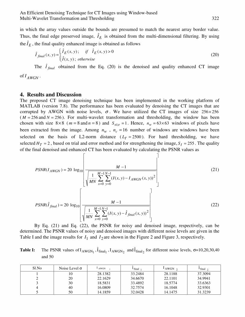
Figure 2: Image results for (a) original CT image 1I (b) AWGN corrupted CT image,1AWGNI with added
noise levels 50 and 40 ,30 ,20 =σ and (c) denoised CT image, 1finalI
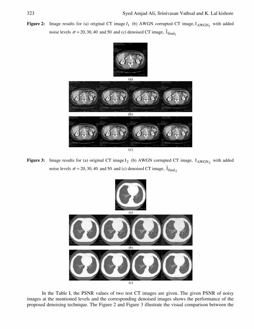
Figure 3: Image results for (a) original CT image 2I (b) AWGN corrupted CT image, 2AWGNI with added
noise levels 50 and 40 ,30 ,20 =σ and (c) denoised CT image, 2finalI
In the Table I, the PSNR values of two test CT images are given. The given PSNR of noisy
images at the mentioned levels and the corresponding denoised images shows the performance of the
proposed denoising technique. The Figure 2 and Figure 3 illustrate the visual comparison between the
An Efficient Denoising Technique for CT Images using Window-based
Multi-Wavelet Transformation and Thresholding 324
original, noisy and the denoised CT images. The results shows that the noise is effectively removed
from the depicted CT images using the proposed image denoising technique.
5. Conclusion In this paper, we have proposed an effective image denoising technique using window-based multi-
wavelet transformation and thresholding. The proposed denoising technique has effectively removed
the AWGN noise from the CT images and improved the quality of the images. The technique has been
evaluated with different levels of AWGN corrupted CT images. The denoising has been performed
well by the proposed technique and also it has offered a good PSNR for the denoised images. The
performance has been well illustrated by comparing the PSNR of the noisy CT images and the
denoised CT images. Moreover, it could be well understood (from the image results given in the
Section 4) that the quality of the CT images has been enhanced well by the proposed denoising
technique. Hence, from the obtained results, it can be concluded that the proposed image denoising
technique effectively removes the AWGN from the CT images with the aid of window-based multi-
wavelet transformation and thresholding and also it enhances the quality of the image based on
filtering techniques. The resultant images are in a good quality which helps for effective and accurate
clinical diagnosis and for other medical imaging applications.
References [1] S. Arivazhagan, S. Deivalakshmi, K. Kannan, B.N.Gajbhiye, C.Muralidhar, Sijo N Lukose,
M.P.Subramanian, “Performance Analysis of Wavelet Filters for Image Denoising”, Advances
in Computational Sciences and Technology, Vol.1 No. 1 (2007), pp. 1–10, ISSN 0973-6107.
[2] R. Sivakumar, "Denoising Of Computer Tomography Images Using Curvelet Transform,"
ARPN Journal of Engineering and Applied Sciences, Vol. 2, No. 1, pp. 21-26, 2007.
[3] Joao M. Sanches, Jacinto C. Nascimento and Jorge S. Marques "Medical Image Noise
Reduction Using the Sylvester-Lyapunov Equation," IEEE transactions on image processing,
Vol. 17, No. 9, pp. 1522-1539, 2008.
[4] G. Landi an E. Loli Piccolomini, "An Algorithm for Image Denoising with Automatic Noise
Estimate," Journal of Mathematical Imaging and Vision, Vol. 34, No. 1, pp. 98–106, 2009.
[5] V.R.Vijaykumar, P.T.Vanathi, P.Kanagasabapathy and D.Ebenezer, "Robust Statistics Based
Algorithm to Remove Salt and Pepper Noise in Images," International Journal of Signal
Processing, Vol. 5, No. 3, 2009
[6] Akshaya. K. Mishra, Alexander Wong, David. A. Clausi and Paul. W. Fieguth, "Adaptive
nonlinear image denoising and restoration using a cooperative Bayesian estimation approach,"
in proceedings of the Sixth Indian Conference on Computer Vision, Graphics & Image
Processing, pp. 621-627, 16-19 December, Bhubaneswar, 2008.
[7] Nguyen Thanh Binh and Ashish Khare, "Adaptive complex wavelet technique for medical
image denoising," in proceedings of the third International Conference on the development of
Biomedical Engineering, pp. 195-198, Vietnam, January 11-14, 2010
[8] S.Arivazhagan, S.Deivalakshmi, K.Kannan, B.N.Gajbhiye, C.Muralidhar, Sijo N.Lukose and
M.P.Subramanian, "Performance Analysis of Image Denoising System for different levels of
Wavelet decomposition," International Journal of Imaging Science and Engineering, Vol.1,
No.3, pp. 104-107, 2007.
[9] Nilamani Bhoi and Sukadev Meher, "Total Variation Based Wavelet Domain Filter for Image
Denoising," in proceedings of the First International Conference on Emerging Trends in
Engineering and Technology, pp. 20-25, July 16 - 18, 2008.
325 Syed Amjad Ali, Srinivasan Vathsal and K. Lal kishore
[10] Joseph Shtok, Michael Elad and Michael Zibulevsky, "Adaptive filtered-back-projection for
computed tomography," in proceedings of the 25th Convention of Electrical and Electronics
Engineers in Israel, pp. 528-532, 3-5 December, Eilat, 2008.
[11] Alexander Wong, Akshaya Mishra, Paul Fieguth and David Clausi, “An adaptive Monte Carlo
approach to nonlinear image denoising," in proceedings of 19th IEEE International Conference
on Pattern Recognition, pp. 1-4, 8-11 December, Tampa, FL, 2008.
[12] H. Rabbani, R. Nezafat and S. Gazor, "Wavelet-Domain Medical Image Denoising Using
Bivariate Laplacian Mixture Model," IEEE Transactions on Biomedical Engineering, Vol. 56,
No. 12, pp. 2826-37, 2009.
[13] Tang Jingtian, Yang Xiaoli and Pan Meisen, "The Application of Independent Component
Analysis in CT Image De-Noising,”, in proceedings of 3rd International Conference on
Bioinformatics and Biomedical Engineering, pp. 1-4, 11-13 June, Beijing, 2009.
[14] Jin Li, Lei Wang and Peihua Bao, "An industrial CT image adaptive filtering method based on
anisotropic diffusion," in proceedings of the IEEE International Conference on Mechatronics
and Automation, pp. 1009 - 1014, 9-12 Aug, 2009.
[15] S. Skiadopoulos, G. Karatrantou, P. Korfiatis, L. Costaridou, P. Vassilakos, D. Apostolopoulos
and G. Panayiotakis, "Evaluating image denoising methods in myocardial perfusion single
photon emission computed tomography (SPECT) imaging," Measurement science &
technology, Vol. 20, No. 10, pp. 104023, 2009.
[16] Guangming Zhang, Xuefeng Xian, Zhiming Cui and Jian Wu, "Medical Image De-noising
Extended Model Based on Independent Component Analysis and Dynamic Fuzzy Function," in
proceedings of the IEEE International Conference on Information Engineering, Vol. 1, pp.209-
212, 2009.
[17] A. Borsdorf, R. Raupach, T. Flohr and J. Hornegger, "Wavelet Based Noise Reduction in CT-
Images Using Correlation Analysis," IEEE Transactions on Medical Imaging, Vol. 27, No.12,
pp. 1685-1703, 2008.
[18] H. Rabbani, "Abdominal CT Image Denoising Based On A Laplace Distribution With Local
Variance In Steerable Pyramid Domain,” in proceedings of International Conference on
Technology and Applications in Biomedicine, pp. 140-143, 30-31 May, Shenzhen, 2008.
[19] Lam, L., Seong-Whan Lee, and Ching Y. Suen, "Thinning Methodologies-A Comprehensive
Survey," IEEE Transactions on Pattern Analysis and Machine, Vol. 14, No. 2, pp. 869-885,
ISSN 0162-8828. DOI: 10.1109/34.161346
[20] Ping S. Huang, Shun-Chi Su, and Te-Ming Tu, “A Destriping and Enhancing Technique
Intelligence, Vol. 14, No. 9, September 1992, page 879, bottom of first column for EROS
Remote Sensing Imagery”, Journal of C.C.I.T., Vol.32, No.2, 2004.
[21] J. Orchard, M. Ebrahimi and A. Wong, "Efficient nonlocal-means denoising using the SVD”, In
Proceedings of the 15th IEEE International Conference on image Processing, pp. 1732-1735,
12-15 October, San Diego, CA, 2008.
A GA-based Window Selection Methodology to
Enhance Window-based Multi-wavelet
transformation and thresholding aided CT image
denoising technique
Prof. Syed Amjad Ali
Professor and Head of ECE Department
Lords Institute of Engineering and
Technology
Himayathsagar, Hyderabad – 8
Dr. Srinivasan Vathsal
Principal
Bhaskar Engineering College
Yenkapally, Moinabad
Ranga reddy Dist
Dr. K. Lal kishore
Rector, Jawahar Lal Nehru
Technological University
Kukatpally
Hyderabad.
Abstract— Image denoising is getting more significance,
especially in Computed Tomography (CT), which is an important
and most common modality in medical imaging. This is mainly
due to that the effectiveness of clinical diagnosis using CT image
lies on the image quality. The denoising technique for CT images
using window-based Multi-wavelet transformation and
thresholding shows the effectiveness in denoising, however, a
drawback exists in selecting the closer windows in the process of
window-based multi-wavelet transformation and thresholding.
Generally, the windows of the duplicate noisy image that are
closer to each window of original noisy image are obtained by the
checking them sequentially. This leads to the possibility of
missing out very closer windows and so enhancement is required
in the aforesaid process of the denoising technique. In this paper,
we propose a GA-based window selection methodology to include
the denoising technique. With the aid of the GA-based window
selection methodology, the windows of the duplicate noisy image
that are very closer to every window of the original noisy image
are extracted in an effective manner. By incorporating the
proposed GA-based window selection methodology, the denoising
the CT image is performed effectively. Eventually, a comparison
is made between the denoising technique with and without the
proposed GA-based window selection methodology.
Keywords-Denoising Technique; Window Selection Methodolog y; Genetic Algorithm (GA); Computed Tomography (CT) image; Closer Windows.
I. INTRODUCTION
Digital images are pivotally involved in the routine applications like satellite television, magnetic resonance imaging and computer tomography. In addition, they are involved in the areas of research and technology, namely, geographical information systems and astronomy. Retrieving original images from incomplete, indirect and noisy images is a serious issue that scientists experience in the aforesaid fields [1]. When the images are captured by the sensors and transmitted in the channel, the noises are added to the images [2]. With the existence of noise, the image gets a mottled, grainy, textured or snowy appearance [3]. Hence, in recent
years, an overwhelming interest has been noticed in the case of recovering an original image from noisy image [4]. The recovery of an image is possible by image denoising. Image denoising can be described as the process of determining the original image from a contaminated image by noise degradation [5].
Generally, image denoising is the action of eliminating undesirable noise from a noised image, by bringing back the image to its un-degraded ideal [6]. The Image denoising techniques can be classified as transform domain methods or spatial domain methods. The transform domain methods transform an image from the spatial domain into another domain (probably, frequency domain or wavelet domain) and suppress noise in the transform domain, whereas, in spatial domain methods, the noise is suppressed in the spatial domain itself [7]. However, the image denoising with multi-wavelet techniques is very effectual due to the potential of capturing the signal energy in a small number of transformation energy values. In comparison with other multi-scale representations, the multi-wavelet transformation offers better spatial and spectral localization of image.
The image denoising finds its applications in fields of medical imaging and preprocessing for computer vision [8]. Medical imaging acquisition technologies and systems bring in noise and artifacts in the images and they should be attenuated by denoising algorithms. The denoising process should not damage anatomical details pertinent to a clinical viewpoint [9]. As a matter of reason, it is hard to put forward a robust method for noise removal which functions well for diverse modalities of medical images [10]. CT is regarded as a general and vital modality in Medical Imaging which is used for clinical diagnosis and computer-aided surgery [11]. In recent years, there have been numerous methods developed and described in literature for denoising [12].
In spite of the existence of several image denoising algorithms over the years, finding a proper solution for noise suppression in situations involving low signal-to-noise ratios
(IJCSIS) International Journal of Computer Science and Information Security, Vol. 7, No. 2, February 2010
280 http://sites.google.com/site/ijcsis/ ISSN 1947-5500
remains a complex task [13]. In the earlier work, an efficient denoising technique for CT images employing window-based Multi-wavelet transformation and thresholding has been presented [32]. There the multi-wavelet has been favored since it betters single wavelets by its characteristics particularly, orthogonality, short support, symmetry, and high degree of vanishing moments. The technique has denoised the CT images degraded by AWGN and enhances the quality of the image. However, a drawback persists in choosing the closer windows in the process of window-based multi-wavelet transformation and thresholding. Normally, the windows of the duplicate noisy image that are closer to each window of original noisy image are acquired by the inspecting them sequentially. This results in the possibility of missing out very closer windows and so enhancement is needed in the aforesaid process of the denoising technique.
Here, we propose a GA-based window selection methodology to incorporate the denoising technique. With the aid of the GA-based window selection methodology, the windows of the duplicate noisy image that are very closer to every window of the original noisy image are extracted in an effectual way. By incorporating the proposed GA-based window selection methodology, the denoising is carried out more successfully. Eventually, a comparison is made between the denoising technique with and without the proposed GA-based window selection methodology. The rest of the paper is organized as follows. Section II briefly reviews the recent research works in the literature and Section III gives a short introduction about the GA. Section IV explains the window selection methodology of the denoising technique proposed in the previous work. Section V describes the proposed GA-based window selection methodology with required illustrations and mathematical formulations. Section VI discusses about the implementation results and Section VII concludes the paper.
II. RELATED WORKS
Lanzolla et al. [14] have evaluated the effect of different noise reduction filters on computed tomography (CT) images. Especially, they have presented a denoising filter on the basis of a combination of Gaussian and Prewitt operators. Simulation results have proved that their presented technique has enhanced the image quality, and then permitted to use low radiation dose protocol in CT examinations. Their work was carried out in association with "G.Moscati" Hospital of Taranto (Italy), that offered all the images and technical materials employed in the proposed algorithm. Bing-gang Ye and Xiao-ming Wu [15] have addressed that the prior detection of small hepatocellular carcinoma (SHCC) has significant clinic value, and wavelet denoising arithmetic research of SHCC CT image, on the basis of image processing technology has aided to diagnose the SHCC focus. In accordance with the wavelet coefficient correlation, their work has reduced the figures and eliminated the feeble or irrelated coefficient of noise of SHCC CT image, and finally removed the noise.
The objective of Jin Li et al. [7] was to lessen the noise and artifacts in the industrial CT image by anisotropic diffusion. Anisotropic diffusion algorithms which could maintain
significant edges sharp and spatially fixed at the same time as filtering noise and small edges eliminated the noise from an image by altering the image through a partial differential equation. In conventional anisotropic diffusions which lead to the loss of image details and cause false contours, 4-neighborhood directions are employed generally except diagonal directions of the image. To remove the drawbacks of the conventional anisotropic diffusion methods, an anisotropic diffusion method for industrial CT image based on the types of gradient directions was presented. In their work, one parameter K is calculated first by the histogram of the gradient. Then Sobel operator was made use of to calculate the directions of gradient. The directions of the gradient were classified. Experimental results have revealed that their presented algorithm could eliminate noise and artifacts from industrial CT volume data sets that were better than the Gaussian filter and other traditional algorithm.
Hossein Rabbani [16] have presented an image denoising algorithm based on the modeling of coefficients in each sub-band of steerable pyramid employing a Laplacian probability density function (PDF) with local variance. That PDF was able to model the heavy-tailed nature of steerable pyramid coefficients and the empirically observed correlation between the coefficient amplitudes. Within that framework, he has described a method for image denoising based on designing both maximum a posteriori (MAP) and minimum mean squared error (MMSE) estimators, which has relied on the zero-mean Laplacian random variables with high local correlation. Despite the simplicity of his spatially adaptive denoising method, both in its concern and implementation, his denoising results has achieved better performance than several published methods such as Bayes least squared Gaussian scale mixture (BLS-GSM) technique that was a state-of-the-art denoising technique.
H.Rabbani et al. [17] have proposed noise reduction algorithms that could be employed to improve image quality in several medical imaging modalities like magnetic resonance and multidetector CT. The acquired noisy 3-D data were first transformed by discrete complex wavelet transform. Employing a nonlinear function, they have modeled the data as sum of the clean data plus additive Gaussian or Rayleigh noise. They employed a mixture of bivariate Laplacian probability density functions for the clean data in the transformed domain. The MAP and minimum mean-squared error (MMSE) estimators enabled them to effectively reduce the noise. In addition, they have calculated the parameters of the model using local information. Experimental results on CT images revealed that among their derived shrinkage functions, generally, BiLapGausMAP has given images with higher peak SNR.
Skiadopoulos et al. [18] have carried out a comparative study between a multi-scale platelet denoising method and the well-established Butterworth filter, which was employed as a pre- and post-processing step on image reconstruction. The comparison was performed with and/or without attenuation correction. Quantitative evaluation was executed by using 1) a cardiac phantom comprising of two different size cold defects, employed in two experiments done to simulate conditions with and without photon attenuation from myocardial surrounding
(IJCSIS) International Journal of Computer Science and Information Security, Vol. 7, No. 2, February 2010
281 http://sites.google.com/site/ijcsis/ ISSN 1947-5500
tissue and 2) a pilot-verified clinical dataset of 15 patients with ischemic defects. Furthermore, an observer preference study was executed for the clinical dataset, based on rankings from two nuclear medicine clinicians. Without photon attenuation conditions, denoising by platelet and Butterworth post-processing methods outplayed Butterworth pre-processing for large size defects. Conversely, for the small size defects and with photon attenuation conditions, all the methods have showed similar denoising performance. Guangming Zhang et al. [19] have proposed an extended model for CT medical image de-noising, which employed independent component analysis and dynamic fuzzy theory. Initially, a random matrix was created to separate the CT image for estimation. Then, dynamic fuzzy theory was applied to set up a series of adaptive membership functions to produce the weights degree of truth. At last, the weights degree was employed to optimize the value of matrix for image reconstruction. By putting to practice their model, the selection of matrix could be optimized scientifically and self-adaptively.
Jessie Q Xia et al. [20] have employed the partial diffusion equation (PDE) based denoising techniques particularly for breast CT at various steps along the reconstruction process and it was noticed that denoising functioned better when applied to the projection data rather than the reconstructed data. Simulation results from the contrast detail phantom have proved that the PDE technique outplayed Wiener denoising and also adaptive trimmed mean filter. The PDE technique has improved its performance features in relation to Wiener techniques when the photon fluence was lowered. With the PDE technique, the sensitivity for lesion detection employing the contrast detail phantom declined by less than 7% when the dose was reduced to 40% of the two-view mammography. For subjective evaluation, the PDE technique was employed to two human subject breast data sets obtained on a prototype breast CT system. The denoised images had great visual characteristics with a considerable lower noise levels and enhanced tissue textures while retaining sharpness of the original reconstructed volume.
A. Borsdorf et al. [21] have proposed a wavelet based structure-preserving method for noise reduction in CT images that could be used together with various reconstruction methods. Their approach was on the basis of presumption that the data could be decomposed into information and temporally uncorrelated noise. The analysis of correlations between the wavelet representations of the input images enabled separating information from noise down to a certain signal-to-noise level. Wavelet coefficients with small correlation were reduced, while those with high correlations were supposed to symbolize structures and are preserved. The ultimate noise-suppressed image was reconstructed from the averaged and weighted wavelet coefficients of the input images. The quantitative and qualitative evaluation on phantom and real clinical data proved that high noise reduction rates of around 40% could be accomplished without considerable loss of image resolution.
III. GENETIC ALGORITHM (GA)
The GA-based approaches have received considerable interest from the academic and industrial communities for coping with optimization problems that have proved to be
difficult by employing conventional problem solving techniques [22][23] [24][25][26]. GAs are computing algorithms designed in correlation to the process of evolution [27], which was proposed in the 1970s in the United States by John Holland [28]. In GA, the search space comprises of solutions which are represented by a string identified as a chromosome. Each chromosome is composed of an objective function called fitness. In GA, the search space consists of solutions which are denoted by a string known as a chromosome. A collection of chromosomes together with their associated fitness is termed as the population. The population, at a particular iteration of the GA, is known as a generation [28] [29] [30].
GA begins to function with numerous possible solutions that are obtained from the randomly generated initial population. Then, it tries to find optimum solutions by employing genetic operators namely selection, crossover and mutation [30]. Selection is a process of selecting a pair of organisms to reproduce. Crossover is a process of swapping the genes between the two individuals that are reproducing. Mutation is the process of randomly modifying the chromosomes [27]. The main aim of mutation is re-establishing lost and exploring variety of data. In accordance with changing some bit values of chromosomes provide different breeds. Chromosome may be better or poorer than old chromosome. If they are poorer than old chromosome, then they are removed from selection step [31]. The process continues until a termination criterion is satisfied and so the GA can converge to an optimal solution.
IV. WINDOW SELECTION METHODOLOGY IN THE DENOISING
TECHNIQUE USING WINDOW-BASED MULTI-WAVELET
TRANSFORMATION AND THRESHOLDING
Prior to detail the proposed GA-based window selection methodology for the CT image denoising technique [32], here, a brief description about the prevailing window-selection methodology used in the technique is given. Let, ),( yxI be the
original CT image and ),( yxIAWGN
be the image affected by
AWGN, where, 10 −≤≤ Mx , 10 −≤≤ Ny . TheAWGN
I is
put to the first stage of the proposed technique, window-based thresholding. The window selection methodology to be described here is one of the components of the first stage of processing of the CT image denoisng technique. In the
methodology, a replica of theAWGN
I , labeled as'AWGNI , is
generated. FromAWGN
I and 'AWGNI , a window of pixels are
considered and put to multi-wavelet transformation. The
process of extracting the windows from the image AWGN
I is
given in the Figure 1.
(IJCSIS) International Journal of Computer Science and Information Security, Vol. 7, No. 2, February 2010
282 http://sites.google.com/site/ijcsis/ ISSN 1947-5500
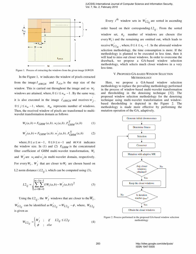
Figure 1. Process of extracting the windows from the given image IAWGN
In the Figure 1, w indicates the window of pixels extracted
from the imageAWGN
I and sizeS is the step size of the
window. This is carried out throughout the image and so iw
windows are attained, where, 10 −≤≤ wni . By the same way,
it is also executed in the image 'AWGNI and receives
'jw ,
10 −≤≤ wnj , where, wn represents number of windows.
Then, the received window of pixels are transformed to multi-wavelet transformation domain as follows
),( . ),( . ),(),( baFbawbaFbaWTGHMiGHMi = (1)
),( . ),( . ),(),(TGHM
''baFbawbaFbaW jGHMj = (2)
where, 10 −≤≤ ma , 10 −≤≤ nb and nm × indicates
the window size. In (1) and (2) GHMF is the concatenated
filter coefficient of GHM multi-wavelet transformation, iW
and 'jW are iw and
'jw in multi-wavelet domain, respectively.
For every iW , 'jW that are closer to iW are chosen based on
L2 norm distance ( ijL2 ), which can be computed using (3),
∑∑−
=
−
=
−=
1
0
1
0
2' |)),(),((|2
m
a
n
b
jiij baWbaWL (3)
Using the ijL2 , the 'jW windows that are closer to the iW ,
'2ijLW can be identified as φ−=
ijij LL WW 2'2 , where,
ijLW 2
is given as
≤
=else
LLifWW Tijj
L ij;
22;'
2φ
(4)
Every th
i window sets in '2ijLW are sorted in ascending
order based on their corresponding ijL2 . From the sorted
window set, cn number of windows are chosen (for
every iW ) and the remaining are omitted out, which leads to
receive'2ikLW , where, 10 −≤≤ cnk . In the aforesaid window
selection methodology, the time consumption is more. If the methodology is planned to be executed in less time, then it will lead to miss out closer windows. In order to overcome the drawback, we propose a GA-based window selection methodology, which selects much closer windows in a very less time.
V. PROPOSED GA-BASED WINDOW SELECTION
METHODOLOGY
Here, we propose a GA-based window selection methodology to replace the prevailing methodology performed in the process of window-based multi-wavelet transformation and thresholding in the denoising technique [32]. The proposed window selection methodology for the denoising technique using multi-wavelet transformation and window-based thresholding is depicted in the Figure 2. The methodology is made more effective by performing the mutation operation of the GA, adaptively.
Figure 2. Process performed in the proposed GA-based window selection
methodology
(IJCSIS) International Journal of Computer Science and Information Security, Vol. 7, No. 2, February 2010
283 http://sites.google.com/site/ijcsis/ ISSN 1947-5500
It is well known that the proposed GA-based window
selection methodology is utilized to obtain cn number of
windows, '2ikLW ; 10 −≤≤ cnk that are closer to every iW .
Once the closer windows are identified, the further process of the denoising is continued using the obtained windows. The proposed methodology is comprised of five functional steps, namely, 1) generation of initial chromosomes, 2) Determination of fitness function, 3) Crossover and Mutation, 4) Selection of closer windows and 5) Termination criteria. They are described below in detail.
A. Generation of initial chromosomes
In the methodology, as the first process, gn initial
chromosomes, each of length cn are generated. The set
representation of initial chromosomes are given as
ilnil c
rrrrR 1210 ,,, −= L ; 10 −≤≤ gnl (5)
where, ilR is the th
l chromosome generated to obtain
windows that are closer to the th
i window of the original noisy
image. Each gene of the generated chromosome ililk Rr ∈ ;
10 −≤≤ cnk , is an arbitrary integer generated within the
interval [ ]1,0 −wn provided that the all the genes of each
chromosome has to satisfy the condition 110 −≠≠≠cnrrr L .
B. Determination of fitness function
A fitness function decides whether the generated chromosomes are fit to survive or not, that can be given as
∑−
=
=
1
0
21
)(
cn
k
ilkc
i Ln
lf (6)
where, )(lfi is the fitness of the th
l chromosome
generated for the th
i window and ilkL2 is the 2L norm
distance determined between the iw and the window indexed
by the th
k gene of the th
l chromosome. The ilkL2 is
determined as follows
∑∑−
=
−
=
−=
1
0
1
0
2' ),(),(2
m
a
n
b
riilk baWbaWLilk
(7)
where, 'ilkrW is the window indexed by ilkr that is
converted to multi-wavelet transformation domain as done in
(1) and (2). From the pn generated chromosomes, 2/pn
chromosomes that have minimum fitness are selected as best chromosomes and they are subjected to the genetic operations, crossover and mutation.
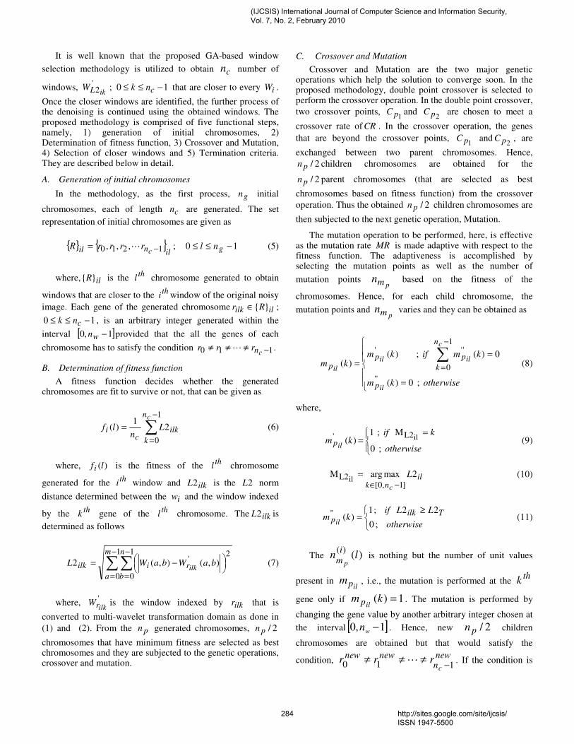
C. Crossover and Mutation
Crossover and Mutation are the two major genetic operations which help the solution to converge soon. In the proposed methodology, double point crossover is selected to perform the crossover operation. In the double point crossover,
two crossover points, 1pC and
2pC are chosen to meet a
crossover rate of CR . In the crossover operation, the genes
that are beyond the crossover points, 1pC and
2pC , are
exchanged between two parent chromosomes. Hence,
2/pn children chromosomes are obtained for the
2/pn parent chromosomes (that are selected as best
chromosomes based on fitness function) from the crossover
operation. Thus the obtained 2/pn children chromosomes are
then subjected to the next genetic operation, Mutation.
The mutation operation to be performed, here, is effective as the mutation rate MR is made adaptive with respect to the fitness function. The adaptiveness is accomplished by selecting the mutation points as well as the number of
mutation points pmn based on the fitness of the
chromosomes. Hence, for each child chromosome, the
mutation points and pmn varies and they can be obtained as
=
==
∑−
=
otherwisekm
kmifkmkm
il
c
ilil
il
p
n
k
ppp
; 0)(
0)( ; )()(
''
1
0
'''
(8)
where,
=
=otherwise
kifkm
ilp ; 0
M ; 1 )( ilL2'
(9)
ilnk
L
c
2maxargM]1,0[
L2il−∈
= (10)
≥
=otherwise
LLifkm
Tilkpil ; 0
22 ; 1)(" (11)
The )()(
lni
m p is nothing but the number of unit values
present in ilpm , i.e., the mutation is performed at the
thk
gene only if 1)( =kmilp . The mutation is performed by
changing the gene value by another arbitrary integer chosen at
the interval [ ]1,0 −wn . Hence, new 2/pn children
chromosomes are obtained but that would satisfy the
condition, newn
newnew
crrr
110 −≠≠≠ L . If the condition is
(IJCSIS) International Journal of Computer Science and Information Security, Vol. 7, No. 2, February 2010
284 http://sites.google.com/site/ijcsis/ ISSN 1947-5500
not satisfied, the mutation is performed in the corresponding child chromosome until it gets satisfied. Once, the mutation operation gets completed, the population pool is filled up by
the selected best 2/pn initial chromosomes and 2/pn new
children chromosomes. Hence, the population pool is
comprised of pn chromosomes and they are subjected to the
selection of closer windows.
D. Selection of closer windows
The closer windows are selected by identifying the
windows which has minimum L2-norm distance with the th
i
window as follows Tilkilkisel LLifrR 22 ; <<< . From the
set iselR , the closer cn windows are selected either by
sorting the iselR elements in ascending order based on the
corresponding 2L norm distance (if cn> |R| isel ) or by
selecting the iselR elements as the best closer windows
(if cisel nR ≤|| ). Thus the selected elements occupy the
set ibestR . Now, with the pn chromosomes in the
population pool, the process is repeated from Step 2 until it satisfies the termination criteria. At every iteration, the
elements in the ibestR are updated, if any windows are
obtained closer than the windows indicated by
the ibestR elements. Hence, when every iteration gets
completed, the closer cn windows are obtained rather than
the windows obtained at the previous iteration.
E. Termination Criteria
The process is repeated until the iteration reaches the
maximum generation maxg . Once the iteration gets reached
the maxg , then the ibestR is checked for the
condition cibest nR =|| . If this condition gets satisfied, the
process is terminated and the ibestR are considered as the
closer cn windows for the th
i window, otherwise, iteration is
continued for another maxg .
Thus, obtained ibestR is the
'2ikLW and it is subjected
to the further steps of the denoising technique, thresholding, reconstruction and enhancement of the image [32].
VI. RESULT AND DISCUSSION
The proposed window selection methodology has been implemented in the working platform of MATLAB (version
7.8). As described in denoising technique [32], 16=cn
number of windows has to be selected for every iw . Hence, in
the proposed methodology, the gene length of 16=cn has
been selected. The methodology has been initialized with a
population size of 10=gn with a maximum generation of
100max =g and the each gene of the chromosome has been
generated in the interval ]3965,0[ (i.e. 6363×=wn ). In the
genetic operations, crossover has been performed by selecting
the crossover points as 51
=pC and 122
=pC and so
5.0=CR has been met by the operation. As the mutation has
been made adaptive, the pmn and so MR change
dynamically. Once the process has been terminated, closer cn
windows have been obtained for every iw . This has been
subjected to the further process of CT image denoising technique using window-based multi-wavelet transformation and thresholding. The proposed methodology has been evaluated by giving some CT images that are affected by
AWGN at different levels ( 50 d 40 ,30 ,20 ,10 an=σ ). The
results obtained for the noisy image, denoised image by the denoising technique with and without the proposed GA-based window selection methodology is given below.
(a)
(b)
(c)
(d)
(e)
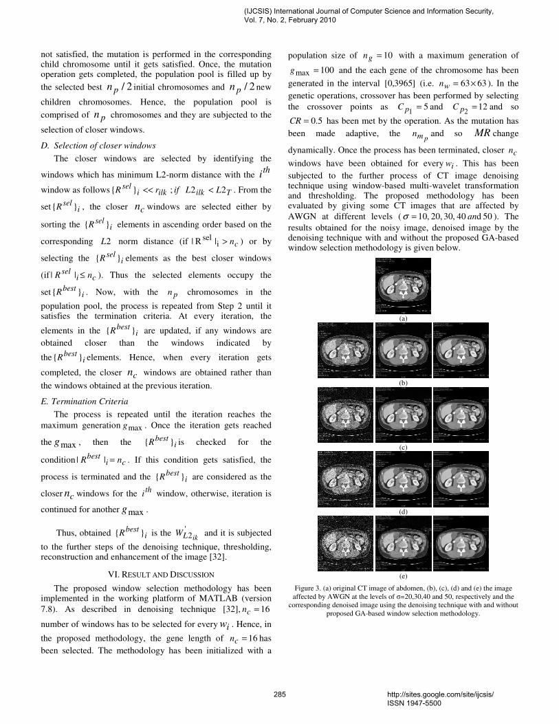
Figure 3. (a) original CT image of abdomen, (b), (c), (d) and (e) the image
affected by AWGN at the levels of σ=20,30,40 and 50, respectively and the
corresponding denoised image using the denoising technique with and without
proposed GA-based window selection methodology.
(IJCSIS) International Journal of Computer Science and Information Security, Vol. 7, No. 2, February 2010
285 http://sites.google.com/site/ijcsis/ ISSN 1947-5500
(a)
(b)
(c)
(d)
(e)
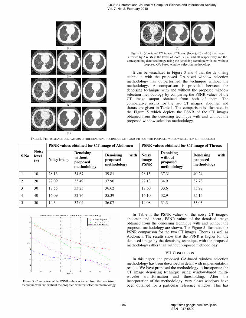
Figure 4. (a) original CT image of Thorax, (b), (c), (d) and (e) the image
affected by AWGN at the levels of σ=20,30, 40 and 50, respectively and the
corresponding denoised image using the denoising technique with and without
proposed GA-based window selection methodology.
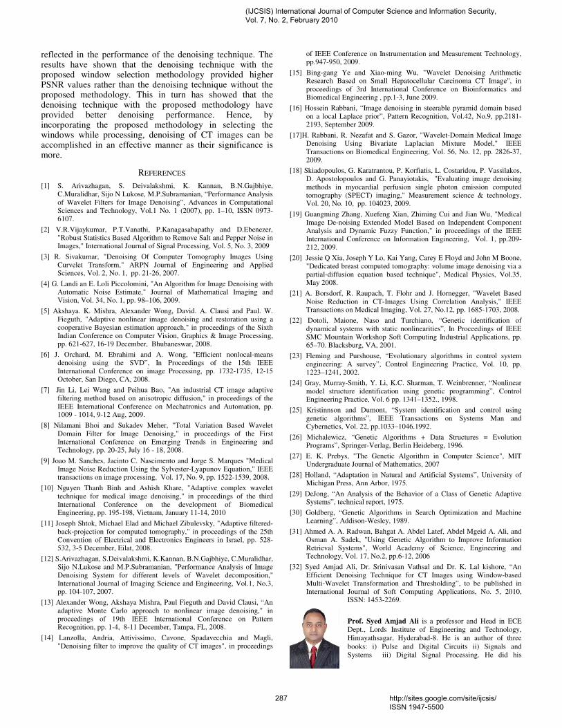
It can be visualized in Figure 3 and 4 that the denoising technique with the proposed GA-based window selection methodology has outperformed the technique without the methodology. A comparison is provided between the denoising technique with and without the proposed window selection methodology by comparing the PSNR values of the CT image output obtained from both of them. The comparative results for the two CT images, abdomen and thorax are given in Table I. The comparison is illustrated in the Figure 5 which depicts the PSNR of the CT images obtained from the denoising technique with and without the proposed window selection methodology.
TABLE I. PERFORMANCE COMPARISON OF THE DENOISING TECHNIQUE WITH AND WITHOUT THE PROPOSED WINDOW SELECTION METHODOLOGY
PSNR values obtained for CT image of Abdomen PSNR values obtained for CT image of Throax
S.No
Noise
level
(σ) Noisy image
Denoising
without
proposed
methodology
Denoising with
proposed
methodology
Noisy
image
PSNR
Denoising
without
proposed
methodology
Denoising with
proposed
methodology
1 10 28.13 34.67 39.81 28.15 37.31 40.24
2 20 22.09 33.49 37.90 22.13 34.9 37.78
3 30 18.55 33.25 36.62 18.60 33.6 35.28
4 40 16.09 32.76 35.39 16.10 32.9 35.15
5 50 14.3 32.04 36.07 14.08 31.3 33.03
Figure 5. Comparison of the PSNR values obtained from the denoising
technique with and without the proposed window selection methodology
In Table I, the PSNR values of the noisy CT images, abdomen and thorax, PSNR values of the denoised image obtained from the denoising technique with and without the proposed methodology are shown. The Figure 5 illustrates the PSNR comparison for the two CT images, Thorax as well as Abdomen. The results show that the PSNR is higher for the denoised image by the denoising technique with the proposed methodology rather than without proposed methodology.
VII. CONCLUSION
In this paper, the proposed GA-based window selection methodology has been described in detail with implementation results. We have proposed the methodology to incorporate the CT image denoising technique using window-based multi-wavelet transformation and thresholding. After the incorporation of the methodology, very closer windows have been obtained for a particular reference window. This has
(IJCSIS) International Journal of Computer Science and Information Security, Vol. 7, No. 2, February 2010
286 http://sites.google.com/site/ijcsis/ ISSN 1947-5500
reflected in the performance of the denoising technique. The results have shown that the denoising technique with the proposed window selection methodology provided higher PSNR values rather than the denoising technique without the proposed methodology. This in turn has showed that the denoising technique with the proposed methodology have provided better denoising performance. Hence, by incorporating the proposed methodology in selecting the windows while processing, denoising of CT images can be accomplished in an effective manner as their significance is more.
REFERENCES
[1] S. Arivazhagan, S. Deivalakshmi, K. Kannan, B.N.Gajbhiye, C.Muralidhar, Sijo N Lukose, M.P.Subramanian, “Performance Analysis of Wavelet Filters for Image Denoising”, Advances in Computational Sciences and Technology, Vol.1 No. 1 (2007), pp. 1–10, ISSN 0973-6107.
[2] V.R.Vijaykumar, P.T.Vanathi, P.Kanagasabapathy and D.Ebenezer, "Robust Statistics Based Algorithm to Remove Salt and Pepper Noise in Images," International Journal of Signal Processing, Vol. 5, No. 3, 2009
[3] R. Sivakumar, "Denoising Of Computer Tomography Images Using Curvelet Transform," ARPN Journal of Engineering and Applied Sciences, Vol. 2, No. 1, pp. 21-26, 2007.
[4] G. Landi an E. Loli Piccolomini, "An Algorithm for Image Denoising with Automatic Noise Estimate," Journal of Mathematical Imaging and Vision, Vol. 34, No. 1, pp. 98–106, 2009.
[5] Akshaya. K. Mishra, Alexander Wong, David. A. Clausi and Paul. W. Fieguth, "Adaptive nonlinear image denoising and restoration using a cooperative Bayesian estimation approach," in proceedings of the Sixth Indian Conference on Computer Vision, Graphics & Image Processing, pp. 621-627, 16-19 December, Bhubaneswar, 2008.
[6] J. Orchard, M. Ebrahimi and A. Wong, "Efficient nonlocal-means denoising using the SVD”, In Proceedings of the 15th IEEE International Conference on image Processing, pp. 1732-1735, 12-15 October, San Diego, CA, 2008.
[7] Jin Li, Lei Wang and Peihua Bao, "An industrial CT image adaptive filtering method based on anisotropic diffusion," in proceedings of the IEEE International Conference on Mechatronics and Automation, pp. 1009 - 1014, 9-12 Aug, 2009.
[8] Nilamani Bhoi and Sukadev Meher, "Total Variation Based Wavelet Domain Filter for Image Denoising," in proceedings of the First International Conference on Emerging Trends in Engineering and Technology, pp. 20-25, July 16 - 18, 2008.
[9] Joao M. Sanches, Jacinto C. Nascimento and Jorge S. Marques "Medical Image Noise Reduction Using the Sylvester-Lyapunov Equation," IEEE transactions on image processing, Vol. 17, No. 9, pp. 1522-1539, 2008.
[10] Nguyen Thanh Binh and Ashish Khare, "Adaptive complex wavelet technique for medical image denoising," in proceedings of the third International Conference on the development of Biomedical Engineering, pp. 195-198, Vietnam, January 11-14, 2010
[11] Joseph Shtok, Michael Elad and Michael Zibulevsky, "Adaptive filtered-back-projection for computed tomography," in proceedings of the 25th Convention of Electrical and Electronics Engineers in Israel, pp. 528-532, 3-5 December, Eilat, 2008.
[12] S.Arivazhagan, S.Deivalakshmi, K.Kannan, B.N.Gajbhiye, C.Muralidhar, Sijo N.Lukose and M.P.Subramanian, "Performance Analysis of Image Denoising System for different levels of Wavelet decomposition," International Journal of Imaging Science and Engineering, Vol.1, No.3, pp. 104-107, 2007.
[13] Alexander Wong, Akshaya Mishra, Paul Fieguth and David Clausi, “An adaptive Monte Carlo approach to nonlinear image denoising," in proceedings of 19th IEEE International Conference on Pattern Recognition, pp. 1-4, 8-11 December, Tampa, FL, 2008.
[14] Lanzolla, Andria, Attivissimo, Cavone, Spadavecchia and Magli, "Denoising filter to improve the quality of CT images", in proceedings
of IEEE Conference on Instrumentation and Measurement Technology, pp.947-950, 2009.
[15] Bing-gang Ye and Xiao-ming Wu, "Wavelet Denoising Arithmetic Research Based on Small Hepatocellular Carcinoma CT Image", in proceedings of 3rd International Conference on Bioinformatics and Biomedical Engineering , pp.1-3, June 2009.
[16] Hossein Rabbani, “Image denoising in steerable pyramid domain based on a local Laplace prior”, Pattern Recognition, Vol.42, No.9, pp.2181-2193, September 2009.
[17]H. Rabbani, R. Nezafat and S. Gazor, "Wavelet-Domain Medical Image Denoising Using Bivariate Laplacian Mixture Model," IEEE Transactions on Biomedical Engineering, Vol. 56, No. 12, pp. 2826-37, 2009.
[18] Skiadopoulos, G. Karatrantou, P. Korfiatis, L. Costaridou, P. Vassilakos, D. Apostolopoulos and G. Panayiotakis, "Evaluating image denoising methods in myocardial perfusion single photon emission computed tomography (SPECT) imaging," Measurement science & technology, Vol. 20, No. 10, pp. 104023, 2009.
[19] Guangming Zhang, Xuefeng Xian, Zhiming Cui and Jian Wu, "Medical Image De-noising Extended Model Based on Independent Component Analysis and Dynamic Fuzzy Function," in proceedings of the IEEE International Conference on Information Engineering, Vol. 1, pp.209-212, 2009.
[20] Jessie Q Xia, Joseph Y Lo, Kai Yang, Carey E Floyd and John M Boone, "Dedicated breast computed tomography: volume image denoising via a partial-diffusion equation based technique", Medical Physics, Vol.35, May 2008.
[21] A. Borsdorf, R. Raupach, T. Flohr and J. Hornegger, "Wavelet Based Noise Reduction in CT-Images Using Correlation Analysis," IEEE Transactions on Medical Imaging, Vol. 27, No.12, pp. 1685-1703, 2008.
[22] Dotoli, Maione, Naso and Turchiano, “Genetic identification of dynamical systems with static nonlinearities”, In Proceedings of IEEE SMC Mountain Workshop Soft Computing Industrial Applications, pp. 65–70. Blacksburg, VA, 2001.
[23] Fleming and Purshouse, “Evolutionary algorithms in control system engineering: A survey”, Control Engineering Practice, Vol. 10, pp. 1223–1241, 2002.
[24] Gray, Murray-Smith, Y. Li, K.C. Sharman, T. Weinbrenner, “Nonlinear model structure identification using genetic programming”, Control Engineering Practice, Vol. 6 pp. 1341–1352., 1998.
[25] Kristinnson and Dumont, “System identification and control using genetic algorithms”, IEEE Transactions on Systems Man and Cybernetics, Vol. 22, pp.1033–1046.1992.
[26] Michalewicz, “Genetic Algorithms + Data Structures = Evolution Programs”, Springer-Verlag, Berlin Heideberg, 1996.
[27] E. K. Prebys, "The Genetic Algorithm in Computer Science", MIT Undergraduate Journal of Mathematics, 2007
[28] Holland, “Adaptation in Natural and Artificial Systems”, University of Michigan Press, Ann Arbor, 1975.
[29] DeJong, “An Analysis of the Behavior of a Class of Genetic Adaptive Systems”, technical report, 1975.
[30] Goldberg, “Genetic Algorithms in Search Optimization and Machine Learning”, Addison-Wesley, 1989.
[31] Ahmed A. A. Radwan, Bahgat A. Abdel Latef, Abdel Mgeid A. Ali, and Osman A. Sadek, "Using Genetic Algorithm to Improve Information Retrieval Systems", World Academy of Science, Engineering and Technology, Vol. 17, No.2, pp.6-12, 2006
[32] Syed Amjad Ali, Dr. Srinivasan Vathsal and Dr. K. Lal kishore, “An Efficient Denoising Technique for CT Images using Window-based Multi-Wavelet Transformation and Thresholding”, to be published in International Journal of Soft Computing Applications, No. 5, 2010,
ISSN: 1453-2269.
Prof. Syed Amjad Ali is a professor and Head in ECE
Dept., Lords Institute of Engineering and Technology,
Himayathsagar, Hyderabad-8. He is an author of three
books: i) Pulse and Digital Circuits ii) Signals and
Systems iii) Digital Signal Processing. He did his
(IJCSIS) International Journal of Computer Science and Information Security, Vol. 7, No. 2, February 2010
287 http://sites.google.com/site/ijcsis/ ISSN 1947-5500
M.Tech(Digital Systems and Computer Electronics) from JNTU,
Kukatpally,Hyderabad. He has presented the papers in national conferences
.He has 16+ years of teaching experience. His area of interest is Image
Processing.
Dr. Srinivasan Vathsal was born in Tiruchirapalli,
Tamilnadu, India in 1947. He obtained his B.E (Hons),
Electrical Engg., in 1968 from Thiagarajar, College of
Engineering, Madurai, India, the M.E (Control Systems) in
1970, from BITS, Pilani, India, and the Ph.D in 1974 from
I.I.Sc., Bangalore. He worked at the VSSC, Trivandrum,
India from 1974-1978 and 1980-1982. During 1982-1984,
he was professor of Electrical Engineering at the PSG
College of Tech., Coimbatore. He was a senior NRC, NASA Research
Associate (1984-1986) at the NASA, GSF Center. He worked at Directorate of
SAT of the DRDL in 1988. He was a principal scientist in navigational
electronics of Osmania University during 1989-1990, as Head, PFA,
directorate of systems in DRDO, Hyderabad during 1990-2005, as Scientist-
G, Director, ER & IPR directorate, DRDO, New Delhi. His current research
interests are fuzzy logic control, neural networks, missile systems and
guidance, radar signal processing and optimal control and image processing.
He was awarded a prize for best essay on comprehensive aeronautical policy
for India. He is a member of the IEEE, AIAA, Aeronautical Society of India
and System Society of India. He has published a number of papers in national
and international journals and conferences. Presently working as a Principal
in Bhaskar Engineering College, Yenakapally, R.R.Dist., Andhra Pradesh.
Dr.K.Lal Kishore did his B.E. from Osmania
University,Hyderabad , M.Tech from Indian Institute of
Science, Bangalore,Ph.D from Indian Institute of Science,
Bangalore. His Fields of Interest are Micro Electronics
and VLSI Engineering.Has more than 25 years of teaching
experience. Worked as a Professor and Head in E.C.E
department, Chairman, Board of Studies for Electronics and Communication
Engineering, Jawaharlal Nehru Technological University, Director, School of
Continuing and Distance Education (SCDE) of JNTU Hyderabad, Principal,
JNTU College of Engineering, Kukatpally, Hyderabad during June 2002 to
30th June 2004, Director, Academic & Planning (DAP) of JNT University,
Director I/c. UGC Academic Staff College of JNT University, Registrar of
JNT University.Convener for ECET (FDH) 2000, 2001 and 2005, conducted
the Common Entrance Test for Diploma Holders for admission into B.Tech.
He has membership in many professional societies like Fellow of IETE,
Member IEEE, Member ISTE and Member ISHM. He Won First Bapu
Seetharam Memorial Award for Research work from Institution of Electronics
and Communication Engineers (IETE), New Delhi in 1986. Received best
teacher award from the Government of Andhra Pradesh for the year 2004.He
has 31 publications to his credit so far.He wrote three text books i) Electronics
Devices and Circuits ii) Electronic Circuit Analysis. iii) Pulse
Circuits.Presently guiding number of Ph.D. students. Now, he is a Rector in
JNTU, Kukatpally, Hyderabad.
(IJCSIS) International Journal of Computer Science and Information Security, Vol. 7, No. 2, February 2010
288 http://sites.google.com/site/ijcsis/ ISSN 1947-5500
International Journal of Digital Content Technology and its Applications
Volume 4, Number 4, July 2010
75
CT Image Denoising Technique using GA aided Window-based
Multiwavelet Transformation and Thresholding with the Incorporation of
an Effective Quality Enhancement Method
1Prof. Syed Amjad Ali,
2Dr. Srinivasan Vathsal and
3Dr. K. Lal kishore
1Professor and Head of ECE Department
Lords Institute of Engineering and Technology, Himayathsagar, Hyderabad – 8, India
Email: [email protected]
Tel: +919490944104 2Principal, Bhaskar Engineering College
Yenkapally, Moinabad, Ranga reddy Dist, India
Email:[email protected]
Tel: +919676863811 3Director (I/c), School of IT, Jawahar Lal Nehru Technological University, Kukatpally,
Hyderabad, India
Email: [email protected]
Tel: +919396438312 doi: 10.4156/jdcta.vol4.issue4.9
Abstract Denoising the CT images removes noise from the CT images and so makes the disease diagnosis
procedure more efficient. The denoised images have a notable level of raise in its PSNR values,
ensuring a smoother image for diagnosis purpose. In the previous work, a CT image denoising
technique using window-based Multi-wavelet transformation and thresholding has been proposed. The
performance of the technique has been improved by Genetic Algorithm (GA)-based window selection
methodology. However, in the perspective of diagnosis, the PSNR values have not much significance;
instead they rely on the quality of the images in the perspective of medical diagnosis. In this paper, a
quality enhancement methodology is proposed to include in the CT image denoising technique using
window-based multi-wavelet transformation and thresholding. The methodology is comprised of an
edge detection technique based on canny algorithm that is performed on the gradient images so that
the images are visualized better for diagnosis. A pair of micro block set is generated from the edge
detected image and it is subjected to unsharp filter to obtain a sharper image. The smoothness of the
image is improved by applying Gaussian filter to the sharper image. Implementation results are given
to demonstrate the superior performance of the proposed quality enhancement technique over various
CT images in terms of medical perspective.
Keywords: CT image, Medical Perspective, Quality Enhancement, Edge Detection, Denoising,
Multi-wavelet transformation, Thresholding
1. Introduction
Roughly every sort of data holds noise. Noise reduction is an essential step for any complicated
algorithms in computer vision and image processing. This problem has survived for a long time and
still there is no proper solution for it. There exists a tradeoff between the removed noise and the
blurring in the image [1]. An image is mostly degraded by noise in its acquisition and transmission [2].
The challenge of eliminating noise from images, and the other major problem of reconstructing a signal
that has been degraded in a certain way have a strong history. Substantial researches are present for
general approaches, and also for particular domains where knowledge about the nature of the signal is
employed to customize or tune the estimation process. In spite of the presence of existing research,
CT Image Denoising Technique using GA aided Window-based Multiwavelet Transformation and
Thresholding with the Incorporation of an Effective Quality Enhancement Method
Prof. Syed Amjad Ali, Dr. Srinivasan Vathsal and Dr. K. Lal kishore
76
there is a gap between the introduction of modern denoising methods, and their broad implementation
outside of very specific applications [3]. The recovery of an image is possible by image denoising.
Image denoising is defined as the method of obtaining the original image from a tainted image by
means of noise degradation [4]. Image denoising is employed to eliminate the additive noise while
preserving the vital signal features [2]. Over the years, several methods have been developed and
detailed in literature for denoising [10]. The Image denoising methods can be categorized as spatial
domain methods or transform domain methods. The transform domain methods convert an image from
the spatial domain into wavelet domain or frequency domain and suppress noise in the transform
domain. But in the case of spatial domain methods, the noise is suppressed within the spatial domain
[5]. Several researchers have proposed hybrid filters for image denoising in the last few years. Kai U.
Barthel et al. (2004) introduced a hybrid wavelet-fractal denoising method. By employing a non
subsampled overcomplete wavelet transform, image is denoted as a set of translation invariant copies
in different frequency subbands [6].
The applications of Image denoising are noticed in the fields of medical imaging and preprocessing
for computer vision [7]. The ultimate goal of medical imaging is to acquire a high resolution image
with necessary details for the diagnosis purpose [15]. Commonly, Medical images possess poor
contrast and complex types of noise because of several transmission, acquisitions, and storage and
display devices. Consequently, it becomes complicated to recommend a robust method for noise
removal that works better for diverse modalities of medical images. In the last decade, a number of
novel methods have materialized for eliminating Gaussian random noise from medical images [8].
There are numerous medical imaging techniques namely the MR and the CT techniques. Both
techniques provide unique advanced characteristics of the organ which has to be imaged [15].
Medical images obtained from Magnetic Resonance Imaging (MRI) and Computed Tomography
(CT) are among the most general tools for diagnosis. The existence of noise not only generates
unwanted visual quality but also lessens the visibility of low-contrast objects. In Medical Imaging, CT
is considered as a common and major modality. CT is employed in clinical diagnosis and computer-
aided surgery [9]. In the case of CT, many mathematical applications can be used to conclude whether
the normal tissue has been infected by the mutations of the cancer cells through diagnosis. The
improvement in image quality of the CT images leads to a proper diagnosis of the defects. Various
methods have been introduced for improving the quality of the CT images.
A general methodology for improving the quality of CT images for diagnosis purpose based on edge
detection using unsharp filter is proposed in this paper. A denoised image is taken as input for
analyzing the efficient operation of the proposed method. First the X and Y gradients of the image are
obtained as a result of applying gradient function to the image. The two components are subjected to
edge detection separately and the visibility of the image is being improved. A micro block set is
obtained from the denoised image, which is then sharpened by applying unsharp filter to it. The micro
block set obtained as output from the unsharp filter is subjected to Gaussian filtering thus resulting in a
smoothened image. The smoothened image is thus converted into a sharper image that makes diagnosis
process easier. The proposed method is a better compromising one between noise smoothing and edge
orientation, that results in the retrieval of a sharper image for diagnosis purpose.
The rest of the paper is organized as follows. Section 2 reviews the related works with respect to the
proposed method. Section 3 gives a brief explanation about the CT image denoising technique using
window-based multi-wavelet transformation and thresholding. Section 4 presents the proposed Quality
enhancement technique which is elaborated in its subsections. Section 5 discusses about the
implementation results and section 6 concludes the paper.
2. Related works
Literature presents with a copious number of researches based on wavelet transformation for the
noise removal in medical images. Several researches in the literature are based on. Some recent and
significant researches are briefly reviewed in this section.
Zhao Yu-qian et al. [11] have introduced a basic mathematical morphological theory and operations,
and a novel mathematical morphological edge detection algorithm was proposed to detect the edge of
International Journal of Digital Content Technology and its Applications
Volume 4, Number 4, July 2010
77
lungs CT image with salt-and-pepper noise. The experimental results showed that the proposed
algorithm was more efficient for medical image denoising and edge detection than the usually used
template-based edge detection algorithms and general morphological edge detection algorithms.
The effect of various noise reduction filters on computed tomography (CT) images has been
analysed by Lanzolla et al. [12].Based on the combination of Gaussian and Prewitt operators they have
bestowed a denoising filter. Experimental values have proved that their presented technique has
permitted to utilize low radiation dose protocol in CT examinations as well as enhanced the image
quality. Their formulation was conceded out in combination with "G.Moscati" Hospital of Taranto
(Italy) which presented all the images and technical materials engaged in the proposed algorithm. Bing-
gang Ye and Xiao-ming Wu [20] have directed that the previous exposure of undersized hepatocellular
carcinoma (SHCC) has important clinic value, and wavelet denoising arithmetic research of SHCC CT
image which is based on image processing technology that facilitate to diagnose the SHCC focus.
Their contribution has minimized the figures and eradicated the weak or irrelated coefficient of noise
of SHCC CT image in synchronization with the wavelet coefficient correlation and at last detached the
noise.
Skiadopoulos et al. [13] have conceded away a relative revise among a multi-scale platelet
denoising method and the genuine Butterworth filter that has exercised as pre- and post-processing
modules on image reconstruction. The assessment was achieved with and/or without attenuation
correction. Assessable estimation was implemented by via 1) a cardiac phantom containing a pair of
various size cold defects, bestowed in two trials performed to replicate circumstances with and without
photon attenuation from myocardial surrounding tissue and 2) a pilot-verified clinical dataset of 15
patients with ischemic defects. In addition, on the basis of rankings from two nuclear medicine
clinicians, an observer preference study was administered for the clinical dataset. Denoising by platelet
and Butterworth post-processing methods overwhelmed Butterworth pre-processing for large size
defects, devoid of photon attenuation conditions. On the contrary, for the small size defects and with
photon attenuation conditions, whole of the methods have demonstrated equal denoising performance.
Guangming Zhang et al. [14] have proposed an enhanced technique for CT medical image de-noising
that bestowed autonomous component analysis and dynamic fuzzy theory. At first, a random matrix
was formed to split the CT image for evaluation. Afterward, dynamic fuzzy theory was practicaed to
set up a sequence of adaptive membership functions to generate the weights degree of truth. At last, the
weights degree was applied to optimize the value of matrix for image reconstruction. Following their
model, helps the selection of matrix to be optimized scientifically and self-adaptively.
F. E. Ali et al. [15] have presented a curvelet based approach for the fusion of magnetic resonance
(MR) and computed tomography (CT) images. The objective of the fusion of an MR image and a CT
image of the same organ was to obtain a single image containing as much information as possible
about that organ for diagnosis. Some attempts made are proposed for the fusion of MR and CT images
using the wavelet transform. Since medical images have several objects and curved shapes, it was
expected that the curvelet transform would be better in their fusion. The simulation results show the
superiority of the curvelet transform to the wavelet transform in the fusion of MR and CT images from
both the visual quality and the peak signal to noise ratio (PSNR) points of view.
Derek LG Hill and David J Hawkes [16] have presented a new algorithm based on surface fitting
that makes use of anatomical knowledge of adjacency of identified anatomical structures to solve the
3D rigid body registration problem. The algorithm has been applied to the registration of MR CT and
vascular images of the head, and has achieved accuracies similar to those obtained using interactive
location of corresponding landmarks. The algorithm presented by them potentially provided a means of
automating the registration process for many applications. Anatomical knowledge combined with the
use of registered reference images provided a means of customising the algorithm for particular clinical
applications. They have shown how it could be done with MR, CT and angiographic images.
A wavelet based structure-preserving method was proposed by Borsdorf et al. [17] for noise
reduction in CT images which could be utilized mutually with different reconstruction methods. Their
technique was based on the deduction that the data may perhaps be decomposed into information and
temporally uncorrelated noise. The investigation of correlations among the wavelet representations of
the input images endowed isolating information from noise down to a definite signal-to-noise level.
Wavelet coefficients with undersized correlation were minimized, at the same time those with high
correlations were subjected to indicate structures and are conserved. The crucial noise-suppressed
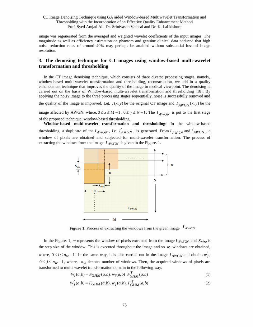
CT Image Denoising Technique using GA aided Window-based Multiwavelet Transformation and
Thresholding with the Incorporation of an Effective Quality Enhancement Method
Prof. Syed Amjad Ali, Dr. Srinivasan Vathsal and Dr. K. Lal kishore
78
image was regenerated from the averaged and weighted wavelet coefficients of the input images. The
magnitude as well as efficiency estimation on phantom and genuine clinical data adduced that high
noise reduction rates of around 40% may perhaps be attained without substantial loss of image
resolution.
3. The denoising technique for CT images using window-based multi-wavelet
transformation and thresholding
In the CT image denoising technique, which consists of three diverse processing stages, namely,
window-based multi-wavelet transformation and thresholding, reconstruction, we add in a quality
enhancement technique that improves the quality of the image in medical viewpoint. The denoising is
carried out on the basis of Window-based multi-wavelet transformation and thresholding [18]. By
applying the noisy image to the three processing stages sequentially, noise is successfully removed and
the quality of the image is improved. Let, ),( yxI be the original CT image and ),( yxIAWGN
be the
image affected by AWGN, where, 10 Mx , 10 Ny . The AWGN
I is put to the first stage
of the proposed technique, window-based thresholding.
Window-based multi-wavelet transformation and thresholding: In the window-based
thresholding, a duplicate of the AWGNI , i.e. 'AWGNI , is generated. From
AWGNI and '
AWGNI , a
window of pixels are obtained and subjected for multi-wavelet transformation. The process of
extracting the windows from the image AWGNI is given in the Figure. 1.
Figure 1. Process of extracting the windows from the given image
In the Figure. 1, w represents the window of pixels extracted from the image AWGNI and sizeS is
the step size of the window. This is executed throughout the image and so iw windows are obtained,
where, 10 wni . In the same way, it is also carried out in the image 'AWGNI and obtains '
jw ,
10 wnj , where, wn denotes number of windows. Then, the acquired windows of pixels are
transformed to multi-wavelet transformation domain in the following way:
),( . ),( . ),(),( baFbawbaFbaW TGHMiGHMi (1)
),( . ),( . ),(),( TGHM
'' baFbawbaFbaW jGHMj (2)
AWGNI
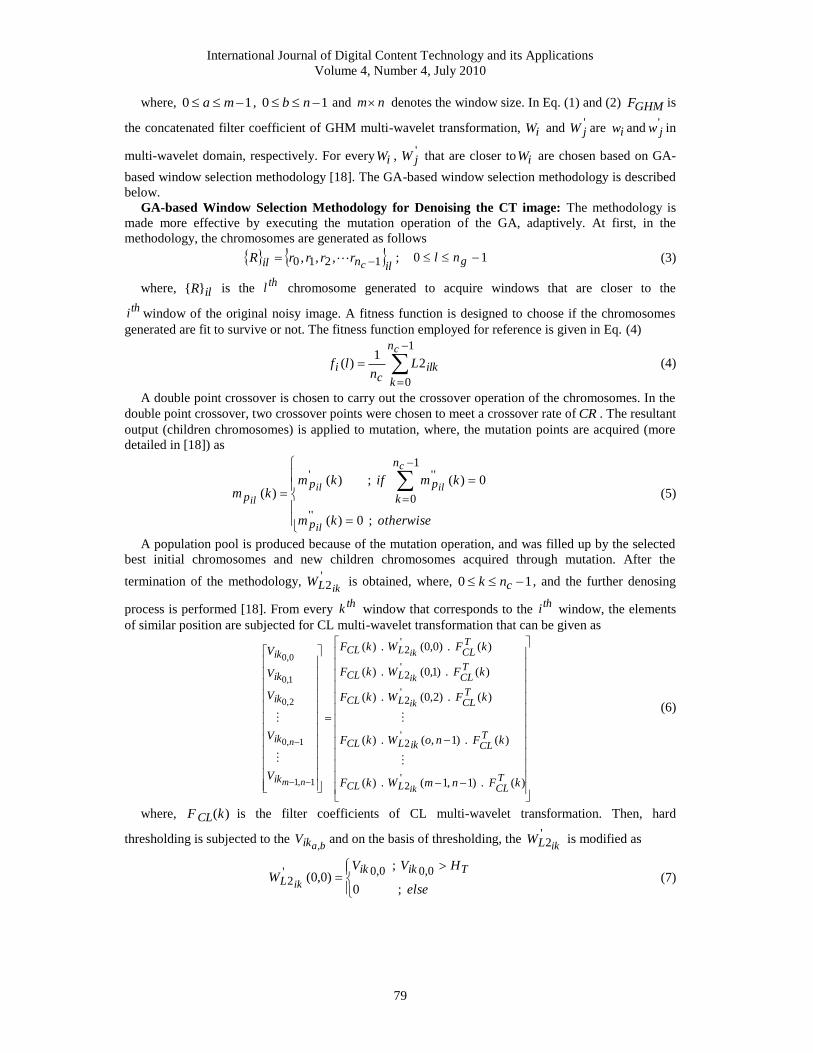
International Journal of Digital Content Technology and its Applications
Volume 4, Number 4, July 2010
79
where, 10 ma , 10 nb and nm denotes the window size. In Eq. (1) and (2) GHMF is
the concatenated filter coefficient of GHM multi-wavelet transformation, iW and 'jW are iw and '
jw in
multi-wavelet domain, respectively. For every iW , 'jW that are closer to iW are chosen based on GA-
based window selection methodology [18]. The GA-based window selection methodology is described
below.
GA-based Window Selection Methodology for Denoising the CT image: The methodology is
made more effective by executing the mutation operation of the GA, adaptively. At first, in the
methodology, the chromosomes are generated as follows
ilnil c
rrrrR 1210 ,,, ; 10 gnl (3)
where, ilR is the thl chromosome generated to acquire windows that are closer to the
thi window of the original noisy image. A fitness function is designed to choose if the chromosomes
generated are fit to survive or not. The fitness function employed for reference is given in Eq. (4)
1
0
21
)(
cn
k
ilkc
i Ln
lf (4)
A double point crossover is chosen to carry out the crossover operation of the chromosomes. In the
double point crossover, two crossover points were chosen to meet a crossover rate of CR . The resultant
output (children chromosomes) is applied to mutation, where, the mutation points are acquired (more
detailed in [18]) as
otherwisekm
kmifkmkm
il
c
ilil
il
p
n
k
ppp
; 0)(
0)( ; )()(
''
1
0
'''
(5)
A population pool is produced because of the mutation operation, and was filled up by the selected
best initial chromosomes and new children chromosomes acquired through mutation. After the
termination of the methodology, '2ikLW is obtained, where, 10 cnk , and the further denosing
process is performed [18]. From every thk window that corresponds to the
thi window, the elements
of similar position are subjected for CL multi-wavelet transformation that can be given as
)( . )1,1( . )(
)( . )1,( . )(
)( . )2,0( . )(
)( . )1,0( . )(
)( . )0,0( . )(
'2
'2
'2
'2
'2
1,1
1,0
2,0
1,0
0,0
kFnmWkF
kFnoWkF
kFWkF
kFWkF
kFWkF
V
V
V
V
V
TCLLCL
TCLikLCL
TCLLCL
TCLLCL
TCLLCL
ik
ik
ik
ik
ik
ik
ik
ik
ik
nm
n
(6)
where, )(kF CL is the filter coefficients of CL multi-wavelet transformation. Then, hard
thresholding is subjected to the baikV,
and on the basis of thresholding, the '2ikLW is modified as
else
HVVW
TikikL ik ; 0
; )0,0(
0,00,0'2 (7)
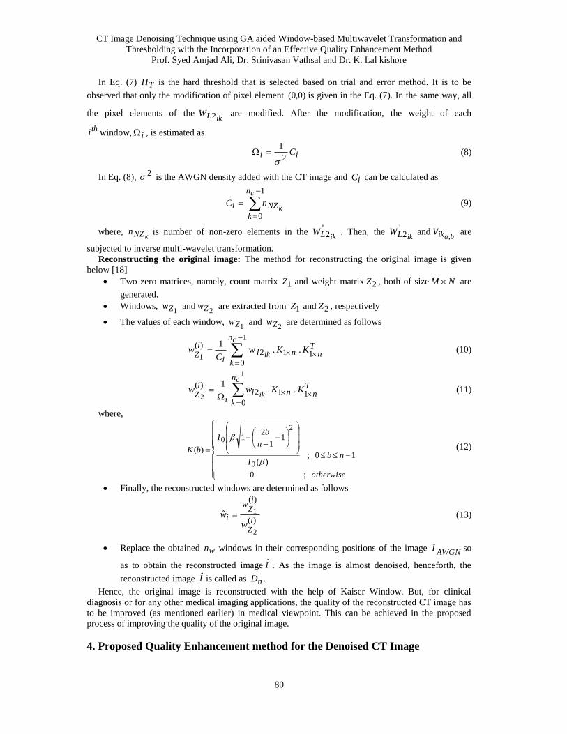
CT Image Denoising Technique using GA aided Window-based Multiwavelet Transformation and
Thresholding with the Incorporation of an Effective Quality Enhancement Method
Prof. Syed Amjad Ali, Dr. Srinivasan Vathsal and Dr. K. Lal kishore
80
In Eq. (7) TH is the hard threshold that is selected based on trial and error method. It is to be
observed that only the modification of pixel element )0,0( is given in the Eq. (7). In the same way, all
the pixel elements of the '2ikLW are modified. After the modification, the weight of each
thi window, i , is estimated as
ii C2
1
(8)
In Eq. (8), 2 is the AWGN density added with the CT image and iC can be calculated as
1
0
c
k
n
k
NZi nC (9)
where, kNZn is number of non-zero elements in the '
2ikLW . Then, the '2ikLW and
baikV,
are
subjected to inverse multi-wavelet transformation.
Reconstructing the original image: The method for reconstructing the original image is given
below [18]
Two zero matrices, namely, count matrix 1Z and weight matrix 2Z , both of size NM are
generated.
Windows, 1Zw and
2Zw are extracted from 1Z and 2Z , respectively
The values of each window, 1Zw and
2Zw are determined as follows
Tnn
n
k
li
iZ
KKC
w
c
ik
1 1
1
0
2)(
. . w1
1 (10)
Tn
n
k
nli
iZ
KKww
c
ik
10
12)(
. .1
1
2 (11)
where,
otherwise
nbI
n
bI
bK
; 0
10 ; )(
11
21
)(
0
2
0
(12)
Finally, the reconstructed windows are determined as follows
)(
)(
2
1ˆi
Z
iZ
iw
w
w (13)
Replace the obtained wn windows in their corresponding positions of the image AWGN
I so
as to obtain the reconstructed image I . As the image is almost denoised, henceforth, the
reconstructed image I is called as nD .
Hence, the original image is reconstructed with the help of Kaiser Window. But, for clinical
diagnosis or for any other medical imaging applications, the quality of the reconstructed CT image has
to be improved (as mentioned earlier) in medical viewpoint. This can be achieved in the proposed
process of improving the quality of the original image.
4. Proposed Quality Enhancement method for the Denoised CT Image
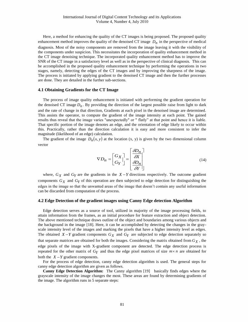
International Journal of Digital Content Technology and its Applications
Volume 4, Number 4, July 2010
81
Here, a method for enhancing the quality of the CT images is being proposed. The proposed quality
enhancement method improves the quality of the denoised CT image nD in the perspective of medical
diagnosis. Most of the noisy components are removed from the image leaving it with the visibility of
the components under suspicion. This necessitates the incorporation of quality enhancement method in
the CT image denoising technique. The incorporated quality enhancement method has to improve the
SNR of the CT image in a satisfactory level as well as in the perspective of clinical diagnosis. This can
be accomplished in the proposed quality enhancement technique by performing the operations in two
stages, namely, detecting the edges of the CT images and by improving the sharpness of the image.
The process is initiated by applying gradient to the denoised CT image and then the further processes
are done. They are detailed in the further sub-sections.
4.1 Obtaining Gradients for the CT Image
The process of image quality enhancement is initiated with performing the gradient operation for
the denoised CT image nD . By providing the direction of the largest possible raise from light to dark
and the rate of change in that direction, Gradients at each pixel in the denoised image are determined.
This assists the operator, to compute the gradient of the image intensity at each point. The gained
results thus reveal that the image varies "unexpectedly" or " flatly” at that point and hence it is liable.
That specific portion of the image denotes an edge, and the orientation of edge likely to occur within
this. Practically, rather than the direction calculation it is easy and more consistent to infer the
magnitude (likelihood of an edge) calculation.
The gradient of the image yxDn , at the location (x, y) is given by the two dimensional column
vector
Y
D
X
D
G
GD
n
n
Y
Xn
(14)
where, XG and YG are the gradients in the YX directions respectively. The outcome gradient
components XG and YG of this operation are then subjected to edge detection for distinguishing the
edges in the image so that the unwanted areas of the image that doesn’t contain any useful information
can be discarded from computation of the process.
4.2 Edge Detection of the gradient images using Canny Edge detection Algorithm
Edge detection serves as a source of tool, utilized in majority of the image processing fields, to
attain information from the frames, as an initial procedure for feature extraction and object detection.
The above mentioned technique draws outline of the object and boundaries among various objects and
the background in the image [18]. Here, it can be accomplished by detecting the changes in the gray-
scale intensity level of the images and marking the pixels that have a higher intensity level as edges.
The obtained YX gradient components XG and YG are subjected to edge detection separately so
that separate matrices are obtained for both the images. Considering the matrix obtained from XG , the
edge pixels of the image with X-gradient component are detected. The edge detection process is
repeated for the other matrix of YG and thus the edge pixel matrices of size nm are obtained for
both the YX gradient components.
For the process of edge detection, canny edge detection algorithm is used. The general steps for
canny edge detection algorithm are given as follows.
Canny Edge Detection Algorithm: The Canny algorithm [19] basically finds edges where the
grayscale intensity of the image changes the most. These areas are found by determining gradients of
the image. The algorithm runs in 5 separate steps:
CT Image Denoising Technique using GA aided Window-based Multiwavelet Transformation and
Thresholding with the Incorporation of an Effective Quality Enhancement Method
Prof. Syed Amjad Ali, Dr. Srinivasan Vathsal and Dr. K. Lal kishore
82
(i) Smoothing: Blurring of the image to remove noise from the image.
(ii) Finding gradients: The edges should be spotted where the gradients of the image has large
magnitudes.
(iii) Non-maximum suppression: Only local maxima should be spotted as edges.
(iv) Double thresholding: Potential edges are determined by thresholding.
(v) Edge tracking by hysteresis: Final edges are determined by suppressing all edges that are not
connected to a very certain (strong) edge.
4.3. Generation of a pair of intermediate matrices from the X-Y Coordinates
For every edge pixels of the gradient components XG and YG , their respective YX coordinates
are obtained and thus an intermediate matrix of 2K size is generated for the gradient components XG
and YG . The constant K denotes the total number of rows of the edge pixels obtained for a gradient
component.
4.4. Generation of a Microblock from the edge detected images
For this process the denoised CT image nD considered as input for the edge detection purpose is
used as the reference image. Considering the first 2K matrix generated with respect to XG , for
every count of the edge pixel K in the generated matrix, its respective YX coordinates from the CT
image nD are retrieved. The process is repeated for all edge pixels denoted by the row’s value K in the
gradient component XG . The resultant output is the required microblock )(1 XMb of 22 size is
generated for XG .
)),(),,(()( 111 yyxxIXM Xb )),(),,(()( 111 yxyxIXM xb
)),(),,(()( 112 yyxxIYM Yb )),(),,(()( 112 yxyxIYM yb (15)
where nD is the denoised input image, xI is the intermediate matrix of the gradient component XG .
The same process is repeated for the next gradient component YG thus yielding a microblock )(2 YMb .
The microblocks generated in this process are given in Eqs. (15).
4.5. Generation of the Quality Enhanced CT Image from the Microblock
The required output image should be obtained such that the visibility of the organs present in the
image should be improved for clinical diagnosis purpose. The edges of the image are clearly plotted
with the help of Canny algorithm and the microblock set generated from the edge oriented image are
subjected to unsharp filter and Gaussian filter for the purpose of sharpening and smoothening the
image and thus the quality of the CT image is improved. The process of sharping and smoothing the
image are explained in the following sections.
4.5.1. Image Sharpening By Unsharp Filter
The use of this step is to translate the “blurred” edges in the image of the gradient magnitudes to
“sharp” edges. In general, this is performed by safeguarding all local maxima in the gradient image,
and eradicating the whole thing as well. The provided algorithm is for every pixel in the gradient
image:
1. Adjacent to the gradient direction to nearest 45 , consequent to the utilization of an 8-
connected neighborhood.
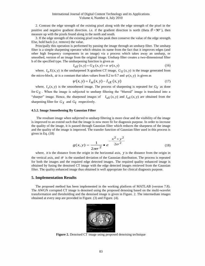
International Journal of Digital Content Technology and its Applications
Volume 4, Number 4, July 2010
83
2. Contrast the edge strength of the existing pixel along with the edge strength of the pixel in the
positive and negative gradient direction. i.e. if the gradient direction is north (theta = 90 ), then
measure up with the pixels found along in the north and south.
3. If the edge strength of the existing pixel reaches peak then conserve the value of the edge strength.
Else, hold back (i.e. remove) the value.
Principally this operation is performed by passing the image through an unsharp filter. The unsharp
filter is a simple sharpening operator which obtains its name from the fact that it improves edges (and
other high frequency components in an image) via a process which takes away an unsharp, or
smoothed, version of an image from the original image. Unsharp filter creates a two-dimensional filter
h of the specified type. The unsharpening function is given as
),( ),(),( yxyxGyxI XuX (16)
where, ),( yxXIu is the unsharpened X-gradient CT image, ),( yxGX is the image generated from
the micro-block, is a constant that takes values from 0.2 to 0.7 and ),( yx is given as
),(),(),( yxIyxIyx sXm (17)
where, ),( yxI s is the smoothened image. The process of sharpening is repeated for YG as done
for XG . When the image is subjected to unsharp filtering the “blurred” image is translated into a
“sharper” image. Hence, the sharpened images of ),( yxIuX and ),( yxIuY are obtained from the
sharpening filter for XG and YG respectively.
4.5.2. Image Smoothening By Gaussian Filter
The resultant image when subjected to unsharp filtering is more clear and the visibility of the image
is improved to an extend such that the image is now more fit for diagnosis purpose. In order to increase
the quality of the image, it is passed through Gaussian filter which reduces the sharpness of the image
and the quality of the image is improved. The transfer function of Gaussian filter used in this process is
given in Eq. (18)
2
22
222
1),(
yx
eyxg
(18)
where, x is the distance from the origin in the horizontal axis, y is the distance from the origin in
the vertical axis, and is the standard deviation of the Gaussian distribution. The process is repeated
for both the images and the required edge detected images. The required quality enhanced image is
obtained by fusing the denoised CT image with the edge detected images retrieved from the Gaussian
filter. The quality enhanced image thus obtained is well appropriate for clinical diagnosis purpose.
5. Implementation Results
The proposed method has been implemented in the working platform of MATLAB (version 7.8).
The AWGN corrupted CT image is denoised using the proposed denosing based on the multi-wavelet
transformation and thresholding and the denoised image is given in Figure. 2. The intermediate images
obtained at every step are provided in Figure. (3) and Figure. (4).
Figure 2. Denoised CT image using proposed denoising technique
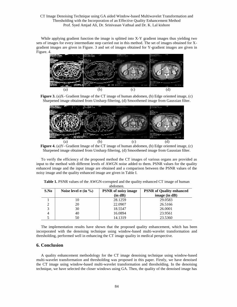
CT Image Denoising Technique using GA aided Window-based Multiwavelet Transformation and
Thresholding with the Incorporation of an Effective Quality Enhancement Method
Prof. Syed Amjad Ali, Dr. Srinivasan Vathsal and Dr. K. Lal kishore
84
While applying gradient function the image is splitted into X-Y gradient images thus yielding two
sets of images for every intermediate step carried out in this method. The set of images obtained for X-
gradient images are given in Figure. 3 and set of images obtained for Y-gradient images are given in
Figure. 4.
(a) (b) (c) (d)
Figure 3. (a)X- Gradient Image of the CT image of human abdomen, (b) Edge oriented image, (c)
Sharpened image obtained from Unsharp filtering, (d) Smoothened image from Gaussian filter.
(a) (b) (c) (d)
Figure 4. (a)Y- Gradient Image of the CT image of human abdomen, (b) Edge oriented image, (c)
Sharpened image obtained from Unsharp filtering, (d) Smoothened image from Gaussian filter.
To verify the efficiency of the proposed method the CT images of various organs are provided as
input to the method with different levels of AWGN noise added to them. PSNR values for the quality
enhanced image and the input image are obtained and a comparison between the PSNR values of the
noisy image and the quality enhanced image are given in Table I.
Table 1. PSNR values of the AWGN-corrupted and the quality enhanced CT image of human
abdomen.
S.No Noise level σ (in %) PSNR of noisy image
(in dB)
PSNR of Quality enhanced
image (in dB)
1 10 28.1259 29.0583
2 20 22.0907 26.5166
3 30 18.5547 26.0001
4 40 16.0894 23.9561
5 50 14.1319 23.5360
The implementation results have shown that the proposed quality enhancement, which has been
incorporated with the denoising technique using window-based multi-wavelet transformation and
thresholding, performed well in enhancing the CT image quality in medical perspective.
6. Conclusion
A quality enhancement methodology for the CT image denoising technique using window-based
multi-wavelet transformation and thresholding was proposed in this paper. Firstly, we have denoised
the CT image using window-based multi-wavelet transformation and thresholding. In the denoising
technique, we have selected the closer windows using GA. Then, the quality of the denoised image has
International Journal of Digital Content Technology and its Applications
Volume 4, Number 4, July 2010
85
been enhanced in terms of medical perspective. The quality enhancement based on edge detection of
the CT image has been performed using Canny edge detection algorithm. The new technique has the
advantages of enhancing the quality of the denoised image and preserving important features and organ
surfaces well. The implementation results show that the noise is removed efficiently and the particular
information is well preserved; meanwhile, the whole visual quality is improved. From the obtained
results, it can be seen that the proposed quality enhancing method effectively improves the quality of
the CT images based on the filtering techniques. It can also be visualized that the quality enhancing
technique has effectively improved the quality of the CT image. The resultant images of the proposed
method are in good quality that assists for accurate clinical diagnosis and also for other medical
imaging applications.
7. References
[1] Aglika Gyaourova, Chandrika Kamath and Imola K. Fodor, "Undecimated wavelet transforms for
image de-noising", Report, Lawrence Livermore National Lab., CA, pp: 18, 2002.
[2] Lakhwinder Kaur, Savita Gupta and R.C. Chauhan, "Image Denoising using Wavelet
Thresholding", Third Conference on Computer Vision, Graphics and Image Processing, pp:
16-18, Dec 2002.
[3] Francisco Estrada, David Fleet and Allan Jepson, "Stochastic Image Denoising", British Machine
Vision Conference, London 2009
[4] Akshaya. K. Mishra, Alexander Wong, David. A. Clausi and Paul. W. Fieguth, "Adaptive nonlinear
image denoising and restoration using a cooperative Bayesian estimation approach," in
proceedings of the Sixth Indian Conference on Computer Vision, Graphics & Image Processing,
pp. 621-627, 16-19 December, Bhubaneswar, 2008.
[5] Jin Li, Lei Wang and Peihua Bao, "An industrial CT image adaptive filtering method based on
anisotropic diffusion," in proceedings of the IEEE International Conference on Mechatronics and
Automation, pp. 1009 - 1014, 9-12 Aug, 2009.
[6] N. P. Anil and S. Natarajan, "A New Technique for Image Denoising Using Fourth Order PDE and
Wiener Filter", International Journal of Applied Engineering Research, Vol. 5, No. 3, pp. 509–516,
2010.
[7] Nilamani Bhoi and Sukadev Meher, "Total Variation Based Wavelet Domain Filter for Image
Denoising," in proceedings of the First International Conference on Emerging Trends in
Engineering and Technology, pp. 20-25, July 16 - 18, 2008.
[8] Nguyen Thanh Binh and Ashish Khare, "Adaptive complex wavelet technique for medical image
denoising", Third International Conference on the Development of Biomedical Engineering,
pp:195-198 , Vietnam ,11-14th
Jan 2010.
[9] Joseph Shtok, Michael Elad and Michael Zibulevsky, "Adaptive filtered-back-projection for
computed tomography," in proceedings of the 25th Convention of Electrical and Electronics
Engineers in Israel, pp. 528-532, 3-5 December, Eilat, 2008.
[10] S.Arivazhagan, S.Deivalakshmi, K.Kannan, B.N.Gajbhiye, C.Muralidhar, Sijo N.Lukose and
M.P.Subramanian, "Performance Analysis of Image Denoising System for different levels of
Wavelet decomposition," International Journal of Imaging Science and Engineering, Vol.1, No.3,
pp. 104-107, 2007.
[11] Zhao Yu-qian, Gui Wei-hua, Chen Zhen-cheng, Tang Jing-tian, Li Ling-yun, "Medical Images
Edge Detection Based on Mathematical Morphology", Proceedings of the 2005 IEEE Engineering
in Medicine and Biology 27th Annual Conference, pp: 6492-6495,Shanghai, China, September 1-4,
2005.
[12] Lanzolla, Andria, Attivissimo, Cavone, Spadavecchia and Magli, "Denoising filter to improve the
quality of CT images", in proceedings of IEEE Conference on Instrumentation and Measurement
Technology, pp.947-950, 2009.
[13] Skiadopoulos, G. Karatrantou, P. Korfiatis, L. Costaridou, P. Vassilakos, D. Apostolopoulos and
G. Panayiotakis, "Evaluating image denoising methods in myocardial perfusion single photon
emission computed tomography (SPECT) imaging," Measurement science & technology, Vol. 20,
No. 10, pp. 104023, 2009.
CT Image Denoising Technique using GA aided Window-based Multiwavelet Transformation and
Thresholding with the Incorporation of an Effective Quality Enhancement Method
Prof. Syed Amjad Ali, Dr. Srinivasan Vathsal and Dr. K. Lal kishore
86
[14] Guangming Zhang, Xuefeng Xian, Zhiming Cui and Jian Wu, "Medical Image De-noising
Extended Model Based on Independent Component Analysis and Dynamic Fuzzy Function," in
proceedings of the IEEE International Conference on Information Engineering, Vol. 1, pp.209-
212, 2009.
[15] F. E. Ali, I. M. El-Dokany, A. A. Saad and F. E. Abd El-Samie, "Curvelet Fusion of MR and CT
Images", Progress In Electromagnetics Research C, Vol. 3, pp; 215–224, 2008.
[16] Derek LG Hill and David J Hawkes, "Medical Image Registration Using Knowledge of Adjacency
of Anatomical Structures", Image and Vision Computing, Vol. 12, pp: 173-178, 1994.
[17] A. Borsdorf, R. Raupach, T. Flohr and J. Hornegger, "Wavelet Based Noise Reduction in CT-
Images Using Correlation Analysis," IEEE Transactions on Medical Imaging, Vol. 27, No.12, pp.
1685-1703, 2008.
[18] Syed Amjad Ali, Srinivasan Vathsal, Lal Kishore, “A GA-based Window Selection Methodology
to Enhance Window-based Multi-Wavelet Transformation and Thresholding aided CT image
Denoising Technique”, IJCSIS International Journal of Computer Science and Information
Security, Vol. 7, No. 2, pp: 280-288, 2010.
[19] Thomas Moeslund, “Canny Edge Detection”, Technical Report, Laboratory of Computer Vision
and Media Technology, Aalborg University, 2009.
[20] Bing-gang Ye and Xiao-ming Wu, "Wavelet Denoising Arithmetic Research Based on Small
Hepatocellular Carcinoma CT Image", in proceedings of 3rd International Conference on
Bioinformatics and Biomedical Engineering , pp.1-3, June 2009.
International Journal of Digital Content Technology and its Applications
Volume 4, Number 4, July 2010
87
Prof. Syed Amjad Ali is a Head in ECE Dept., Lords Institute of Engineering and
Technology, Himayathsagar, Hyderabad-8. He is an author of three books: i) Pulse and
Digital Circuits ii) Signals and Systems iii) Digital Signal Processing. He did his
M.Tech (Digital Systems and Computer Electronics) from JNTU, Kukatpally,
Hyderabad. He has presented the papers in National conferences and published the
research papers in International journals. He has 16+ years of teaching experience. His
area of interest is Image Processing.
Dr. Srinivasan Vathsal was born in Tiruchirapalli, Tamilnadu, India in 1947. He
obtained his B.E (Hons), Electrical Engg., in 1968 from Thiagarajar, College of
Engineering, Madurai, India, the M.E (Control Systems) in 1970, from BITS, Pilani,
India, and the Ph.D in 1974 from I.I.Sc., Bangalore. He worked at the VSSC,
Trivandrum, India from 1974-1978 and 1980-1982. During 1982-1984, he was
professor of Electrical Engineering at the PSG College of Tech., Coimbatore. He was a
senior NRC, NASA Research Associate (1984-1986) at the NASA, GSF Center. He
worked at Directorate of SAT of the DRDL in 1988. He was a principal scientist in navigational
electronics of Osmania University during 1989-1990, as Head, PFA, directorate of systems in DRDO,
Hyderabad during 1990-2005, as Scientist-G, Director, ER & IPR directorate, DRDO, New Delhi. His
current research interests are fuzzy logic control, neural networks, missile systems and guidance, radar
signal processing and optimal control and image processing. He was awarded a prize for best essay on
comprehensive aeronautical policy for India. He is a member of the IEEE, AIAA, Aeronautical Society
of India and System Society of India. He has published a number of papers in national and
international journals and conferences. Presently working as a Principal in Bhaskar Engineering
college, Yenakapally, R.R.Dist., Andhra Pradesh.
Dr.K.Lal Kishore did his B.E. from Osmania University, Hyderabad, M.Tech from
Indian Institute of Science, Bangalore, Ph.D from Indian Institute of Science, Bangalore.
His Fields of Interest are Micro Electronics and VLSI Engineering. Has more than 25
years of teaching experience. Worked as a Professor and Head in E.C.E department,
Chairman, Board of Studies for Electronics and Communication Engineering,
Jawaharlal Nehru Technological University, Director, School of Continuing and
Distance Education (SCDE) of JNTU Hyderabad, Principal, JNTU College of Engineering, Kukatpally,
Hyderabad during June 2002 to 30th
June 2004, Director, Academic & Planning (DAP) of JNT
University, Director I/c. UGC Academic Staff College of JNT University, Registrar of JNT University.
Convener for ECET (FDH) 2000, 2001 and 2005, conducted the Common Entrance Test for Diploma
Holders for admission into B.Tech. He has membership in many professional societies like Fellow of
IETE, Member IEEE, Member ISTE and Member ISHM. He Won First Bapu Seetharam Memorial
Award for Research work from Institution of Electronics and Communication Engineers (IETE), New
Delhi in 1986. Received best teacher award from the Government of Andhra Pradesh for the year
2004.He has 31 publications to his credit so far. He wrote three text books i) Electronics Devices and
Circuits ii) Electronic Circuit Analysis. iii) Pulse Circuits. Presently guiding number of Ph.D. students.
Now, he is a Director (I/c), school of Information and technology in JNTU, Kukatpally, Hyderabad.