Embed Size (px)
Citation preview

AMERICAN ACADEMY OF PEDIATRICSCommittee on Genetics
Health Care Supervision for Children With Williams Syndrome
ABSTRACT. This set of guidelines is designed to assistthe pediatrician to care for children with Williams syn-drome diagnosed by clinical features and with regionalchromosomal microdeletion confirmed by fluorescencein situ hybridization.
ABBREVIATIONS. WS, Williams syndrome; FISH, fluorescence insitu hybridization.
INTRODUCTION
Williams syndrome (WS, also Williams-Beuren syndrome), now recognized to becaused by a microdeletion of chromosome
7, is a multisystem disorder first identified as a dis-tinct clinical entity in 1961.1 It is present at birth andaffects boys and girls equally. As routine geneticamniocentesis does not typically detect chromosomemicrodeletions, children with WS usually come tothe attention of pediatricians during infancy or child-hood. Initially thought to be a rare genetic disorder,increased awareness of the clinical features and es-tablishment of a reliable diagnostic test have re-vealed WS to be one of the more commonly recog-nized genetic disorders in childhood. Williamssyndrome is characterized by dysmorphic facies(100%), cardiovascular disease (most commonly sup-ravalvar aortic stenosis [80%]), mental retardation(75%), a characteristic cognitive profile (90%), andidiopathic hypercalcemia (15%)2–5 (Table 1).
The diagnosis historically has been made on thebasis of clinical criteria (Fig 1), but recently it hasbeen shown that 99% of patients with WS have ahemizygous submicroscopic deletion of 7q11.23detectable by fluorescence in situ hybridization(FISH).6–8 Chromosome analysis and the WilliamsSyndrome Chromosomal Region FISH test are rec-ommended for confirmation of the diagnosis. (Achild with the clinical features of WS and a negativeFISH result should be referred to a clinical geneticistfor further evaluation.) The deleted portion of thechromosome includes the ELN gene that codes forthe structural protein elastin, an important compo-nent of the elastic fibers found in the connectivetissue of many organs. The elastin deletion explainssome of the characteristics of WS, such as some of thefacial features, hoarse voice, bladder and bowel di-verticula, cardiovascular disease, and orthopedicproblems. The pathogenesis of other characteristics,
such as hypercalcemia, mental retardation, andunique personality traits, remains unexplained. Onepossibility is that the loss of 1 or more genes contig-uous to the ELN gene contributes to the phenotype.
The pediatrician can use knowledge of the clinicalmanifestations (Table 1) and natural history of WS toanticipate medical problems and to educate the fam-ily. Most children with WS are described as havingsimilar facial features.4,9 Although these features areoften subtle, they tend to become more distinctivewith advancing age. Facial features often includeperiorbital fullness, short nose with bulbous nasaltip, long philtrum, wide mouth, full lips, and mildmicrognathia. Infants have full cheeks and a flatfacial profile, whereas older children and adults of-ten have a long narrow face and a long neck.10,11
Blue- and green-eyed children with WS have a prom-inent “starburst” pattern to their irides (stellateiris).12 Mild prenatal growth deficiency and a post-natal growth rate about 75% of normal are consis-tently observed features of the condition.8,13
The majority of children with WS have cardiovas-cular anomalies.1,2,4 The most common cardiovascu-lar defect is supravalvar aortic stenosis, an oftenprogressive condition that may require surgical re-pair.10,11 Peripheral pulmonary artery stenosis is of-ten present in infancy and usually improves overtime. Coarctation of the aorta, renal artery stenosis,and systemic hypertension are complications thatwhen present may worsen over time.4,11,14,15 Becausethe elastin protein is an important component ofelastic fibers in the arterial wall, any artery maybecome narrowed.
Idiopathic infantile hypercalcemia is an intriguingfeature of WS that can contribute to the presence ofextreme irritability, vomiting, constipation, and mus-cle cramps associated with this condition.4,9 Symp-tomatic hypercalcemia usually resolves during child-hood, but lifelong abnormalities of calcium andvitamin D metabolism may persist. Hypercalciuria iscommon and predisposes to nephrocalcinosis. Thecause of the abnormality in calcium metabolism isunknown.
An infant with WS often has difficulty feeding andmay be brought for medical care because of gastro-esophageal reflux, colic, or failure to thrive.4,9,16
Other medical problems include Chiari I malforma-tion, strabismus,12 hyperopia,12 chronic otitis media,hypodontia, malocclusion, bowel or bladder diver-ticula, hernias, joint laxity, joint contractures,17 ky-phosis, lordosis, renal or urinary tract malforma-tions,14,15 hypothyroidism, and rectal prolapse.
Children with WS have a unique cognitive and
The recommendations in this statement do not indicate an exclusive courseof treatment or serve as a standard of medical care. Variations, taking intoaccount individual circumstances, may be appropriate.PEDIATRICS (ISSN 0031 4005). Copyright © 2001 by the American Acad-emy of Pediatrics.
1192 PEDIATRICS Vol. 107 No. 5 May 2001 by guest on December 30, 2019www.aappublications.org/newsDownloaded from
behavioral profile.3,5,18 Cognitive, motor, and lan-guage delay are universal, and in 75% of the chil-dren, mental retardation is ultimately diagnosed.19,20
Older children demonstrate a relative strength inlanguage and auditory memory, with a significant
weakness in visuospatial cognition.5,18 Behavioralproblems may include hypersensitivity to sound,sleep problems, attention-deficit/hyperactivity dis-order,20 and anxiety. Overfriendliness and an empa-thetic nature are commonly observed.17
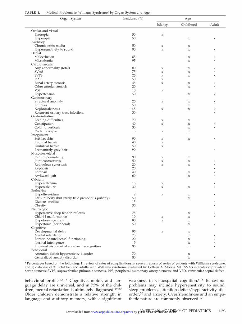
TABLE 1. Medical Problems in Williams Syndrome* by Organ System and Age
Organ System Incidence (%) Age
Infancy Childhood Adult
Ocular and visualEsotropia 50 xHyperopia 50 x x
AuditoryChronic otitis media 50 x xHypersensitivity to sound 90 x x x
DentalMalocclusion 85 x xMicrodontia 95 x x
CardiovascularAny abnormality (total) 80 x x xSVAS 75 x x xSVPS 25 x x xPPS 50 xRenal artery stenosis 45 x x xOther arterial stenosis 20 x xVSD 10 xHypertension 50 x x
GenitourinaryStructural anomaly 20 x x xEnuresis 50 xNephrocalcinosis ,5 x x xRecurrent urinary tract infections 30 x
GastrointestinalFeeding difficulties 70 x xConstipation 40 x x xColon diverticula 30 x xRectal prolapse 15 x x
IntegumentSoft lax skin 90 x x xInguinal hernia 40 xUmbilical hernia 50 xPrematurely gray hair 90 x
MusculoskeletalJoint hypermobility 90 x xJoint contractures 50 x x xRadioulnar synostosis 20 x x xKyphosis 20 xLordosis 40 x xAwkward gait 60 x x
CalciumHypercalcemia 15 x xHypercalciuria 30 x x x
EndocrineHypothyroidism 2 x x xEarly puberty (but rarely true precocious puberty) 50 xDiabetes mellitus 15 xObesity 30 x
NeurologicHyperactive deep tendon reflexes 75 x xChiari I malformation 10 x x xHypotonia (central) 80 x xHypertonia (peripheral) 50 x x
CognitiveDevelopmental delay 95 x xMental retardation 75 x xBorderline intellectual functioning 20 x xNormal intelligence 5 x xImpaired visuospatial constructive cognition 95 x x
BehavioralAttention-deficit hyperactivity disorder 70 xGeneralized anxiety disorder 80 x x
* Percentages based on the following: 1) review of rates of complications in several reports of series of patients with Williams syndrome,and 2) database of 315 children and adults with Williams syndrome evaluated by Colleen A. Morris, MD. SVAS indicates supravalvaraortic stenosis; SVPS, supravalvular pulmonic stenosis, PPS, peripheral pulmonary artery stenosis; and VSD, ventricular septal defect.
AMERICAN ACADEMY OF PEDIATRICS 1193 by guest on December 30, 2019www.aappublications.org/newsDownloaded from

The medical care of children with WS requires anunderstanding of the natural history of the disorder,awareness of potential clinical complications, andongoing assessment and periodic review at appro-priate ages (Fig 2). Because the clinical manifesta-
tions during the neonatal period are variable, thediagnosis may not be suspected during early infancy.Accordingly, this statement includes a series of eval-uations that should be considered at the time thediagnosis is suspected clinically; the diagnosis
Fig 1. Williams syndrome diagnostic scoring table: clinical diagnosis.
1194 HEALTH CARE SUPERVISION FOR CHILDREN WITH WILLIAMS SYNDROME by guest on December 30, 2019www.aappublications.org/newsDownloaded from

Fig
2.H
ealt
hsu
perv
isio
nfo
rch
ildre
nw
ith
Will
iam
ssy
ndro
me*
.
AMERICAN ACADEMY OF PEDIATRICS 1195 by guest on December 30, 2019www.aappublications.org/newsDownloaded from

should be confirmed by FISH analysis. The evalua-tions include the following:• Complete physical and neurologic examination• Growth parameters plotted on WS growth charts
(Fig 3A–F)• Cardiology evaluation
–Full clinical evaluation by a cardiologist withexpertise and experience in pediatric patients thatincludes 4-limb blood pressure measurements andechocardiography
• Genitourinary system evaluation–Ultrasonography of bladder and kidneys–Renal function studies (serum urea nitrogen andcreatinine levels)
–Urinalysis• Calcium determinations (serum calcium, spot
urine calcium, and creatinine levels) (Table 2)• Thyroid function tests• Ophthalmologic evaluation• Multidisciplinary developmental evaluation (old-
er than 2 years)• FISH to determine ELN deletion
Referral to a clinical geneticist should be consid-ered for individualized assessment and recommen-dations; a more extensive discussion of the clinicalmanifestations, natural history, recurrence risks, andfuture reproductive options; and evaluation of ge-netic risks for other family members.
SPECIAL CONSIDERATIONS FOR THE CHILDDIAGNOSED WITH WS
1. Do not give multivitamin preparations to childrenwith WS because of the potential deleterious ef-fects of vitamin D. Recommend diligent use ofsunscreen to minimize autologous production ofvitamin D.
2. Perform periodic cardiovascular evaluations, evenafter a baseline examination with normal findings.
3. Baseline cardiology evaluation should be per-formed by a cardiologist with pediatric expertiseand experience.
4. Screen for the development of hypertension peri-odically according to guidelines of the AmericanAcademy of Pediatrics.
5. Establish a medical home with clear emphasis oncontinuity of care and the role of the family mem-bers as partners in the ongoing management andcare of the child.
HEALTH SUPERVISION FROM BIRTH TO 1 YEAR(INFANCY)
Examination1. Review and note clinical features and confirm
diagnosis with FISH analysis2. Routine health maintenance examinations and
baseline evaluation3. Growth and developmental evaluations using
WS growth charts (Fig 3A–F)4. Baseline cardiology evaluation by a cardiologist
with pediatric expertise and experience5. Review feeding issues (reflux, refusal, disor-
dered suck or swallow, vomiting or symptoms ofcolic).
6. Consider pediatric ophthalmologic evaluationfor strabismus, amblyopia, and refractive errors
7. Check for inguinal hernia8. Objective hearing assessment at 6 to 12 months
(recurrent otitis media is common)9. Blood pressure measurement (both arms) annu-
ally and careful evaluation of femoral pulses10. Early recognition and management of constipa-
tion11. Pediatric anesthesia consultation for any child
requiring surgery (several reports of unexpecteddeaths have been associated with the administra-tion of anesthesia)22
Laboratory1. Williams Syndrome Chromosomal Region FISH
to confirm clinical diagnosis2. Serum creatinine level3. Urinalysis4. Calcium levels
a. Serum*b. Spot urine test to determine calcium-creatinine
ratio†5. Thyroid screen for newborns (according to state
mandate)6. Baseline ultrasonographic examination of the
bladder and kidneys
Anticipatory Guidance1. Individual support for the family (by family,
friends, clergy), support groups, or both (see list)2. Review increased risk for otitis media3. Feeding (difficulty in transition to textured foods)4. Do not prescribe multivitamin preparations con-
taining vitamin D5. Refer to early childhood intervention program
HEALTH SUPERVISION FROM 1 TO 5 YEARS(EARLY CHILDHOOD)
Examination1. Annual health maintenance examinations and
baseline evaluation (including careful ausculta-tion of chest and abdomen for murmurs orbruits)
2. Developmental evaluation and growth evalua-tion using WS growth charts (Fig 3A–F)
3. Annual cardiology evaluation from 1 to 5 years4. Feeding issues: watch for rectal prolapse and
avoid constipation with stool softeners if neces-sary
5. Annual hearing and vision screening; objectiveaudiologic evaluation and an ophthalmologicevaluation before age 3 years
6. Orthopedic issues: musculoskeletal and neuro-logic assessments to evaluate joints, muscle tone,spasticity, and hyperactive reflexes17
*If hypercalcemia is found, dietary calcium restriction should be imple-mented and diet should be monitored in conjunction with a pediatricdietician/nutritionist. Referral to a pediatric renal specialist should beconsidered.†If hypercalciuria is found, 2 repeated urine studies of the calcium-creati-nine ratio (morning and afternoon) should be performed. If the level is stillelevated, repeat measurement of the serum calcium level and perform renalultrasonography for nephrocalcinosis. Assess dietary calcium intake.21
1196 HEALTH CARE SUPERVISION FOR CHILDREN WITH WILLIAMS SYNDROME by guest on December 30, 2019www.aappublications.org/newsDownloaded from

7. Pediatric anesthesia consultation for any childrequiring surgery (several reports of unexpecteddeaths have been associated with the administra-tion of anesthesia)22
8. Annual blood pressure measurement (botharms) and careful examination of femoral pulses
9. Multidisciplinary developmental assessmentand treatment in early intervention programs(0–3 years) or school based programs (3 yearsand older)1,5,19
10. Dental referral
Laboratory1. Yearly urinalysis2. Annual total calcium measurement if the level
was elevated at baseline or as needed if the childbecomes symptomatic; if level was normal, mea-sure every 2 to 3 years
3. Urinary calcium-creatinine ratio every 2 years4. Thyroid function test every 4 years
Fig 3A. Williams syndrome—stature, females.
Height for patients withWilliams syndrome (61 fe-males, 47 males). Normalcurves—dashed lines; af-fected patients—solid lines.Reprinted with permissionfrom Saul RA, Geer JS,Seaver LH, Phelan MC,Sweet KM, Mills MS. GrowthReferences: Third Trimester toAdulthood. Greenwood, SC:Greenwood Genetic Center;1998.
AMERICAN ACADEMY OF PEDIATRICS 1197 by guest on December 30, 2019www.aappublications.org/newsDownloaded from
5. Serum creatinine level every 4 years
Anticipatory Guidance1. Individual support for the family (by family,
friends, clergy), support groups, or both2. Review increased risk for otitis media3. Ongoing feeding and dietary assessments
4. Therapy as needed (physical, speech and language,and occupational, including sensory integration)
5. Review constipation as a possible problem6. Children with unexplained fever should be eval-
uated for urinary tract infection7. Discuss developmental status, early intervention
programs, and preschool programs
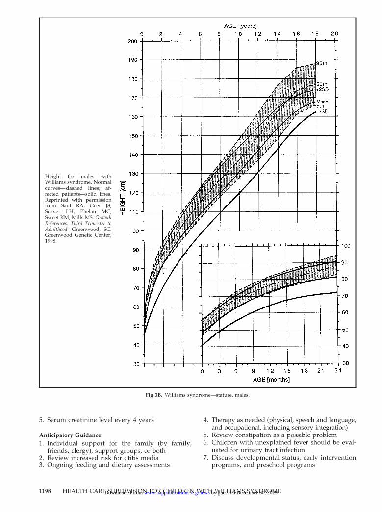
Fig 3B. Williams syndrome—stature, males.
Height for males withWilliams syndrome. Normalcurves—dashed lines; af-fected patients—solid lines.Reprinted with permissionfrom Saul RA, Geer JS,Seaver LH, Phelan MC,Sweet KM, Mills MS. GrowthReferences: Third Trimester toAdulthood. Greenwood, SC:Greenwood Genetic Center;1998.
1198 HEALTH CARE SUPERVISION FOR CHILDREN WITH WILLIAMS SYNDROME by guest on December 30, 2019www.aappublications.org/newsDownloaded from

HEALTH SUPERVISION FROM 5 YEARS TO12 YEARS (LATE CHILDHOOD)
Examination1. Annual health maintenance examinations and
baseline evaluation2. Developmental evaluation and growth evalua-
tion using WS growth charts (Fig 3A–F)3. Annual blood pressure measurements (both
arms) and careful evaluation of femoral pulses
4. Cardiology evaluation as indicated by previousclinical findings. If results of previous evalua-tions are negative, repeated cardiology evalua-tion (for arterial stenoses, hypertension) shouldbe performed at puberty
5. Ophthalmologic evaluation for strabismus andhyperopia
6. Orthopedic problems (eg, joint limitation, ky-phosis, lordosis, scoliosis, and spasticity)
Fig 3C. Williams syndrome—weight, females.
Weight for females with Williams syn-drome. Normal curves—dashed lines; af-fected patients—solid lines. Reprintedwith permission from Saul RA, Geer JS,Seaver LH, Phelan MC, Sweet KM, MillsMS. Growth References: Third Trimester toAdulthood. Greenwood, SC: GreenwoodGenetic Center; 1998.
AMERICAN ACADEMY OF PEDIATRICS 1199 by guest on December 30, 2019www.aappublications.org/newsDownloaded from

7. Hearing and vision screening annually8. Pediatric anesthesia consultation for any child
requiring surgery (several reports of unexpecteddeaths have been associated with the administra-tion of anesthesia22)
9. School readiness and placement and IndividualEducational Plan at 5 years
10. Developmental and psychoeducational assess-ment; formal evaluation for attention-deficit hy-peractivity disorder, anxiety, or both and discus-sion of treatment options23
Laboratory1. Yearly urinalysis2. Thyroid function tests every 4 years3. Annual total calcium level if baseline result was
elevated or child becomes symptomatic; other-wise measure level every 4 years
4. Urinary calcium-creatinine ratio every 2 years5. Serum creatinine level every 2 to 4 years
Anticipatory Guidance1. School readiness and placement
Fig 3D. Williams syndrome—weight, males.
Weight for males with Williams syn-drome. Normal curves—dashed lines; af-fected patients—solid lines. Reprintedwith permission from Saul RA, Geer JS,Seaver LH, Phelan MC, Sweet KM, MillsMS. Growth References: Third Trimester toAdulthood. Greenwood, SC: GreenwoodGenetic Center; 1998.
1200 HEALTH CARE SUPERVISION FOR CHILDREN WITH WILLIAMS SYNDROME by guest on December 30, 2019www.aappublications.org/newsDownloaded from

2. Therapy as needed (physical, speech and lan-guage, and occupational, including sensory inte-gration)
3. Long-term vocational planning4. Discuss sexuality and adolescence; puberty is of-
ten early in WS, but true precocious puberty israre
5. Discuss diet and exercise as obesity may becomeapparent in late childhood
6. Discuss treatment options for anxiety (counseling,relaxation techniques, and medications)
7. Estate planning for parents of a child with specialneeds
HEALTH SUPERVISION FROM 13 YEARS TO18 YEARS (ADOLESCENCE)
Progressive medical problems including hyperten-sion, progressive joint limitations, recurrent urinary
Fig 3E. Williams syndrome—head circumference, females.
Head circumference for femaleswith Williams syndrome. Nor-mal curves—dashed lines; af-fected patients—solid lines. Re-printed with permission fromSaul RA, Geer JS, Seaver LH,Phelan MC, Sweet KM, MillsMS. Growth References: Third Tri-mester to Adulthood. Greenwood,SC: Greenwood Genetic Center;1998.
AMERICAN ACADEMY OF PEDIATRICS 1201 by guest on December 30, 2019www.aappublications.org/newsDownloaded from
tract infections, and gastrointestinal problems arecommon beginning in this age group and continuingthroughout adult life.
Examination1. Annual health maintenance examinations and
baseline evaluation; blood pressure measure-ment (both arms)
2. Developmental evaluation and growth evalua-tion using WS growth charts (Fig 3A–F)
3. Cardiology evaluation if indicated by previousclinical findings
4. Pediatric anesthesia consultation for any childrequiring surgery (several reports of unexpecteddeaths have been associated with the administra-tion of anesthesia22)
5. Consider ophthalmologic evaluation for hyper-opia
6. Orthopedic problems (eg, joint limitation, ky-phosis, lordosis, scoliosis, and spasticity)
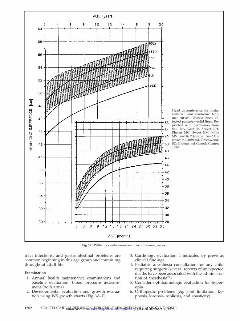
Fig 3F. Williams syndrome—head circumference, males.
Head circumference for maleswith Williams syndrome. Nor-mal curves—dashed lines; af-fected patients—solid lines. Re-printed with permission fromSaul RA, Geer JS, Seaver LH,Phelan MC, Sweet KM, MillsMS. Growth References: Third Tri-mester to Adulthood. Greenwood,SC: Greenwood Genetic Center;1998.
1202 HEALTH CARE SUPERVISION FOR CHILDREN WITH WILLIAMS SYNDROME by guest on December 30, 2019www.aappublications.org/newsDownloaded from

7. Hearing and vision screening annually8. Developmental and psychoeducational assess-
ment; school placement and resource enhance-ment; vocational training; social skills trainingfor peer interaction10,11
9. Gastrointestinal issues: consider diverticulitisand diverticulosis, cholelithiasis, and chronicconstipation in adolescents with abdominal pain
10. Screen for generalized anxiety disorder19
Laboratory1. Yearly urinalysis2. Thyroid function test every 4 years3. Total calcium level only if adolescent becomes
symptomatic, otherwise, every 4 years4. Urinary calcium-creatinine ratio every 2 years5. Bladder and renal ultrasonography at puberty
and every 5 years thereafter6. Serum creatinine level every 2 to 4 years
Anticipatory Guidance1. School placement2. Therapy as needed (physical, occupational,
speech, and language)3. Discuss diagnosis with the adolescent; support
groups for the adolescent (see American Acad-emy of Pediatrics statement on “Transition ofCare Provided for Adolescents With SpecialNeeds”)24
4. Discuss sexuality and reproductive issues5. Encourage career counseling6. Foster independence7. Assist in transition to adult care (especially for
cardiology care). Many pediatricians feel com-fortable continuing to provide primary care wellinto young adulthood
8. Encourage daily exercise to include range of mo-tion
9. Encourage prompt medical attention for urinarytract or gastrointestinal symptoms
10. Mental health issues
Committee on Genetics, 2000–2001Christopher Cunniff, MD, ChairpersonJaime L. Frias, MDCelia I. Kaye, MD, PhDJohn Moeschler, MDSusan R. Panny, MDTracy L. Trotter, MD
LiaisonsFelix de la Cruz, MD, MPH
National Institute of Child Health and HumanDevelopment
John Williams III, MDAmerican College of Obstetricians andGynecologists
James W. Hanson, MDAmerican College of Medical Genetics
Cynthia A. Moore, MD, PhDCenters for Disease Control and Prevention
Michele Lloyd-Puryear, MD, PhDHealth Resources and Services Administration
Section LiaisonH. Eugene Hoyme, MD
Section on Genetics
ConsultantsPaige Kaplan, MDRon Lacro, MDKaren Levine, PhDMartin Levinson, MDCarolyn Mervis, PhDColleen A. Morris, MDBeth A. Pletcher, MDBarbara Pober, MDLaurie Sadler, MDPaul Wang, MD
StaffLauri A. Hall
REFERENCES1. Williams JC, Barratt-Boyes BG, Lowe JB. Supravalvular aortic stenosis.
Circulation. 1961;24:1311–13182. Beuren AJ. Supravalvular aortic stenosis: a complex syndrome with and
without mental retardation. Natl Found March Dimes Birth Defects OrigArt Ser. 1972;8:45–56
3. Burn J. Williams syndrome. J Med Genet. 1986;23:389–3954. Morris CA, Demsey SA, Leonard CO, Dilts C, Blackburn BL. Natural
history of Williams syndrome: physical characteristics. J Pediatr. 1988;113:318–326
5. Udwin O, Yule W. A cognitive and behavioural phenotype in Williamssyndrome. J Clin Exp Neuropsychol. 1991;13:232–244
6. Ewart AK, Morris CA, Atkinson D, et al. Hemizygosity at the elastinlocus in a developmental disorder, Williams syndrome. Nat Genet.1993;5:11–16
7. Lowery MC, Morris CA, Ewart A, et al. Strong correlation of elastindeletions, detected by FISH, with Williams syndrome: evaluation of 235patients. Am J Hum Genet. 1995;57:49–53
8. Wu Y-Q, Sutton VR, Nickerson E, et al. Delineation of the commoncritical region in Williams syndrome and clinical correlation of growth,heart defects, ethnicity, and parental origin. Am J Med Genet. 1998;78:82–89
9. Martin ND, Snodgrass GJ, Cohen RD. Idiopathic infantilehypercalcemia: a continuing enigma. Arch Dis Child. 1984;59:605–613
10. Lopez-Rangel E, Maurice M, McGillivray B, Friedman JM. Williamssyndrome in adults. Am J Med Genet. 1992;44:720–729
11. Morris CA, Leonard CO, Dilts C, Demsey SA. Adults with Williamssyndrome. Am J Med Gen Suppl. 1990;6:102–107
12. Greenberg F, Lewis RA. The Williams syndrome: spectrum and signif-icance of ocular features. Ophthalmology. 1988;95:1608–1612
13. Saul RA, Stevenson RE, Rogers RC, Skinner SA, Prouty LA, FlanneryDB. Williams syndrome. In: Proceedings of the Greenwood Genetic Center.Greenwood, SC: Greenwood Genetic Center; 1988:204–209
14. Pankau R, Partsch C-J, Winter M, Gosch A, Wessel A. Incidence andspectrum of renal abnormalities in Williams-Beuren syndrome. Am JMed Genet. 1996;63:301–304
15. Pober BR, Lacro RV, Rice C, Mandell V, Teele RL. Renal findings in 40individuals with Williams syndrome. Am J Med Genet. 1993;46:271–274
16. Morris CA, Mervis CB. Williams syndrome. In: Goldstein S, ReynoldsCR, eds. Handbook of Neurodevelopmental and Genetic Disorders in Children.New York, NY: The Guilford Press; 1999;555–590
17. Kaplan P, Kirschner M, Watters G, Costa MT. Contractures in patientswith Williams syndrome. Pediatrics. 1989;84:895–899
TABLE 2. Normal Values for Random Urinary Calcium-Creatinine Ratios21
Age Calcium-Creatinine Ratio (mg/mg ratio)(95th Percentile for Age)
,7 mo 0.867–18 mo 0.619 mo–6 y 0.42Adults 0.22
AMERICAN ACADEMY OF PEDIATRICS 1203 by guest on December 30, 2019www.aappublications.org/newsDownloaded from

18. Wang PP, Hesselink JR, Jernigan TL, Doherty S, Bellugi U. Specificneurobehavioral profile of Williams’ syndrome is associated with neo-cerebellar hemispheric preservation. Neurology. 1992;42:1999–2002
19. Chapman CA, du Plessis A, Pober BR. Neurologic findings in childrenand adults with Williams syndrome. J Child Neurol. 1996;11:63–65
20. Pober BR, Filiano JJ. Association of Chiari I malformations and Williamssyndrome. Pediatr Neurol. 1995;12:84–88
21. Sargent JD, Stukel TA, Kresel J, Klein RZ. Normal values for randomurinary calcium to creatinine ratios in infancy. J Pediatr. 1993;123:393–397
22. Bird LM, Billman GF, Lacro RV, et al. Sudden death in Williamssyndrome: report of ten cases. J Pediatr. 1996;129:926–931
23. Power TJ, Blum NJ, Jones SM, Kaplan PE. Brief report: response to
methylphenidate in two children with Williams syndrome. J Autism DevDis. 1997;27:79–87
24. American Academy of Pediatrics, Committee on Children With Disabil-ities. Transition of care provided for adolescents with special health careneeds. Pediatrics. 1996;98:1203–1206
RESOURCES FOR PARENTSMarch of Dimes, 1275 Mamaroneck Ave, White Plains, NY 10605;
Telephone: 914/428–7100; http. //www.modimes.orgThe Williams Syndrome Association, PO Box 297, Clawson, MI 48017;
Telephone: 248/541–3630; http://www.williams-syndrome.orgWilliams Syndrome Foundation, University of California, Irvine, CA 92679;
Telephone: 949/824–7259; http://www.wsf.org
1204 HEALTH CARE SUPERVISION FOR CHILDREN WITH WILLIAMS SYNDROME by guest on December 30, 2019www.aappublications.org/newsDownloaded from
2001;107;1192Pediatrics Committee on Genetics
Health Care Supervision for Children With Williams Syndrome
ServicesUpdated Information &
http://pediatrics.aappublications.org/content/107/5/1192including high resolution figures, can be found at:
Referenceshttp://pediatrics.aappublications.org/content/107/5/1192#BIBLThis article cites 22 articles, 6 of which you can access for free at:
Subspecialty Collections
http://www.aappublications.org/cgi/collection/genetics_subGeneticsshttp://www.aappublications.org/cgi/collection/committee_on_geneticCouncil on Geneticshttp://www.aappublications.org/cgi/collection/current_policyCurrent Policyfollowing collection(s): This article, along with others on similar topics, appears in the
Permissions & Licensing
http://www.aappublications.org/site/misc/Permissions.xhtmlin its entirety can be found online at: Information about reproducing this article in parts (figures, tables) or
Reprintshttp://www.aappublications.org/site/misc/reprints.xhtmlInformation about ordering reprints can be found online:
by guest on December 30, 2019www.aappublications.org/newsDownloaded from

2001;107;1192Pediatrics Committee on Genetics
Health Care Supervision for Children With Williams Syndrome
http://pediatrics.aappublications.org/content/107/5/1192located on the World Wide Web at:
The online version of this article, along with updated information and services, is
1073-0397. ISSN:60007. Copyright © 2001 by the American Academy of Pediatrics. All rights reserved. Print
the American Academy of Pediatrics, 141 Northwest Point Boulevard, Elk Grove Village, Illinois,has been published continuously since 1948. Pediatrics is owned, published, and trademarked by Pediatrics is the official journal of the American Academy of Pediatrics. A monthly publication, it
by guest on December 30, 2019www.aappublications.org/newsDownloaded from