Embed Size (px)
Citation preview
Alpha-Domain Structures
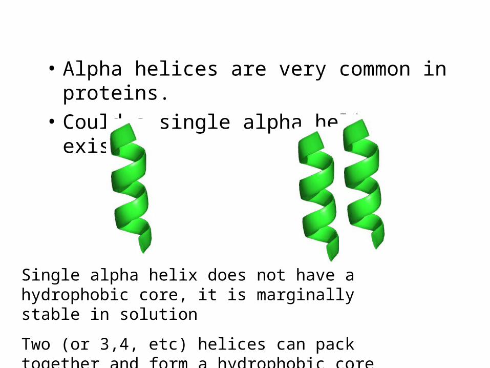
• Alpha helices are very common in proteins.
• Could a single alpha helix exist?
Single alpha helix does not have a hydrophobic core, it is marginally stable in solution
Two (or 3,4, etc) helices can pack together and form a hydrophobic core
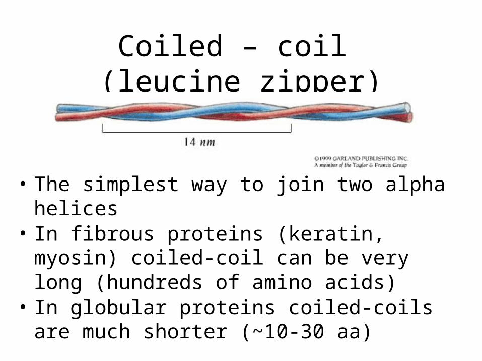
Coiled – coil (leucine zipper)
• The simplest way to join two alpha helices• In fibrous proteins (keratin, myosin) coiled-coil
can be very long (hundreds of amino acids)• In globular proteins coiled-coils are much shorter
(~10-30 aa)
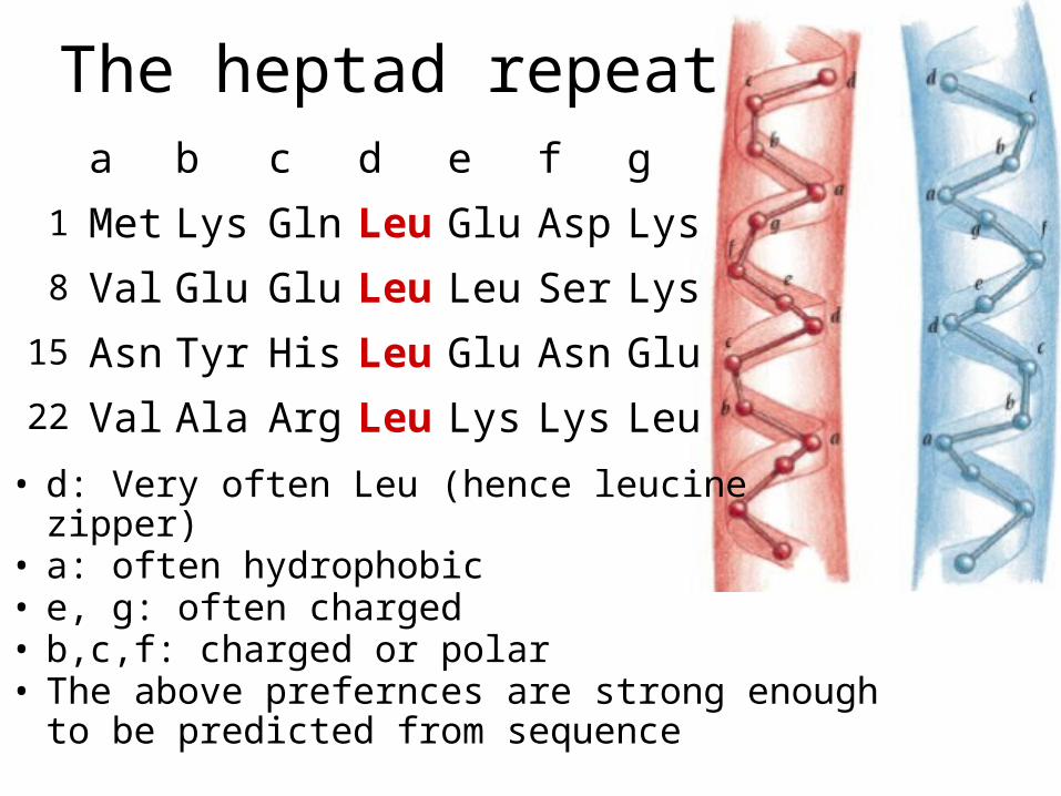
The heptad repeat
• d: Very often Leu (hence leucine zipper)• a: often hydrophobic• e, g: often charged• b,c,f: charged or polar• The above prefernces are strong enough to be
predicted from sequence
a b c d e f g
Met Lys Gln Leu Glu Asp Lys
Val Glu Glu Leu Leu Ser Lys
Asn Tyr His Leu Glu Asn Glu
Val Ala Arg Leu Lys Lys Leu
1
8
15
22
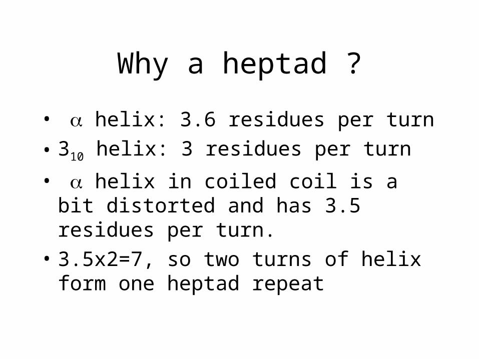
Why a heptad ?
• helix: 3.6 residues per turn
• 310 helix: 3 residues per turn
• helix in coiled coil is a bit distorted and has 3.5 residues per turn.
• 3.5x2=7, so two turns of helix form one heptad repeat
Original concept
(“zipper”)
Real life
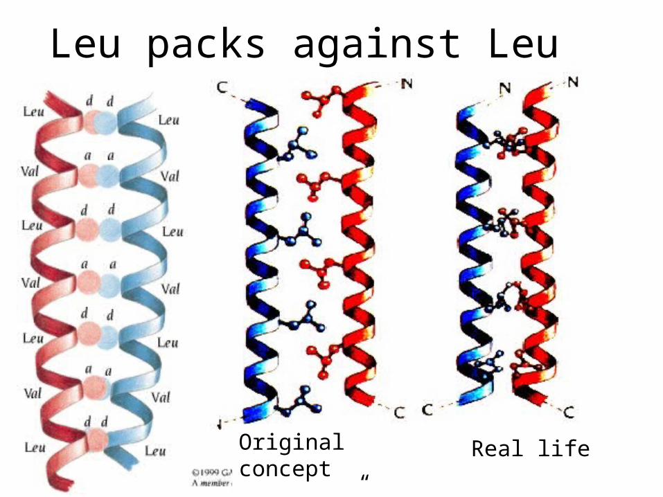
Leu packs against Leu
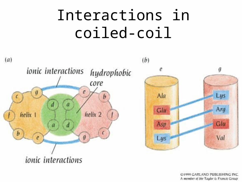
Interactions in coiled-coil
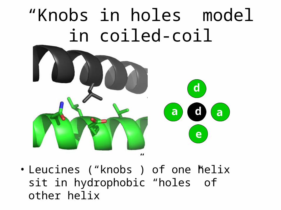
“Knobs in holes” model in coiled-coil
• Leucines (“knobs”) of one helix sit in hydrophobic “holes” of other helix
da a
d
e
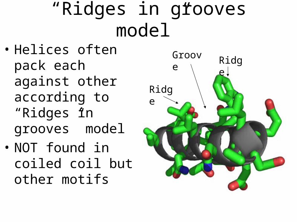
“Ridges in grooves model”
• Helices often pack each against other according to “Ridges in grooves” model
• NOT found in coiled coil but other motifs
Ridge
RidgeGroove

• Depending on actual amino acid sequence, ridges may be formed of residues which are 3 or 4 amino acids apart
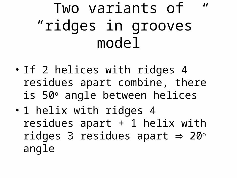
• If 2 helices with ridges 4 residues apart combine, there is 50o angle between helices
• 1 helix with ridges 4 residues apart + 1 helix with ridges 3 residues apart 20o angle
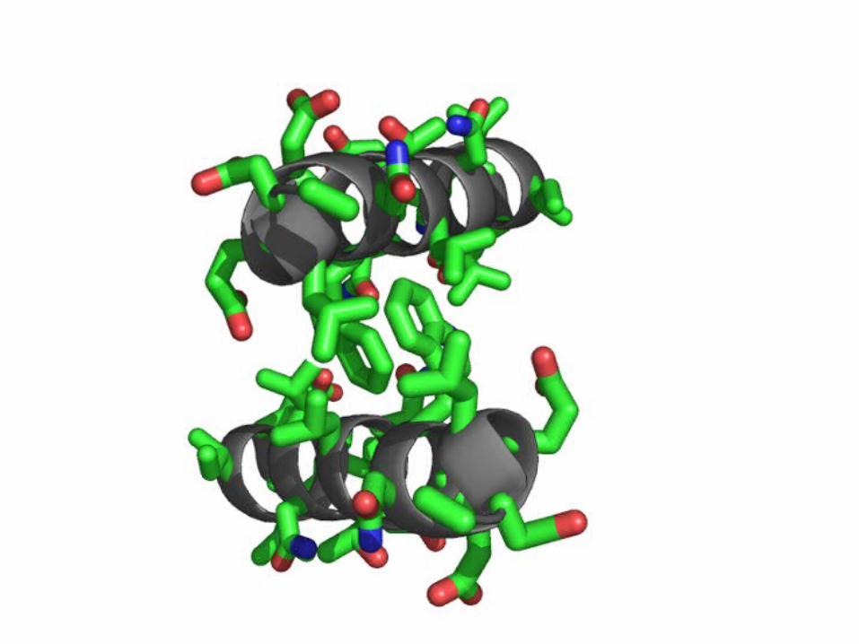
Two variants of “ridges in grooves” model
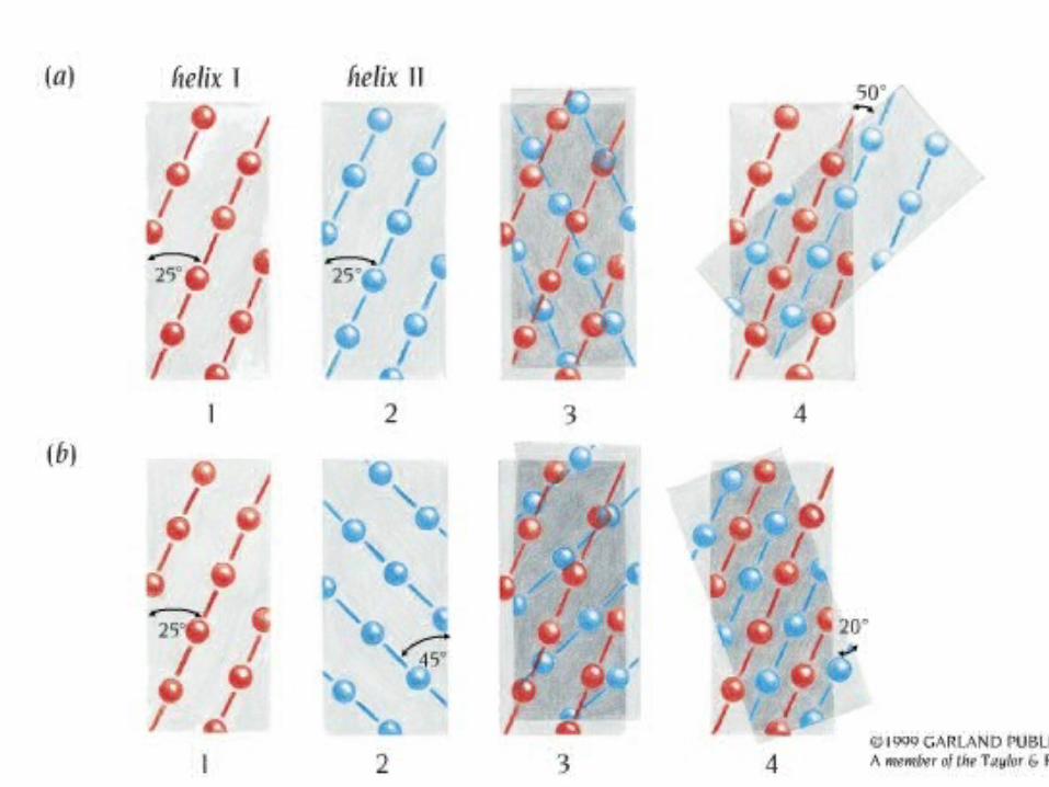
Four helix bundle• The most usual way of packing alpha
helices in globular proteins
• Usually “ridges in grooves” model
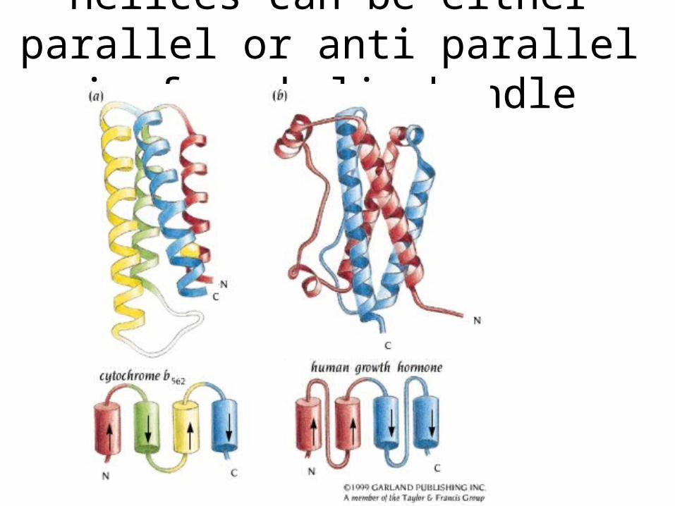
Helices can be either parallel or anti parallel in four helix bundle

Two leucine zippers can form a four helix bundle
• Two helices form leucine zipper
• Two zippers pack as “ridges and grooves”
• Note that usually two helices in 4hb do not make a leu zipper, this is just a special case
Leu zipper
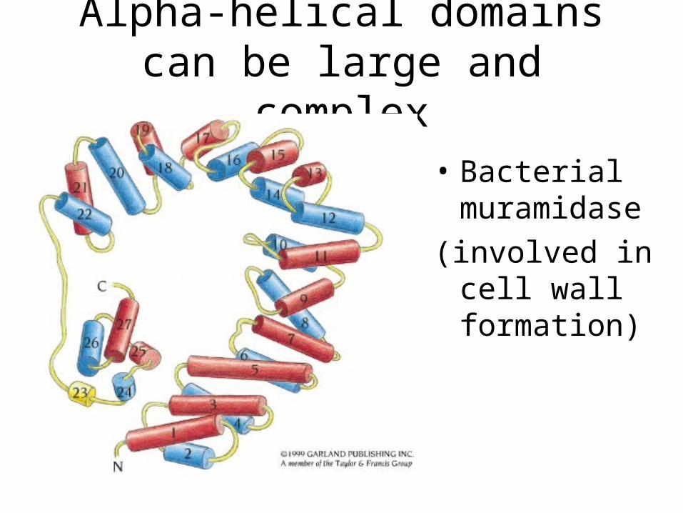
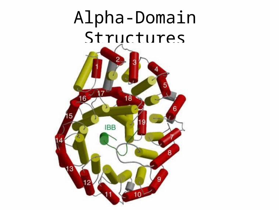
Alpha-helical domains can be large and complex
• Bacterial muramidase
(involved in cell wall formation)
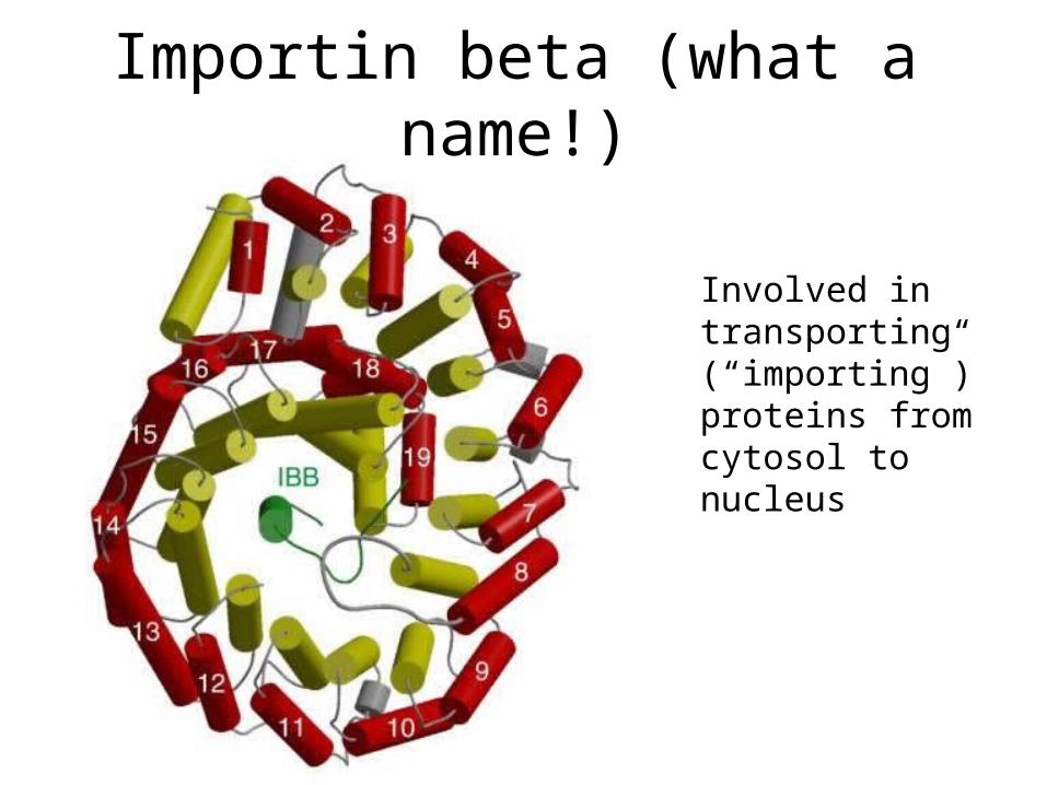
Importin beta (what a name!)
Involved in transporting (“importing”) proteins from cytosol to nucleus
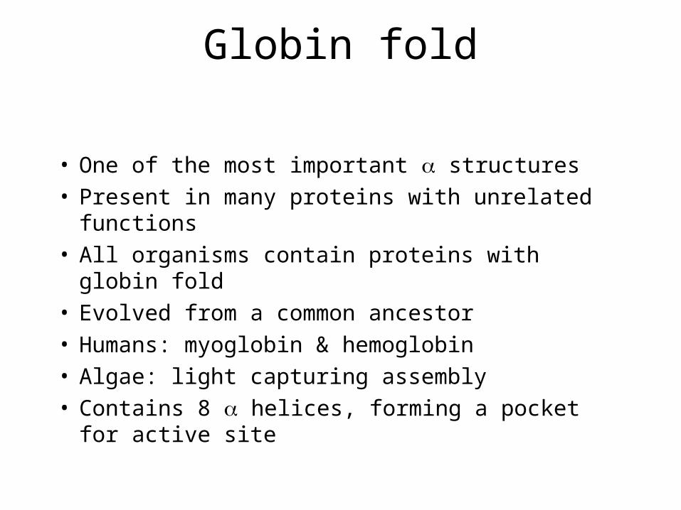
Globin fold
• One of the most important structures• Present in many proteins with unrelated functions • All organisms contain proteins with globin fold• Evolved from a common ancestor• Humans: myoglobin & hemoglobin• Algae: light capturing assembly• Contains 8 helices, forming a pocket for active
site
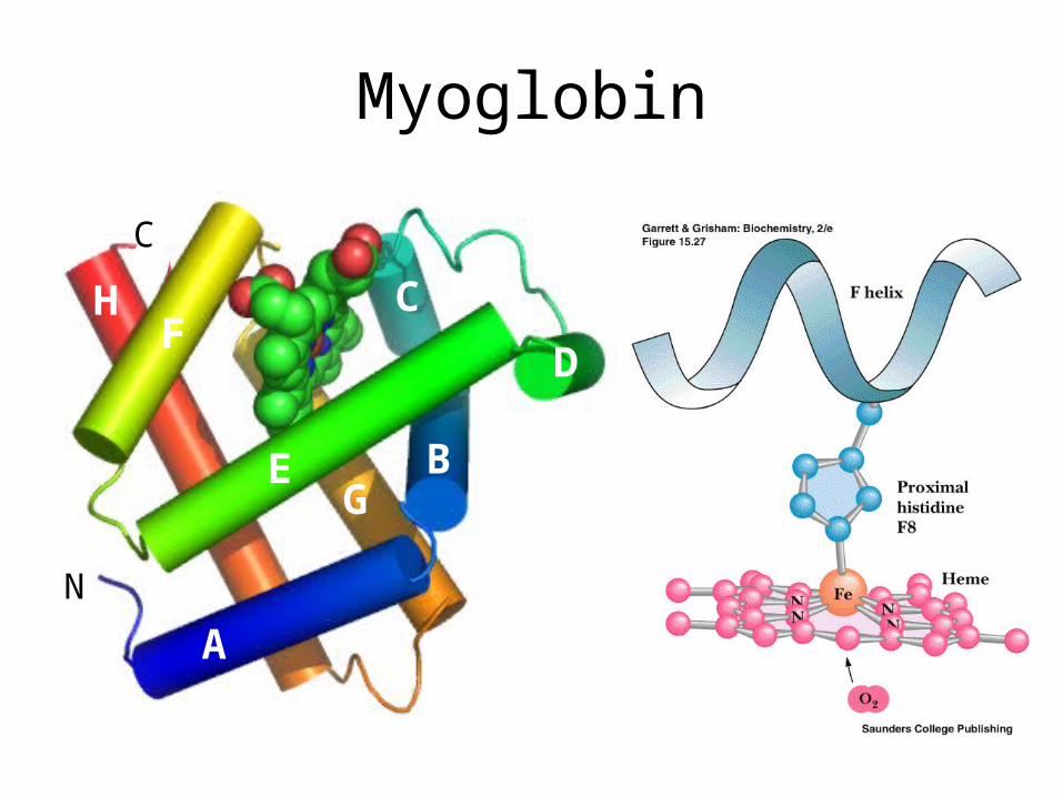
Myoglobin
A
B
C
D
E
FH
G
N
C
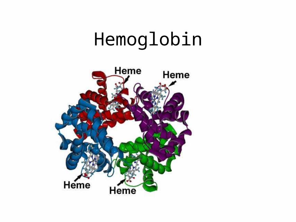
Hemoglobin
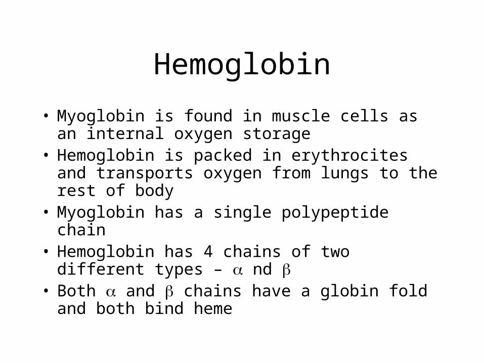
• Myoglobin is found in muscle cells as an internal oxygen storage
• Hemoglobin is packed in erythrocites and transports oxygen from lungs to the rest of body
• Myoglobin has a single polypeptide chain• Hemoglobin has 4 chains of two different types –
nd • Both and chains have a globin fold and both
bind heme
Hemoglobin
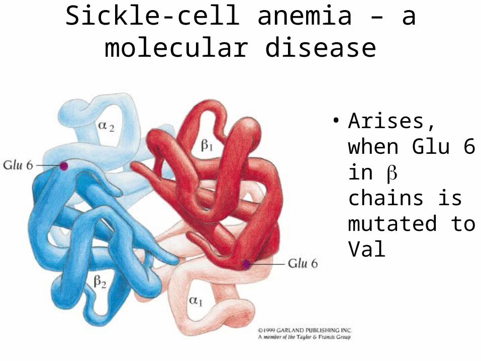
Sickle-cell anemia – a molecular disease
• Arises, when Glu 6 in chains is mutated to Val
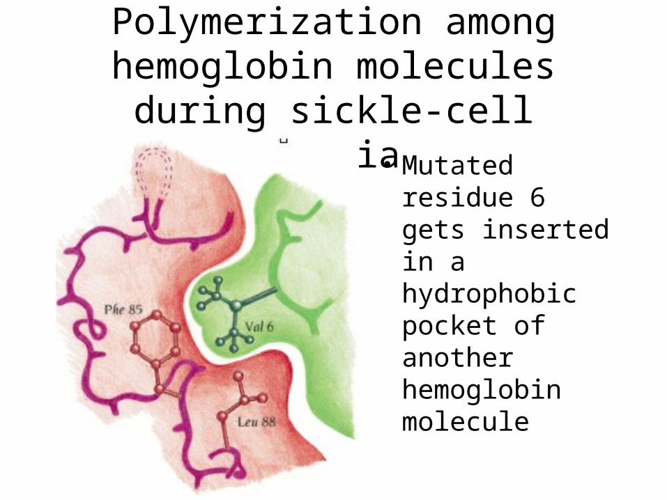
Polymerization among hemoglobin molecules during sickle-cell anemia
• Mutated residue 6 gets inserted in a hydrophobic pocket of another hemoglobin molecule
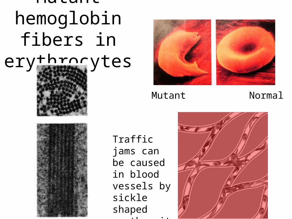
Mutant hemoglobin
fibers in erythrocytes
Mutant Normal
Traffic jams can be caused in blood vessels by sickle shaped erythrocites
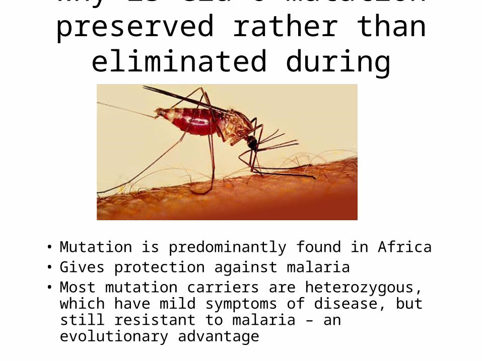
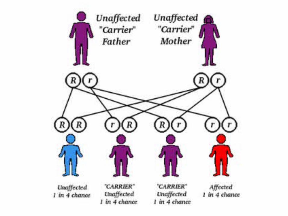
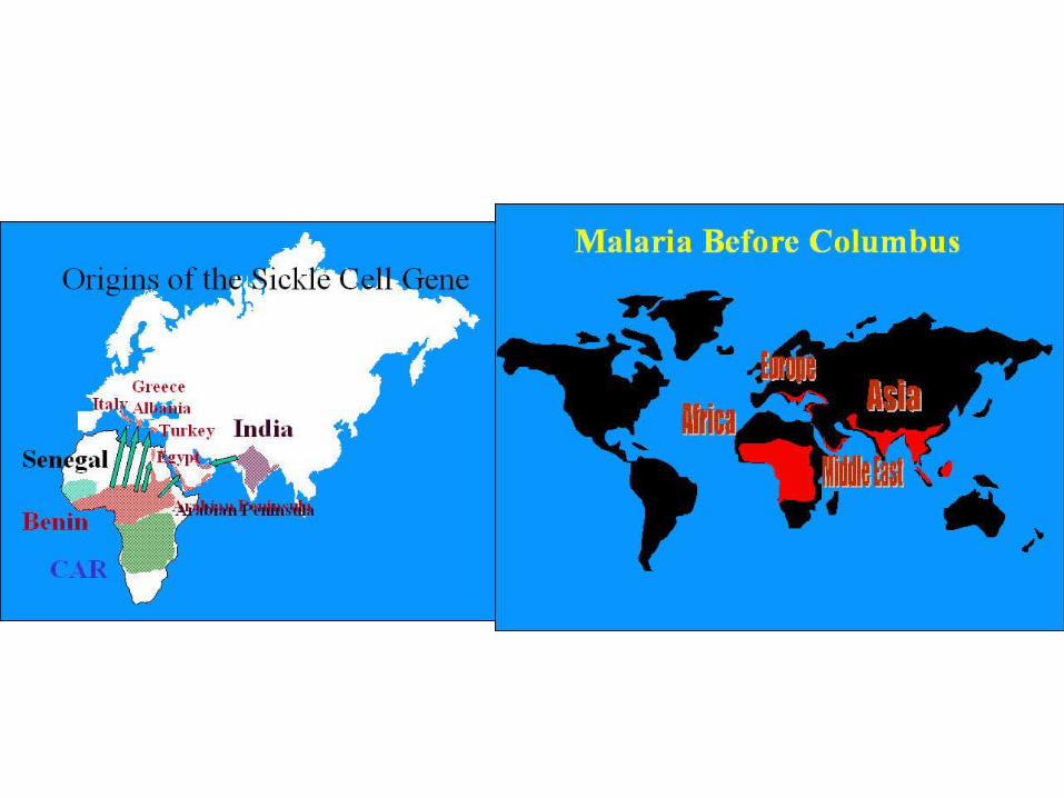
Why is Glu 6 mutation preserved rather than eliminated during evolution?
• Mutation is predominantly found in Africa• Gives protection against malaria• Most mutation carriers are heterozygous, which
have mild symptoms of disease, but still resistant to malaria – an evolutionary advantage