Embed Size (px)
Citation preview
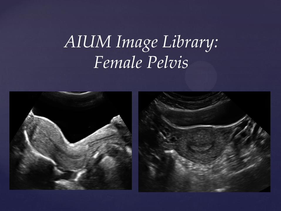
AIUM Image Library:Female Pelvis
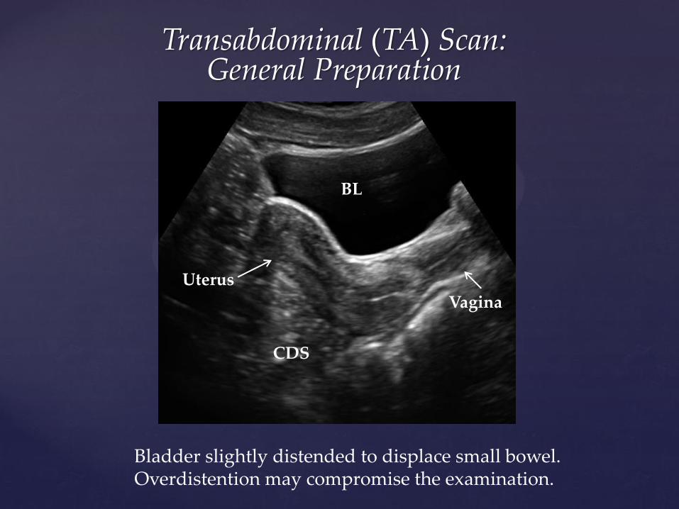
Transabdominal (TA) Scan: General Preparation
Bladder slightly distended to displace small bowel. Overdistention may compromise the examination.
Uterus
BL
Vagina
CDS
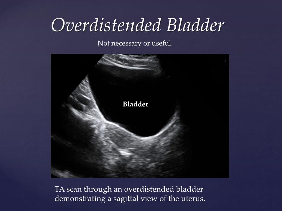
Overdistended Bladder Not necessary or useful.
Bladder
TA scan through an overdistended bladder demonstrating a sagittal view of the uterus.
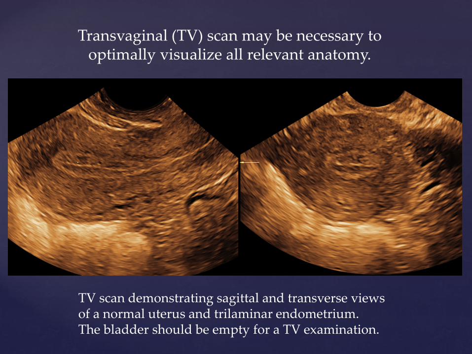
Transvaginal (TV) scan may be necessary to optimally visualize all relevant anatomy.
TV scan demonstrating sagittal and transverse views of a normal uterus and trilaminar endometrium. The bladder should be empty for a TV examination.
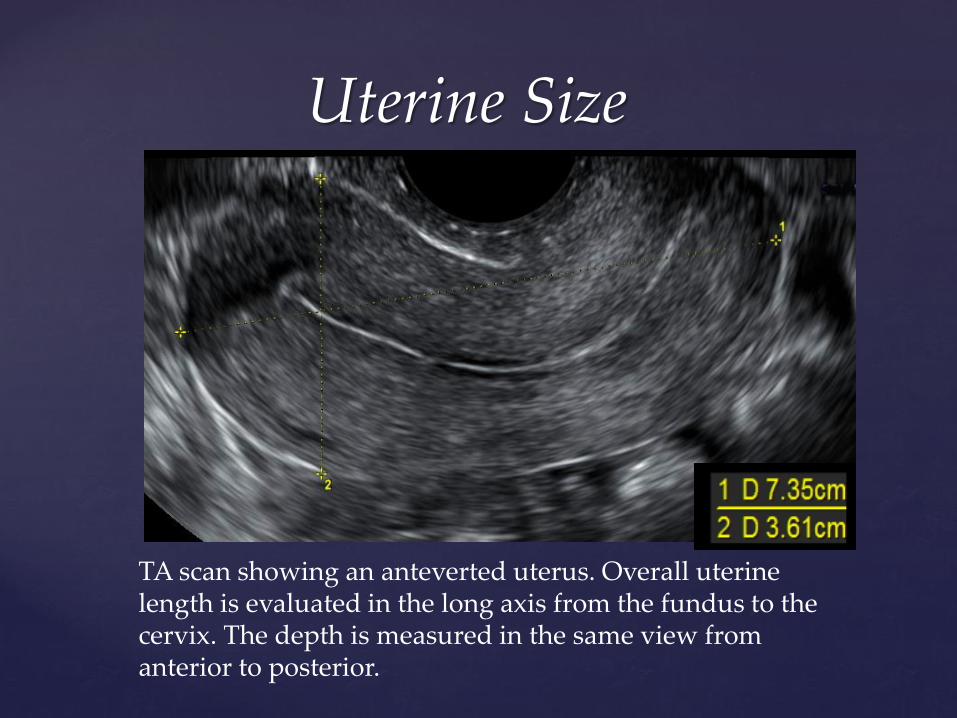
Uterine Size
TA scan showing an anteverted uterus. Overall uterine length is evaluated in the long axis from the fundus to the cervix. The depth is measured in the same view from anterior to posterior.
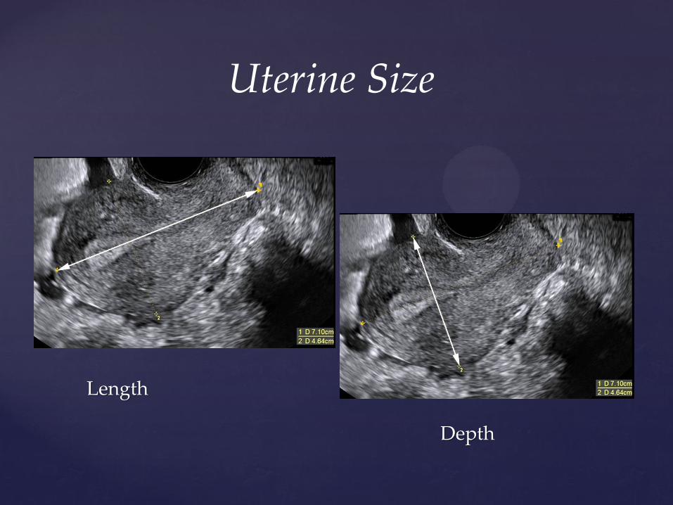
Length
Depth
Uterine Size
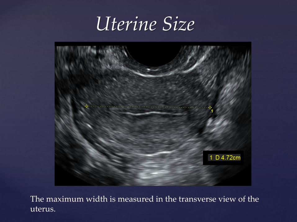
Uterine Size
The maximum width is measured in the transverse view of the uterus.
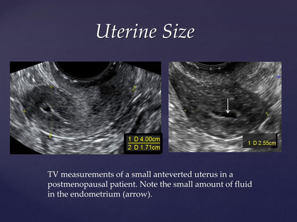
Uterine Size
TV measurements of a small anteverted uterus in a postmenopausal patient. Note the small amount of fluid in the endometrium (arrow).
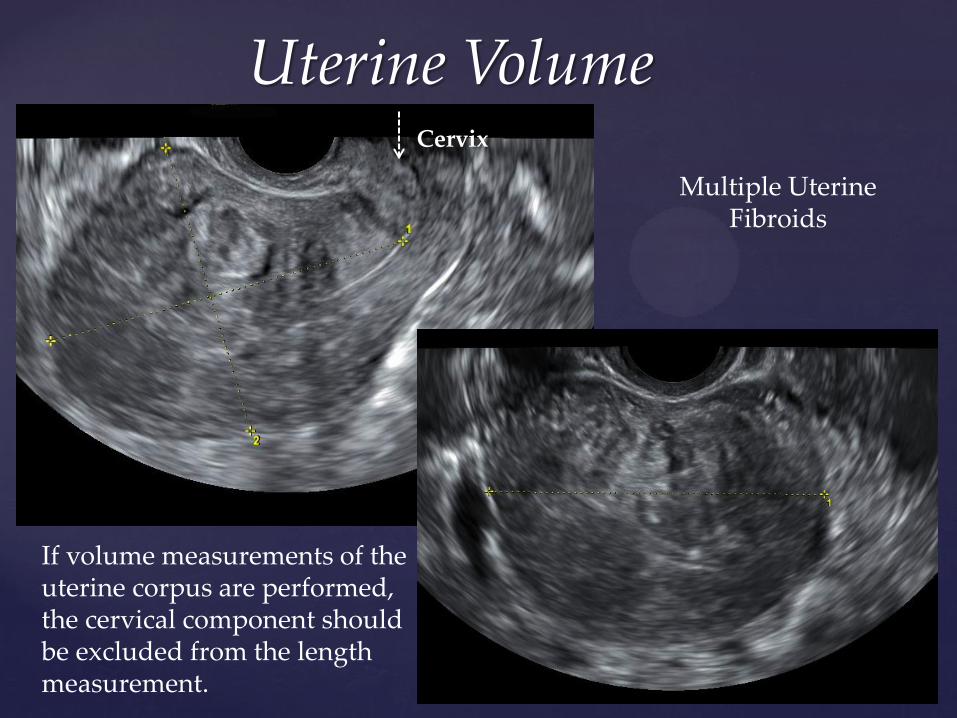
Uterine Volume
If volume measurements of the uterine corpus are performed, the cervical component should be excluded from the length measurement.
Cervix
Multiple Uterine Fibroids
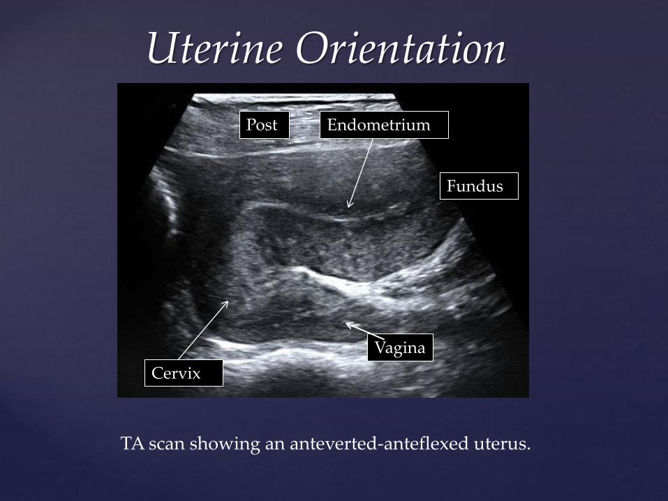
Uterine Orientation
TA scan showing an anteverted-anteflexed uterus.
Cervix
Fundus
Post
Vagina
Endometrium
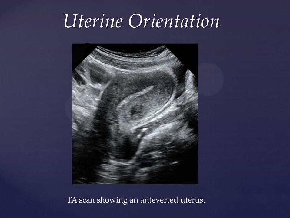
Uterine Orientation
TA scan showing an anteverted uterus.
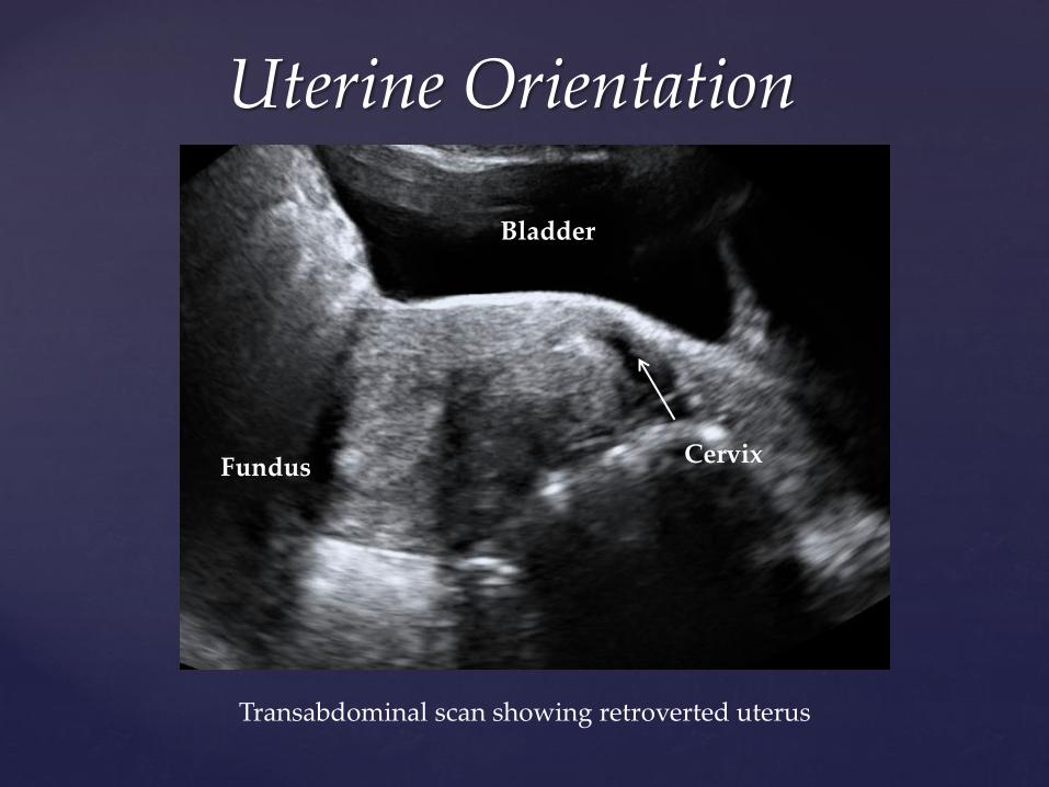
Uterine Orientation
Transabdominal scan showing retroverted uterus
Bladder
Cervix Fundus
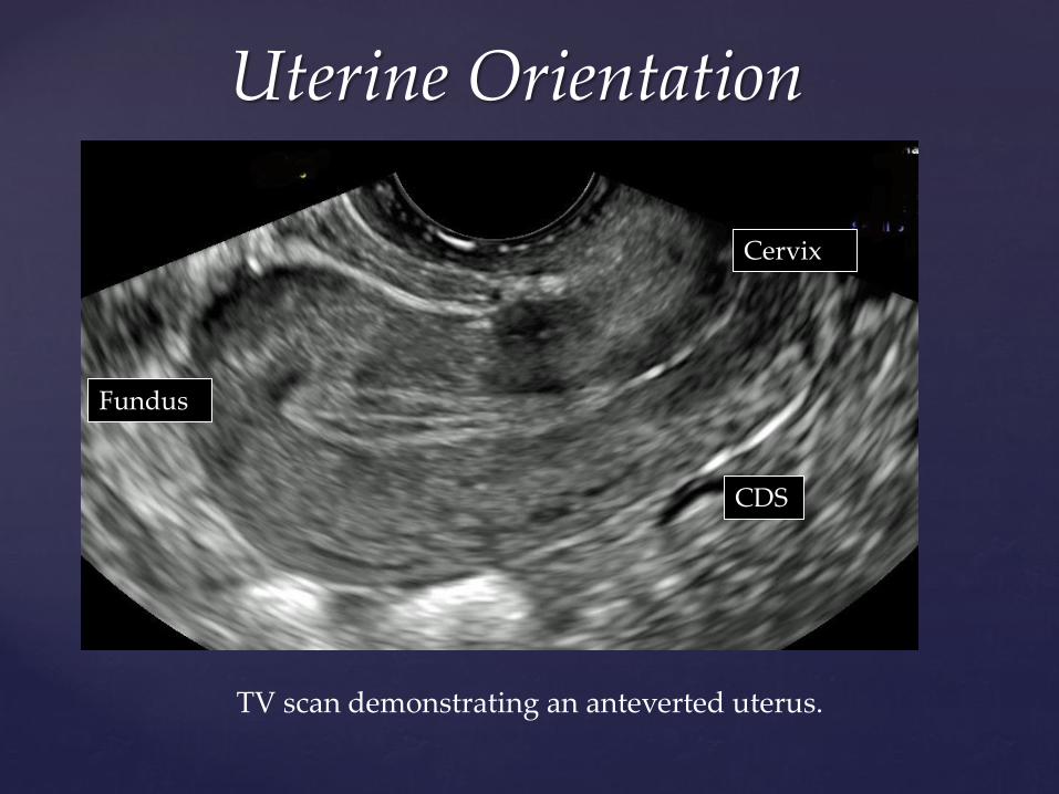
Uterine Orientation
TV scan demonstrating an anteverted uterus.
Cervix
Fundus
CDS
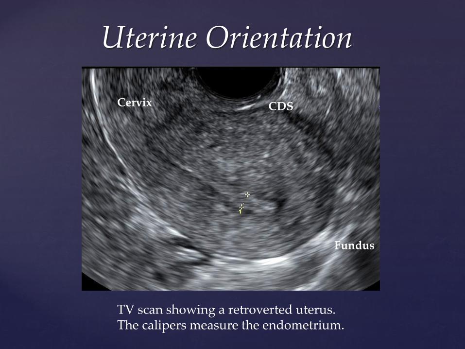
Uterine Orientation
Cervix
Fundus
CDS
TV scan showing a retroverted uterus. The calipers measure the endometrium.
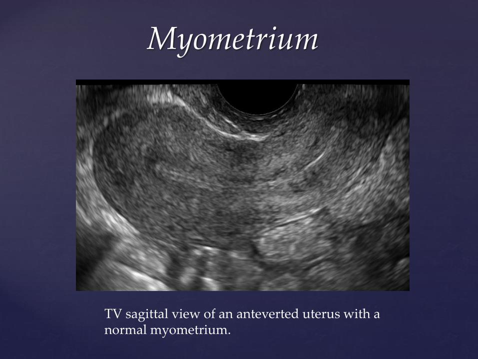
Myometrium
TV sagittal view of an anteverted uterus with a normal myometrium.
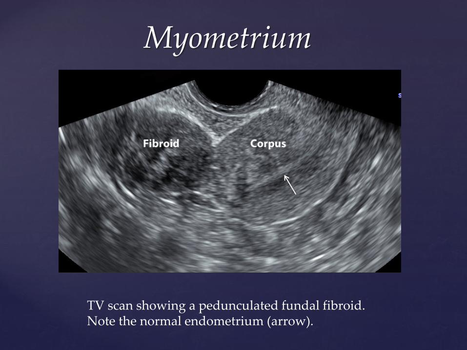
Myometrium
TV scan showing a pedunculated fundal fibroid. Note the normal endometrium (arrow).
Myometrium
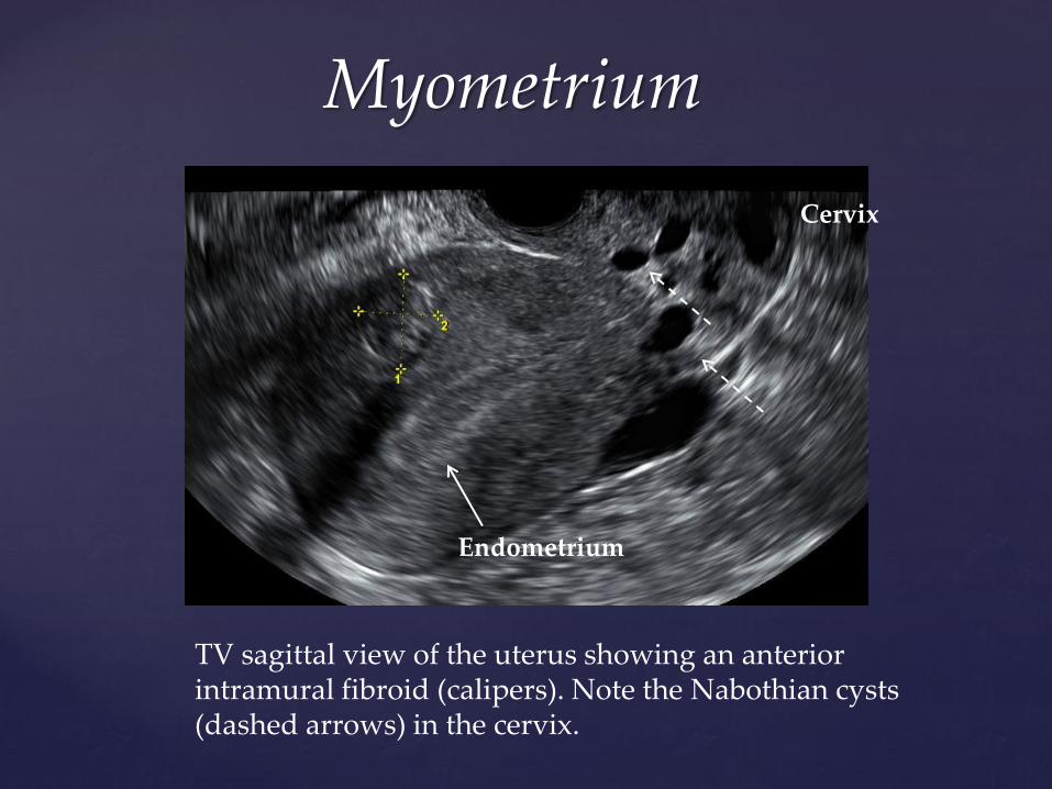
TV sagittal view of the uterus showing an anterior intramural fibroid (calipers). Note the Nabothian cysts (dashed arrows) in the cervix.
Cervix
Endometrium
Myometrium
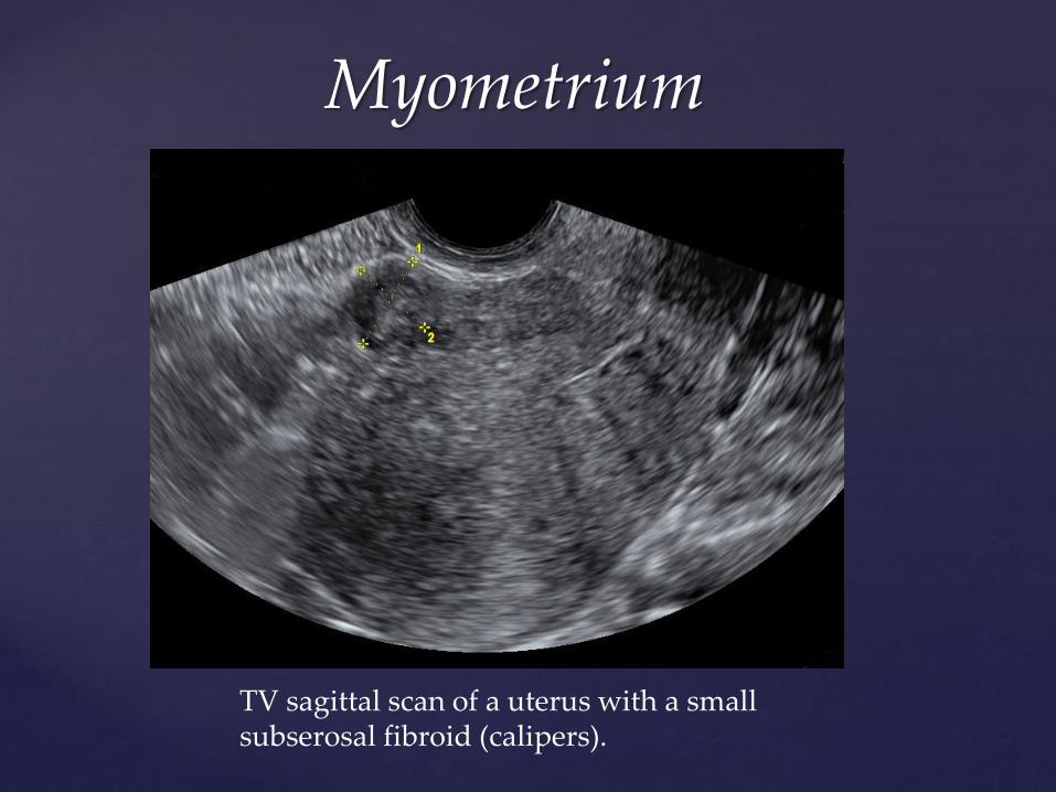
TV sagittal scan of a uterus with a small subserosal fibroid (calipers).
Myometrium
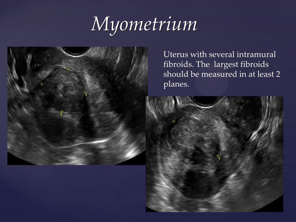
Uterus with several intramural fibroids. The largest fibroids should be measured in at least 2 planes.
Myometrium
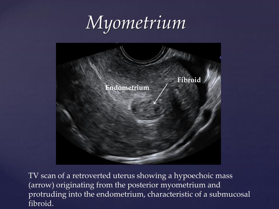
TV scan of a retroverted uterus showing a hypoechoic mass (arrow) originating from the posterior myometrium and protruding into the endometrium, characteristic of a submucosal fibroid.
Fibroid Endometrium
Myometrium
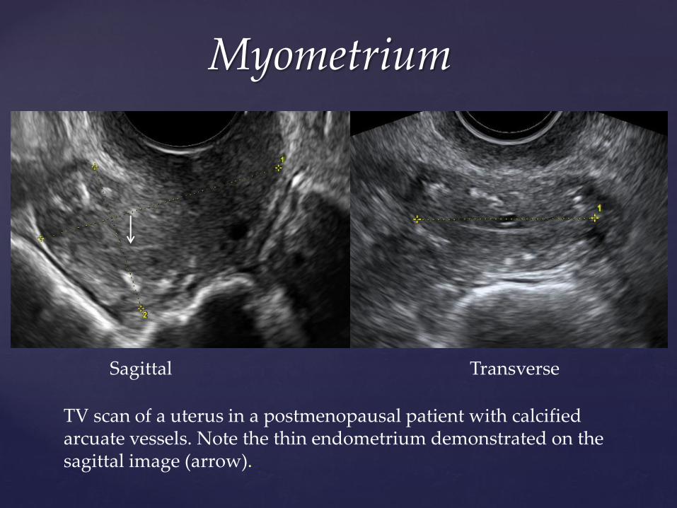
TV scan of a uterus in a postmenopausal patient with calcified arcuate vessels. Note the thin endometrium demonstrated on the sagittal image (arrow).
Sagittal Transverse
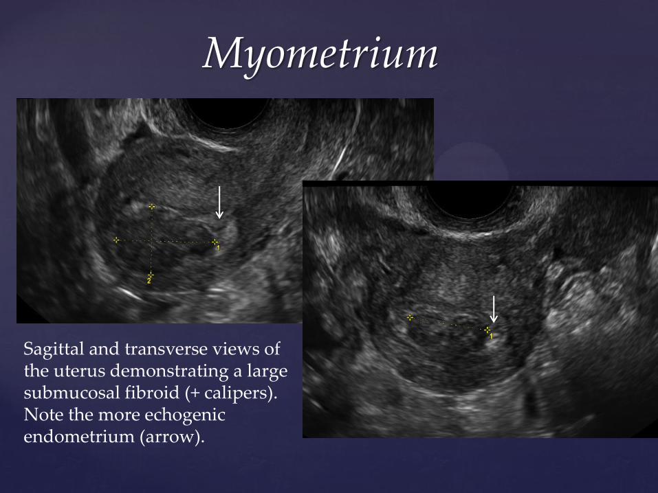
Myometrium
Sagittal and transverse views of the uterus demonstrating a large submucosal fibroid (+ calipers). Note the more echogenic endometrium (arrow).
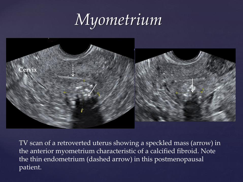
Myometrium
TV scan of a retroverted uterus showing a speckled mass (arrow) in the anterior myometrium characteristic of a calcified fibroid. Note the thin endometrium (dashed arrow) in this postmenopausal patient.
Cervix
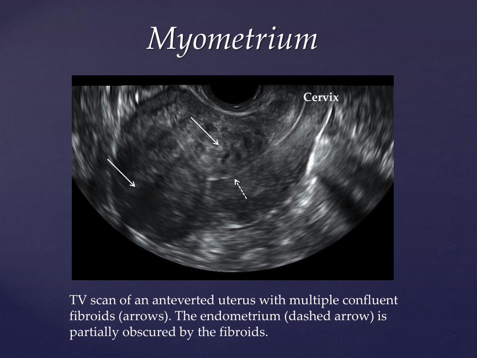
Myometrium
TV scan of an anteverted uterus with multiple confluent fibroids (arrows). The endometrium (dashed arrow) is partially obscured by the fibroids.
Cervix
Myometrium
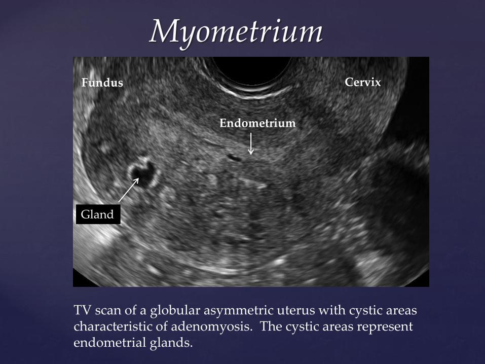
TV scan of a globular asymmetric uterus with cystic areas characteristic of adenomyosis. The cystic areas represent endometrial glands.
Cervix Fundus
Endometrium
Gland
Myometrium
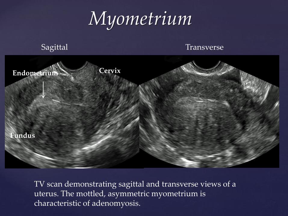
TV scan demonstrating sagittal and transverse views of a uterus. The mottled, asymmetric myometrium is characteristic of adenomyosis.
Endometrium
Sagittal Transverse
Cervix
Fundus
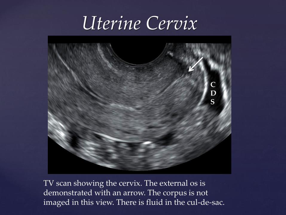
Uterine Cervix
TV scan showing the cervix. The external os is demonstrated with an arrow. The corpus is not imaged in this view. There is fluid in the cul-de-sac.
CDS
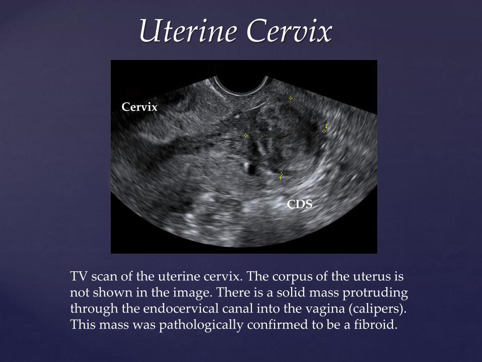
Uterine Cervix
TV scan of the uterine cervix. The corpus of the uterus is not shown in the image. There is a solid mass protruding through the endocervical canal into the vagina (calipers). This mass was pathologically confirmed to be a fibroid.
CDS CDS
Cervix
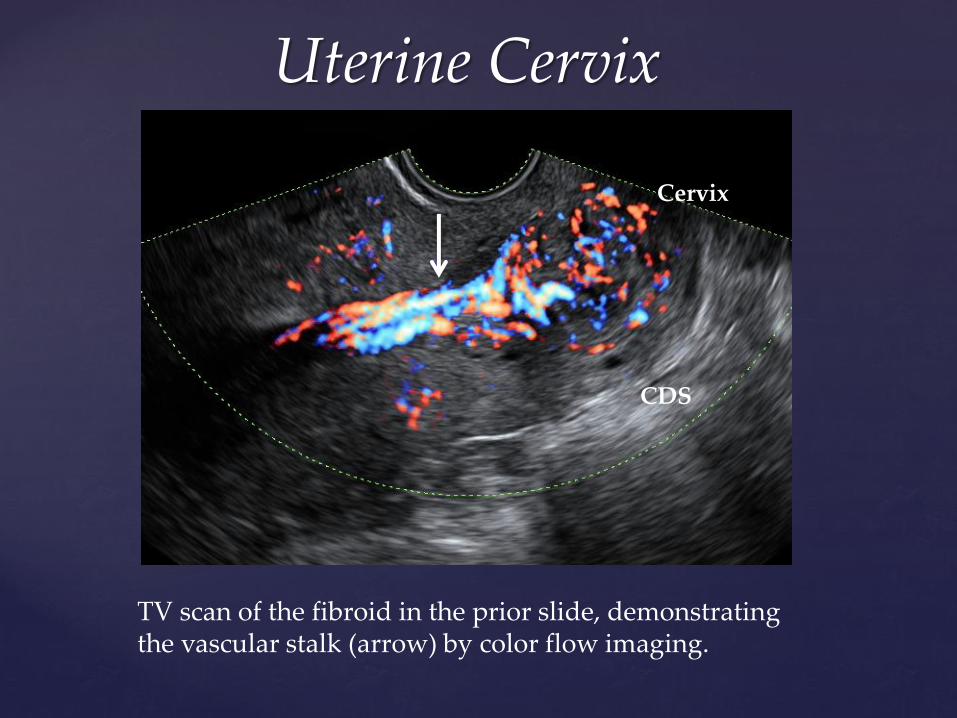
Uterine Cervix
TV scan of the fibroid in the prior slide, demonstrating the vascular stalk (arrow) by color flow imaging.
CDS
Cervix
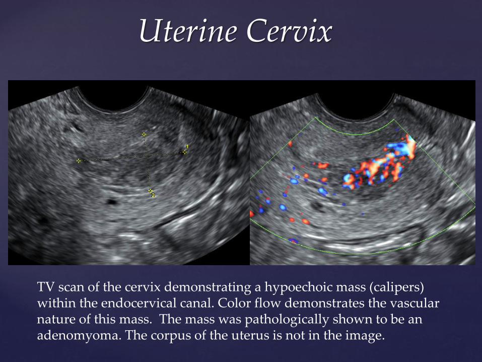
Uterine Cervix
TV scan of the cervix demonstrating a hypoechoic mass (calipers) within the endocervical canal. Color flow demonstrates the vascular nature of this mass. The mass was pathologically shown to be an adenomyoma. The corpus of the uterus is not in the image.
CDS
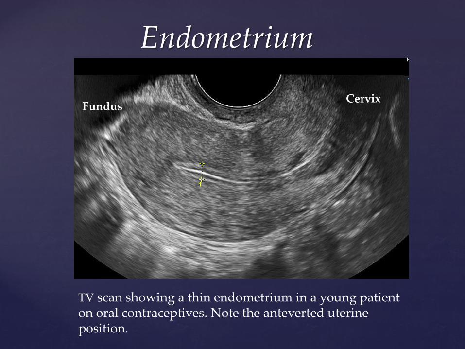
Endometrium
TV scan showing a thin endometrium in a young patient on oral contraceptives. Note the anteverted uterine position.
Cervix Fundus
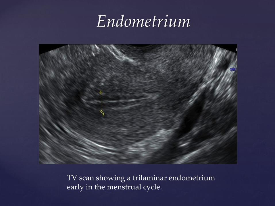
Endometrium
TV scan showing a trilaminar endometrium early in the menstrual cycle.
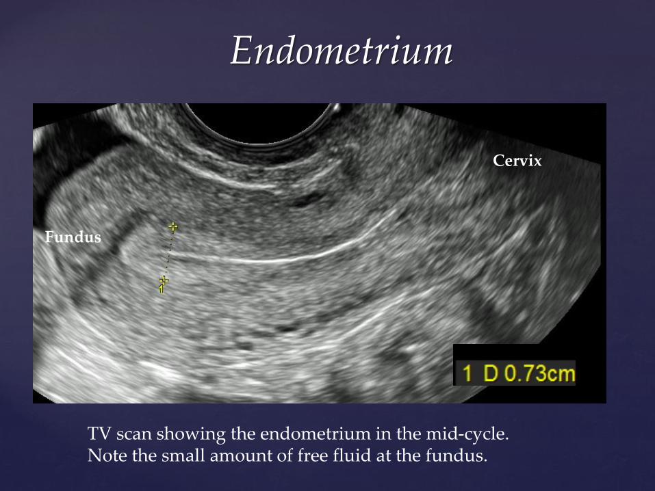
Endometrium
TV scan showing the endometrium in the mid-cycle. Note the small amount of free fluid at the fundus.
Cervix
Fundus
Endometrium
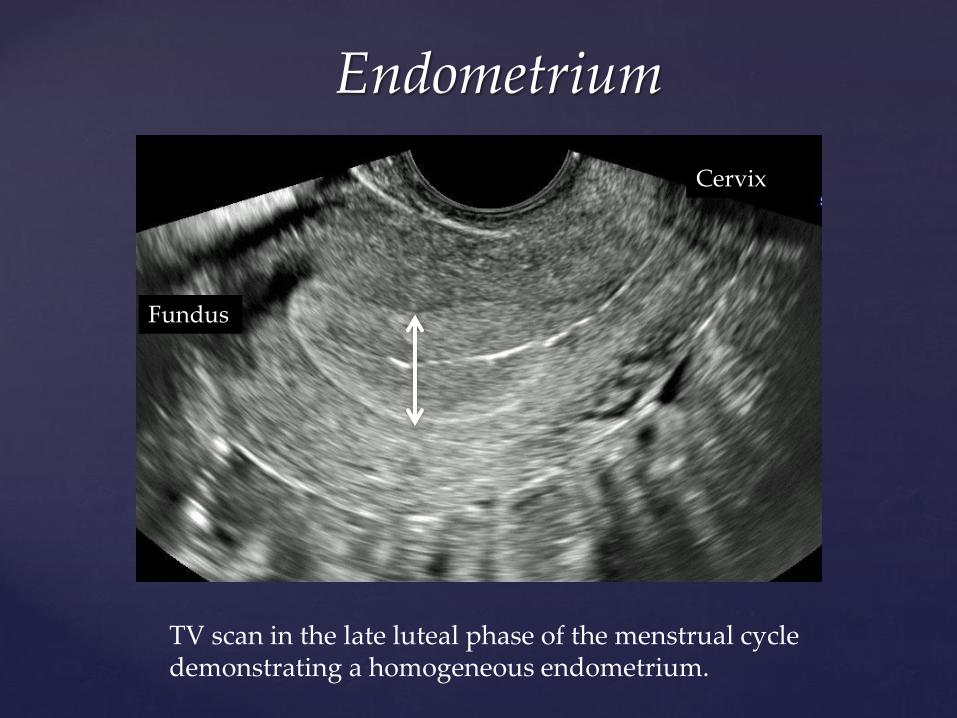
TV scan in the late luteal phase of the menstrual cycle demonstrating a homogeneous endometrium.
Cervix
Fundus
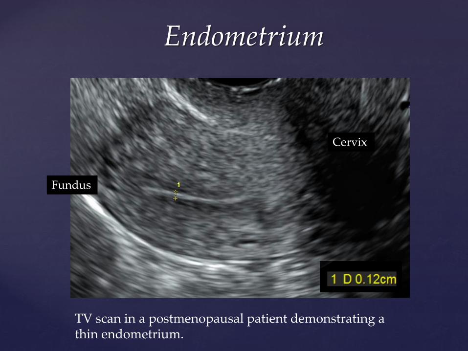
Endometrium
TV scan in a postmenopausal patient demonstrating a thin endometrium.
Fundus
Cervix
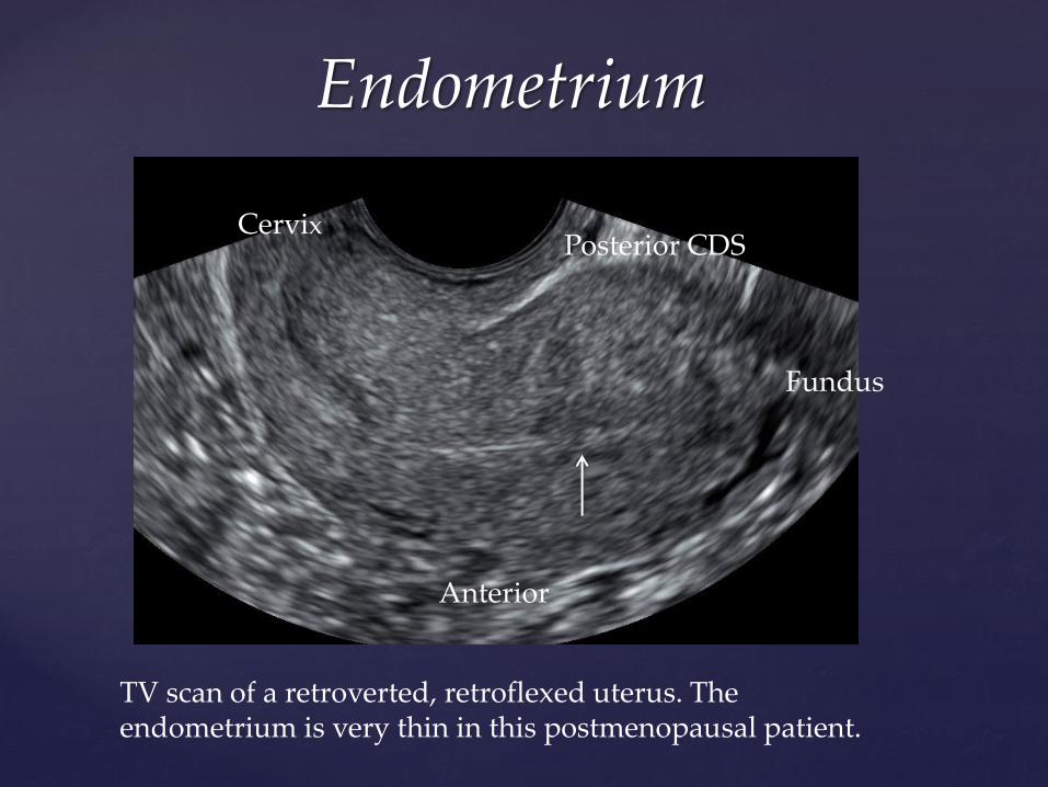
Endometrium
TV scan of a retroverted, retroflexed uterus. The endometrium is very thin in this postmenopausal patient.
Cervix
Fundus
Anterior
Posterior CDS
Endometrium
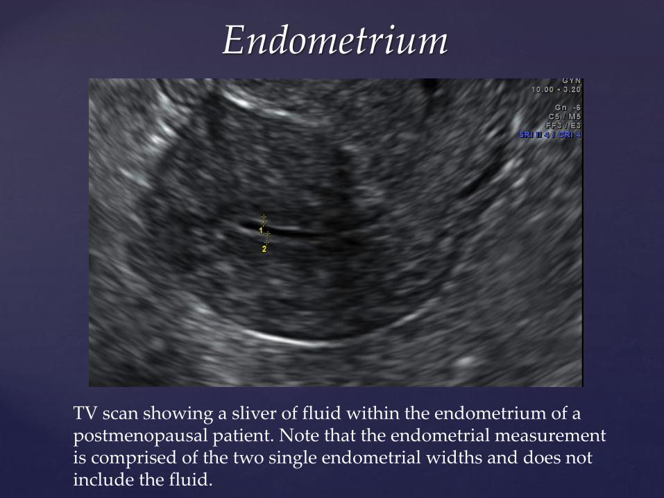
TV scan showing a sliver of fluid within the endometrium of a postmenopausal patient. Note that the endometrial measurement is comprised of the two single endometrial widths and does not include the fluid.
Endometrium
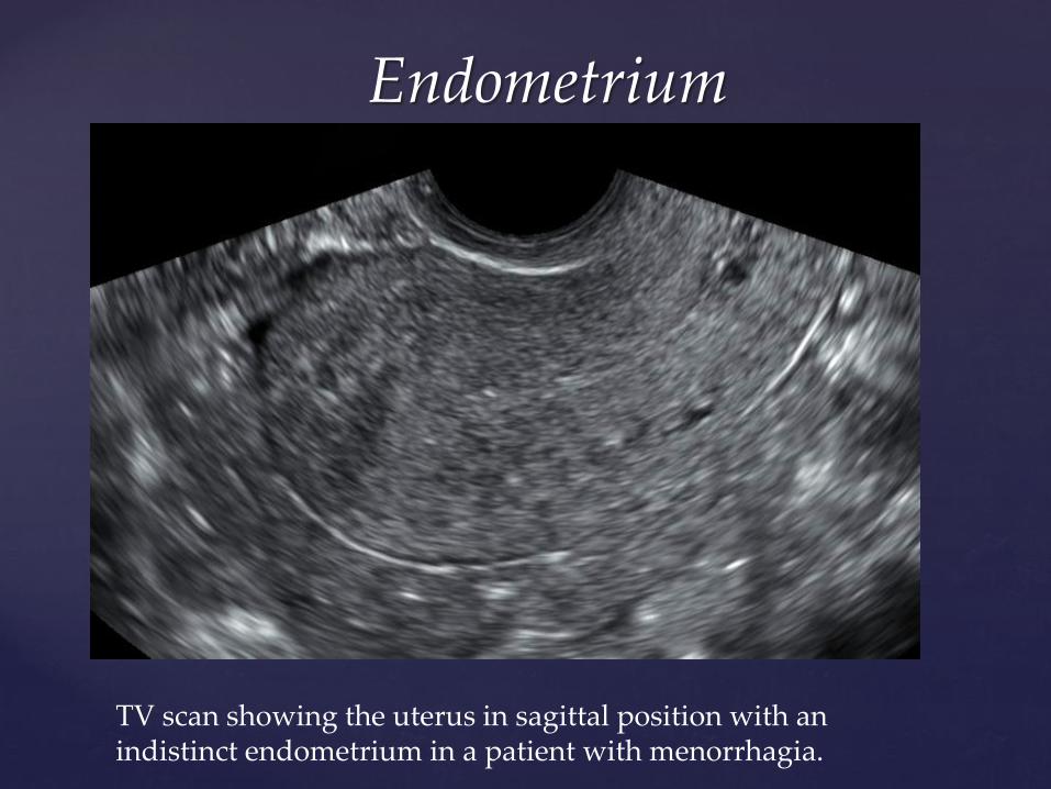
TV scan showing the uterus in sagittal position with an indistinct endometrium in a patient with menorrhagia.
Endometrium
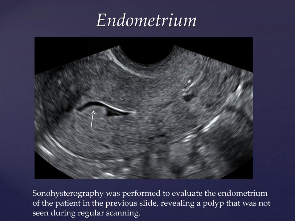
Sonohysterography was performed to evaluate the endometrium of the patient in the previous slide, revealing a polyp that was not seen during regular scanning.
Endometrium
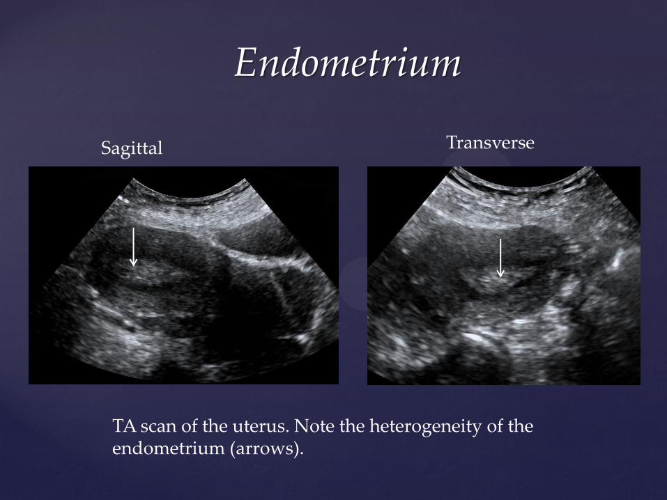
TA scan of the uterus. Note the heterogeneity of the endometrium (arrows).
Sagittal Transverse
Endometrium
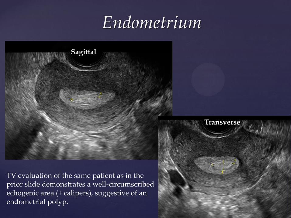
TV evaluation of the same patient as in the prior slide demonstrates a well-circumscribed echogenic area (+ calipers), suggestive of an endometrial polyp.
Sagittal
Transverse
Endometrium
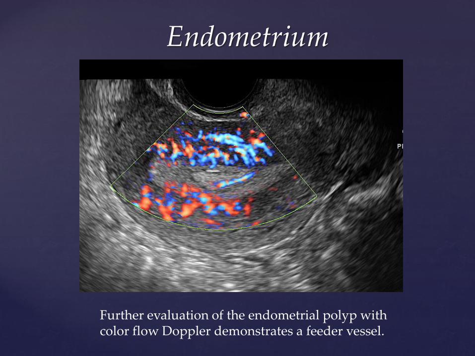
Further evaluation of the endometrial polyp with color flow Doppler demonstrates a feeder vessel.
Endometrium
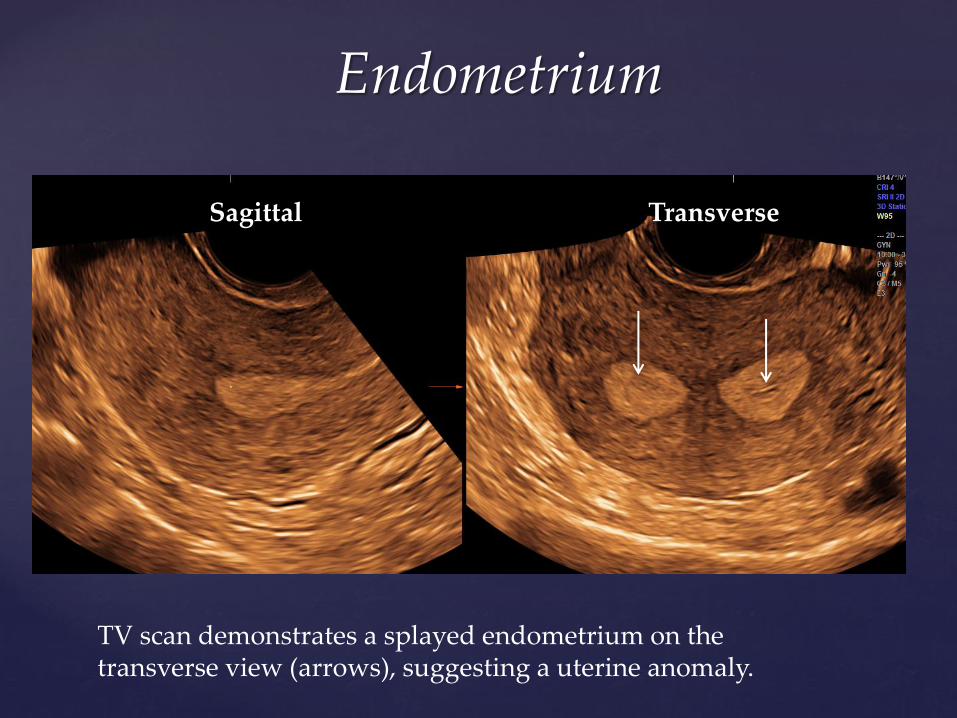
TV scan demonstrates a splayed endometrium on the transverse view (arrows), suggesting a uterine anomaly.
Sagittal Transverse
Endometrium
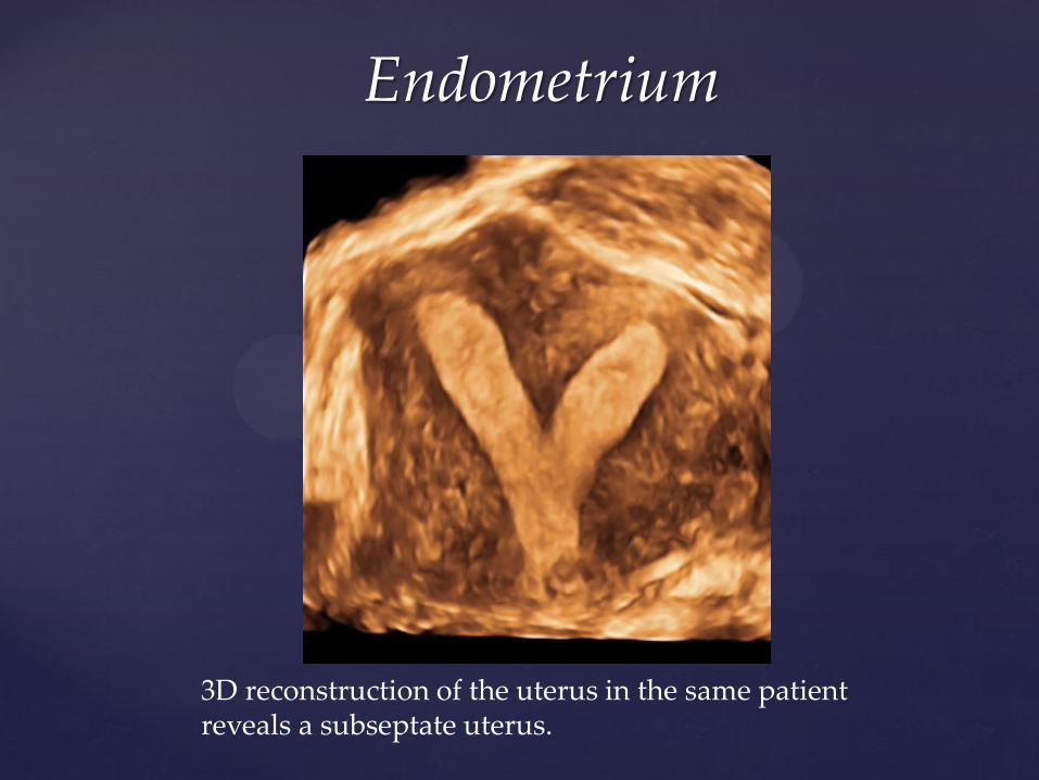
3D reconstruction of the uterus in the same patient reveals a subseptate uterus.
Endometrium
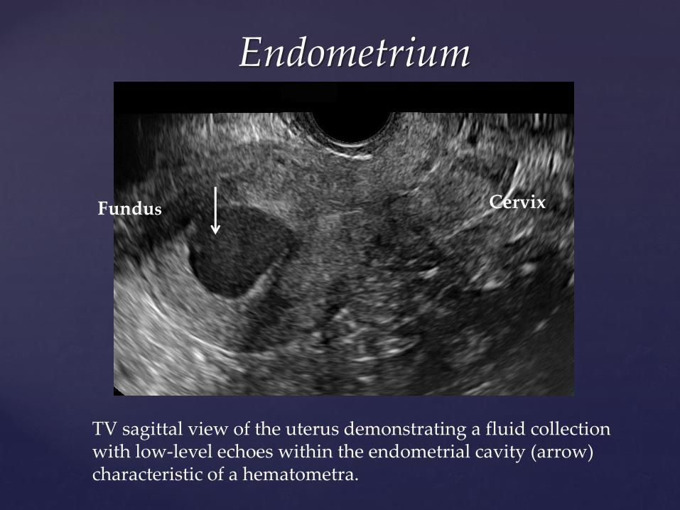
TV sagittal view of the uterus demonstrating a fluid collection with low-level echoes within the endometrial cavity (arrow) characteristic of a hematometra.
Cervix Fundus
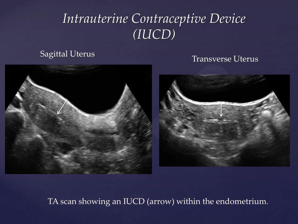
Intrauterine Contraceptive Device (IUCD)
TA scan showing an IUCD (arrow) within the endometrium.
Sagittal Uterus Transverse Uterus
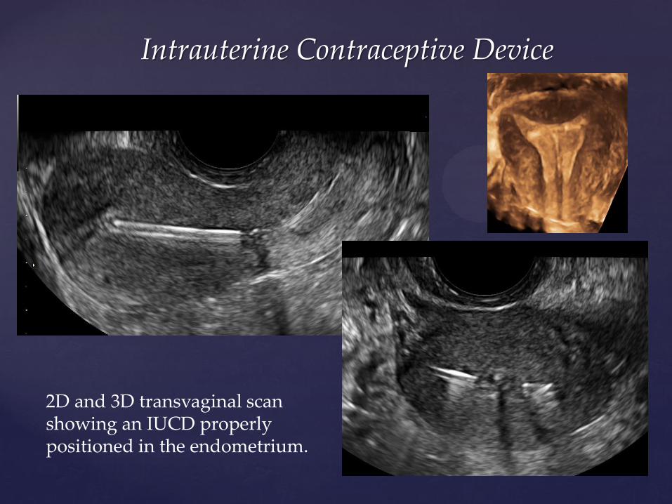
Intrauterine Contraceptive Device
2D and 3D transvaginal scan showing an IUCD properly positioned in the endometrium.
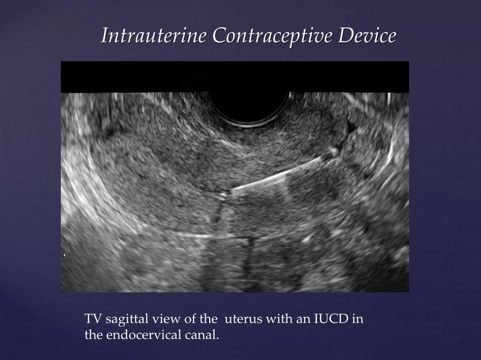
Intrauterine Contraceptive Device
TV sagittal view of the uterus with an IUCD in the endocervical canal.
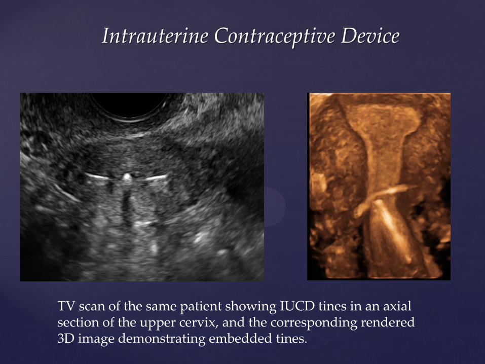
Intrauterine Contraceptive Device
TV scan of the same patient showing IUCD tines in an axial section of the upper cervix, and the corresponding rendered 3D image demonstrating embedded tines.
Ovaries
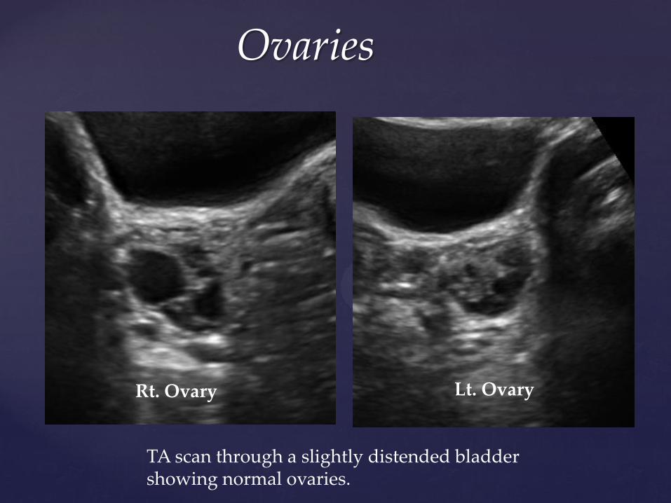
Rt. Ovary Lt. Ovary
TA scan through a slightly distended bladder showing normal ovaries.
Ovaries
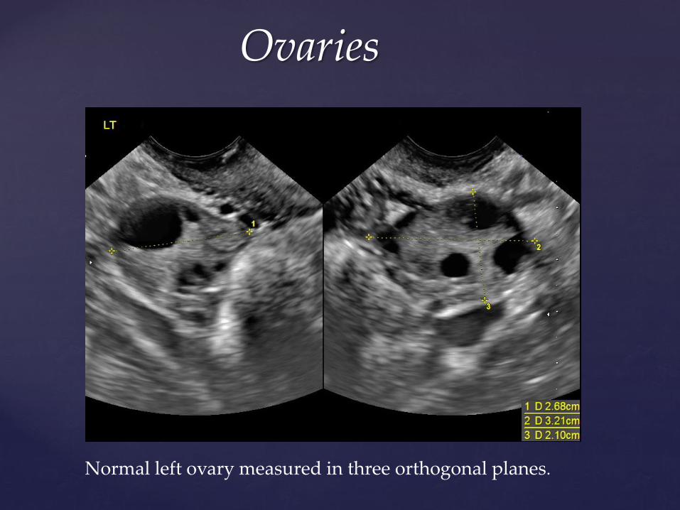
Normal left ovary measured in three orthogonal planes.
Ovaries
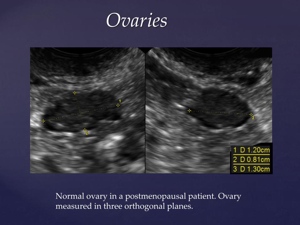
Normal ovary in a postmenopausal patient. Ovary measured in three orthogonal planes.
Ovaries
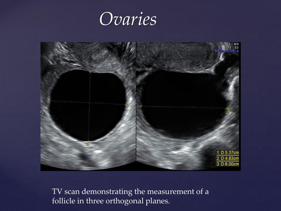
TV scan demonstrating the measurement of a follicle in three orthogonal planes.
Ovaries
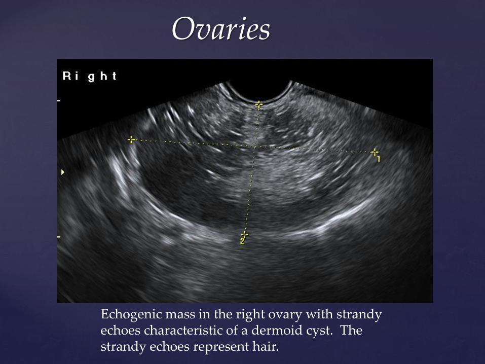
Echogenic mass in the right ovary with strandy echoes characteristic of a dermoid cyst. The strandy echoes represent hair.
Ovaries
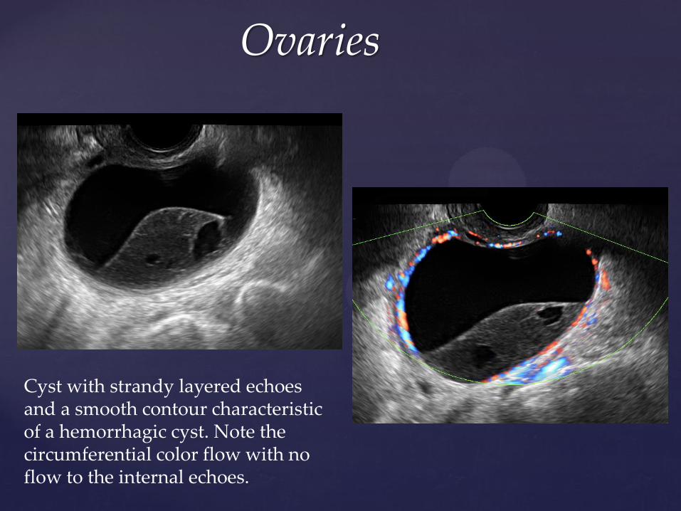
Cyst with strandy layered echoes and a smooth contour characteristic of a hemorrhagic cyst. Note the circumferential color flow with no flow to the internal echoes.
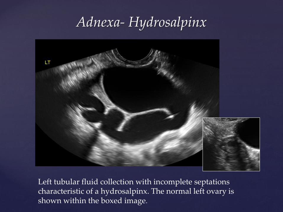
Adnexa- Hydrosalpinx
Left tubular fluid collection with incomplete septations characteristic of a hydrosalpinx. The normal left ovary is shown within the boxed image.
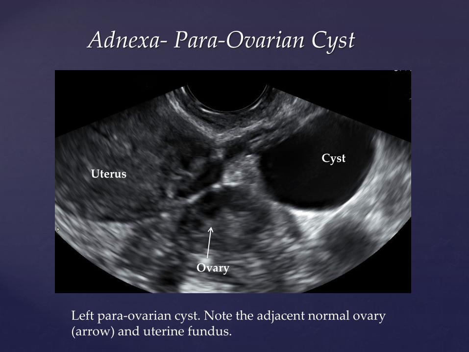
Adnexa- Para-Ovarian Cyst
Left para-ovarian cyst. Note the adjacent normal ovary (arrow) and uterine fundus.
Ovary
Uterus
Cyst
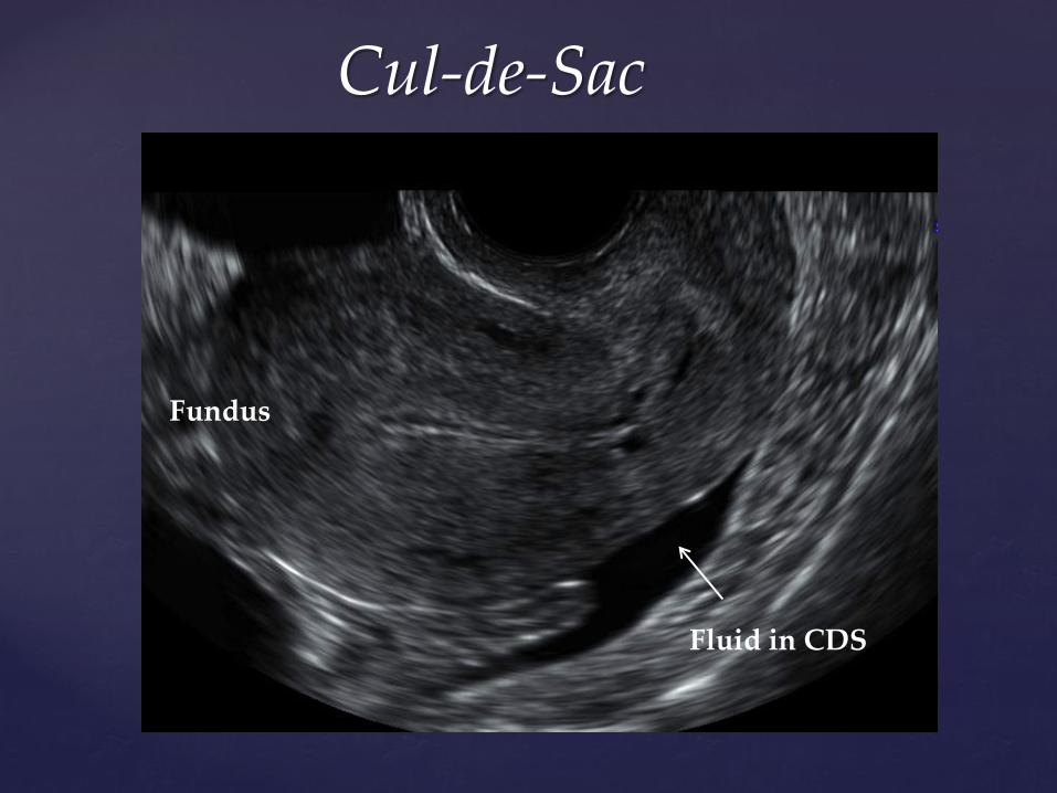
Cul-de-Sac
Fundus
Fluid in CDS
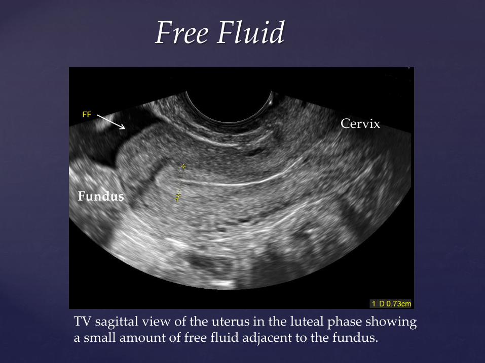
Free Fluid
Fundus
Cervix
TV sagittal view of the uterus in the luteal phase showing a small amount of free fluid adjacent to the fundus.
Free Fluid Free Fluid
Free Fluid
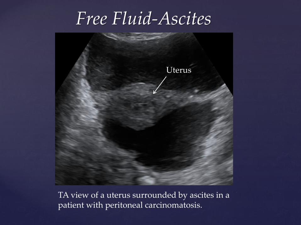
Free Fluid-Ascites
TA view of a uterus surrounded by ascites in a patient with peritoneal carcinomatosis.
Uterus
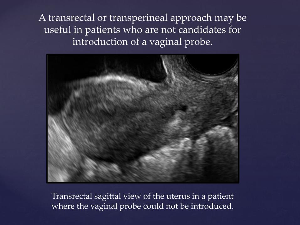
A transrectal or transperineal approach may be useful in patients who are not candidates for
introduction of a vaginal probe.
Transrectal sagittal view of the uterus in a patient where the vaginal probe could not be introduced.