Embed Size (px)
Citation preview
© 2017 Dental Press Journal of Orthodontics Dental Press J Orthod. 2017 Mar-Apr;22(2):106-17106
BBO Case Report
Agenesis of mandibular second premolar in
patient with dental bimaxillary protrusion
Carlos Alberto Estevanell Tavares1
The present study reports the treatment carried out in a patient with mandibular second premolar agenesis associated with early loss of a deciduous second molar, deep overbite, severe overjet and dentoalveolar bimaxillary protrusion, which led to lip incompetence and a convex facial profile. The main objectives of this treatment were: to eliminate the spaces in man-dibular arch, correct overbite, as well as eliminate bimaxillary protrusion and lip incompetence, thus leading to a balanced profile. The case was presented to the Brazilian Board of Orthodontics and Dentofacial Orthopedics (BBO) as part of the requirements to obtain the title of BBO diplomate.
Keywords: Tooth agenesis. Corrective Orthodontics. Tooth extraction.
How to cite this article: Tavares CAE. Agenesis of mandibular second premolar in patient with dental bimaxillary protrusion. Dental Press J Orthod. 2017 Mar-Apr;22(2):106-17. DOI: http://dx.doi.org/10.1590/2177-6709.22.2.106-117.bbo
» The author reports no commercial, proprietary or financial interest in the products or companies described in this article.
Contact address: Carlos Alberto Estevanell TavaresRua Furriel Luiz Antonio Vargas, 250, conj. 1404, Porto Alegre/RS – CEP: 90.470-130 - E-mail: [email protected]
» Patients displayed in this article previously approved the use of their facial and in-traoral photographs.
Submitted: January 18, 2017 - Revised and accepted: March 03, 2017
1 Professor, Specialization course in Orthodontics, Associação Brasileira de Odontologia-RS (Rio Grande do Sul, Brazil).
DOI: http://dx.doi.org/10.1590/2177-6709.22.2.106-117.bbo
INTRODUCTIONTooth agenesis is part of orthodontic routine, being
found among 4-5% of the overall population, without including third molars. Such an incidence increases to 9% when patients seeking orthodontic treatment are taken into consideration.1
With a prevalence of 2.5-4%, mandibular second premolars are the most commonly affected teeth, fol-lowed by third molars.2,3 In most patients, agenesis af-fects both sides of the dental arch (nearly 60% of cases), whereas it affects a single tooth only on a smaller scale.3
With advances in Orthodontics, a number of thera-peutic options became available for agenesis treatment, namely: spontaneous space closure, autotransplantation, implants, mini-implants, anchorage for space closure.4 The orthodontic mechanics of choice must consider whether agenesis affects both sides or not. Arguments in favor of contralateral tooth extraction are more com-mon than unilateral space closure, due to potential dif-ficulties involved not only in mesially displacing molars to an edentulous area, but also in achieving a satisfactory occlusion in the posterior region.5
O presente caso clínico relata o tratamento de uma paciente com agenesia de segundo pré-molar inferior associada à perda pre-coce do segundo molar decíduo, sobremordida profunda, sobressaliência exagerada e biprotrusão dentoalveolar, que causavam incompetência labial e perfil facial convexo. Os objetivos do tratamento foram eliminar os espaços presentes na arcada inferior, corrigir a sobremordida, eliminar a biprotrusão e a incompetência labial, harmonizando o perfil. Esse caso foi apresentado ao Board Brasileiro de Ortodontia e Ortopedia Facial (BBO) como parte dos requisitos para obtenção do título de Diplomado pelo BBO.
Palavras-chave: Agenesia dentária. Ortodontia corretiva. Extração dentária.

© 2017 Dental Press Journal of Orthodontics Dental Press J Orthod. 2017 Mar-Apr;22(2):106-17107
Tavares CAE BBO case report
Figure 1 - Initial facial and intraoral photographs.
CASE REPORTA 11-year and 6-month-old female patient sought
orthodontic treatment with chief complaint of dental protrusion and lip incompetence. Her family medical history presented with no major occurrences nor re-ported any trauma affecting her teeth or face.
DIAGNOSISFacial examination revealed a rather decreased nasola-
bial angle and an everted lower lip, in addition to lip in-competence. Smile line was considered adequate (Fig 1).
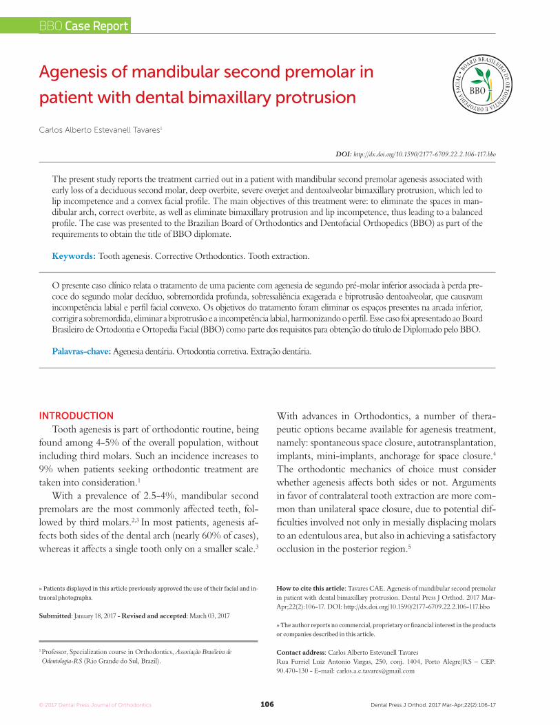
Intraoral examination revealed 6-mm overjet and 60% of overbite, in addition to the presence of 5-mm space in the mandibular arch and minimal crowd-ing in the maxillary arch. The #45 tooth was miss-ing and #47 tooth was found to be impacted, whereas
#46 tooth was 3.0 mm mesially displaced, in relation to #36 and #43 teeth, and distally displaced in relation to #33 tooth. Occlusal relationship was of Class I for molars and Class II for canines on the right side, and Class I for molars and edge-to-edge for canines on the left side (Figs 1 and 2).
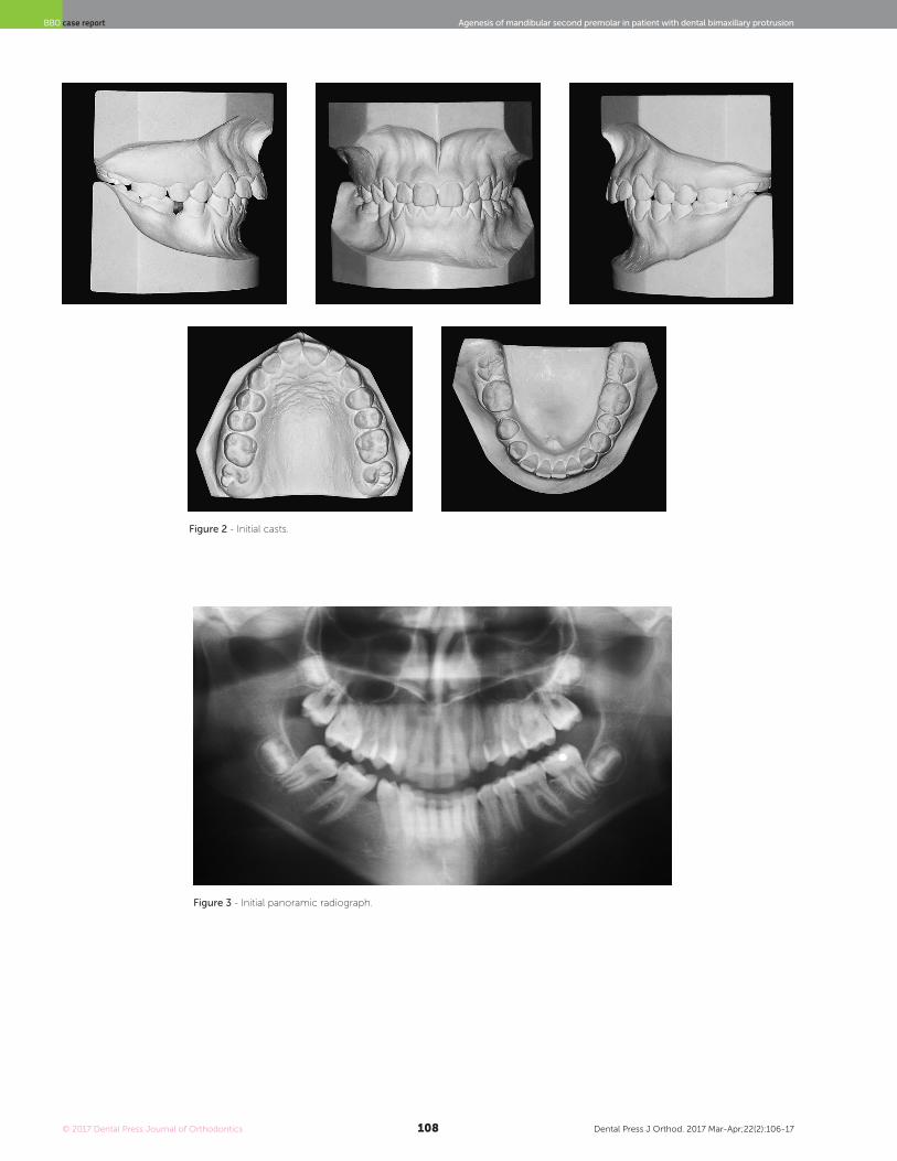
Radiographic analysis revealed #45 tooth congeni-tally missing, in addition to four developing third mo-lars and absence of caries or other pathologies. Cepha-lometric analysis suggested Class II skeletal pattern (ANB = 5o), predisposition to vertical facial growth (FMA = 29o, Y-axis = 64o) and bimaxillary protrusion, with lower and upper lips 2 and 3 mm forward in relation to the S-line, respectively (1.NA = 31o, 1.NB = 32.5o, and IMPA = 98o) (Figs 3 and 4).
© 2017 Dental Press Journal of Orthodontics Dental Press J Orthod. 2017 Mar-Apr;22(2):106-17108
Agenesis of mandibular second premolar in patient with dental bimaxillary protrusionBBO case report
Figure 2 - Initial casts.
Figure 3 - Initial panoramic radiograph.
© 2017 Dental Press Journal of Orthodontics Dental Press J Orthod. 2017 Mar-Apr;22(2):106-17109
Tavares CAE BBO case report
TREATMENT PLANTreatment objectives were as follows: to reduce
overjet and overbite, achieve a Class I canine rela-tionship, upright incisors, correct #47 tooth impac-tion, carry out space closure and achieve a symmet-rical mandibular arch. As regards facial aesthetics, treatment objectives were: to decrease lip protrusion and increase lip seal.
Treatment plan included extraction of #14, #24 and #35 teeth, orthodontic appliance placement with 0.022 x 0.028-in slot metal brackets, and a se-quence of 0.014-in, 0.016-in and 0.017 x 0.022-in NiTi archwires, followed by 0.018 x 0.025-in and 0.019 x 0.025-in SS archwires, in addition to retrac-tion of #13, #23, #34, #44, #33 and #43 teeth with elastomeric chain. Subsequently, after tooth re-traction, 0.019 x 0.025-in archwires with teardrop loops were used for incisors retraction and closure of remaining spaces. For finishing and torque con-trol, maxillary and mandibular 0.019 x 0.025-in SS archwires were manufactured to be used in coordina-tion. After fixed appliance debonding, a fixed canine-to-canine lingual bar retainer was placed in the man-
dibular arch, whereas a wrapround removable reten-tion was placed in the maxillary arch.
Alternative treatment included distalization me-chanics, such as extraoral appliance and intermax-illary elastics, in addition to keeping agenesis space unchanged for future prosthetic rehabilitation. Now-adays, the first choice for prosthetic rehabilitation of a congenitally missing premolar is implant treat-ment.6 However, implants are not stable before fa-cial growth completion. Ostler and Kokich7 assessed long-term changes in the bone crest after deciduous second molar extraction, and showed that the bone crest decreased in 25% during the first four years after extraction and 5% during the following three years, thus totaling 30% in seven years. Resorption is more often at the buccal surface of the bone crest and although the authors have showed that gingival crest width is enough to receive implants after this period, they would have to be placed more lingually than ideal.8 Another alternative would be carrying out bone graft. Since the patient presented with bimaxil-lary protrusion, extraction of #14, #24 and #35 teeth was considered as being more favorable.
Figure 4 - Initial lateral cephalogram (A) and cephalometric tracing (B).
BA
© 2017 Dental Press Journal of Orthodontics Dental Press J Orthod. 2017 Mar-Apr;22(2):106-17110
Agenesis of mandibular second premolar in patient with dental bimaxillary protrusionBBO case report
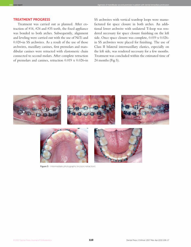
TREATMENT PROGRESS Treatment was carried out as planned. After ex-
traction of #14, #24 and #35 teeth, the fixed appliance was bonded to both arches. Subsequently, alignment and leveling were carried out with the use of NiTi and 0.020-in SS archwires. As a result of the use of those archwires, maxillary canines, first premolars and man-dibular canines were retracted with elastomeric chain connected to second molars. After complete retraction of premolars and canines, retraction 0.019 x 0.026-in
SS archwires with vertical teardrop loops were manu-factured for space closure in both arches. An addi-tional lower archwire with unilateral T-loop was ren-dered necessary for space closure finishing on the left side. Once space closure was complete, 0.019 x 0.026-in SS archwires were placed for finishing. The use of Class II bilateral intermaxillary elastics, especially on the left side, was rendered necessary for a few months. Treatment was concluded within the estimated time of 24 months (Fig 5).
Figure 5 - Intermediate photographs (incisors retraction).
© 2017 Dental Press Journal of Orthodontics Dental Press J Orthod. 2017 Mar-Apr;22(2):106-17111
Tavares CAE BBO case report
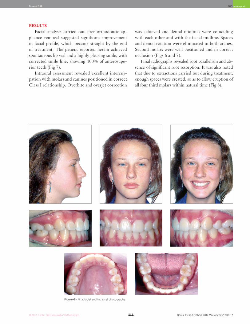
RESULTSFacial analysis carried out after orthodontic ap-
pliance removal suggested significant improvement in facial profile, which became straight by the end of treatment. The patient reported herein achieved spontaneous lip seal and a highly pleasing smile, with corrected smile line, showing 100% of anterosupe-rior teeth (Fig 7).
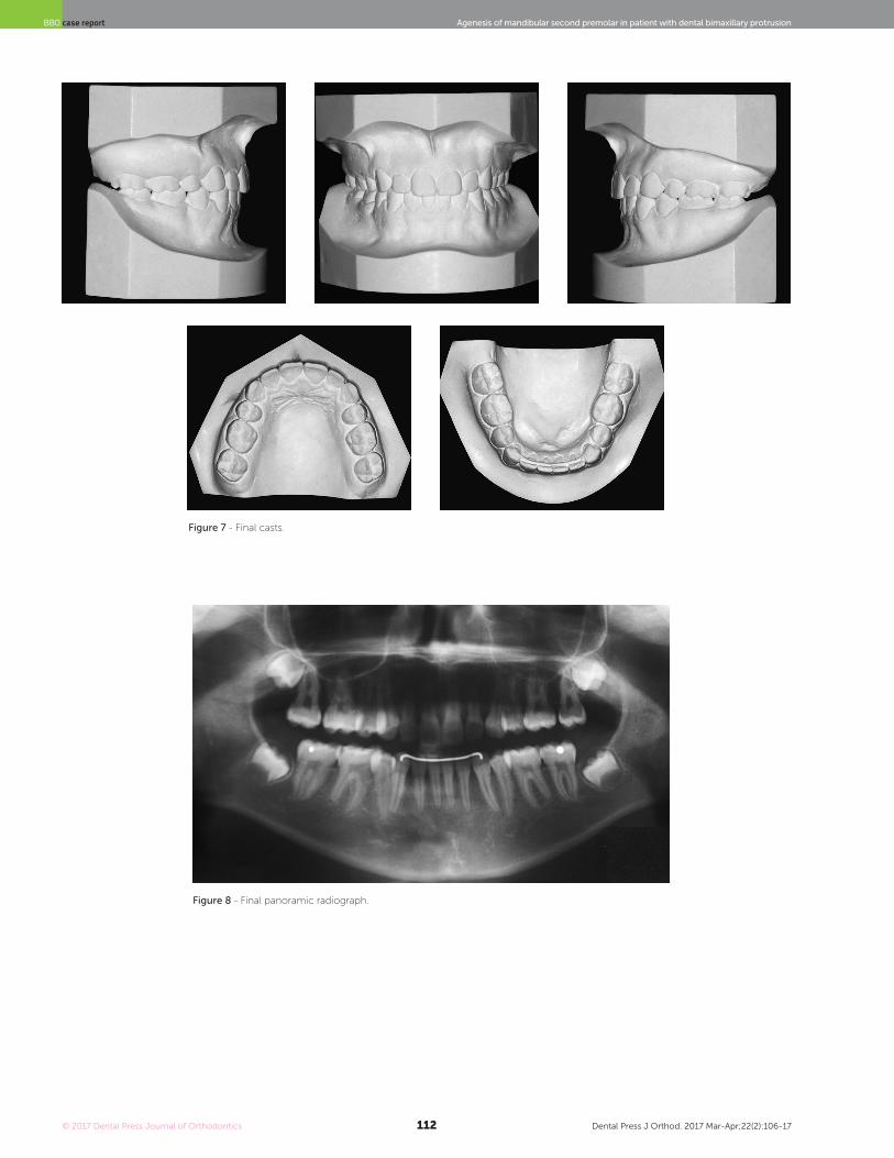
Intraoral assessment revealed excellent intercus-pation with molars and canines positioned in correct Class I relationship. Overbite and overjet correction
was achieved and dental midlines were coinciding with each other and with the facial midline. Spaces and dental rotation were eliminated in both arches. Second molars were well positioned and in correct occlusion (Figs 6 and 7).
Final radiographs revealed root parallelism and ab-sence of significant root resorption. It was also noted that due to extractions carried out during treatment, enough spaces were created, so as to allow eruption of all four third molars within natural time (Fig 8).
Figure 6 - Final facial and intraoral photographs.
© 2017 Dental Press Journal of Orthodontics Dental Press J Orthod. 2017 Mar-Apr;22(2):106-17112
Agenesis of mandibular second premolar in patient with dental bimaxillary protrusionBBO case report
Figure 7 - Final casts.
Figure 8 - Final panoramic radiograph.
© 2017 Dental Press Journal of Orthodontics Dental Press J Orthod. 2017 Mar-Apr;22(2):106-17113
Tavares CAE BBO case report
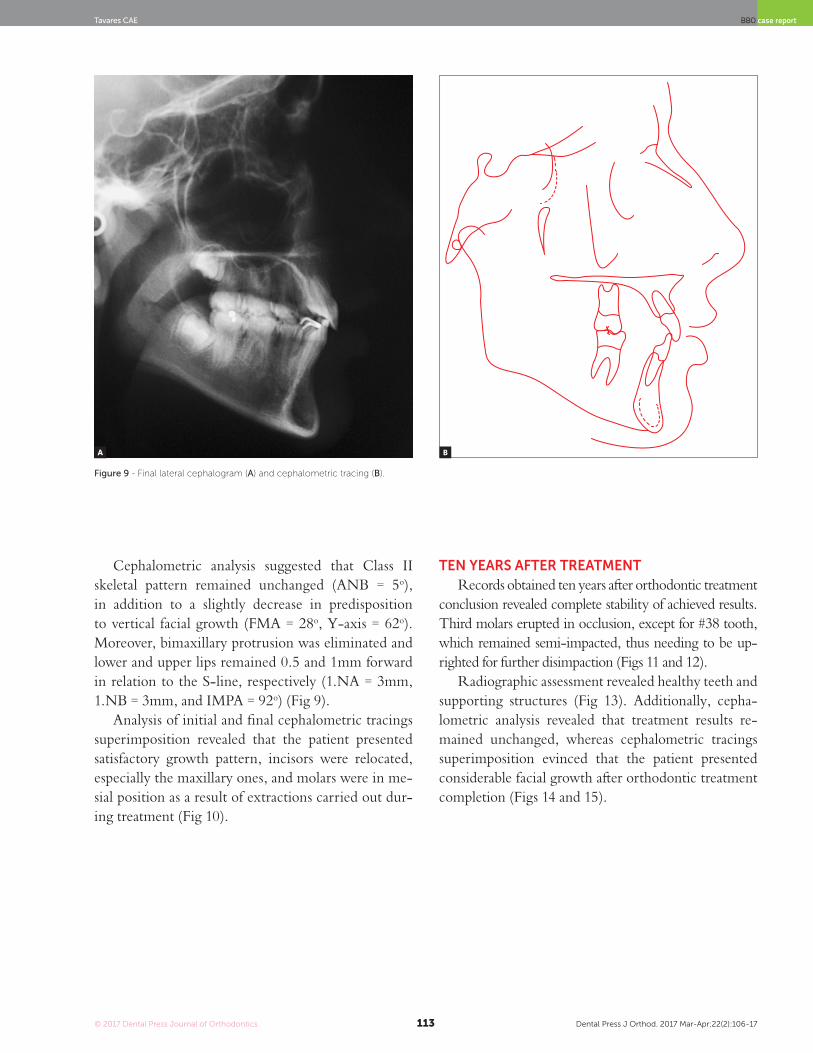
Figure 9 - Final lateral cephalogram (A) and cephalometric tracing (B).
BA
Cephalometric analysis suggested that Class II skeletal pattern remained unchanged (ANB = 5o), in addition to a slightly decrease in predisposition to vertical facial growth (FMA = 28o, Y-axis = 62o). Moreover, bimaxillary protrusion was eliminated and lower and upper lips remained 0.5 and 1mm forward in relation to the S-line, respectively (1.NA = 3mm, 1.NB = 3mm, and IMPA = 92o) (Fig 9).
Analysis of initial and final cephalometric tracings superimposition revealed that the patient presented satisfactory growth pattern, incisors were relocated, especially the maxillary ones, and molars were in me-sial position as a result of extractions carried out dur-ing treatment (Fig 10).
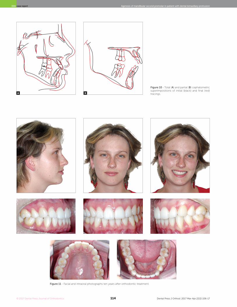
TEN YEARS AFTER TREATMENTRecords obtained ten years after orthodontic treatment
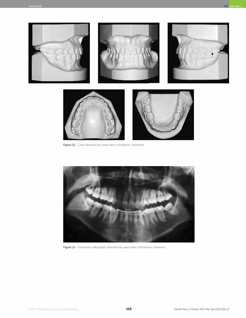
conclusion revealed complete stability of achieved results. Third molars erupted in occlusion, except for #38 tooth, which remained semi-impacted, thus needing to be up-righted for further disimpaction (Figs 11 and 12).
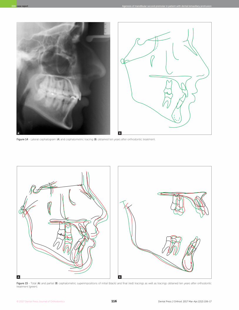
Radiographic assessment revealed healthy teeth and supporting structures (Fig 13). Additionally, cepha-lometric analysis revealed that treatment results re-mained unchanged, whereas cephalometric tracings superimposition evinced that the patient presented considerable facial growth after orthodontic treatment completion (Figs 14 and 15).
© 2017 Dental Press Journal of Orthodontics Dental Press J Orthod. 2017 Mar-Apr;22(2):106-17114
Agenesis of mandibular second premolar in patient with dental bimaxillary protrusionBBO case report
Figure 10 - Total (A) and partial (B) cephalometric superimpositions of initial (black) and final (red) tracings. BA
Figure 11 - Facial and intraoral photographs ten years after orthodontic treatment.
© 2017 Dental Press Journal of Orthodontics Dental Press J Orthod. 2017 Mar-Apr;22(2):106-17115
Tavares CAE BBO case report
Figure 12 - Casts obtained ten years after orthodontic treatment.
Figure 13 - Panoramic radiograph obtained ten years after orthodontic treatment.
© 2017 Dental Press Journal of Orthodontics Dental Press J Orthod. 2017 Mar-Apr;22(2):106-17116
Agenesis of mandibular second premolar in patient with dental bimaxillary protrusionBBO case report
Figure 14 - Lateral cephalogram (A) and cephalometric tracing (B) obtained ten years after orthodontic treatment.
BA
Figure 15 - Total (A) and partial (B) cephalometric superimpositions of initial (black) and final (red) tracings as well as tracings obtained ten years after orthodontic treatment (green).
A B
© 2017 Dental Press Journal of Orthodontics Dental Press J Orthod. 2017 Mar-Apr;22(2):106-17117
Tavares CAE BBO case report
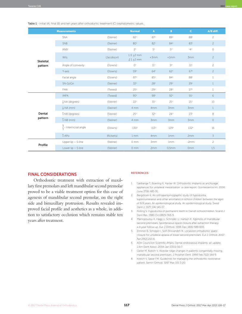
Table 1 - Initial (A), final (B) and ten years after orthodontic treatment (C) cephalometric values..
Measurements Normal A B C A/B diff.
Skeletalpattern
SNA (Steiner) 82o 87o 89o 88o 2
SNB (Steiner) 80o 82o 84o 83o 2
ANB (Steiner) 2o 5o 5o 4o 0
Wits (Jacobson)♀ 0 ±2 mm
♂ 1 ±2 mm+3mm +1mm 3mm 2
Angle of convexity (Downs) 0o 11o 9o 11o 2
Y-axis (Downs) 59o 64o 62o 67o 2
Facial angle (Downs) 87o 85o 84o 88o 1
SN-GoGn (Steiner) 32o 28o 29o 29o 1
FMA (Tweed) 25o 29o 28o 27o 1
Dental pattern
IMPA (Tweed) 90o 98o 92o 91o 6
1.NA (degrees) (Steiner) 22o 31o 21o 21o 10
1-NA (mm) (Steiner) 4 mm 4mm 3mm 3mm 1
1.NB (degrees) (Steiner) 25o 32o 24o 23o 8
1-NB (mm) (Steiner) 4 mm 3mm 3mm 3mm 0
11
- Interincisal angle (Downs) 130o 113o 129o 132o 16
1-APo (Ricketts) 1 mm 4mm 1mm 2mm 3
ProfileUpper lip — S-line (Steiner) 0 mm 3mm 1mm -2mm 2
Lower lip — S-line (Steiner) 0 mm 2mm 0,5mm 0mm 1,5
FINAL CONSIDERATIONSOrthodontic treatment with extraction of maxil-
lary first premolars and left mandibular second premolar proved to be a viable treatment option for this case of agenesis of mandibular second premolar, on the right side and bimaxillary protrusion. Results revealed im-proved facial profile and aesthetics as a whole, in addi-tion to satisfactory occlusion which remains stable ten years after treatment.
REFERENCES
1. Gedrange T, Boening K, Harzer W. Orthodontic implants as anchorage
appliances for unilateral mesialization: a case report. Quintessence Int. 2006
June;37(6):485-91.
2. Bergstrom K. An orthopantomographic study of hypodontia,
supernumeraries and other anomalies in school children between the ages
of 8-9 years. An epidemiological study. An epidemiological study. Swed
Dent J. 1977;1(4):145-57.
3. Rolling S. Hypodontia of permanent teeth in Danish schoolchildren. Scand J
Dent Res. 1980 Oct;88(5):365-9.
4. Mamopoulou A, Hägg U, Schröder U, Hansen K. Agenesis of mandibular
second premolars. Spontaneous space closure after extraction therapy:
a 4-year follow-up. Eur J Orthod. 1996 Dec;18(6):589-600.
5. Zimmer B, Schelper I, Seifi-Shirvandeh N. Localized orthodontic space
closure for unilateral aplasia of lower second premolars. Eur J Orthod. 2007
Apr;29(2):210-6.
6. ADA Councilon Scientific Affairs. Dental endosseous implants: an update.
J Am Dent Assoc. 2004 Jan;135(1):92-7.
7. Ostler M, Kokich V. Alveolar ridge changes in patients congenitally missing
mandibular second premolars. J Prosthet Dent. 1994 Feb;71(2):144-9.
8. Kokich V, Spear FM. Guidelines for managing the orthodontic-restorative
patient. Semin Orthod. 1997 Mar;3(1):3-20.