Embed Size (px)
Citation preview

Advances in ophthalmic drug delivery Article
Accepted Version
Morrison, P. W. J. and Khutoryanskiy, V. V. (2014) Advances in ophthalmic drug delivery. Therapeutic Delivery, 5 (12). pp. 12971315. ISSN 20415990 doi: https://doi.org/10.4155/tde.14.75 Available at http://centaur.reading.ac.uk/38436/
It is advisable to refer to the publisher’s version if you intend to cite from the work. See Guidance on citing .Published version at: http://www.futurescience.com/page/journal/tde/aims.jsp
To link to this article DOI: http://dx.doi.org/10.4155/tde.14.75
Publisher: Future Science
All outputs in CentAUR are protected by Intellectual Property Rights law, including copyright law. Copyright and IPR is retained by the creators or other copyright holders. Terms and conditions for use of this material are defined in the End User Agreement .
www.reading.ac.uk/centaur
CentAUR
Central Archive at the University of Reading
Reading’s research outputs online

1
Advances in Ophthalmic Drug Delivery 1
Peter W. J. Morrison, Vitaliy V. Khutoryanskiy* 2
School of Pharmacy, University of Reading, Whiteknights, PO Box 224, Reading, RG6 6AD, 3
United Kingdom. E-mail: [email protected]; Tel: +44(0)1183786119 4
Abstract: 5
Various strategies for ocular drug delivery are considered; from basic formulation techniques for 6
improving availability of drugs; viscosity enhancers and mucoadhesives aid drug retention and 7
penetration enhancers promote drug transport into the eye. The use of drug loaded contact lenses 8
and ocular inserts allows drugs to be better placed where they are needed for more direct 9
delivery. Developments in ocular implants gives a means to overcome the physical barriers that 10
traditionally prevented effective treatment. Implant technologies are under development allowing 11
long term drug delivery from a single procedure, these devices allow posterior chamber diseases 12
to be effectively treated. Future developments could bring artificial corneas to eliminate the need 13
for donor tissue and one-off implantable drug depots lasting the patient’s lifetime. 14
Key Terms 15
Bandage contact lens: Device designed to fit directly onto the front of the eye to offer 16
protection during the healing process, for example, after corneal surgery. 17
Container molecule: Molecular structures with cavities that can accommodate another molecule 18
via guest – host complexation. 19

2
Hydrotrope: Water-soluble compound that improves the aqueous solubility of hydrophobic or 20
poorly water-soluble compounds. 21
In situ gelling system: Liquid formulations that turn in to gel upon dosage form administration. 22
These phase transitions can typically be triggered by changes in temperature, pH or electrolyte 23
interaction. 24
Mucoadhesive: Defined as a compound, usually a polymer, with the ability to adhere to mucosal 25
tissue. 26
Ocular insert: A drug-loaded device designed to reside within the ocular cul-de-sac, attach to 27
the conjunctiva or directly onto the cornea. 28
Ocular implant: Dosage forms implanted directly into the ocular globe; these can be devices 29
that bring ‘quality of life benefit’ such as intraocular lenses used for crystalline lens replacement. 30
Implantable devices are also used for sustained and controlled drug delivery to the posterior 31
segment. 32
‘Smart’ DDS: Responsive drug delivery systems where a favourable change takes place in 33
response to some form of stimulus, for example, change in temperature, pH, ionic interactions or 34
stimulation from a light source. 35
Introduction 36
Ocular drug delivery is hampered by the physiological barriers presented by the eyes. These 37
include, blinking and wash out by tears, nasolacrimal drainage, non-productive losses and 38
impermeability of the cornea. [1,2] 39

3
Some of the various structures of the eye are detailed in Figure 1, highlighting the intricate 40
complexity of this organ. The conjunctiva (not shown for clarity) is the mucosa lining the inside 41
surface of the eyelids and the external surface of the front of the eye up to the limbus, the edge of 42
the cornea. 43
44
45
Figure 1. A sketch showing some of the key features of the human eye. 46
Despite the easy accessibility of the eye for administering medication, in many ways it is an 47
isolated organ with several barriers imposing challenges to drug delivery, tear mechanisms, the 48
physical barriers of its membranes, blood-aqueous and blood-retinal barriers.[3] 49

4
Topical, systemic and intraocular are the three main routes for administering ophthalmic 50
medication; each has their own advantages and disadvantages. Topical drug delivery is the most 51
accepted route accounting for ~90% aqueous ophthalmic formulations. Advantages are their 52
relative simplicity to formulate, minimal storage limitations and ease of drug instillation by most 53
patients. Disadvantages include limited drug concentration for lipophilic agents, pre-corneal 54
losses and the barrier function of the cornea.[4,5] For effective systemic delivery a relatively 55
high drug concentration needs to be circulating in the blood plasma in order to achieve a 56
therapeutically effective dose within the eye. Sustained release oral drugs can be suitable for 57
glaucoma patients, allowing for continuous and effective treatment, however this method 58
exposes the whole body to the drug often giving rise to undesired side effects.[6] Intraocular 59
drug delivery by intravitreal injection is an invasive procedure carrying a degree of risk such as 60
retinal hemorrhage or detachment, especially if the technique needs to be repeated when treating 61
chronic disorders. However, it is very effective at getting drugs to the posterior segment.[3] 62
The cornea is the main route for topically applied drugs to gain access into the eye and the 63
conjunctival/scleral route can also be efficient. [7,8] Drops are the most accepted means to apply 64
medication to this organ;[9] they are easy to apply by most patients and they are convenient. 65
However, regardless of the ease of access to the eye for topical application of medication, 66
efficient ocular drug delivery is hampered by a series of clearance mechanisms that protect the 67
ocular structures from foreign matter. Upon administration of traditional eye drops they are 68
immediately diluted in the tear film followed by very quick elimination by action of blinking, 69
wash out by tears, and nasolacrimal drainage. [10,11] After instilling eye drops, there remains a 70
very short time where any residual medication is in contact with the cornea during which time 71
there is opportunity for the drug to penetrate into the eye; however, due to poor corneal 72
5
permeability only a very small portion of active pharmaceutical ingredient will be capable of 73
crossing the cornea. Of the applied dose, only 1% or less will successfully reach the intended 74
target in most cases, the rest will be systemically absorbed via the conjunctiva or nasolacrimal 75
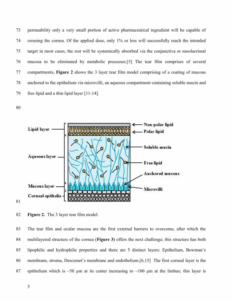
mucosa to be eliminated by metabolic processes.[5] The tear film comprises of several 76
compartments, Figure 2 shows the 3 layer tear film model comprising of a coating of mucous 77
anchored to the epithelium via microvilli, an aqueous compartment containing soluble mucin and 78
free lipid and a thin lipid layer [11-14]. 79
80
81
Figure 2. The 3 layer tear film model. 82
The tear film and ocular mucosa are the first external barriers to overcome, after which the 83
multilayered structure of the cornea (Figure 3) offers the next challenge; this structure has both 84
lipophilic and hydrophilic properties and there are 5 distinct layers: Epithelium, Bowman’s 85
membrane, stroma, Descemet’s membrane and endothelium.[6,15] The first corneal layer is the 86
epithelium which is ~50 μm at its center increasing to ~100 μm at the limbus; this layer is 87

6
lipophilic, offering ~90% resistance to hydrophilic drugs and ~10% to hydrophobic preparations. 88
Immediately underneath the epithelium is the Bowman’s membrane, a transitional acellular 89
structure ~8-14 μm in thickness. Next we find the hydrophilic stroma; this is a gel-like structure 90
with around 80 % water, consisting of collagen, mucopolysaccharides and proteins and it forms 91
the main bulk of the cornea, some 90 % of its total thickness. Next there is the Descemet’s 92
membrane, a tough membrane of around 6 μm thickness supporting the endothelium, a single 93
layer of loose, epithelia-like cells important in regulating stromal hydration, and this layer is 94
deposited by endothelial cells. The correct level of hydration is important for the cornea to 95
remain clear and transparent.[6,15,16] 96
97

7
98
Figure 3. Micrograph of a section of bovine cornea showing the multi-layered structure typical 99
of mammalian corneas. Scale bar = 100 μm. 100
The corneal epithelial barrier also has different zones; the basement layer consists of newly 101
formed cells firmly attached to the Bowman’s layer, here they are columnar in shape. As new 102
cells are formed the preceding basement cells are pushed forwards, becoming polyhedral in 103
shape, eventually as they are moved towards the corneal surface where they become polygonal 104
squamous cells. These superficial epithelial cells have Ca2+
dependent membrane adherent 105
regions; zonula occludens, zonula adherens and desmosomes forming tight junctions.[17] 106
Taken together, these tightly bound cell membrane regions and the lipophilic nature of the 107

8
epithelium make the structure an extremely efficient barrier that resists intrusion of foreign 108
material including potentially therapeutic compounds; this creates a major challenge for ocular 109
drug delivery.[6,11,18] 110
Strategies for enhancing ocular drug delivery 111
Despite traditional eye drops being convenient and simple to use, they are not very efficient and 112
only a small amount of the dose is effectively delivered to its intended target, most is lost due to 113
clearance mechanisms. There are however certain strategies that can be employed to improve the 114
bioavailability of drugs. First, solubility enhancers can be used, to improve drug concentrations 115
within the formulation; more medication in the dosage form can mean increased bioavailability. 116
This strategy could allow a smaller droplet to be applied, which would be less susceptible to loss 117
by drainage due to induced reflex tearing and blinking.[6] Second, the formulation can be 118
designed in a form that resists clearance; these dosage forms are retained for a longer period, 119
therefore they have more time to interact with ocular tissue. Next, drug penetration enhancers 120
can be incorporated into the formulation to assist their transit across the cornea.[19] Ocular 121
inserts are another area of active research and development. With this method a drug-loaded 122
device resides in the cul-de-sac under the eyelids or fits directly on the cornea like a contact lens; 123
these devices are often designed with controlled release in mind.[20,21] Drug delivery into the 124
cornea and anterior chamber is difficult enough; delivering an effective therapeutic dose to the 125
posterior segment is a major challenge, in many cases it is not possible to deliver sufficient 126
medication to the posterior structures via the topical route.[22] For diseases of the retina, such as 127
age-related macular degeneration (AMD), diabetic retinopathy, and retinitis pigmentosa and 128
related ocular neovascular disease there is often a need to resort to invasive methods for drug 129
delivery. Angiogenesis inhibitor medication via intravitreal injection is an option for getting 130

9
drugs to the posterior segment but these are often effective for the short term and need repeat 131
injections, which carries risks such as hemorrhage, endophthalmitis, ocular hypertension and 132
retinal detachment.[22-26] Ocular implants are devices that penetrate the sclera or reside within 133
the deeper ocular structures to deliver drugs for an extended period, sometimes many years, 134
minimising the need for repeat injections.[23] Implantable devices that are not designed to 135
deliver drugs are also employed to improve the ‘quality of life’ for patients with certain 136
conditions, for example, intraocular lenses. However, drugs to counter postoperative bacterial 137
infection are often included in these devices for short term protection.[27,28] These various 138
strategies will be discussed in more detail in the following sections. 139
Solubility enhancers: 140
Discovery of potentially therapeutic compounds is accelerating through developments in 141
genomics, combinational chemistry and the ability to use high throughput screening. High 142
proportions of newly screened compounds prove to be hydrophobic and are poorly water-143
soluble.[29] For efficacious performance in the physiological environment drug candidates need 144
to interact within an aqueous media, the interstitial fluids within tissues. 145
Drugs used for treatment of ocular disorders often have low aqueous solubility and eye drops are 146
only in contact with ocular tissue for a short time. Formulations that are developed to increase 147
the amount of available drug in solution could improve its bioavailability, therefore solubility 148
enhancement is an important strategy to use when developing ocular medication. Solubility 149
enhancement can be achieved by employing hydrotropic compounds. Evstigneev et al.[30] and 150
Coffman and Kildsig [31,32] reported the effectiveness of caffeine, urea and nicotinamide and its 151
derivatives as efficient hydrotropes for enhancing the solubility of riboflavin, a vitamin with poor 152

10
aqueous solubility of less than 0.1 mg mL-1
which is used as a photosensitive drug for the 153
treatment of keratoconus. Cyclodextrins are a class of cyclic supramolecular compounds that 154
have been well studied for dissolution enhancement of low solubility drugs; Loftsson and 155
Stefansson discussed the use of cyclodextrins for complexation with steroids, carbonic anhydrase 156
inhibitors, pilocarpine and cyclosporins in eye drop formulations which are well tolerated.[33] 157
Morrison et al.[34] investigated cyclodextrins for their hydrotropic properties and were able to 158
show that β-cyclodextrin achieved solubility enhancement of more than 140% for riboflavin. 159
Whilst the above mentioned studies achieved modest solubility enhancements, research by Kim 160
et al. [29] investigating the performance of two hydrotropes; N,N-diethylnicotinamide (DENA) 161
and N,N-dimethylbenzamide (DMBA) with 13 poorly water-soluble drugs and these compounds 162
were shown to have superior hydrotropic action between 1000- to 10000- fold. 163
Supramolecular structures are sub-micron sized molecules within the realm of nanotechnology 164
and many of these assemblies have solubility enhancement properties. This technology is 165
becoming an important tool within the pharmaceutical industry with substantial investment 166
within the global market. Dendrimers, microemulsions, nanoparticles, nanosuspensions and 167
liposomes belong to this class of compound and are proving to be useful structures to improve 168
bioavailability, all of which are at the forefront of research in ocular drug delivery.[1,2,35-41] 169
Micelles are aggregates of amphiphilic molecules forming self-assembled spheres in aqueous 170
media. They have a monolayer ‘shell’ of polar groups with their associated fatty acid ‘tails’ 171
forming the core. These are useful carriers of hydrophobic drugs within the core albeit with 172
limited efficiency due to a high amphiphile / drug ratio.[42] The work of Qu et al.[43] involved 173
chemical modification of chitosan by increasing their hydrophobicity and this allowed them to 174
produce 100 – 300 nm sized micellar clusters which could achieve up to an order of magnitude 175

11
enhancement in hydrophobic drug bioavailability compared to micelles produced using triblock 176
copolymers. In ocular drug formulations they were able to show an initial prednisolone 177
concentration in the aqueous humor equivalent to that found when using a 10-fold dose of 178
prednisolone suspension. 179
An approach taken by Kulkarni et al. [44] was to take the poorly soluble drug, indomethacin, and 180
using simple chemistry, convert this drug into its sodium salt. They found that this improved its 181
aqueous solubility and the drug was stable at physiological pH and compatible with excipients 182
used for ocular drug formulation. 183
Penetration enhancement: 184
Materials that modify the corneal epithelia can allow enhancement of drug permeation and this 185
can be achieved using various strategies. Benzalkonium chloride (BAC) is commonly used as a 186
preservative in ocular drug formulations, this together with other compounds; cetylpyridinium 187
chloride (CPC), ethylenediaminetetraacetic acid (EDTA), polyoxyethylene strearyl ether (PSE) 188
and polyethoxylated castor oil (PCO) are compounds with penetration enhancing properties. 189
Their mode of action is due to destabilisation of the tear film and the protection given by its 190
mucus component (for BAC), and ultrastructural alterations [17] and solubilisation of cellular 191
membranes for the other enhancers. Useful as they are for penetration enhancement they can also 192
induce irritation and damage to ocular epithelium even at low concentrations. Chung et al. [45] 193
and Burgalassi et al. [46] investigated these materials confirming their irritation and cytotoxicity 194
effects. Liu et al. [47] state that penetration enhancers should be: 195
Non-toxic; 196
Non-irritant to the eye; 197
12
Inert and compatible to other excipients within the formulation; 198
Fast acting and reversible action; 199
Effective at low concentration. 200
In their report they discuss the use of several penetration enhancers for ocular drugs; BAC, 201
EDTA, surfactants, heteroglycosides, bile salts, polycarbophil-cysteine conjugates and boric 202
acid, all of which have been used in ophthalmic formulations despite the fact that even at low 203
concentrations they can cause ocular irritation.[47] Morrison et al. [17] investigated drug 204
penetration enhancement using EDTA and two analogues EGTA and EDDS and they found that 205
this was achieved by sequestering Ca2+
and therefore loosen tight junctions which depend on the 206
availability of these ions. 207
Gelucires are glycerides composed of mono-, di- and triglycerides with mono- and diesters of 208
polyethylene glycol. They are amphiphilic with surface active properties.[48] Gelucire 44/14 has 209
a melting temperature of 44°C and a hydrophilic – lipophilic balance of 14, hence its name. It is 210
a compound known for its permeation enhancing properties and is ‘generally regarded as safe’ 211
(GRAS). Liu et al. [47] investigated Gelucire 44/14 for its permeability enhancing performance 212
in vitro and in vivo for various ophthalmic drugs and demonstrated that it enhanced transcorneal 213
permeability of drugs with a range of hydrophilicity / lipophilicity whilst remaining non-214
irritating. Loftsson and Stefansson [33] reviewed cyclodextrins for enhanced topical delivery of 215
steroids for ophthalmic formulation and the cyclodextrin-drug complexes were found to be well 216
tolerated in eye drop formulations. Cyclodextrins and their drug complexes are too large to 217
partition into the cornea and until recently it was generally thought that they kept the drug in 218
solution at the eye surface where the drug was able to diffuse into the tissue,[47,49] or by 219

13
modulation of the aqueous diffusion layer on the corneal surface.[50] Morrison et al. [34] 220
investigated the use of cyclodextrins as ocular drug delivery excipients for permeability 221
enhancement of riboflavin for the treatment of keratoconus. They have shown that cyclodextrin 222
forms complexes with riboflavin and release their drug payload by preferential take up of 223
cholesterol from corneal epithelial cell membranes. The removal of cholesterol renders the 224
epithelium permeable, allowing enhanced drug penetration. Figure 4 shows β-cyclodextrin 225
induced histological changes to the epithelium of bovine corneas (b,d,f), compared to those 226
without cyclodextrin exposure (a,c,e). β-Cyclodextrin induced loosening of the epithelium 227
appears to increase with exposure time of 15, 45 and 75 minutes (b,d,f respectively), and this 228
correlates with increased riboflavin penetration without complete destruction of this barrier. 229
230
Figure 4. Micrographs of bovine cornea cross-sections showing differences between areas that 231
were exposed to β-cyclodextrin (b,d,f) or not (a,c,e), at 15, 45 and 75 minutes. Scale bar = 100 232
μm. Adapted with permission from: Morrison et al.[34] Cyclodextrin-mediated enhancement of 233
riboflavin solubility and corneal permeability. Molecular Pharmaceutics. 10, 756-762 (2013). 234

14
Retention strategies: 235
Pre-corneal losses have a major impact on ocular drug delivery; it follows that if the drug 236
formulation stays in contact with the intended tissue for longer it is more likely to penetrate the 237
target site to afford its desired action. Adopting an approach for formulation retention is one 238
way to minimize this problem and this can be achieved by several means. Various retention 239
approaches will be discussed in the following section: 240
Viscosity enhancing polymers; 241
Natural and synthetic polymers prove useful for their viscosity enhancing properties in ocular 242
drug formulations for improving residence time. These materials absorb water to form 243
viscoelastic gels which prove to be suitable vehicles for drug delivery, and they include 244
derivatives of cellulose, poly(vinyl alcohol), poly(vinyl pyrrolidone), carbomers (weakly 245
crosslinked poly(acrylic acids)), and the natural mucopolysaccharide; hyaluronic acid, a 246
component of the vitreous humour.[51,52] Mechanisms for release of incorporated drugs are 247
determined by their chemical structure, network arrangement and swelling properties.[53] 248
Ocular drug delivery formulations incorporating viscosity enhancing polymers resist lacrimal 249
drainage when residing in the lower conjunctival cul-de-sac. However, disadvantages with this 250
approach are an initial blurring of vision due to changes in refractive index at the corneal surface, 251
and difficulty instilling a precise dose.[24,54,71] 252
In situ gelling systems; 253
‘In situ’ gelling systems undergo phase transition from liquid to gel under physiological 254
conditions and this technique has advantage over the simpler viscosity enhancing systems. Phase 255
transition can be mediated by physiological temperature, pH or electrolyte composition at the 256
cornea surface. 257

15
Thermogelling systems include polaxomers,[55,56] pluronics and tetronics,[57]. Ur-Rehman et 258
al. [58] investigated combined formulations of polaxamer 407 with chitosan as thermogelling 259
delivery systems for ocular, vaginal, orthodontal and parenteral drug administration; this process 260
allowed site specific tunable drug delivery with enhanced gel strength and mucoadhesive 261
properties. Gratieri et al. [59,60] also worked with polaxamer/chitosan gel forming systems, their 262
aim was to develop phase transition gels with improved mechanical and mucoadhesive 263
properties. They investigated poly(ethylene oxide) – poly(propylene oxide) - poly(ethylene 264
oxide) triblock polymers (PEO-PPO-PEO) with chitosan of various polymer ratios and found 265
that the polymer/chitosan ratio of 16:1 w/w offered optimum gelation temperature of 32°C, 266
good resistance to shearing forces at 35°C and good retention due to mucoadhesion. Poly(N-267
isopropylacrylamide) is a well-researched thermogelling polymer with a lower critical solution 268
temperature (LCST) of 32°C, an ideal temperature for thermosensitive applications for ocular 269
drug delivery, although the polymer precipitates above the LCST forming a stiff gel which can 270
be uncomfortable for ocular drug delivery applications.[61] It also shows reduced transparency 271
above LCST,[62] which would be undesirable for eye-drop formulations. Cao et al.[61] 272
investigated thermogelling poly(N-isopropylacrylamide)-chitosan formulation and found it to be 273
a suitable system for ocular delivery of water-soluble drugs, but it is not clear whether they have 274
solved the ‘reduced transparency’ issue with their development. Mayol et al. [56] investigated 275
thermogelling polaxamers (F127 and F68) and found that on their own their gelling properties 276
were not ideal but could be optimized by addition of the naturally occurring mucoadhesive 277
polysaccharide, hyaluronic acid. They consider that this approach can be exploited for a range of 278
sustained drug delivery scenarios and they are especially suited for ocular drug delivery. PH-279
mediated systems include Carbopol®,[63] and cellulose acetate phthalate. [64] Electrolyte 280

16
triggered gelling systems make the transition from liquid to gel by induction of crosslinking in 281
the gelling system mediated by cations present in the tear fluid, and these include gellan gum 282
(Gelrite®), carrageenan,[65-67] and sodium alginate.[68] 283
Mucoadhesives; 284
Mucoadhesion is the interaction between a compound, usually a polymer, natural or synthetic, 285
with mucosa or associated mucus.[53,69] Mucoadhesive drug delivery depends on the interplay 286
between the dosage form and mucus covered mucosal epithelial membranes, residence time 287
increases due to this interaction, allowing more time for the drug to penetrate its intended site of 288
action.[69,70] Mucosal adhesion of dosage forms can be explained using a combination of 289
theories:[71,72] 290
Electronic theory, where interaction is due to electron transfer between the dosage form 291
and mucosal surface. 292
Adsorption theory, attraction mechanisms are via electrostatic effects, hydrogen bonds 293
and Van der Waals forces. Hydrophobic effects are also implicated, more so when the 294
mucoadhesive polymers are amphiphilic. Covalent bonding can also come into effect 295
between some specific polymers and mucins. 296
Wetting theory, mostly applies to liquid mucoadhesives where there are structural 297
similarities between the polymer and mucin, these effects reduce surface tension and 298
allow the mucoadhesive polymer to spread on the mucosal surface. 299
Diffusion theory, considers the interpenetration of polymer into the mucus and diffusion 300
of soluble mucins into the mucoadhesive. 301

17
Neither of the above mentioned theories can be used to explain mucoadhesion on their own, 302
more, they each play a part to varying degrees within any given scenario.[71-74] In considering 303
a typical series of events involving a mucoadhesive – mucosa interaction; first of all the wetting 304
theory comes into play with wetting and associated swelling of the dosage form; next physical 305
interactions involving electronic and adsorption theories take place forming non-covalent bonds 306
between the system components; diffusion theory then comes into play when further non-307
covalent bonds during interpenetration of polymer-protein chains during which physical and 308
covalent (chemical) bonds form again involving electronic and adsorption theories.[71,72] 309
With traditional ocular drug delivery systems residence time is determined by tear turnover, but 310
for mucoadhesive systems this becomes governed by mucus turnover, hence drug retention and 311
bioavailability is substantially increased.[51] Mucoadhesive polymer films could potentially 312
provide a suitable platform to deliver ocular drugs, Khutoryanskaya et al.[75] investigated the 313
use of complexes and blends of poly(acrylic acid) (PAA) and methylcellulose (MC) to produce 314
polymeric films as vehicles for ocular drug delivery. PAA has excellent mucoadhesive properties 315
due to an ability to form hydrogen bonds with mucin, although it has limited application for 316
transmucosal drug delivery due to being very hydrophilic, thus quick dissolving; it also has poor 317
mechanical properties and can cause irritation to delicate mucosa. MC has favourable properties 318
that are applied in transmucosal delivery systems; it has excellent biocompatibility profiles but 319
has poor mucoadhesive properties. The researchers used a polymer blend approach with different 320
combinations of PAA / MC under a range of pH and optimized a formulation bringing together 321
the favourable properties of both polymers. In vitro studies of drug-loaded polymer films 322
determined their release profiles and they found that films enriched in MC had significantly 323
slower drug release profiles than films enriched in PAA. This could allow a tunable drug 324

18
delivery system depending on whether rapid or sustained release is required. They further 325
investigated in vivo retention of the polymer films using rabbits and found that 100% MC films 326
were retained for up to 50 minutes but successful application was hampered by poor 327
mucoadhesive properties. 100% PAA films were strongly mucoadhesive but retention was poor 328
due to quick dissolution. They concluded that polymer blends had good bioadhesive qualities and 329
showed better retention of 30-60 minutes compared to the films composed of individual 330
polymers. [75] 331
Nanoparticles; 332
Nanoparticle drug delivery systems are more generally described as submicron sized structures; 333
these systems were described by Nagarwal et al.[19] as 10 to 1000 nm particles in which drugs 334
could be loaded by attachment to the matrix or dissolved within, encapsulated or entrapped 335
within the structure giving a versatile drug delivery system. Hans and Lowman [76] discuss 336
biodegradable polymeric nanoparticles for drug delivery, they suggest that surface modified 337
biodegradable solid nanoparticles have an advantage regarding controlled release, principally for 338
targeted drug delivery for the treatment of specific organs, in particular for extended drug 339
delivery to the cornea and conjunctiva.[76] Ibrahim et al.[77] describe a mucoadhesive 340
nanoparticle system as a carrier for gatafloxacin/prednisolone biotherapy for treatment of 341
bacterial keratitis, a serious corneal condition which could lead to blindness without rapid and 342
appropriate intervention. The drug loaded nanoparticle systems they describe were produced 343
from Eudragit® RS 100 and RL 100 and were coated with the bioadhesive polymer hyaluronic 344
acid. Nanoparticles within the suspensions produced using these systems were in the range of 345
315 nm to 973 nm. For ocular drug delivery, supramolecular structures, complexes and 346
composites belong to nanoparticulate systems and these can include microemulsions, liposomes, 347
19
niosomes, dendrimers and cyclodextrins.[1,2,36-41] Kassam et al.[78] investigated the use of 348
nanosuspensions for ophthalmic delivery of three virtually insoluble glucocorticoid drugs in 349
aqueous media; hydrocortisone, prednisolone and dexamethasone. Their findings show an 350
enhancement to the rate and extent of ophthalmic drug absorption together with improved drug 351
performance compared with aqueous solutions and microcrystalline suspensions. De Campos et 352
al.[79] investigated the interaction of poly(ethylene glycol)- or chitosan- coated colloidal 353
nanocapsules with ocular mucosa; they conclude from ex vivo studies that the systems they 354
developed enhanced permeation of dye through the cornea. Evidence from confocal microscopy 355
shows their systems penetrated the epithelium of rabbit cornea via the transcellular pathway and 356
they found that PEG-coated colloids had an enhanced rate of transport across the whole 357
epithelium; whilst chitosan-coated nanocapsules were retained in the superficial epithelial layers. 358
They suggest these systems could be designed as colloidal drug carriers targeting a specific 359
purpose, that is, to attach to the cornea or penetrate into or through it. This implies these systems 360
should prove useful of treating conditions of the cornea and deeper structures within the eye. 361
Diseases of the posterior section of the eye include macular degeneration, diabetic retinopathy, 362
retinitis pigmentosa and related ocular neovascular disease. Topical delivery of drugs to the 363
posterior section of the eye is particularly challenging due not least to ocular barrier function and 364
internal clearance mechanisms within the anterior chamber. Recent developments in the field of 365
nanoparticles involve submicron-sized liposomes (ssLips) and these are proving useful for 366
topical drug delivery systems in the form of eye drops for the treatment of posterior segment 367
diseases. Studies by Hironaka et al. and Inikuchi et al. [80,81] show successful delivery of 368
coumarin-6 to the retina via non-corneal and non-systemic pathways using eye drops. The 369

20
assumption can be made that posterior section delivery is via penetration through the sclera 370
using ssLips [8,41] (emphasis highlights conclusion of the authors of this review). 371
Ocular inserts: 372
Ocular inserts are drug loaded devices placed in the upper or lower cul-de-sac and in some cases, 373
directly on the cornea; their purpose is to act as a controlled release drug reservoir. These 374
systems can be insoluble devices that need to be removed after a given period of time or they can 375
be designed to dissolve, erode or biodegrade at the ocular surface. Early forms of ocular inserts 376
have been used since the middle ages and were given the arabic term al-kohl. By the nineteenth 377
century, paper patches soaked with drug solutions were used and in the early twentieth century 378
glycerinated gelatin systems were in use.[82] It is not clear how effective these early devices 379
were, however, drug delivery by this means has developed and devices can be of soluble 380
ophthalmic drug inserts (SODI) or insoluble polymers, mucoadhesives or soluble natural 381
materials such as collagen (e.g. from porcine sclera).[4] Ideally these devices could be applied 382
and left in place with no further intervention thereafter. Ocular inserts need to be discreet and 383
comfortable to gain patient acceptance. Sustained release ophthalmic inserts are defined as 384
sterile devices which can be drug impregnated thin, single or multi-layered films, solid or 385
semisolid materials. The objective being to extend ocular contact time thus improving 386
bioavailability. Development of ocular inserts that bring reliable controlled release drug delivery 387
and patient comfort offers a considerable challenge. The main classes of devices are insoluble, 388
soluble and biodegradable inserts.[83] Ocusert® was the first relatively successful product for 389
delivery of pilocarpine for the treatment of ocular hypertension and has been commercialised 390
since 1974. Ocusert® consists of a pilocarpine-alginate reservoir sandwiched between thin 391
ethylene-vinyl acetate films, the devices are designed to deliver pilocarpine at either 20µg per 392

21
hour or 40 µg per hour. Some disadvantages of this system were unreliable control of intraocular 393
pressure, leakage, folding, difficulty inserting the devices and ejection or irritation.[82,84] 394
Ocufit SR® are sustained release rod shaped devices made from silicone elastomer, designed to 395
reside in the lower conjunctival fornix; these devices are well tolerated and expulsion is 396
significantly less than with oval or flat inserts. Minidisc ocular therapeutic system (OTS) by 397
Bausch & Lomb are drug-loaded polymer discs with similar shape as contact lenses but are 398
smaller (4-5 mm); they were designed to reside on the sclera in the upper or lower fornix and 399
deliver the antibiotics gentamicin or sulfisoxazole between 3-14 days depending on the system. 400
The company produces non-erodible hydrophobic and hydrophilic systems and erodible devices 401
based on hydroxypropyl cellulose. The inserts are comfortable and easy to use for most patients. 402
Smith & Nephew Pharmaceutical Ltd patented what they term ‘new ophthalmic delivery system’ 403
(NODS®); these devices offer precision pilocarpine delivery for glaucoma patients from 404
poly(vinyl alcohol) (PVA) film flags. These devices attach to the mucosal surface of the lower 405
conjunctival sac where it takes up fluid from the tears, swells and delivers its drug payload at a 406
pre-determined rate into the lacrimal fluid as it slowly dissolves.[82] Mydriasert® are insoluble 407
devices marketed by IOLTech for the delivery of phenylephrine and tropicamide to induce 408
sustained mydriasis during surgery or for examination of the fundus (interior ocular surface).[3] 409
Human amniotic membrane has been used for corneal transplant to treat corneal disorders and 410
ulcerative ocular conditions. Resch et al. [85,86] investigated its use as drug loaded ocular 411
devices to deliver ofloxacin in vitro and they concluded that single layer human amniotic 412
membrane had a significant reservoir capacity capable of delivering the drug for up to 7 hours in 413
vitro. They propose that drug pretreatment of amniotic membrane could be beneficial when using 414

22
this tissue for ocular transplant when treating infectious keratitis.[85,86] Table 1 lists some 415
advantages and disadvantages for using ocular inserts. [20,82,87] 416
Table 1. Advantages and disadvantages using ocular inserts.
Advantages Disadvantages
Increased residence time / bioavailability
Precision dosing with controlled release,
avoids pulsate drug delivery
Minimal systemic absorption
Administration frequency reduced
Conjunctival / scleral route to internal
target
Better shelf life and no preservatives
Combinational therapeutic approaches
Physical and psychological obstacles of
placing solid objects on the eye, foreign
body sensation
Movement around the eye could interfere
with vision
Potential accidental loss
Some devices difficult to insert or
remove
Potential burst release upon insertion
prior to controlled delivery
417
Recent developments in ocular insert drug delivery systems: 418
Colo et al. [88] investigated the effect of adding chitosan hydrochloride (CH-HCl) to 419
mucoadhesive erodible ocular inserts produced from poly(ethylene oxide) (PEO) of various 420
molecular weight for delivery of ofloxacin. They added 10, 20 and 30 % medicated CH-HCl 421
microparticles to PEO formulations made from 900 kDa or 2000 kDa. Erosion of the devices 422
was accelerated proportional to CH-HCl content. The lower molecular weight PEO proved more 423
suitable for prolonged drug release. They conclude that inclusion of CH-HCl in the devices aids 424
erosion and enhances corneal permeability of ofloxacin when compared to devices not 425
containing CH-HCl. Hornof et al. [89] developed mucoadhesive devices based on thiolated 426
poly(acrylic acid) (PAA) and these were evaluated in human in vivo studies. Their aim was to 427

23
develop mucoadhesive ocular inserts for controlled delivery of ophthalmic drugs using 428
fluorescein as a fluorescent tracer to determine release rates from the devices in humans. They 429
compared mean fluorescein concentrations in the tear film and cornea as a function of time after 430
instillation of eye drops and inserts composed of thiolated and unmodified PAA. The thiolated 431
polymer inserts formed a soft, insoluble hydrogel and were well tolerated by volunteers. Their 432
findings show this material offers a promising platform for ocular drug delivery for a prolonged 433
duration. Mishra and Gilhotra [63] designed and characterized a bioadhesive in-situ gelling 434
ocular insert for the delivery of gatifloxacin using a mixture of sodium alginate with chitosan, 435
which was plasticized with glycerin. They combined sodium alginate for its gelling properties, 436
with chitosan for its bioadhesive qualities, formulations of various proportions were prepared 437
and films were produced using the solvent casting technique as described by Pandit et al. [90] 438
Using this system they found an accumulative drug release of 95-99% during 8-12 hours and the 439
formulation consisting of 2% alginate with 1% chitosan had the most sustained release of 12 440
hours. They conclude that this system allowed production of uniform in situ gelling polymer 441
films suitable for controlled release of gatifloxacin for the treatment of bacterial keratitis and 442
conjunctivitis.[63] Natamycin is a polyene antibiotic used for the treatment of fungal blepharitis, 443
bacterial keratitis and conjunctivitis and it has the ability to reduce intraocular pressure. 444
Rajasekaran et al.[91] compared the controlled release performance of natamycin from ocular 445
inserts they designed from a variety of polymeric materials; Eudragit® L-100, S-100, RL-100, 446
hydroxypropyl methyl cellulose phthalate (HMCP) and cellulose acetate phthalate (CAP) in 447
different proportions with poly(ethylene glycol-400) (PEG-400) as a plasticizer. Their aim was 448
to develop devices for in situ sustained drug delivery and their approach was to prepare 449
polymeric films using the solvent casting method. 1 cm discs were cut from the films to be used 450
24
as inserts; these were evaluated for their physicochemical properties such as drug concentration, 451
weight, folding durability, thickness, moisture absorption and vapour transmission rate. FTIR 452
studies established that there was no chemical interaction between the drug and polymers used. 453
In vitro studies were conducted to determine their drug release kinetics; devices made from CAP, 454
HPMCP and Eudragit® S-100 released all of their drug payload within 10-15 hours, whilst 455
inserts made from increased concentrations of Eudragit® RL-100 continued release for 18-23 456
hours; best performance was shown for formulations consisting of 3% Eudragit® RL-100 and 457
1% Eudragit® L-100. They conclude that nataycin loaded ocular inserts produced from 3% 458
Eudragit® RL-100 and 1% Eudragit® L-100 plasticised with 33% PEG-400 are capable of 459
controlled drug delivery up to 23 hours. 460
Contact lenses for drug delivery 461
Contact lenses are hard or soft polymeric devices designed to fit directly onto the cornea to 462
correct refractive abnormalities; they can be produced from hydrophilic or hydrophobic 463
polymers. Hydrogel contact lenses are realistic products to act as ocular drug delivery systems; 464
they are able to imbibe a large volume of aqueous solution relative to their anhydrous form. If 465
the aqueous solution that hydrates the contact lens contains sufficient pharmaceutically active 466
material this will be able to diffuse from the polymer matrix into the tear film bathing the eye 467
and subsequently interact with the ocular tissue. However, there still remains a need to retain the 468
drug within the devices sufficiently to provide sustained release. 469
The idea of using hydrogel contact lenses as drug delivery devices was first suggested by 470
Wichterle et al. [29,92] in their 1965 patent, in which they suggest the inclusion of medication 471
upon lens hydration to offer extended drug availability during wear. Contact lens design 472
determines how they are to be used; daily, weekly and monthly disposable options are 473

25
available.[92] Early approaches to contact lens aided drug delivery relied on absorbance of drug 474
loaded solution during pre-wear soaking. Conventional contact lenses have limited drug loading 475
potential and drug delivery using this method proves unreliable, giving an initial ‘burst release’ 476
followed by rapid decline over a relatively short period.[20,93] Other methodologies include 477
molecular imprinting technology, drug loaded coating or addition of a sandwhich layer of drug-478
loaded polymer, inclusion of drug-loaded nanoparticles and cyclodextrin grafting.[28] 479
Molecular imprinting technology is a technique whereby the polymer formulation is modified to 480
give it a higher affinity towards drug molecules, thus increasing their drug loading potential and 481
prolonging delivery [94-96]. Hiratani et al. [93] took this approach in developing a system 482
employing methacrylic acid, N,N-diethylacrylamide and the drug timolol; from this system they 483
were able to achieve sustained timolol release for almost 48 hours in vitro. Alvarez-Lorenzo et 484
al. [97] applied the same strategy to produce norfloxacin-loaded poly(hydroxyethyl 485
methacrylate) contact lenses and they report that reservoir capacity was enhanced by up to 300 486
fold compared with pHEMA lenses without molecular imprinting technology. Hyatt et al.[98] 487
investigated the release profiles of gentamicin and vancomycin from fibrin coated and fibrin 488
sandwiched contact lenses in vitro; their aim was to develop a system that could offer controlled 489
and sustained drug delivery for a minimum period of 8 hours. They conclude that the fibrin 490
gel/lens systems performed better for extended delivery of gentamicin compared to normal 491
lenses soaked with the antibiotic solution, however, their performance for delivering vancomycin 492
was poor compared to soaked lenses. Lenses incorporating fibrin showed potential for treating 493
microbial keratitis. Ciolino et al.[99,100] investigated poly(lactic-co-glycolic acid) (PLGA) 494
coatings and sandwiched films with contact lenses as potential drug delivery devices. They found 495
that contact lenses incorporating PLGA film retained antifungal properties up to 3 weeks in vitro, 496

26
and their prototype ciprofloxacin eluting contact lens demonstrated controlled release at 497
therapeutically active concentrations for up to 4 weeks in vitro. Although fibrin or PLGA film 498
sandwiched and coated lenses bring sustained drug delivery benefits, the lenses are opaque; 499
therefore they require clear ‘window’ in the centre of the lens allowing the patient to see during 500
treatment.[97-100] Inclusion of drug loaded nanoparticles within the polymer matrix of contact 501
lens is an effective strategy for prolonged drug delivery. This approach can allow sustained 502
release which can be tuned towards the patient’s needs, anything between a few hours to several 503
weeks. Gulsen and Chauhan [101] conducted a pilot study to determine the effectiveness of 504
nanoparticle laden pHEMA. The nanoparticles were based on oil-in-water microemulsion 505
loaded with lidocaine, a hydrophobic drug; the droplets were then encapsulated in a silica shell 506
which stabilized the nanoparticles and these were incorporated in the hydrogel matrix during 507
polymerization. Hydrophobic lidocaine has a slight and finite solubility in water; therefore it is 508
able to slowly diffuse from the nanoparticles into the aqueous phase of the gel matrix where it 509
would then be able to further diffuse into the tear film. The nanoparticle-laden hydrogels 510
remained clear and drug release studies in vitro showed an initial burst release followed by slow 511
and steady release thereafter; by day 10 virtually all the drug had been released. They conclude 512
that the nanoparticle-loaded hydrogels could be suitable for controlled drug delivery for several 513
days at therapeutically effective concentrations. Gulsen and Chauhan [102] followed up their 514
previous investigation of nanoparticle-laden pHEMA by developing four more microemulsion 515
based formulations, type 1 and 2 were based on canola oil with Tween® 80 and Panadon SDK, 516
with or without a stabilizing silica shell, and type 3 and 4 were based on hexadecane with Brij® 517
97 with or without a stabilizing silica shell; they incorporated lidocaine as a model drug. Type 1 518
formulation was opaque due to the poor solubility of Tween® 80 in HEMA, type 2 formulation 519

27
lost some transparency but was not opaque indicating that the silica shell reduced interaction 520
between the surfactant and HEMA. Type 3 showed minimal transparency reduction but was not 521
as transparent as pHEMA, type 4 showed no observable loss of transparency due to stabilization 522
afforded by the silica shell. Release studies in vitro determined that formulations based on 523
hexadecane with Brij® 97 were suitable for sustained drug delivery at therapeutic rates for up to 524
8 days, Tween®80 based formulation was deemed unsuitable due to poor stability and particle 525
aggregation. Gulsen and Chauhan speculate that furthering this work to develop ‘smart’ 526
particulate based systems which could respond to pH or temperature change could minimise 527
burst release and decaying release rates.[101,102] The approach followed by Jung and Chauhan 528
[103] was to develop a timolol loaded nanoparticle / HEMA based contact lens system. Their 529
aim was to produce nanoparticles without using surfactant due to opacity issues when these are 530
used with HEMA. Using thermal polymerization techniques they formed drug loaded 531
nanoparticles based on crosslinking monomers; propoxylated glycerol triacrylate (PGT) and 532
ethylene glycol dimethacrylate (EGDMA) and incorporated these in pHEMA hydrogels. Their 533
product was a transparent drug loaded hydrogel with temperature dependent release rates 534
between 2-4 weeks. They conclude their system maintains drug stability under refrigerated 535
conditions and the temperature change promotes drug release upon insertion of the lenses into 536
the eyes. Figure 5 shows how nanoparticles could release entrapped drug molecules into the pre- 537
and post-tear films. 538

28
539
Figure 5. Drug diffusion from nanoparticles encapsulated within hydrogel contact lens. The 540
scale used in this image has been exaggerated for clarity. 541
Drug loading capacity of hydrogel contact lenses can be enhanced by the inclusion of ‘container 542
molecules’. Cyclodextrins, with their ‘guest-host’ properties have been investigated for this 543
purpose. Complexation between cyclodextrins and drug molecules is a dynamic process due to 544
the weak non-covalent interactions in play. The strategy followed by dos Santos et al.[104] was 545
to synthesise methacrylated β-cyclodextrin and use it to form co-polymer with HEMA and 546
EGDMA, the polymers formed had clear gel properties. Drug loading was achieved by soaking 547
the anhydrous polymers in solutions of acetazolamide or hydrocortisone for 4 days. The 548
performance of these methacrylated β-cyclodextrin hydrogels was studied in vitro and they were 549
found to offer tunable drug loading/release rates with capacity for sustained drug delivery over 550
several days. They followed up this study with development of another hydrogel formulation 551

29
using β-cyclodextrin grafted onto pHEMA-co-GMA (glycidyl methacrylate). This system was 552
able to enhance diclofenac loading by 1300% and could sustain drug release for 2 weeks in 553
lacrimal fluid. They conclude that these systems could have potential for pharmaceutical 554
applications in soft contact lenses and other medicated devices.[105] Xu et al.[106] produced 555
hydrogel films and contact lenses from HEMA, mono-methacrylated β-cyclodextrin and 556
trimethylolpropane trimethacrylate. Puerarin was incorporated as a model drug by soaking in 557
drug solution to hydrate the gel. In vitro studies determined loading and release rates were 558
dependent on β-cyclodextrin content. In vivo studies using rabbits showed the gels offered 559
sustained drug release with superior performance compared to commercial puerarin eyedrops. 560
The devices had excellent mechanical properties and the researchers propose the material is 561
suitable for drug delivery from re-usable daily wear contact lenses. 562
Ocular implants: 563
Treating the posterior segment 564
Historically, the posterior segment has been exceptionally difficult to treat due to the many 565
barriers that obstruct ingress of foreign matter into the eye. The development of ocular implants 566
have allowed these external barriers to be overcome. Modern devices allow long term treatments 567
for otherwise impossible to treat conditions, many devices provide medication for years from a 568
single procedure. [107,112] 569
Drug eluting intraocular lenses 570
Intraocular lens (IOL) surgery is a well-established and safe procedure routinely performed 571
worldwide; however as with any surgical technique there is always risk from infection or other 572

30
complications, for example, postoperative inflammation, posterior capsule opacification (PCO) 573
and secondary cataracts caused by epithelial cell adhesion and proliferation in the posterior lens 574
capsule. Introduction of preventative medication during surgery is subject to decay or 575
elimination before it can be effective. Much research is currently carried out for development of 576
drug eluting IOL’s to minimise postoperative problems, and also to address concurrent 577
pathologies. IOL / drug combinations can be achieved by pre-insertion soaking in concentrated 578
drug solution (only useful for drugs with a high affinity for the polymer), coating with layers of 579
drug/polymer, chemical grafting of drugs, drug impregnation using super critical fluids and 580
attaching inserts onto the haptics (the ‘arms’ of the IOL).[28] A study by Kleinmann et al.[113] 581
determined that commercial hydrophilic acrylic lenses (C-flex, Rayner intraocular lenses) [114] 582
have affinity for fourth generation fluoroquinolones and were able to release this drug above the 583
minimum inhibitory concentration in rabbits for at least 12 hours. They conclude C-flex/drug 584
combination is safe and effective for delivery of these antibiotics. Davis et al.[115] investigated 585
concentrations of 4 antibiotics (moxifloxacin, gatifloxacin, linezolid and ceruroxime) in aqueous 586
and vitreous humour samples from rabbit eyes. Drug released from implanted hydrophilic IOL’s 587
was analysed using HPLC to determine drug concentration in the ocular fluid samples. The 588
IOL’s used were STAAR Nanoflextm
Colamer®, 40% water content material comprised of a 589
collagen, pHEMA blend,[116] pre-soaked in antibiotic solution. Ocular fluid samples were 590
taken for analysis at intervals up to 24 hours. It was established that the antibiotics studied were 591
above the minimum inhibitory concentration in the aqueous humour for at least 6 hours, notably, 592
gatifloxacin concentrations remained above this level at 24 hours after implantation.[116] 593
Layer-by-layer deposition is a technique used for coating opposing charge polymers to rigid 594
hydrophobic IOL’s, a drug can be incorporated during this process. Coating pHEMA based 595

31
hydrophilic IOL’s by immersion in octadecyl isocyanate can be an effective method to give 596
controlled release from norfloxacin containing IOL’s. Grafting drug molecules onto the IOL 597
surface can provide a permanently active surface to prevent cell adhesion, or allow release of 598
drugs by some external trigger, for example light irradiation. High drug concentrations within a 599
polymeric matrix can be achieved using supercritical CO2 as a means to force drugs into the 600
polymer without the need for organic solvent.[28] Duarte et al.[117] employed supercritical CO2 601
technology to impregnate p(MMA-EHA-EGDMA), a suitable polymer for IOL manufacture, 602
with flurbiprofen, an anti-inflammatory drug used for intraocular delivery. Their experiments 603
found the process allowed higher drug impregnation and release studies showed the system to be 604
effective for up to 3 months. The approach employed by Garty et al. [27] was to produce 605
norfloxacin loaded pHEMA cylinders in 1.0 mm diameter microglass tubes with 0.09 mm 606
stainless steel wire through the centre during room temperature polymerization. When fully 607
polymerized the hydrogel was ejected from the tube and the wire removed leaving a tubular 608
hydrogel structure, this was washed with sterilized water to remove unreacted components. The 609
gel was cut into 1.0 mm lengths and lyophilized. Next they added a hydrophobic coating using 610
octadecyl isocyanate to control drug release. The devices were used as sleeves placed over IOL 611
haptics and this assembly was used in lens replacement procedures in the rabbit model. Results 612
from in vivo studies showed the devices offered sustained drug delivery above the minimum 613
inhibitory concentration for over 4 weeks. They conclude that these controlled release devices 614
are effective at sustained delivery of therapeutic levels of drugs within the anterior chamber post 615
operatively. Incorporation of drugs with IOL’s has predominantly aimed at postoperative 616
delivery of antibiotics and anti-inflammatory medication. 617

32
Drug delivery by intravitreal injection 618
There are many debilitating and sight threatening conditions resulting from posterior segment 619
diseases and in most cases the only way these can be treated is by invasive procedures, for 620
example ‘intravitreal injection’. In the main this still remains so, however, developments have 621
brought a diverse range of effective implantable drug delivery systems targeting posterior 622
segment disease and the various options will now be considered. [22] The most common means 623
to place drugs in the posterior chamber employs injection into the vitreous humour; this provides 624
a high concentration of drug where it is needed and minimises systemic complications. Xu et al. 625
investigated the diffusion of polystyrene nanoparticles of various size and surface chemistries in 626
fresh bovine vitreous and they were able to achieve tuneable drug transport within the posterior 627
chamber depending the designed properties of the nanoparticle [118]. However, many conditions 628
require repeated treatment and this can cause intraocular problems, for example, cataract, retinal 629
detachment, haemorrhage, endophthalmitis and ocular hypertension. 630
Intraocular implants 631
In an attempt to overcome the problem of frequent injections biodegradable and non-632
biodegradable drug depot devices which can offer long term drug release into the posterior 633
chamber have been developed and further research in this area is ongoing. Solutions, liposomes, 634
micelles, nanoparticles and vectosomes are suitable for intravitreal injection although these 635
dosage forms only give short term drug availability, generally days to several weeks.[23,119] 636
Biodegradable and non-biodegradable drug depot devices have been developed and further 637
research in this area is ongoing. Implantable devices for long term drug delivery are on the 638
market or currently undergoing clinical trial. Vitrasert® is a drug depot device for sustained 639
delivery of ganciclovir via a rate limiting poly(vinyl acetate)/ethylene vinyl acetate (PVW/EVA) 640
33
membrane for up to 8 months.[22,119,120] Retisert® intraocular inserts were approved by the 641
FDA in 2005. They are inserts for delivery of the corticosteroid, fluocinolone acetonide for 642
treatment of posterior uveitis, a serious sight threatening condition. The devices are designed for 643
long term drug release up to 30 months.[121] Vitrisert® and Retisert® inserts are non-644
degradable and require surgical implantation and removal.[22] Medidur® are implantable 645
devices for delivering fluocinolone acetonide for up to 36 months. This device consists of a 646
narrow cylindrical polyimide tube loaded with the drug and PVA-based end caps provide rate 647
limiting drug delivery. The 3.5 mm long device is inserted through a 25-g needle carried out 648
under local anaesthesia and creates a self-healing wound eliminating the need for surgery.[122] 649
Implants employing biodegradable polymers are promising systems for intraocular drug delivery. 650
Sivaprasad et al. [123] report the use of the Posurdex® biodegradable polymer device for 651
treatment of macula oedema using dexamethasone. This drug has a half-life of less than 24 hours 652
therefore it provides only limiting management of this condition by injecting the drug. However, 653
dexamethasone containing Posurdex® devices were shown to deliver the drug at a constant rate 654
for up to 4 months, these devices have been re-named Ozurdex® and are marketed by Allergan 655
Inc. [124] In vivo studies using monkeys showed the system was effective at reducing retinal 656
vasculopathy and neuropathy.[125] Surodex® is a poly(lactic-glycolic acid) device to be 657
inserted in the anterior or posterior chamber at the time of cataract surgery to deliver 658
dexamethasone for up to 10 days. Tan et al. [126] conducted a randomized clinical trial to 659
evaluate the effectiveness of the Surodex® insert as a safe and effective treatment of intraocular 660
inflammation in post-cataract surgery. Their study employed flare meter readings to determine 661
inflammation and this showed that measured values were lower in all readings from the 662
Surodex® group compared to those treated post operatively with dexamethasone eye drops, they 663

34
conclude that implantation of a single Surodex® device at the time of cataract surgery reduces 664
post-surgery inflammation [126,127]. 665
Future perspectives: 666
In this review the various strategies for enhancing bioavailability of ophthalmic drugs have been 667
considered; how drug bioavailability can be improved using solubility, retention and 668
permeability enhancers has been explored. Drug loaded contact lenses allow localised delivery 669
directly to the cornea, where the lenses offer controlled release whilst isolating the post corneal 670
tear film from lachrymal clearance. Nanoparticle technology is allowing drug delivery to the 671
posterior chamber via topically applied formulations. Future research is likely to bring 672
discoveries of materials with superior performance compared with those in current use. 673
The use of ocular inserts for extended and intimate contact between the dose form and ocular 674
tissue proves to be a beneficial strategy and the use of ocular implants allows all external barriers 675
to be overcome, giving direct access to internal tissues whilst minimising side effects. Many of 676
these approaches have been developed in recent decades and continue to be improved upon with 677
new innovations. Looking to the future innovative advances to delay or prevent blindness could 678
be made; developments in two main areas could be speculated; the cornea and vitreous humour. 679
First, corneal disease has a major influence on visual health; corneal tissue engineered constructs 680
are being developed to test new ocular drugs. Future development of artificial corneas could 681
become a possibility to replace diseased ones without the need for donor tissue, which is a scarce 682
commodity.[127,128] Another area for advanced drug delivery is the posterior segment; 683
vitrectomy is an invasive but well-established procedure for many posterior segment disorders. A 684
synthetic material is used to replace natural vitreous humour. The possibility of developing 685
synthetic materials for whole or partial vitrectomy as a drug depot could allow long term 686

35
controlled release for decades. A one off procedure would be more favourable than many less 687
effective ones over the course of a lifetime.[129,130] 688
Executive summary: 689
Strategies to enhance the bioavailability of drugs are; 690
Drug solubility and penetration enhancement 691
Many ocular drugs have low aqueous solubility; this can be improved using hydrotropic 692
compounds. Formulating for higher drug concentration means increased availability. 693
Inclusion of penetration enhancers within a formulation improves drug partitioning into 694
tissue. 695
Drug retention strategies 696
Viscosity enhancing polymers, in situ gels and bioadhesives allow eye drop formulation 697
to resist pre-corneal losses and they retain intimate contact with ocular tissue longer 698
giving the dose form more time to penetrate ocular membranes. 699
Drug delivery from ocular inserts are a means to place the dose form in immediate 700
contact with ocular mucosa, this strategy allows controlled and sustained drug release for 701
an extended period. 702
Ocular implants 703
Implantable devices are designed to penetrate the ocular membranes or reside entirely 704
within the eye. This strategy overcomes all external barriers and can offer short term 705
medication or deliver medication for several years when treating chronic conditions. 706

36
Future perspectives 707
A speculative outlook considered the possibility of innovative technologies developing 708
synthetic tissues to enable testing new drugs and possibly even produce artificial corneas 709
for transplant. The idea of developing novel materials for vitreous humour replacement as 710
lifetime drug delivery depots could potentially become realised. 711
712
References 713
Papers of special note have been highlighted as; 714
* of interest 715
**of considerable interest 716
1. Tseng CL, Chen KH, Su WY, Lee YH, Wu CC, Lin FH. Cationic gelatin nanoparticles for 717
drug delivery to the ocular surface: in vitro and in vivo evaluation. Journal of Nanomaterials. 718
http://dx.doi.org/10.1155/2013/238351 (2013). 719
2. Gupta H, Aqul M, Khar RK, Ali A, Bhatnagar A, Mittal G. Sparfloxacin-loaded PLGA 720
nanoparticles for sustained ocular drug delivery. Nanomedicine. 6(2), 324-333 (2010). 721
3. Yasukawa T, Tabata Y, Kimura H, Ogura Y. Recent advances in intraocular drug delivery 722
systems. Recent Patents on Drug Delivery & Formulation, 5(1), 1-10 (2011). 723
4. Bourlais CL, Acar L, Zia H, Sado PA, Needham T, Leverge R. Ophthalmic drug delivery 724
systems – recent advances. Progress in Retinal and Eye Research, 17(1), 33-58 (1998) 725
5. Abdulrazik M, Beher-Cohen F, Benita S. Drug delivery systems for enhanced ocular 726
absorption. In: Enhancement in drug delivery. Touitou E, Barry BW (Eds), CRC Press, Florida, 727
USA, 489-525 (2007). 728

37
6. Washington N, Washington C, Wilson CG. Ocular drug delivery. In: Physiological 729
Pharmaceutics: Barriers to Drug Absorption, 2nd
ed. CRC Press: Florida, USA, 249-270 (2001). 730
7. Das S, Suresh PK. Drug delivery to the eye: special reference to nanoparticles. International 731
Journal of Drug Delivery. 2, 12-21 (2010). 732
8. Schoenwald RD, Deshpande GS, Rethwisch DG, Barfknecht CF. Penteration into the anterior 733
chamber via the conjunctival/scleral pathway. Journal of Ocular Pharmacology and 734
Therapeutics. 13(1), 41-59 (1997). 735
9. Chandran S, Roy A, Saha RN. Effect of pH and formulation variables on in vitro 736
transcorneal permeability of flurbiprofen: a technical note. AAPS PharmSciTech. 9(3), 1031-737
1037 (2008). 738
10. Kumaran KSGA, Karthika K, Padmapreetha J. Comparative review on conventional and 739
advanced ocular drug delivery formulations. International Journal of Pharmacy and 740
Pharmaceutical Sciences. 2(4), 1-5 (2010). 741
11. Kaur IP, Batra A. Ocular Penetration Enhancers. In: Enhancement in Drug Delivery. 742
Touitou E, and Barry BW. (Eds.) CRC Press, Florida, USA, 527-547 (2007). 743
12. Wilson CG, Semenova EM, Hughes PM, Olenik O. Eye structure and physiological 744
functions. In: Enhancement in drug delivery. Touitou E, Barry BW (Eds), CRC Press, Florida, 745
USA, 473-487 (2007). 746
13. Bron AJ, Tiffany JM, Gouveia SM, Yokoi N, Voon LW. Functional aspects of the tear film 747
lipid layer. Experimental Eye research. 78(3), 347-360 (2004). 748
14. McCulley JP, Shine W. A compositional based model for tear the film lipid layer. 749
Transactions of the American Ophthalmological Society. 95, 79-88 (1997). 750

38
15. Wilson CG, Zhu YP, Kumala P, Rao LS, Dhillon B. Ophthalmic drug delivery. In: Drug 751
Delivery and Targeting for Pharmacists and Pharmaceutical Scientists. Hillery AM, Lloyd AW, 752
Swarbrick J (Eds.) CRS Press, Florida, USA, 2001 329-353 (2001). 753
16. Jarvinen T, Jarvinen K. Prodrugs for improved ocular drug delivery. Adv. Drug. Deliver. 754
Rev. 19(2), 203-224 (1996). 755
17. Morrison PWJ, Khutoryanskiy VV. Enhancement in corneal permeability of riboflavin using 756
calcium sequestering compounds. International Journal of Pharmaceutics. 757
DOI:10.1016/j.ijpharm.2014.06.007 (2014). 758
18. Jain-Vakkalagadda B, Pal D, Gunda S, Nashed Y, Ganapathy V, Mitra AK. Identification of 759
a Na+-Dependent Cationic and Neutral Amino Acid Transporter, B
0,+, in Human and Rabbit 760
Cornea. Molecular Pharmaceutics. 1(5), 338-346 (2004). 761
19. Nagarwal RC, Kant S, Singh PN, Maiti P, Pandit JK. Polymeric nanoparticulate system: A 762
potential approach for ocular drug delivery. Elsevier, Journal of Controlled Release. 136(1), 2-763
13 (2009). 764
20. Kumari A, Sharma PK, Garg VK, Garg G. Ocular inserts – Advancement in therapy of eye 765
diseases. Journal of Advanced Pharmaceutical Technology and Research. 1(3), 291-296 (2010). 766
21. Hu X, Hao L, Wang H, Yang X, Zhang G, Wang G, Zhang X. Hydrogel contact lens for 767
extended delivery of ophthalmic drugs. International Journal of Polymer Science. 2011, 1-9 768
(2011). 769
22. del Amo EM, Urtti A. Current and future ophthalmic drug delivery systems: A shift to the 770
posterior segment. Drug Discovery Today. 13(3-4), 135-143 (2008). 771
23. Thrimawithana TR, Young S, Bunt CR, Green C, Alany RG. Drug delivery to the posterior 772
segment of the eye. Drug Discovery Today. 16(5-6), 270-277 (2011). 773
39
24. Ding S. Recent developments in ophthalmic drug delivery. Pharmaceutical Science and 774
Technology Today, 1(8), 328-335 (1998). 775
25. Dong A. Current and potential therapies for ocular neovascular diseases. Clinical and 776
Experimental Pharmacology. 3(4), (2013). 777
26. Hironaka K, Inokuchi Y, Tozuka Y, Shimazawa M, Hara H, Takeuchi H. Design and 778
evaluation of a liposomal delivery system targeting the posterior segment of the eye. Journal of 779
Controlled Release, 136, 247-253 (2009). 780
** Designed a topical drug delivery system using sub-micron sized liposomes (ss-Lips) as 781
carriers targeting the retina from a non-invasive system. 782
27. Garty S, Shirakawa R, Warsen A, Anderson EM, Noble ML, Bryers JD, Ratner BD, Shen 783
TT. Sustained antibiotic release from an intraocular lens-hydrogel assemble for cataract surgery. 784
Investigative Ophthalmology & Visual Science, 52(9) 6109-6116 (2011). 785
** Developed a drug delivery device designed to attach to the haptics of IOL’s, potentially 786
useful for inclusion of other drugs for post-surgery 787
28. Gonzalez-Chomon C, Concheiro A, Alvarez-Lorenzo C. Drug-eluting intraocular lenses. 788
Materials, 4, 1927-1940 (2011). 789
29. Kim JY, Kim S, Papp M, Park K, Pinal R. Hydrotropic solubilisation of poorly water-soluble 790
drugs. Journal of Pharmaceutical Sciences, 99(9), 3953-3965 (2010). 791
30. Evstigneev MP, Evstigneev VP, Santiago AA, Davies DB. Effect of a mixture of caffeine 792
and nicotinamide on the solubility of vitamin (B2) in aqueous solution. European Journal of 793
Pharmaceutical Science. 28(2), 59-66 (2006). 794
31. Coffman RE, Kildsig DO. Effect of Nicotinamide and Urea on the Solubility of Riboflavin 795
in Various Solvents. Journal of Pharmaceutical Sciences. 85(9), 951-954 (1996). 796

40
32. Coffman RE, Kildsig DO. Hydrotropic solubilisation – mechanistic studies. Journal of 797
Pharmaceutical Research. 13(10), 1460-1463 (1996). 798
33. Loftsson T, Stafansson E. Cyclodextrins in eye drop formulations: enhanced topical delivery 799
of corticosteroids to the eye. Acta Ophthalmologica Scandinavica, 80, 144-150 (2002). 800
34. Morrison PWJ, Connon CJ, Khutoryanskiy VV. Cyclodextrin-Mediated Enhancement of 801
Riboflavin Solubility and Corneal Permeability, Molecular Pharmaceutics, 10, 756-762 (2013). 802
* Developed methods to enhance drug solubility and corneal permeability. Proposes a 803
mechanism for corneal epithelial permeability enhancement using cyclodextrin. 804
35. Sahoo SK, Labhasetwar V. Nanotech approaches to drug delivery and imaging. Drug 805
Discovery Today, 8(24), 1112-1120 (2003). 806
36. Sahoo SK, Dilnawaz F, Krishnakumar S. Nanotechnology in ocular drug delivery. Drug 807
Discovery Today, 13(3-4), 144-151 (2008). 808
37. Gupta H, Aqil M, Khar RK, Bhatnagar A, Mittal G. Nanoparticles laden in situ gel for 809
sustained ocular drug delivery. Journal of Pharmacy and BioAllied Sciences. 5(2), 162-165 810
(2013). 811
38. Zhou HY, Hao JL, Wang S, Zheng Y, Zhang WS. Nanoparticles in ocular drug delivery. 812
International Journal of Ophthalmology. 6(3), 390-396 (2013). 813
39. Lai SK, Wang YY, Hanes J. Mucus-penetrating nanoparticles for drug and gene delivery to 814
mucosal tissues. Advanced Drug delivery Reviews.61(2), 158-171 (2009). 815
40. Das S, Suresh PK, Desmukh R. Design of eudragit RL 100 nanoparticles by 816
nanoprecipitation method for ocular drug delivery. Nanomedicine. 6(2), 318-323 (2010). 817
41. Das S, Suresh PK. Drug delivery to the eye: special reference to nanoparticles. International 818
Journal of Drug Delivery. 2, 12-21 (2010). 819

41
42. Rangel-Yagui CO, Pessoa-Jr A, Tavares LC. Micellar solubilisation of drugs. Journal of 820
Pharmacy and Pharmaceutical Sciences, 8(2), 147-163 (2005). 821
43. Qu X, Khutoryanskiy VV, Stewart A, Rahman S, Papahadjopoulos-Sternberg B, Dufes C, 822
McCarthy D, Wilson CG, Lyons R, Carter KC, Schatzlein A, Uchegbu F. Carbohydrate-based 823
micelle clusters which enhance hydrophobic drug bioavailability by up to 1 order of magnitude. 824
Biomacromolecules, 7(12), 3452-3459 (2006). 825
44. Kulkarni S, Gupta SP, Upmanya N, Tonpay SD. Solubility enhancement of water insoluble 826
drug for ophthalmic formulation. International Journal of Drug Delivery, 3, 141-148 (2011). 827
45. Chung SH, Lee SK, Cristol SM, Lee ES, Lee DW, Seo KY, Kim EK. Impact of short-term 828
exposure of commercial eyedrops preserved with benzalkonium chloride on precorneal mucin. 829
Molecular Vision. 12, 415-421 (2006). 830
46. Burgalassi S, Chetoni P, Monti D, Saettone F. Cytotoxicity of potential ocular permeation 831
enhancers evaluated on rabbit and human corneal epithelial cell lines. Toxicology Letters. 122, 1-832
8 (2001). 833
47. Liu R, Liu Z, Zhang C, Zhang B. Gelucire44/14 as a novel absorption enhancer for drugs 834
with different hydrophobicities: in vitro and in vivo improvement on transcorneal permeation. 835
Journal of Pharmaceutical Sciences, 100(8), 3186-3195 (2011). 836
** Reveals a non-irritating strategy for permeability enhancement of topically applied 837
ocular drug delivery. 838
48. Antunes ABF, De Geest BG, Vervaet C, Remon JP. Gelucire 44/14 based immediate release 839
formulations for poorly water-soluble drugs. Drug Development and Industrial Pharmacy, 840
39(5), 791-798 (2013). 841

42
49. Uekama K, Hirayama F, Irie T. Cyclodextrin drug carrier systems. Chemical Reviews. 98, 842
2045-2076 (1998). 843
50. Tirucherai GS, Mitra AK. Effect of hydroxypropyl beta cyclodextrin complexation on 844
aqueous solubility, stability, and corneal permeation of acyl ester prodrugs of ganciclovir. AAPS 845
PharmSciTech, 4(3), 1-12 (2003). 846
51. Kaur IP, Smitha R. Penetration enhancers and ocular bioadhesives: two new avenues for 847
ophthalmic drug delivery. Drug Development and Industrial Pharmacy, 28(4), 353-369 (2002). 848
52. Shahwal VK. Ocular drug delivery: an overview. International Journal of Biomedical and 849
Advance Research. 2(5), 167-187 (2011). 850
53. Nazar H, Roldo M, Fatouros DG, van der Merwe SM, Tsibouklis J. Hydrogels in mucosal 851
delivery. Therapeutic Delivery, 3(4), 535-555 (2012). 852
54. Shaikh R, Raghu T, Singh R, Garland MJ, Woolfson AD, Donnelly F. Mucoadhesive drug 853
delivery systems. Journal of Pharmacy and BioAllied Sciences, 3(1), 89-100 (2011). 854
55. Miller SC, Donovan MD. Effect of polaxomer 407 gel on miotic activity of pilocarpine 855
nitrate in rabbits. International Journal of Pharmaceutics, 12(2-3), 147-152 (2004). 856
56. Mayol L, Quaglia F, Borzacchiello A, Ambrosio L, Rotonda MIL. A novel 857
polaxamers/hyaluronic acid in situ forming hydrogel for drug delivery: rheological, 858
mucoadhesive and in vitro release properties. European Journal of Pharmaceutics and 859
Biopharmaceutics. 70(1), 199-206 (2008). 860
57. Desai SD, Blanchard J, In vitro evaluation of pluronic F127 based controlled release ocular 861
delivery systems for pilocarpine. Journal Pharmaceutical Sciences. 87(2), 226-230 (1998). 862

43
58. Ur-Rehman T, Tavelin S, Grobner G. Chitosan in situ gelation for improved drug loading 863
and retention in polaxamer 407 gels. International Journal of Pharmaceutics. 409(1-2), 19-21 864
(2011). 865
59. Gratieri T, Gelfuso GM, Rocha EM, Sarmento VH, de Freitas O, Lopez RFV. A 866
polaxamer/chitosan in situ forming gel with prolonged retention time for ocular delivery. 867
European Journal of Pharmaceutics and Biopharmaceutics. 75(2), 186-193 (2010). 868
60. Gratieri T, Gelfuso GM, de Freitas O, Rocha EM, Lopez RFV. Enhancing and sustaining the 869
topical ocular delivery of fluconazole using chitosan solution and polaxamer/chitosan in situ 870
forming gel. European Journal of Pharmaceutics and Biopharmaceutics. 79(2), 320-327 (2011). 871
61. Cao Y, Zhang C, Shen W, Cheng Z, Yu L, Ping Q. Poly(N-isopropylacrylamide)-chitosan as 872
thermosensitive in situ gel-forming system for ocular drug delivery. Journal of controlled 873
release. 120(3), 186-194 (2007). 874
62. Fujishige S. Phase transition of aqueous solutions of poly(N-isopropylmethacrylamide). The 875
Journal of Physical Chemistry, 93(8). 3311-3313 (1989). 876
63. Mishra DN, Gilhotra RM. Design and characterization of bioadhesive in-situ gelling ocular 877
inserts of gatifloxacin sesquihydrate. Daru Journal of Pharmaceutical Sciences, 16(1), 1-8 878
(2008) 879
64. Gurny R, Ibrahim H, Buri P. The development and use of in situ formed gels triggered by 880
pH. In: Biopharmaceutics of Ocular Drug Delivery. Edman P (Ed), CRC Press, Florida, USA, 881
(1993). 882
65. Moorhouse R, Colegrove GT, Sandford PA. Baird JK, Kang KS. PS-60: a new gel-forming 883
polysaccharide. In: Solution Properties of Polysaccharides. Brand DA (Ed), ACS Symposium 884
Series, Washington DC, USA, 111-124 (1981). 885

44
66. Rupenthal ID, Alany RG, Green CR. Ion-activated in situ gelling systems for antisense 886
oligodeoxynucleotide delivery to the ocular surface. Molecular Pharmaceutics, 8(6), 2282-2290 887
(2011). 888
67. Sultana Y, Aquil M, Ali A. Ion-activated, Gelrite®-based in situ ophthalmic gels of 889
pefloxacin mesylate: comparison with conventional eye drops. Drug Delivery. 13(3) 215-219 890
(2006). 891
68. Smidsrod O, Skjakbraek G. Alginate as immobilization matrix for cells. Trends in 892
Biotechnology. 8, 71-78 (1990). 893
69. Yadav VK, Gupta AB, Kumar R, Yadav JS, Kumar B. Mucoadhesive polymers: means of 894
improving the mucoadhesive properties of drug delivery system. Journal of Chemical and 895
Pharmaceutical Research. 2(5), 418-432 (2010). 896
70. Boddupalli BM, Mohammed ZNK, Nath RA, Banji D. Mucoadhesive drug delivery system: 897
An overview. Journal of advanced Pharmaceutical technology and research. 1(4), 381-387 898
(2010). 899
71. Khutoryanskiy VV. Advances in mucoadhesion and mucoadhesive polymers. 900
Macromolecular Bioscience, 11(6), 748-764 (2011). 901
72. Smart JD. The basics and underlying mechanisms of mucoadhesion. Advanced Drug 902
Delivery Reviews. 57(11), 1556-1568 (2005). 903
73. Baudner BC, O’Hagan DT. Bioadhesive delivery systems for mucosal vaccine delivery. 904
Journal of Drug Targeting. 18(10), 752-770 (2010). 905
74. Vinod KR, Reddy T, Sandhya S, Banji D, Reddy V. Critical review on mucoadhesive drug 906
delivery system. Hygeia, Journal for Drugs and Medicines. 4(1), 7-28 (2012). 907

45
75. Khutoryanskaya OV, Morrison PWJ, Seilkanov SK, Mussin MN, Ozmukhametova EK, 908
Rakhypbekov TK, Khutoryanskiy VV. Hydrogen-bonded complexes and blends of poly(acrylic 909
acid) and methylellulose: Nanoparticles and mucoadhesive films for ocular delivery of 910
riboflavin. Macromolecular Bioscience. 14(2), 225-234 (2014). 911
76. Hans ML, Lowman AM. Biodegradable nanoparticles for drug delivery and targeting. 912
Current opinion in Solid State Materials Science. 6, 319-327 (2002). 913
77. Ibrahim HK, El-Leithy IS, Makky AA. Mucoadhesive nanoparticles as carrier systems for 914
prolonged ocular delivery of gatifloxacin / prednisolone biotherapy. Molecular Pharmaceutics, 915
7(2), 576-585 (2010). 916
78. Kassem MA, Rahman AAA, Ghorab MM, Ahmed MB, Khalil RM. Nanosuspension as an 917
ophthalmic delivery system for certain glucocorticoid drugs. International Journal of 918
Pharmaceutics. 340(1-2), 126-133 (2007). 919
79. De Campos AM, Sanchez A, Gref R, Calvo P, Alonso MJ. The effect of a PEG versus a 920
chitosan coating on the interaction of drug colloidal carriers with ocular mucosa. European 921
Journal of Pharmaceutical Sciences. 20(1), 73-81 (2003). 922
80. Hironaka K, Inokuchi Y, Tozuka Y, Shimazawa M, Hara H, Takeuchi H. Design and 923
evaluation of a liposomal delivery system targeting the posterior segment of the eye. Journal of 924
Controlled Release. 136(3), 247-253 (2009). 925
81. Inokuchi Y, Hironaka K, Fujisawa T, Tozuka Y, Tsuruma K, Shimazawa M, Takeuchi H, 926
Hara, H. Physicochemical properties affecting retinal drug / coumarin-6 delivery from 927
nanocarrier systems via eyedrop administration. Investigative Ophthalmology and Visual 928
Science. 51(6), 3162- 3170 (2010). 929

46
82. Seatone MF, Salminen L. Ocular inserts for topical delivery. Advanced Drug Delivery 930
Reviews. 16(1), 95-106 (1995). 931
83. Rathore KS, Nema RK. Review on ocular inserts. International Journal of PharmTech 932
Research. 1(2), 164-169 (2009). 933
84. Kuno N, Fujii S. Recent advances in ocular drug delivery systems. Polymers. 3(1), 193-221 934
(2011). 935
85. Resch MD, Resch BE, Csizmazia E, Imre L, Nemeth J, Revesz P, Csanyi E. Permeability of 936
human amniotic membrane to ofloxacin in vitro. Investigative Ophthalmology and Visual 937
Science. 51(2), 1024-1027 (2010). 938
86. Resch MD, Resch BE, Csizmazia E, Imre L, Nemeth J, Revesz PS, Csanyi E. Drug 939
Reservoir Function of Human Amniotic Membrane. Journal of Ocular Pharmacology and 940
Therapeutics. 27(4), 323-326 (2011). 941
87. Karthikeyen D, Bhowmick M, Pandey VP, Nandhakumar J, Sengottuvelu S, Sonkar S, 942
Sivakumar T. The concept of ocular inserts as drug delivery systems: An Overview. Asian 943
Journal of Pharmaceutics. 2(4), 192-200 (2008). 944
88. Colo GD, Zambito Y, Burgalassi S, Serafini A, Seattone MF. Effect of chitosan on in vitro 945
release and ocular delivery of ofloxacin from erodible inserts based on poly(ethylene oxide). 946
International Journal of Pharmaceutics. 248(1-2), 115-122 (2002). 947
89. Hornof M, Weyenberg W, Ludwig A, Bernkop-Schnurch A. Mucoadhesive ocular insert 948
based on thiolated poly(acrylic acid): development and in vivo evaluation in humans. Journal of 949
Controlled Release. 89(3), 419-428 (2003). 950

47
** Developed a well-tollerated cross-linked mucoadhesive drug delivery system based on 951
thiolated poly(acrylic acid) which offer sustained drug release with good resistance to 952
elimination. 953
90. Pandit JK, Harikumar SL, Mishra DN, Balasubramaniam J. Effect of physical cross-linking 954
on in vitro and ex vivo permeation of indomethacin from polyvinyl alcohol ocular inserts. 955
Indian Journal of Pharmaceutical Sciences. 65(2), 146-151 (2003). 956
91. Rajasekaran A, Sivakumar V, Karthika K, Preetha JP, Abirami T. Design and evaluation of 957
polymeric controlled release natamycin ocular inserts. Kathmandu University Journal of 958
Science, Engineering and Technology. 6(1), 108-115 (2010). 959
92. Wichterle O, Lim D. Cross-linked hydrophilic polymers and articles made therefrom. US 960
3220960 (1965). 961
93. Hiratani H, Alvarez-Lorenzo C. Timolol uptake and release by imprinted soft contact lenses 962
made of N,N- diethylacrylamide and methacrylic acid. Journal of Controlled Release. 83(2), 963
223-230 (2006). 964
94. White CJ, Byrne ME. Molecularly imprinted therapeutic contact lenses. Expert Opinion on 965
Drug Delivery Reviews. 7(6), 765-780 (2010). 966
95. Salian VD, Vaughan AD, Byrne ME. The role of living/controlled radical polymerization in 967
the formation of improved imprinted polymers. Journal of Molecular Recognition. 25(6), 361–968
369 (2012). 969
96. Byrne ME, Park K, Peppas NA. Molecular imprinting within hydrogels. Advanced Drug 970
Delivery Reviews. 54(1), 149-161 (2002). 971
97. Alvarez-Lorenzo C, Yanez F, Barreiro-Iglesias R, Concheiro A. Imprinted soft contact 972
lenses as norfloxacin delivery systems. Journal of Controlled Release. 113(3), 236-244 (2006). 973

48
98. Hyatt AJT, Rajan MS, Burling K, Ellington MJ, Tassoni A, Martin KR. Release of 974
vancomycin and gentamicin from contact lens versus a fibrin coating applied to a contact lens. 975
Investigative Ophthalmology and Visual Science. 53(4), 1946-1952 (2012). 976
99. Ciolino JB, Hoare TR, Iwata NG, Behlau I, Dohlman CH, Langer R, Kohane DS. A drug-977
eluting contact lens. Investigative Ophthalmology and Visual Science. 50(7), 3346-3352 978
(2009). 979
100. Ciolino JB, Hudson SP, Mobbs AN, Hoare TR, Iwata NG, Fink GR, Kohane DS. A 980
prototype antifungal contact lens. Investigative Ophthalmology and Visual Science. 52(9), 981
6286-6291 (2011). 982
101. Gulsen D, Chauhan A. Ophthalmic drug delivery through contact lenses. Investigative 983
Ophthalmology and Visual Science. 45(7), 2342-2347 (2004). 984
102. Gulsen D, Chauhan A. Dispersion of microemulsion drops in HEMA hydrogel: a potential 985
ophthalmic drug delivery vehicle. International Journal of Pharmaceutics. 292(1-2), 95-117 986
(2005). 987
103. Jung HJ, Chauhan A. Temperature sensitive contact lenses for triggered ophthalmic drug 988
delivery. Biomaterials. 33(7), 2289-2300 (2012). 989
104. dos Santos JFR, Couceiro R, Concheiro A, Torres-Labandeira JJ, Alvarez-Lorenzo C. 990
Poly(hydroxyethyl methacrylate-co-methacryalated-β-cyclodextrin) hydrogels: Synthesis, 991
cytocompatibility, mechanical properties and drug loading/release properties. Acta 992
Biomaterialia. 4(3), 745-755 (2008). 993
105. dos Santos JFR, Alvarez-Lorenzo C, Silva M, Balsa L, Couceiro J, Torres-Labandeira JJ, 994
Concheiro A. Soft contact lenses functionalized with pendant cyclodextrins for controlled drug 995
delivery. Biomaterials. 30(7), 1348-1355 (2009). 996

49
106. Xu J, Li X, Sun F. Cyclodextrin-containing hydrogels for contact lenses as a platform for 997
drug incorporation and release. Acta Biomaterialia. 6(2), 486-493 (2010). 998
107. Guadana R, Jwala J, Boddu SHS, Mitra AK. Recent perspectives in ocular drug delivery. 999
Pharmaceutical Research. 26(5), 1197-1216 (2009). 1000
108. Edelhauser HF, Rowe-Redelman CL, Robinson MR, Dawson DG, Chader GJ, Grossniklaus 1001
HE,Rittenhouse KD, Wilson CG, Weber DA, Kuppermann BD, Csaky KG, Olsen TW, 1002
Kompella UB, Holers VM, Hageman GS,Gilger BC, Campochiaro PA, Whicup SM, Wong WT. 1003
Ophthalmic drug delivery systems for the treatment of retinal diseases: basic research to clinical 1004
applications. Investigative Ophthalmology and Visual Science. 51(11), 5403-5420 (2010). 1005
109. Williams, GA, Haller JA, Kuppermann BD, Blumenkranz MS, Weinberg DV, Chou C, 1006
Whitcup SM. Dexamethasone posterior-segment drug delivery system in the treatment of 1007
macular edema resulting from uveitis or Irvine-Gass syndrome. American Journal of 1008
Ophthalmology. 147(6), 1048-1054 (2009). 1009
110. Haller JA, Kuppermann BD, Blumenkranz MS, Williams AD, Weinberg DV, Chou C, 1010
whitcup SM. Randomized controlled trial of an intravitreous dexamethasone drug delivery 1011
system in patients with diabetic macular edema. JAMA Ophthalmology. 28(3), 289-296 (2010). 1012
111. Bergeles C, Kummer MP, Kratochvil BE, Framme C, Nelson BJ. Steerable intravitreal 1013
inserts for drug delivery: in vitro and ex vivo mobility experiments. Medical Image Computing 1014
and Computer Assisted Intervention. 14(1), 33-40 (2011). 1015
112. Zhang G, Feng X, Wabner K, Fandrey C, Naqwi A, Weidmann T, Olsen TW. Intraocular 1016
nanoparticle drug delivery: a pilot study using an aerosol during pars plana vitrectomy. 1017
Investigative Ophthalmology and Visual Science. 48(11), 5243-5249 (2007). 1018

50
113. Kleinmann G, Apple DJ, Chew J, Hunter B, Stevens S, Larson S, Mamalis N, Olson RJ. 1019
Hydrophilic acrylic intraocular lens as a drug-delivery system for fourth-generations 1020
fluoroquinolones. Journal of cataract refractive Surgery. 32(10), 1717-1721 (2006). 1021
114. Rayner C-flex® / Superflex® Monofocal IOL. Online, UrL: 1022
http://www.rayner.com/products/c-flex-superflex [Accessed: 24/02/2014]. 1023
115. Davis LT, Kumar N, Nijm LM, Ulanski LJ, Tu, EY, Fiscella RG, Peterson RJ, Glickman 1024
RD. An adaptable HPLC method for the analysis of frequently used antibiotics in ocular 1025
samples. Journal of Chromatography B. 878(26), 2421-2426 (2010). 1026
116. STAAR Surgical. Online, Url: http://staar.com/products/collamer-iols/ [Accessed: 1027
24/02/2014]. 1028
117. Duarte AR, Simplico AL, Vega-Gnzalez A, Subra- Paternault P, Coimbra P, Gil MH, De 1029
Sousa HC. Impregnation of an intraocular lens for ophthalmic drug delivery. Current Drug 1030
Delivery. 5(2), 102-107 (2008). 1031
118. Conway BR. Recent patents on ocular drug delivery systems. Recent Patents on Drug 1032
Delivery and Formulation. 2(1), 1-8 (2008). 1033
119. Vitrasert®. Online, Url: http://www.bioportfolio.com/resources/drug/18877/Vitrasert.html 1034
[Accessed: 24/02/2014]. 1035
120. Retisert®. Online, Url: http://www.psivida.com/products-retisert.html [Accessed: 1036
24/02/2014]. 1037
121. Medidur insert technology. Mruthyunjaya P, Jaffe GJ. Online, Url: 1038
http://www.retinalphysician.com/printarticle.aspx?articleID=100862 [Accessed: 24/02/2014]. 1039
122. Sivaprasad S, Mcluskey P, Lightman S. Intravitreal steroids in the management of macular 1040
oedema. Acta Ophthalmologica Scandinavica. 84(6), 722-733 (2006). 1041

51
123. Stuart A. The promise of implantable drug delivery systems. American Academy of 1042
Ophthalmology. Online, URL: http://www.aao.org?publications/eyenet/201003/feature.cfm 1043
[Accessed: 14/06/2014]. 1044
124. Posurdex implant technology. Kuppermann BD. (2007). Online, Url: 1045
http://www.retinalphysician.com/printarticle.aspx?articleID=100863 [Accessed: 24/02/2014]. 1046
125. Tan DT, Chee SP, Lim l, Lim AS. Randomised clinical trial of a new dexamethasone 1047
delivery system (Surodex) for treatment of post-cataract surgery inflammation. Ophthalmology. 1048
106(2), 223-231 (1999). 1049
126. Gulati V, Pahuia S, Fan S, Toris CB. An experimental steroid responsive model of ocular 1050
inflammation in rabbits using an SLT frequency doubled Q switched Nd:YAG laser. Journal of 1051
Ocular Pharmacology and Therapeutics. 29(7), 663-669 (2013). 1052
127. Cheng Y, Xu Z, Ma M, Xu T. Dendrimers as drug carriers:applications in different routes 1053
of drug administration. Journal of Pharmaceutical Sciences. 97(1), 123-143 (2008). 1054
128. Duan X, Sheardown H. Dendrimer cross-linked collagen as a corneal tissue engineering 1055
scaffold: mechanical properties and corneal epithelial cell interactions. Biomaterials. 27(26), 1056
4608-4617 (2006). 1057
129. Patel CC, Roy H. Pars plana vitrectomy. (2013). Online. Url: 1058
http://emedicine.medscape.com/article/1844160-overview#a01 [Accessed: 27/02/14]. 1059
130. Banerjee, Carvalho E. Nanoparticle in-situ gels as vitreous humor substitutes for ocular 1060
diseases. WO2011135400A1 (2011). 1061