Embed Size (px)
Citation preview
Section 4
Generalized epilepsies
Adult Epileps y, F irs t Edition. Edited by G regory D . C as cino and J os eph I. S irven.© 2011 J ohn Wiley & Sons , Ltd. P ublis hed 2011 by J ohn Wiley & Sons , Ltd. ISBN: 978-0-470-74122-1
11 Idiopathicgeneralized epilepsiesRaj D. ShethDepartment of Neurology, Division of Epilepsy, Mayo Clinic, FL, USA;Division of Pediatric Neurology, Nemours Children’s Clinic, Jacksonville,FL, USA
11.1 Overview of the generalized epilepsiesThe generalized epilepsies can be broadly categorized as idiopathic generalizedepilepsy and symptomatic generalized epilepsy. The latter category includes“cryptogenic” generalized epilepsy. Cryptogenic refers to the presence of subtleneurological deficits, intellectual deficiency, or motoric and cognitive delaysin the absence of an identifiable neurological lesion or disease. The prognosisand management of cryptogenic epilepsy closely follows that of symptomaticepilepsy. The idiopathic epilepsies are usually familial and are presumed to havea genetic basis.
There are several practical reasons to properly classify these disorders since anappropriate diagnosis carries significant prognostic and therapeutic implications.As an example, childhood absence epilepsy would typically be expected to dis-appear by age 10 years, whereas juvenile absence epilepsy is usually a lifelongcondition. Improper classification of staring spells as complex partial epilepsymay result in the use of carbamazepine for the treatment of absence epilepsy, acondition that the medication usually worsens. Furthermore, failure to accurately
Adult Epileps y, F irs t Edition. Edited by G regory D . C as cino and J os eph I. S irven.© 2011 J ohn Wiley & Sons , Ltd. P ublis hed 2011 by J ohn Wiley & Sons , Ltd. ISBN: 978-0-470-74122-1
174 IDIOPATHIC GENERALIZED EPILEPSIES
classify epilepsy syndromes may limit the ability to decide on the appropriatetiming of medication withdrawal.
11.2 IntroductionThe idiopathic generalized epilepsies comprise a diverse group of disorders thatinclude common features of generalized spike-waves on EEG associated with anormal neurological examination and absent cognitive deficits. Although cerebralimaging is not necessary for the diagnosis, the presence of lesions on neuroimagingshould suggest either partial epilepsy or symptomatic generalized epilepsy. Theidiopathic generalized epilepsies can occur across the life spectrum; although, theyusually do not have their onset in the neonatal period or in the mid-life years orin the elderly.
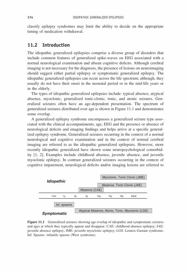
The types of idiopathic generalized epilepsies include: typical absence, atypicalabsence, myoclonic, generalized tonic-clonic, tonic, and atonic seizures. Gen-eralized seizures often have an age-dependent presentation. The spectrum ofgeneralized seizures distributed over age is shown in Figure 11.1 and demonstratessome overlap.
A generalized epilepsy syndrome encompasses a generalized seizure type asso-ciated with the clinical accompaniments, age, EEG and the presence or absence ofneurological deficits and imaging findings and helps arrive at a specific general-ized epilepsy syndrome. Generalized seizures occurring in the context of a normalneurological and cognitive examination and in the context of normal cerebralimaging are referred to as the idiopathic generalized epilepsies. However, morerecently idiopathic generalized have shown some neuropsychological comorbid-ity [1, 2]. Examples include childhood absence, juvenile absence, and juvenilemyoclonic epilepsy. In contrast generalized seizures occurring in the context ofcognitive impairment, neurological deficits and/or imaging lesions are referred to
1mo 1y 2y 5y 10y 15y 18y Adult
Inf. spasms
Atypical Absence, Atonic, Tonic, Myoclonic (LGS)
Absence (CAE)
Absence, Tonic Clonic (JAE)
Myoclonic, Tonic Clonic (JME)Idiopathic
Symptomatic
Figure 11.1 Generalized seizures showing age overlap of idiopathic and symptomatic seizuresand ages at which they typically appear and disappear. CAE: childhood absence epilepsy; JAE:juvenile absence epilepsy; JME: juvenile myoclonic epilepsy; LGS: Lennox-Gastaut syndrome;Inf. Spasms: infantile spasms (West syndrome).
11.3 DIFFERENTIATING GENERALIZED SEIZURES FROM PARTIAL SEIZURES 175
as the symptomatic generalized epilepsies. An intermediate group referred to asthe cryptogenic generalized epilepsies may have normal imaging and laboratoryinvestigations but are associated with developmental, cognitive, and neurologi-cal deficits. Examples of symptomatic or cryptogenic epilepsy include West andLennox-Gastaut syndrome.
Generalized seizures can occur in the context of any of these three broadcategories. Differentiating between them carries important therapeutic and prog-nostic information.
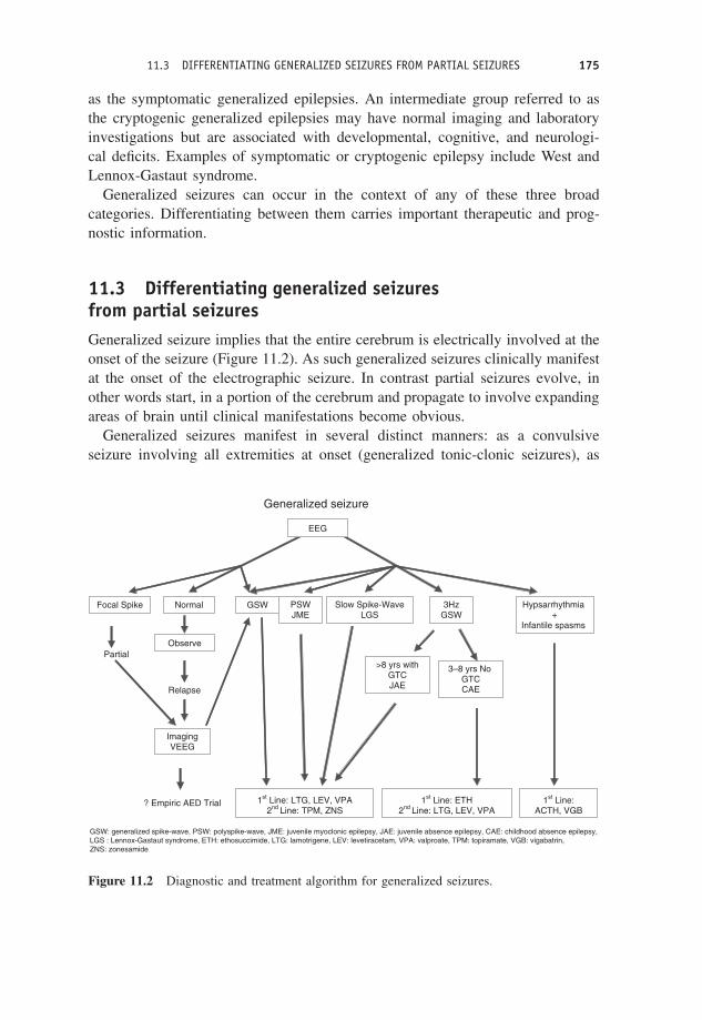
11.3 Differentiating generalized seizuresfrom partial seizuresGeneralized seizure implies that the entire cerebrum is electrically involved at theonset of the seizure (Figure 11.2). As such generalized seizures clinically manifestat the onset of the electrographic seizure. In contrast partial seizures evolve, inother words start, in a portion of the cerebrum and propagate to involve expandingareas of brain until clinical manifestations become obvious.
Generalized seizures manifest in several distinct manners: as a convulsiveseizure involving all extremities at onset (generalized tonic-clonic seizures), as
PSWJME
Slow Spike-WaveLGS
Focal Spike 3HzGSW
Hypsarrhythmia +
Infantile spasms
Normal GSW
>8 yrs withGTCJAE
3–8 yrs No GTCCAE
1st Line:ACTH, VGB
1st Line: LTG, LEV, VPA2nd Line: TPM, ZNS
Observe
ImagingVEEG
Partial
Relapse
? Empiric AED Trial
Generalized seizure
1st Line: ETH2nd Line: LTG, LEV, VPA
GSW: generalized spike-wave, PSW: polyspike-wave, JME: juvenile myoclonic epilepsy, JAE: juvenile absence epilepsy, CAE: childhood absence epilepsy,LGS : Lennox-Gastaut syndrome, ETH: ethosuccimide, LTG: lamotrigene, LEV: levetiracetam, VPA: valproate, TPM: topiramate, VGB: vigabatrin,ZNS: zonesamide
EEG
Figure 11.2 Diagnostic and treatment algorithm for generalized seizures.
176 IDIOPATHIC GENERALIZED EPILEPSIES
a sudden brief whole body jerk (myoclonic seizures), as staring spells (absenceseizures), as a slump (atonic seizures) or as a propulsive movement that drops thepatient to the ground (tonic seizures). Consciousness is almost always impaired atthe onset and typically returns instantaneously after seizure offset. The exceptionis generalized tonic-clonic convulsive seizures where there is often a pronouncedpostictal period.
11.4 Clinical and EEG characteristics of generalized seizures
Typical absence seizures
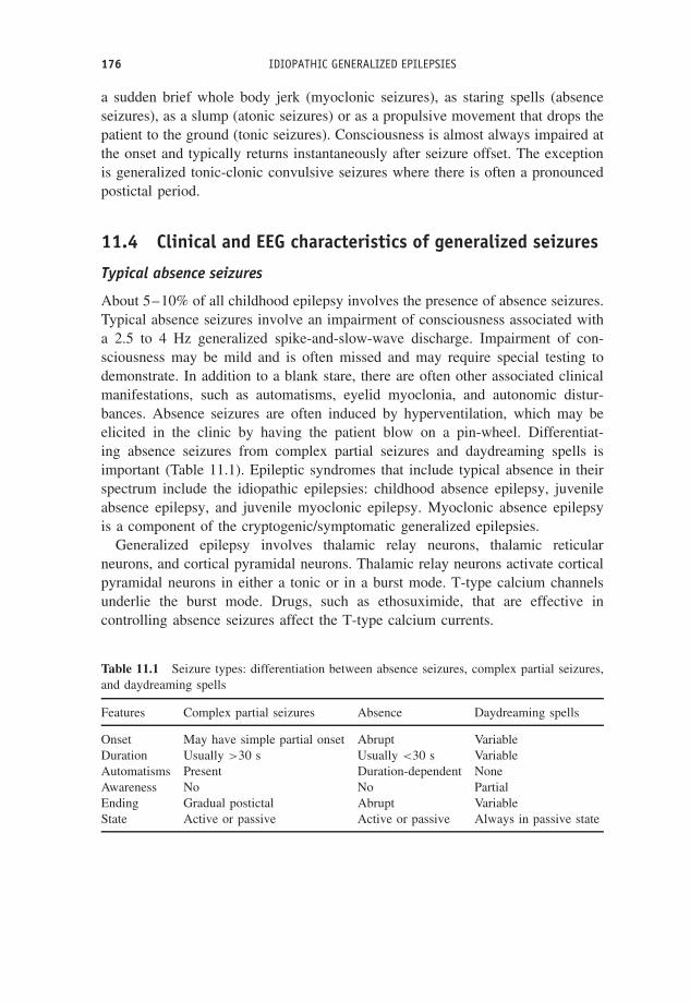
About 5–10% of all childhood epilepsy involves the presence of absence seizures.Typical absence seizures involve an impairment of consciousness associated witha 2.5 to 4 Hz generalized spike-and-slow-wave discharge. Impairment of con-sciousness may be mild and is often missed and may require special testing todemonstrate. In addition to a blank stare, there are often other associated clinicalmanifestations, such as automatisms, eyelid myoclonia, and autonomic distur-bances. Absence seizures are often induced by hyperventilation, which may beelicited in the clinic by having the patient blow on a pin-wheel. Differentiat-ing absence seizures from complex partial seizures and daydreaming spells isimportant (Table 11.1). Epileptic syndromes that include typical absence in theirspectrum include the idiopathic epilepsies: childhood absence epilepsy, juvenileabsence epilepsy, and juvenile myoclonic epilepsy. Myoclonic absence epilepsyis a component of the cryptogenic/symptomatic generalized epilepsies.
Generalized epilepsy involves thalamic relay neurons, thalamic reticularneurons, and cortical pyramidal neurons. Thalamic relay neurons activate corticalpyramidal neurons in either a tonic or in a burst mode. T-type calcium channelsunderlie the burst mode. Drugs, such as ethosuximide, that are effective incontrolling absence seizures affect the T-type calcium currents.
Table 11.1 Seizure types: differentiation between absence seizures, complex partial seizures,and daydreaming spells
Features Complex partial seizures Absence Daydreaming spells
Onset May have simple partial onset Abrupt VariableDuration Usually >30 s Usually <30 s VariableAutomatisms Present Duration-dependent NoneAwareness No No PartialEnding Gradual postictal Abrupt VariableState Active or passive Active or passive Always in passive state
11.4 CLINICAL AND EEG CHARACTERISTICS OF GENERALIZED SEIZURES 177
Atypical absence seizures
Atypical absence seizures are much less common then typical absence seizures.They are often seen in the Lennox-Gastaut syndrome where they may accompanyatonic and tonic seizures. Clinically, atypical absence is associated with develop-mental delay or mental retardation whereas mental function in typical absence isnormal. The EEG is useful in differentiating atypical absence from typical absenceseizures by the presence of diffuse slow (<2.5 Hz) spike-and-wave on EEG. Inaddition to this, the backgrounds compared to normal background frequenciesseen in typical absence are slow and disorganized.
Generalized tonic-clonic seizures
A generalized convulsion may result from the culmination of partial complexseizures as it propagates to involve the entire cerebrum, when it is referred toas secondarily generalized tonic-clonic seizures. Primary generalized tonic-clonicseizures are by definition generalized right at onset of the seizure.
Complications associated with generalized tonic-clonic seizures include, oraland head trauma, stress fractures during the tonic phase, aspiration pneumonia,pulmonary edema, and sudden unexpected death in epilepsy (SUDEP). The inter-ictal EEG shows either a normal background or runs of occipital delta activityand may show either fragmented diffuse spike-wave or polyspike-wave dischargesor frank generalized spike-wave discharges. Ictal EEG findings at onset includehigh amplitude anteriorly dominant generalized spike-wave discharges, diffusefast frequencies that evolve to generalized spike-wave discharges or polyspike-wave discharges. Once the seizure is clinically manifest muscle activity preventsdetermination of EEG changes. Postictally the EEG often demonstrates diffuseslowing and slow spike-wave discharges.
Myoclonus and myoclonic seizures
Myoclonus, a sudden involuntary brief shock-like muscle contraction originatingin the CNS, is a nonepileptic phenomenon and unassociated with epileptiformdischarges. Myoclonic seizures on the other hand are similar movements butassociated with epileptiform discharges (often fast polyspike-and-wave discharge)and arise from the cerebral cortex. Consciousness may be impaired but is oftenpreserved. They often precede generalized tonic-clonic seizures, commonly aseries of myoclonic seizure jerks are followed by a generalized convulsive seizure.When present in the context of a normal exam and imaging, they are strongly sug-gestive of juvenile myoclonic epilepsy. However, myoclonic seizures may be anaccompaniment of the more ominous Lennox-Gastaut syndrome. In young children
178 IDIOPATHIC GENERALIZED EPILEPSIES
they may occur in the context of myoclonic-astatic epilepsy of Doose syndrome.Clinically, they can be separated from sleep myoclonus in that they often occurwith awakening or randomly throughout the day while the patient is awake.
Tonic and atonic seizures
Both tonic and atonic seizures are typically seen in the symptomatic generalizedepilepsies, occurring in up to 90% of patients with Lennox-Gastaut syndrome.They most characteristically result in a drop attack, where the patient is eitherpropelled to the ground head first in tonic seizures, or slumps to the ground withatonic seizures. The duration of both seizure types is usually under one minute andcan be associated with autonomic changes, including facial flushing, tachycardia,hypertension, and papillary changes. The EEG correlates of the seizures typicallyinvolve a sudden interruption of the slow background for the duration of theseizures. This activity is somewhat similar to the electrodecremental responseseen on EEG in infantile spasms.
11.5 Generalized epilepsy syndromes
Childhood absence epilepsy
Childhood absence epilepsy accounts for up to 10% of children with epilepsy.Absence seizures usually have an onset around 3–4 years and often disappear byage 10 years [3]. The absence seizures are associated with normal intelligence andneurological function, although there is concern that subtle learning and attentionaldisorders are associated comorbidities. The cause is predominantly genetic, withabout one-third of the families of children reporting a family history of similarseizures [4]. Siblings of children have about a 10% chance of developing epilepsy.Typical childhood absence is not associated with generalized tonic-clonic seizures.
The pathogenic mechanisms of absence epilepsy are unknown; although animalmodels suggest activation of the thalamo-cortical pathways in a rhythmic mannerresulting in hypersynchronous discharges [5]. Response to antiepileptic medica-tion is usually excellent with over 70% of patients achieving complete control ofabsence seizures with the first medication used. A recently completed childhoodabsence study indicated that valproate has been reported to be equally efficaciousto ethosuximide in the treatment of absence seizures, although it may have moreside effects [6–8]. Lamotrigine had the least side effects but was not as effective incontrolling absence seizures [9]. Ethosuximide has undergone a number of studiesas adjunct therapy and first-line treatment and has been found to have the best bal-ance between efficacy and adverse effects. A common side effect is gastrointestinalupset. In rare instances, it can cause aplastic anemia, and hepatic or renal fail-ure. Second-line agents include topiramate and zonisamide. Levetiracetam may be
11.5 GENERALIZED EPILEPSY SYNDROMES 179
effective for juvenile absence with some data demonstrating efficacy for absenceseizures per se [10, 11].
There are patients with typical childhood absence who do not respond to max-imal doses of monotherapy and there is some evidence that a combination ofvalproate and ethosuximide or lamotrigine may be effective. Possibly 10% ofchildhood absence epilepsy does not respond to polytherapy.
The EEG is not only helpful in diagnosing absence but is also useful in evaluat-ing the effectiveness of treatment. Typically, medication is increased until clinicalseizures disappear and then increased until the EEG no longer demonstrates 3 Hzgeneralized spike-wave discharges. Fragmented discharges during sleep that lastless than three seconds and occurring during sleep may be considered as acceptablecontrol by some.
Juvenile absence epilepsy
Juvenile absence epilepsy is relatively common, with seizures beginning aroundpuberty. Typically, onset is between 10 and 17 years of age with intelligence, neu-rologic function, EEG background, and cerebral imaging being normal. The EEGshows generalized spike-and-wave discharges with normal background activity.The generalized spike-and-wave are often 3 Hz or occur at a slightly faster ratewith typical bursts lasting 4–15 seconds. Juvenile absence epilepsy was first dis-tinguished from childhood absence in the 1950s when Janz and Christian identifiedthe occurrence of generalized tonic-clonic seizures as a differentiating feature. Fur-thermore, unlike childhood absence which shows a female preponderance, juvenileabsence occurs equally in males and females. The absence seizures may not benoticed until the occurrence of a generalized tonic-clonic seizure which leads to anEEG and to the diagnosis. Absence seizures usually occur sporadically comparedto childhood absence where they occur multiple times a day.
Response to anti-absence medication is good, with 80% of patients becomingseizure-free. Ethosuximide is effective against the absence seizures, but is noteffective in controlling generalized tonic-clonic seizures. As such ethosuximide israrely used. Valproate, lamotrigine, and levetiracetam are usually effective agents.
Juvenile myoclonic epilepsy
This syndrome is the most common primary generalized idiopathic epilepsywith important implications for prognostication and treatment [12]. Typically,patients experience a series of myoclonic jerks that may at times be asymmetric.These myoclonic jerks may occur in clusters in the morning while awake,or randomly throughout the day. They may be activated by flickering lightsor sleep deprivation. Attention is sought when the patient experiences a firstgeneralized tonic-clonic seizure, often after sleep deprivation. The neurological
180 IDIOPATHIC GENERALIZED EPILEPSIES
exam and cerebral imaging studies if obtained are normal. The EEG demonstratesgeneralized polyspike-and-wave discharges. Often these discharges are activatedby photic light stimulation, or can occur during sleep. They may be associatedwith a myoclonic jerk. Myoclonic seizures should be differentiated from sleepmyoclonus which occurs as normal phenomena during drowsiness. Sleepmyoclonus is not associated with polyspike-and-wave discharges on EEG.
Treatment is most effective with valproate, although more recently other med-ications such as levetiracetam or lamotrigine have been shown to be effective.Lamotrigine, although initially thought to exacerbate myoclonic seizures, has notbeen shown to do this in randomized studies, although it may not be as effec-tive as valproate in controlling myoclonic seizures. Carbamazepine and phenyotinare effective against the generalized tonic-clonic seizures, although as in absenceseizures, they may exacerbate myoclonic seizures.
11.6 Treating generalized seizuresWhile there are several treatment options for generalized seizures, as indicatedearlier, there are medications that should be avoided. Absence epilepsy is noteffectively treated with phenyotin, gabapentin, and carbamazepine. Exacerbationsof absence following the initiation of carbamazepine have been well documented.Typically, myoclonic seizures do not respond well to phenyotin, carbamazepine,or lamotrigine. As with absence epilepsy some patients may have their myoclonicseizures exacerbated by these medications. While not effective generally againstabsence and myoclonic seizures these medications are effective in controllingthe generalized tonic-clonic seizures that may accompany the primary general-ized epilepsies.
11.7 Treatment algorithmThere is a paucity of well-designed, randomized clinical trials for patients withgeneralized seizures. The American Academy of Neurology has issued broadrecommendations for the treatment of generalized seizures. Generally, whereverpossible monotherapy should be used.
A diagnostic and treatment algorithm for treating generalized seizures is pre-sented in Figure 11.2. A starting point in the treatment algorithm is an EEG. TheEEG is usually essential to appropriately classify seizures, including avoiding themisidentification of secondarily generalized seizures as being primarily general-ized seizures. The EEG may show focal spikes in which case the diagnosis of truegeneralized epilepsy needs to be questioned. A normal EEG does not rule out ageneralized seizure and in this case it may be appropriate to observe the patient.If there is a relapse in seizures then a video-EEG and an imaging study may
11.7 TREATMENT ALGORITHM 181
be useful. If the video-EEG shows generalized spike-waves then the diagnosis isconfirmed and treatment proceeds from there. If the video-EEG including cap-turing sleep fails to show epileptiform discharges then the diagnosis may bequestioned or an empiric antiepileptic medication trial considered.
If the EEG shows a traditional pattern thereby confirming a primary generalizedseizure then the next step is to differentiate between generalized polyspike-waves(usually seen in juvenile myoclonic epilepsy), 3 Hz generalized spike-waves (usu-ally seen in either childhood or juvenile absence epilepsy), or slow spike-waves(usually less than 2.5 Hz, seen in symptomatic Lennox-Gastaut syndrome). TheEEG may show hypsarrythmia, which in the presence of infantile spasms is diag-nostic of West syndrome.
Cerebral imaging is not usually required if an epilepsy syndromic diagnosis isarrived at. However, imaging should be considered if there are focal epileptiformdischarges on EEG, atypical features, or if a definitive epilepsy syndrome cannotbe arrived at. Other indications for imaging include the presence of either cognitivedeficits or focal deficits seen on neurological examination.
First-line agents for the treatment of generalized epilepsy, especially those asso-ciated with generalized spike-wave, polyspike-wave, and slow spike-wave, includelevetiracetam, lamotrigine, or valproate. Valproate is the most effective agent intreating the spectrum of generalized seizures. However, the potential for neuraltube defects should be considered when considering its use in females of child-bearing age. Second-line agents include topiramate and zonisamide. Medicationsto be avoided include carbamazepine, phenyotin, and oxcarbazepine. These agentsare effective against convulsive generalized tonic-clonic seizure, although as men-tioned earlier they may exacerbate absence or myoclonic seizures.
If the EEG shows typical 3 Hz generalized spike-wave then a diagnosis ofabsence epilepsy can be made. Childhood absence is usually not associated withconvulsive seizures and ethosuximide is usually a first-line agent that can be used.If the child has a diagnosis of juvenile absence epilepsy then there is an increasedrisk of accompanying generalized convulsive seizures against which ethosuximidehas not been shown effective. In these situations a first-line agent is as for thetreatment of primary generalized seizures as discussed above. The presence ofhypsarrythmia in an infant raises suspicion for West syndrome with accompanyinginfantile spasms. Agents that are effective against West syndrome include ACTHand vigabatrin. The academy has also indicated the use of vigabatrin as a first-linetherapy for the treatment of infantile spasm. This presents an added option to thelong-established use of ACTH. ACTH has many adverse effects associated with itsuse, including weight increase, hyperglycemia, hypertension, and the possibilityof associated cerebral atrophy. Vigabatrin has been reported to constrict visualfields and be associated with reversible MRI white matter changes. Felbamatehas a broad spectrum of activity in generalized seizures, but rare reports of fatal
182 IDIOPATHIC GENERALIZED EPILEPSIES
aplastic anemia and hepatic failure limit its use to a third-line agent where othertreatment alternatives have failed.
11.8 Prognosis/outcomesThe EEG can help guide treatment. When seizures are effectively controlled clin-ically, an EEG showing residual generalized epileptiform discharges usually indi-cates the need for an increased dose of medication. Juvenile myoclonic epilepsyand juvenile absence epilepsy typically do not remit with age, whereas childhoodabsence epilepsy usually remits by age 10 years. Infantile spasms often transitionto Lennox-Gastaut syndrome associated with slow spike-and-wave discharges.
References1. Hermann, B.P., Jones, J.E., Sheth, R. et al. (2008) Growing up with epilepsy: a two-year
investigation of cognitive development in children with new onset epilepsy. Epilepsia, 49(11), 1847–1858.
2. Hermann, B.P., Jones, J., Sheth, R., and Seidenberg, M. (2007) Cognitive and magneticresonance volumetric abnormalities in new-onset pediatric epilepsy. Semin Pediatr Neurol ,14 (4), 173–180.
3. Wirrell, E.C., Camfield, C.S., Camfield, P.R., Gordon, K.E., and Dooley, J.M. (1996) Long-term prognosis of typical childhood absence epilepsy: remission or progression to juvenilemyoclonic epilepsy. Neurology , 47 (4), 912–918.
4. Delgado-Escueta, A.V., Medina, M.T., Serratosa, J.M. et al. (1999) Mapping and positionalcloning of common idiopathic generalized epilepsies: juvenile myoclonus epilepsy andchildhood absence epilepsy. Adv Neurol , 79, 351–374.
5. Pulsipher, D.T., Seidenberg, M., Guidotti, L. et al. (2009) Thalamofrontal circuitry andexecutive dysfunction in recent-onset juvenile myoclonic epilepsy. Epilepsia, 50 (5),1210–1219.
6. Montouris, G. and Abou-Khalil, B. (2009) The first line of therapy in a girl with juvenilemyoclonic epilepsy: should it be valproate or a new agent? Epilepsia, 50 (Suppl 8), 16–20.
7. Sheth, R.D., Wesolowski, CA., Jacob, J.C. et al. (1995) Effect of carbamazepine and val-proate on bone mineral density. J Pediatr , 127 (2), 256–262.
8. Sheth, R.D. and Montouris, G. (2008) Metabolic effects of AEDs: impact on body weight,lipids and glucose metabolism. Int Rev Neurobiol , 83, 329–346.
9. Coppola, G., Licciardi, F., Sciscio, N. et al. (2004) Lamotrigine as first-line drug in child-hood absence epilepsy: a clinical and neurophysiological study. Brain Dev , 26 (1), 26–29.
10. Abou-Khalil, B. (2008) Levetiracetam in the treatment of epilepsy. Neuropsychiatr DisTreat , 4 (3), 507–523.
11. Noachtar, S., Andermann, E., Meyvisch, P. et al. (2008) Levetiracetam for the treatmentof idiopathic generalized epilepsy with myoclonic seizures. Neurology , 70 (8), 607–616.
12. Camfield, C.S. and Camfield, P.R. (2009) Juvenile myoclonic epilepsy 25 years after seizureonset: a population-based study. Neurology , 73 (13), 1041–1045.


![Recommendations for the treatment of epilepsy in adult and … · 2020. 10. 13. · epilepsy)[19,22] Vigabatrincanalsobeusedasadd-on treatmentbutonlyaslastchoice becauseofitsunfavorablesafety](https://img.dokumen.tips/doc/110x75/60ac60deababab702a673921/recommendations-for-the-treatment-of-epilepsy-in-adult-and-2020-10-13-epilepsy1922.jpg)