Embed Size (px)
Citation preview
4999
Abstract. – OBJECTIVE: The present study was designed to evaluate the effect of Adi-ponectin (APN) against alveolar epithelial apop-tosis in chronic obstructive pulmonary disease (COPD) rat models.
MATERIALS AND METHODS: Thirty-six male Sprague-Dawley (SD) rats were randomly as-signed to three groups: Sham group, COPD group, and COPD + APN group (2.5 ug/kg/day). To assess the effect of APN, histopathological evaluations, lung function, and the apoptotic in-dex (AI) of alveolar septal cells, were performed. In addition, the levels of oxidative stress and en-doplasmic reticulum stress were measured.
RESULTS: HE staining demonstrated that APN inhibited pathological injury in COPD rats. In addition, APN could restore the levels of su-peroxide dismutase (SOD) and malondialde-hyde (MDA) in serum. APN also inhibited the levels of endoplasmic reticulum stress pathway including CHOP, phospho-JNK and Caspase-12 in alveolar epithelial cell. Furthermore, APN significantly inhibited the protein levels of Caspase-3 and apoptosis in alveolar epithelial cell of COPD rats.
CONCLUSIONS: Our findings suggested that APN might effectively ameliorate the progres-sion of COPD via inhibiting the endoplasmic re-ticulum stress-induced alveolar epithelial apop-tosis in rats.
Key Words:Adiponectin, CDPD, Endoplasmic reticulum stress,
Alveolar epithelial cell apoptosis.
Introduction
Chronic obstructive pulmonary disease (COPD) is a kind of disease that can be prevented and treated, characterized by persistent airflow limitation in progressive development, which is related to the enhanced chronic inflammatory
response of airways and lung tissues to tobacco smoke and other harmful gases or particles; the acute exacerbation and complications affect the course of disease. According to the large-sample survey in China in 2007, the prevalence rate of COPD was 8.2% in people aged above 40 years old1. COPD has a high disability and death rate and has become the third leading cause of death in the world2. The repeated acute exacerbation of COPD caused by virus or bacterial infection, etc.3,4, accelerates the progression of disease, re-duces the life quality of patients, and increases the death rate5-7.
Major pathological changes of COPD include the bronchial inflammation and emphysema8, and it is currently believed that COPD is related to the chronic inflammation, oxidative stress and pro-tease-anti-protease system imbalance in airway and lungs. In recent years, studies have found that the increased apoptosis of alveolar epithe-lial cells, airway epithelial cells, and other lung tissue cells, are also an important pathogenesis of COPD9,10, among which oxidative stress is an important pathogenesis of COPD11,12. The oxidant and antioxidant system in normal human body is in an equilibrium state, but oxidative stress will be caused in the body when the number of reac-tive oxygen species increases greatly far beyond the scavenging capacity of antioxidant in lung tissues. This will lead to oxidative stress in the body, thus resulting in airway injury, inflamma-tory response, protein carbonation, anti-protease inactivation, lipid peroxidation, apoptosis, etc., and further inducing the occurrence and develop-ment of COPD13.
Endoplasmic reticulum is an important or-ganelle in eukaryotes, as well as the main site for intracellular protein synthesis, processing and folding, transport and intracellular calcium storage, which feels the changes in intracellular
European Review for Medical and Pharmacological Sciences 2017; 21: 4999-5007
Y.-L. ZHAO, F. LI, Y.-W. LIU, Y.-J. SHI, Z.-H. LI, G.-K. CAO, W. ZHU
Intensive Care Unit, The First People’s Hospital of Xuzhou, Xuzhou, Jiangsu, China
Corresponding Author: Yuliang Zhao, MD; e-mail: [email protected]
Adiponectin attenuates endoplasmic reticulum stress and alveolar epithelial apoptosis in COPD rats
Y.-L. Zhao, F. Li, Y.-W. Liu, Y.-J. Shi, Z.-H. Li, G.-K. Cao, W. Zhu
5000
environment in time to maintain the balance of intracellular environment14. Some stimulating factors, such as infection, hypoxia, starvation, oxidative stress, calcium balance disorder and physical and chemical stimulation, can induce the acute stress response in cells, increase the synthesis of structural protein or secretory pro-tein, increase the protein synthesis load in en-doplasmic reticulum, change the internal envi-ronment in endoplasmic reticulum, make the unfolded and misfolded protein gather in endo-plasmic reticulum lumen, damage the normal function of endoplasmic reticulum and lead to endoplasmic reticulum stress (ERS)15,16. ERS is a self-protection response of cells to the outside harmful stimulation. However, when ERS is too serious or the harmful stimulation lasts too long, the expressions of apoptosis-related genes will be upregulated, the apoptotic signaling path-ways will be initiated, and the damaged cells will be apoptotic eventually17. ERS can induce apoptosis through three ways: (1) activation and transcription of CCAAT/enhancer binding protein homologous protein (CHOP) gene; (2) activation of C-Jun N-terminal kinase (JNK) pathway; (3) activation of Caspase-12 specific in endoplasmic reticulum18. In recent years, studies have confirmed that ERS and its related apopto-sis play important roles in the pathogenesis of COPD. Adiponectin (APN) has been found as a kind of protein mainly secreted by adipocytes in recent years, and its anti-inflammatory, anti-ath-erosclerosis, glucolipid metabolism-regulating and memory and cognitive disorder-regulating effects, have been reported in many studies19-22. At present, its biological characteristics have been attached increasingly more importance, and some studies have shown that APN can inhibit oxidative stress and protect bronchial epithelial cells and myocardial tissues23,24. How-ever, there are few studies on the relationship between APN and COPD, so APN was used to intervene in COPD rats in this study.
Materials and Methods
Animal Model and Grouping36 male SD rats (140-160 g) were provided by
the First People’s Hospital of Xuzhou Animal Center. They were arisen in polycarbonate cag-es and kept with 12-hour light-dark cycle and continuous access to food and water. Instillation of LPS together with cigarette smoke exposure
were used to establish the COPD model in rats as previously described25,26. Briefly, after the rats were anesthetized with chloral hydrate (250 mg/kg), the LPS (1 mg/mL, 0.2 mL, Sigma-Aldrich, St. Louis, MO, USA) was dripped through in-tratracheal instillation on the 1st, 15th and 30th day during this experiment. All rats, except the sham group, were placed in a sealed box and exposed twice a day to smoke from 6 commer-cial unfiltered cigarettes (ChungHua Cigarettes, Shanghai, China). The treatment was sustained for 60 days. Rats in sham group were treated with air instead. Rats were divided into three groups: (1) Sham group: rats were intraperitone-ally injected with normal saline (1 mL/day, ip) for 60 days; (2) COPD group: COPD rats were treated with normal saline (1 mL/day, ip) for 60 days (1 mL, ip); (3) COPD with APN group: COPD rats were treated with APN for 60 days (2.5 μg/kg/day dissolved in 1 mL normal saline, ip, Sigma-Aldrich, St. Louis, MO, USA). The study was approved by the Animal Ethics Com-mittee of The First People’s Hospital of Xuzhou Animal Center.
Measurement of Lung Function6 rats from each group were randomly selected
for measurements of lung function; after they were anesthetized with chloral hydrate, the tra-chea cannula was performed and connected with the small animal ventilator. The variables were observed by a spirometer, which is recommended for small animals; FEV0.3/FVC% and PEF were measured.
Measurement of SOD and MDABlood was collected and separated in a re-
frigerated centrifuge and the samples were cen-trifuged at 3000 rpm for 20 min at 4°C. The activities of SOD and the content of MDA were measured according to the manufacturer’s in-struction (Beyotime, Shanghai, China).
Histopathological AnalysisThe middle or upper lobe of the right lung was
excised from each rat and fixed in 4% formalin. The samples were cut into 5 μm sections followed with embedded in paraffin. Hematoxylin-Eosin (HE) was used to stain the paraffin sections. Af-ter that, a light microscope was used to visualize the pathological conditions in the lung tissues on randomized sections. Measuring the mean linear intercept (MLI) and mean alveoli number (MAN) was used to assess emphysema27.
Adiponectin attenuates alveolar epithelial apoptosis
5001
Tunel AssayThe apoptosis of alveolar epithelial was detect-
ed by tunel assay according to the manufacturer (Roche, Basel, Switzerland). Horseradish perox-idase (HP)-mediated diaminobenzidine reaction was used to visualize the TUNEL-positive cells, following the counterstain. Fields were photo-graphed at a magnification of 200× and were randomly selected. The apoptosis index was used to measure the degree of apoptosis.
Western BlotThe lung tissues were frozen and stored at
-70°C. Next, they were homogenized and added with lysis buffer in which phosphatase and pro-tease inhibitors were included. Protein centra-tion was measured by bicinchoninic acid (BCA) method, and sodium dodecyl sulphate-polyacryl-amide gel electrophoresis (SDS-PAGE) was used to separate the tissue lysates. After blotting on-to polyvinylidene difluoride (PVDF) membrane, samples were incubated with specific antibodies against Chop, phospho-JNK, JNK, Caspase-12, Caspase-3 and β-actin (1:1000) (Cell Signaling Technology, Danvers, MA, USA). Next, the sam-ples were incubated at 4°C overnight. The re-spective secondary antibodies conjugated to HRP were incubated for 1 h, followed by three-time washing. The membrane was then incubated with
ECL (Millipore, Billerica, MA, USA) for lumi-nescence generation. The proteins were visual-ized and detected, and the grey level of each pro-tein was normalized against to the β-actin. The results were expressed as fold increase compared with the Sham.
Statistical AnalysisAll results are presented as the means ± stan-
dard deviation (SD). Statistical analyses were per-formed using both GraphPad Prism 6.02 (Graph-Pad Software, La Jolla, CA, USA) and PASW Statistics 18.0 (SPSS Inc., Chicago, IL, USA). One-way ANOVA was used to compare differ-ences among groups. An unpaired t-test was used for the comparison between 2 groups. A value of p < 0.05 was considered statistically significant.
Results
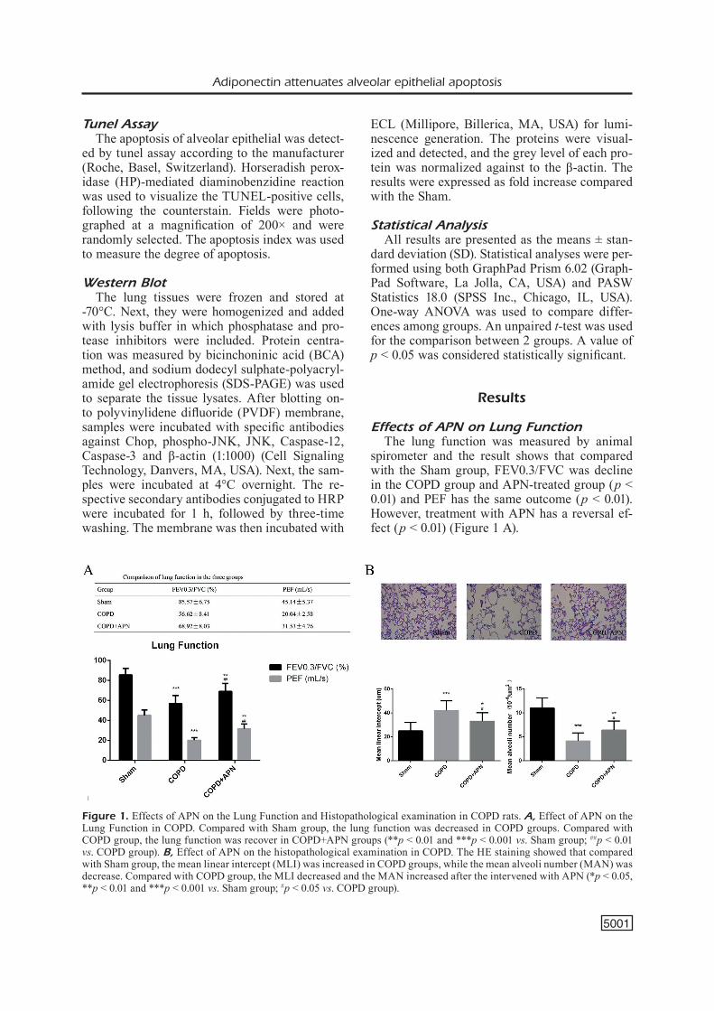
Effects of APN on Lung FunctionThe lung function was measured by animal
spirometer and the result shows that compared with the Sham group, FEV0.3/FVC was decline in the COPD group and APN-treated group (p < 0.01) and PEF has the same outcome (p < 0.01). However, treatment with APN has a reversal ef-fect (p < 0.01) (Figure 1 A).
Figure 1. Effects of APN on the Lung Function and Histopathological examination in COPD rats. A, Effect of APN on the Lung Function in COPD. Compared with Sham group, the lung function was decreased in COPD groups. Compared with COPD group, the lung function was recover in COPD+APN groups (**p < 0.01 and ***p < 0.001 vs. Sham group; ##p < 0.01 vs. COPD group). B, Effect of APN on the histopathological examination in COPD. The HE staining showed that compared with Sham group, the mean linear intercept (MLI) was increased in COPD groups, while the mean alveoli number (MAN) was decrease. Compared with COPD group, the MLI decreased and the MAN increased after the intervened with APN (*p < 0.05, **p < 0.01 and ***p < 0.001 vs. Sham group; #p < 0.05 vs. COPD group).
Y.-L. Zhao, F. Li, Y.-W. Liu, Y.-J. Shi, Z.-H. Li, G.-K. Cao, W. Zhu
5002
Effects of APN on the Histopathological Examination
The histopathological examination was mea-sured by HE staining; the result analyzed from lung tissue demonstrated that emphysema his-tologically advanced in COPD groups; in con-trast, the histological markers of emphysema, such as MLI and PEF, were normal in Sham group. After intervening with APN, the section from COPD-APN group showed MLI and PEF were significantly lower than COPD group (p < 0.05). The result indicated APN has the effect of inhibiting pathological injury from COPD (Figure 1B).
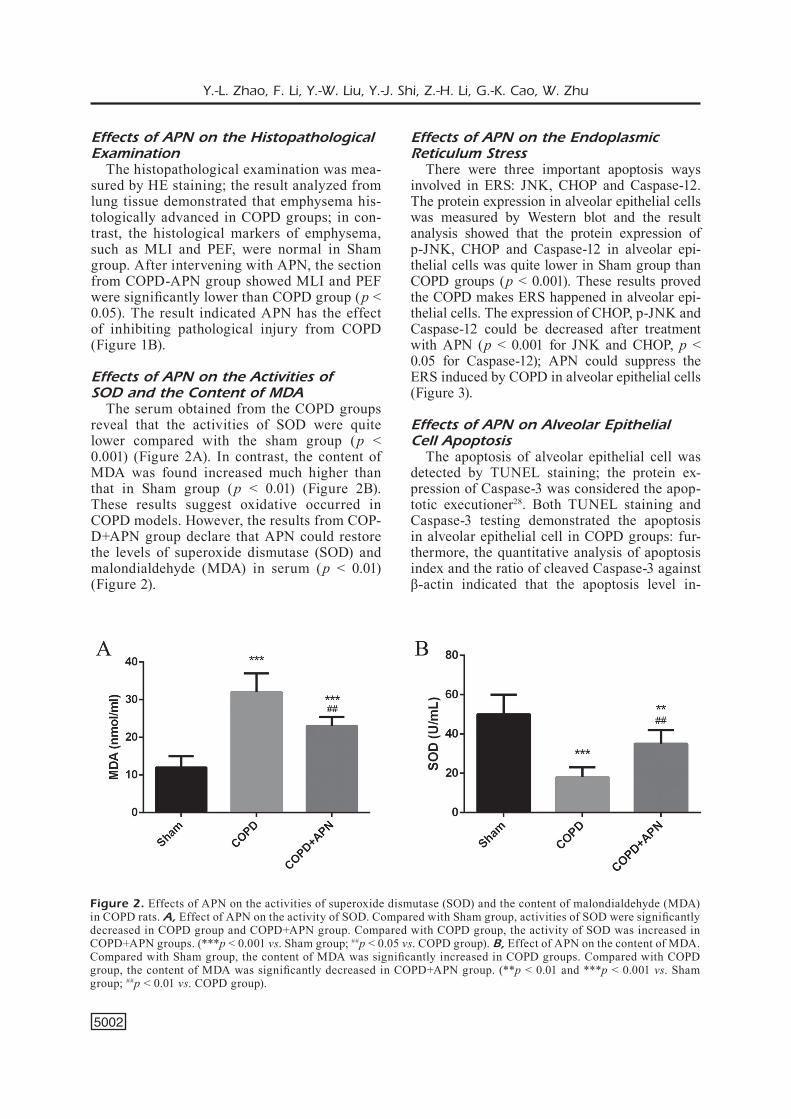
Effects of APN on the Activities of SOD and the Content of MDA
The serum obtained from the COPD groups reveal that the activities of SOD were quite lower compared with the sham group (p < 0.001) (Figure 2A). In contrast, the content of MDA was found increased much higher than that in Sham group (p < 0.01) (Figure 2B). These results suggest oxidative occurred in COPD models. However, the results from COP-D+APN group declare that APN could restore the levels of superoxide dismutase (SOD) and malondialdehyde (MDA) in serum (p < 0.01) (Figure 2).
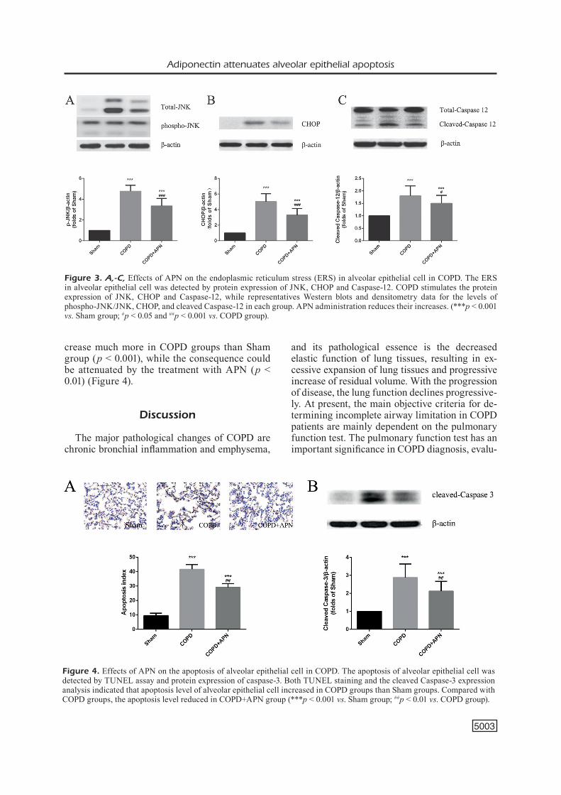
Effects of APN on the Endoplasmic Reticulum Stress
There were three important apoptosis ways involved in ERS: JNK, CHOP and Caspase-12. The protein expression in alveolar epithelial cells was measured by Western blot and the result analysis showed that the protein expression of p-JNK, CHOP and Caspase-12 in alveolar epi-thelial cells was quite lower in Sham group than COPD groups (p < 0.001). These results proved the COPD makes ERS happened in alveolar epi-thelial cells. The expression of CHOP, p-JNK and Caspase-12 could be decreased after treatment with APN (p < 0.001 for JNK and CHOP, p < 0.05 for Caspase-12); APN could suppress the ERS induced by COPD in alveolar epithelial cells (Figure 3).
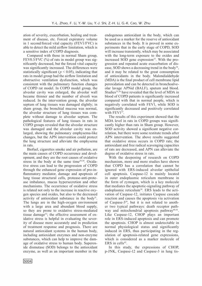
Effects of APN on Alveolar Epithelial Cell Apoptosis
The apoptosis of alveolar epithelial cell was detected by TUNEL staining; the protein ex-pression of Caspase-3 was considered the apop-totic executioner28. Both TUNEL staining and Caspase-3 testing demonstrated the apoptosis in alveolar epithelial cell in COPD groups: fur-thermore, the quantitative analysis of apoptosis index and the ratio of cleaved Caspase-3 against β-actin indicated that the apoptosis level in-
Figure 2. Effects of APN on the activities of superoxide dismutase (SOD) and the content of malondialdehyde (MDA) in COPD rats. A, Effect of APN on the activity of SOD. Compared with Sham group, activities of SOD were significantly decreased in COPD group and COPD+APN group. Compared with COPD group, the activity of SOD was increased in COPD+APN groups. (***p < 0.001 vs. Sham group; ##p < 0.05 vs. COPD group). B, Effect of APN on the content of MDA. Compared with Sham group, the content of MDA was significantly increased in COPD groups. Compared with COPD group, the content of MDA was significantly decreased in COPD+APN group. (**p < 0.01 and ***p < 0.001 vs. Sham group; ##p < 0.01 vs. COPD group).
Adiponectin attenuates alveolar epithelial apoptosis
5003
crease much more in COPD groups than Sham group (p < 0.001), while the consequence could be attenuated by the treatment with APN (p < 0.01) (Figure 4).
Discussion
The major pathological changes of COPD are chronic bronchial inflammation and emphysema,
and its pathological essence is the decreased elastic function of lung tissues, resulting in ex-cessive expansion of lung tissues and progressive increase of residual volume. With the progression of disease, the lung function declines progressive-ly. At present, the main objective criteria for de-termining incomplete airway limitation in COPD patients are mainly dependent on the pulmonary function test. The pulmonary function test has an important significance in COPD diagnosis, evalu-
Figure 3. A,-C, Effects of APN on the endoplasmic reticulum stress (ERS) in alveolar epithelial cell in COPD. The ERS in alveolar epithelial cell was detected by protein expression of JNK, CHOP and Caspase-12. COPD stimulates the protein expression of JNK, CHOP and Caspase-12, while representatives Western blots and densitometry data for the levels of phospho-JNK/JNK, CHOP, and cleaved Caspase-12 in each group. APN administration reduces their increases. (***p < 0.001 vs. Sham group; #p < 0.05 and ##p < 0.001 vs. COPD group).
Figure 4. Effects of APN on the apoptosis of alveolar epithelial cell in COPD. The apoptosis of alveolar epithelial cell was detected by TUNEL assay and protein expression of caspase-3. Both TUNEL staining and the cleaved Caspase-3 expression analysis indicated that apoptosis level of alveolar epithelial cell increased in COPD groups than Sham groups. Compared with COPD groups, the apoptosis level reduced in COPD+APN group (***p < 0.001 vs. Sham group; ##p < 0.01 vs. COPD group).
Y.-L. Zhao, F. Li, Y.-W. Liu, Y.-J. Shi, Z.-H. Li, G.-K. Cao, W. Zhu
5004
ation of severity, exacerbation, healing and treat-ment of disease, etc. Forced expiratory volume in 1 second/forced vital capacity (FEV1/FVC) is able to detect the mild airflow limitation, which is a sensitive index of COPD diagnosis.
Compared with those in normal Sham group, FEV0.3/FVC (%) of rats in model group was sig-nificantly decreased, but the forced vital capacity was significantly increased. The differences were statistically significant (p < 0.05), indicating that rats in model group had the airflow limitation and obstructive ventilation dysfunction, which was consistent with the pulmonary function changes of COPD rat model. In COPD model group, the alveolar cavity was enlarged, the alveolar wall became thinner and the number of alveoli was reduced. In the intervention group, the alveolar septum of lung tissues was damaged slightly; in sham group, the bronchial mucosa was normal, the alveolar structure of lung tissues was com-plete without damage to alveolar septum. The pathological features of lung tissues in rats in COPD groups revealed that the alveolar structure was damaged and the alveolar cavity was en-larged, showing the pulmonary emphysema-like changes, but the APN intervention could protect the lung structure and alleviate the emphysema in rats.
Biofuel, cigarettes smoke and air pollution, are the main causes of COPD occurrence and devel-opment, and they are the root causes of oxidative stress in the body at the same time29,30. Oxida-tive stress can lead to the progression of COPD through the enhanced gene expression of pro-in-flammatory mediator, damage and apoptosis of lung tissue structural cells, protease-anti-prote-ase imbalance, mucus hypersecretion and other mechanisms. The occurrence of oxidative stress is related not only to the increase in reactive oxy-gen species and oxides, but also to the decreased activity of antioxidant substance in the body31. The lungs are in the high-oxygen environment with a large area and abundant blood supply, so they are prone to oxidative stress-mediated tissue damage32; the effective assessment of ox-idative stress is helpful in evaluating the sever-ity of disease more accurately and in prediction of treatment response and prognosis. There are natural antioxidant systems in the human body, including antioxidant enzymes and non-enzyme substances, which can help to improve the dam-age of oxidative stress to human body. Superox-ide dismutase (SOD) belongs to the antioxidant enzyme, as well as an important member in the
endogenous antioxidant in the body, which can be used as a marker for the reserve of antioxidant substances in the body. It is proved in some ex-periments that in the early stage of COPD, SOD will increase transiently, which may be associated with the long-term exposure to the oxides and increased SOD gene expression33. With the pro-gression and repeated acute exacerbation of dis-ease, SOD shows a decreasing trend in the body34, and it may be related to the great consumption of antioxidants in the body. Malondialdehyde (MDA) is the final product of cell membrane lipid peroxidation and can be detected in bronchoalve-olar lavage APNid (BALF), sputum and blood. Studies35,36 have revealed that the level of MDA in blood of COPD patients is significantly increased compared with that in normal people, which is negatively correlated with FEV1, while SOD is significantly decreased and positively correlated with FEV137.
The results of this experiment showed that the MDA level in rats in COPD groups was signifi-cantly higher than rats in Sham group. However, SOD activity showed a significant negative cor-relation, but there were some restitute trends after APN intervention. The above results suggested that oxidative stress occurs in COPD rats; the antioxidant and free radical scavenging capacities of rats are decreased, and APN can alleviate the degree of oxidative stress in rats.
With the deepening of research on COPD mechanism, more and more studies have shown that COPD has a correlation that cannot be ignored with ERS-induced alveolar epithelial cell apoptosis. Caspase-12 is mainly located in outer endoplasmic reticulum membrane in the form of zymogen, which is a key molecule that mediates the apoptotic-signaling pathway of endoplasmic reticulum38. ERS leads to the acti-vation of Caspase-12, initiates Caspase cascade reaction and causes the apoptosis via activation of Caspase-339, but it is not related to anoth-er two typical pathways: death receptor path-way and mitochondrial apoptosis pathway40,41. Like Caspase-12, CHOP plays an important role in ERS-induced apoptosis and can promote the apoptosis. CHOP is almost undetectable in normal physiological status and significantly induced in ERS, thus participating in the reg-ulation of apoptosis-related gene expressions, which is considered as a marker molecule of ERS in cells42.
In this study, the expressions of CHOP, p-JNK, Caspase-12 and Caspase-3 in lung tis-
Adiponectin attenuates alveolar epithelial apoptosis
5005
sues were detected via Western blot, and the apoptosis of alveolar epithelial cells was de-tected using the TUNEL method. The experi-mental results revealed that CHOP, p-JNK, and Caspase-12 were expressed in alveolar epithelial cells, and the gray level results present clear growth trends in COPD groups, while the ex-pression of Caspase-3 and apoptosis index in COPD groups were also significantly increased by comparing with Sham group. Therefore, this experiment showed that ERS-induced cell apop-tosis in lung tissues did occur in COPD rats, and APN can alleviate the degree of ERS-in-duced cell apoptosis in rats. APN, also known as adipocyte complement regulatory protein, is a kind of cytokine with the molecular weight of 28-30 kDa, mainly synthesized and secreted by adipocytes, which has anti-inflammatory and/or pro-inflammatory activity43. At present, studies on the exact relationship between APN and lung function in COPD are still controversial and further investigations are needed44-46.
Conclusions
This study provided some references for the effect of APN in the process of COPD through the attenuation of endoplasmic reticulum stress and alveolar epithelial apoptosis.
AcknowledgementsThis study was supported by Xuzhou Social Development and Clinical Medical Technology Project (No: KC16SL126) and Xuzhou Science and Technology Planning Project (XM13B055).
Conflict of InterestThe Authors declare that they have no conflict of interests.
References
1) Zhong n, Wang C, Yao W, Chen P, Kang J, huang S, Chen B, Wang C, ni D, Zhou Y, Liu S, Wang X, Wang D, Lu J, Zheng J, Ran P. Prevalence of chronic ob-structive pulmonary disease in China: a large, population-based survey. Am J Respir Crit Care Med 2007; 176: 753-760.
2) TeRZano C, oRioLo F. Lung characteristics in elder-ly males and females patients with COPD: dif-ferences and optimal use of dry powder inhal-ers (DPIs). Eur Rev Med Pharmacol Sci 2017; 21: 2708-2716.
3) Wu X, Chen D, gu X, Su X, Song Y, Shi Y. Preva-lence and risk of viral infection in patients with acute exacerbation of chronic obstructive pul-monary disease: a meta-analysis. Mol Biol Rep 2014; 41: 4743-4751.
4) CuKiC V. The most common detected bacteria in sputum of patients with the acute exacerbation of COPD. Mater Sociomed 2013; 25: 226-229.
5) BaKeR CL, Zou Kh, Su J. Risk assessment of read-missions following an initial COPD-related hospi-talization. Int J Chron Obstruct Pulmon Dis 2013; 8: 551-559.
6) KiRaZ K, goKDeniZ T, KaLaYCiogLu e, BoReKCi a, aKYoL S, BaYKan ao, aCeLe a, KaRaKoYun S, SeKeR T, guR M. Epicardial fat thickness is associated with sever-ity of disease in patients with chronic obstructive pulmonary disease. Eur Rev Med Pharmacol Sci 2016; 20: 4508-4515.
7) ViLLaR Bi, CaRRiLLo MR, Regi BM, MaRZo CM, aRCu-Sa Vn, SegunDo YM. [Factors associated with the quality of life in patients with chronic obstructive pulmonary disease]. Aten Primaria 2014; 46: 179-187.
8) YoShiDa T, TuDeR RM. Pathobiology of ciga-rette smoke-induced chronic obstructive pul-monary disease. Physiol Rev 2007; 87: 1047-1082.
9) KiRKhaM Pa, CaRaMoRi g, CaSoLaRi P, PaPi aa, eD-WaRDS M, ShaMJi B, TRianTaPhYLLoPouLoS K, huSSain F, PinaRT M, Khan Y, heineMann L, STeVenS L, YeaDon M, BaRneS PJ, Chung KF, aDCoCK iM. Oxidative stress-induced antibodies to carbonyl-modified protein correlate with severity of chronic obstruc-tive pulmonary disease. Am J Respir Crit Care Med 2011; 184: 796-802.
10) LouheLainen n, STaRK h, MaZuR W, RYTiLa P, DJu-KanoViC R, KinnuLa VL. Elevation of sputum matrix metalloproteinase-9 persists up to 6 months after smoking cessation: a research study. BMC Pulm Med 2010; 10: 13.
11) RahMan i. Pharmacological antioxidant strategies as therapeutic interventions for COPD. Biochim Biophys Acta 2012; 1822: 714-728.
12) Yao h, RahMan i. Current concepts on oxidative/carbonyl stress, inflammation and epigenetics in pathogenesis of chronic obstructive pulmonary disease. Toxicol Appl Pharmacol 2011; 254: 72-85.
13) Van KLaVeRen RJ, RoeLanT C, BoogaeRTS M, PYPe JL, DeMeDTS M, neMeRY B. Protective effects of the lazaroid U-74389G against hyperoxia in rat type II pneumocytes. Pulm Pharmacol Ther 1998; 11: 23-30.
14) WaLTeR P, Ron D. The unfolded protein response: from stress pathway to homeostatic regulation. Science 2011; 334: 1081-1086.
15) goMeZ e, PoWeLL ML, BeVingTon a, heRBeRT TP. A decrease in cellular energy status stimulates PERK-dependent eIF2alpha phosphorylation and regulates protein synthesis in pancreatic be-ta-cells. Biochem J 2008; 410: 485-493.
Y.-L. Zhao, F. Li, Y.-W. Liu, Y.-J. Shi, Z.-H. Li, G.-K. Cao, W. Zhu
5006
16) KiTaguChi Y, TaRaSeViCiene-STeWaRT L, hanaoKa M, na-TaRaJan R, KRaSKauSKaS D, VoeLKeL nF. Acrolein in-duces endoplasmic reticulum stress and caus-es airspace enlargement. PLoS One 2012; 7: e38038.
17) Zheng X, Zheng X, Wang X, Ma Z, guPTa SV, BoTu-San i, TaKeDa T, BJoRKLunD a, inoue M, CaTRina SB, BRiSMaR K, PoeLLingeR L, PeReiRa TS. Acute hypox-ia induces apoptosis of pancreatic beta-cell by activation of the unfolded protein response and upregulation of CHOP. Cell Death Dis 2012; 3: e322.
18) FuLDa S, goRMan aM, hoRi o, SaMaLi a. Cellular stress responses: cell survival and cell death. Int J Cell Biol 2010; 2010: 214074.
19) ohaShi K, PaRKeR JL, ouChi n, higuChi a, ViTa Ja, goKCe n, PeDeRSen aa, KaLThoFF C, TuLLin S, SaMS a, SuMMeR R, WaLSh K. Adiponectin promotes macrophage polarization toward an anti-inflam-matory phenotype. J Biol Chem 2010; 285: 6153-6160.
20) BeRg ah, CoMBS TP, Du X, BRoWnLee M, SCheReR Pe. The adipocyte-secreted protein Acrp30 enhanc-es hepatic insulin action. Nat Med 2001; 7: 947-953.
21) Song J, Lee Je. Adiponectin as a new paradigm for approaching Alzheimer’s disease. Anat Cell Biol 2013; 46: 229-234.
22) Wang h, Yan WJ, Zhang JL, Zhang FY, gao C, Wang YJ, BonD LW, Tao L. Adiponectin partially rescues high glucose/high fat-induced impairment of mito-chondrial biogenesis and function in a PGC-1al-pha dependent manner. Eur Rev Med Pharmacol Sci 2017; 21: 590-599.
23) naKaniShi K, TaKeDa Y, TeTSuMoTo S, iWaSaKi T, TSuJino K, KuhaRa h, Jin Y, nagaToMo i, KiDa h, goYa S, Ki-JiMa T, MaeDa n, FunahaShi T, ShiMoMuRa i, TaChiBa-na i, KaWaSe i. Involvement of endothelial apopto-sis underlying chronic obstructive pulmonary dis-ease-like phenotype in adiponectin-null mice: im-plications for therapy. Am J Respir Crit Care Med 2011; 183: 1164-1175.
24) Zhang gg, Teng X, Liu Y, Cai Y, Zhou YB, Duan Xh, Song JQ, Shi Y, Tang CS, Yin Xh, Qi YF. Inhibition of endoplasm reticulum stress by ghrelin protects against ischemia/reperfusion injury in rat heart. Peptides 2009; 30: 1109-1116.
25) Qi Y, Shang JY, Ma LJ, Sun BB, hu Xg, Liu B, Zhang gJ. Inhibition of AMPK expression in skeletal muscle by systemic inflammation in COPD rats. Respir Res 2014; 15: 156.
26) Wang Y, Xue C, Dong F, Peng Y, Zhang Y, Jin M, Zang B, Tan L. Hydroxysafflor yellow a attenuates small airway remodeling in a rat model of chron-ic obstructive pulmonary disease. Biol Pharm Bull 2014; 37: 1591-1598.
27) ThuRLBeCK WM. Measurement of pulmonary em-physema. Am Rev Respir Dis 1967; 95: 752-764.
28) BRenneR D, MaK TW. Mitochondrial cell death ef-fectors. Curr Opin Cell Biol 2009; 21: 871-877.
29) ChuRCh DF, PRYoR Wa. Free-radical chemistry of cigarette smoke and its toxicological implications. Environ Health Perspect 1985; 64: 111-126.
30) LaKhDaR R, DenDen S, KaSSaB a, LeBan n, Knani J, LeFRanC g, MiLeD a, ChiBani JB, KheLiL ah. Update in chronic obstructive pulmonary disease: role of antioxidant and metabolizing gene polymor-phisms. Exp Lung Res 2011; 37: 364-375.
31) TageR M, PieCYK a, KohnLein T, ThieL u, anSoRge S, WeLTe T. Evidence of a defective thiol status of alveolar macrophages from COPD patients and smokers. Chronic obstructive pulmonary disease. Free Radic Biol Med 2000; 29: 1160-1165.
32) VLahoS R, BoZinoVSKi S. Glutathione peroxidase-1 as a novel therapeutic target for COPD. Redox Rep 2013; 18: 142-149.
33) haRJu T, KaaRTeenaho-WiiK R, SiRVio R, PaaKKo P, CRa-Po JD, ouRY TD, Soini Y, KinnuLa VL. Manganese superoxide dismutase is increased in the airways of smokers’ lungs. Eur Respir J 2004; 24: 765-771.
34) aRJa C, SuRaPaneni KM, RaYa P, aDiMooLaM C, BaLi-SeTTY B, KanaLa KR. Oxidative stress and antioxi-dant enzyme activity in South Indian male smok-ers with chronic obstructive pulmonary disease. Respirology 2013; 18: 1069-1075.
35) ahMaD a, ShaMeeM M, huSain Q. Altered oxi-dant-antioxidant levels in the disease prognosis of chronic obstructive pulmonary disease. Int J Tuberc Lung Dis 2013; 17: 1104-1109.
36) WoZniaK a, goReCKi D, SZPinDa M, MiLa-KieRZenKoW-SKa C, WoZniaK B. Oxidant-antioxidant balance in the blood of patients with chronic obstructive pul-monary disease after smoking cessation. Oxid Med Cell Longev 2013; 2013: 897075.
37) guo F, Kuang JL. [Superoxide dismutase gene polymorphisms and functional activity in chronic obstructive pulmonary disease]. Zhonghua Jie He He Hu Xi Za Zhi 2011; 34: 424-428.
38) SZegeZDi e, FiTZgeRaLD u, SaMaLi a. Caspase-12 and ER-stress-mediated apoptosis: the story so far. Ann N Y Acad Sci 2003; 1010: 186-194.
39) BeCK D, nieSSneR h, SMaLLeY KS, FLaheRTY K, PaRaiSo Kh, BuSCh C, SinnBeRg T, VaSSeuR S, ioVanna JL, DRieS-Sen S, SToRK B, WeSSeLBoRg S, SChaLLeR M, BieDeRMann T, BaueR J, LaSiThioTaKiS K, WeiDe B, eBeRLe J, SChiTTeK B, SChaDenDoRF D, gaRBe C, KuLMS D, MeieR F. Vemu-rafenib potently induces endoplasmic reticulum stress-mediated apoptosis in BRAFV600E mela-noma cells. Sci Signal 2013; 6: ra7.
40) naKagaWa T, Zhu h, MoRiShiMa n, Li e, Xu J, YanK-neR Ba, Yuan J. Caspase-12 mediates endoplas-mic-reticulum-specific apoptosis and cytotoxicity by amyloid-beta. Nature 2000; 403: 98-103.
41) hiToMi J, KaTaYaMa T, TaniguChi M, honDa a, iMai-ZuMi K, TohYaMa M. Apoptosis induced by endo-plasmic reticulum stress depends on activation of caspase-3 via caspase-12. Neurosci Lett 2004; 357: 127-130.
42) enDo M, MoRi M, aKiRa S, goToh T. C/EBP homolo-gous protein (CHOP) is crucial for the induction of

Adiponectin attenuates alveolar epithelial apoptosis
5007
caspase-11 and the pathogenesis of lipopolysac-charide-induced inflammation. J Immunol 2006; 176: 6245-6253.
43) TiLg h, MoSChen aR. Adipocytokines: mediators linking adipose tissue, inflammation and immuni-ty. Nat Rev Immunol 2006; 6: 772-783.
44) BaRneS PJ, ShaPiRo SD, PauWeLS Ra. Chronic ob-structive pulmonary disease: molecular and cellu-lar mechanisms. Eur Respir J 2003; 22: 672-688.
45) Di STeFano a, CaPeLLi a, LuSuaRDi M, BaLBo P, VeC-Chio C, MaeSTReLLi P, MaPP Ce, FaBBRi LM, DonneR CF, SaeTTa M. Severity of airflow limitation is associat-ed with severity of airway inflammation in smok-ers. Am J Respir Crit Care Med 1998; 158: 1277-1285.
46) ShaPiRo SD. Evolving concepts in the pathogene-sis of chronic obstructive pulmonary disease. Clin Chest Med 2000; 21: 621-632.