Embed Size (px)
Citation preview
Adhesion Molecules And Lymphocyte Homing
Sharon S. Evans, Ph.D. Dept. Immunology, RPCI Jan. 29 & Feb. 3, 2015
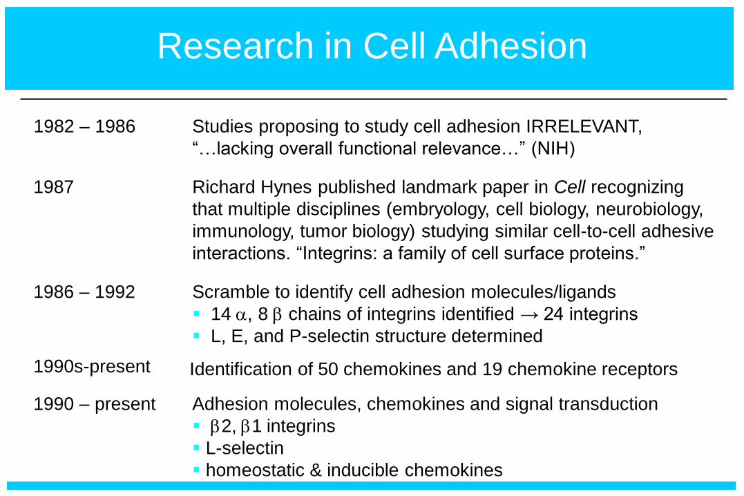
Research in Cell Adhesion
1982 – 1986 Studies proposing to study cell adhesion IRRELEVANT,
“…lacking overall functional relevance…” (NIH)
1987 Richard Hynes published landmark paper in Cell recognizing
that multiple disciplines (embryology, cell biology, neurobiology,
immunology, tumor biology) studying similar cell-to-cell adhesive
interactions. “Integrins: a family of cell surface proteins.”
1986 – 1992 Scramble to identify cell adhesion molecules/ligands
14 a, 8 b chains of integrins identified → 24 integrins
L, E, and P-selectin structure determined
1990 – present Adhesion molecules, chemokines and signal transduction
b2, b1 integrins
L-selectin
homeostatic & inducible chemokines
1990s-present Identification of 50 chemokines and 19 chemokine receptors
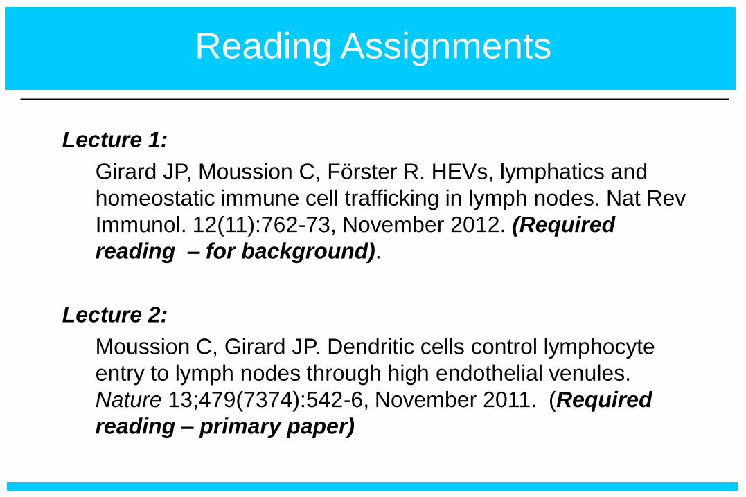
Reading Assignments
Lecture 1:
Girard JP, Moussion C, Förster R. HEVs, lymphatics and
homeostatic immune cell trafficking in lymph nodes. Nat Rev
Immunol. 12(11):762-73, November 2012. (Required
reading – for background).
Lecture 2:
Moussion C, Girard JP. Dendritic cells control lymphocyte
entry to lymph nodes through high endothelial venules.
Nature 13;479(7374):542-6, November 2011. (Required
reading – primary paper)
Be able to read and understand field of adhesion and
leukocyte trafficking.
Working knowledge of experimental approaches used
to dissect role of adhesion molecules/chemokines in
leukocyte trafficking.
Understand structure/function relationship of trafficking
molecules that direct leukocyte migration under
homeostatic versus inflammatory conditions.
Objectives

Outline
General introduction to lymphocyte trafficking – homeostatic
recirculation vs active recruitment.
Homeostatic trafficking of lymphocytes across specialized high
endothelial venules (HEVs)
Structure/function of adhesion molecules involved in initial tethering/rolling
(L-selectin/PNAd)
Structure/function of chemokine/chemokine receptors that mediate activation
(CCR7/CCL21)
Structure/function of adhesion molecules required for firm
arrest/transendothelial migration (LFA-1/ICAM-1 & α4β7 integrin/MAdCAM-1)
Leukocyte recruitment in acute and chronic inflammation.
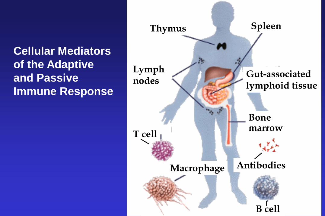
Thymus Spleen
Lymph nodes
Gut-associated lymphoid tissue
Bone marrow
T cell
Antibodies
B cell
Macrophage
Cellular Mediators
of the Adaptive
and Passive
Immune Response
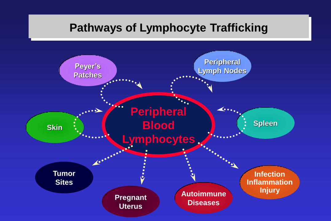
Peripheral
Blood
Lymphocytes
Infection Inflammation
Injury Autoimmune
Diseases
Peripheral
Lymph Nodes Peyer’s
Patches
Spleen Skin
Pathways of Lymphocyte Trafficking
Tumor
Sites
Pregnant
Uterus
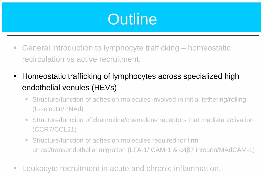
Outline
General introduction to lymphocyte trafficking – homeostatic
recirculation vs active recruitment.
Homeostatic trafficking of lymphocytes across specialized high
endothelial venules (HEVs)
Structure/function of adhesion molecules involved in initial tethering/rolling
(L-selectin/PNAd)
Structure/function of chemokine/chemokine receptors that mediate activation
(CCR7/CCL21)
Structure/function of adhesion molecules required for firm
arrest/transendothelial migration (LFA-1/ICAM-1 & α4β7 integrin/MAdCAM-1)
Leukocyte recruitment in acute and chronic inflammation.
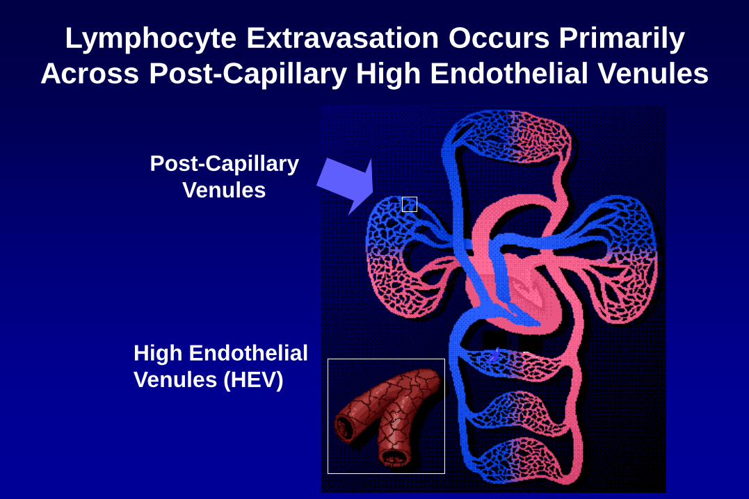
Lymphocyte Extravasation Occurs Primarily
Across Post-Capillary High Endothelial Venules
High Endothelial
Venules (HEV)
Post-Capillary
Venules
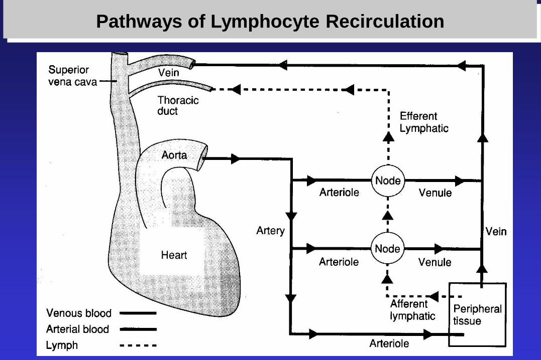
Pathways of Lymphocyte Recirculation
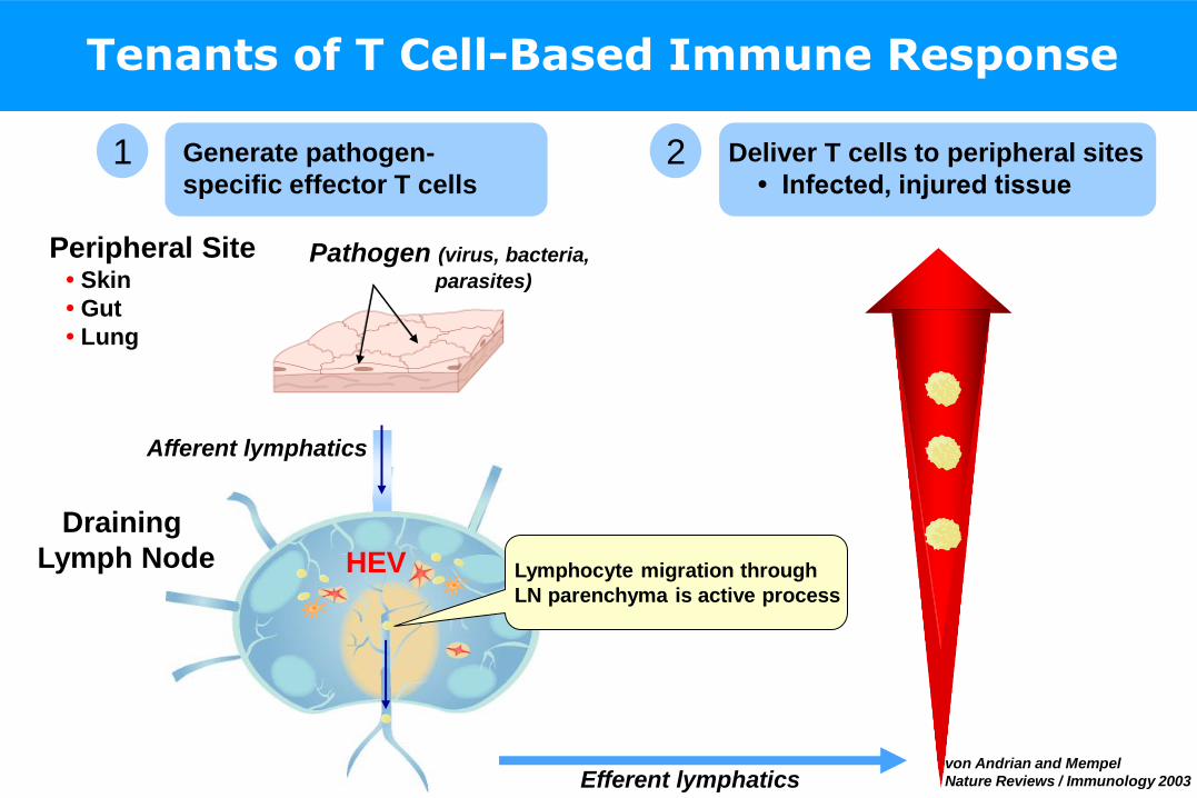
Pathogen (virus, bacteria,
parasites)
HEV
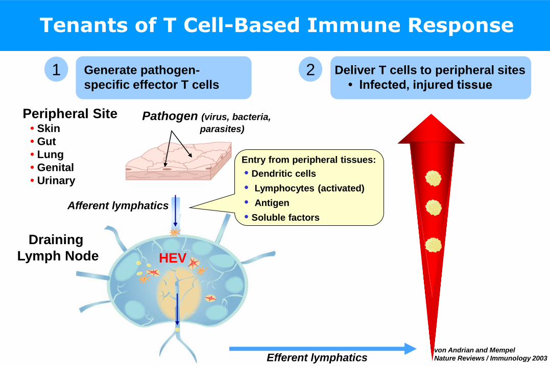
Tenants of T Cell-Based Immune Response
1 Generate pathogen-
specific effector T cells
Draining
Lymph Node
Afferent lymphatics
Efferent lymphatics
Deliver T cells to peripheral sites
• Infected, injured tissue
Peripheral Site • Skin
• Gut
• Lung
• Genital
• Urinary
von Andrian and Mempel
Nature Reviews / Immunology 2003
2
Entry from peripheral tissues:
• Dendritic cells
• Lymphocytes (activated)
• Antigen
• Soluble factors
Pathogen (virus, bacteria,
parasites)
HEV
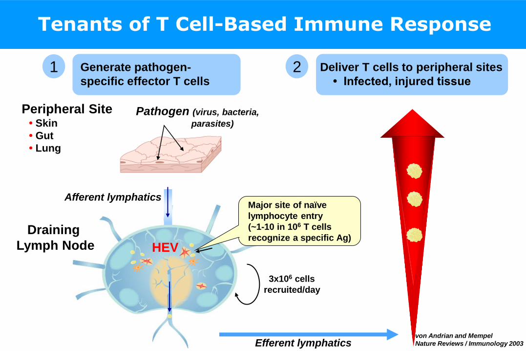
Tenants of T Cell-Based Immune Response
1 2 Generate pathogen-
specific effector T cells
Draining
Lymph Node
Afferent lymphatics
Efferent lymphatics
Deliver T cells to peripheral sites
• Infected, injured tissue
Peripheral Site • Skin
• Gut
• Lung
3x10⁶ cells
recruited/day
Major site of naïve
lymphocyte entry
(~1-10 in 106 T cells
recognize a specific Ag)
von Andrian and Mempel
Nature Reviews / Immunology 2003
Pathogen (virus, bacteria,
parasites)
HEV
Tenants of T Cell-Based Immune Response
1 2 Generate pathogen-
specific effector T cells
Draining
Lymph Node
Afferent lymphatics
Efferent lymphatics
Deliver T cells to peripheral sites
• Infected, injured tissue
Peripheral Site • Skin
• Gut
• Lung
Lymphocyte migration through
LN parenchyma is active process
von Andrian and Mempel
Nature Reviews / Immunology 2003
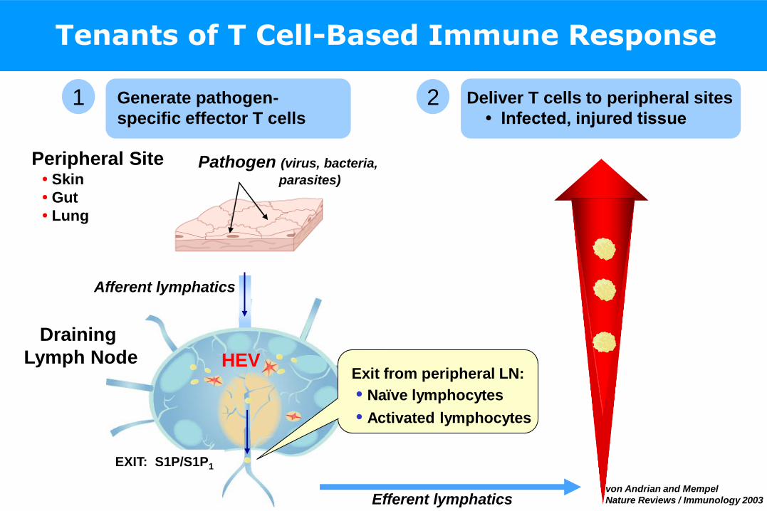
Pathogen (virus, bacteria,
parasites)
HEV
Tenants of T Cell-Based Immune Response
1 2 Generate pathogen-
specific effector T cells
Draining
Lymph Node
Afferent lymphatics
Efferent lymphatics
Deliver T cells to peripheral sites
• Infected, injured tissue
Peripheral Site • Skin
• Gut
• Lung
Exit from peripheral LN:
• Naïve lymphocytes
• Activated lymphocytes
EXIT: S1P/S1P1
von Andrian and Mempel
Nature Reviews / Immunology 2003
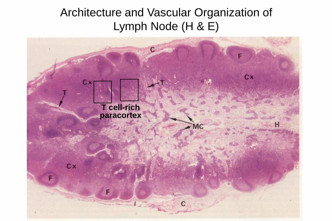
Architecture and Vascular Organization of
Lymph Node (H & E)
T cell-rich paracortex
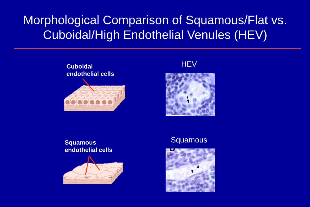
Morphological Comparison of Squamous/Flat vs.
Cuboidal/High Endothelial Venules (HEV)
HEV Cuboidal
endothelial cells
Squamous
endothelial cells
Squamous
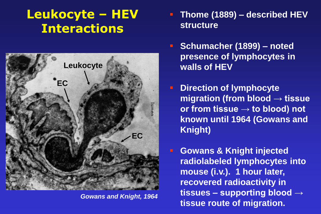
Leukocyte – HEV Interactions
Gowans and Knight, 1964
EC
EC
Leukocyte
Thome (1889) – described HEV
structure
Schumacher (1899) – noted
presence of lymphocytes in
walls of HEV
Direction of lymphocyte
migration (from blood → tissue
or from tissue → to blood) not
known until 1964 (Gowans and
Knight)
Gowans & Knight injected
radiolabeled lymphocytes into
mouse (i.v.). 1 hour later,
recovered radioactivity in
tissues – supporting blood →
tissue route of migration.
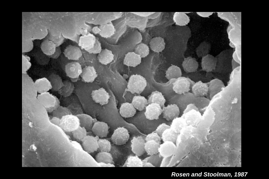
Rosen and Stoolman, 1987
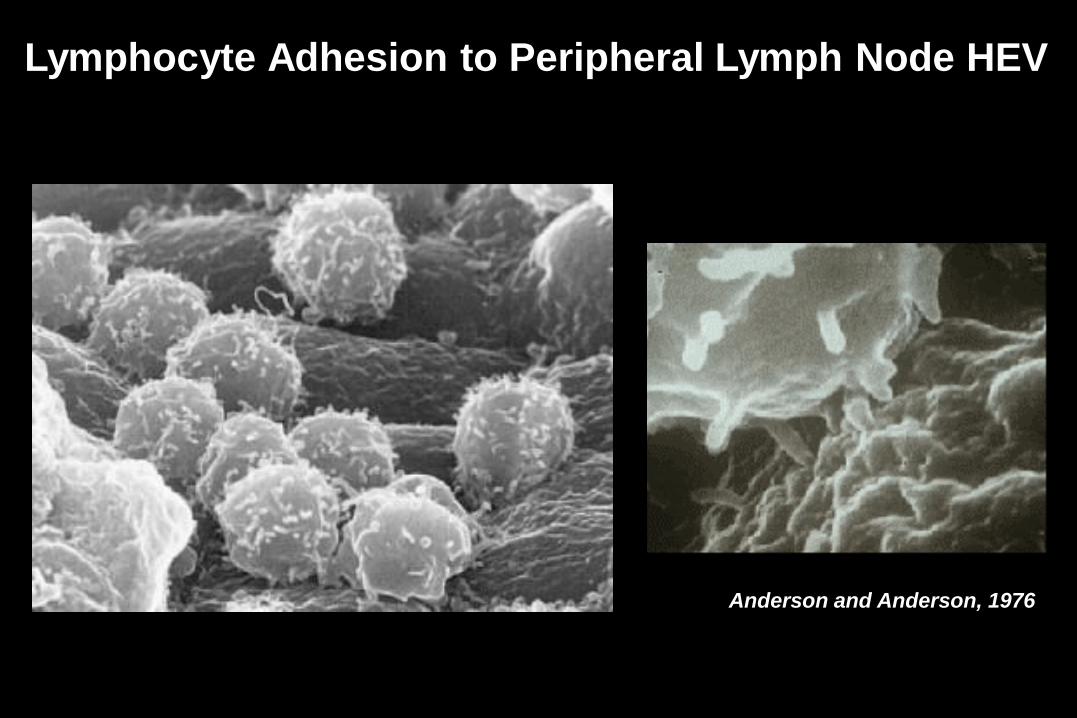
Anderson and Anderson, 1976
Lymphocyte Adhesion to Peripheral Lymph Node HEV
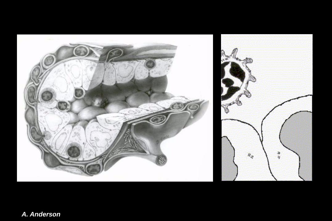
A. Anderson
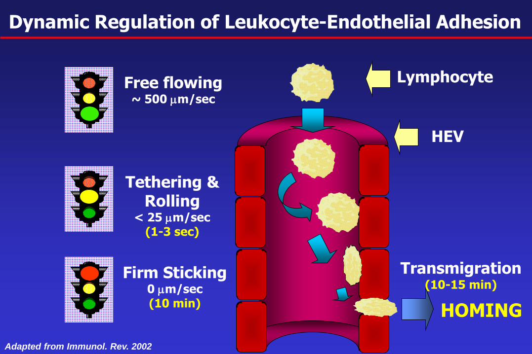
HOMING
Free flowing ~ 500 mm/sec
Tethering & Rolling
< 25 mm/sec (1-3 sec)
Firm Sticking 0 mm/sec (10 min)
Dynamic Regulation of Leukocyte-Endothelial Adhesion
Transmigration (10-15 min)
Lymphocyte
HEV
Adapted from Immunol. Rev. 2002
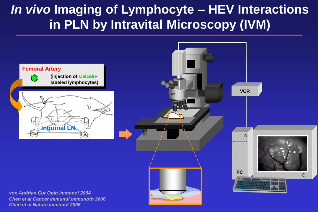
In vivo Imaging of Lymphocyte – HEV Interactions
in PLN by Intravital Microscopy (IVM)
Femoral Artery
(injection of Calcein-
labeled lymphocytes)
Inguinal LN
VCR
PC
von Andrian Cur Opin Immunol 2004
Chen et al Cancer Immunol Immunoth 2006
Chen et al Nature Immunol 2006
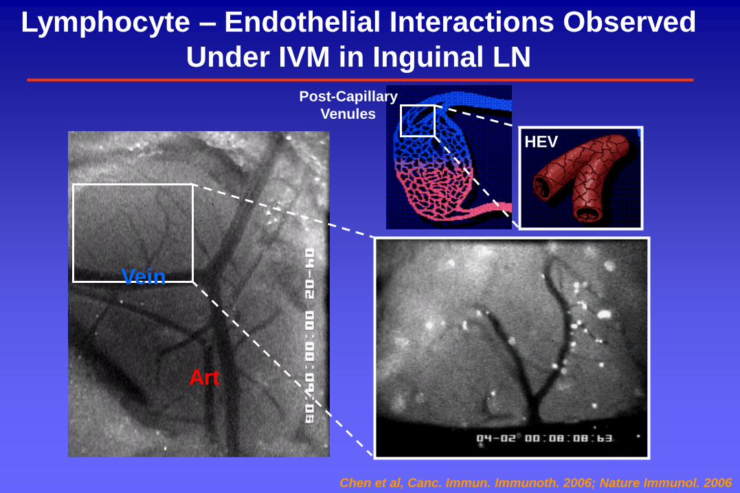
Lymphocyte – Endothelial Interactions Observed
Under IVM in Inguinal LN
Vein
Art
Chen et al, Canc. Immun. Immunoth. 2006; Nature Immunol. 2006
Post-Capillary
Venules
HEV
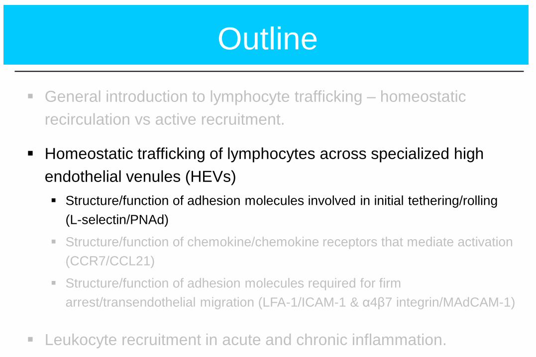
Outline
General introduction to lymphocyte trafficking – homeostatic
recirculation vs active recruitment.
Homeostatic trafficking of lymphocytes across specialized high
endothelial venules (HEVs)
Structure/function of adhesion molecules involved in initial tethering/rolling
(L-selectin/PNAd)
Structure/function of chemokine/chemokine receptors that mediate activation
(CCR7/CCL21)
Structure/function of adhesion molecules required for firm
arrest/transendothelial migration (LFA-1/ICAM-1 & α4β7 integrin/MAdCAM-1)
Leukocyte recruitment in acute and chronic inflammation.
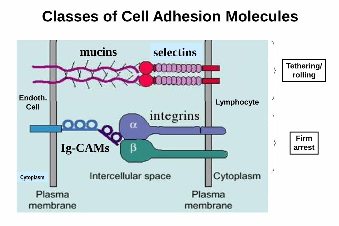
Ig-CAMs
mucins
Cytoplasm
Classes of Cell Adhesion Molecules
mucins selectins
Lymphocyte Endoth.
Cell
Tethering/
rolling
Firm
arrest
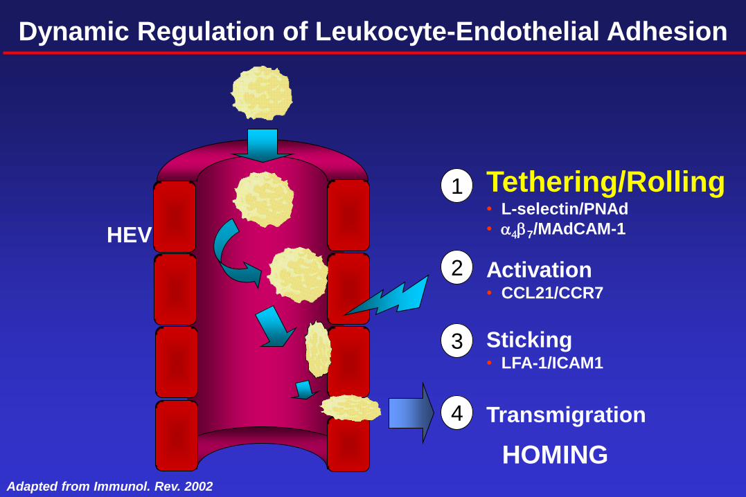
1
2
3
Tethering/Rolling • L-selectin/PNAd
• a4b7/MAdCAM-1
Activation • CCL21/CCR7
Sticking • LFA-1/ICAM1
Transmigration
4
Adapted from Immunol. Rev. 2002
Dynamic Regulation of Leukocyte-Endothelial Adhesion
HOMING
HEV
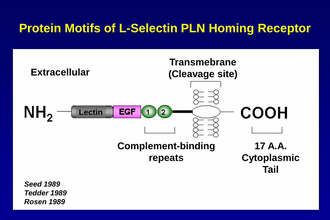
Complement-binding
repeats
Transmebrane
(Cleavage site)
17 A.A.
Cytoplasmic
Tail
Seed 1989
Tedder 1989
Rosen 1989
Protein Motifs of L-Selectin PLN Homing Receptor
Extracellular
Lectin
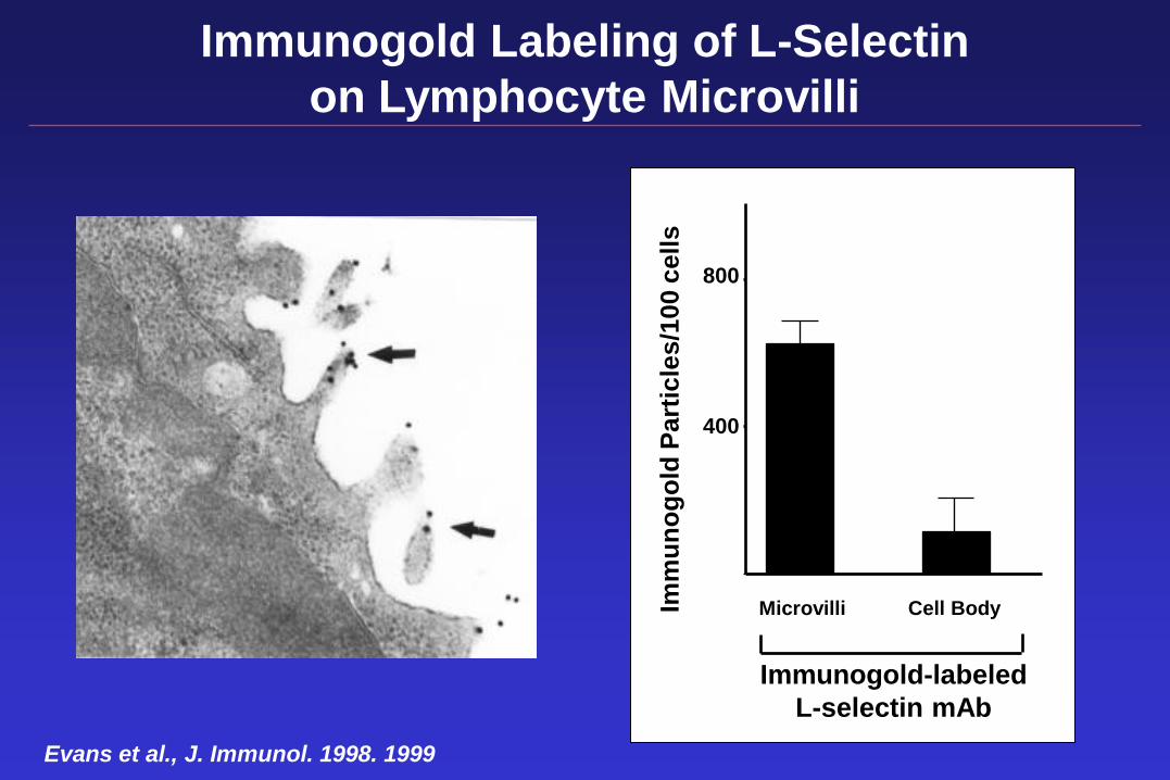
Immunogold Labeling of L-Selectin
on Lymphocyte Microvilli
Microvilli Cell Body
Im
mu
no
go
ld P
art
icle
s/1
00
ce
lls
Immunogold-labeled
L-selectin mAb
Evans et al., J. Immunol. 1998. 1999
800
400
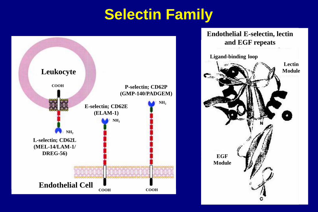
Endothelial Cell
Leukocyte
COOH
NH2
L-selectin; CD62L
(MEL-14/LAM-1/
DREG-56)
E-selectin; CD62E
(ELAM-1)
NH2
COOH COOH
NH2
P-selectin; CD62P
(GMP-140/PADGEM)
Endothelial E-selectin, lectin
and EGF repeats
Ligand-binding loop
EGF
Module
Lectin
Module
Selectin Family
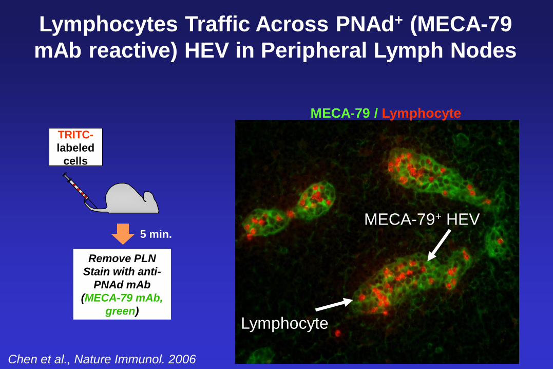
Remove PLN
Stain with anti-
PNAd mAb
(MECA-79 mAb,
green)
5 min.
TRITC-
labeled
cells
Lymphocytes Traffic Across PNAd+ (MECA-79
mAb reactive) HEV in Peripheral Lymph Nodes
MECA-79+ HEV
Lymphocyte
MECA-79 / Lymphocyte
Chen et al., Nature Immunol. 2006
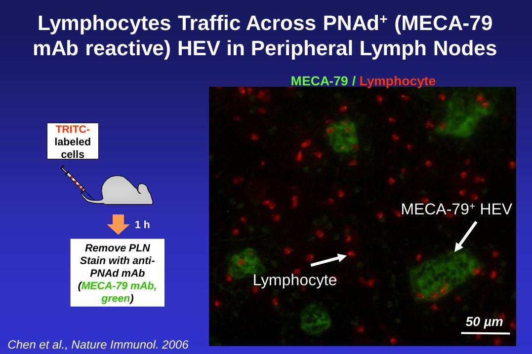
MECA-79+ HEV
Lymphocyte
50 µm
MECA-79 / Lymphocyte
Remove PLN
Stain with anti-
PNAd mAb
(MECA-79 mAb,
green)
1 h
TRITC-
labeled
cells
Chen et al., Nature Immunol. 2006
Lymphocytes Traffic Across PNAd+ (MECA-79
mAb reactive) HEV in Peripheral Lymph Nodes
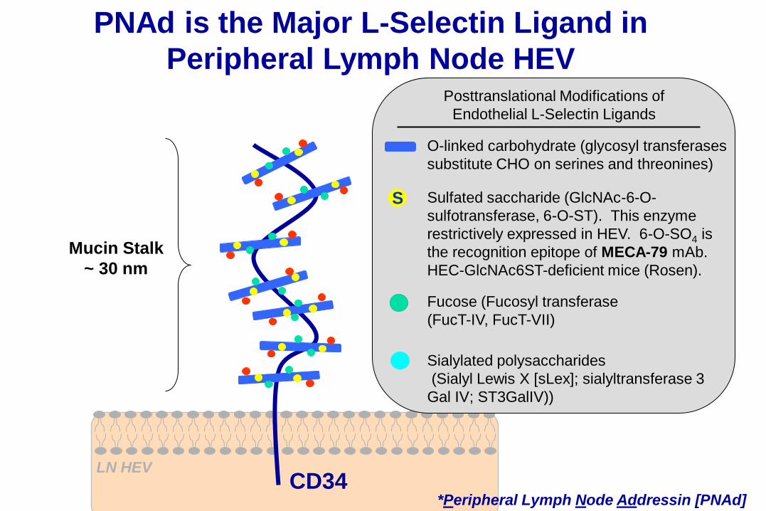
LN HEV
PNAd is the Major L-Selectin Ligand in
Peripheral Lymph Node HEV
Mucin Stalk
~ 30 nm
O-linked carbohydrate (glycosyl transferases
substitute CHO on serines and threonines)
Fucose (Fucosyl transferase
(FucT-IV, FucT-VII)
Sialylated polysaccharides
(Sialyl Lewis X [sLex]; sialyltransferase 3
Gal IV; ST3GalIV))
Sulfated saccharide (GlcNAc-6-O-
sulfotransferase, 6-O-ST). This enzyme
restrictively expressed in HEV. 6-O-SO4 is
the recognition epitope of MECA-79 mAb.
HEC-GlcNAc6ST-deficient mice (Rosen).
Posttranslational Modifications of
Endothelial L-Selectin Ligands
*Peripheral Lymph Node Addressin [PNAd] CD34
S
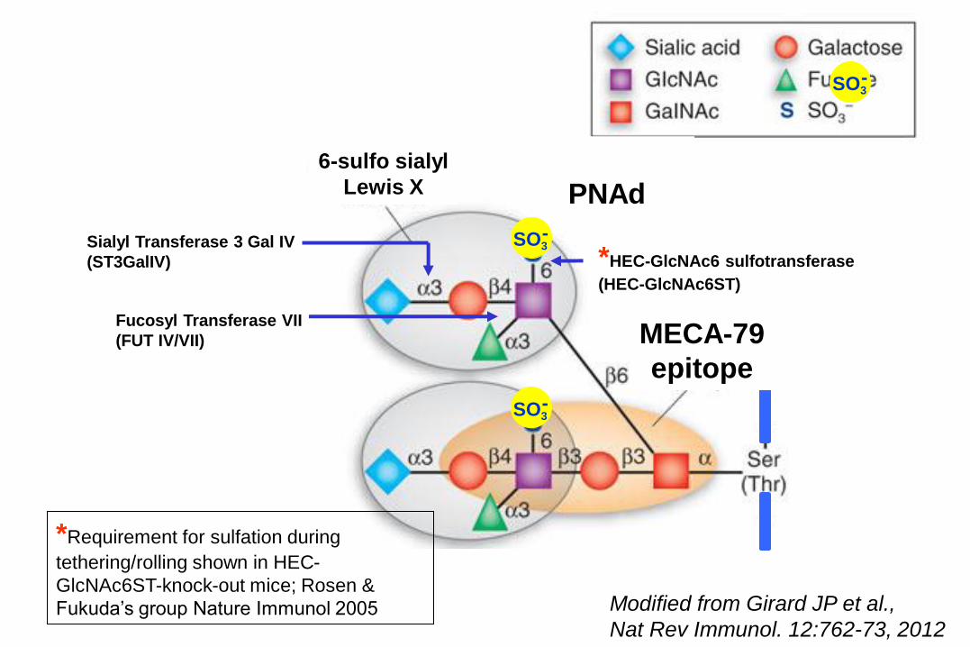
*HEC-GlcNAc6 sulfotransferase
(HEC-GlcNAc6ST)
*Requirement for sulfation during
tethering/rolling shown in HEC-
GlcNAc6ST-knock-out mice; Rosen &
Fukuda’s group Nature Immunol 2005
SO3 -
6-sulfo sialyl
Lewis X
MECA-79
epitope
SO3 -
SO3 -
Fucosyl Transferase VII
(FUT IV/VII)
Sialyl Transferase 3 Gal IV
(ST3GalIV)
Modified from Girard JP et al.,
Nat Rev Immunol. 12:762-73, 2012
PNAd
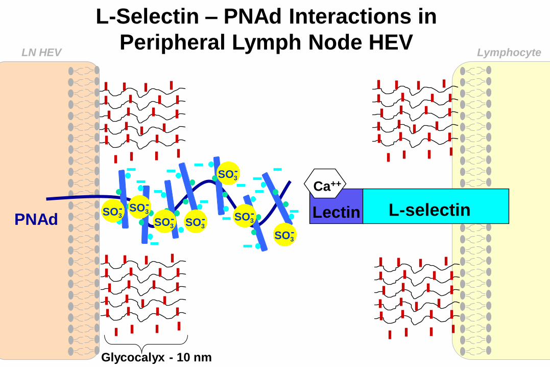
L-Selectin – PNAd Interactions in
Peripheral Lymph Node HEV
LN HEV
PNAd
Glycocalyx - 10 nm
L-selectin Lectin
Ca++
Lymphocyte
SO3 -
SO3 -
SO3 -
SO3 -
SO3 -
SO3 -
SO3 -
PNAd
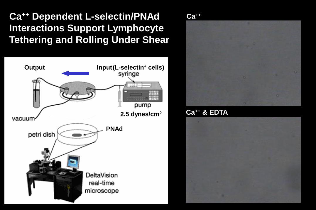
Input Output (L-selectin+ cells)
Ca++ & EDTA 2.5 dynes/cm2
Ca++ Ca++ Dependent L-selectin/PNAd
Interactions Support Lymphocyte
Tethering and Rolling Under Shear
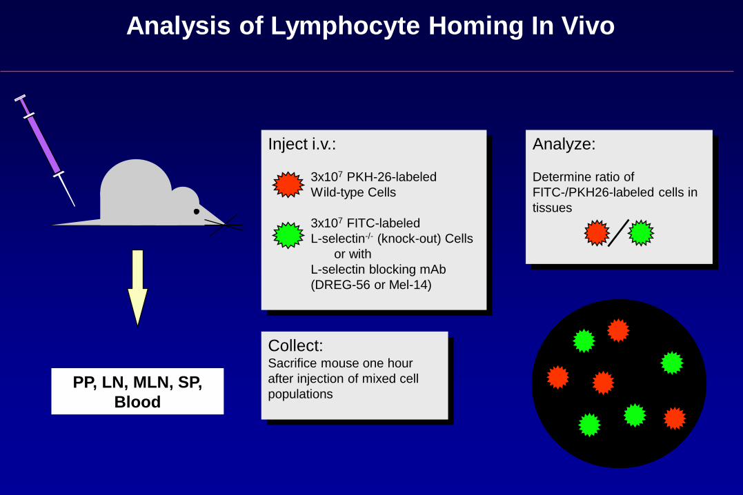
Analysis of Lymphocyte Homing In Vivo
Inject i.v.:
3x10⁷ PKH-26-labeled
Wild-type Cells
3x10⁷ FITC-labeled
L-selectin-/- (knock-out) Cells
or with
L-selectin blocking mAb
(DREG-56 or Mel-14)
Collect: Sacrifice mouse one hour
after injection of mixed cell
populations
Analyze:
Determine ratio of
FITC-/PKH26-labeled cells in
tissues
PP, LN, MLN, SP,
Blood
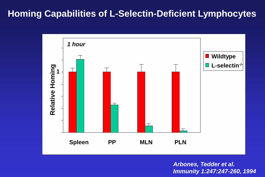
Homing Capabilities of L-Selectin-Deficient Lymphocytes
Arbones, Tedder et al.
Immunity 1:247:247-260, 1994
Spleen PP MLN PLN
Wildtype
L-selectin-/-
Rela
tiv
e H
om
ing
1
1 hour
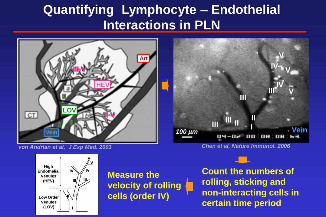
Count the numbers of
rolling, sticking and
non-interacting cells in
certain time period
Measure the
velocity of rolling
cells (order IV)
Quantifying Lymphocyte – Endothelial
Interactions in PLN
II II
III III
III III
IV
IV
V
V
V
I 100 µm Vein
von Andrian et al, J Exp Med. 2003
HEV
Art
III-V
III-V
Vein
I I
II
LOV
Chen et al, Nature Immunol. 2006
I
II II
III III
IV IV
V V
Low Order
Venules
(LOV)
High
Endothelial
Venules
(HEV)
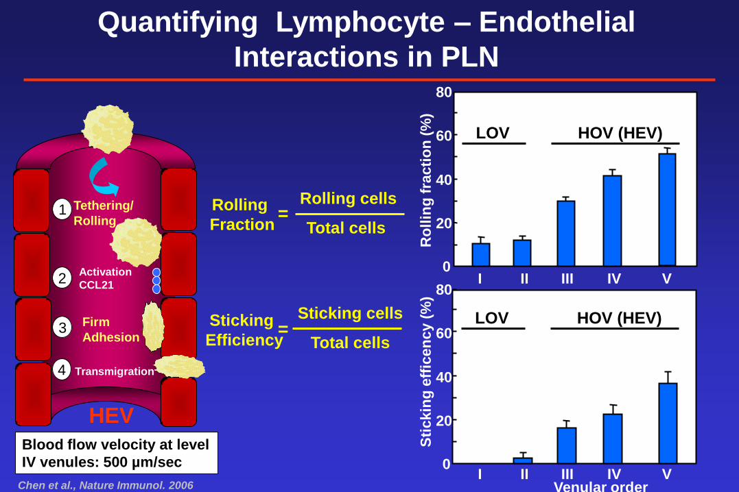
Rolling
Fraction =
Sticking
Efficiency =
0
20
40
60
80
Venular order
Sti
ckin
g e
ffic
en
cy (
%)
Ro
llin
g f
racti
on
(%
)
0
20
40
60
80
I II III V IV
I II III V IV
HEV
1
3
2
4
Firm
Adhesion
Transmigration
Activation
CCL21
Tethering/
Rolling
Rolling cells
Total cells
Sticking cells
Total cells
Quantifying Lymphocyte – Endothelial
Interactions in PLN
LOV HOV (HEV)
LOV HOV (HEV)
Chen et al., Nature Immunol. 2006
Blood flow velocity at level
IV venules: 500 µm/sec
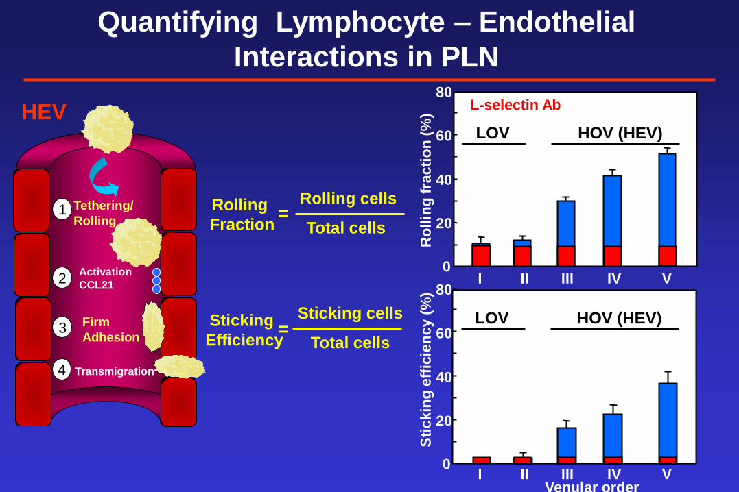
Rolling
Fraction =
Ro
llin
g f
racti
on
(%
)
0
20
40
60
80
I II III V IV
HEV
1
3
2
4
Firm
Adhesion
Transmigration
Activation
CCL21
Tethering/
Rolling
Rolling cells
Total cells
Quantifying Lymphocyte – Endothelial
Interactions in PLN
LOV HOV (HEV)
= Sticking cells
Total cells
Sticking
Efficiency
IV Venular order
0
20
40
60
80
Sti
ckin
g e
ffic
ien
cy (
%)
I II III V
LOV HOV (HEV)
L-selectin Ab
Venular order
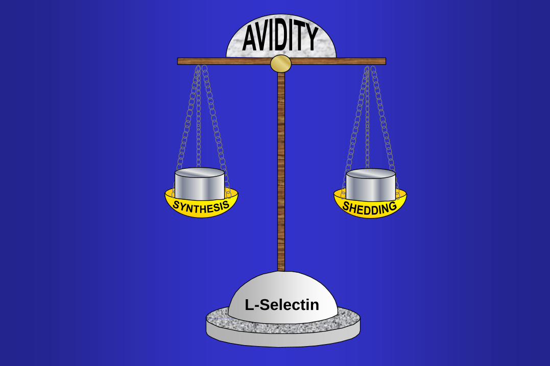
L-Selectin
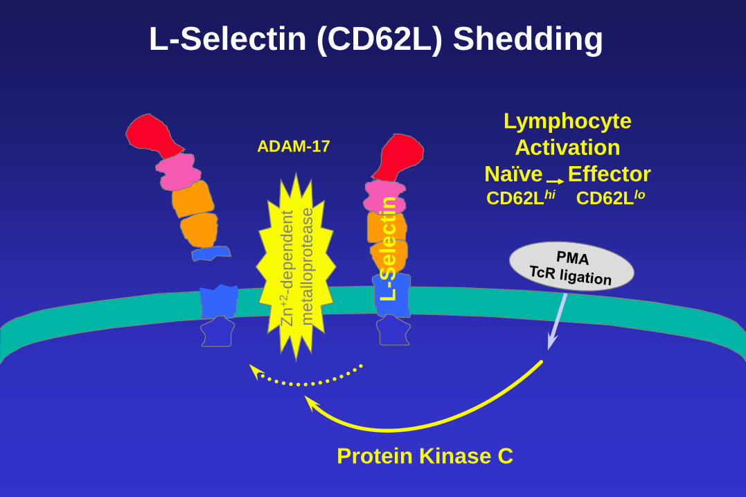
L-S
ele
cti
n
Protein Kinase C
Zn
+2-d
ep
en
de
nt
me
tallo
pro
tease
L-Selectin (CD62L) Shedding
Lymphocyte
Activation
Naïve Effector
CD62Lhi CD62Llo
ADAM-17
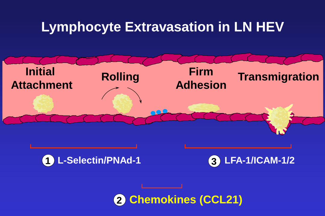
Lymphocyte Extravasation in LN HEV
L-Selectin/PNAd-1
Chemokines (CCL21)
LFA-1/ICAM-1/2 1
2
3
Initial
Attachment Rolling Firm
Adhesion Transmigration
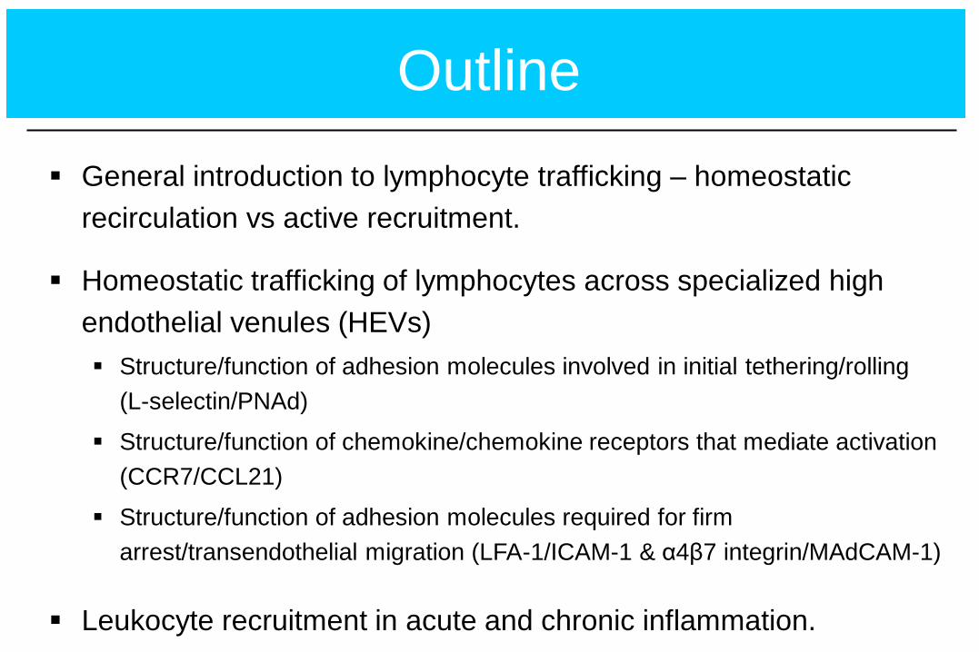
Outline
General introduction to lymphocyte trafficking – homeostatic
recirculation vs active recruitment.
Homeostatic trafficking of lymphocytes across specialized high
endothelial venules (HEVs)
Structure/function of adhesion molecules involved in initial tethering/rolling
(L-selectin/PNAd)
Structure/function of chemokine/chemokine receptors that mediate activation
(CCR7/CCL21)
Structure/function of adhesion molecules required for firm
arrest/transendothelial migration (LFA-1/ICAM-1 & α4β7 integrin/MAdCAM-1)
Leukocyte recruitment in acute and chronic inflammation.