Embed Size (px)
Citation preview

Acute Kidney Injury
July 28, 2015 Katharine Dahl, MD
Definition
Rapid decrease in GFR (minutes-days) ↓
Metabolic waste rate of production>rate of excretion
↓ Rise in serum markers of renal function:
urea and creatinine

Manifestations
• Azotemia progressing to uremia • Hyperkalemia • Metabolic acidosis • Volume overload • Hyperphosphatemia • Accumulation and toxicity of medications
excreted by the kidney
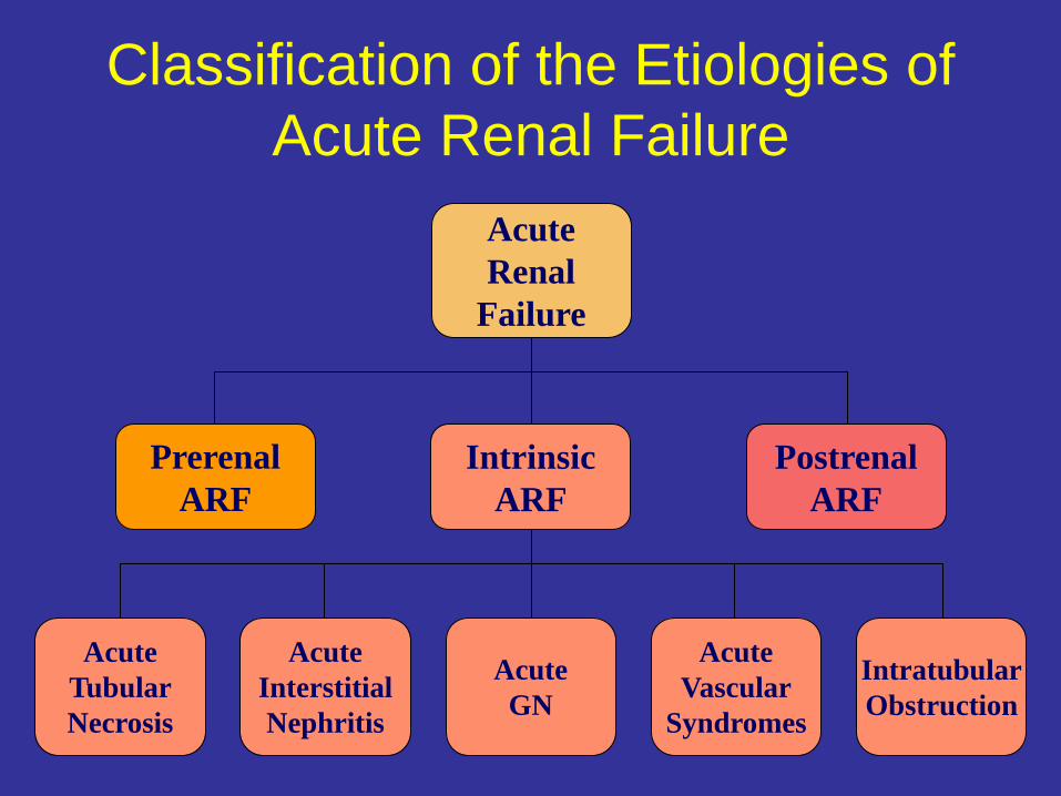
Acute Tubular Necrosis
Acute Interstitial Nephritis
Acute GN
Acute Vascular
Syndromes
Intratubular Obstruction
Classification of the Etiologies of Acute Renal Failure
Prerenal ARF
Postrenal ARF
Intrinsic ARF
Acute Renal
Failure
Prerenal Azotemia
• Functional response to renal hyperfusion • Renal structure and microstructure are
preserved • Complete recovery may be seen in 1-2
days after relief of offending lesion (normal perfusion)

Postrenal Azotemia
• Obstruction of the urinary tract • Few microscopic changes initially,
– Early hydronephrosis – Pelvic dilation – Distention or blunting of the renal papilla
• Or no microscopic changes • Complete recovery may be seen in 1-2
days after relief of offending lesion (normal outflow)

Intrinsic Renal Azotemia
• Parenchymal injury of the blood vessels, glomeruli, tubules, or interstitium
• Recovery may be prolonged
Ischemic Acute Renal Failure: prerenal azotemia and acute
tubular necrosis • Prerenal azotemia and ATN account for
more than half of the cases of ARF seen in hospitalized patients
• Often, the contribution of ischemia is unrecognized because the patient is normotensive

Prerenal Acute Renal Failure: Clinical Presentation
• BUN:Creatinine ratio – > 20:1
• Urine indices – Oliguria
• usually < 500 mL/24 hours; but may be non-oliguric – Elevated urine concentration
• UOsm > 700 mmol/L • specific gravity > 1.020
– Evidence of high renal sodium avidity • UNa < 20 mmol/L • FENa < 0.01
– Inactive urine sediment

Response to Hypoperfusion: Renin-Angiotensin System
Renin Angiotensinogen → AT-I
AT-I → AT-II Proximal mechanism Distal mechanism Constricts efferent arteriole Stimulates zona glomerulosa ↑ glom. hydrostatic pressure to secrete aldosterone. & ↓ renal blood flow ↑ Na channels in distal & FF ↑ (FF=GFR/RPF) collecting tubules ↑ Na reabsorption Stimulates Na-K-ATPase pump at
basolateral membrane
Role of Antidiuretic Hormone • Synthesized in the hypothalamus • Stored in posterior pituitary • Release stimulated by
– hyperosmolalty (sensitive, 1% change stimulates release) – volume depletion (less sensitive – only released if enough
change in volume to lower blood pressure; renin/sympathetic system will be stimulated first).
• Binds to V2 receptor on the basolateral membrane of the principal cells in the collecting tubules → apical membrane insertion of aquaporin-2 channels
• Binds to V1a receptors on the vascular smooth muscle cells

Renal Nerves
• Sympathetic nerves directly increase afferent and efferent arterial tone – ↓ both RPF & GFR, favoring sodium retention
• Directly stimulate proximal tubules and thick ascending limb segments
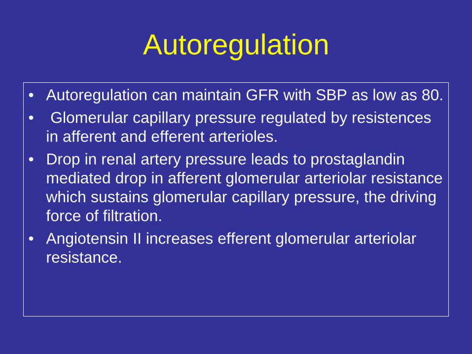
Autoregulation • Autoregulation can maintain GFR with SBP as low as 80. • Glomerular capillary pressure regulated by resistences
in afferent and efferent arterioles. • Drop in renal artery pressure leads to prostaglandin
mediated drop in afferent glomerular arteriolar resistance which sustains glomerular capillary pressure, the driving force of filtration.
• Angiotensin II increases efferent glomerular arteriolar resistance.

Renal Response to Ischemia: autoregulation
Image from Annals of Thoracic Surgery, 65:993-8, April 1998
Prostaglandins dilate afferent arteriole
Angiotensin II constricts the efferent arteriole

Causes of Autoregulation Failure: NSAIDs
Image from Annals of Thoracic Surgery, 65:993-8, April 1998
Prostaglandins dilate afferent arteriole
Angiotensin II, Norepinephrine constrict the efferent arteriole unopposed
NSAIDS
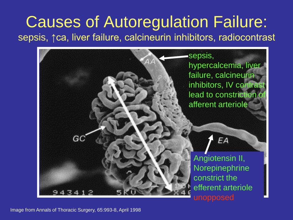
Causes of Autoregulation Failure: sepsis, ↑ca, liver failure, calcineurin inhibitors, radiocontrast
Image from Annals of Thoracic Surgery, 65:993-8, April 1998
sepsis, hypercalcemia, liver failure, calcineurin inhibitors, IV contrast lead to constriction of afferent arteriole
Angiotensin II, Norepinephrine constrict the efferent arteriole unopposed
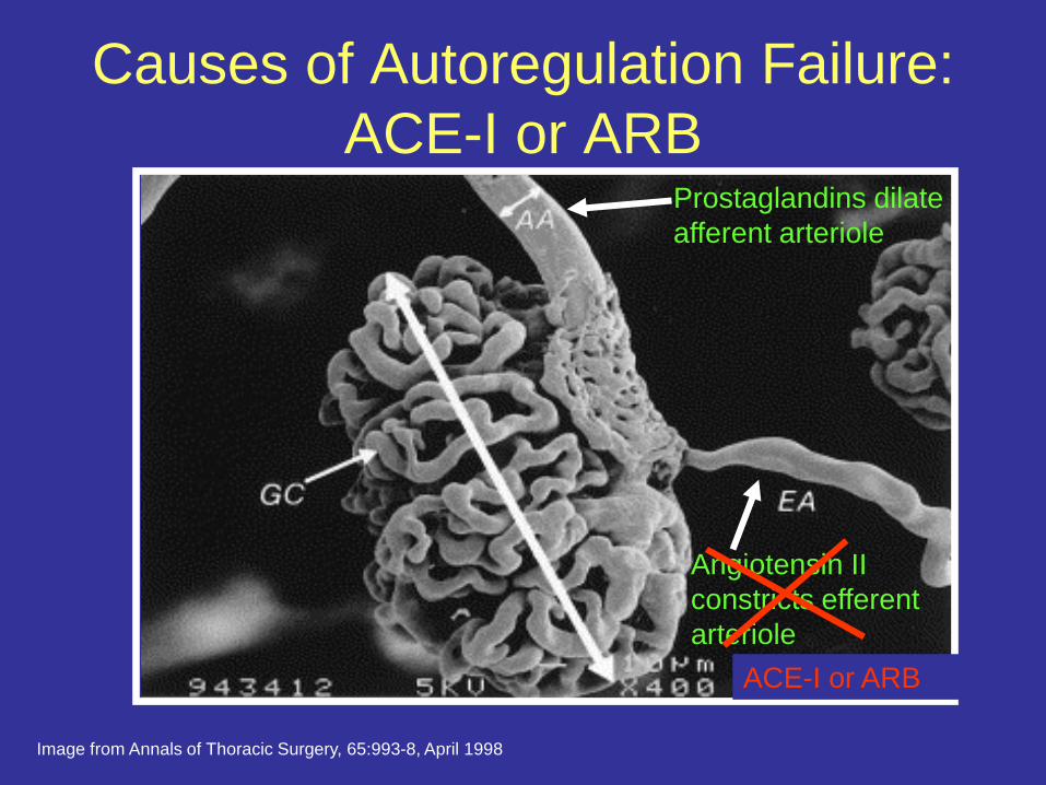
Causes of Autoregulation Failure: ACE-I or ARB
Image from Annals of Thoracic Surgery, 65:993-8, April 1998
Prostaglandins dilate afferent arteriole
Angiotensin II constricts efferent arteriole
ACE-I or ARB
Normotensive Ischemic Acute Renal Failure
• Increased renal susceptibility to modest reductions in perfusion pressure
• Caused by failure to decrease afferent arteriolar resistance or failure to increase efferent arteriolar resistance

Factors Increasing Susceptibility to Renal Hypoperfusion:
Failure to Decrease Afferent Arteriolar Resistance
• Structural changes in small arteries/arterioles – Old age – Atherosclerosis – Chronic hypertension – Chronic kidney disease – Malignant hypertension
• Reduction in vasodilatory prostaglandins – NSAIDs – COX-2 inhibitors
• Afferent arteriolar constriction – Sepsis – Hypercalcemia – Hepatorenal syndrome – Cyclosporine or tacrolimus – Radiocontrast agents

Factors Increasing Susceptibility to Renal Hypoperfusion
Failure to Constrict Efferent Arteriole:
• ACE-I • ARB
Structural changes • Renal artery stenosis
Image from VascularWeb.org Image from Annals of Thoracic Surgery, 65:993-8, April 1998
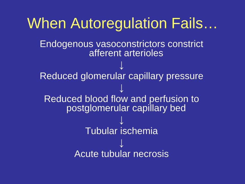
When Autoregulation Fails… Endogenous vasoconstrictors constrict
afferent arterioles ↓
Reduced glomerular capillary pressure ↓
Reduced blood flow and perfusion to postglomerular capillary bed
↓ Tubular ischemia
↓ Acute tubular necrosis
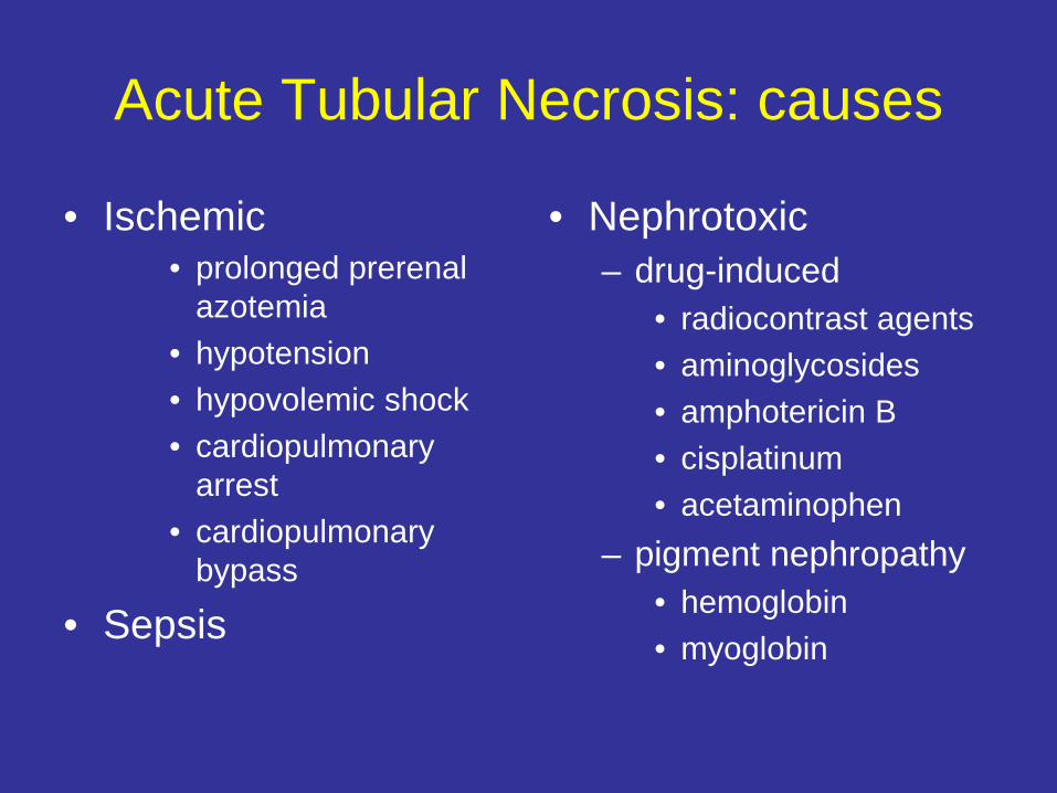
Acute Tubular Necrosis: causes
• Ischemic • prolonged prerenal
azotemia • hypotension • hypovolemic shock • cardiopulmonary
arrest • cardiopulmonary
bypass
• Sepsis
• Nephrotoxic – drug-induced
• radiocontrast agents • aminoglycosides • amphotericin B • cisplatinum • acetaminophen
– pigment nephropathy • hemoglobin • myoglobin

Pathophysiology of ATN: Tubular Epithelial Cell Injury and
Repair
Loss of polarity Normal Epithelium
Migration , Dedifferentiation of Viable Cells
Differentiation & Reestablishment of polarity
Sloughing of viable and dead cells with luminal obstruction
Ischemia/ Reperfusion
Apoptosis Necrosis
Cell death
Adhesion molecules Na+/K+-ATPase
Proliferation

Acute Tubular Necrosis: Clinical Presentation
• Urine indices – Urine volume
• may be oliguric or non-oliguric – Isosthenuric urine concentration
• UOsm ≅ 300 mmol/L • specific gravity ≅ 1.010
– Evidence of renal sodium wasting • UNa > 40 mmol/L • FENa > 0.02
– Urine sediment • tubular epithelial cell casts • Pigmented or “muddy brown” granular casts
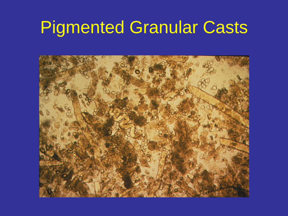
Pigmented Granular Casts
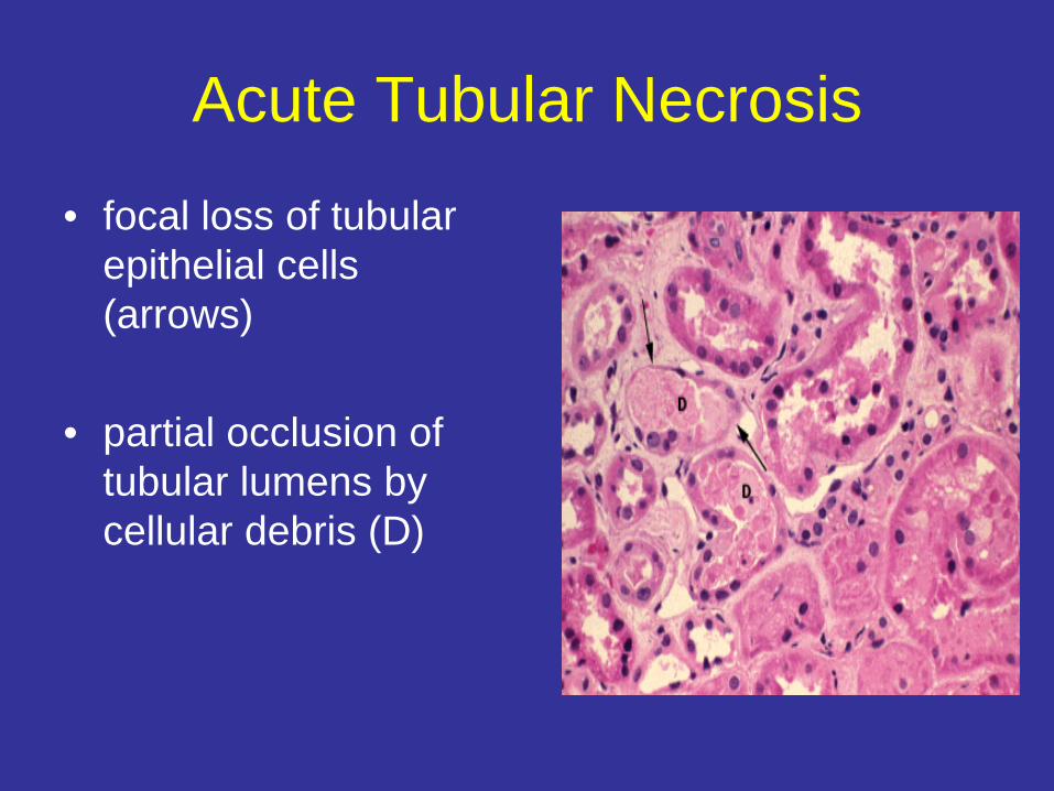
Acute Tubular Necrosis
• focal loss of tubular epithelial cells (arrows)
• partial occlusion of
tubular lumens by cellular debris (D)
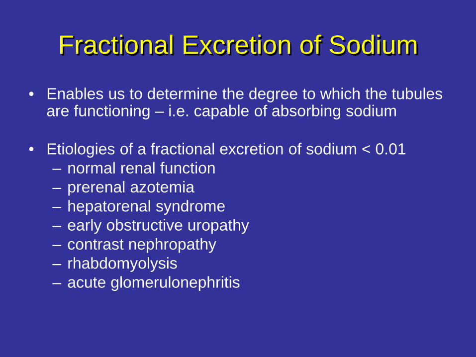
Fractional Excretion of Sodium
• Enables us to determine the degree to which the tubules are functioning – i.e. capable of absorbing sodium
• Etiologies of a fractional excretion of sodium < 0.01 – normal renal function – prerenal azotemia – hepatorenal syndrome – early obstructive uropathy – contrast nephropathy – rhabdomyolysis – acute glomerulonephritis
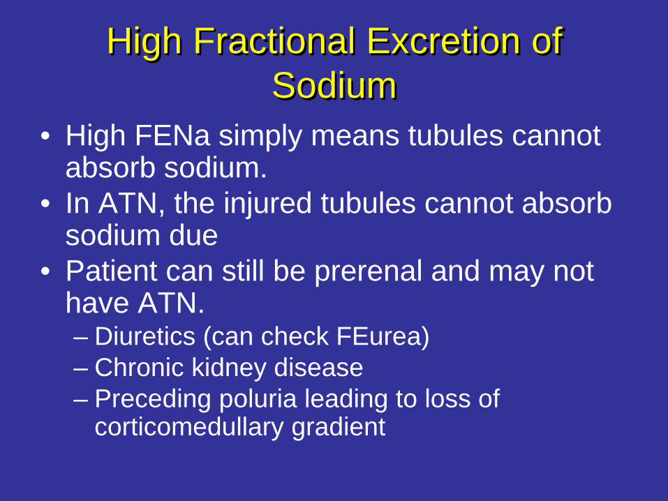
High Fractional Excretion of Sodium
• High FENa simply means tubules cannot absorb sodium.
• In ATN, the injured tubules cannot absorb sodium due
• Patient can still be prerenal and may not have ATN. – Diuretics (can check FEurea) – Chronic kidney disease – Preceding poluria leading to loss of
corticomedullary gradient
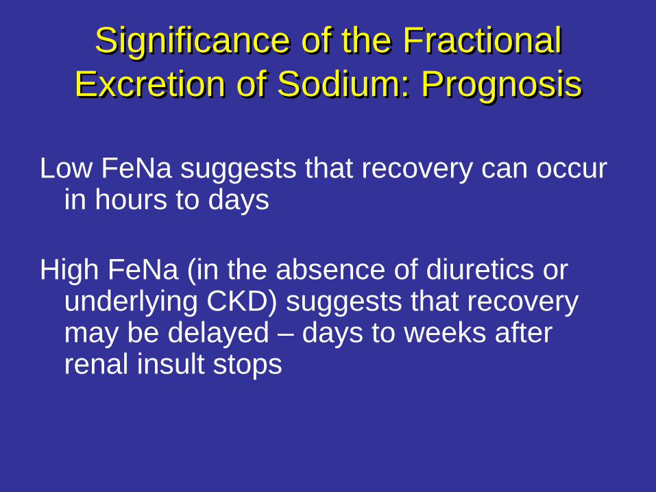
Significance of the Fractional Excretion of Sodium: Prognosis
Low FeNa suggests that recovery can occur
in hours to days High FeNa (in the absence of diuretics or
underlying CKD) suggests that recovery may be delayed – days to weeks after renal insult stops
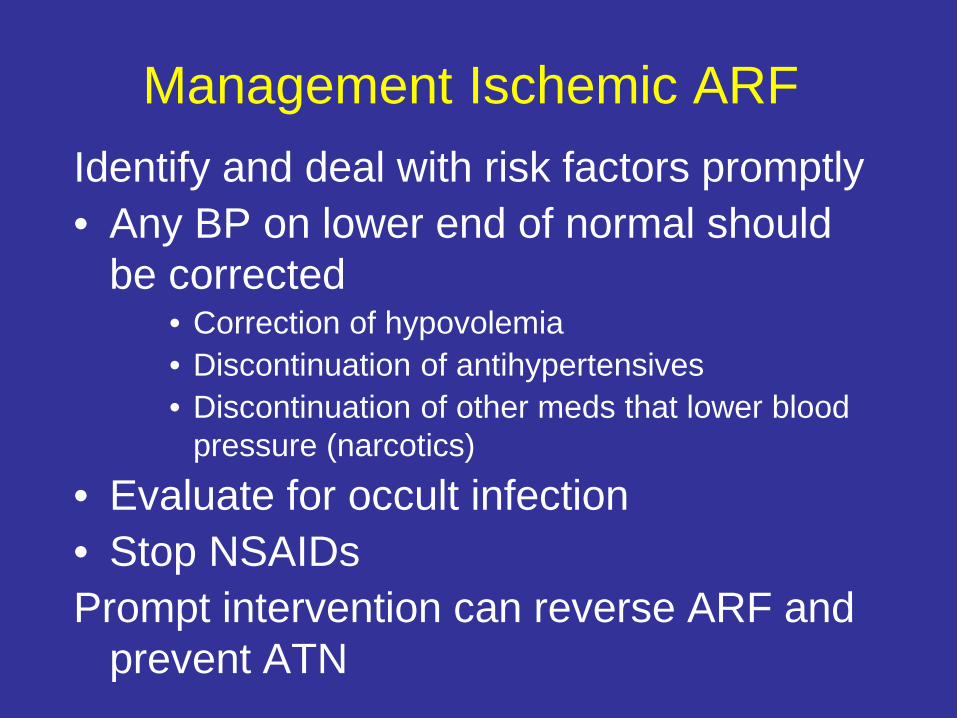
Management Ischemic ARF Identify and deal with risk factors promptly • Any BP on lower end of normal should
be corrected • Correction of hypovolemia • Discontinuation of antihypertensives • Discontinuation of other meds that lower blood
pressure (narcotics)
• Evaluate for occult infection • Stop NSAIDs Prompt intervention can reverse ARF and
prevent ATN

Acute Tubular Necrosis: Treatment
• Supportive therapy: avoid volume depletion, hypotension, nsaids, unnecessary anesthesia, surgery, or contrast
• Adjust doses of renally excreted medications • Acute dialysis for:
– volume overload – metabolic acidosis – hyperkalemia – uremic syndrome: pericarditis, encephalopathy

Mortality in Acute Tubular Necrosis
Chertow et al: Arch Int Med 1995; 155:1505-1511
0%
20%
40%
60%
80%
100%
0 1 2 3 4Number of Failed Non-Respiratory Organ Systems

Acute Renal Failure and Sepsis
• 51% of patients with septic shock with positive blood cultures have ARF
• Combination of ARF and sepsis is associated with 70% mortality
• Arterial vasodilation mediated by nitric oxide predisposes patients to ARF
• Metabolic acidosis and down-regulation of vasoactive hormone receptors leads to resistance to norepinephrine and angiogensin II

Acute Renal Failure and Sepsis: Role of Vasopressin
• Activates V1a receptors in vascular smooth muscle • Modulates ATP-Sensitive K channels • Attenuates the effect of nitric oxide • Potentiates other adrenergic and vasoconstrictor
agents • Constricts the efferent but not the afferent arteriole,
thus preserving renal perfusion • Levels high for the 1st hr of vasodilatory shock, but
then pituitary stores are depleted

0
5
10
15
20
25
30
35 Septic Shock
(n=19) Cardiogenic Shock
(n=12)
3.1 ± 0.4
22.7 ± 2.2
AVP pg/ml
Vasopressin levels in shock

Vasopressin Immunoreactivity in Pituitary of Dog
Control Vasodilatory Shock

Vasopressin in hormone replacement doses improves renal
function in septic shock

Intrinsic Renal Azotemia
• ATN • Acute Interstitial Nephritis • Acute Glomerulonephritis

Acute Interstitial Nephritis
• Acute renal failure due to lymphocytic infiltration of the interstitium
• Classic triad of – fever – rash – eosinophilia

Acute Interstitial Nephritis: Clinical Presentation
• History – preceding illness or drug exposure
• Physical examination – fever – rash
• Serum Findings – Eosinophilia
• Urine findings – non-nephrotic range proteinuria – possibly hematuria – sterile pyuria (WBC’s without infection) – WBC casts – eosinophiluria

Acute Interstitial Nephritis: causes
• Drug-induced – penicillins – cephalosporins – sulfonamides – rifampin – phenytoin – furosemide – NSAIDs
• Malignancy • Idiopathic
• Infection-related – bacterial – viral – rickettsial – tuberculosis
• Systemic diseases – SLE – sarcoidosis – Sjögren’s syndrome – tubulointerstitial nephritis
and uveitis
Acute Interstitial Nephritis: Treatment
• Discontinue offending drug • Treat underlying infection • Treat systemic illness • Glucocorticoid therapy may be used in patients
who fail to respond to more conservative therapy
Rapidly Progressive Glomerulonephritis
• Clinical manifestations: – Macrohematuria – Oliguria – Edema – or insidious onset: fatigue and edema – With anti-GBM disease or pauci-immune
glomerulonephritis (Granulomatosis with poyangiitis), may have pulmonary symptoms

Rapidly Progressive Glomerulonephritis:classifications
• Type I: anti GBM disease • Type II: Immune complex
– IgA – Post-infectious GN – Lupus nephritis – Cryoglobulinemia
• Type III: pauci-immune – Usually ANCA positive, but can be negative
Post-Renal Failure:obstruction
• Risk factors – BPH – Abdominal cancer – Nephrolithiasis with baseline chronic kidney
disease – Abdominal compartment syndrome (maybe)
• Diagnosis – Check bladder scan for post-void residual – Renal ultrasound
CASES

Case 1 An 87-year old female nursing home resident has been
admitted for altered mental status. Records from the nursing home state that she has been more withdrawn lately, not eating, but with no specific complaints.
PMH: HTN, arthritis, dementia, osteoarthritis occasional
UTIs. Meds: HCTZ, lisinopril, naprosyn prn In ER: pt is awake and moaning, but otherwise
noncommunicative.
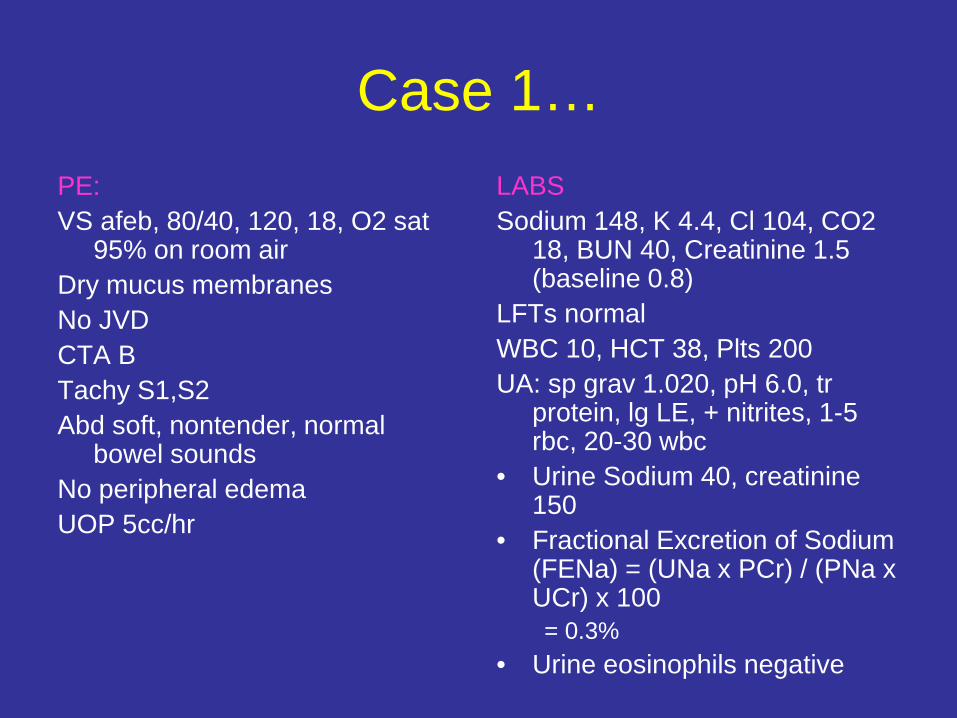
Case 1… PE: VS afeb, 80/40, 120, 18, O2 sat
95% on room air Dry mucus membranes No JVD CTA B Tachy S1,S2 Abd soft, nontender, normal
bowel sounds No peripheral edema UOP 5cc/hr
LABS Sodium 148, K 4.4, Cl 104, CO2
18, BUN 40, Creatinine 1.5 (baseline 0.8)
LFTs normal WBC 10, HCT 38, Plts 200 UA: sp grav 1.020, pH 6.0, tr
protein, lg LE, + nitrites, 1-5 rbc, 20-30 wbc
• Urine Sodium 40, creatinine 150
• Fractional Excretion of Sodium (FENa) = (UNa x PCr) / (PNa x UCr) x 100 = 0.3%
• Urine eosinophils negative
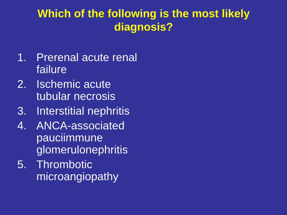
Which of the following is the most likely diagnosis?
1. Prerenal acute renal
failure 2. Ischemic acute
tubular necrosis 3. Interstitial nephritis 4. ANCA-associated
pauciimmune glomerulonephritis
5. Thrombotic microangiopathy
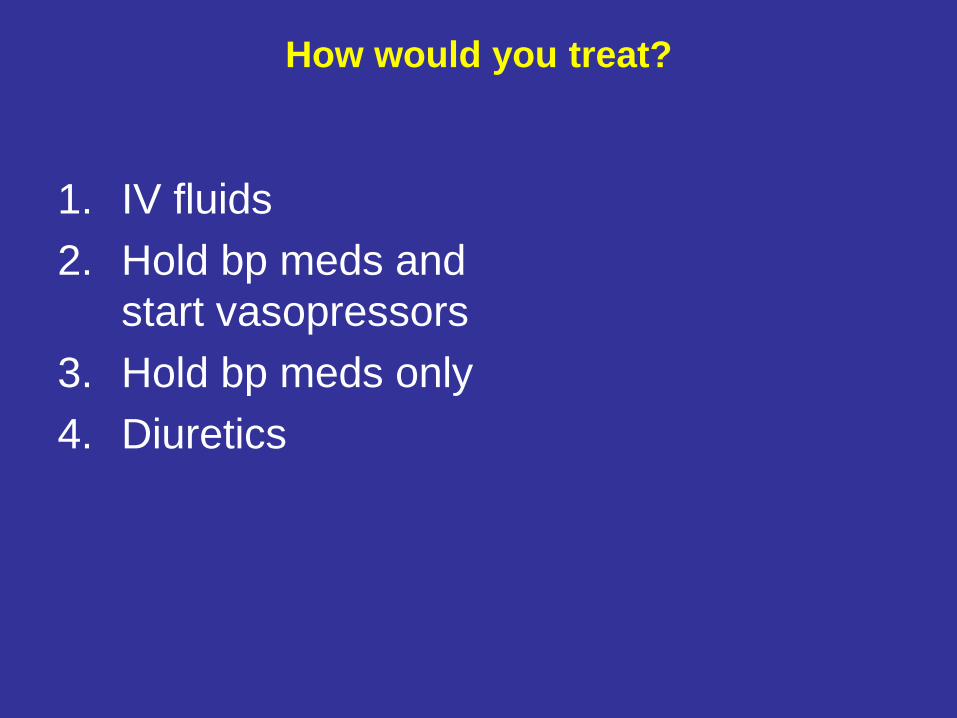
How would you treat?
1. IV fluids 2. Hold bp meds and
start vasopressors 3. Hold bp meds only 4. Diuretics

Case 1 continued… Hospital course: she is admitted to the ICU, given
aggressive hydration and antibiotics for UTI. Over the next 2 days, her weight increases from 66kg to 72kg. BP 120/80, hr 88. She is still lethargic.
Sodium 143, BUN 35, Creatinine 2.6. UOP 0. Urine Sodium 80, Urine creatinine 60 Fractional Excretion of Sodium (FENa) = (UNa x PCr) /
(PNa x UCr) x 100 = 2.4%
Urine eosinophils negative Urine Culture >100,000 E. Coli

Now which of the following is the most likely diagnosis?
1. Prerenal acute renal failure
2. Ischemic acute tubular necrosis
3. Interstitial nephritis
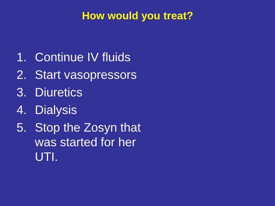
How would you treat?
1. Continue IV fluids 2. Start vasopressors 3. Diuretics 4. Dialysis 5. Stop the Zosyn that
was started for her UTI.
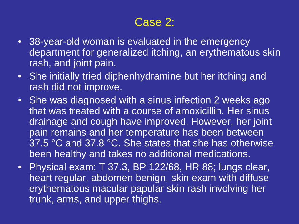
Case 2: • 38-year-old woman is evaluated in the emergency
department for generalized itching, an erythematous skin rash, and joint pain.
• She initially tried diphenhydramine but her itching and rash did not improve.
• She was diagnosed with a sinus infection 2 weeks ago that was treated with a course of amoxicillin. Her sinus drainage and cough have improved. However, her joint pain remains and her temperature has been between 37.5 °C and 37.8 °C. She states that she has otherwise been healthy and takes no additional medications.
• Physical exam: T 37.3, BP 122/68, HR 88; lungs clear, heart regular, abdomen benign, skin exam with diffuse erythematous macular papular skin rash involving her trunk, arms, and upper thighs.

Laboratory Studies
• Hemoglobin 12.5 • WBC 9.8 • Platelets 325 • Blood urea nitrogen 36 • Creatinine 2.6 • Sodium 138 meq/L • Potassium 4.4 meq/L • Bicarbonate 26 meq/L • Urinalysis pH 5, specific gravity 1.020, 2+ blood, trace
protein, 4+ leukocyte esterase, 20–25 leukocytes and several leukocyte casts/hpf, 3–5 intact erythrocytes/hpf, Hansel stain shows eosinophils
• Urine culture is negative
Which of the following is the most likely diagnosis in this patient?
1. Thrombotic
thrombocytopenic purpura
2. Antineutrophil cytoplasmic autoantibody–associated vasculitis
3. Acute tubular necrosis 4. Acute interstitial
nephritis 5. Membranous
glomerulopathy
Which of the following conditions can present with eosinophiluria?
1. Atheroembolic disease
2. Postinfectious glomerulonephritis
3. Rapidly progressive glomerulonephritis
4. Pyelonephritis 5. All of the above

Why not ANCA-associated vasculitis?
• Antineutrophil cytoplasmic autoantibody–associated small-vessel vasculitis also should be considered in patients with kidney failure and concomitant arthralgias, skin rash, and fever.
• Lack of dysmorphic erythrocytes or erythrocyte casts makes this diagnosis unlikely.

Why not TTP?
• Thrombotic thrombocytopenic purpura should be considered in patients with fever, skin rash, and kidney failure.
• Absence of concomitant anemia, mental
status changes, and thrombocytopenia makes this diagnosis less likely.
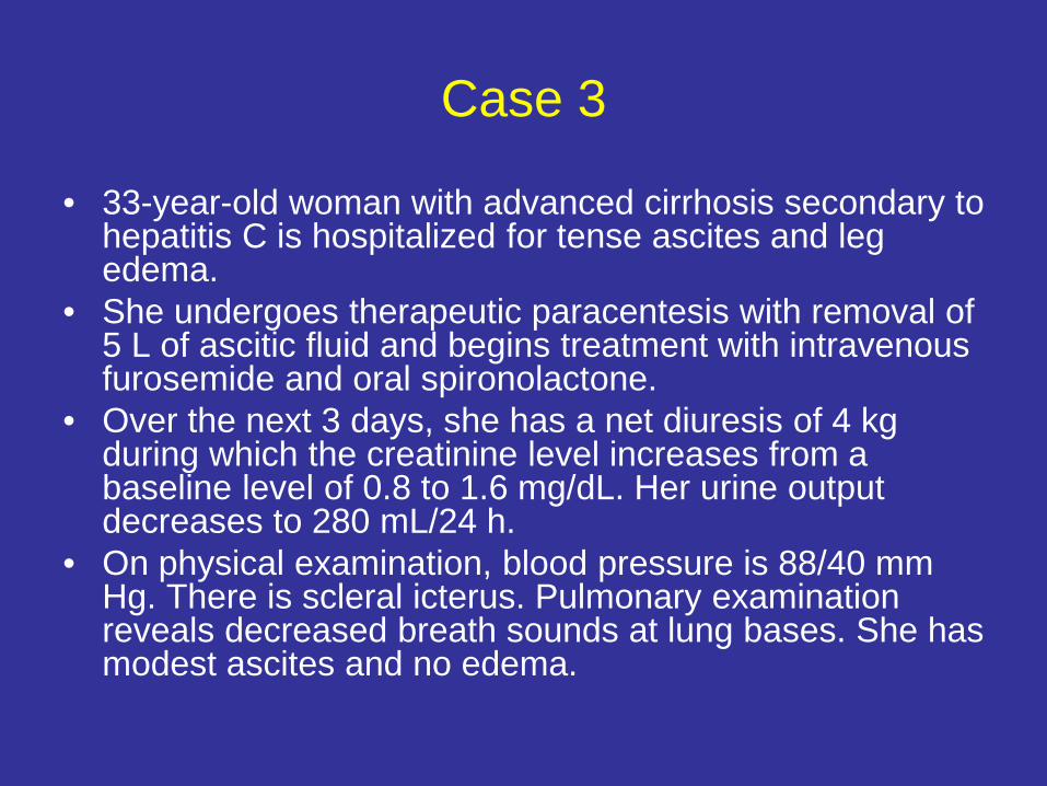
Case 3
• 33-year-old woman with advanced cirrhosis secondary to hepatitis C is hospitalized for tense ascites and leg edema.
• She undergoes therapeutic paracentesis with removal of 5 L of ascitic fluid and begins treatment with intravenous furosemide and oral spironolactone.
• Over the next 3 days, she has a net diuresis of 4 kg during which the creatinine level increases from a baseline level of 0.8 to 1.6 mg/dL. Her urine output decreases to 280 mL/24 h.
• On physical examination, blood pressure is 88/40 mm Hg. There is scleral icterus. Pulmonary examination reveals decreased breath sounds at lung bases. She has modest ascites and no edema.

Case 3...
Labs: INR 2.7 Blood urea nitrogen 21 mg/dL Creatinine 1.6 mg/dL Sodium 118 meq/L Potassium 3.4 meq/L Chloride 83 meq/L Bicarbonate 26 meq/L Total bilirubin 25 mg/dL Albumin 2.1 g/dL (23 g/L) Urinalysis Several granular and epithelial casts/hpf Urine sodium 12 meq/L Fractional Excretion of Sodium 0.8%
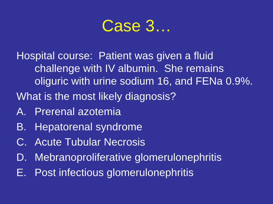
Case 3…
Hospital course: Patient was given a fluid challenge with IV albumin. She remains oliguric with urine sodium 16, and FENa 0.9%.
What is the most likely diagnosis? A. Prerenal azotemia B. Hepatorenal syndrome C. Acute Tubular Necrosis D. Mebranoproliferative glomerulonephritis E. Post infectious glomerulonephritis

Large volume paracentesis
• >5L volume removal carries greater risk of hemodynamic compromise and renal ischemia
• Albumin solution 6-8g/L of fluid removed is recommended

Hepatorenal syndrome: criteria
• Presence of liver disease • AKI (creat increase 0.3 or more within 48hrs) • Absence of other cause of renal failure • Bland urine sediment
• Urine RBCs <50/hpf • Urine Protein <500mg/day
• Lack of improvement after volume expansion with IV albumin (1g/kg of body weight per day – up to 100g/day) for 2 days

CASE 4 A 56 year old man is admitted to
the hospital with shortness of breath and lower extremity edema.
PMH: HTN, coronary artery disease s/p stents, diabetes mellitus, benign prostatic hypertrophy
PE: afebrile, BP 75/45, HR 100, RR 28, Bibasilar rales on lung exam, S1, S2, and S3 gallop, point of maximal impulse laterally displaced, jugular venous pressure is 10cm H20, abdomen is normal, 2+ lower extremity edema.
LABS: sodium 125,
potassium 3.8, chloride 89, bicarbonate 25, blood urea nitrogen 46, creatinine 2.4, albumin 3.0, urinalysis 1.020/5.0/1+ protein/no blood, urine protein to creatinine ratio 1.1g/g. Urine sodium 14, FENa 0.6%. CXR: bilateral infiltrates, cardiomegaly
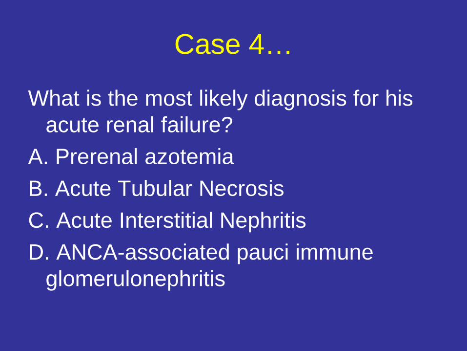
Case 4…
What is the most likely diagnosis for his acute renal failure?
A. Prerenal azotemia B. Acute Tubular Necrosis C. Acute Interstitial Nephritis D. ANCA-associated pauci immune
glomerulonephritis

Summary:take-home points
• Careful history – Look for factors increasing renal susceptibility to ischemia
• Physical exam cues – Careful investigation of blood pressure trends over the days
leading up to the insult. – Consider causes of normotensive ischemic renal failure
• Lab data: – BUN:Cr ratio – Urine sodium & creatinine (FENa) – Urinalysis – Urine eosinophils

Summary:take-home points (cont.)
• Pre-renal does not always mean volume depleted, hemodynamic compromise is a frequent culprit (CHF, too many bp meds, sepsis)
• Consider causes of normotensive ischemic renal failure
• If no response to volume/bp correction, check renal ultrasound for obstruction.
• If there is an active urine sediment, may need biopsy. Low-threshhold for expedient biopsy if renal function deteriorating.
Summary:take-home points (cont.)
• In septic shock: – Early aggressive intervention – Consider adding vasopressin early on to
improve renal perfusion and to reduce norepinephrine resistance