Embed Size (px)
Citation preview
Acute Hepatitis
Including Acute Liver Failure
Ahmed ElsharkawyLiver Unit, Queen Elizabeth Hospital, Birmingham
Stefan HübscherInstitute of Immunology & Immunotherapy, University of Birmingham
Department of Cellular Pathology, Queen Elizabeth Hospital, Birmingham
Case 1
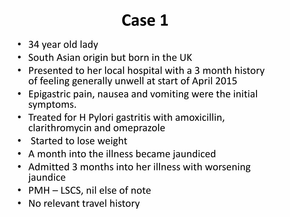
Case 1
• 34 year old lady• South Asian origin but born in the UK• Presented to her local hospital with a 3 month history
of feeling generally unwell at start of April 2015• Epigastric pain, nausea and vomiting were the initial
symptoms. • Treated for H Pylori gastritis with amoxicillin,
clarithromycin and omeprazole• Started to lose weight• A month into the illness became jaundiced• Admitted 3 months into her illness with worsening
jaundice• PMH – LSCS, nil else of note• No relevant travel history
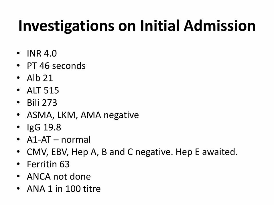
Investigations on Initial Admission
• INR 4.0• PT 46 seconds• Alb 21 • ALT 515• Bili 273• ASMA, LKM, AMA negative• IgG 19.8• A1-AT – normal• CMV, EBV, Hep A, B and C negative. Hep E awaited.• Ferritin 63• ANCA not done• ANA 1 in 100 titre

CT at Local Hospital
• Liver diffusely abnormal• Demonstrates extensive variable enhancement
characteristics with large areas of poor enhancement that are generally ill defined
• No discrete mass lesions• Overall liver a little large• Periportal oedema present• Hepatic veins are difficult to discern• No biliary dilatation• No intra-abdominal lymphadenopathy
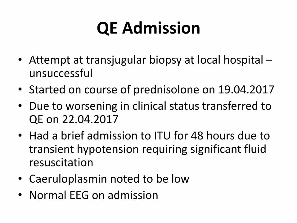
QE Admission
• Attempt at transjugular biopsy at local hospital –unsuccessful
• Started on course of prednisolone on 19.04.2017
• Due to worsening in clinical status transferred to QE on 22.04.2017
• Had a brief admission to ITU for 48 hours due to transient hypotension requiring significant fluid resuscitation
• Caeruloplasmin noted to be low
• Normal EEG on admission

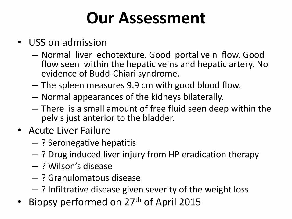
Our Assessment
• USS on admission– Normal liver echotexture. Good portal vein flow. Good
flow seen within the hepatic veins and hepatic artery. No evidence of Budd-Chiari syndrome.
– The spleen measures 9.9 cm with good blood flow.– Normal appearances of the kidneys bilaterally.– There is a small amount of free fluid seen deep within the
pelvis just anterior to the bladder.
• Acute Liver Failure – ? Seronegative hepatitis– ? Drug induced liver injury from HP eradication therapy– ? Wilson’s disease– ? Granulomatous disease– ? Infiltrative disease given severity of the weight loss
• Biopsy performed on 27th of April 2015

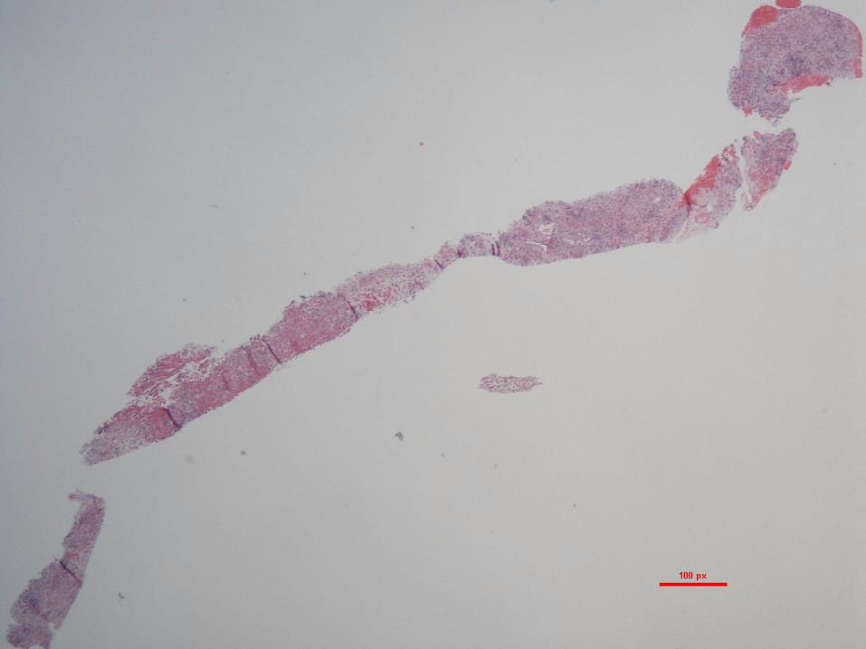
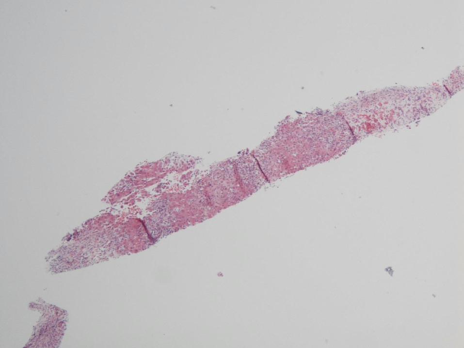
Liver Biopsy

Liver Biopsy in Acute Hepatitis
Histological Approach
1. Is this acute or chronic damage?
2. How severe is the damage?
3. What is the cause?

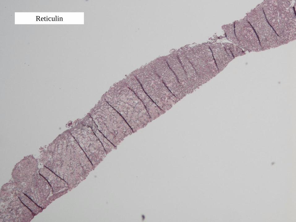
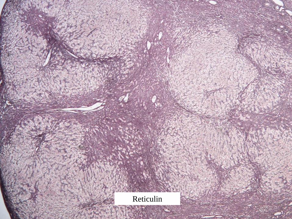
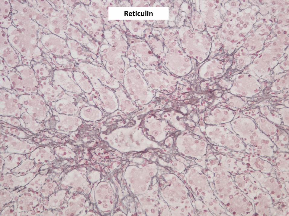
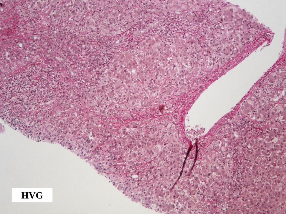
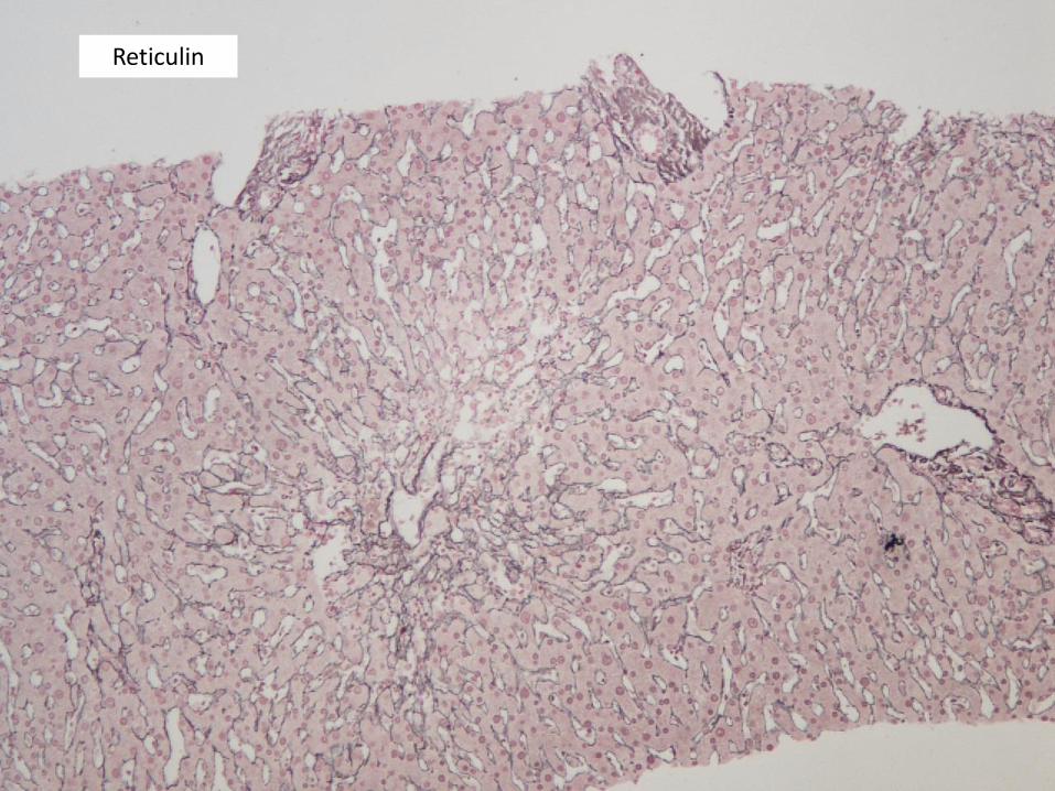
Reticulin
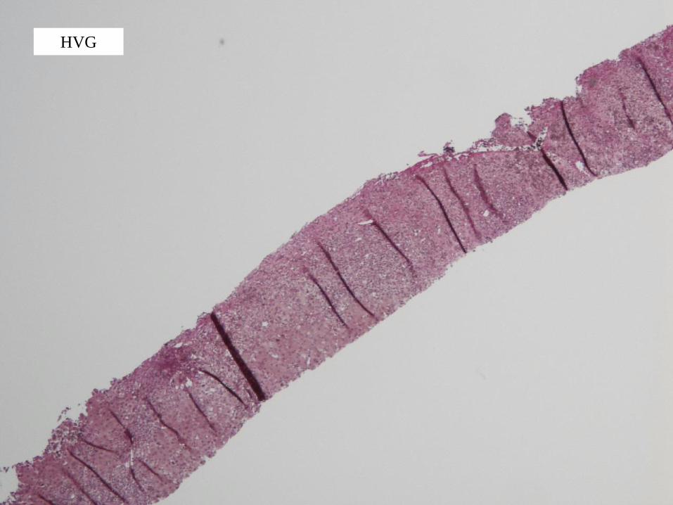
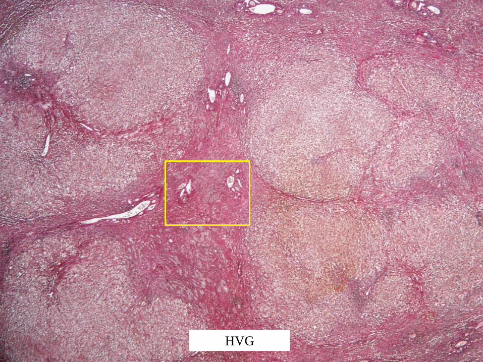
HVG

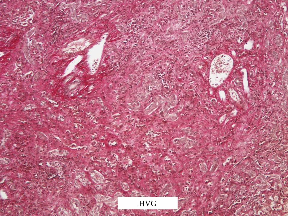
HVG

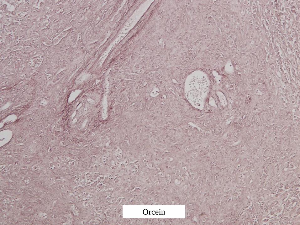
Orcein
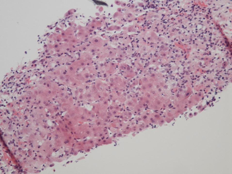
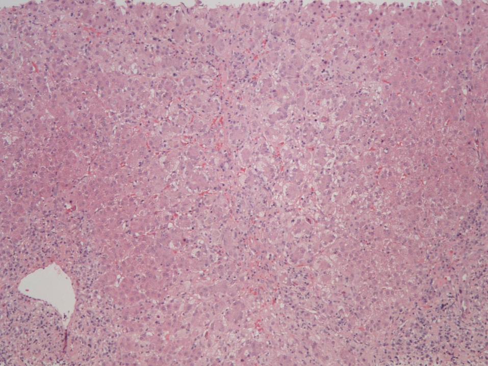
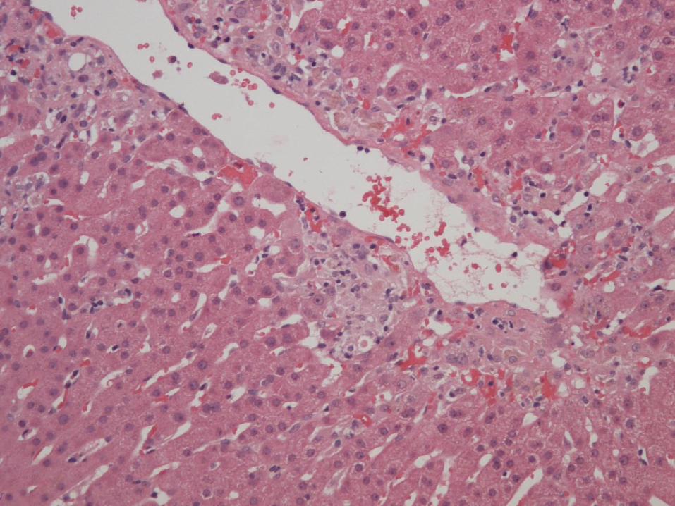
Case 1 – Liver Biopsy
Histological Findings
• Widespread recent hepatocyte necrosis
– Bridging necrosis and nodule formation
– Panacinar necrosis
• Bile ductular reaction ++
• Surviving hepatocyte nodules show spotty inflammation
Case 1 – Liver Biopsy
Diagnosis
• Acute/subacute hepatitis
• Bridging and panacinar necrosis
• No strong aetiological pointers
– Consider viral agents, drugs and autoimmune hepatitis in differential
diagnosis
– No evidence of underlying chronic liver disease or malignancy
1. Is this acute or chronic damage?
2. How severe is the damage?
3. What is the cause?

Clinical Progress
• Remained stable for first 9-10 days• No clinical signs of hepatic encephalopathy• No biochemical improvement at all• EEG on day 10 showed slowing of brain waves
consistent with hepatic encephalopathy• MDT discussion on day 10 with decision to list for
super-urgent liver transplant • Day 11 – more drowsy and requiring 20%
dextrose to maintain sugars.• Transferred back to ITU day 11• Transplant done day 13 of admission with us

Explant
Case 1 – Hepatectomy Specimen
Macroscopic Findings
• Shrunken liver, weight 475g
• Capsular surface knobbly and wrinkled
• Cut surface shows brown areas alternating with yellow areas
– Brown areas most extensive in left lobe
Liver Biopsy in the Assessment of Medical Liver Disease
• Hepatectomy specimens obtained at liver transplantation
provide valuable insights into diseases, which have a
heterogeneous distribution within the liver
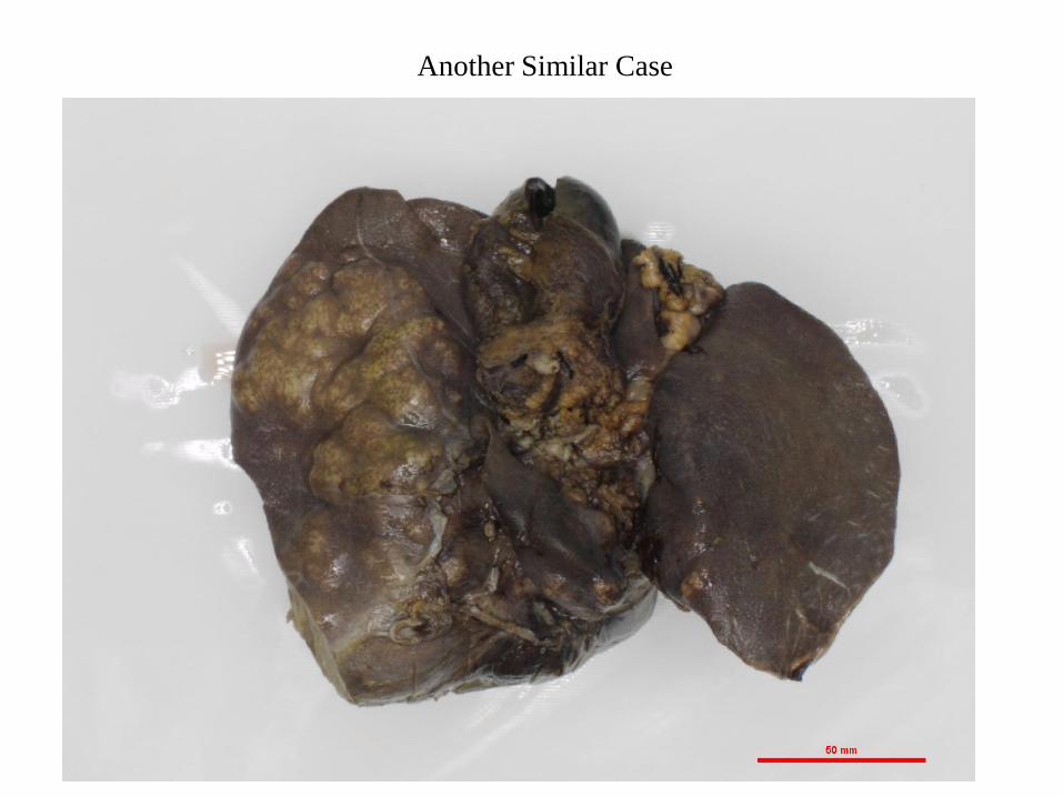
Another Similar Case
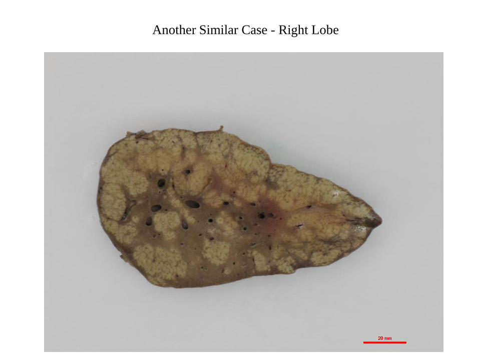
Another Similar Case - Right Lobe
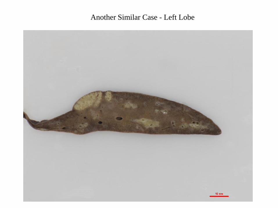
Another Similar Case - Left Lobe
Case 1 – Hepatectomy Specimen
Histological Findings
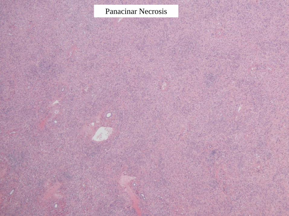
Panacinar Necrosis
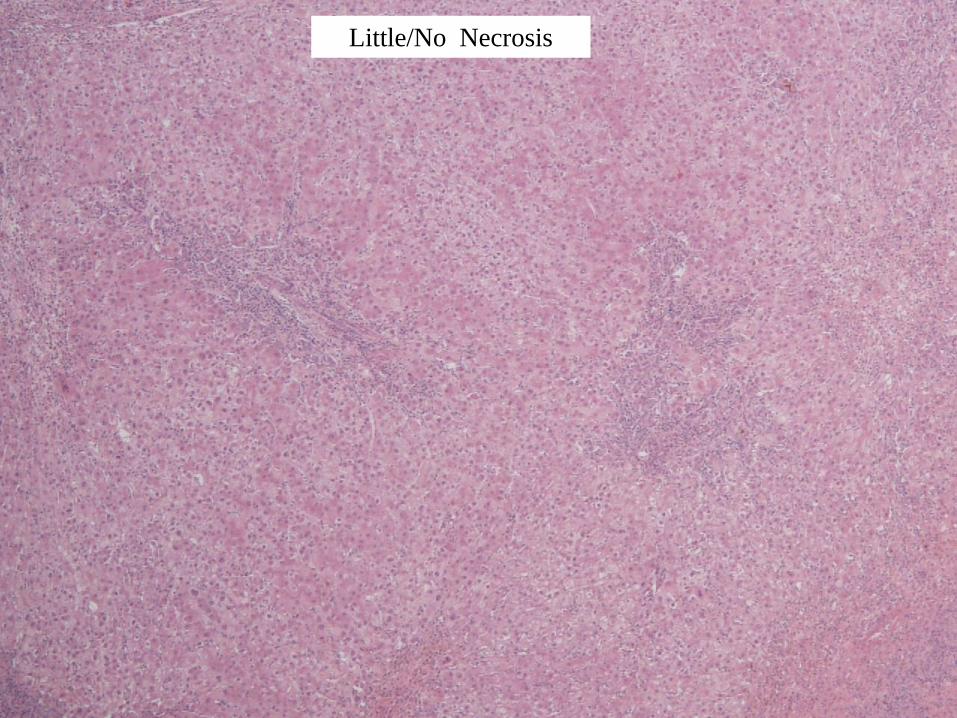
Little/No Necrosis
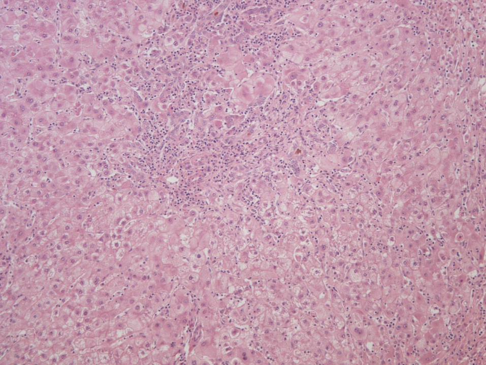

Nodules with Bridging
Could this be cirrhotic?
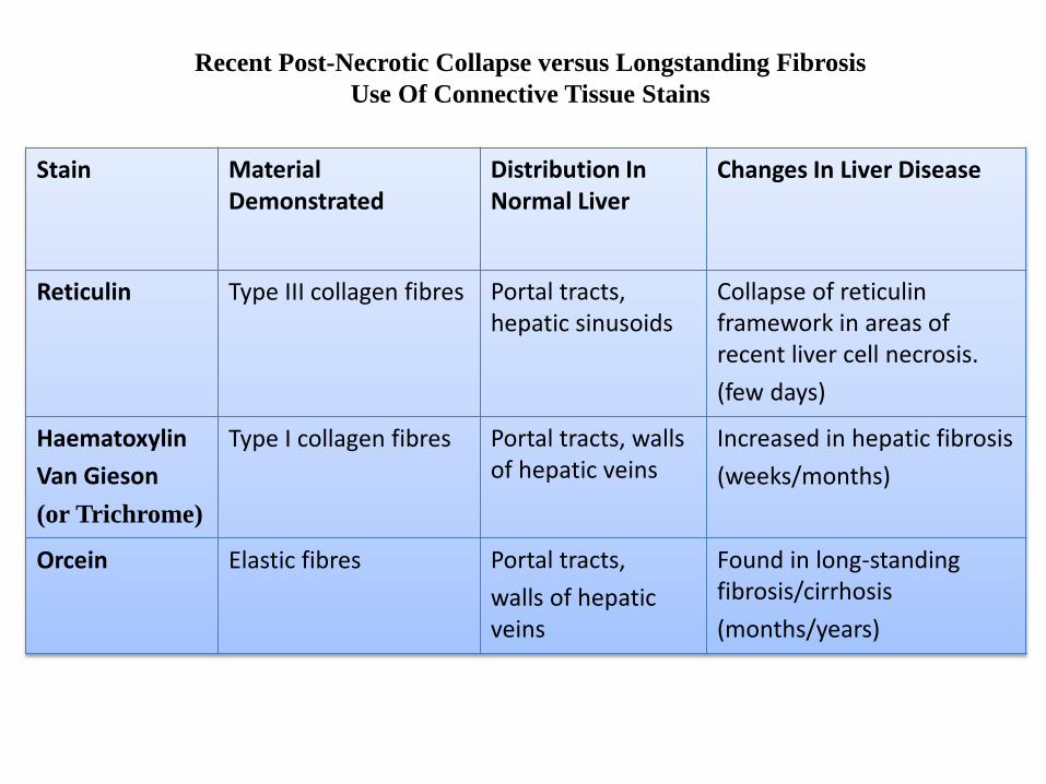
Recent Post-Necrotic Collapse versus Longstanding Fibrosis
Use Of Connective Tissue Stains
Stain Material Demonstrated
Distribution In Normal Liver
Changes In Liver Disease
Reticulin Type III collagen fibres Portal tracts, hepatic sinusoids
Collapse of reticulin framework in areas of recent liver cell necrosis.
(few days)
Haematoxylin
Van Gieson
(or Trichrome)
Type I collagen fibres Portal tracts, walls of hepatic veins
Increased in hepatic fibrosis
(weeks/months)
Orcein Elastic fibres Portal tracts,
walls of hepatic veins
Found in long-standing fibrosis/cirrhosis
(months/years)
Reticulin
HVG
HVG
Orcein
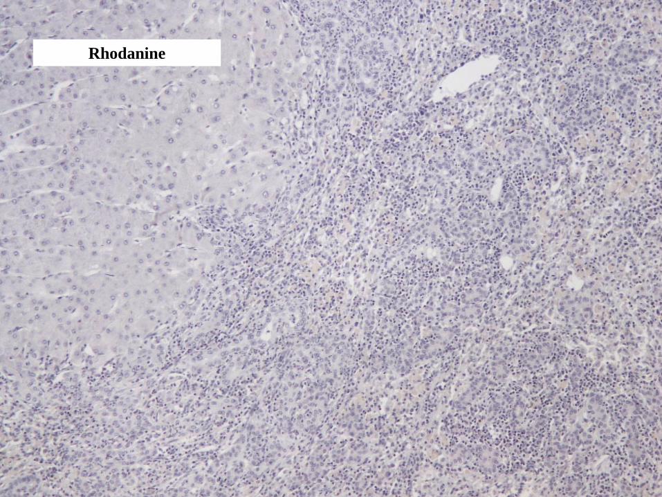
Rhodanine
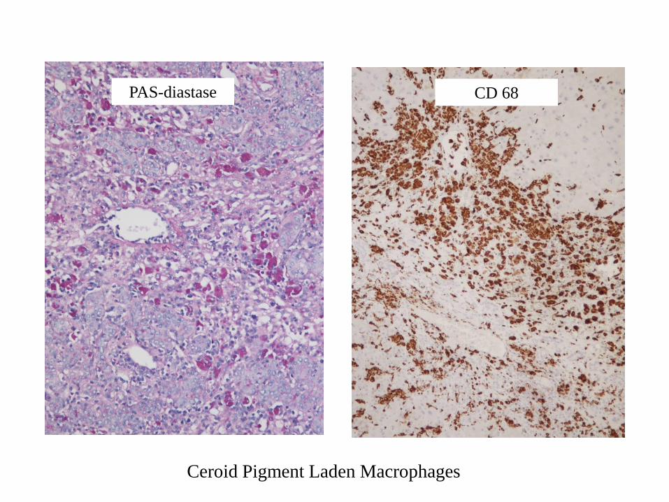
Ceroid Pigment Laden Macrophages
CD 68PAS-diastase

Ductular Reaction – Keratin 7
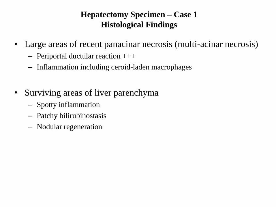
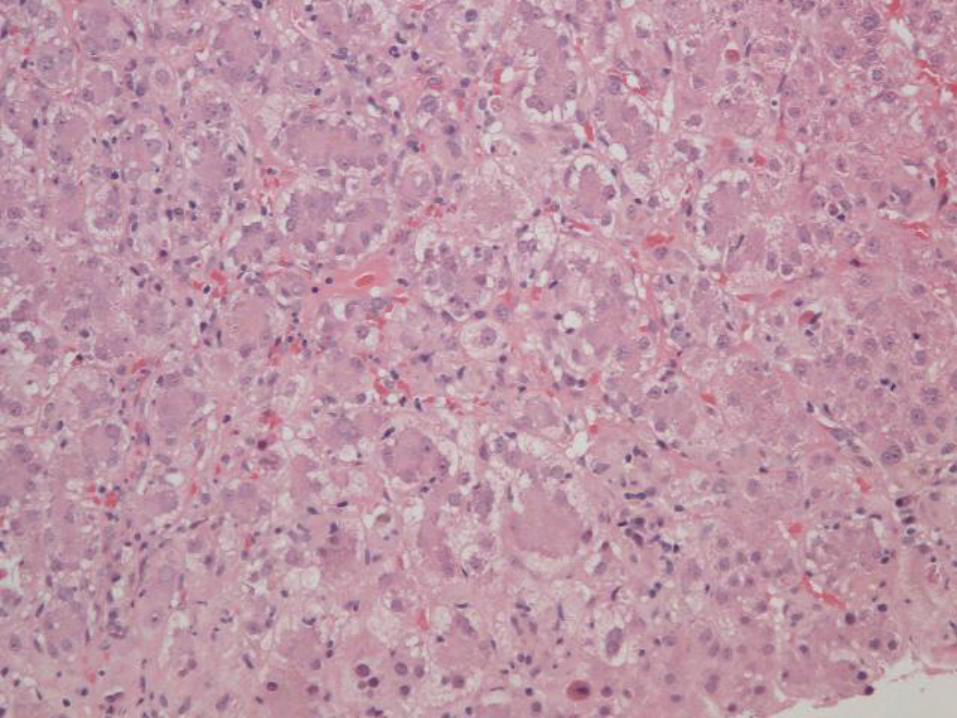
Hepatectomy Specimen – Case 1
Histological Findings
• Large areas of recent panacinar necrosis (multi-acinar necrosis)
– Periportal ductular reaction +++
– Inflammation including ceroid-laden macrophages
• Surviving areas of liver parenchyma
– Spotty inflammation
– Patchy bilirubinostasis
– Nodular regeneration
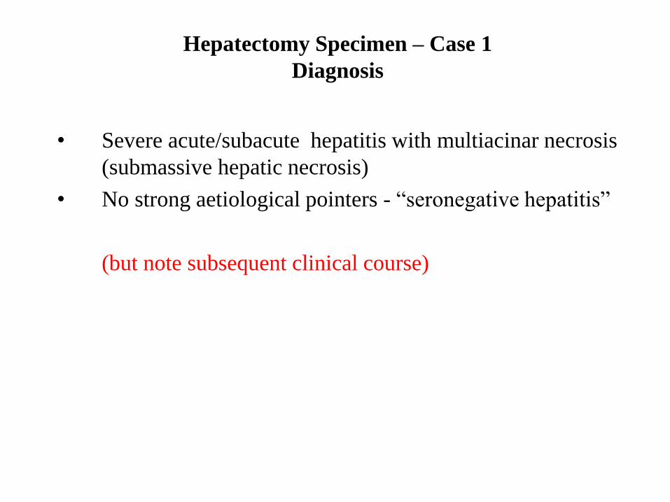
Hepatectomy Specimen – Case 1
Diagnosis
• Severe acute/subacute hepatitis with multiacinar necrosis
(submassive hepatic necrosis)
• No strong aetiological pointers - “seronegative hepatitis”
(but note subsequent clinical course)
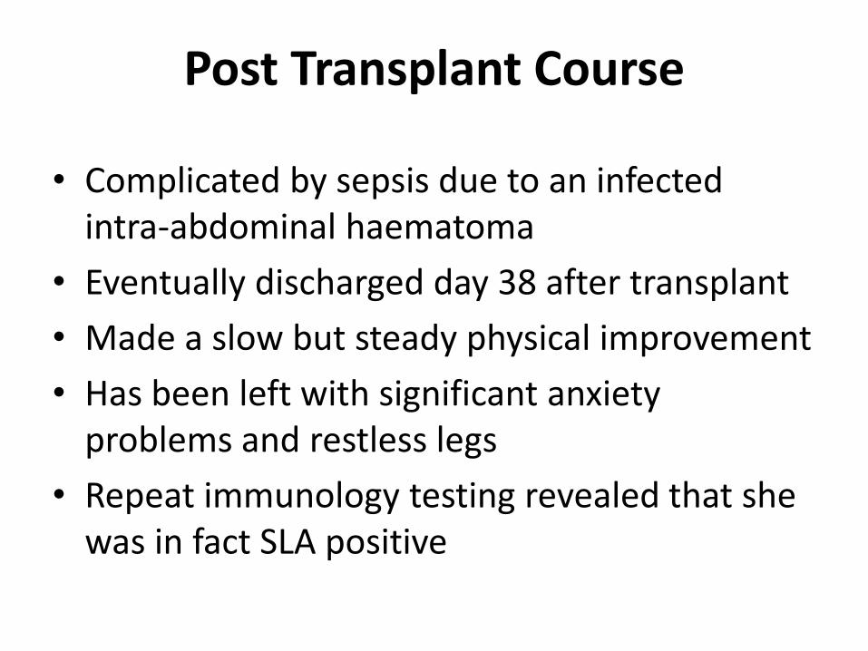
Post Transplant Course
• Complicated by sepsis due to an infected intra-abdominal haematoma
• Eventually discharged day 38 after transplant
• Made a slow but steady physical improvement
• Has been left with significant anxiety problems and restless legs
• Repeat immunology testing revealed that she was in fact SLA positive
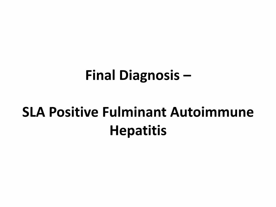
Final Diagnosis –
SLA Positive Fulminant Autoimmune Hepatitis

Seronegative vs Autoimmune Hepatitis
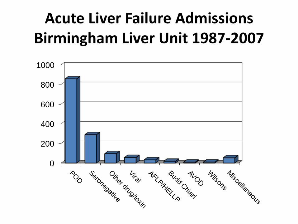
Acute Liver Failure Admissions Birmingham Liver Unit 1987-2007
0
200
400
600
800
1000
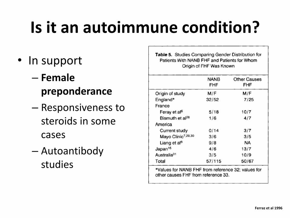
Is it an autoimmune condition?
• In support
– Female preponderance
– Responsiveness to steroids in some cases
– Autoantibody studies
Ferraz et al 1996
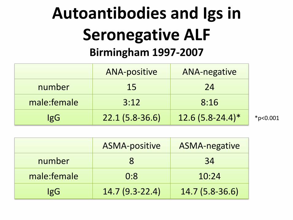
ANA-positive ANA-negative
number 15 24
male:female 3:12 8:16
IgG 22.1 (5.8-36.6) 12.6 (5.8-24.4)*
ASMA-positive ASMA-negative
number 8 34
male:female 0:8 10:24
IgG 14.7 (9.3-22.4) 14.7 (5.8-36.6)
*p<0.001
Autoantibodies and Igs in Seronegative ALF
Birmingham 1997-2007

Autoantibodies and Immunoglobulins in ALF
• King’s College Hospital Liver Unit 1999-2004
• 73 acute liver failure patients (not consecutive)– paracetamol poisoning n=20
– drug-induced (non-paracetamol) n=16
– viral n=21 (14 HBV)
– cryptogenic (seronegative) n=16
• serum immunoglobulins
• non-organ specific autoantibodies– ANA, ASM, LKM-1, AMA
• anti-SLA (soluble liver antigen)
• application of “autoimmune diagnostic score”
Bernal et al 2007
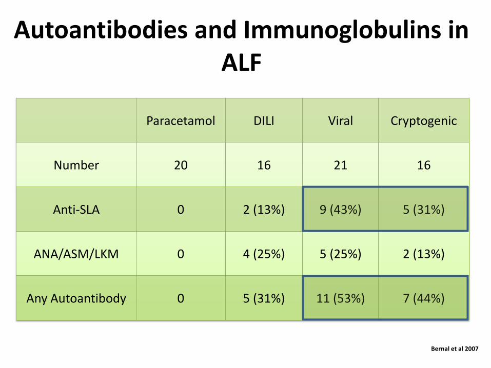
Paracetamol DILI Viral Cryptogenic
Number 20 16 21 16
Anti-SLA 0 2 (13%) 9 (43%) 5 (31%)
ANA/ASM/LKM 0 4 (25%) 5 (25%) 2 (13%)
Any Autoantibody 0 5 (31%) 11 (53%) 7 (44%)
Autoantibodies and Immunoglobulins in ALF
Bernal et al 2007
Case 2
Case 2
• 45 year old lady• Admitted to QE on 7th September 2016• Transfer from her local hospital – concern about
PT prolongation (44 at the time of transfer)• 2 week history of jaundice, pruritis and lethargy• 6 pounds weight loss• Background HT, OA and previous umbilical hernia
repair• Meds – omeprazole, lisinopril, movicol, naproxen
(regular use for 12 months), ondansteron, cyclizine, paracetamol
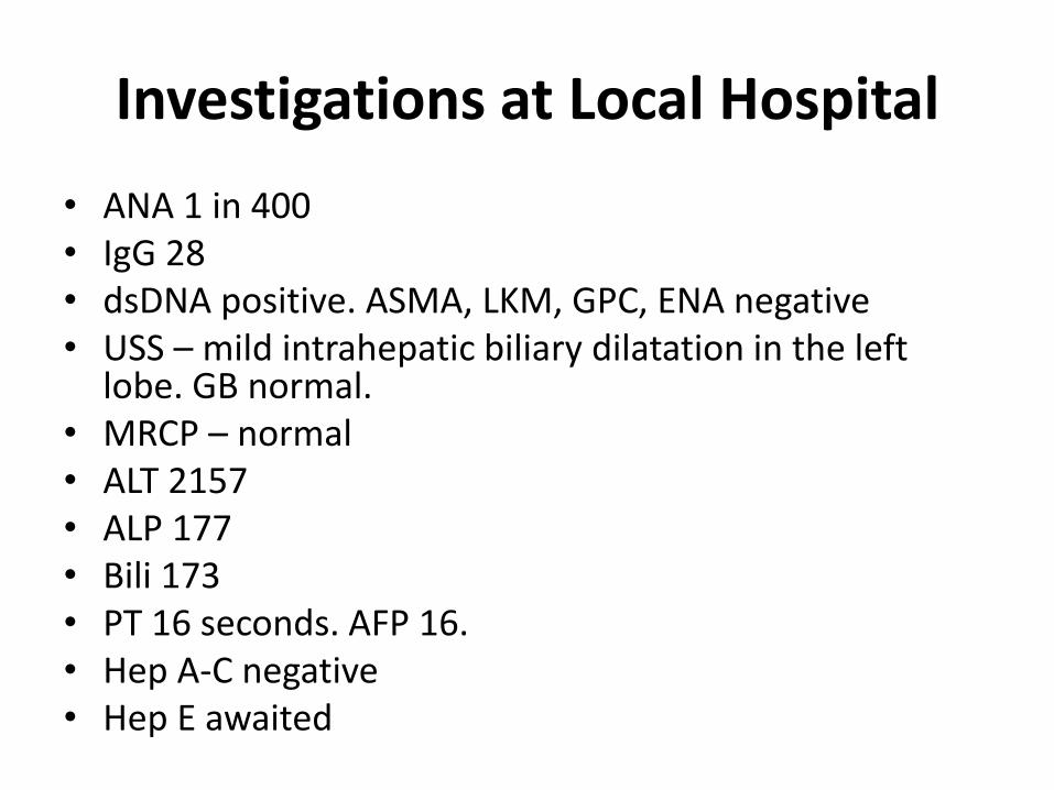
Investigations at Local Hospital
• ANA 1 in 400• IgG 28• dsDNA positive. ASMA, LKM, GPC, ENA negative• USS – mild intrahepatic biliary dilatation in the left
lobe. GB normal.• MRCP – normal• ALT 2157• ALP 177• Bili 173• PT 16 seconds. AFP 16. • Hep A-C negative• Hep E awaited
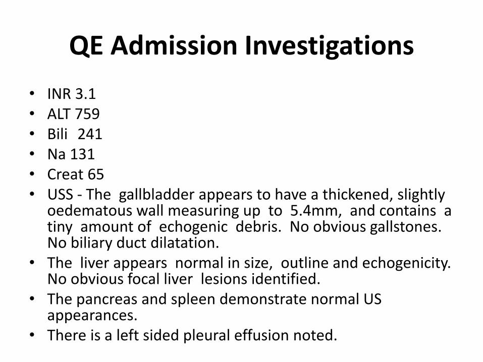
QE Admission Investigations
• INR 3.1• ALT 759• Bili 241• Na 131• Creat 65• USS - The gallbladder appears to have a thickened, slightly
oedematous wall measuring up to 5.4mm, and contains a tiny amount of echogenic debris. No obvious gallstones. No biliary duct dilatation.
• The liver appears normal in size, outline and echogenicity. No obvious focal liver lesions identified.
• The pancreas and spleen demonstrate normal US appearances.
• There is a left sided pleural effusion noted.
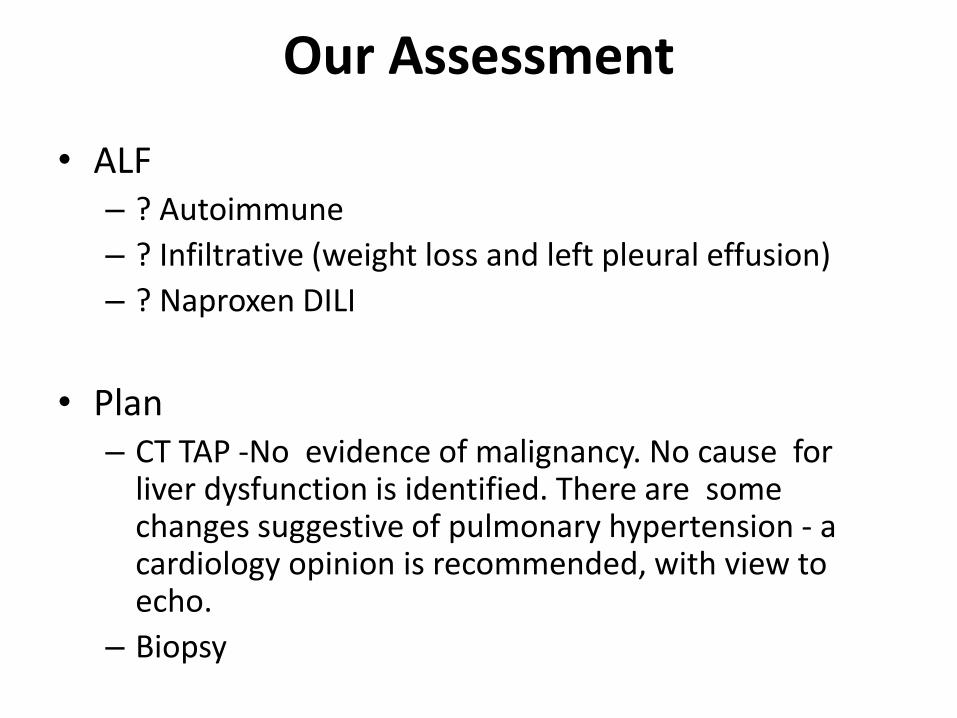
Our Assessment
• ALF– ? Autoimmune
– ? Infiltrative (weight loss and left pleural effusion)
– ? Naproxen DILI
• Plan– CT TAP -No evidence of malignancy. No cause for
liver dysfunction is identified. There are some changes suggestive of pulmonary hypertension - a cardiology opinion is recommended, with view to echo.
– Biopsy

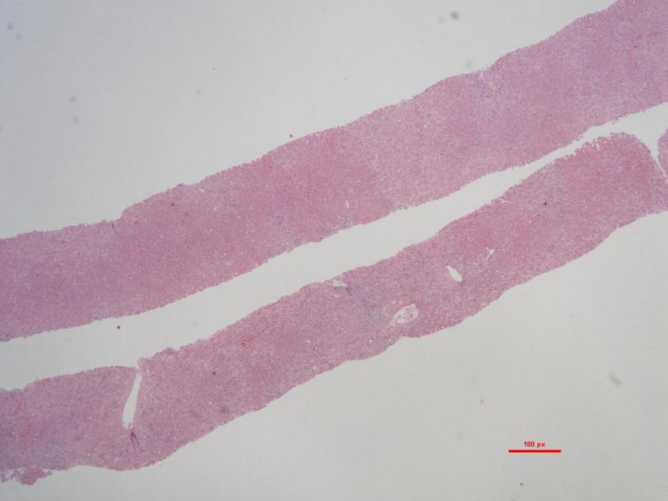
Case 2 – Liver Biopsy
Reticulin


HVG
HVG

HVG
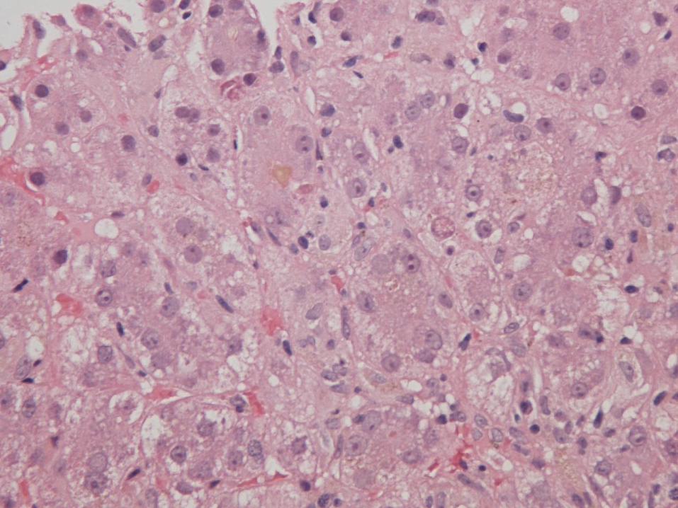
Case 2 – Liver Biopsy
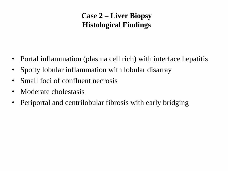
Histological Findings
• Portal inflammation (plasma cell rich) with interface hepatitis
• Spotty lobular inflammation with lobular disarray
• Small foci of confluent necrosis
• Moderate cholestasis
• Periportal and centrilobular fibrosis with early bridging
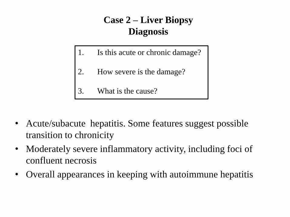
Case 2 – Liver Biopsy
Diagnosis
• Acute/subacute hepatitis. Some features suggest possible
transition to chronicity
• Moderately severe inflammatory activity, including foci of
confluent necrosis
• Overall appearances in keeping with autoimmune hepatitis
1. Is this acute or chronic damage?
2. How severe is the damage?
3. What is the cause?
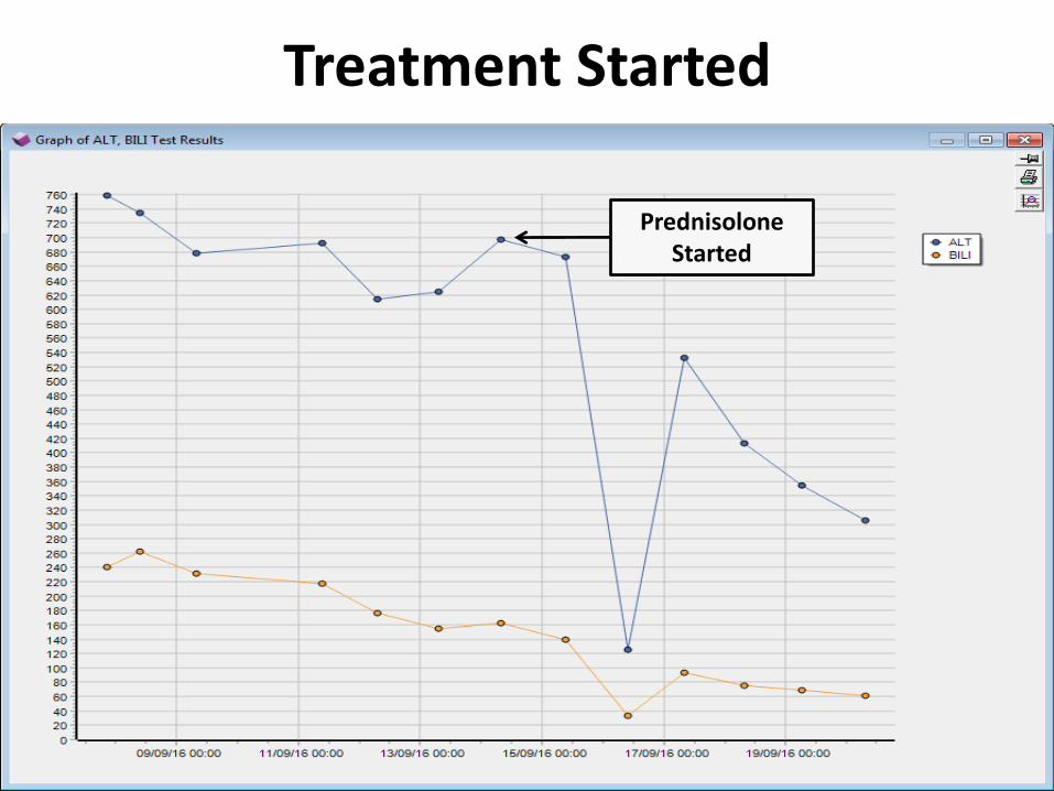
Treatment Started
PrednisoloneStarted
Progress
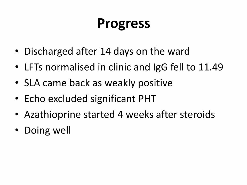
• Discharged after 14 days on the ward
• LFTs normalised in clinic and IgG fell to 11.49
• SLA came back as weakly positive
• Echo excluded significant PHT
• Azathioprine started 4 weeks after steroids
• Doing well

Final Diagnosis –
Steroid Responsive Acute Autoimmune Hepatitis

Autoimmune Hepatitis - Acute Presentation
Incidence & Diagnostic Criteria
30- 40% of cases present as acute hepatitis /acute liver failure
(Czaja & Freese 2002, Manns 2010, Lohse 2011, Gleeson 2012, Lohse 2015)
Autoantibodies unreliable in the diagnosis of acute AIH
• Autoantibodies and hypergammaglobulinaemia may not be present at the time of
presentation with acute AIH (Lohse 2011)
• Autoantibodies present in up to 40% of patients with other causes of acute liver
failure - e.g viral or drug-induced (Bernal 2007)

Autoimmune Hepatitis - Acute Presentation
Histological Features
1. Acute presentation of chronic liver disease
• 14-35% have features of chronic hepatitis (Fujiwara 2011,
Yasui 2011)
• 10-95% have bridging fibrosis or cirrhosis (Nikias 1994,
Burgart 1995, Miyake 2010, Fujiwara 2011)
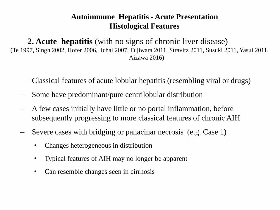
Autoimmune Hepatitis - Acute Presentation
Histological Features
2. Acute hepatitis (with no signs of chronic liver disease) (Te 1997, Singh 2002, Hofer 2006, Ichai 2007, Fujiwara 2011, Stravitz 2011, Susuki 2011, Yasui 2011,
Aizawa 2016)
– Classical features of acute lobular hepatitis (resembling viral or drugs)
– Some have predominant/pure centrilobular distribution
– A few cases initially have little or no portal inflammation, before
subsequently progressing to more classical features of chronic AIH
– Severe cases with bridging or panacinar necrosis (e.g. Case 1)
• Changes heterogeneous in distribution
• Typical features of AIH may no longer be apparent
• Can resemble changes seen in cirrhosis
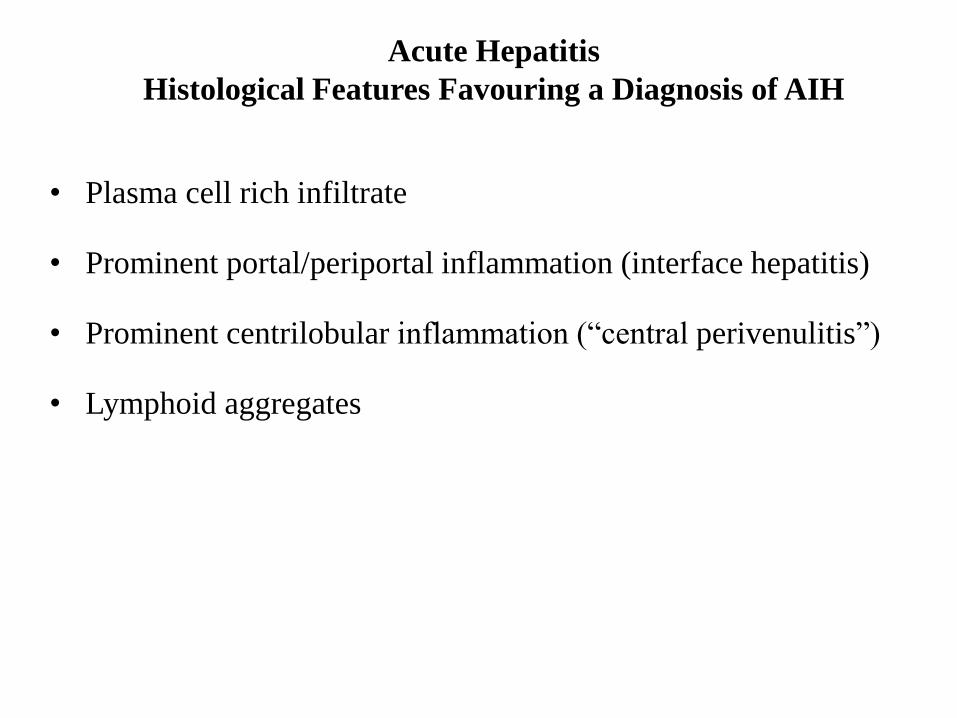
Acute Hepatitis
Histological Features Favouring a Diagnosis of AIH
• Plasma cell rich infiltrate
• Prominent portal/periportal inflammation (interface hepatitis)
• Prominent centrilobular inflammation (“central perivenulitis”)
• Lymphoid aggregates

Case 3
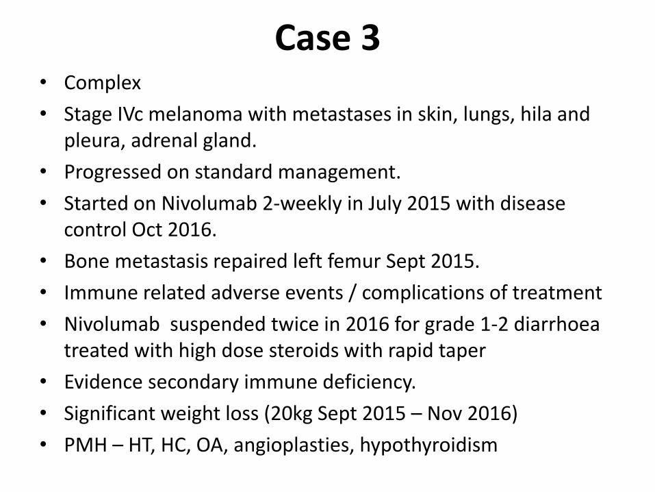
Case 3• Complex
• Stage IVc melanoma with metastases in skin, lungs, hila and pleura, adrenal gland.
• Progressed on standard management.
• Started on Nivolumab 2-weekly in July 2015 with disease control Oct 2016.
• Bone metastasis repaired left femur Sept 2015.
• Immune related adverse events / complications of treatment
• Nivolumab suspended twice in 2016 for grade 1-2 diarrhoea treated with high dose steroids with rapid taper
• Evidence secondary immune deficiency.
• Significant weight loss (20kg Sept 2015 – Nov 2016)
• PMH – HT, HC, OA, angioplasties, hypothyroidism

Mid November 2016
• Routine clinic visit
• LFTs normal up to then
• Suddenly– ALT 390
– ALP 37
– Bili 5
– INR 1.1
• Autoimmune profile negative, IgG 4.08, Hep B and C negative, EBV negative, low CMV titre of 1069 copies/ml
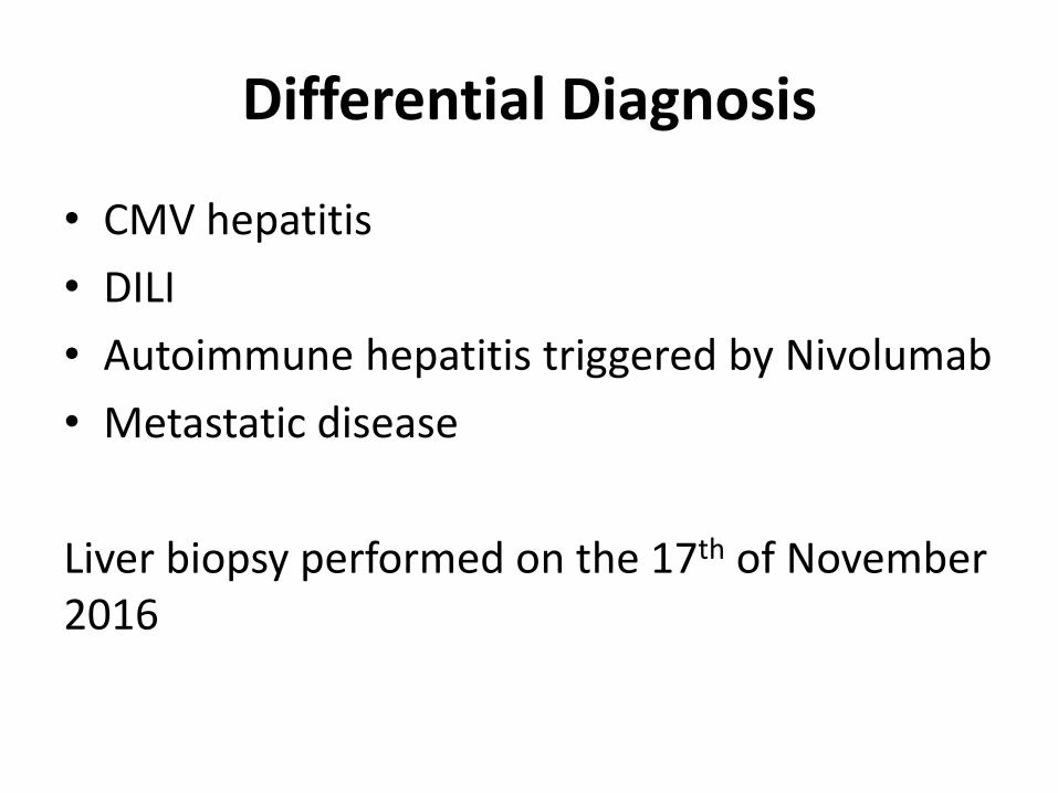
Differential Diagnosis
• CMV hepatitis
• DILI
• Autoimmune hepatitis triggered by Nivolumab
• Metastatic disease
Liver biopsy performed on the 17th of November 2016
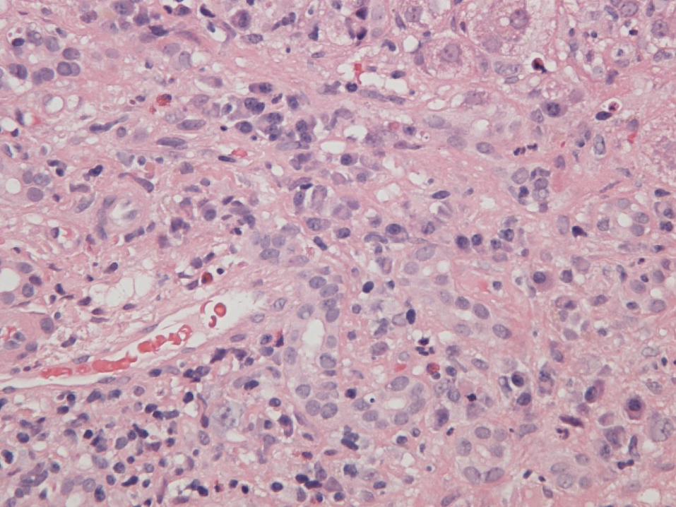
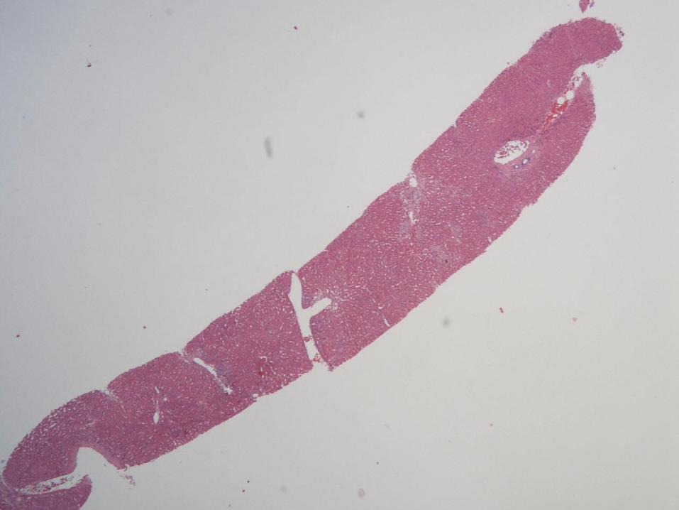
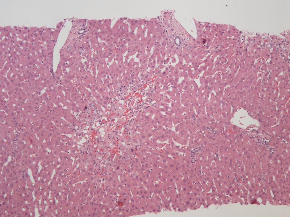
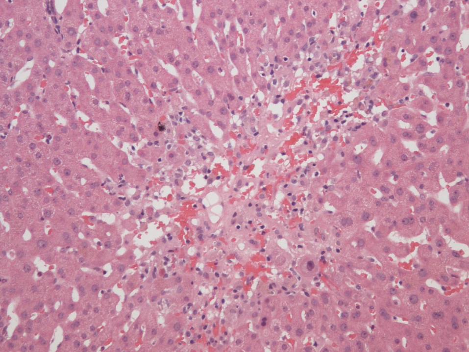
Case 3 – Liver Biopsy
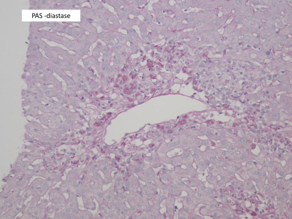
PAS -diastase
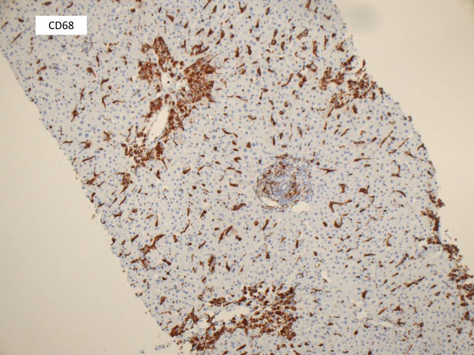
CD68

CD8
Reticulin
HVG
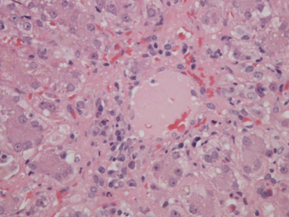
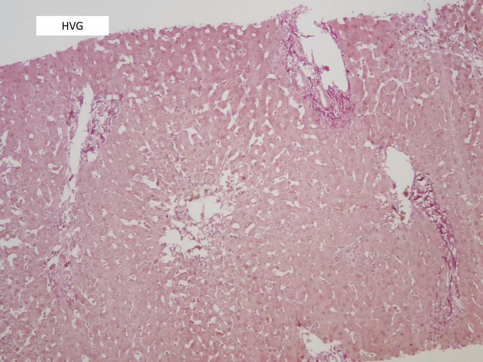
Case 3 – Liver Biopsy
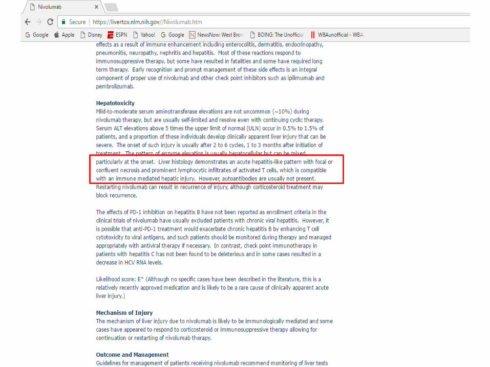
Histological Findings
• Perivenular inflammation and necrosis (“central perivenulitis”)
• Little/no inflammation elsewhere in liver parenchyma or in
portal tracts
Case 3 – Liver Biopsy
Diagnosis
• Acute injury with centrilobular inflammation (“central
perivenulitis”)
• Confluent zone 3 necrosis
• In keeping with drug-induced liver injury (DILI)
1. Is this acute or chronic damage?
2. How severe is the damage?
3. What is the cause?
Drug-induced Acute Hepatitis
• Drugs account for approximately 10% of cases of acute hepatitis and acute liver failure (Ramachandran & Kakar 2009, Reuben 2010)
• Acute hepatitis/cholestatic hepatitis are two commonest pattern of DILI
– Present in 50% of 249 cases reviewed by DILI Network (Kleiner 2014)
• Many agents implicated – antimicrobial drugs commonest
Histological features
• Frequently indistinguishable from other causes of acute hepatitis (e.g. viral hepatitis, autoimmune hepatitis)
• Features favouring a drug aetiology:
– Predominantly centrilobular (zone 3) inflammation
– Disproportionately severe / well-circumscribed necrosis (relatively little inflammation – lobular and/or portal)
– Unusual patterns of necrosis - e.g periportal (zone 1) necrosis
– Unusually prominent cholestasis
– Eosinophils, granulomas

Progress
• Started on IV methylprednisolone on the day before the biopsy
• Switched to prednisolone on day 3 after the biopsy
• Discharged
• Been in since for symptom control of diarrhoea
• No issues with LFTs

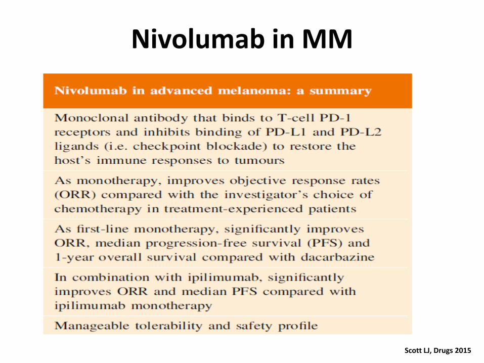
Nivolumab in MM
Scott LJ, Drugs 2015
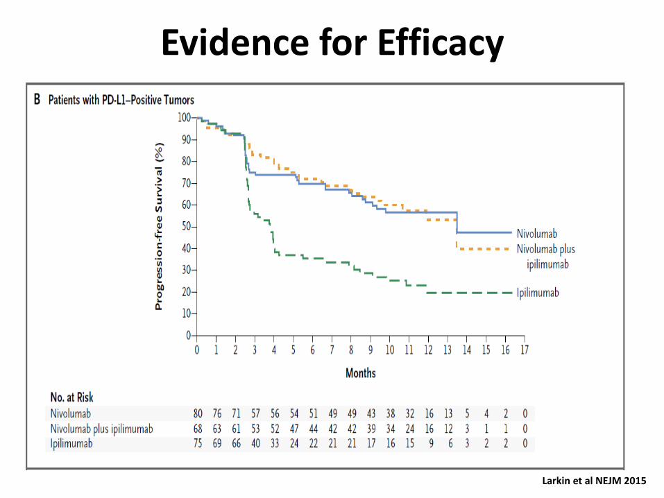
Evidence for Efficacy
Larkin et al NEJM 2015
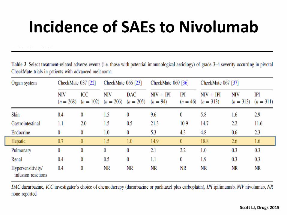
Incidence of SAEs to Nivolumab
Scott LJ, Drugs 2015

Final Diagnosis –
Steroid Responsive Nivolumabinduced liver injury
Changing Role of Liver Biopsy in Acute Hepatitis
• Many of the classical morphological studies of acute hepatitis were carried out before the main causes had been discovered
• Most cases of acute hepatitis now diagnosed on the basis of clinical, biochemical and serological findings and liver biopsy is rarely indicated
• Liver biopsy may still be carried out in cases where the clinical presentation is atypical or the cause is uncertain
– Confirm diagnosis of acute hepatitis
– Determine disease severity
– Identify possible aetiological factors (including cases of acute liver injury not related to hepatitis)
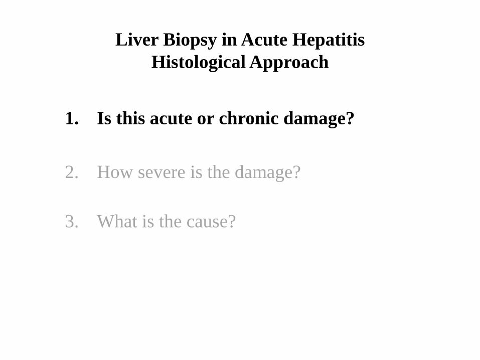
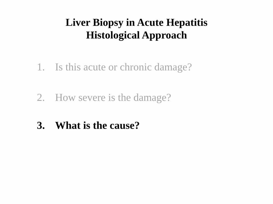
Liver Biopsy in Acute Hepatitis
Histological Approach
1. Is this acute or chronic damage?
2. How severe is the damage?
3. What is the cause?
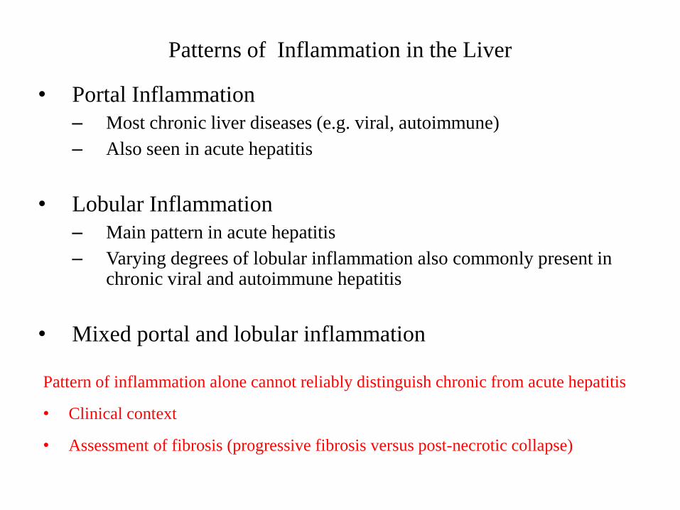
Patterns of Inflammation in the Liver
• Portal Inflammation
– Most chronic liver diseases (e.g. viral, autoimmune)
– Also seen in acute hepatitis
• Lobular Inflammation
– Main pattern in acute hepatitis
– Varying degrees of lobular inflammation also commonly present in chronic viral and autoimmune hepatitis
• Mixed portal and lobular inflammation
Pattern of inflammation alone cannot reliably distinguish chronic from acute hepatitis
• Clinical context
• Assessment of fibrosis (progressive fibrosis versus post-necrotic collapse)
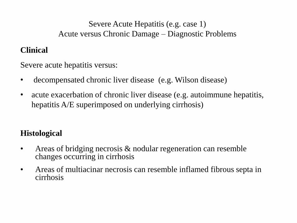
Severe Acute Hepatitis (e.g. case 1)
Acute versus Chronic Damage – Diagnostic Problems
Clinical
Severe acute hepatitis versus:
• decompensated chronic liver disease (e.g. Wilson disease)
• acute exacerbation of chronic liver disease (e.g. autoimmune hepatitis,
hepatitis A/E superimposed on underlying cirrhosis)
Histological
• Areas of bridging necrosis & nodular regeneration can resemble changes occurring in cirrhosis
• Areas of multiacinar necrosis can resemble inflamed fibrous septa in cirrhosis

Multiacinar Necrosis in Severe Acute Hepatitis (e.g. case 1)
Acute versus Chronic Damage - Helpful pointers
• Clinical context
• Identification of normal vascular relationships
• Use of connective tissue stains to determine age of lesions

Liver Biopsy in Acute Hepatitis
Histological Approach
1. Is this acute or chronic damage?
2. How severe is the damage?
3. What is the cause?
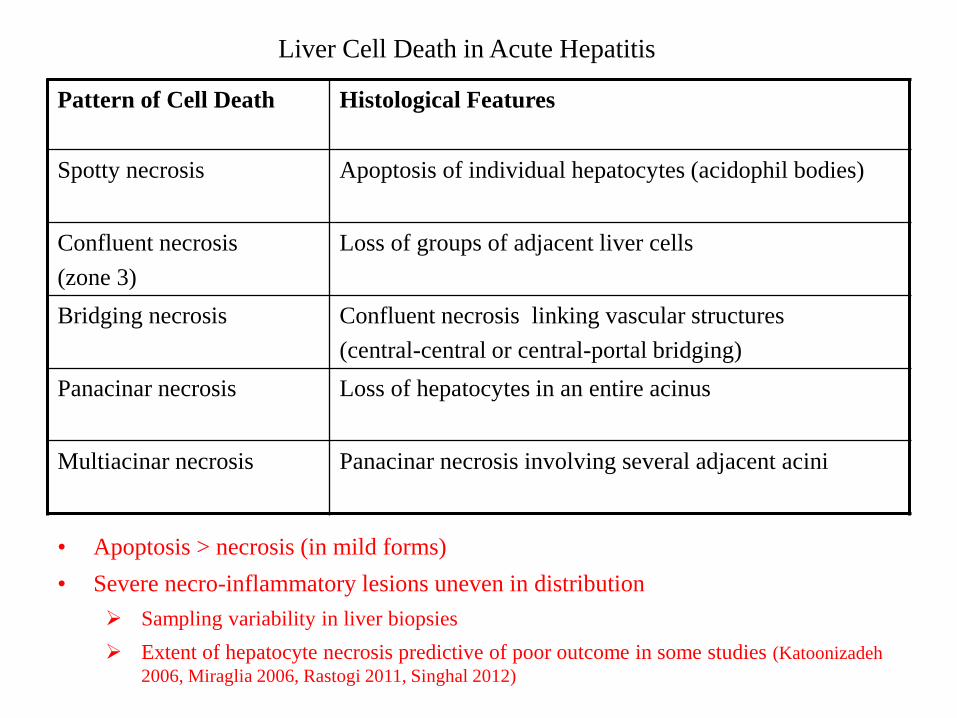
Liver Cell Death in Acute Hepatitis
Pattern of Cell Death Histological Features
Spotty necrosis Apoptosis of individual hepatocytes (acidophil bodies)
Confluent necrosis
(zone 3)
Loss of groups of adjacent liver cells
Bridging necrosis Confluent necrosis linking vascular structures
(central-central or central-portal bridging)
Panacinar necrosis Loss of hepatocytes in an entire acinus
Multiacinar necrosis Panacinar necrosis involving several adjacent acini
• Apoptosis > necrosis (in mild forms)
• Severe necro-inflammatory lesions uneven in distribution
Sampling variability in liver biopsies
Extent of hepatocyte necrosis predictive of poor outcome in some studies (Katoonizadeh
2006, Miraglia 2006, Rastogi 2011, Singhal 2012)
Liver Biopsy in Acute Hepatitis
Histological Approach
1. Is this acute or chronic damage?
2. How severe is the damage?
3. What is the cause?
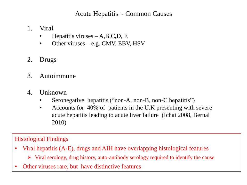
Acute Hepatitis - Common Causes
1. Viral• Hepatitis viruses – A,B,C,D, E
• Other viruses – e.g. CMV, EBV, HSV
2. Drugs
3. Autoimmune
4. Unknown• Seronegative hepatitis (“non-A, non-B, non-C hepatitis”)
• Accounts for 40% of patients in the U.K presenting with severe
acute hepatitis leading to acute liver failure (Ichai 2008, Bernal
2010)
Histological Findings
• Viral hepatitis (A-E), drugs and AIH have overlapping histological features
Viral serology, drug history, auto-antibody serology required to identify the cause
• Other viruses rare, but have distinctive features

Liver biopsy rarely identifies a previously unsuspected aetiology
• Biopsies mostly obtained from people in whom main recognised causes
have been excluded (“seronegative hepatitis”)
• Biopsy sometimes provides aetiological pointers, including cases
presenting with acute liver injury not due to acute hepatitis
– Decompensated chronic liver disease (e.g. Wilson’s disease)
– Another cause of acute liver damage (e.g. ischaemic necrosis, severe
alcoholic hepatitis, paracetamol toxicity)
– Hepatic infiltration (usually lymphoma, rarely carcinoma)
• Liver usually enlarged
Acute Hepatitis - Aetiological Considerations

The End