Embed Size (px)
Citation preview
16
Acute Complications of Hemodialysis
Gülsüm Özkan and Şükrü Ulusoy Karadeniz Technical University, School of Medicine, Department of Nephrology
Turkey
1. Introduction
Chronic kidney disease (CKD) is a common public health problem, which occurs in many countries with an increasing prevalence. Over 50 million people throughout the world are known to have CKD, and of these, more than 1 million require renal replacement therapies such as dialysis and renal transplantation. In recent years , the rising incidence of diabetes and hypertension, the most common two causes of CKD, cause an increase in the prevalence of CKD. Hemodialysis, which is one of the renal replacement therapies, is a life-saving treatment. In the absence of this therapy, more than a million patients worldwide would have died within weeks. Hemodialysis was successfully performed for the first time in 1944 by Willem Kollf in patients with renal failure. However, hemodialysis is accompanied by several complications. During the first years following the introduction of hemodialysis, complications were common due to the technical drawbacks associated with the dialysis machines and water systems. Currently, the advances in technology, particularly those in the last 20 years, have reduced the complications. However, complications caused by the reasons other than the dialysis machine and water system remain as a significant cause of morbidity and mortality in hemodialysis patients. Cardiovascular complications are currently the most common complication of hemodialysis. Among these complications, the rate of symptomatic intradialytic hypotension ranges between 20% and 50%, and it remains an important problem (Cruz DN et al., 1997). Another concern is the hemodialysis-associated arrhythmias, the rate of which was reported to be 5% to 75%. The common and lethal types of arrhythmias include ventricular arrhythmias and ectopies. The rate of hemodialysis-associated complex ventricular arrhythmia is around 35% (Burton JO et al., 2008). The second most common type of arrhythmia is the atrial fibrillation, the rate of which is 27% (Genovesi S et al., 2008). Sudden cardiac death accounts for 62% of cardiac-related deaths and it is usually attributed to arrhythmias (Herzog CA et al., 2008). The first year of hemodialysis is of vital importance with respect to sudden cardiac deaths, which was determined in 93 of 1000 patients in the first year of hemodialysis (Shastri S et al., 2010). While cramps were observed in 24%-86% of the cases during the first years following the introduction of dialysis therapy, recently it has been shown that only 2% of the patients having ≥2 hemodialysis sessions in a week suffer from cramps (Kobrin SM et al., 2007). Other common complications include nausea, vomiting with a rate of 5%-15%, headache with a rate of 5%-10% and itching with a rate of 5%-10%( Jesus AC et al., 2009; Mettang T et al., 2002). Although cramps, nausea-vomiting, headache and itching do not result in mortality, they substantially deteriorate the quality of life of the patients. Although more
www.intechopen.com
Technical Problems in Patients on Hemodialysis
252
common during the first years following the introduction of dialysis, Disequilibrium syndrome and complications associated with dialyser, water systems and dialysis machines are currently uncommon but may have fatal consequences. Hemodialysis cause many complications despite the advances in technology. It is of great importance to prevent the complications before they occur. Particularly, early recognition and correction of life-threatening complications save lives. Some complications may not threaten the patients’ life but deteriorate the quality of life of the patients. The treatment of these complications provides a longer life and a better quality of life for the patients. Acute complications of hemodialysis can be classified as follows:
Complications associated with hemodialysis equipment
Hemodialysis device-related complications Membrane-related complications Water system-related complications Vascular acces-related complications
Cardiovascular complications
Hypotension Hypertension Arrhythmias Pericardial effusion Sudden death Chest pain
Neurological complications
Disequilibrium syndrome Cerebrovascular accident Consciousness changes Headache Seizure Tremor
Complications associated with use of anticoagulant therapy
Heparin associated thrombocytopenia Bleeding diathesis
Electrolyte abnormalities
Hematologic complications
Others
Nausea Vomiting Itching
2. Complications associated with hemodialysis equipment
2.1 Hemodialysis device-related complications
The basic principles of hemodialysis were established many years ago. Technology that developed over many years enabled hemodialysis machines to better meet the needs of
www.intechopen.com
Acute Complications of Hemodialysis
253
patients and reduce the amount of complications. However, the number of hemodialysis patients is increasing today and especially those with comorbid disorders need hemodialysis treatment. As a result of this situation, more research is being made to further develop the hemodialysis machine technology. The functions of a hemodialysis machine include taking the patient’s blood from the access by using a blood pump and extracorporeal tubing, passing it through the dialyzer and returning it to the patient, preparing the dialysate using purified water and concentration, circulating the dialysate along the dialyzer system and ultrafiltrating it, and enabling the blood and diaysate to circulate safely by means of control and alarm systems (Ward& Ronco,2006). Such control and alarm systems include an air detector, pressure monitor (for artery and vein pressure), heat detector, blood leakage detector, conductivity monitor, and ultrafiltration control systems. With the developments in technology, some dialysis machines now display blood flow rates corrected port the pressure at the pump inlet using software algorithm (Depner et al.,1990). Moreover, low pressure-sensitive blood tubing sets have been produced recently (Ahmed et al.,2004). Despite all these developments, it is of vital importance to know and prevent the complications associated with the HD machines and equipment.
2.1.1 Air embolism
One of the much-feared fatal complications of the hemodialysis therapy is the air embolism. There are ultrasonographic air detectors in hemodialysis machine trapping air bubbles to prevent air embolism. Such detectors sense the air bubbles in certain volumes and diameters and activate the control systems. The most common cause of air embolism is air entering in the system mostly from the pre-pump section where there is a negative pressure system and the access points of artery needles (Barak et al.,2008). The symptoms of an air embolism depend on the position of the patient at that moment. If he/she is in a sitting position, neurologic complications occur because the embolus will go into the cerebral system whereas symptoms such as shortness of breath and chest pain occur when the embolus goes into the lungs in the supine position. The first step in treatment is to clamp the vein tubing and stop the pump. The patient then should be laid on his/her left with his/her head and chest facing downwards and 100% oxygen should be given. If the embolus is in the heart, it can be removed with a needle percutaneously and a hyperbaric oxygen therapy may also be used. The clinical signs and therapies we mentioned above are for large air emboli. Besides this, creation of micro-bubbles is also possible during a hemodialysis therapy. The contemporary hemodialysis machines cannot detect any doses of air infusions less than 0,1 ml/k/minutes in bolus infusions and 0,03 ml/kg/minutes in continuous infusions and thus fail to activate the alarm system (Polaschegg, 2007). Therefore, the hemodialysis machines today remain ineffective in preventing micro-bubbles to enter the venous system. Micro-bubbles usually do not result in acute symptoms in patients, but are thought to cause pulmonary hypertension in the lungs and chronic changes in the brain in the long run. Various filters have been developed to prevent micro-bubbles to penetrate the venous system during hemodialysis. However, routine use of such filters has not approved as they cause an extra resistance before the blood flow and the patient’s blood becomes exposed to various chemicals contained in the filters (Barak et al.,2008). There are efforts in recent years to develop new technologies to detect and eliminate micro-bubbles through ultrasonographic methods. Works on the issue is still in progress (Palanchon et al., 2001; Versluis et al., 2010).
www.intechopen.com
Technical Problems in Patients on Hemodialysis
254
2.1.2 Complications resulting from manual setup of the machines or not following the instruction manual
Despite the technological developments in hemodialysis machine, some complications arise due to failure to follow their instruction manuals or setting the alarm limits manually by individuals (Davenport, 2006). For example, one of the errors is to set up artery tubing which does not fit the diameters of the blood pump. This may result in hemolysis by increasing the pre-pumping pressure. Another error occurs when lowering the temperature of the dialysate especially in patients with intradialytic hypotension. In such a case when the hemodialysis machine is reset to stop the alarm, the temperature changes may go unnoticed even at very high or low levels due to a problem in the machine. Very low temperatures make the patient feel cold and very high temperatures may cause serious hemolysis. Most of the hemodialysis machine can automatically perform disinfection through heat or chemicals, but if the user manually restricts the disinfection process, this may cause hemolysis and the resulting symptoms as some of the compounds used in disinfection cannot be removed adequately. In some instances, the venous needle comes loose, but the hemodialysis machine cannot sense this and give the necessary alarm in time, or when the venous alarm limits are changed or the alarm is disabled by the user, an abundant loss of blood from the patient may not be sensed. Considering the above mentioned complications, it would be advisable not to disable the alarm systems of the hemodialysis machine or in cases of necessity to employ close monitoring. In a recent study, the effect of the age and maintenance status of a hemodialysis machine on the satisfactoriness of dialysis was examined. The study showed that technical maintenance of the machines in regular intervals had a significant effect on the efficacy of the hemodialysis therapy (Azar, 2009). Therefore, it should remembered that in order to reduce the number of complications and to give the patient the targeted dose of dialysis, calibrations and service maintenance of hemodialysis machine should be regularly made, the machines should be used according to their instruction manuals, and as manual adjustments may harm the patient, the patients in such situations should be monitored closely.
2.2 Membrane-related complications
During hemodialysis, the patient’s blood passes through many extracorporeal compartments. These include the dialyzer, the blood tubing set, the chemicals used during sterilization of the dialyzer and the dialysate. The dialyzer contains a dialysis membrane and sterilization products used during its manufacturing. Dialyzers come in two geometries as hollow-fiber and parallel plate dialyzers according to their membrane structure. In hollow-fiber dialyzers with thousands of tiny hollow fibers, blood flows into the compartment at one end of the cylinder-shaped case and passes through thousands of tiny capillaries. Dialysis solution flows in the opposite direction of the blood flow around the capillaries. Blood passing through the capillaries is collected in the compartment at the other end of the dialyzer and returned to the patient. Membranes also come in various types with respect to the material used in them; they can be cellulosic, cellulose/synthetic (semi-synthetic), synthetic and bioactive (in dialyzers covered by vitamin E). They can be referred to as being reusable or not and biocompatible or not in the terminology. The most commonly used ones are the synthetic membranes today.(Twardowski, 2008).
www.intechopen.com
Acute Complications of Hemodialysis
255
2.2.1 Dialyzer reactions
Hemodialysis-related anaphylactoid reaction was first reported in 1975. A well-documented prospective study on its incidence is not available. However, according to the data from the Food and Drug Administration, a severe hypersensitivity reaction was reported in 3.5 of 100.000 dialysis sessions in 1982 (Ebo et al., 2006). Such reactions consisted of a series of incidences involving both anaphylactic reactions and reactions with unknown causes. The classification made by Daugirdas JT and associates is the one most commonly used for these reactions. The classification involves Type-A (hypersensitivity) reactions and Type-B (non-specific) reactions (Daugirdas & Ing, 1988).
Type-A reactions
The symptoms may start with dyspnea, fear of death, and a sensation of heat in the fistula site or the whole body and end with a complete anaphylactic episode. In less severe cases, there may be symptoms such as itching, coughing, sneezing, nasal discharge, nausea and vomiting. These generally occur at the very beginning of dialysis, but may also appear between the 15th and 20th minutes. Such reactions are seen more in patients with atopy and/or eosinophilia (Walter &Taraba, 1991). The criteria developed by Daugirdas and Ing. are mostly used in diagnosis. The major criteria include the reaction occurring in the first 20 minutes after the beginning of dialysis, dyspnea, sensation of burning or heating-up in the access site or diffused to the whole body and angioedema whereas the minor criteria include recurrence of the reaction during the next dialysis session when the same class or type dialyzer is used, urticaria, rhinorrhea or lacrimation, abdominal cramps and itching. Diagnosis is made when three major or two major and 1 minor criteria are met (Daugirdas & Ing, 1988). It is mostly caused by sterilization using ethylene oxide, other reasons being the use of an AN69 membrane, reuse, complementary fragment release and eosinophilia (Shaldon & Koch, 1995).
Treatment
The dialysis must immediately be discontinued and the blood in the blood tubing set must not be given back to the patient. Antihistaminic, adrenalin or steroid may be administered depending on the severity of the reaction.
Prevention
It can be considered to sufficiently wash the dialyzers before using them for each patient, to use a dialyzer sterilized by γ-rays or steam if the reaction was due to the use of a dialyzer sterilized by ethylene oxide, to use a membrane that activates the complement more mildly or to make a transition from those using Angiotensin Converting Enzyme (ACE) inhibitor to those using Angiotensin Receptor Blockers (ARB) (Dumler et al., 1987; Daugirdas & Ing, 1988).
Type-B reactions
Their primary symptoms are chest pain and lower back pain. They appear after 20 to 40 minutes after the beginning of dialysis. The symptoms alleviate or disappear in the progressing hours of the dialysis. Complement activation may be blamed of them although the etiology is not fully known (Jaber&Pereira, 1997). The treatment is similar to that in type-A reactions and is adapted depending on the intensity of the symptoms.
www.intechopen.com
Technical Problems in Patients on Hemodialysis
256
2.2.2 Hemodialysis-related hypoxemia
During hemodialysis, Pa O2 drops to approximately 10-20 mmHg. While such decrease does not lead to significant clinical problems in patients with normal oxygenation, may produce catastrophic results in those with poor oxygenation (De Backer et al.,1983; Hakim & Lowrie,1982). One of the factors that is blamed in the etiology of hypoxemia that emerge during hemodialysis is dialysate containing acetate (De Backer et al.,1983). However, it was demonstrated that it could also be observed in dialysate with bicarbonate. Dialysate with acetate may induce hypoxia in two ways, first by increased oxygen consumption during acetate bicarbonate conversion and second by intradialytic loss of CO2 (Dolan et al.,1981; Oh et al.,1985). The biocompatibility of the membrane used is one of the most frequently blamed factors in hypoxemia (Graf et al.,1980). Especially the use of an acetate-containing dialysate together with a Cuprophan membrane increases hypoxemia (Vanholder et al.,1987). Hypocapnia associated with intradialytic loss of CO2 and adaptation to chronic metabolic acidosis lead to periodic shortness of breath and a tendency to sleep apnea syndrome (De Broe & De Backer, 1989).
Treatment and prevention
Increasing the level of CO2 in the dialysate by directly adding CO2 to it or by using a dialysate containing bicarbonate, Using biocompatible membranes (De Backer et al.,1983; Hakim & Lowrie,1982), Making appropriate ventilator settings for the patients who are known to have hypoxemia prior to the dialysis and are administered mechanical ventilation, nocturnal hemodialysis may be appropriate for those with sleep apnea syndrome (Hanly&Pierratos, 2001),
2.2.3 Disadvantages of first-use dialyzers
New dialyzer syndrome, neutropenia and complement activation as well as reactions associated with the use of ethylene oxide are seen more often.
2.2.4 Disadvantages of reuse dialyzers
Reactions associated with the compounds used in chemical disinfection, side-effects of the volatile gases used during sterilization, allergic reactions, residual chemical infusion, sterilization in insufficient concentrations, pyogenic reactions, variations in the permeability of the membrane and failure to perform an efficient dialysis are seen more often (Twardowski, 2006).
2.3 Water system-related complications
Patients receiving hemodialysis therapy become exposed to 18000 to 36000 liters of water a year during hemodialysis. The formation of dialysate involves water purification, distribution of the purified water to individual hemodialysis machines, concentrate preparation (acidic and basic concentrate) and finally mixing the concentrates with the purified water. While the acidic concentrate is not suitable for bacterial growth, the basic concentrate creates an environment suitable for bacterial growth. For this reason, dry powder cartridges are being used as basic concentrates recently; this allows online preparation of fluid bicarbonate in individual dialysis machines (Ward, 2004). A large portion of the water used in preparing the dialysate is the purified water produced in the water system. In case the hemodialysis water system fails to produce the proper water, patients can be exposed to various chemicals, bacteria and toxic contaminations (Montanari
www.intechopen.com
Acute Complications of Hemodialysis
257
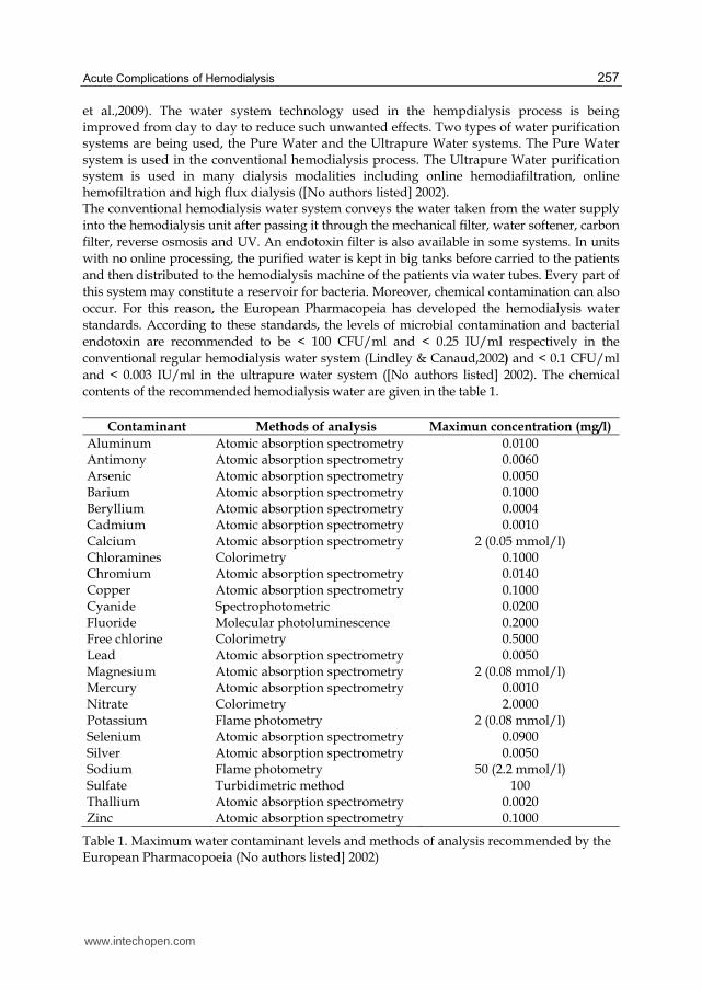
et al.,2009). The water system technology used in the hempdialysis process is being improved from day to day to reduce such unwanted effects. Two types of water purification systems are being used, the Pure Water and the Ultrapure Water systems. The Pure Water system is used in the conventional hemodialysis process. The Ultrapure Water purification system is used in many dialysis modalities including online hemodiafiltration, online hemofiltration and high flux dialysis ([No authors listed] 2002). The conventional hemodialysis water system conveys the water taken from the water supply into the hemodialysis unit after passing it through the mechanical filter, water softener, carbon filter, reverse osmosis and UV. An endotoxin filter is also available in some systems. In units with no online processing, the purified water is kept in big tanks before carried to the patients and then distributed to the hemodialysis machine of the patients via water tubes. Every part of this system may constitute a reservoir for bacteria. Moreover, chemical contamination can also occur. For this reason, the European Pharmacopeia has developed the hemodialysis water standards. According to these standards, the levels of microbial contamination and bacterial endotoxin are recommended to be < 100 CFU/ml and < 0.25 IU/ml respectively in the conventional regular hemodialysis water system (Lindley & Canaud,2002) and < 0.1 CFU/ml and < 0.003 IU/ml in the ultrapure water system ([No authors listed] 2002). The chemical contents of the recommended hemodialysis water are given in the table 1.
Contaminant Methods of analysis Maximun concentration (mg/l)
Aluminum Atomic absorption spectrometry 0.0100 Antimony Atomic absorption spectrometry 0.0060 Arsenic Atomic absorption spectrometry 0.0050 Barium Atomic absorption spectrometry 0.1000 Beryllium Atomic absorption spectrometry 0.0004 Cadmium Atomic absorption spectrometry 0.0010 Calcium Atomic absorption spectrometry 2 (0.05 mmol/l) Chloramines Colorimetry 0.1000 Chromium Atomic absorption spectrometry 0.0140 Copper Atomic absorption spectrometry 0.1000 Cyanide Spectrophotometric 0.0200 Fluoride Molecular photoluminescence 0.2000 Free chlorine Colorimetry 0.5000 Lead Atomic absorption spectrometry 0.0050 Magnesium Atomic absorption spectrometry 2 (0.08 mmol/l) Mercury Atomic absorption spectrometry 0.0010 Nitrate Colorimetry 2.0000 Potassium Flame photometry 2 (0.08 mmol/l) Selenium Atomic absorption spectrometry 0.0900 Silver Atomic absorption spectrometry 0.0050 Sodium Flame photometry 50 (2.2 mmol/l) Sulfate Turbidimetric method 100 Thallium Atomic absorption spectrometry 0.0020 Zinc Atomic absorption spectrometry 0.1000
Table 1. Maximum water contaminant levels and methods of analysis recommended by the European Pharmacopoeia (No authors listed] 2002)
www.intechopen.com

Technical Problems in Patients on Hemodialysis
258
In spite of these standards, the complications related to production of water is still being significant with the increased use of high-flux dialyzers, which increase the back-filtration of the dialysate, and the increased use of online hemodiafiltration process, which is based on allowing the blood compartment to contact large amounts of purified water (Brunet & Berland, 2000). The problems associated with the water purification process lead to short and long term complications. In short term complications, a serious septic episode may develop accompanied by tremble, fever, nausea, myalgia, headache, debility and even hypotension and shock when the patient is exposed to excessive amounts of bacteria or endotoxins (Dinarello et al.,1987). Therefore, it is of vital importance to detect in time any contaminant in the dialysis fluid or any formation of biofilm in the parts of the system (Glorieux et al.,2009). Some problems arise if the chemical contents of the water system are not in desired limits. For example, a low level of sodium may cause hypotension, cramps and hemolysis while a high level of sodium may result in thirstiness and a disequilibrium-like episode. While low and high levels of potassium lead to cardiac arrhythmia, low levels of calcium cause hypotension, hyperparathyroidism, fasciculation, tetany and petechiae. Low levels of magnesium may cause hyperparathyroidism and high levels of it may lead to osteoporosis and osteomalacia, nausea, visual disorders, muscle weakness, ataxia and hypotension (Floege & Lonnemann, 2000)). It is advisable to check the levels of certain chemicals or contaminants when some symptoms and signs exist. For example, in the case of anemia, the levels of aluminum, chloramines, nitrate, lead, copper, zinc and silicon; in the case of hypertension, the levels of calcium, magnesium and sodium; in the case of hypotension, the levels of bacteria, endotoxins and nitrate; in the case of muscle weakness, the levels of calcium and magnesium; in the case of nausea and vomiting, the levels of bacteria, endotoxins, chloramines, pH, nitrate, sulfate, calcium, magnesium, copper and zinc; and in the case of a neurological disorder, the levels of aluminum, lead, calcium and magnesium may be checked (Hoenich& Levin, 2003). Taking samples from the water system for microbiological and chemical analysis may be done once a week when the water system is newly set up, but the sampling should not be done immediately after the sterilization process. The frequency of analysis may be decreased after making sure that water quality, but it is recommended not to exceed once a month. The quality of the water tank should be checked at least twice a year. The water system should be sterilized in certain intervals. The frequency of such sterilization should comply with the instructions of the manufacturer. It is also advisable to replace the active carbon filters and the membranes in the reverse osmosis (RO) unit as frequently as advised by the manufacturer (Hoenich& Levin, 2003). The samples for microbiological inspection should be taken into sterile cups of 50 ml from the RO unit, softening unit and water tank, and then from the water system components right before the connection to the dialysis machine. If endotoxins will be checked, samples should be taken into cups with no endotoxines and placed in the culture medium within 30 minutes (Alter et al., 2004). In conclusion, the necessary care should be taken for the quality of water used in hemodialysis, the allowed levels of chemical contaminants should be maintained, the limits of the European Pharmacopeia should not be exceeded in the levels of microbiological contaminants, and proper samples should be taken and analyzed from various sections of the dialysis water system unit in regular intervals.
www.intechopen.com
Acute Complications of Hemodialysis
259
2.4 Vascular acces-related complications
Hemodialysis therapy requires a safe vascular access from which an adequate blood flow can be obtained. This is made possible by using arteriovenous fistulae (AVF) or synthetic grafts (AVG) made of poytetrafluoroethylene in chronic dialysis patients whereas central venous catheters (CVC) are used in patients with acute or chronic kidney failure who must urgently undergo dialysis.
2.4.1 Use of central venous catheters, their complications and treatment
Although the The National Kidney Foundation Kidney Disease Outcomes Quality Initiative (NKF KDOQI) recommends that the use of catheters in hemodialysis should remain below 10%, they are being used today in increasing amounts reaching a level of 21% (Chan, 2008; Pisoni et al.,2002). The reason for their being used so much is because they are placed easily, can be used immediately and enable a pain-free dialysis (Chan,2008). There are mainly two classes of CVCs. One is temporary, dual lumen, mostly non-tunneled catheters and the other is long-term tunneled catheters. The temporary catheters are usually preferred in patients whose hemodialysis must be started immediately, whose fistula has not matured yet or whose fistula cannot be used due to a problem. The long-term tunneled catheters, on the other hand, are used in patients for whom an AVF cannot be opened or whose fistula is thought to take long to mature (Wadelek, 2010). Hemodialysis catheters are placed in internal jugular, external jugular or femoral veins respectively. In recent years, subclavian vein catheters are not recommended because of the high possibility of stenosis. However, if catheters cannot be placed in the above mentioned veins, a temporary catheter may be placed in the subclavian vein opposite the AVF (Trerotola et al.,1997).
2.4.1.1 Early complications that develop during and after catheter placing
The catheter-related complications in hemodialysis usually develop during catheter placing. Such complications include cardiac arrhythmia, pneumothorax, pleural or mediastinal hematoma, air emboli, thoracic tract injury, nerve injury in the neck or thorax, puncture of the cardiac cavities or cardiac arrest (Chan,2008). In a study made by Stuart RK et al atrial arrhythmia was seen in 41% of the cases and ventricular ectopia in 25% of the cases during placing of CVCs. Ventricular ectopia was more common in shorter patients in that study. Ventricular ectopia was seen in 43% of the cases when catheters were being placed in the right subclavian vein while it was seen in 10% of the cases when catheters were being placed in other areas. The patient’s age and cardiac disease history, the procedure type or the levels of potassium did not affect the development of arrhythmia, but it was demonstrated that over-insertion of the guide wire triggered arrhythmia depending on the body structure of the patient (Stuart et al.,1990). The most important factor in preventing development of arrhythmia is to avoid over-insertion during catheter placing (Fiaccadori et al.,1996). First of all, the guide wire should be pulled back in a case of symptomatic dysrhythmia. A vagal maneuver should be attempted in supraventricular arrhythmia and if the arrhythmia persists, iv administration of adenosine or calcium channel blockers may be considered. A synchronized cardioversion may be attempted in patients with hypotension, lung edema or ischemic chest pain (Yavascan et al.,2009). While pneumothorax was being observed in 1-6% of the cases when placing CVCs previously (Moini et al.,2009), the prevalence of it has been reduced considerably today as catheters are now placed with the help of ultrasonography. Farrell J et al, for example, did
www.intechopen.com

Technical Problems in Patients on Hemodialysis
260
not observe any pneumothorax when placing 460 internal jugular dialysis catheters (Farrell et al.,1997). Carotid artery puncture and hematoma during placing of CVCs occur less frequently when they are placed with the help of USG as is the case in other complications. For example, in the study carried out by Farrell J et al, carotid artery puncture was seen 7.6% of the whole patient group and hematoma in 12% of it whereas carotid artery puncture was not observed in patients whose catheters were placed under USG (Farrell et al.,1997). In the study where Oguzkurt et al made 220 internal jugular vein catheterizations under USG, 78% of the patients were under risk in terms of catheter complications such as hematologic complications and incompatibility. Yet, a 100% technical success was achieved in that study and only 4% minor complications developed. Carotid artery puncture was observed in 1.8% of the cases, leakage-like bleeding around the catheter in 1.4% of the cases and minor hematoma in 0.4% of the cases (Oguzkurt et al.,2005). Air embolus is a rare complication that is seen during placing of catheters. It is only mentioned as case reports in the literature (Heckmann et al.,2000; Yu & Levy,1997). Intense cardiovascular and pulmonary changes typically occur after air emboli. Symptoms usually vary according to the amount of air, its diffusion in the body and its location (Orebaugh, 1992). Cerebral air emboli may also develop in patients with left-to-right shunts (Yu & Levy,1997). When treating it, air intake should be stopped immediately, air should be aspirated from the right ventricle if the catheter is still in place, the patient should be brought to an upside-down, on-the-left-side position and resuscitation process including cardiopulmonary resuscitation and oxygen support should be initiated (Heckmann et al.,2000). Therefore, while the occurrence of complications is around 6% even in competent hands (Bour & Weaver, 1990), it comes down to 0.8% with the use of USG (Trerotola et al., 1997). The said complication percentage is less in jugular vein catheterization than in subclavian vein catheterization (Feldman, 1996). In addition to the complications developing during placing of catheters, the early catheter dysfunctions are usually associated with the patient’s position, mechanical kink, bending of the catheter outside the right atrium and formation of fibrin sheaths. Fibrin sheaths are formed as a result of a pathologic process that follows the placing of a catheter, which is a foreign object for the body, in the vein and a damage occurring in vein endothelium. In general, a fibrin sheath develops within the first 24 hours after the placing of a catheter and in addition to inadequate functioning of the catheter, it may also result in a thrombus, catheter infection and pulmonary emboli after the catheter is removed (Alomari & Falk A, 2007). After any catheter malposition or kink formation is ruled out by radiological exam, 10 mg of saline is given through the catheter. Then the saline is aspirated. Fluid may be injected when a fibrin sheath is diagnosed, but it cannot be aspirated. Although formation of a fibrin sheath is observed as much as 100%, only a portion of them becomes symptomatic (Faintuch&Salazar, 2008). Prevention and treatment of fibrin sheath is similar to those of thrombosis (see below). In treating malpositioned catheters, repositioning or if appropriate replacement of the catheter through the sheath may be considered (National Kidney Foundation: K/DOQI Clinical Practice Guidlines for Vascular Access (NKF KDOQI), 2006).
2.4.1.2 Complications of central venous catheter at later stages
The CVC complications in later periods include thrombosis, infection and stenosis (Chan,2008).
www.intechopen.com
Acute Complications of Hemodialysis
261
2.4.1.2.1 Thrombosis in central venous catheter
Development of a thrombosis as a later-period complication of CVC is one of the significant causes of catheter malfunction. Catheter thromboses are divided into extrinsic and intrinsic thromboses. Extrinsic thromboses include mural thrombosis, central vein thrombosis and atrial thrombosis and intrinsic thromboses include intraluminal, catheter-type thrombosis and fibrin sheaths (Floege &Lonnemann, 2000). The percentage of catheter thromboses that may require removal of catheters is reported to be between 17 and 33% (Chan,2008). The risk factors involved in a catheter thrombosis are formation of a fibrin sheath, venous stasis, catheter malposition, a patient-related predisposing factor that creates tendency towards thrombosis, and failure to make sufficient heparinisation during the hemodialysis (Mandolfo et al., 2002; Dinwiddie, 2004). Central vein thrombosis is important in that it may prevent efficient dialysis in the clinic, produce tendency towards catheter infection and cause mortality and morbidity. Antiplatelet and anticoagulant drugs have been used in various trials to prevent catheter thrombosis. Buturovic et al compared heparin, citrate and polygeline for their efficacy in preventing catheter thrombosis and found that duration of using catheters was longer in the group taking citrate than in other groups (Buturovic et al., 1998). In a study conducted by Filiopoulos V et al, the efficacy of two groups of catheter lock solutions (gentamicin/heparin and taurolidine/citrate) in preventing catheter infection and thrombosis was assessed. Catheter-related bacteremia and thrombosis were seen in similar rates in both groups and catheters could be used for 3 months on the average without any thrombosis (Filiopoulos et al., 2011). Another trial investigated the efficacy of tPA in reducing catheter thrombosis and infection. The trial evaluated the difference between the use of heparin as a catheter lock solution 3 times a week and the use of rt-PA once a week plus heparin in the other days. It was observed after a 6-month monitoring that the rate of catheter-related thrombosis and bacteremia decreased with the use of rt-PA (Hemmelgarn et al.,2011). Another study assessed the efficacy of a solution containing 0.24 M (7.0%) of sodium citrate, 0.15% methylene blue, 0.15% methylparaben, and 0.015% propylparaben (C-MB-P) against heparin and revealed that the group taking C-MB-P experienced less catheter-related infection and thrombosis (Maki et al.,2010). It can be concluded that the use of catheter lock solutions may be appropriate in preventing catheter-related infection and thrombosis. In treating catheter thrombosis, thrombolytics are administered using either an intraluminal lytic, intradialytic lock protocol, or an intracatheter thrombolytic infusion or interdialytic lock (NKF KDOQI,2006). It is recommended that the use of anticoagulants after a thrombolytic treatment is decided on the basis of a potential benefit and harm assessment because they have plenty of side-effects (Mondolfo & Gallieni, 2010).
2.4.1.2.2 Central venous catheter infections
Bacteremia is seen in patients using CVCs 7.6 times as much when compared to patients using AVFs (Hoen et al., 1998). The average prevalence of catheter-related bacteremia is 3-4 episodes / 1000 catheter days; this rate is slightly higher in non-tunneled catheters (NKF KDOQI,2006; Battistella et al.,2011). Catheter-related bacteremia may often result in serious infections such as endocarditis, osteomyelitis, epidural abscess and septic arthritis (Hoen et al., 1998). In conclusion, the rate of mortality in patients using CVC was found to be 2.3 times as much in diabetic ones and 1.83 times as much in non-diabetic ones as compared to those using fistulas. The use of CVC also causes an increase in the frequency of hospitalization and thus in costs (Ishani et al., 2005; Inrig et al.,2006).
www.intechopen.com
Technical Problems in Patients on Hemodialysis
262
Central venous catheter infections are classified mainly in 3 groups. 1. Infection in catheter exit-side: A generally exudative lesion localized at the catheter
exit-side (suspicion) and growth in the culture taken from this lesion (definite diagnosis)
2. Infection in tunnels: Signs of infection such as pain and swelling along the tunel of the catheter and purulent discharge from the exit-side (suspicion) and growth in the culture (definite diagnosis)
3. Catheter-related bacteremia: Growth in 2 or more blood cultures, but no infection signs exit-side the catheter (suspicion) and colonial unit growth 10 or more times as much in the catheter culture taken concurrently with the blood culture (Division of Nosocomial and Occupationl Infectious Diseases, Bureau of Infectious Diseases, Laboratory Centre for Disease Control, Health Canada. (CCDR) 1997; NKF KDOQI,2006).
The factors increasing the risk of a catheter infection include diabetes, peripheral atherosclerosis, previous history of bacteremia, being aged female gender, nasal carriage of Staphylococcus Aureus, long term use of a catheter, the catheter being used very frequently for infusion of various medications, and presence of local infections (NKF KDOQI,2006). In order to reduce the risk of infection, the catheter exit-side should be checked by an experienced nurse or physician for infection symptoms in every dialysis session, the catheter outlet site should be dressed after every dialysis session and staff should observe the rules of asepsis and wear a mask when dealing with catheters (NKF KDOQI,2006). Catheter lock therapies involving antibiotics may be effective in preventing catheter infections. In the study made on the issue by Battistella et al in recent years, it was demonstrated that a tropical ointment containing polysporin triple ointment (500 U/g of bacitracin, 0.25 mg/g of gramicidin and 10000 U/g of polymixin B) (Lok et al., 2003; Battistella et al., 2011). The treatment of infected hemodialysis catheters depends on the type and duration of the infection. All the catheter-related infections other than the infection in the exit-side should be treated with a parenteral antibiotherapy suitable for the suspected organisms. If there is growth in the culture taken from the exit-side, again a suitable antibiotherapy should be initiated. When the causative organism is isolated, the antibiotherapy should be adjusted accordingly. The catheters that are thought to have been infected should be replaced as soon as possible, often within 72 hours. A blood culture should be taken for checking a week after the completion of the antibiotics treatment (NKF KDOQI,2006).
2.4.1.2.3 Stenosis associated with the use of central venous stenosis
Prevalence of central venous stenosis (CVS) was reported to be as much as 30% in the literature (Lumsdenet al., 1997). The risk factors for developing a stenosis include a history of placing more than one catheter, the location of the placed catheter in the body and the catheter being with the patient for a long time. There is also a risk of stenosis when the catheter is placed in the subclavian vein (Agarwal al., 2007). While the prevalence of CVS after placing the catheter in the subclavian vein is 42%, it remains around 10% after placing it in the internal jugular vein (Schillinger et al.,1991). Central venous stenosis is asymptomatic; it can be detected coincidentally or it may give signs depending on the site where it is placed. A subclavian vein stenosis usually causes a swelling in the arm of the same side and the breast tissue. The bilateral innominate vein stenosis in particular may lead to a vena cava superior syndrome. Insertion of an AVF on
www.intechopen.com
Acute Complications of Hemodialysis
263
the side of stenosis and administration of hemodialysis cause an increase in the symptoms and signs. The stenosis restricts blood flow in the hemodialysis access in the clinic and results in an insufficient hemodialysis (Agarwal et al., 2007; Kundu, 2010). Occurrence of AVF complications particularly in patients with a subclavian vein stenosis is more common and the gold standard for its diagnosis is the digital subtraction venography (Lumsden et al., 1997). A percutaneous transluminal angioplasty with or without a stent is recommended for treating the stenosis (NKF KDOQI,2006). Nonetheless, the CVCs are important instruments as they enable urgent initiation of a treatment in some hemodialysis patients and maintain a long-term therapy in the others. Therefore, in order to prevent catheter withdrawal we mentioned earlier and the complications that result in morbidity or even mortality in patients, it is necessary that blood is easily aspirated from the catheter of a patient at the beginning of a hemodialysis session, sufficient blood flow is attained during the session and the patient’s hemodialysis efficiency is monitored. When there is a deviation in the monitoring parameters it is important to check the catheter for any dysfunction and to employ an appropriate treatment approach. Moreover, the target should be that the percentage of CVC usage in the population of hemodialysis patients is less than 10%.
2.4.2 Use, complications and treatment of arteriovenous fistula/graft
2.4.2.1 Arteriovenous fistula
Use of AVF is recommended as it is superior in enabling sufficient blood flow in hemodialysis patient group and has fewer complications. NKF DOQI targets the percentage of AVF usage in hemodialysis units to be 65% (NKF KDOQI,2006). AVFs are more commonly preferred to AVGs. The reason for AVFs to be preferred more than grafts is that they have longer access life because there are fewer incidences of thrombosis or infection and fewer procedures requiring punctures, and the cost is less. It was shown in various studies that access-related complications were 3 to 7 times more in AVGs than in fistulae (Di Iorio et al., 2004; Enzler et al.,1996; Gibson et al., 2001a; Gibson et al., 2001b). The access potency was found in a study to be 85% in negative AVF while it was 40% in grafts (Hodges et al., 1997). Besides its advantages, AVFs also involve some complications. While a maturation defect in AVFs lead to venous stenosis and thrombosis, low dialysis blood flow and inefficiency in dialysis, the high flow rate in fistulae may cause a high-output heart failure. Besides these, access-related infections, steal syndrome and aneurism are other complications associated with AVFs. Arteriovenous fistulae are required to mature in 6 weeks on the average. The factors influencing development of a maturation defect include age, DM, obesity and female gender (Allon et al., 2000; Enzler et al.,1996; Lin et al., 1998). A fistulography may be attempted in cases involving immature fistulae (NKF KDOQI,2006). A cause-oriented treatment may be employed. AVF thrombosis is the major cause of access failure. An average of 0.5 to 0.8 fistula thrombosis is observed per patient in a year (Fan & Schwab, 1992). The cause in 85% of the cases is venous stenosis resulting from neointimal hyperplasia (Bent et al., 2011). The other reasons that create a tendency to fistula thrombosis are excessive compression on the fistula after dialysis, hypotension, hypovolemia, susceptibility to hypercoagulation, arterial
www.intechopen.com
Technical Problems in Patients on Hemodialysis
264
stenosis and the fistula being made subject to a prolonged compression for some reason. The thromboses observed especially in the first month after the implantation of a fistula relate to the fistula implantation technique used and the use of fistula before its maturation (Fan & Schwab, 1992). When treating a fistula thrombosis, a thrombectomy should be employed as soon as possible. The thrombectomy may be conducted using surgical or percutaneous interventional techniques (Bent et al., 2011). AVF stenosis is the most common cause of a fistula failure. Since a fistula-related stenosis may result in susceptibility to thrombosis, dialysis failure and consequently loss of the fistula, its early diagnosis and treatment is very important (Chandra et al.,2010, Tessitore et al.,2004). The Doppler USG is a noninvasive and reliable technique for its diagnosis (Chandra et al.,2010; Sands et al.,1999). Various studies have been done to determine at what stage of the stenosis the treatment should start. While some of these studies produced results evidencing that an angioplasty or a surgical intervention at the early stenosis stage prolonged fistula survival (Schwabet al.,2001;Tessitore et al.,2003,), other studies defended that an early intervention was not advantageous (Turmel-Rodrigues et al.,2000). The commonly accepted approach today is that the fistula should be treated via PTA or surgically if the stenosis is more than 50% and shows clinical signs (NKF KDOQI,2006). The ischemia that develops as a result of diversion of the arterial flow to the access site is referred to as the steal syndrome. Although a steal syndrome is seen rarely, it produces significant clinical results. The risk factors are female gender, diabetes mellitus, old age, a history of an operation in the extremity which previously had an AVF and the use of a brachial artery rather than a radial artery in making a fistula (Maliket al., 2008). A short time after making the AVF, patients may experience chilling, pain, numbness and paleness in the fingers of their extremity where the fistula is located and after a few months, necrosis or permanent nerve damages may occur in the fingers (Akoh, 2009). Diagnosis of steal syndrome involves hearing the history and carrying out a physical examination followed by an arteriogram to support the diagnosis and viewing the extremity via duplex Doppler ultrasound (DDU). Surgical methods such as access banding, ligation, angioplasty, bypass and sympathectomy may be used in treating it (Berman et al.,1997; Jean-Baptiste et al., 2004; Schanzer et al., 1992). Native AVF infections are seen less frequently than in CVCs and AVGs (Inrig et al., 2006; Hoen et al.,1998). In the case of an infection, an antibiotherapy should be administered in periods up to 6 weeks due to the risk of developing an infective endocarditis (NKF KDOQI,2006; Tordoir et al.,2007). In preventing arteriovenous fistula complications, it is recommended to brief, patients with a GFR under 30 ml/min. / 1.73 m2 about a permanent renal replacement therapy, to avoid any vascular puncture (for placing a catheter or taking blood) in the veins that are suitable for making an AVF and the large veins on that side in stage 4 and 5 patients, to make the AVF 6 months before the starting of hemodialysis when possible, to obtain patient histories and physically examine patients before making an AVF, to examine the upper extremity veins and arteries via a duplex USG and to view the central veins of those patients with a previous central vein catheterization history. The aseptic techniques should be adhered to in all vascular access cannulations. In order for an AVF to be ready, there must be a flow of more than 600 ml/min, and the fistula vein diameter must be at least 0.6 cm and its depth should not exceed 0.6 cm. It should be checked by an experienced physician or nurse at least once a month for any signs of dysfunction, which include any change in the characteristics
www.intechopen.com
Acute Complications of Hemodialysis
265
of fistula trill and murmur during a physical examination, an increase in swelling, redness and heat in the arm carrying the fistula, and not being able to stop bleeding for a long time after pulling out the fistula needle. Direct flow measurement and dublex USG are preferred diagnostic methods in these cases. A fistulagraphy may also be carried out as an advanced diagnostic test (NKF KDOQI,2006; Tordoir et al.,2007).
2.4.2.2 AV graft
The use of grafts as vascular access in hemodialysis patient group varies from country to country. It is most common in the USA, but quite uncommon in the European countries (Hirth et al., 1996). Studies demonstrated that its primary and secondary potency is less as compared to native AVF and it involved more complications than native AVFs. Since it may involve more mortality and morbidity for this reason, the use of grafts as vascular access is only recommended for the patients who are problematic in making native AVFs (Coburn&Carney,1994; Di Iorio et al.,2004; Enzler et al.,1996; Gibson et al., 2001a; Gibson et al., 2001b). AVG may be used as an access in elderly patients, those with comorbid diseases, those whose vascular structures are impaired or those who require an early access. Grafts usually become ready for hemodialysis approximately in 3 weeks. All the complications seen in native AVFs may also bee seen in AVGs. However, frequency of such complications is more in grafts (Coburn&Carney,1994; Di Iorio et al.,2004; Enzler et al.,1996; Gibson et al., 2001a; Gibson et al., 2001b). There are some points to pay attention to in grafts that differ from the treatments of native AVF complications. These include spontaneous bleeding, suspecting graft rapture in the case of a fast increase in the diameter of pseudoaneurysm and a severe degenerative change in the graft material and considering an urgent surgery in this situation, the initial treatment of a graft infection needing to cover gram negatives and positives, then selection of a suitable antibiotherapy according to culture result, incision and drainage also possibly being useful, and replacing the graft material in prolonged infections. Furthermore, when an edema lasts more than 2 weeks in patients with AVGs, a fistulography should be made and if any stenosis is found, it should be treated via either surgery or PTA (NKF KDOQI,2006).
3. Cardiovascular complications of hemodialysis
Prevalence of cardiovascular diseases in dialysis patients increased as compared to the normal population. The most important reason of this increase is the increased number of incidences of diabetes mellitus (DM) and hypertension in this patient group. Cardiovascular diseases accounts for approximately 45% of the causes of mortality in dialysis patients (Shastri&Sarnak,2010). Besides the patient-related factors, the hemodialysis therapy itself brings about a number of cardiovascular complications.
3.1 Hypotension
The frequency of intradialytic hypotension (IDH) in patients receiving hemodialysis therapy has been assessed in various studies. For example, in a study made by Andrulli et al on 123 hemodialysis patients, IDH was considered to prevail if there was a decrease of 30 mmHg or more in Systolic blood pressure (SBP) or if IDH appeared symptomatically and the prevalence of IDH in the group that has a tendency to hypotension was found to be 44% (Andrulli et al., 2002). In another study made by Emily S et al, IDH was found in 608 (24%) of 2559 dialysis
www.intechopen.com
Technical Problems in Patients on Hemodialysis
266
patients (Emili et al., 1999). Although the figures change in different studies, IDH is observed between 20 and 50% of the cases and continues to be an important problem (Cruz et al.,1997; Daugirdas, 2001). IDH is a significant clinical issue as it involves impairment in the quality of life, increased treatment costs and loss of time and effort on the part of the employees and leads to incidences of high mortality and morbidity such as cardiovascular, cerebrovascular and mesentery ischemia in the patients of the risk group (Emili et al., 1999; Daugirdas, 2001). There are two mechanisms suggested in the pathogenesis of IDH. First is failure to keep the plasma volume at an optimum level and the second is cardiovascular abnormalities. The first mechanism is related to excessive weight gain that requires low serum osmolarity and large volume ultrafiltration and the second to autonomic dysfunction, a shift of blood flow to the gastrointestinal area during eating, a decrease in vasoconstructive compounds and an increase in vasodilatatory compounds, vasodilatation associated with acetate-based dialysate, and impairment of compensatory response due to hypertrophy or ischemia (Emili et al., 1999). The causes of intradialytic hypotension are shown in the table 2. IDH may be accompanied by symptoms such as cramps, dizziness, nausea, vomiting, excessive fatigue and debility or it may show no symptoms at all (Perazella,2001). It becomes more symptomatic in the aged and women, and in the presence of a cardiac disease, autonomic neuropathy and DM (Perazella,2001, Davenport,2006). IDH-based extremity interactions may be seen in a chronic hypotensive patient when there is a drop in blood pressure < 30 mmHg and in a normotensive or hypertensive patient when the drop in blood pressure is > 30 mmHg (Schreiber, 2001).
Factor associated with the patient
Excessive interdialytic weight gain (more than 3% of body weight) Myocardial infarction Left ventricular hypertrophy Diastolic dysfunction Aritmia Pericardial tamponade Autonomic neuropathy Taking antihipertensive or other medications that lower blood pressure before dialysis İnterdialitic food consumption Factros associated with the hemodialysis High ultrafiltration rate Dialysis with acetate High dialysate temperature Electrolyte abnormalities Factors associated with the doctor Incorrect calculation of dry body weight
Table 2. Intradialytic hypotension causes
Prevention and treatment of intradialytic hypotension
Educating the patient should be the first consideration in preventing IDH. The patient should be educated to restrict his/her salt consumption so that the interdialytic weight gain is limited to 3% of his/her weight, to avoid taking any antihypertensive drugs before dialysis, and to avoid eating during dialysis (Schreiber, 2001). If the patient has anemia, it should be corrected. The patient’s dry weight should be reassessed and the temperature of
www.intechopen.com
Acute Complications of Hemodialysis
267
the dialisate should be optimized (Maggiore et al., 1982; Maggiore Q et al., 2002). A bicarbonate-based dialysate may be preferred (Sopngano et al.,1988; Velez et al.,1984), a dialysate with high calcium content may be used if the patient’s calcium situation allows it (Maynard et al.,1986) and a sodium profiling may be carried out in the relevant patients (Emili et al.,1999; Schreiber, 2001). Nevertheless, conflicting results were obtained in the meta-analyses made for sodium profile applications. Therefore, it is recommended that it should be carried out in a way to avoid sodium overloading in selected patients (Stiller et al.,2001). Ultrafiltration profiles and relative blood volume measurements may relieve hemodialysis-related hypotension (Donauer et al.,2000; Andrulli et al.,2002). In patients with sudden symptomatic hypotension, the patient’s hemodynamic stability should be achieved first. To do this, the UF is closed, the patient is brought to a trendelenburg position and then a bolus of iv fluid is administered. These fluids may be normal saline, hypertonic saline, albumin, mannitol or hydroxyethylstarch (HES) (Schreiber, 2001). They may be given one by one or in incremental profiles (Emili et al.,1999). Additional dialysis sessions should be considered in patients gaining kilos more than 3% of their weight and those who are susceptible to hypotension. However, most of the patients do not agree with extra sessions. Therefore, a medical treatment may be considered for the patients with increased hypotension episodes in spite of all these measures. The treatment agents that were evidenced to have positive effects in pharmacologic therapy are carnitine, sertraline and midodrine (Perazella, 2001). L-carnitine is a naturally available amino acid and assumes the duty of carrying long-chain fatty acids to the mitochondria. It is either synthesized endogenously in the kidneys and liver or taken in by a diet. It may be insufficient in patients with chronic kidney failure. It was demonstrated in many studies that an iv administration of 20 mg/kg during each dialysis reduced the intradialytic hypotension (Perazella, 2001; Lynch et al.,2008). There are also studies showing that the use of sertraline, which is a reuptake inhibitor of the selective serotonin, in doses of 50 to 100 mg/day also reduced IDH (Perazella, 2001; Yalcin et al.,2003). Midodrine is an α1 agonist. Hypotension was shown to be reduced with its use 30 minutes before hemodialysis (the initial dose of 2.5 mg is increased to 30 mg by titration) in patients with IDH (Perazella, 2001; Cruz et al.,1997).
3.2 Hypertension
Hypertension (HT) is the most frequently observed complication in chronic hemodialysis patients. Over 80% of the patients have HT histories and the blood pressures of two thirds of these are not under control. The target values of blood pressure in hemodialysis patients are not clear today. While K-DOQI recommends a pre-dialysis blood pressure target of 140/90 mmHg and post-dialysis blood pressure target of 130/180 mmHg, such recommendation is not based on strong evidence (Hemodialysis Adequacy 2006 Work Group,2006). As a result of many observational studies, it was shown that low blood pressure (BP) increased mortality; the lowest mortality was in those with a pre-dialysis blood pressure between 140-160/70-90 mmHg and the highest mortality was in those patients with > 180/100 mmHg (Agarwal,2005; Lacson &Lazarus, 2007; Peixoto & Santos,2010). An intradialytic increase of >10 mmHg BP was observed in 12 to 13% of hemodialysis patients (Inrig et al.2007, Inrig et al., 2009). In another study made by the same researchers, it was shown that intradialytic hypertension caused mortality in patients with a pre-dialysis hypotension (<120/80 mmHg) (Inrig et al., 2009). Most of them are hypertensions associated with high blood volume in the etiology of hemodialysis patients. Considering the prolonged patient survey among the hemodialysis
www.intechopen.com
Technical Problems in Patients on Hemodialysis
268
population in the Tassin dialysis center and the absence of any hypertension, lack of hypertension can be thought of as a synonym of normovolemia and presence of hypertension as a synonym of hypervolemia (Kooman, 2009). The volume balance in hemodialysis patients are adjusted by daily sodium intake, amount of urine, and removal of excess fluid through ultrafiltration. An imbalance of these factors leads to hypertension and a poor cardiovascular outcome (Hörl et al., 2002). A study showed that a weight gain of more than 4.8% between two dialysis sessions increased mortality (Foley et al.,1998). The patients need to be brought back to their dry weights to treat a HT associated with excess volume. The dry weight is clinically determined by measuring the BP and considering the presence of any signs of excess volume and the patient’s tolerance to ultrafltration (Sherman, 2002). However, it should be noted here that there could be excessive volume in patients without any signs of hypervolemia (Mitch&Wilcox, 1982). Therefore, besides physical examination, chest radiography, vena cava echography and bioimpedance techniques can be employed in assessing the volume status and the changes in blood volume during dialysis can be monitored by observing the level of natriuretic peptide and the changes in hematocrit and proteins (Koomanet al., 2009). Besides excessive volume, the other causes of hypertension in hemodialysis patients include arterial stiffness associated with atherosclerosis, salt-related decline in NO formation, overactivation of the sympathetic nervous system, activation of the rennin-angiotensin-aldosterone system, presence of other vasoconstructive agents, inadequacy of vasodilatatory compounds, erythropoietin therapy and genetic tendency (Hemodialysis Adequacy 2006 Work Group,2006).
Treatment
It is being argued in recent years that home measurements or ambulatory blood pressure measurements may be more realistic in diagnosing hypertension (Peixoto & Santos,2010). As no definite target BP values are provided to hemodialysis patients, it is suggested that the pre-hemodialysis BP is kept between 130-160 mmHg / 80-100 mmHg (140/90 mmHg) until more objective data is published (Hemodialysis Adequacy 2006 Work Group,2006; Peixoto & Santos,2010). In hypertensive HD patients, the first consideration should be whether the patient is in his/her dry weight. If not, intake of Na chloride should be restricted to 5 gm, weight gain between two dialyses to 1 kg during the week and to 1.5-2 kg at weekends; sodium profiling and use of dialisate with high amounts of sodium should be avoided; and the patients should not be made to lose more than 1-2 kg in a week when bringing them to their dry weight (Hemodialysis Adequacy 2006 Work Group,2006). Additional dialysis sessions and prolongation of dialysis time may also be helpful to attain the dry weight (Culleton et al.,2007). Some patients persist to remain hypertensive even though they are brought to their dry weight. In such cases, antihypertensive therapies are required. The rennin-angiotensin-aldosterone system (RAAS) blockers may be preferred as a first step. KDOQI recommends that they are to be preferred especially in patients with diabetics and/or heart failure (K/DOQI Workgroup, 2005). However, angiotensin converting enzyme inhibitors (ACE), which are RAAS blockers, are dialyzable with the exception of fosinopril. Therefore, a transition should be made to non-dialyzable fosinopril or angiotensin receptor blockers (ARB) in patients with intradialytic hypertension and patients taking RAS blockers should be monitored for any side-effects. There is not adequate number of studies made on aldosterone antagonists or Alliskrein. Beta blockers may be preferred in patients with coronary arterial disease and heart failure. Since water-soluble B blockers are dialyzable, additional doses should be given after
www.intechopen.com
Acute Complications of Hemodialysis
269
dialysis. Calcium channel blockers are not dialyzable and can safely be used. In conclusion, RAS blockers may be used as the first line and alternative drugs such as combined alpha and beta blockers, calcium channel blockers and direct vazodilatators may be employed as the second line in patients with heart failure (Inrig,2010; Hörl MP& Hörl WH,2004).
3.3 Arrhythmias
Arrhythmias are complications that are frequently observed in patients attending to hemodialysis. They mostly occur during and after dialysis. Prevalence of arrhythmia varies between 17 to 76% (Buemi et al.,2009). Prevalence of arrhythmia was reported to be 5 to 75% in another study (Genovesi et al., 2008). ECG abnormalities were found in 65% of the patients in a study made by Abe S and associates (Abe et al.,1996). In a study made by Severi S et al, an increase in the heart rate was seen in 30% of the patients at the end of the hemodialysis (Severi et al.,2001). The differences in the figures of the studies are associated with patient characteristics and arrhythmia types. Arrhythmia etiology of the hemodialysis patient group is multi-factorial. The dialysis therapy itself may lead to changes that can alter excitability of myocardium. Dialysis may be pro-arrhythmic as it changes the fluid composition in the body, the PH and the concentrations of heat and electrolytes. Patients with chronic kidney diseases who are undergoing a dialysis therapy are prone to arrhythmia since they usually have ischemic heart disease, left ventricle hypertrophy or autonomic neuropathy in high prevalence. Finally, the drugs used by some of the patients receiving anti-arrhythmic therapy may also be dialyzable. Such patients may, for this reason, be susceptible to arrhythmia during or after hemodialysis (Kimura et al.,1989; Weber et al., 1984). Prevalence of atrial fibrillation as one of the arrhythmia types was reported to be 27%, which is way above 0.5-1% seen in the general population (Genovesi et al.,2008). Another two types of arrhythmia, the complex ventricular arrhythmia and premature ventricular complexes, in particular increase the mortality and morbidity. Complex ventricular arrs (those defined as having a Lown score of 3 and more) prevail at a rate of 35% in the HD patient group (Burton et al.,2008).
Treatment
NKF-DOQI recommends that due to the susceptibility of hemodialysis patients to arrhythmia, every dialysis patient should undergo a 12-lead ECG regardless of his/her age and if an arrhythmia is found, he/she should be treated as in the normal population. In case of an atrial fibrillation, B blockers, calcium channel blockers and amiodarone may be used for controlling the rate (K/DOQI Workgroup, 2005). While the indications of using anticoagulants in preventing a stroke in patients with atrial fibrillation in the general population are distinct, this issue is controversial in the hemodialysis population because this patient group is prone to bleeding (Sood et al., 2009). In the trial made by Quinn RR et al, the cost of using either acetylsalicylic acid or warfarin in hemodialysis patients with atrial fibrillation was compared and no difference was found between the two costs (Quinn et al., 2007). At present, an anticoagulant therapy can be applied in a similar way as in the normal population, but the susceptibility of patients to bleeding and the reactions with other medication they use should be borne in mind and the patients should be closely monitored (Abbott et al.,2007). Doses should be adjusted depending on whether or not the drugs used in treatment are dialyzable and have potential side-effects or some drugs should be avoided altogether (K/DOQI Workgroup, 2005).
www.intechopen.com
Technical Problems in Patients on Hemodialysis
270
3.4 Pericarditis
We come across pericarditis in hemodialysis patients in two ways. The first is in the form of a uremic pericarditis. This type of pericarditis can be seen before starting the dialysis or in the first 8 weeks of dialysis. It is usually associated with uremia. The other type of pericarditis is a dialysis-related pericarditis that can be seen any time after the patient starts the dialysis. Although its definite cause is not known, insufficient dialysis and excess volume are the most blamed factors in pathogenesis (Rostand&Rutsky,1990; Rutsky& Rostand, 1987). Prevalence of pericarditis in dialysis patients is reported to be between 2 and 21% (Lange& Hillis,2004; Banerjee& Davenport,2006). They can be clinically present as complaints such as a nonspecific chest pain, muscle weakness and coughing, but they can also come in as a hypotension and heart failure. A reduction of heart sounds and pericardial rubbing, and in serious cases, hypotension can be observed depending on the intensity of effusion during a physical examination. Classical ECG changes may not appear in uremic pericarditis. A final diagnosis is made using an ECHO (Shastri &Sarnak,2010).
Treatment
The treatment depends on the symptoms and the diameter of the effusion. A small scale asymptomatic effusion does not usually necessitate taking urgent measures. Those having a large amount of pericardial fluid may need to undergo an urgent drainage by way of pericardiotomy if it is hemodynamically unstable or an intensive hemodialysis therapy for 7 to 14 days and avoidance of heparinization during hemodialysis if it is hemodynamically stable. Glucocorticoid and non-steroidal anti-inflammatory drugs are usually ineffective (Banerjee& Davenport,2006; Shastri &Sarnak,2010). In uremic pericarditis, a response can be obtained from an intensive hemodialysis therapy in >85% (76-100%) of the patients and in dialysis-related pericarditis, in <60% (12.5-66%) of the patients (Alpert & Ravenscraft,2003).
3.5 Sudden cardiac death
Sudden cardiac death is held responsible for 62% of the cardiac-related deaths and is usually attributed to arrs (Herzog et al.,2008). The first year of hemodialysis is significant in terms of sudden cardiac deaths and a sudden death was reported in 93 of 1000 patients in the first year (Shastri &Sarnak,2010). Ischemic heart diseases, cardiomyopathy, fast ion change and electrolyte during hemodialysis, changes in PH, microvascular diseases or endothelial dysfunction are blamed in its pathogenesis (Shastri &Sarnak,2010).
Treatment
It is the same as in the normal population. It is advisable that an external defibrillator is made available in hemodialysis units and the staff is trained in using it (K/DOQI Workgroup, 2005). There is not adequate data on the use of B blockers (Pun et al.,2007) and internal defibrillators (de Bie et al.,2009).
3.6 Myocardiac infarction
An increase is observed in the prevalence of coronary incidences in patients with end-stage renal failure and in mortality following a myocardial infarction (MI) (Herzog et al., 1998; Winkelmayer et al., 2006). A cardiac-related death is seen 10 to 20 times as much in this patient group as compared to the normal population (Foley et al., 1998).
www.intechopen.com
Acute Complications of Hemodialysis
271
Acute MI is diagnosed in the normal population at the presence of a high cardiac enzyme level, classical chest pain and ECG changes. However, there are some differences in MI presentation and laboratory findings of hemodialysis patient group. This situation causes a delay in diagnosing MI in this group and thus a less frequent use of the thrombolytic and early coronary angiography/coronary stent applications for treatment of MI as compared to the normal population. For example, MI-associated classical chest pain is seen less in patients with renal failure in correlation with the intensity of such renal failure. The cause of this is thought to be the impairment of sensory and autonomic nerve functions seen in patients with renal failure. It was demonstrated in a study made by Komukai et al that as the renal function disorders increased, the prevalence of painless MI also increased (Komukai et al., 2007). In another study conducted by Pitsavos C et al, it was shown that MI patients with renal failure admitted to the hospital late and the possible reason for such late admission was thought to be the less occurence of alerting symptoms such as chest pain in this patient group (Pitsavos et al., 2007). Late admissions certainly mean that coronary interventions are less and mortality is more. Cardiac troponin T (cTnT) and creatine kinase-MB, which are two of the enzymes used in the verification of a myocardial infarction diagnosis, were seen in high levels in the hemodialysis patient group without the presence of coronary ischemia. cTnT in particular was shown to be as high as 17 to 23.8% in this patient group (Chew, 2008). This situation leads to controversies in diagnosing MI in the hemodialysis patient group. Researches are still in progress to find an ideal marker that supports an MI diagnosis. For the time being, it is advisable to monitor the cardiac enzyme levels in patients clinically suspected of having an MI. Another point to take into consideration is that 15 to 40% of the patients are seen to have ST depression during hemodialysis (Abe et al., 1996; Conlon et al., 1998). Dialysis therapy itself may cause subclinical myocardial ischemia in this patient group which is prone to atherosclerosis and left ventricular hypertrophy (Selby& McIntyre, 2007). There is not sufficient data about the reliability of conducting a dialysis within 48 hours after a myocardial infarction (MI). In such a case, the volume status of the patient should be assessed together with its biochemical parameters and the hemodialysis therapy should be adjusted in a way to avoid hypotension (Coritsidis et al., 2009). The treatment of acute MI is recommended to be the same as in the normal population (K/DOQI Workgroup, 2005).
4. Neurologic complications
Neurologic complications may develop in the patients of end-stage renal failure due to a multiple metabolic disorder caused by a chronic kidney disease and due to the dialysis procedure. These complications may appear in the form of variations in consciousness, headache, nausea, vomiting, myoclonus, tremor, focal and generalized seizures, cerebrovascular events (infarct and bleeding) and disequilibrium syndrome.
4.1 Disequilibrium syndrome
Dialysis Disequilibrium syndrome (DDS) was first defined by Kennedy AC (Kennedy ,1970, Chen et al.,2007). Although the pathogenesis of DDS is controversial, the first theory blamed in etiology is the fast urea removal theory. According to this theory, the fast removal of urea from plasma in patients who newly started a hemodialysis therapy creates an osmotic gradient between the brain cells and plasma and the fluid enters the brain cells due to this osmotic gradient (Kennedy,1970; Attur et al.,2008; Chen et al.,2007; Trinh-Trang-Tanet
www.intechopen.com

Technical Problems in Patients on Hemodialysis
272
al.,2005). Another theory is the idiogenic osmole effect. According to this theory, the diffusion of bicarbonate from the dialysate to plasma increases PH. Bicarbonate transforms into carbon dioxide (CO2) outside the cell. Blood with CO2 penetrates the brain barrier and enters the brain cells, causing an intracellular acidosis. This event then causes the cell proteins to break down to form idiogenic osmoles. An increase of idiogenic osmoles in the cell in turn results in an osmotic gradient and eventually causes the fluid to enter the cell (Arieff et al.,1976). DDS usually develops as a result of fast reduction of urea in patients with severe uremia. Risk factors include young age, a history of head trauma or cerebrovascular event, and an electrolyte imbalance such as a malign hypertension and hyponatremia (Trinh-Trang-Tanet al.,2005; Patel et al.,2008). DDS is a diagnosis of exclusion, because its clinical signs resemble other neurologic complications. DDS is an acute neurologic complication of dialysis. It generally starts towards the end of dialysis or after it ends. Its symptoms and signs can be fatigue, slight headache, HT, nausea, vomiting, blurred vision, and muscle cramps, and it can cause arrhythmia, confusion, tremor, seizure, and coma. DDS may rarely result in death due to a brain edema (Patel et al.,2008). To prevent a Dialysis Disequilibrium syndrome, the initial dialysis session may be performed using a slow flow and in a shorter time, sodium level may be raised in the dailysate and osmotic active compounds may be administrated. In a slow-flow shortened dialysis, it may be useful to limit the time to 2 hours and the blood flow rate to 200 ml/min and to use a dialyzer with a small surface area (Levin&Goldstein,1996; Sang et al.,1997). The target rate of urea reduction may be 0.4 to 0.45 for the first session. There are studies showing that adding urea to the dialysate is useful in preventing DDS (Hampl et al.,1983, Patel et al.,2008, Levin&Goldstein,1996; Sang et al.,1997). The aim in raising the level of Na in the dialysate is to reduce the osmolarity difference resulting from a fast urea removal by an increase in plasma Na. Na profile applications and use of fixed high-Na dialysate can be attempted in this respect, but they are not evidenced to be effective. Therefore, use of dialysate containing 143-146 mmmol/L is recommended in patients under DDS risk (Patel et al.,2008; Levin&Goldstein,1996, Sang et al.,1997). Administration of osmotic active compounds follows the same logic. Various studies showed that osmolarity change and DDS were reduced by administering a dialysate with high glucose content and 1 gr/kg mannitol (Rodrigo et al.,1977; Rosa et al.,1981).
4.2 Headache
The International Headache Society (ICHD, 2004) included the hemodialysis headache in the headache classification. To be able to mention a hemodialysis headache, the headache should prevail in at least half of the hemodialysis sessions, there should be 3 acute headache attacks meeting at least two criteria and the headache should be relieved within 72 hours after the hemodialysis (Gladstone& Dodick,2004,; Goksel et al.,2006). Although its prevalence is not certain, it was found to be 30% by Goksel et al and 48% by Göksan et al (Goksel et al.,2006; Göksan et al.,2004). Jesus AC et al, on the other hand, found a much lower prevalence of 6.7% in 2009 (Jesus et al.,2009). Although its physiopathology is not fully clear, the factors triggering headache may be hypertension, hypotension, low level of sodium, decreased serum osmolarity, low level of plasma rennin, pre- and post-dialysis Bun values and low levels of magnesium (Bana et al.,1972; Bana& Graham,1976; Göksan et al.,2004; Goksel et al.,2006).
www.intechopen.com

Acute Complications of Hemodialysis
273
Treatment
After making sure that there is no migraine, cerebrovascular event or intracranial mass, the first step in treatment is to investigate if there is a hemodialysis headache. If a hemodialysis headache is suspected, the factors that are thought to trigger the headache should be reviewed and the necessary electrolyte replacements or a modification in the treatment modality should be made.
4.3 Cerebrovascular event
A cerebrovascular event constitutes the 3rd most common cause of death in the normal population. Patients with chronic kidney failure have an increased rate of cerebrovascular event risk as compared to the normal population (K/DOQI Workgroup, 2005). Although there is not any clear information on its prevalence, a study made in Japan revealed that cerebrovascular events were seen at a rate of 8% and cerebral hemorrhage was seen more frequently (Kawamura et al.,1998). According to the American data, the rates of hemorrhagic stroke and ischemic stroke were found equal, which were 5 to 10 times more than those seen in the normal population (Selinger et al.,2003). It was found in another study made in 2009 that ischemic stroke was seen more frequently (Sozio et al.,2009). There are just a few studies make on the etiologic risk factors of cerebrovascular event. Hypertension was defined as a risk factor in a study conducted in Japan (Iseki&Fukiyama,1996). In a study made by Selinger SL et al, the risk factors for cerebrovascular event were determined to be hypertension, low hemoglobin level and indicators of malnutrition (low weight, low level of albumin) (Selinger et al.,2003). The frequency of carotid artery atherosclerosis increased in patients with end-stage renal failure. This in turn may increase the rate of ischemic strokes. Hypertension, routine heparin use during dialysis therapy and tendency to bleeding diathesis in this patient group may result in an increase in hemorrhagic strokes (Selinger et al.,2003).
Screening and treatment
Since subclinical vascular disease is common in the dialysis population, ultrasonographic measurement of carotid artery elasticity during screening may be helpful (Pascazio et al.,1996). Likewise, since an increase in the thickness of carotid intima media is attributed to the increase in cardiovascular events, this can also be used during screening (Benedetto et al.,2001). Screening is not recommended in asymptomatic patients (K/DOQI Workgroup,2005). Screening of patients for cerebrovascular event is the same as recommended for the normal population. It only differs from the normal population in that it should be remembered to educate the patients who are planned to be given a heparinisation or thrombolytic therapy about the increased bleeding diathesis. Differentiation between the hemorrhagic and ischemic stroke should also be made before initiating the hemodialysis therapy (because heparinisation will be wrong in hemorrhagic stroke) (K/DOQI Workgroup).
5. Complications associated with use of anticoagulant therapy
5.1 Heparin-induced thrombocytopenia
Heparin is frequently used as an anticoagulant in hemodialysis therapy due to its low cost and short half life, but a heparin-induced thrombocytopenia (HIT) is a situation restricting the use of heparin and resulting in a significant amount of mortality. HIT is classified as Type-I and Type-II.
www.intechopen.com
Technical Problems in Patients on Hemodialysis
274
Type-I HIT is a commonly seen form. It develops as a result of a direct reaction between heparin and thrombocytes. It usually appears as a slight decline in the number of thrombocytes in the early stage of heparin administration and the number of thrombocytes goes back to normal despite the repeated heparin applications (Kapa& Qian, 2009). Type-II HIT is less common, its incidence being between 0.5 to 5% (Jang &Hursting,2005). It is an antibody immune response against platelet factor 4 and heparin complex (Visentin et al., 1994; Suranyi&Chow, 2010). HIT generally appears with an acute systemic reaction, thrombocytopenia, thrombosis, skin necrosis and venous gangrene in the extremities 5 to 30 minutes after a bolus application of unfractioned heparin (Syed&Reilly, 2009). In diagnosing a heparin-induced thrombocytopenia, the following criteria are used: thrombocytopenia appering 5-10 days after the initiation of heparin therapy, presence of any thrombotic event, having a normal number of thrombocytes before the heparin therapy, 50% decline in the number of thrombocytes from baseline, absence of any other reason to cause thrombocytopenia, thrombocytes returning to normal when heparin use is suspended and presence of HIT antibody seroconversion. A possible diagnosis of HIT is made after the scoring of these criteria (Warkentin, 2004). The risk factors for developing a heparin-induced thrombocytopenia show racial differences and may vary according to the type and source of heparin used. For example, when a low molecule weight heparin (LMWH) is used, less HIT is observed as compared to the use of unfractioned heparin (UFH) (Prandoni et al., 2005). It was shown in a meta-analysis of 5 trials that unfractioned bovine heparin caused more HIT than the classical unfractioned porcine heparin (Syed&Reilly, 2009). As the results of HIT antibodies can take time to be obtained, the treatments of high-risk patients in particular should not be delayed. When treating it, first all heparin therapies including flash and catheter lock therapies should be discontinued and then alternative non-heparin anticoagulant therapies should be initiated. However, a warfarin therapy should not be attempted until the number of thrombocytes returns to normal (Syed&Reilly, 2009). LMWH may be continued in the low-risk patients until the HIT antibody results are obtained (Warkentin et al., 2003). Patients who developed HIT may be made subject to a non-heparin dialysis using flush or citrate anticoagulation in intervals or using the direct thrombin inhibitors lepirudin and argatroban or the Factor Xa inhibitor danaparoid (Matsuo&Wanaka, 2008, Syed&Reilly, 2009).
5.2 Bleeding diathesis
Bleeding is the most important factor restricting the use of heparin in hemodialysis treatment. Taking all methods of use into account, the level of bleeding is 10%-15%, while that of hemorrhage-associated morbidity is above 15% (Davenport et al.,1994, Martin et al.,1994; van de Wetering et al.,1996) Gastrointestinal (GIS) bleeding is observed in one-third of uremic patients. Upper GIS bleeding is more frequent in uremic patients undergoing hemodialysis in particular (Galbusera et al., 2009). Kutsumi et al. reported that 17% of patients presenting to the emergency department with GIS bleeding had received hemodialysis treatment (Kutsumi et al., 1998). Other reported hemorrhagic complications include hemorrhagic stroke, subdural hematoma, spontaneous retroperitoneal bleeding, spontaneous subcapsular hematoma of the liver, intraocular hemorrhage, and hemorrhagic
www.intechopen.com
Acute Complications of Hemodialysis
275
pericarditis with cardiac tamponade (Remuzzi, 1989; Galbusera et al. 2009). Of these, hemorrhagic stroke and subdural hematoma are more prevalent in the hemodialysis population compared to the normal population. Hemorrhagic stroke incidence is 5-10 times greater than in the normal population (Seliger et al., 2003; Toyoda et al., 2005), while the incidence of subdural hematoma was 20 times greater in a study by Power et al. For these reasons, the mortality rate in this population group is 40% higher than in the normal population (Power et al., 2010). One study regarding the frequency of hemorrhagic complications and correlation with mortality determined 48 hemorrhagic complications in 37 patients undergoing 78 continuous renal replacements. Six of the 40 major hemorrhages were intra-abdominal, 18 involved bleeding around the catheter, 3 were GIS bleeding, 12 were oronasopharyngeal and 1 intracerebral. One intracerebral case, 1 intra-abdominal case and 1 with gastrointestinal bleeding died (van de Wetering et al., 1996). In conclusion, the use of anticoagulant therapy in patients undergoing hemodialysis increases the tendency toward hemorrhage. The frequency of hemorrhage in vital organs in particular increases. The appropriate approach to preventing the progress of hemorrhagic complications in hemorrhagic patients during hemodialysis treatment is the restriction or avoidance of anticoagulants during hemodialysis. Systemic heparin as an alternative to anticoagulation in this patient group may be administered as regional anticoagulation with heparin and protamine, low-dose heparin, regional anticoagulation with citrate and hemodialysis without anticoagulation with intermittent saline flushes (Galbusera et al., 2009; Yixiong et al., 2010)
6. Electrolyte disorders
6.1 Impaired potassium balances
Chronic hemodialysis patients are usually predisposed to hyperkalemia at the beginning of dialysis sessions. The first reason for such tendency to hyperkalemia is this patient group does not have residual urine whose major duty is to remove potassium (K) from the body. Another reason is that K passes from inside the cells to outside to correct the acid-base balance and an increase being present in nitrogenous catabolites and inhibition of these Na / K ATPase (Weiner & Wingo, 1998). K is normally in balance inside and outside the cells. A small change in K in the extracellular area causes big changes in resting membrane potential (RMP). The myocardial tissue is affected the most from this situation. A decline in RMP may result in fetal arrhythmias. For this reason, if there is a change in ECG, hyperkalemia should be treated urgently (Browning & Channer, 1981). To prevent a cardiac interaction, iv calcium may be administered. It generally starts affecting in 1 to 3 minutes; if not, a second dose may be given (Schwarts, 1978). K starts dropping after dialysis therapy begins in the hemodialysis patient group, but if the interdialytic hyperkalemia persists, dietary compatibility should be questioned in these patients. Potassium binding resins may also be given to these patients (Acker et al., 1998). During dialysis, potassium is removed 85% by diffusion and 15% by convection. Hypokalemia is seen more often in dialysis patients and especially in those whose pre-dialysis K levels are normal and who are administered a sodium profile technique (Buemi et al., 2009). Hypokalemia creates tendency to arrhythmia just like hyperkalemia. In order to avoid hypokalemia, the level of K in the dialysate should be arranged for each patient and the intracellular and extracellular shifts of K should be borne in mind. It is especially
www.intechopen.com
Technical Problems in Patients on Hemodialysis
276
recommended in recent years to avoid using dialysates with very low levels of K during dialysis in order to prevent excessive decrease of K. For example, it is recommended to use a dialysate containing 2 mEq/L of K in patients whose serum K level is 4-6 mEq/L at the beginning of dialysis (Zehnder et al., 2001), and a dialysate containing 3 mEq/L of K in patients whose pre-dialysis K is in normal intervals (Weisberg& Rachoin, 2010).
6.2 Impaired calcium balances
Calcium plays an important role in the contraction of skeletal, smooth and cardiac muscle. The major factors causing increases and decreases in the Ca level in the hemodialysis patient group are secondary hyperparathyroidism and vitamin D used in treating it or its analogs, calcimimetics, and phosphorus binders containing calcium and magnesium. Since there is no urination in these patients, the excess calcium taken through diet may also cause increases in the serum calcium. In addition to these factors, the changes in Ca level are also associated with the concentration of calcium in the dialysate during hemodialysis therapy (Saha et al., 1996). Dialysates containing 3.5 mEq/L of Ca have been used for many years to help treat secondary hyperparathyroidism. However, this approach resulted in an increase both in hypercalcemia and adynamic bone disease. The relationship between a high serum calcium and mortality has been demonstrated in many studies conducted over years (Block et al., 2004; Kalantar-Zadeh et al., 2006). Due to the relationship between hypercalcemia and mortality, K-DOQI recommended that the target for serum Ca should be 8.4-9.5 mg/dl and for dialisate Ca to be 2.5 mEq/l (Hemodialysis Adequacy 2006 Work Group,2006). A tendency to hypocalcemia is seen in the hemodialysis patient group in recent years as a result of decreases in the use of calcimimetics, phosphorus binders with no Ca content and dialisates with low levels of Ca, and in the dietary intakes of Ca (Hemodialysis Adequacy 2006 Work Group,2006; Kalantar-Zadeh et al., 2006; Stevens et al.,2010). It was demonstrated in the study carried out recently by Miller JE and associates that the Ca values of < 9 mg/dl and >10 mg/dl were associated with increased mortality (Miller et al., 2010). Hypercalcemia causes mortality through tendency to arrhythmia, hypertension and vascular calcification, and hypocalcemia through tendency to arrhythmia. As a result, both hypercalcemia and hypocalcemia can cause an increase in mortality. The most important approach in the prevention and treatment of this electrolyte imbalance will be to assess each patient individually and prescribe hemodialysis accordingly.
6.3 Impaired sodium balances
The Na balance in the hemodialysis patient group is closely associated with the Na level in the dialysate and the volume situation. The Na balance in the HD patient group, on the other hand, is mainly maintained by the balance between the amount taken through the diet and the amount removed through dialysis. Various studies showed that HD patient group has been taking salt as much as taken by the normal population (Maduell& Navarro, 2000; Lambie et al., 2005). Intake of excessive salt in turn leads to an increase in thirstiness and interdialytic weight gain (Santos&Peixoto, 2010). In a study made by Peixoto AJ and associates, 100 stable HD patients were observed for a period of 12 months and it was seen that the patients had lower pre-dialysis levels of salt, but they were stable in the 12-month period (Peixoto&Santos, 2010). During hemodialysis, Na is removed mainly through diffusion and in fewer amounts through convection (Lambie et al., 2005). The level of Na being high or low in the dialysate directly affects the level of Na in patients. It is recommended, therefore, that the Na level in
www.intechopen.com
Acute Complications of Hemodialysis
277
the dialysate is kept between 139 and 144 mEq/L to prevent development of hyponatremia or hypernatremia in patients (Henrich et al., 1982, Swartz et al., 1982). An Na modeling is made today in some patient groups (high level of Na at the beginning of dialysis and low levels in later hours). Such modeling is particularly made for patients with intradialytic hypotension, cramps or severe uremia or for those whose hemodynamics is not stable. However, modeling will not be appropriate for patients with hypernatremia or intradialytic hypertension (Palmer, 2001). Finally, preparation of individual HD prescriptions should be remembered in adjusting the Na balance.
7. Hematological complications
7.1 Hemolysis
Hemolysis was observed at a level of 2% in the initial years of the chronic dialysis program (Maher&Schreiner, 1965), and this has now declined considerably. Various factors lead to hemolysis. These include those arising through oxidizing agents and reducing agents, osmolar insults, thermal and mechanical injury or excessive uremia at initiation of dialysis (Abtahi et al., 2007; Sweet et al., 1996) Oxidizing agents result from contamination of the dialysate with copper, zinc, chloramine or nitrate. These agents lead to hemolysis by establishing oxidant damage in erythrocytes (Kjellstrand et al., 1974; Carlson&Shapiro 1970; Calderaro & Heller 2001; Blomfield et al., 1969). Reducing injury generally arises because of the formaldehyde used in the dialyzer sterilization process (Fonseca et al., 2004). Osmolar injury generally develops secondary to hypotonic dialysate use (Said et al., 1977). Thermal injury is observed when dialysate temperature reaches levels higher than body temperature (Berkes et al., 1975). Mechanical injury may develop in association with maloccluded blood pumps, arterial line collapse, kinked or obstructed hemodialysis tubing of the use of subclavian hemodialysis catheter (Abtahi et al., 2007; Sweet et al., 1996) Acute hemolytic reaction symptoms include malaise, nausea, chest pain, shortness of breath, abdominal pain, back pain, emesis, cyanosis and headache. A positive pink test (pink-appearing serum) is seen in massive hemolysis. Pink test positivity is due to the almost total loss of haptoglobin, elevated levels of serum lactate dehydrogenase and the presence of free hemoglobin (Malinauskas, 2008; Murcutt, 2007). Acute hemolysis is a life-threatening condition that may lead to such complications as anemia, hyperkalemia, vasoconstriction in plasma hemoglobin and pancreatitis. In treatment, dialysis must be brought to a conclusion and patients should not be given blood in sets. Emergency resuscitation should be performed depending on the patient’s clinical condition, electrolyte imbalance and hemoglobin decrease evaluated, and appropriate treatments administered. The etiological factors leading to hemolysis must subsequently be investigated and eliminated.
7.2 Neutropenia
Neutropenia may be observed in correlation with membrane biocompatibility during hemodialysis. It generally begins within 2-3 min of the start of dialysis and reaches a maximum 10-15 min subsequently (Cheung et al., 1994; Deppisch et al.,1990; Twardowski, 2006). It generally reverts to normal levels after dialysis. Neutropenia observed during hemodialysis is associated with neutrophils accumulating on the hemodialysis membrane surfaces and with sequestration in the lungs in particular (Dodd et al., 1983). C5 and C5ades
Arg binding to specific receptors and alterations to various receptors on the neutrophil
www.intechopen.com

Technical Problems in Patients on Hemodialysis
278
surface are held responsible in the pathogenesis of dialysis-induced neutropenia. In addition, several studies have shown a correlation between complement activation and leukopenia. (Hakim et al., 1984; Huang et al., 2009; Lee et al., 1984; Takemoto et al., 2011) Temporary neutropenia does not generally lead to significant clinical problems. However, it is regarded as maybe one of the factors causing a predisposition to infection observed in hemodialysis patients.
8. Others
8.1 Nausea and vomiting
Nausea and vomiting is encountered in the hemodialysis patient group at rates up to 10% (Bregman H et al., 1994). While nausea and vomiting can be part of dialysis-related complications such as disequilibrium syndrome, hypotension, allergic reactions and electrolytic imbalance, they may also accompany acute coronary syndrome, cerebrovascular events and infections. Patients with nausea and vomiting should be examined for the causes of these events. One of the points to remember is that besides the factors enumerated above, prevalence of dyspeptic complaints and gastritis, duodenitis, peptic ulcer and colelithiasis has also increased in the dialysis patient group (Jain J & Thiele D, 2006). Therefore, hemodialysis-related complications should be set aside in the hemodialysis patients with nausea and vomiting, and the patients should be assessed in terms of any cerebrovascular and cardiovascular events, and infection. If these causes are absent, presence of other gastrointestinal symptoms should be assessed and a gastroscopy should be conducted. Nausea and vomiting not associated with hemodynamics may benefit from 5 to 10 mg of metoclopramide before dialysis (Bregman H et al., 1994).
8.2 Itching
Itching is one of the most frequently encountered symptoms in chronic kidney disease. Complaints of itching was found in 50 to 60% of the patients with end-stage renal failure who are undergoing a dialysis therapy (Narita et al., 2008). Although the etiology of uremic itching is not fully clarified, the factors held responsible include xerosis (Morton et al., 1996), peripheral neuropathy (Johansson et al.,1989), increases in divalent ions such as calcium, magnesium and phosphorus (Blachley et al., 1985), high level of parathyroid hormones (Morachiello et al., 1991) increases in the level of, and sensitivity to, histamine (Stockenhuber et al., 1987) and dialysis-related factors (Schwartz&Iaina 1999). In diagnosing uremic itching, first the other causes of itching should be ruled out. Various tropical and systemic therapies have been tried in treating it for many years, but there have been patients who did not benefit from any of such therapies. For this reason, both the pathogenesis and the treatment of itching continue to be researched today. The recommended tropical therapy at present involves moisturizing creams and creams containing capsaicin (Breneman et al., 1992). Other treatment methods include phototherapy, acupuncture, a low-protein diet, long-chain fatty acids, lidocaine, orally activated charcoal, cholestramine, efficient dialysis, heparin, opioid antagonists, erythropoietin, parathyroidectomy, serotonin antagonists, thalidomide, antihistaminics and nicergoline (Schwartz&Iaina, 2000).
8.3 Cramps
Muscle cramps were being seen at a rate of 24 to 86% in the years when hemodialysis was first introduced (Kobrin&Berns,2007; Chou et al., 1985). Today, the intradialytic cramp rate
www.intechopen.com

Acute Complications of Hemodialysis
279
fell down to 2% in a week owing to the improvements in the dialysis technology (Ahsan et al., 2004). Although cramps are mostly seen in the lower extremities, they can also occur in the abdomen, arms and hands. Pathogenesis of muscle cramps is not fully clarified, but electromyographic research indicates that they more likely to originate from the neurons rather than the muscle itself (McGee, 1990). Subnormal muscle metabolism is considered as the most important factor in cramp etiology (Chang et al., 2002). For this reason, hypotension, changes in plasma osmolarity, hyponatremia, carnitine deficiency, hypomagnesemia and tissue hypoxia are thought to cause development of cramps (Ahsan et al., 2004; Chou et al., 1985; Khajehdehi et al., 2001). In these aforesaid situations, the muscle metabolism is impaired and cramps develop. Muscle cramps may lead to early finishing of dialysis sessions, failure to carry out sufficient ultrafiltration and finally dialysis inefficiency. Hypertonic glucose, saline and mannitol may be administered in the acute treatment of cramps (Canzanello et al., 1991). Non-medical measures that can be taken to prevent cramps include avoidance of intradialytic hypotension and osmolarity changes, and regular exercise. There are studies showing that administration of 320 mg quinine sulfate 1 or 2 hours before hemodialysis therapy decreased muscle cramps (Kaji et al., 1976; Roca et al., 1992). However, use of quinine sulfate has side-effects such as cinchonism, optical atrophy, thrombocytopenia, arrhythmia, hemolytic uremic syndrome and interaction with drugs such as digoxin and warfarin (Wolf et al., 1992; Goldenberg&Wexler, 1988; Kojouri et al., 2001; Pedersen et al., 1985). There are also studies showing that muscle cramp development is reduced by administering 400 mg/day vitamin E, 250 mg/day vitamin C (Khajehdehi et al., 2001), 12 gm of creatinin monohydrate before dialysis (Chang et al.,2002), prozosin (0.25-1 mg) (Sidhom et al., 1994) and L-carnitine (Bellinghieri et al., 1983). However, the safety of using vitamin C above 200 mg for a long time is not proven (Kobrin et al., 2007).
9. References
Abbott, K.C., Neff, R.T., Bohen, E.M. & Narayan, R. (2007). Anticoagulation for chronic atrial fibrillation in hemodialysis patients: which fruit from the decision tree? Am J Kidney Dis, 50,3, 345-8.
Abe, S., Yoshizawa, M., Nakanishi, N., Yazawa, T., Yokota, K., Honda, M. & Sloman, G. (1996). Electrocardiographic abnormalities in patients receiving hemodialysis. Am Heart J, 131,6,1137-44
Abtahi, M., Uzan, M. & Souid, M. (2007). Hemolysis-induced acute pancreatitis secondary to kinked hemodialysis blood lines. Hemodial Int, 11, 1, 38-41
Acker, C.G., Johnson, J.P., Palevsky, P.M. & Greenberg, A. (1998). Hyperkalemia in hospitalized patients: causes, adequacy of treatment, and results of an attempt to improve physician compliance with published therapy guidelines. Arch Intern Med, 27,158,917-24.
Agarwal, R. (2005). Hypertension and survival in chronic hemodialysis patients--past lessons and future opportunities. Kidney Int, 67, 1, 1-13
Agarwal, A.K., Patel, B.M. & Haddad NJ. (2007). Central vein stenosis: a nephrologist's perspective. Semin Dial,. 20,1,53-62.
Ahmed, J., Besarab, A., Lubkowski, T. & Frinak, S. (2004). Effect of differing blood lines on delivered blood flow during hemodialysis. Am J Kidney Dis, 44,3,498-508
www.intechopen.com
Technical Problems in Patients on Hemodialysis
280
Ahsan, M., Gupta, M., Omar, I., Frinak, S., Gendjar, S., Osman-Malik, Y.& Yee, J. (2004).Prevention of hemodialysis-related muscle cramps by intradialytic use of sequential compression devices: a report of four cases. Hemodial Int, 1,8,283-6.
Akoh, J.A. (2009). Prosthetic arteriovenous grafts for hemodialysis. J Vasc Access, 10,3,137-47 Allon, M., Ornt, D.B., Schwab, S.J., Rasmussen, C., Delmez, J.A., Grene, T., Kusek, J.W.,
Martin, A.A. & Minda, S. (2000). Factors associated with the prevalence of arteriovenous fistulas in hemodialysis patients in the HEMO study. Hemodialysis (HEMO) Study Group. Kidney Int, 58, 5, 2178-85
Alomari, A.I. & Falk, A. (2007). The natural history of tunneled hemodialysis catheters removed or exchanged: a single-institution experience. J Vasc Interv Radiol, 18,2, 227-35
Alpert, M.A., & Ravenscraft, M.D. (2003). Pericardial involvement in end-stage renal disease. Am J Med Sci, 325, 4, 228-36
Alter, M.J., Tokars, J.I., Arduıno M.J. & Favero M.S.(2004). Nosocomial ınfections associated with hemodialysis, In: Hospital Epidemiology and Infection Control, Mayhall, C.G.1139-1160 Lippincott Williams& Wılkıns, ISBN: 0781742587, Philadelphia
Andrulli, S., Colzani, S., Mascia, F., Lucchi, L., Stipo, L., Bigi, M.C., Crepaldi, M., Redaelli, B., Albertazzi, A. & Locatelli, F. (2002). The role of blood volume reduction in the genesis of intradialytic hypotension. Am J Kidney Dis, 40, 6, 1244-54
Arieff, A.I., Guisado, R., Massry, S.G. & Lazarowitz, V.C. (1976). Central nervous system pH in uremia and the effects of hemodialysis. J Clin Invest, 58,2,306-11
Attur, R.P., Kandavar, R., Kadavigere, R. & Baig, W.W. (2008). Dialysis disequilibrium syndrome presenting as a focal neurological deficit. Hemodial Int, 12,3,313-5
Azar, A.T. (2009). The influence of maintenance quality of hemodialysis machines on hemodialysis efficiency. Saudi J Kidney Dis Transpl, 20, 1, 49-5
Bana, D.S., Yap, A.U.& Graham, J.R.(1972). Headache during hemodialyis. Headache, 12:1-14 Bana, D.S. & Graham, J.R.(1976). Renin response during hemodialysis headache. Headache,
16,168-172 Barak, M., Nakhoul, F. & Katz, Y. (2008). Pathophysiology and clinical implications of
microbubbles during hemodialysis. Semin Dial, 21,3,232-8 Battistella, M., Bhola, C. & Lok, C.E. (2011). Long-term Follow-up of the Hemodialysis
Infection Prevention With Polysporin Ointment (HIPPO) Study: A Quality Improvement Report. Am J Kidney Dis, 57, 3, 432-41
Banerjee, A. & Davenport, A.(2006). Changing patterns of pericardial disease in patients with end-stage renal disease. Hemodial Int, 10, 249-55.
Benedetto, F.A., Mallamaci, F., Tripepi, G.&Zoccali, C. (2001). Prognostic value of ultrasonographic measurement of carotid intima media thickness in dialysis patients. J Am Soc Nephrol, 12, 11, 2458-64
Breneman, D.L., Cardone, J.S., Blumsack, R.F., Lather, R.M., Searle, E.A. & Pollack V.E. (1992). Topical capsaicin for treatment of hemodialysis-related pruritus. J Am Acad Dermatol, 26,1,91-4
Bellinghieri, G., Savica, V., Mallamace, A., Di Stefano, C., Consolo, F., Spagnoli, L.G., Villaschi, S., Palmieri, G., Corsi, M.&Maccari, F. (1983). Correlation between increased serum and tissue L-carnitine levels and improved muscle symptoms in hemodialyzed patients. Am J Clin Nutr, 38, 4, 523-31.
www.intechopen.com

Acute Complications of Hemodialysis
281
Bent, C.L., Sahni, V.A. & Matson, M.B. (2011). The radiological management of the thrombosed arteriovenous dialysis fistula. Clin Radiol, 66, 1,1-12.
Berman, S.S., Gentile, A.T., Glickman, M.H., Mills, J.L., Hurwitz, R.L., Westerband, A., Marek, J.M., Hunter, G.C., McEnroe, C.S., Fogle, M.A. & Stokes, G.K. (1997). Distal revascularization-interval ligation for limb salvage and maintenance of dialysis access in ischemic steal syndrome. J Vasc Surg, 26, 3, 393-402
Berkes, S.L., Kahn, S.I., Chazan, J.A. & Garella, S. (1975). Prolonged hemolysis from overheated dialysate. Ann Intern Med, 83,3, 363-4
Blachley, J.D., Blankenship, D.M., Menter, A., Parker, T.F. 3rd. & Knochel, J.P. (1985). Uremic pruritus: skin divalent ion content and response to ultraviolet phototherapy. Am J Kidney Dis, 5:237-41.
Block, G.A., Klassen, P.S., Lazarus, J.M., Ofsthun, N., Lowrie, E.G. & Chertow, G.M. (2004). Mineral metabolism, mortality, and morbidity in maintenance hemodialysis. J Am Soc Nephrol, 15, 8, 2208-18.
Blomfield, J., McPherson, J. & George, C.R. (1969). Active uptake of copper and zinc during haemodialysis. Br Med J, 19,2,141-5
Bour, E.S. & Weaver, A.S. (1990). Experience with the double lumen silastic catheter for hemoaccess. Surg Gynecol Obstet, 171, 33-39.
Bregman, H., Daugirdas, J.T.& Ing, T.S. (Daugirdas, JT, Ing, TS (Eds)), (1994). Complications during hemodialysis. In: Handbook of Dialysis, Little, Brown, ISBN 0-190852954-6, New York
Browning, J.J. & Channer, K.S. (1981). Hyperkalaemic cardiac arrhythmia caused by potassium citrate mixture. Br Med J, 283,1366
Brunet, P. & Water, Y.B. (2000). quality and complications of haemodialysis. Nephrol Dial Transplant, 15, 578-580
Buemi, M., Coppolino. G., Bolignano. D., Sturiale. A., Campo. S., Buemi, A., Crascì, E. & Romeo, A. (2009). Arrhythmias and hemodialysis: role of potassium and new diagnostic tools. Ren Fail, 31,1,75-80.
Burton, J.O., Korsheed, S., Grundy, B.J. & McIntyre, C.W. (2008). Hemodialysis-induced left ventricular dysfunction is associated with an increase in ventricular arrhythmias. Ren Fail, 30, 7, 701-9
Buturović, J., Ponikvar, R., Kandus, A., Boh, M., Klinkmann, J. & Ivanovich, P. (1998). Filling hemodialysis catheters in the interdialytic period: heparin versus citrate versus polygeline: a prospective randomized study. Artif Organs, 22,11, 945-7.
Canzanello, V.J., Hylander-Rossner, B., Sands, R.E., Morgan, T.M., Jordan, J.&Burkart, J.M. (1991).Comparison of 50% dextrose water, 25% mannitol, and 23.5% saline for the treatment of hemodialysis-associated muscle cramps. ASAIO Trans,37, 4, 649-52
Calderaro, R.V. & Heller, L. (2001). Outbreak of hemolytic reactions associated with chlorine and chloramine residuals in hemodialysis water. Rev Saude Publica, 35,5,481-6.
Carlson DJ& Shapiro FL. (1970). Methemoglobinemia from well water nitrates: a complication of home dialysis. Ann Intern Med, 73,5,757-9.
Can, M.R. (2008).Hemodialysis central venous catheter dysfunction. Sem Dial,21, 516-521. Chandra, A.P., Dimascio, D., Gruenewald, S., Nankivell., B, Allen, R.D. & Swinnen, J. (2010).
Colour duplex ultrasound accurately identifies focal stenoses in dysfunctional autogenous arteriovenous fistulae. Nephrology (Carlton), 15,3,300-6.
www.intechopen.com
Technical Problems in Patients on Hemodialysis
282
Chang, C.T., Wu, C.H., Yang, C.W., Huang, J.Y. & Wu, M.S. (2002). Creatine monohydrate treatment alleviates muscle cramps associated with haemodialysis. Nephrol Dial Transplant, 17,11,1978-81
Chen, C.L., Lai, P.H., Chou. K.J., Lee, P.T., Chung, H.M. & Fang, H.C. (2007). A preliminary report of brain edema in patients with uremia at first hemodialysis: evaluation by diffusion-weighted MR imaging. AJNR Am J Neuroradiol,28,1,68-71
Cheung, A.K., Faezi-Jenkin, B. & Leypoldt, J.K. (1994). Effect of thrombosis on complement activation and neutrophil degranulation during in vitro hemodialysis. J Am Soc Nephrol, 5,1,110-5.
Chew, H.C. (2008). Cardiac troponin T in acute coronary syndrome with renal insufficiency. Asian Cardiovasc Thorac Ann, 16, 4, 284-7.
Chou, C.T., Wasserstein, A., Schumacher, H.R. Jr. & Fernandez, P. (1985). Musculoskeletal manifestations in hemodialysis patients. J Rheumatol, 12,1149-53.
Coburn, M.C. &Carney, W.I. Jr. (1994). Comparison of basilic vein and polytetrafluoroethylene for brachial arteriovenous fistula. J Vasc Surg, 20,6,896-902
Conlon, P.J., Krucoff, M.W., Minda, S., Schumm, D. & Schwab, S.J. (1998). Incidence and long-term significance of transient ST segment deviation in hemodialysis patients. Clin Nephrol, 49, 4,236-9.
Coritsidis, G., Sutariya, D., Stern, A., Gupta, G., Carvounis, C., Arora, R., Balmir, S. &Acharya, A. (2009). Does timing of dialysis in patients with ESRD and acute myocardial infarcts affect morbidity or mortality? Clin J Am Soc Nephrol, 4,8,1324-30
Cruz, D.N., Mahnensmith, R.L. & Perazella, M.A. (1997). Intradialytic hypotension: is midodrine beneficial in symptomatic hemodialysis patients? Am J Kidney Dis, 30,6,772-9.
Culleton, B.F., Walsh, M., Klarenbach, S.W., Mortis, G., Scott-Douglas, N., Quinn, R.R., Tonelli, M., Donnelly, S., Friedrich, M.G, Kumar, A., Mahallati, H., Hemmelgarn, B.R. & Manns, B.J. (2007). Effect of frequent nocturnal hemodialysis vs conventional hemodialysis on left ventricular mass and quality of life: a randomized controlled trial. JAMA, 298,1291-9.
Daugirdas, J.T.,&Ing, T.S. (1988). First-use reactions during hemodialysis: a definition of subtypes. Kidney Int, 24, 37–S43
Daugirdas, J.T. (2001). Pathophysiology of dialysis hypotension: an update. Am J Kidney Dis, 38,11-7
Davenport, A., Will, E.J. & Davison, A.M. (1994). Comparison of the use of standard heparin and prostacyclin anticoagulation in spontaneous and pump-driven extracorporeal circuits in patients with combined acute renal and hepatic failure. Nephron,66, 4, 431-7
Davenport, A. (2006). Intradialytic complications during hemodialysis. Hemodial Int, 10,2,162-7.
De Backer, W.A., Verpooten, G.A., Borgonjon, D.J., Vermeire, P.A., Lins, R.R. &De Broe, M.E. (1983). Hypoxemia during hemodialysis: effects of different membranes and dialysate compositions. Kidney Int, 23,5,738-43.
De Broe, M.E. &De Backer, W.A. (1989).Pathophysiology of hemodialysis-associated hypoxemia. Adv Nephrol Necker Hosp, 18, 297-315
www.intechopen.com
Acute Complications of Hemodialysis
283
de Bie, M.K., van Dam, B., Gaasbeek, A., van Buren, M., van Erven, L., Bax, J.J., Schalij, M.J., Rabelink, T.J. & Jukema, J.W. (2009). The current status of interventions aiming at reducing sudden cardiac death in dialysis patients. Eur Heart J, 30,13,1559-64
Depner, T.A., Rizwan, S. & Stasi, T.A. (1990). Pressure effects on roller pump blood flow during hemodialysis. ASAIO Trans, 36,3,456-9
Deppisch, R., Schmitt, V., Bommer, J., Hänsch, G.M., Ritz, E. & Rauterberg, E.W. (1990). Fluid phase generation of terminal complement complex as a novel index of bioincompatibility. Kidney Int, 37, 2, 696-706
Di Iorio, B.R., Bellizzi, V., Cillo, N., Cirillo, M., Avella, F., Andreucci, V.E. & De Santo, N.G. (2004). Vascular access for hemodialysis: the impact on morbidity and mortality. J Nephrol, 17, 1,19-25
Dinarello, C.A., Lonnemann, G., Maxwell, R. & Shaldon, S. (1987). Ultrafiltration to reject human interleukin-1-inducing substances derived from bacterial cultures. J Clin Microbiol, 25,7,1233-8.
Dinwiddie, L.C. (2004). Managing catheter dysfunction for better patient outcomes: a team approach. Nephrol Nurs J, 31,6, 653-60
Division of Nosocomial and Occupationl Infectious Diseases, Bureau of Infectious Diseases, Laboratory Centre for Disease Control, Health Canada. (1997). Preventing infections associated with indwelling intravascular access devices. Can Commun DisRep, 23, 1-32
Dodd, N.J., Gordge, M.P., Tarrant, J., Parsons, V. & Weston, M.J. (1983). A demonstration of neutrophil accumulation in the pulmonary vasculature during haemodialysis. Proc Eur Dial Transplant Assoc, 20, 186-9.
Dolan, M.J., Whipp, B.J., Davidson, W.D., Weitzman, R.E. & Wasserman, K. (1981). Hypopnea associated with acetate hemodialysis: carbon dioxide-flow-dependent ventilation. N Engl J Med, 305,2,72-5
Donauer, J., Kölblin, D., Bek, M., Krause, A. & Böhler, J. (2000). Ultrafiltration profiling and measurement of relative blood volume as strategies to reduce hemodialysis-related side effects. Am J Kidney Dis,36,115-23.
Dumler, F., Zasuwa, G. & Levin, N.W. (1987). Effect of dialyzer reprocessing methods on complement activation and hemodialyzer-related symptoms. Artif Organs, 11,128-31
Ebo, D.G., Bosmans, J.L., Couttenye, M.M. & Stevens, W.J. (2006). Haemodialysis-associated anaphylactic and anaphylactoid reactions. Allergy, 61, 211-20.
Emili, S., Black, N.A., Paul, R.V., Rexing, C.J. & Ullian, M.E. (1999). A protocol-based treatment for intradialytic hypotension in hospitalized hemodialysis patients. Am J Kidney Dis, 33, 1107-14.
Enzler, M.A., Rajmon, T., Lachat, M. & Largiadèr, F. (1996). Long-term function of vascular access for hemodialysis. Clin Transplant, 10,511-5.
Faintuch, S. & Salazar, G.M. (2008). Malfunction of dialysis catheters: management of fibrin sheath and related problems. Tech Vasc Interv Radiol, 11,3,195-200.
Fan, P.Y.&Schwab, S.J. (1992).Vascular access: concepts for the 1990s. J Am Soc Nephrol, 3, 1, 1-11.
Farrell, J., Walshe, J., Gellens, M. & Martin, K.J. (1997). Complications associated with insertion of jugular venous catheters for hemodialysis: the value of postprocedural radiograph. Am J Kidney Dis, 30, 5, 690-2
www.intechopen.com
Technical Problems in Patients on Hemodialysis
284
Feldman, H.I., Kobrin, S. & Wasserstein, A. (1996). Hemodialysis vascular access morbidity. J Am Soc Nephrol,7,4, 523-35.
Fiaccadori, E., Gonzi, G., Zambrelli, P. &Tortorella, G. (1996). Cardiac arrhythmias during central venous catheter procedures in acute renal failure: a prospective study. J Am Soc Nephrol, 7,7,1079-84.
Filiopoulos, V., Hadjiyannakos, D., Koutis, I., Trompouki, S., Micha, T., Lazarou, D. & Vlassopoulos, D. (2011). Approaches to prolong the use of uncuffed hemodialysis catheters: results of a randomized trial. Am J Nephrol, 33, 3, 260-8.
Foley, R.N., Herzog, C.A. &Collins, A.J. (2002). United States Renal Data System Blood pressure and long-term mortality in United States hemodialysis patients: USRDS Waves 3 and 4 Study. Kidney Int, 62,5,1784-90
Foley, R.N, Parfrey, P.S. & Sarnak, M.J. (1998). Epidemiology of cardiovascular disease in chronic renal disease. J Am Soc Nephrol, 9, 16-23.
Floege, J., Lonnemann, G. (2000).Complications Related to Water Treatment, Substitution Fluids, and Dialysate Composition. In: Complications of Dialysis, (Lameire N, Mehta R.L.(29-40)), Markel Dekker,(0-8247-8871-0) New York
Fonseca, H.E., Chiba, A.K., Junior, A.F., Draibe, S.A. &Bordin, J.O. (2004). Anti-N-like and anti-Form red cell antibodies in chronic hemodialysis patients. Ren Fail, 26, 5, 553-6
Galbusera, M., Remuzzi, G. &Boccardo, P. (2009). Treatment of bleeding in dialysis patients. Semin Dial, 22,3,279-86
Genovesi, S., Vincenti, A., Rossi, E., Pogliani, D., Acquistapace, I., Stella, A. & Valsecchi, M.G. (2008). Atrial fibrillation and morbidity and mortality in a cohort of long-term hemodialysis patients. Am J Kidney Dis, 51, 2, 255-62.
Gibson, K.D., Caps, M.T., Kohler, T.R., Hatsukami, T.S., Gillen, D.L., Aldassy, M., Sherrard, D.J. & Stehman-Breen, C.O. (2001). Assessment of a policy to reduce placement of prosthetic hemodialysis access. Kidney Int, 59, 6, 2335-45.
Gibson, K.D., Gillen, D.L., Caps, M.T., Kohler, T.R., Sherrard, D.J. & Stehman-Breen, C.O. (2001). Vascular access survival and incidence of revisions: a comparison of prosthetic grafts, simple autogenous fistulas, and venous transposition fistulas from the United States Renal Data System Dialysis Morbidity and Mortality Study. J Vasc Surg, 34, 4, 694-700.
Gladstone, J.P. & Dodick, D.W. (2004). Revised 2004 International Classification of Headache Disorders: new headache types.Can J Neurol Sci, 31,3,304-14
Glorieux, G., Schepers, E., Schindler, R., Lemke, H.D., Verbeke, F., Dhondt, A. Lameire, N. &Vanholder, R. (2009). A novel bio-assay increases the detection yield of microbiological impurity of dialysis fluid, in comparison to the LAL-test. Nephrol Dial Transplant, 24, 2,548-54
Goksel, B.K., Torun, D., Karaca, S., Karatas, M., Tan, M., Sezgin, N., Benli, S., Sezer, S. & Ozdemir, N. (2006). Is low blood magnesium level associated with hemodialysis headache? Headache, 46,1, 40-5.
Goldenberg, A.M. & Wexler, L.F. (1988). Quinine overdose: review of toxicity and treatment. Clin Cardiol, 11,10,716-8.
Göksan, B., Karaali-Savrun, F., Ertan, S. &Savrun, M. (2004).Haemodialysis-related headache. Cephalalgia, 24,4, 284-7.
Graf, H., Stummvoll, H.K., Haber, P. & Kovarik, J. (1980). Pathophysiology of dialysis related hypoxaemia. Proc Eur Dial Transplant Assoc, 17,155-61.
www.intechopen.com

Acute Complications of Hemodialysis
285
Hakim, R.M.,& Lowrie, E.G. (1982).Hemodialysis-associated neutropenia and hypoxemia: the effect of dialyzer membrane materials. Nephron, 32,1,32-9
Hakim, R.M., Fearon, D.T. & Lazarus, J.M. (1984). Biocompatibility of dialysis membranes: effects of chronic complement activation. Kidney Int, 26, 2, 194-200
Hampl, H., Klopp, H.W., Michels, N., Mahiout, A., Schilling, H., Wolfgruber, M., Schiller, R., Hanefeld, F. & Kessel, M. (1983). Electroencephalogram investigations of the disequilibrium syndrome during bicarbonate and acetate dialysis. Proc Eur Dial Transplant Assoc, 19, 351-9
Hanly, P.J. & Pierratos, A. (2001). Improvement of sleep apnea in patients with chronic renal failure who undergo nocturnal hemodialysis. N Engl J Med, 344,2,102-7.
Heckmann, J.G., Lang, C.J., Kindler, K., Huk, W., Erbguth, F.J. & Neundörfer, B. (2000). Neurologic manifestations of cerebral air embolism as a complication of central venous catheterization. Crit Care Med, 28,5,1621-5.
Hemmelgarn, B.R., Moist, L.M., Lok, C.E., Tonelli, M., Manns, B.J., Holden, R.M., LeBlanc, M., Faris, P., Barre, P., Zhang, J. & Scott-Douglas, N. (2011). Prevention of Dialysis Catheter Lumen Occlusion with rt-PA versus Heparin Study Group. Prevention of dialysis catheter malfunction with recombinant tissue plasminogen activator. N Engl J Med 364, 4, 303-12
Hemodialysis Adequacy 2006 Work Group. (2006). Clinical practice guidelines for hemodialysis adequacy, update 2006. Am J Kidney Dis, 48, 2-90
Henrich, W.L., Woodard, T.D. & McPhaul, J.J. Jr. (1982). The chronic efficacy and safety of high sodium dialysate: double-blind, crossover study. Am J Kidney Dis, 2,3,349-53
Herzog, C.A., Mangrum, J.M., & Passman, R. (2008). Sudden cardiac death and dialysis patients. Semin Dial, 21, 300-7
Herzog, C.A., Ma, J.Z. &Collins, A.J. (1998). Poor long-term survival after acute myocardial infarction among patients on long-term dialysis. N Engl J Med, 339,12, 799-805
Hirth, R.A., Turenne, M.N., Woods, J.D., Young, E.W., Port, F.K., Pauly, M.V. & Held, P.J. (1996). Predictors of type of vascular access in hemodialysis patients. JAMA, 276,16,1303-8
Hodges,T.C., Fillinger, M.F., Zwolak, R.M., Walsh, D.B., Bech, F. & Cronenwett, J.L. (1997). Longitudinal comparison of dialysis access methods: risk factors for failure. J Vasc Surg, 26, 6, 1009-19.
Hoen, B., Paul-Dauphin, A., Hestin, D. & Kessler, M. (1998). EPIBACDIAL: a multicenter prospective study of risk factors for bacteremia in chronic hemodialysis patients. J Am Soc Nephrol, 9, 5, 869-76.
Hoenich, N.A.&Levin, R. (2003). The implications of water quality in hemodialysis. Semin Dial, 16, 6, 492-7
Hörl, M.P. & Hörl, W.H. (2002). Hemodialysis-associated hypertension: pathophysiology and therapy. Am J Kidney Dis,39, 2, 227-44.
Hörl, M.P.& Hörl W.H. (2003). Hypertension and dialysis. Kidney Blood Press Res, 26, 2, 76-81 Hörl, M.P. & Hörl, W.H. (2004). Drug therapy for hypertension in hemodialysis patients.
Semin Dial,17,4, 288-94. Huang, Z., Gao, D., Letteri, J.J. & Clark, W.R. (2009). Blood-membrane interactions during
dialysis. Semin Dial, 22, 6, 623-8 Inrig, J.K., Reed, S.D., Szczech, L.A., Engemann, J.J., Friedman, J.Y., Corey, G.R., Schulman,
K.A., Reller, L.B. & Fowler, V.G. Jr. (2006). Relationship between clinical outcomes
www.intechopen.com
Technical Problems in Patients on Hemodialysis
286
and vascular access type among hemodialysis patients with Staphylococcus aureus bacteremia. Clin J Am Soc Nephrol, 1,3,518-24
Inrig, J.K., Oddone, E.Z., Hasselblad, V., Gillespie, B., Patel, U.D., Reddan, D., Toto, R., Himmelfarb, J., Winchester, J.F., Stivelman, J., Lindsay, R.M. & Szczech, L.A. (2007). Association of intradialytic blood pressure changes with hospitalization and mortality rates in prevalent ESRD patients. Kidney Int, 71, 5, 454-61
Inrig, J.K., Patel, U.D., Toto, R.D. & Szczech, L.A. (2009). Association of blood pressure increases during hemodialysis with 2-year mortality in incident hemodialysis patients: a secondary analysis of the Dialysis Morbidity and Mortality Wave 2 Study. Am J Kidney Dis, 54, 5, 881-90
Inrig, J.K. (2010). Antihypertensive agents in hemodialysis patients: a current perspective. Semin Dial, 23, 3, 290-7.
Iseki, K. & Fukiyama, K. (1996). Predictors of stroke in patients receiving chronic hemodialysis. Kidney Int, 50, 5, 1672-5
Ishani, A., Collins, A.J., Herzog, C.A. & Foley, R.N. (2005). Septicemia, access and cardiovascular disease in dialysis patients: the USRDS Wave 2 study. Kidney Int, 68, 1, 311-8.
Jaber, B.L. & Pereira, B.J.G. (1997). Dialysis reactions. Semin Dial, 10, 158-165 Jain, J. &Thiele, D., (2006) Gastrointestinal and hepatic manifestations of systemic diseases.
(In Feldman. M. & Friedman, L, Brandt LJ. (Eds)). Sleisenger and Fordtran’s Gastrointestinal and Liver Disease.8th ed. vol 1. Philadelphia, PA: Saunders Elsevier
Jang, I.K. & Hursting, M.J. (2005). When heparins promote thrombosis: review of heparin-induced thrombocytopenia. Circulation, 111, 20, 2671-83.
Jean-Baptiste, R.S. & Gahtan, V. (2004). Distal revascularization-interval ligation (DRIL) procedure for ischemic steal syndrome (ISS) after arteriovenous fistula placement. Surg Technol Int, 12, 201-5.
Jenson, B.M., Dobbe, S.A., Squillace, D.P. & McCarthy, J.T. (1994). Clinical benefits of high and variable sodium concentration dialysate in hemodialysis patients. ANNA J, 21:115-20
Jesus, A.C., Oliveira, H.A., Paixão, M.O., Fraga, T.P., Barreto, F.J. &Valença, M.M. (2009). Clinical description of hemodialysis headache in end-stage renal disease patients. Arq Neuropsiquiatr, 67, 4, 978-81
Johansson, O., Hilliges, M. & Ståhle-Bäckdahl, M. (1989). Intraepidermal neuron-specific enolase (NSE)-immunoreactive nerve fibres: evidence for sprouting in uremic patients on maintenance hemodialysis. Neurosci Lett, 8, 99:281-6
Kaji, D.M., Ackad, A., Nottage, W.G. & Stein, R.M. (1976). Prevention of muscle cramps in haemodialysis patients by quinine sulphate. Lancet, 10, 2, 66-7
Kalantar-Zadeh, K., Kuwae, N., Regidor, D.L., Kovesdy, C.P., Kilpatrick, R.D., Shinaberger, C.S., McAllister, C.J., Budoff, M.J., Salusky, I.B. & Kopple, J.D. (2006). Survival predictability of time-varying indicators of bone disease in maintenance hemodialysis patients. Kidney Int, 70, 4, 771-80
Kapa, S. & Qian, Q. (2009). 84-year-old woman with hemodialysis-associated shortness of breath. Mayo Clin Proc, 84, 2, 187-90
Kawamura, M., Fijimoto, S., Hisanaga, S., Yamamoto, Y. & Eto, T. (1998). Incidence, outcome, and risk factors of cerebrovascular events in patients undergoing maintenance hemodialysis. Am J Kidney Dis, 31, 6, 991-6.
www.intechopen.com

Acute Complications of Hemodialysis
287
K/DOQI, Workgroup. (2005).K/DOQI clinical practice guidelines for cardiovascular disease in dialysis patients. Am J Kidney Dis. 2005, 45, 1-153
Ken-ichi, Kimura., Kaoru, Tabie., Yasushi, Asano. & Saichi, Hosoda. (1989). Cardiac Arrhythmias in Hemodialysis Patients A Study of Incidence and Contributory Factors Nephron, 53:201–207
Kennedy, A.C. (1970). Dialysis disequilibrium syndrome. Electroencephalogr Clin Neurophysiol, 29, 2, 213
Khajehdehi, P., Mojerlou, M., Behzadi, S. & Rais-Jalali, G.A. (2001). A randomized, double-blind, placebo-controlled trial of supplementary vitamins E, C and their combination for treatment of haemodialysis cramps. Nephrol Dial Transplant, 16,7, 1448-51
Kjellstrand, C.M., Eaton, J.W., Yawata, Y., Swofford, H., Kolpin, C.F., Buselmeier, T.J., von Hartitzsch, B. & Jacob, H.S. (1974). Hemolysis in dialized patients caused by chloramines. Nephron, 13, 6, 427-33
Kobrin, S.M. & Berns, J.S. (2007). Quinine--a tonic too bitter for hemodialysis-associated muscle cramps? Semin Dial, 20, 396-401
Kojouri, K., Vesely, S.K. & George, J.N. (2001). Quinine-associated thrombotic thrombocytopenic purpura-hemolytic uremic syndrome: frequency, clinical features, and long-term outcomes. Ann Intern Med, 135, 12, 1047-51
Komukai, K., Ogawa, T., Yagi, H., Date, T., Suzuki, K., Sakamoto, H., Miyazaki, H., Takatsuka, H., Shibayama, K., Ogawa, K., Kanzaki, Y., Kosuga, T., Kawai, M., Hongo, K., Yoshida, S., Taniguchi, I. & Mochizuki, S. (2007). Renal insufficiency is related to painless myocardial infarction. Circ J, 71, 9, 1366-9
Koman, J.P., van der Sande, F.M. & Leunissen, K.M. (2009). Wet or dry in dialysis--can new technologies help? Semin Dial, 22,1, 9-12
Kundu, S. (2010). Central venous disease in hemodialysis patients: prevalence, etiology and treatment. J Vasc Access,11,1,1-7
Kutsumi, H, Fujimoto, S & Rokutan, K. (1998). Risk factors for gastrointestinal bleeding]. Nippon Rinsho, 56 , 9, 2309-13
Lacson, E. Jr. & Lazarus, J.M. (2007). The association between blood pressure and mortality in ESRD-not different from the general population? Semin Dial, 20,510-7
Lambie, S.H., Taal, M.W., Fluck, R.J. & McIntyre, C.W. (2005). Online conductivity monitoring: validation and usefulness in a clinical trial of reduced dialysate conductivity. ASAIO J, 51, 1, 70-6
Lange, R.A. & Hillis, L.D. (2004). Clinical practice. Acute pericarditis. N Engl J Med, 351,21, 2195-202
Lee, J., Hakim, R.M. & Fearon, D.T. (1984). Increased expression of the C3b receptor by neutrophils and complement activation during haemodialysis. Clin Exp Immunol, 56, 1, 205-14
Levin, A. & Goldstein, M.B. (1996). The benefits and side effects of ramped hypertonic sodium dialysis. J Am Soc Nephrol, 7, 2, 242-6
Lin, S.L., Huang, C.H., Chen, H.S., Hsu, W.A., Yen, C.J. &Yen, T.S. (1998). Effects of age and diabetes on blood flow rate and primary outcome of newly created hemodialysis arteriovenous fistulas. Am J Nephrol, 18, 2, 96-100
Lindley, E. & Canaud, B. (2002). New European guidelines for microbiological quality of dialysis fluid: a review. Nephrol News Issues, 16, 7, 46-8
www.intechopen.com
Technical Problems in Patients on Hemodialysis
288
Lok, C.E., Stanley, K.E, Hux, J.E., Richardson, R., Tobe, S.W. & Conly, J. (2003). Hemodialysis infection prevention with polysporin ointment. J Am Soc Nephrol, 14, 1,169-79
Lumsden, A,B., MacDonald, M.J., Isiklar, H., Martin, L.G., Kikeri, D., Harker, L.A. & Allen R.C. Central venous stenosis in the hemodialysis patient: incidence and efficacy of endovascular treatment. Cardiovasc Surg. 1997, 5, 5, 504-9
Lynch, K.E., Feldman, H.I., Berlin, J.A., Flory, J., Rowan, C.G. & Brunelli, S.M. (2008). Effects of L-carnitine on dialysis-related hypotension and muscle cramps: a meta-analysis. Am J Kidney Dis, 52, 5, 962-71
Maduell, F. &Navarro, V. (2000). Dietary salt intake and blood pressure control in haemodialysis patients. Nephrol Dial Transplant, 15, 12, 2063
Maggiore, Q., Pizzarelli, F., Sisca, S., Zoccali, C., Parlongo, S., Nicolò, F. & Creazzo, G. (1982). Blood temperature and vascular stability during hemodialysis and hemofiltration. Trans Am Soc Artif Intern Organs, 28, 523-7
Maggiore, Q., Pizzarelli, F., Santoro, A., Panzetta, G., Bonforte, G., Hannedouche, T., Alvarez de Lara, M.A., Tsouras, I., Loureiro, A., Ponce, P., Sulkovà, S., Van Roost, G., Brink, H. & Kwan, J.T. (2002). Study Group of Thermal Balance and Vascular Stability. The effects of control of thermal balance on vascular stability in hemodialysis patients: results of the European randomized clinical trial. Am J Kidney Dis, 40, 2, 280-90
Maher, J.F. & Schreiner, G.E. (1965). Hazards and complications of dialysis. N Engl J Med, 273, 7, 370-7
Maki, D.G., Ash,S.R., Winger, R.K. & Lavin, P. for the AZEPTIC Trial Investigators. (2010). A novel antimicrobial and antithrombotic lock solution for hemodialysis catheters: A multi-center, controlled, randomized trial. Crit Care Med
Malinauskas, R.A. (2008). Decreased hemodialysis circuit pressures indicating postpump tubing kinks: a retrospective investigation of hemolysis in five patients. Hemodial Int, 12, 3, 383-93
Malik, J., Tuka, V., Kasalova, Z., Chytilova, E., Slavikova, M., Clagett, P., Davidson, I., Dolmatch, B., Nichols, D. & Gallieni, M. (2008). Understanding the dialysis access steal syndrome. A review of the etiologies, diagnosis, prevention and treatment strategies. J Vasc Access. 9, 3, 155-66
Malinauskas, R.A. (2008). Decreased hemodialysis circuit pressures indicating postpump tubing kinks: a retrospective investigation of hemolysis in five patients. Hemodial Int, 12, 3, 383-93
Mandolfo, S., Piazza, W. & Galli, F. (2002). Central venous catheter and the hemodialysis patient: a difficult symbiosis. J Vasc Access, 3, 2, 64-73
Mandolfo, S. & Gallieni, M. (2010). Use of oral anticoagulants to prevent central venous catheter-related thrombosis in hemodialysis. G Ital Nefrol, 27, 5, 490-7
Martin, P.Y., Chevrolet, J.C., Suter, P. & Favre, H. (1994). Anticoagulation in patients treated by continuous venovenous hemofiltration: a retrospective study. Am J Kidney Dis, 24, 5, 806-12
Matsuo, T. & Wanaka, K. (2008). Management of uremic patients with heparin-induced thrombocytopenia requiring hemodialysis. Clin Appl Thromb Hemost, 14, 4, 459-64
Maynard, J.C., Cruz, C., Kleerekoper, M. & Levin, N.W. (1986 ). Blood pressure response to changes in serum ionized calcium during hemodialysis. Ann Intern Med, 104,3,358-61
www.intechopen.com
Acute Complications of Hemodialysis
289
McGee, S.R. (1990). Muscle cramps. Arch Intern Med, 150, 511-8 Miller, J.E., Kovesdy, C.P, Norris, K.C., Mehrotra, R., Nissenson, A.R., Kopple, J.D. &
Kalantar-Zadeh, K. (2010) Association of cumulatively low or high serum calcium levels with mortality in long-term hemodialysis patients. Am J Nephro, 32,5,403-13
Mitch, W.E. & Wilcox, C.S. (1982) Disorders of body fluids, sodium and potassium in chronic renal failure. Am J Med, 72,3,536-50
Moini, M., Rasouli, M.R., Kenari, M.M. & Mahmoodi, H.R.(2009) Non-cuffed dual lumen catheters in the external jugular veins versus other central veins for hemodialysis patients. Saudi J Kidney Dis Transp,. 20,1,44-8
Montanari, L.B., Sartori, F.G., Cardoso, M.J., Varo, S.D., Pires, R.H., Leite, C.Q., Prince, K. & Martins, C.H.(2009) Microbiological contamination of a hemodialysis center water distribution system. Rev Inst Med Trop Sao Paulo, 51,1,37-43
Morachiello, P., Landini, S., Fracasso, A., Righetto, F., Scanferla, F., Toffoletto, P., Genchi, R. & Bazzato, G.(1991) Combined hemodialysis-hemoperfusion in the treatment of secondary hyperparathyroidism of uremic patients. Blood Purif, 9,3,148-52
Morton, C.A., Lafferty, M., Hau, C., Henderson, I., Jones, M. & Lowe, J.G.(1996) Pruritus and skin hydration during dialysis. Nephrol Dial Transplant, 11,2031-6
Murcutt, G. (2007) Guarding against hidden haemolysis during dialysis: an overview. Summary of the EDTNA/ERCA Journal Club discussion Spring. J Ren Care. 334,191-5
Narita, I., Iguchi, S., Omori, K. &Gejyo, F. (2008)Uremic pruritus in chronic hemodialysis patients. J Nephrol. 21,161-5
National Kidney Foundation: K/DOQI (2006). Clinical Practice Guidlines for Vascular Access Am J Kidney Dis, 48,5248-5257
National Kidney Foundation. K/DOQI (2005). Clinical practice guidelines for cardiovascular disease in dialysis patients. Am J Kidney Dis, 45:1-153
[No authors listed] Nephrol Dial Transplant. (2002)17 :45-62. Section IV. Dialysis fluid purity. Oguzkurt, L., Tercan, F., Kara, G., Torun, D., Kizilkilic, O. & Yildirim, T.(2005) US-guided
placement of temporary internal jugular vein catheters: immediate technical success and complications in normal and high-risk patients. Eur J Radiol, 55,1,125-9
Oh, M.S., Uribarri, J., Del Monte, M.L., Heneghan, W.F., Kee, C.S., Friedman, E.A. & Carroll, H.J.(1985) A mechanism of hypoxemia during hemodialysis. Consumption of CO2 in metabolism of acetate. Am J Nephrol, 5,5,366-71
Orebaugh, S.L.(1992) Venous air embolism: Clinical and experimental considerations. Crit Care Med 20,1169-1177
Palanchon, P., Bouakaz, A., van Blankenstein, J.H., Klein, J., Bom, N. & de Jong, N.(2001) New technique for emboli detection and discrimination based on nonlinear characteristics of gas bubbles. Ultrasound Med Biol, 27,6,801-8
Pascazio, L., Bianco, F., Giorgini, A., Galli, G., Curri, G. & Panzetta, G.(1996) Echo color Doppler imaging of carotid vessels in hemodialysis patients: evidence of high levels of atherosclerotic lesions. Am J Kidney Dis, 28,5,713-20
Patel, N., Dalal, P. & Panesar, M.(2008) Dialysis disequilibrium syndrome: a narrative review. Semin Dial, 21,5,493-8
Palmer, B.F.(2001) Individualizing the dialysate in the hemodialysis patient. Semin Dial, 1,41-9
www.intechopen.com

Technical Problems in Patients on Hemodialysis
290
Pedersen, K.E., Lysgaard Madsen, J., Klitgaard, N.A., Kjaer, K. & Hvidt, S.(1985) Effect of quinine on plasma digoxin concentration and renal digoxin clearance. Acta Med Scand, 218,2,229-32
Peixoto, A.J. & Santos, S.F.(2010) Blood pressure management in hemodialysis: what have we learned? Curr Opin Nephrol Hypertens, 19,561-6
Peixoto, A.J., Gowda, N., Parikh, C.R. & Santos, S.F.(2010) Long-term stability of serum sodium in hemodialysis patients. Blood Purif, 29,3,264-7
Perazella, M.A.(2001) Pharmacologic options available to treat symptomatic intradialytic hypotension. Am J Kidney Dis, 38,S26-36
Pisoni, R.L., Young, E.W., Dykstra, D.M., Greenwold, R.N., Hecking, E., Gillespie, B., Wolfe, R.A., Goodkin & D.A., Held, P.J.(2002) Vascular access use in Europe and the United States: results from the DOPPS. Kidney Int, 61,305-316
Pitsavos, C., Kourlaba, G., Panagiotakos, D.B., Kogias, Y., Mantas, Y., Chrysohoou, C. & Stefanadis, C. ( 2007 ) Association of creatinine clearance and in-hospital mortality in patients with acute coronary syndromes: the GREECS study. Circ J, 71,1,9-14
Polaschegg , H.D.(2007) Hemodialysis machine air detectors need not detect microbubbles. Artif Organs, 31,12,911-2
Power ,A., Hamady, M., Singh, S., Ashby, D., Taube, D. & Duncan, N.(2010) High but stable incidence of subdural haematoma in haemodialysis--a single-centre study. Nephrol Dial Transplant, 25,7, 2272-5
Prandoni, P., Siragusa, S., Girolami, B. & Fabris, F. (2005) BELZONI Investigators Group. The incidence of heparin-induced thrombocytopenia in medical patients treated with low-molecular-weight heparin: a prospective cohort study. Blood, 1,106,9, 3049-54
Pun, P.H., Lehrich, R.W., Smith, S.R. & Middleton, J.P.(2007) Predictors of survival after cardiac arrest in outpatient hemodialysis clinics. Clin J Am Soc Nephrol, 2,3,491-500
Quinn, R.R., Naimark, D.M., Oliver, M.J. & Bayoumi, A.M.(2007) Should hemodialysis patients with atrial fibrillation undergo systemic anticoagulation? A cost-utility analysis. Am J Kidney Dis, 50,3,421-32
Remuzzi, G. (1989) Bleeding disorders in uremia: pathophysiology and treatment. Adv Nephrol Necker Hosp, 18,171-86
Roca, A.O, Jarjoura, D., Blend, D., Cugino, A., Rutecki, G.W., Nuchikat, P.S. & Whittier, F.C.(1992) Dialysis leg cramps. Efficacy of quinine versus vitamin E. ASAIO J, 38,3,481-5
Rodrigo, F., Shideman, J., McHugh, R., Buselmeier, T. & Kjellstrand, C.(1977 )Osmolality changes during hemodialysis. Natural history, clinical correlations, and influence of dialysate glucose and intravenous mannitol. Ann Intern Med, 86,5,554-61
Rosa, A.A., Shideman, J., McHugh, R., Duncan, D. & Kjellstrand, C.M.(1981) The importance of osmolality fall and ultrafiltration rate on hemodialysis side effects. Influence of intravenous mannitol. Nephron, 27,3,134-41
Rostand, S.G. & Rutsky, E.A.(1990) Pericarditis in end-stage renal disease. Cardiol Clin, 8,4,701-7
Rutsky, E.A &Rostand, S.G.(1987)Treatment of uremic pericarditis and pericardial effusion. Am J Kidney Dis. 10,1,2-8
www.intechopen.com
Acute Complications of Hemodialysis
291
Saha , H., Harmoinen, A., Pietilä, K., Mörsky, P. & Pasternack, A.(1996) Measurement of serum ionized versus total levels of magnesium and calcium in hemodialysis patients. Clin Nephrol, 46,5,326-31
Said, R., Quintanilla, A., Levin, N. & Ivanovich, P.(1997) Acute hemolysis due to profound hypo-osmolality. A complication of hemodialysis. J Dial, 1,5,447-52
Sands, J.J., Jabyac, P.A., Miranda, C.L. & Kapsick, B.J.(1999) Intervention based on monthly monitoring decreases hemodialysis access thrombosis. ASAIO J, 45,3,147-50
Sang, G.L, Kovithavongs, C., Ulan, R. & Kjellstrand, C.M.(1997) Sodium ramping in hemodialysis: a study of beneficial and adverse effects. Am J Kidney Dis, 29,5,669-77
Santos, S.F. & Peixoto, A.J.(2010) Sodium balance in maintenance hemodialysis. Semin Dial, 23,6,549-55
Schanzer, H., Skladany, M. & Haimov, M .(1992) Treatment of angioaccess-induced ischemia by revascularization. J Vasc Surg, 16,6,861-4
Schillinger, F., Schillinger, D., Montagnac, R. & Milcent, T.(1991) Post catheterisation vein stenosis in haemodialysis: comparative angiographic study of 50 subclavian and 50 internal jugular accesses. Nephrol Dial Transplant, 10,722-4
Schreiber, M.J. Jr.(2001) Clinical case-based approach to understanding intradialytic hypotension. Am J Kidney Dis, 38,37-47
Schwab, S.J., Oliver, M.J., Suhocki, P. & McCann, R.(2001) Hemodialysis arteriovenous access: detection of stenosis and response to treatment by vascular access blood flow. Kidney Int. 59,1,358-62
Schwartz, A.B. (1978) Potassium-related cardiac arrhythmias and their treatment. Angiology, 29,3,194-205
Schwartz, I.F. & Iaina, A .(1999) Uraemic pruritus. Nephrol Dial Transplant, 14,4,834-9 Schwartz, I.F. & Iaina, A.(2000) Management of uremic pruritus. Semin Dial, 13,177-80 Seliger, S.L., Gillen, D.L., Longstreth, W.T. Jr., Kestenbaum, B. & Stehman-Breen, C.O. (2003)
Elevated risk of stroke among patients with end-stage renal disease. Kidney Int, 64,2,603-9
Seliger, S.L., Gillen, D.L., Tirschwell, D., Wasse, H., Kestenbaum, B.R. & Stehman-Breen, C.O.(2003) Risk factors for incident stroke among patients with end-stage renal disease. J Am Soc Nephrol, 14,10,2623-31
Seliger, S.L., Gillen, D.L., Longstreth, W.T. Jr., Kestenbaum, B. & Stehman-Breen, C.O.(2003) Elevated risk of stroke among patients with end-stage renal disease. Kidney Int, 64,2,603-9
Selby, N.M. & McIntyre, C.W.(2007) The acute cardiac effects of dialysis. Semin Dial, 20,3,220-8
Severi, S., Cavalcanti, S., Mancini, E. & Santoro, A.(2001) Heart rate response to hemodialysis-induced changes in potassium and calcium levels. J Nephrol.14,488-96
Shaldon, S. & Koch, K.M.(1995) Biocompatibility in hemodialysis: clinical relevance in 1995. Artif Organs, 19,5,395-7
Shastri, S. & Sarnak, M.J.(2010) Cardiovascular disease and CKD: core curriculum 2010. Am J Kidney Dis, 56,2,399-417
Sherman, R.A.(2002) Intradialytic hypotension: an overview of recent, unresolved and overlooked issues. Semin Dial, 15,3,141-3
www.intechopen.com
Technical Problems in Patients on Hemodialysis
292
Sidhom, O.A., Odeh, Y.K., Krumlovsky, F.A., Budris, W.A., Wang, Z., Pospisil, P.A., Atkinson, & A.J. Jr. (1994) Low-dose prazosin in patients with muscle cramps during hemodialysis. Clin Pharmacol Ther, 56,4,445-51
Sood, M.M., Komenda, P., Sood, A.R., Rigatto, C. & Bueti, J.(2009) The intersection of risk and benefit: is warfarin anticoagulation suitable for atrial fibrillation in patients on hemodialysis? Chest,136,4,1128-33
Sozio, S.M., Armstrong, P.A., Coresh, J., Jaar, B.G., Fink, N.E., Plantinga, L.C., Powe, N.R. & Parekh, R.S.(2009) Cerebrovascular disease incidence, characteristics, and outcomes in patients initiating dialysis: the choices for healthy outcomes in caring for ESRD (CHOICE) study. Am J Kidney Dis, 54,3,468-77
Spongano, M., Santoro, A., Ferrari, G., Badiali, F., Rossi, M., Parrino, A., Lamberti, C., Sarti, E. & Zucchelli, P.(1988) Continuous computerized monitoring of hemodynamic parameters during acetate dialysis, bicarbonate dialysis, and acetate-free biofiltration. Artif Organs, 12,6,476-81
Stevens, L.A., Li, S., Wang, C., Huang, C., Becker, B.N., Bomback, A.S., Brown, W.W., Burrows, N.R., Jurkovitz, C.T., McFarlane, S.I., Norris, K.C., Shlipak, M., Whaley-Connell, AT., Chen, S.C., Bakris, G.L. & McCullough, P.A.(2010) Prevalence of CKD and comorbid illness in elderly patients in the United States: results from the Kidney Early Evaluation Program (KEEP). Am J Kidney Dis, 55,3,Suppl 2,S23-33
Stiller, S., Bonnie-Schorn, E., Grassmann, A., Uhlenbusch-Körwer, I. & Mann, H.(2001). A critical review of sodium profiling for hemodialysis. Semin Dial, 14,337-47
Stockenhuber, F., Sunder-Plassmann, G. & Balcke, P. (1987) Increased plasma histamine levels in chronic renal failure. N Engl J Med, 317,386
Stuart, R.K., Shikora, S.A., Akerman, P., Lowell, J.A., Baxter, J.K., Apovian, C., Champagne, C., Jennings, A., Keane-Ellison, M. & Bistrian, B.R.(1990) Incidence of arrhythmia with central venous catheter insertion and exchange. JPEN J Parenter Enteral Nutr, 14,2,152-5
Suranyi , M. & Chow, J.S.(2010). Review: anticoagulation for haemodialysis. Nephrology (Carlton), 15,4,386-92
Swartz, R.D., Somermeyer, M.G. & Hsu, C.H.(1982) Preservation of plasma volume during hemodialysis depends on dialysate osmolality. Am J Nephrol, 2,4,189-94
Sweet, S.J., McCarthy, S., Steingart, R. & Callahan, T.(1996) Hemolytic reactions mechanically induced by kinked hemodialysis lines. Am J Kidney Dis, 27,2,262-6
Syed, S. & Reilly, R.F.(2009) Heparin-induced thrombocytopenia: a renal perspective. Nat Rev Nephrol, 5,9,501-11
Takemoto, Y., Naganuma, T. & Yoshimura, R.(2011) Biocompatibility of the dialysis membrane. Contrib Nephrol, 68,139-45
Tessitore, N., Mansueto, G., Bedogna, V., Lipari, G., Poli, A., Gammaro, L., Baggio, E., Morana, G., Loschiavo, C., Laudon, A., Oldrizzi, L. & Maschio, G.(2003) A prospective controlled trial on effect of percutaneous transluminal angioplasty on functioning arteriovenous fistulae survival. J Am Soc Nephrol,14,6,1623-7
Tessitore, N., Lipari, G., Poli, A., Bedogna, V., Baggio, E., Loschiavo, C., Mansueto, G. & Lupo, A.(2004) Can blood flow surveillance and pre-emptive repair of subclinical stenosis prolong the useful life of arteriovenous fistulae? A randomized controlled study. Nephrol Dial Transplant,19,9,2325-33
www.intechopen.com
Acute Complications of Hemodialysis
293
Tordoir, J., Canaud, B., Haage, P., Konner, K., Basci, A., Fouque, D., Koman, J., Martin-Malo, A., Pedrini, L., Pizzarelli, F., Tattersall, J., Vennegoor, M., Wanner, C., ter Wee, P. & Vanholder, R.(2007) EBPG on Vascular Access. Nephrol Dial Transplant
Toyoda, K., Fujii, K., Fujimi, S., Kumai, Y., Tsuchimochi, H., Ibayashi, S. & Iida, M. (2005) Stroke in patients on maintenance hemodialysis: a 22-year single-center study. Am J Kidney Dis, 45,6,1058-66
Trerotola, S.O., Johnson, M.S., Haris, V.J., Shah, H., Ambrosius, W.T., McKusky, M.A. & Kraus, M.A.(1997) Outcome of tunneled hemodialysis catheters placed via the right internal jugular vein by interventional radiologists. Radiology, 203,489-495
Trinh-Trang-Tan, M.M., Cartron, J.P. & Bankir, L.(2005) Molecular basis for the dialysis disequilibrium syndrome: altered aquaporin and urea transporter expression in the brain. Nephrol Dial Transplant, 20,9,1984-8
Turmel-Rodrigues, L., Pengloan, J., Baudin, S., Testou, D., Abaza, M., Dahdah, G., Mouton & A., Blanchard, D.(2000) Treatment of stenosis and thrombosis in haemodialysis fistulas and grafts by interventional radiology. Nephrol Dial Transplant,15,12,2029-36
Twardowski, Z.J.(2008) History of hemodialyzers' designs. Hemodial Int,12,173-210 Twardowski, Z.J.(2006) Dialyzer reuse--part II: advantages and disadvantages. Semin Dial,
19,3,217-26 van de Wetering, J., Westendorp, R.G., van der Hoeven, J.G., Stolk, B., Feuth, J.D. & Chang,
P.C.(1996) Heparin use in continuous renal replacement procedures: the struggle between filter coagulation and patient hemorrhage. J Am Soc Nephrol 7,1,145-50
Vanholder, R.C., Pauwels, R.A., Vandenbogaerde, J.F., Lamont, H.H., Van der Straeten, M,E, & Ringoir, S,M.(1987) Cuprophan reuse and intradialytic changes of lung diffusion capacity and blood gases. Kidney Int, 32,1,117-22
Velez, R.L., Woodard, T.D. & Henrich, W.L.(1984) Acetate and bicarbonate hemodialysis in patients with and without autonomic dysfunction. Kidney Int, 26,1, 59-65.
Versluis, M., Goertz, D.E., Palanchon, P., Heitman, I.L., van der Meer, S.M., Dollet, B., de Jong, N. & Lohse, D.(2010) Microbubble shape oscillations excited through ultrasonic parametric driving. Phys Rev E Stat Nonlin Soft Matter Phys, 82,2 Pt 2,026321.
Visentin, G.P., Ford, S.E., Scott, J.P. & Aster, R.H.(1994) Antibodies from patients with heparin-induced thrombocytopenia/thrombosis are specific for platelet factor 4 complexed with heparin or bound to endothelial cells. J Clin Invest, 93,1,81-8
Wadełek, J.(2010)Haemodialysis catheters.Anestezjol Intens Ter,42,4,213-7 Walter, J. & Taraba, I.(1991) Dialysis hypersensitivity. Nephrol Dial Transplant, 3,47-9 Ward, R.A. & Ronco, C.(2006) Dialyzer and machine technologies: application of recent
advances to clinical practice. Blood Purif, 24,1,6-10 Ward, R.A. (2004) Ultrapure dialysate. Semin Dial, 17,6,489-97 Warkentin, T.E.(2004) Heparin-induced thrombocytopenia: diagnosis and management.
Circulation, 2,110,18 Warkentin, T.E., Aird, W.C. & Rand, J.H.(2003) Platelet-endothelial interactions: sepsis, HIT,
and antiphospholipid syndrome. Hematology Am Soc Hematol Educ Program, 497-519 Weber, H., Schwarzer, C., Stummvoll, H.K., Joskowics, G., Wolf, A., Steinbach, K. & Kaindl,
F.(1984) Chronic Hemodialysis: High Risk Patients for Arrhythmias? Nephron, 37,180–185
www.intechopen.com
Technical Problems in Patients on Hemodialysis
294
Weiner, I.D. & Wingo, C.S.(1998) Hyperkalemia: a potential silent killer. J Am Soc Nephrol, 9,8,1535-43
Weisberg, L.S. & Rachoin, J.S.(2010) The safety of low-potassium dialysis. Semin Dial, 23,6,556-60
Whittier, W.L. ( 2009) Surveillance of hemodialysis vascular access. Semin Intervent Radiol, 26,2,130-8
Winkelmayer, W.C., Charytan, D.M., Levin, R. & Avorn, J.(2006) Poor short-term survival and low use of cardiovascular medications in elderly dialysis patients after acute myocardial infarction.Am J Kidney Dis, 47,2,301-8
Wolf, L.R., Otten, E.J. & Spadafora, M.P.(1992) Cinchonism: two case reports and review of acute quinine toxicity and treatment. J Emerg Med, 10,3,295-301
Yalcin, A.U., Kudaiberdieva, G., Sahin, G., Gorenek, B., Akcar, N., Kuskus, S., Bayrak, F. & Timuralp, B.(2003) Effect of sertraline hydrochloride on cardiac autonomic dysfunction in patients with hemodialysis-induced hypotension. Nephron Physiol, 93,21-8
Yavascan, O., Mir, S. & Tekguc, H.(2009) Supraventricular tachycardia following insertion of a central venous catheter. Saudi J Kidney Dis Transpl, 20,6,1061-4
Yu, A.S. & Levy, E.(1997) Paradoxical cerebral air embolism from a hemodialysis catheter. Am J Kidney Dis, 29,3,453-5
Yixiong, Z., Jianping, N., Yanchao, L. & Siyuan, D. (2010) Low dose of argatroban saline flushes anticoagulation in hemodialysis patients with high risk of bleeding. Clin Appl Thromb Hemost, 16,4,440-5
Zehnder, C., Gutzwiller, J.P., Huber, A., Schindler, C. & Schneditz, D.(2001) Low-potassium and glucose-free dialysis maintains urea but enhances potassium removal. Nephrol Dial Transplant, 16,1,78-84
www.intechopen.com

Technical Problems in Patients on HemodialysisEdited by Prof. Maria Goretti Penido
ISBN 978-953-307-403-0Hard cover, 312 pagesPublisher InTechPublished online 07, December, 2011Published in print edition December, 2011
InTech EuropeUniversity Campus STeP Ri Slavka Krautzeka 83/A 51000 Rijeka, Croatia Phone: +385 (51) 770 447 Fax: +385 (51) 686 166www.intechopen.com
InTech ChinaUnit 405, Office Block, Hotel Equatorial Shanghai No.65, Yan An Road (West), Shanghai, 200040, China
Phone: +86-21-62489820 Fax: +86-21-62489821
This book provides an overview of technical aspects in treatment of hemodialysis patients. Authors havecontributed their most interesting findings in dealing with hemodialysis from the aspect of the tools andtechniques used.Each chapter has been thoroughly revised and updated so the readers are acquainted withthe latest data and observations in the area, where several aspects are to be considered. The book iscomprehensive and not limited to a partial discussion of hemodialysis. To accomplish this we are pleased tohave been able to summarize state of the art knowledge in each chapter of the book.
How to referenceIn order to correctly reference this scholarly work, feel free to copy and paste the following:
Gu lsu m O zkan and Su kru Ulusoy (2011). Acute Complications of Hemodialysis, Technical Problems in Patientson Hemodialysis, Prof. Maria Goretti Penido (Ed.), ISBN: 978-953-307-403-0, InTech, Available from:http://www.intechopen.com/books/technical-problems-in-patients-on-hemodialysis/acute-complications-of-hemodialysis

© 2011 The Author(s). Licensee IntechOpen. This is an open access articledistributed under the terms of the Creative Commons Attribution 3.0License, which permits unrestricted use, distribution, and reproduction inany medium, provided the original work is properly cited.