Embed Size (px)
Citation preview

Activation of signal transduction pathways involving trkA, PLCQ-1,PKC isoforms and ERK-1/2 by tetanus toxin
Carles Gil, Imane Cha|«b-Oukadour, Patricia Pelliccioni, Jose Aguilera*Departament de Bioqu|mica i de Biologia Molecular, Facultat de Medicina, Universitat Auto©noma de Barcelona, E-08193 Bellaterra (Barcelona),
Catalunya, Spain
Received 11 August 2000; accepted 22 August 2000
Edited by Marco Baggiolini
Abstract Previous reports have demonstrated that tetanustoxin (TeTx) induces activation and down-regulation of proteinkinase C (PKC). In the present work the differential activation ofPKC isoforms and of signal transduction pathways, includingnerve growth factor receptor trkA, phospholipase CQQ-1 (PLCQQ-1), and extracellular regulated kinases 1 and 2 (ERK-1/2) byTeTx in a synaptosome-enriched P2 fraction from rat brain isreported. TeTx induces clear translocation from the soluble(cytosolic) compartment to the particulate (membranous) com-partment of PKC-LL, -QQ and -NN isoforms, whereas PKC-OO showed aslight decrease of its soluble fraction immunoreactivity. On thecontrary, the PKC-jj isoform shows no consistent response,whereas down-regulation of total PKC-KK immunoreactivity isshown. Immunoprecipitation assays against phosphotyrosineshow an increase of trkA and PLCQQ-1 phosphorylation. More-over, trkA activation is corroborated using phospho-specificantibodies against phosphorylated trkA. On the other hand,TeTx-induced stimulation of mitogen-activated protein (MAP)kinase activity is observed, this event also being detected byWestern analysis using phospho-specific antibodies against ERK-1/2. ß 2000 Federation of European Biochemical Societies.Published by Elsevier Science B.V. All rights reserved.
Key words: Clostridial neurotoxin; Synaptosome;Signal transduction; Rat brain
1. Introduction
Tetanus toxin (TeTx), a holoprotein with Zn2�-dependentproteasic activity produced by some strains of the Clostridiumtetani bacilli, is one of the most known lethal toxins [1]. TeTxis synthesized as a single polypeptide chain of 150 kDa and issubsequently activated by a bacterial endopeptidase to gener-ate a dipeptide toxin. The main targets of TeTx are both thecentral and peripheral nervous systems (for review, see [2]),where inhibition of neurotransmitter release occurs, by meansof selective cleavage of Synaptobrevin II, a protein involved insynaptic vesicle docking and neurotransmitter release [3,4].Other molecular mechanisms underlying TeTx toxicity haverecently been proposed, such as the activation of Ca2�-depen-dent, GTP-modulated transglutaminase [5], or those describedby our group, i.e. activation and subsequent down-regulationof protein kinase C (PKC) after intracerebral injection ofTeTx into adult and neonatal rat brain [6,7]. The increaseof inositol phospholipid hydrolysis in rat cerebral cortex prep-
arations treated with TeTx indicates an activation of phos-pholipases, an e¡ect directly related to PKC activation [8].
Nerve growth factor (NGF) belongs to the neurotrophinfamily [9] and exerts its e¡ects through the interaction withthe trkA membrane receptor (reviewed in [10]), activating theprotein kinase intrinsic to the receptor leading to autophos-phorylation in tyrosine (Tyr) residues of the receptor. Thisallows recognition of the receptor by several intracellular sig-naling proteins that contain src homology (SH2) domains,such as phospholipase CQ-1, shc proteins and the p85 PI-3kinase subunit (reviewed in [11]). Such interactions lead tothe activation of Ras (a low molecular weight G protein)and of Raf kinase, which in turn phosphorylates and activatesMEK (MAP kinase kinase or ERK kinase) [12]. This enzymethen phosphorylates ERK (extracellular regulated kinase), afamily of Ser/Thr protein kinases of which the best character-ized members are ERK-1 (p44) and ERK-2 (p42), being phos-phorylated in Thr and Tyr residues [13].
PKC is a family of protein Ser/Thr kinases, especially abun-dant in the nervous system, composed of homologous iso-forms which can be divided into three subfamilies, based ontheir requirement for calcium, their susceptibility to phorbolester-induced down-regulation and their structure [14]. Mem-bers of the classical group (cPKC), K, LI, LII and Q isoforms,are dependent on calcium and diacylglycerol for activation.On the other hand, the novel isoforms (nPKC), N, O, a andR, lack the C2 calcium binding domain common to the cPKCmembers and are thus calcium-independent. Atypical (aPKC)j, S, V and W isoforms also lack the C2, as well as one of therepeated Cys-rich zinc binding motifs within the C1 domain[15], being unable to bind phorbol esters, such as 12-O-tetra-decanoyl-phorbol-13-acetate (TPA). They are, therefore, resis-tant to phorbol ester-induced down-regulation. TPA, as wellas the physiological activators, can cause the translocation ofPKC activity from the cytosolic fraction to the membrane[16], initially resulting in activation, but followed, in somecases, by down-regulation, by means of a not yet well-knownproteolytic pathway [17]. This e¡ect is not only seen in re-sponse to TPA but also to some physiological events, such asdopamine D1 receptor stimulation in rat hippocampal slices[18].
In the present paper, the existence of a di¡erential activa-tion of PKC isoenzymes by TeTx, as well as the stimulation ofPLCQ-1 Tyr phosphorylation has been determined. SincePLCQ-1 activation occurs as a result of its interaction withTyr kinase membrane receptors, we have addressed trkA ac-tivation through its Tyr content. The parallel activation ofERK-1/2 is another ¢nding that points to the triggering ofsignal transduction pathways by TeTx.
0014-5793 / 00 / $20.00 ß 2000 Federation of European Biochemical Societies. Published by Elsevier Science B.V. All rights reserved.PII: S 0 0 1 4 - 5 7 9 3 ( 0 0 ) 0 2 0 0 2 - 0
*Corresponding author. Fax: (34)-93-581 1573.E-mail: [email protected]
FEBS 24113 8-9-00
FEBS 24113 FEBS Letters 481 (2000) 177^182
2. Materials and methods
2.1. MaterialsPolyclonal antibodies against PKC isoforms K, Q and j, as well as
Protein A-agarose beads were purchased from Boehringer MannheimGmbH (Mannheim, Germany). The speci¢c antibody against phos-photyrosine (Clone PY20) was obtained from Zymed LaboratoriesInc. (San Francisco, CA, USA). Monoclonal antibody against phos-pholipase CQ-1 and polyclonal anti-trkA were from Upstate Biotech-nology (Lake Placid, NY, USA). Phospho-p44/42 MAP kinase (ERK-1/2) E10 monoclonal antibody and polyclonal phospho-speci¢c trkA(Tyr-490) antibody were from New England BioLabs (Beverly, MA,USA). Monoclonal antibody against ERK-1 and anti-mouse and anti-rabbit secondary antibodies conjugated with horseradish peroxidase,as well as monoclonal antibodies against PKC isoforms L, N and Owere from Transduction Laboratories (Lexington, KY, USA). DespiteERK-1 antibody being described as speci¢c for this kinase, it alsodetects ERK-2, although with less potency. [Q-32P]ATP was fromAmersham International (Buckinghamshire, UK). NGF 2.5S was sup-plied by Alomone Labs (Jerusalem, Israel) and TeTx by List Biolog-ical (Campbell, CA, USA). All other reagents were of the highestgrade possible from standard commercial sources.
2.2. Preparation of synaptosomes from rat brainAll experiments were performed with a crude synaptosomal fraction
(P2) prepared from Sprague-Dawley, 4^6 weeks old rat brains accord-ing to [19] with slight modi¢cations. The whole brain was homoge-nized in 40 vol. (wt./vol.) of phosphate bu¡er at pH 7.4 supplementedwith 0.32 M sucrose. Homogenization was performed with 12 strokes(900 rpm) using a Potter homogenizer with a Te£on pestle (0.1^0.15mm clearance). The homogenate was centrifuged at 4³C for 5 min at1000Ug. The resultant supernatant was centrifuged at 12 000Ug for20 min. The crude synaptosomal pellet obtained from one brain wasgently resuspended in 7 ml of Krebs^Ringer bicarbonate bu¡er con-taining 125 mM NaCl, 3 mM KCl, 1.2 mM MgSO4, 1.2 mM CaCl2,22 mM NaHCO3, 1 mM NaH2PO4 and 10 mM glucose, and thebu¡er was gassed before use with a mixture of 95% O2 and 5%CO2 for 20 min and adjusted to pH 7.4.
2.3. Subcellular fractionationWhen subcellular fractionation was required, synaptosomes were
collected by centrifugation after each treatment and resuspended in0.5 ml of homogenization bu¡er containing 20 mM Tris^HCl (pH7.5), 2 mM EDTA, 0.5 mM EGTA, 2 mM dithiothreitol (DTT),1 mM Na3VO4, 50 mM NaF, 2 mM phenylmethyl sulphonyl £uoride(PMSF), 10 Wg/ml leupeptin and 25 Wg/ml aprotinin and disrupted bysonication in a Dynatech Sonic Dismembrator. The homogenate wascentrifuged for 1 h at 100 000Ug to separate the soluble fraction,corresponding to the cytosolic compartment, from the particulatefraction, corresponding to the membranous compartment. The pre-cipitated fraction was further resuspended to the original volume us-ing homogenization bu¡er supplemented with Triton X-100 (0.3%¢nal concentration) and subsequently sonicated and incubated for1 h at 4³C. The extract was centrifuged for 1 h at 100 000Ug andthe resulting supernatant was considered as the particulate fraction.Protein concentration was determined according to Bradford (1976)using bovine serum albumin (BSA) as the standard.
2.4. Immunoprecipitation and immunoblotting analysisThe synaptosome suspension was diluted to a ¢nal protein concen-
tration of 1 mg/ml and split into the necessary aliquots. After treat-ment, synaptosomes were collected by centrifugation and the reactionmedium was eliminated. Next, 1 ml of homogenization bu¡er supple-
mented with 0.3% Triton X-100 was added and synaptosomes weredisrupted by sonication (3U10 s). For immunoprecipitation, 1 mg ofprotein was incubated by gently rocking at 4³C overnight in the pres-ence of 4 Wg of antibody. The immunocomplex was then captured byadding 50 Wl of washed Protein A-agarose bead slurry (25 Wl ofpacked beads) and gently rocked at room temperature for 2 h. Theagarose beads were collected by pulsing in a microcentrifuge and thesupernatant was then drained o¡. The beads were washed three timeswith ice-cold PBS and resuspended in 100 Wl 2Ureducing sample bu¡-er and boiled for 2 min. Next, the agarose beads were separated bypulsing, and 15 Wl of each sample were analyzed in polyacrylamide gelelectrophoresis in the presence of sodium dodecyl sulfate (SDS^PAGE), according to Laemmli (1970). All of the electrophoresis re-agents were of analytical grade (Pharmacia Biotech, Upsala, Sweden).The separated proteins were transferred to a polyvinylidene £uoride(PVDF) membrane (from Millipore, Bedford, MA, USA), using aMini TransBlot Cell II (Bio-Rad, CA, USA) at 100 V for 1 h. Theblotting bu¡er used contained 48 mM Tris, 39 mM glycine, 1.3 mMSDS and 20% methanol (pH 8.3). The membrane ¢lters were blockedfor 1 h with PBS supplemented with 0.1% Tween 20 and 5% defatted
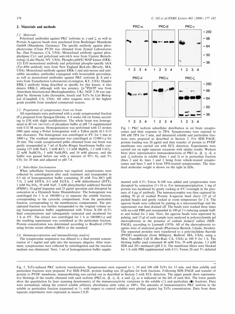
Fig. 1. PKC isoform subcellular distribution in rat brain synapto-somes and their response to TPA. Synaptosomes were exposed to100 nM TPA for 5 min, and denatured soluble and particulate frac-tions were prepared as described in Section 2. For SDS^PAGE,protein loading was 20 Wg/ml and then transfer of protein to PVDFmembrane was carried out with ECL detection. Experiments werecarried out on eight separate occasions with similar results. Westernblots show representative immunodetections of PKC-K, -L, -Q, -N, -Oand -j isoforms in soluble (lines 1 and 3) or in particulate fraction(lines 2 and 4), lines 1 and 2 being from vehicle-treated synapto-somes and lines 3 and 4 from TPA-treated synaptosomes. The stan-dard molecular weight is shown on the right in kDa.
CFig. 2. TeTx-induced PKC isoform translocation. Synaptosomes were exposed to 1, 10 and 100 nM TeTx for 15 min, and then soluble andparticulate fractions were prepared. For SDS^PAGE, protein loading was 20 Wg/lane for both fractions. Following SDS^PAGE and transfer ofprotein to PVDF membrane, immunoblotting was carried out as described in Section 2 with ECL detection. The upper panels show representa-tive blottings of the results obtained with each isoform (PKC-K, -L, -Q, -N, -O and -j), as is indicated on the left of each blot. The lower panelsshow the quantitation by laser scanning densitometry of the immunoreactivity variations in the soluble (a) and particulate (b) fractions. Datawere normalized, taking the control soluble arbitrary absorbance units value as 100%. The amounts of immunoreactive PKC isoform in thesoluble or particulate fraction (expressed as %, with respect to control soluble) were plotted against log TeTx concentration. Data from threeseparate experiments were expressed as means þ S.D.
FEBS 24113 8-9-00
C. Gil et al./FEBS Letters 481 (2000) 177^182178

powdered milk. Next, the membranes were incubated overnight withthe corresponding antibody diluted in blocking bu¡er. Then, themembrane ¢lters were incubated for 1 h with a secondary antibodyconjugated with horseradish peroxidase diluted in blocking bu¡er.Several washes with PBS/0.1% Tween 20 were performed betweeneach one of the steps. The Western blots were developed using Super-
Signal West Pico Chemiluminescent Substrate from Pierce (Rockford,IL, USA) and exposed to Amersham ECL ¢lms. The computer-as-sisted analysis of the bands was performed with a Bio-Rad GS700system (Bio-Rad, CA, USA), and data were processed with a Bio-RadMolecular Analyst image program using a DELL work station. Re-peated scans were taken for ¢lm non-linearity corrections.
FEBS 24113 8-9-00
C. Gil et al./FEBS Letters 481 (2000) 177^182 179

2.5. MAP kinase assayMAP kinase assays were performed as described [20], with modi¢-
cations. Brie£y, 15 Wl of soluble fraction (at a protein concentration of40 Wg/ml) was added to 35 Wl of a solution containing MBP (0.3mg/ml), 20 mM Tris^HCl pH 7.5, 200 WM ATP (with [Q32]ATP at aspeci¢c radioactivity of 10 000 cpm/pmol), 25 mM magnesium acetate,2 WM PKi, 200 WM Na3VO4 and 200 WM EGTA. The phosphoryla-tion reaction was conducted at 37³C for 10 min. In order to terminatethe reaction, 40 Wl of the reaction solution were absorbed in phos-phocellulose P81 Whatman paper (2 cmU2 cm), rinsed in 0.5% phos-phoric acid and washed ¢ve times (1 min each). After that, the radio-activity incorporated was determined by liquid scintillation counting.
3. Results
3.1. Redistribution of PKC isoforms by TPA in the rat brainsynaptosomal fraction
The classical PKC isoforms (PKC-K, -L and -Q), as well asPKC-N, were detected in rat brain synaptosomes, appearing inSDS^PAGE at an apparent molecular weight of 80 kDa,whereas novel PKC-O showed an apparent molecular massof 110 kDa, and atypical PKC-j a molecular mass of 68kDa. The speci¢city of the antibody against the PKC-j iso-form has been tested through blocking the antibody with thecorresponding antigenic peptide, amino acids 577^592 ofPKC-j, since it is the only antibody that recognizes morethan one band (Fig. 1). The incubation of 100 nM TPA for5 min induces translocation from the soluble fraction to theparticulate fraction of the classical isoforms (PKC-K, -L, -Q),as well as the translocation of the novel PKC-N, indicatingactivation of these isoforms by TPA. A large amount ofPKC-O was found associated to the particulate fraction underbasal conditions in synaptosomes, and no translocation of thisisoform was detected, although a visible diminution in thesoluble fraction was seen. In contrast, no e¡ect was observedin the case of the PKC-j, as has been extensively described(Fig. 1).
3.2. E¡ect of TeTx treatment on speci¢c PKC isoformredistribution
Clear translocation of PKC-L, -Q and -N isoforms was ob-served when synaptosomes were incubated with increasingconcentrations of TeTx, during 15 min. PKC-L is the moresensitive isoform, being translocated at 1^10 nM TeTx, where-as at 100 nM the total immunoreactivity decreases, revealingthe appearance of down-regulation. The response of PKC-Qisoform was similar, although down-regulation appears at ahigher TeTx concentration (100 nM) (Fig. 2). On the otherhand, PKC-N showed a sustained translocation, withoutdown-regulation at any concentration tested. In the case ofthe PKC-K isoform, a diminution in its total immunoreactiv-ity (40% of diminution in the soluble fraction and 41% in theparticulate fraction) was detected at the highest concentrationtested (100 nM). No consistent translocation of PKC-O andPKC-j following exposure of synaptosomes to TeTx wasfound. Despite this, a slight diminution in the immunoreac-tivity corresponding to the soluble fraction of PKC-O wasobserved (38% diminution), whereas no increase in the partic-ulate fraction occurs. This absence of translocation cannot beattributed to either a saturation of the immunoblot techniqueor an overexposure of the ¢lm during ECL detection. In orderto obtain internal control in PKC translocation experimentsTPA was included separately in each experiment (not shown).
3.3. TeTx stimulates tyrosine phosphorylation of trkA and ofPLCQ-1
To determine the possible e¡ect of TeTx on cellular signaltransduction pathways that involve Tyr phosphorylation, im-munoprecipitation experiments using anti-phosphotyrosineantibodies (clone PY20) were performed. In each experimentwhole synaptosomal homogenates were immunoprecipitatedusing PY20 antibodies, and the contents of two proteinswhich are phosphorylated in Tyr, i.e. trkA and PLCQ-1,were determined, using speci¢c antibodies, by Western blot.NGF was used as control of a typical signal transductionactivator (Fig. 3A). A single band corresponding to thetrkA receptor appeared at, approximately, 140 kDa of molec-ular weight and a single band corresponding to PLCQ-1 at 150kDa, both in accordance with the literature. This e¡ect wasnot unspeci¢c, since the amount of signal due to FGFR-1(¢broblast growth factor receptor-1) Tyr phosphorylationwas not altered (results not shown). The amount of Tyr phos-phorylation which appeared in trkA and in PLCQ-1 is time-dependent, the onset of the signal being rapid and detectableafter 1 min of treatment (Fig. 3A). In the two phosphopro-teins tested, the maximal amounts of signal were seen in 10min, whereas after 30 min the phosphorylation level had de-creased. The PLCQ-1 phosphorylation was further supportedby our ¢nding that TeTx induces a modest but signi¢cantpolyphosphoinositide hydrolysis increase in rat brain synap-tosomes, which is in agreement with PLC phosphorylationand activation (manuscript submitted). Moreover, Westernanalysis using a phospho-speci¢c antibody recognizing trkA
Fig. 3. TeTx-induced Tyr phosphorylation of trkA and PLCQ-1.A: Synaptosomes in suspension in Krebs^Ringer bu¡er supple-mented with 10 mM glucose were incubated at 30³C with NGF(50 nM) as positive control for 5 min or with 100 nM TeTx at dif-ferent times. Equal amounts of lysate from each time point, ob-tained by sonication and with a protein concentration of 1 mg/ml,were immunoprecipitated, as described in Section 2, with 4 Wg ofantibody PY20 directed against phosphotyrosine residues, and blotswere probed with anti-trkA or anti-PLCQ-1. Each experiment wasrepeated three times, and similar results were obtained. B: Time-course of the TeTx-stimulated phosphorylation of trkA in Tyr-490.The upper blot was incubated with phospho-speci¢c antibody forphosphorylated trkA (Tyr-490). The lower blot shows immunoreac-tivity obtained with antibody speci¢c for trkA, in a manner inde-pendent of its phosphorylation state. Blots are representative ofthree separate experiments.
FEBS 24113 8-9-00
C. Gil et al./FEBS Letters 481 (2000) 177^182180

phosphorylated in Tyr-490 reveals a rapid increase of TeTx-induced trkA phosphorylation, while signal decreasing is de-tected at 30 min after treatment (Fig. 3B). Equal loading ofprotein between samples was con¢rmed by detection withphosphorylation state-independent anti-trkA. This result isin agreement with the signal which appeared in immunopre-cipitation experiments.
3.4. Enhancement of ERK-1/2 phosphorylation status and ofMAP kinase activity
A series of Western analysis using an antibody speci¢c fordually Thr- and Tyr-phosphorylated ERK-1 (p44) and ERK-2(p42) was performed (Fig. 4). In time-course experiments,where synaptosomes were treated with TeTx (100 nM), aninduction of ERK phosphorylation was seen. The strongestsignal appeared after 10 min of treatment, being maintained inthe 30 min treatment. Equal loading of protein between sam-ples was con¢rmed by detection with phosphorylation state-independent anti-ERK-1, although this antibody recognizesboth p44 and p42 in the assayed conditions. The determina-tion of MAPK activity present in the synaptosomal solublefraction, using myelin basic protein (MBP) as substrate, showsan increase in MBP phosphorylation induced by TeTx, thisbeing slight at 10 nM (154%) and more marked at 100 nM(245%) (Fig. 5).
4. Discussion
The in£uence of TeTx over some members of signal trans-duction pathways has been described in several works. The¢rst work on this topic described the decrease of PKC activ-ity, in soluble fraction as well as in particulate fraction, inTeTx-treated macrophages and in the spinal cord of micewith generalized tetanus [21]. Our group described TeTx-in-duced PKC activation/translocation and down-regulationafter intraventricular toxin administration into adult rats [6].In perinatal rat brain, TeTx causes PKC activation parallelwith an increase of serotonin turnover [22,7]. Later, treatmentof rat brain primary neuronal cultures with the neurotoxinrevealed PKC activity translocation and increases in poly-
phosphoinositide hydrolysis [8]. In the present work, a de-tailed investigation focusing on which PKC isoforms are af-fected by TeTx is performed, observing a puzzling diminutionin total PKC-K immunoreactivity, which could be related to ahigh degree of stimulus-triggered PKC hydrolysis detected inrat brain synaptosomes [23]. PKC translocation observed insome isoforms in this work is directly related to the activationof PLCQ-1, since the phospholipase products (diacylglyceroland inositol 1,4,5-triphosphate, as a calcium mobilizer) areactivators of PKC and induce its translocation. Moreover,PLCQ-1 phosphorylation is in consonance with polyphospho-inositide hydrolysis enhancement by TeTx observed in pri-mary neuron cultures from fetal rat brain and in slices fromadult rat cerebral cortex [8]. This phospholipase C activityincrease, which is modest but signi¢cant, is very similar tothat exerted by NGF in rat brain synaptosomes [23]. In thesame work, an NGF-activated PKC-Q and -N translocationwas described, although PKC-L translocation was slight anddi¤cult to detect. TeTx-stimulated inositol phospholipid hy-drolysis has also been detected in rat brain synaptosomes, in acomparable level to that exerted by NGF (results not shown).NGF is a signaling molecule that acts through PLCQ activa-tion [24], this speci¢c activation being related to the modestincrease of polyphosphoinositide hydrolysis. Additionally,PKC activation in synaptosomes could be related to serotonintransport inhibition caused by TeTx or by its HC fragment[25], since serotonin transporter phosphorylation by PKCcauses a diminution in its transport capacity [26]. Studiesfocusing on HC-TeTx-induced redistribution of PKC iso-enzymes are now in progress in our group. On the otherhand, TeTx-induced phosphorylation of trkA in Tyr-490points to a parallel association of shc proteins with thetrkA, an important event implicated in neuronal di¡erentia-tion [27]. This putative association is strongly supported byERK-1/2 phosphorylation observed in this work, since the shcadapter proteins are the ¢rst link of a pathway that results inERK-1/2 activation [12]. In addition, the activation of MAPKpathway could explain the changes in the C-fos and Fos-likeimmunoreactivity observed in the cortex of rats with tetanustoxin-induced epilepsy [28], since growth factor-induced c-fosexpression is activated through transcription factors, such asElk-1 or SAP-1, that are targets of the ras/MAPK [29] orPKC mediated phosphorylation [30].
Early studies show that TeTx binds to gangliosides contain-
Fig. 5. MAPK activity induction by TeTx, at 1, 10 and 100 nMconcentration. The MAPK activity present in the soluble fractionwas assessed at a ¢nal protein concentration of 10 Wg/ml using[Q-32P]ATP and MBP as substrate, as described in Section 2. The re-sults are expressed as means þ S.E.M. of the percentage of three sep-arate experiments. Di¡erences between groups were assessed usingthe one-way ANOVA test (P6 0.001) and one-way Dunnett's testfor multiple comparisons. `*' represents signi¢cantly di¡erent fromthe control group (P6 0.05).
Fig. 4. Activation of ERK-1/2 by TeTx. Synaptosomes were incu-bated at 30³C with TeTx (100 nM) for di¡erent times in A or wereincubated at increasing TeTx concentration for 30 min in B. Forimmunoblot 20 Wg/lane of total synaptosomal lysates were loaded ineach SDS^PAGE. Upper blots were analyzed using antibody speci¢cfor fully Thr- and Tyr-phosphorylated ERK-1 (p44) and ERK-2(p42) (phospho-p44/42 MAP kinase E10 monoclonal antibody).Lower blots show the immunoreactivity obtained for samples as inupper blots with antibodies speci¢c for ERK-1 and -2, independentof their phosphorylation state. All blots are representative of threeseparate experiments.
FEBS 24113 8-9-00
C. Gil et al./FEBS Letters 481 (2000) 177^182 181

ing `1b' structure, such as GT1b or GD1b with a high a¤nity[31]. Ganglioside binding has also been demonstrated forType A and for Type E Clostridium botulinum neurotoxins[32]. On the other hand, the involvement of a protein compo-nent has been postulated, since TeTx binding to cells andneuronal membranes is sensitive to proteases [33]. Accordingto this, a two receptor model has been postulated in which theinitial toxin binding to gangliosides is followed by lateralmovement in the membrane until the toxin reaches a proteinreceptor, then given internalization [34]. Despite e¡orts made,the nature of the neurospeci¢c protein receptor of TeTx is stillunknown, although cross-linking experiments in NGF-di¡er-entiated PC12 cells suggest that an N-glycosylated proteinwith an apparent molecular weight of 15^20 kDa is involvedin the neurospeci¢c binding of TeTx [35,36]. Recently, thebinding of the HC fragment from BoNT/A, which shares thefeature with TeTx and with trk receptors of being retroaxo-nally transported [37], to three proteins of approximately 150,120 and 75 kDa of molecular mass in rat synaptosomes hasbeen reported [38].
In summary, this work determines di¡erential translocationof synaptosomal PKC isoforms in response to TeTx. Like-wise, this PKC activation is directly related to PLCQ-1 phos-phorylation and to trkA activation, assessed by means of itsphosphotyrosine content. Furthermore, the downstream acti-vation of MAP kinases ERK-1 and -2 also exists, displayingpart of a signal transduction pathway, whose complexity mustbe assessed, triggered by TeTx. On the other hand, the inter-action of TeTx with p140 trkA represents a mechanism bywhich the toxin could take advantage of trkA mobility in itstropism to the central nervous system. Further work, which isin progress in our group, will shed light on the speci¢c rela-tions between activation of signal proteins by TeTx and themanner of action of this lethal toxin on the nervous system.
Acknowledgements: This work was supported in part by Grant PB97-0169 from the Ministerio de Educacion y Cultura, Direccion Generalde Ensen¬anza Superior e Investigacion Cient|¢ca. We thank SalvadorBartolome from L.A.B. for technical assistance in image scanning and¢gure presentation.
References
[1] Simpson, L.L. (1989) Botulinum Neurotoxin and Tetanus Toxin,Academic Press, San Diego.
[2] Niemann, H., Blasi, J. and Jahn, R. (1994) Trends Cell Biol. 4,179^185.
[3] Schiavo, G., Benfenati, F., Poulain, B., Rossetto, O., DeLaureto,P.P., DasGupta, B.R. and Montecucco, C. (1992) Nature 359,832^835.
[4] Schiavo, G., Poulain, B., Rossetto, O., Benfenati, F., Tauc, L.and Montecucco, C. (1992) EMBO J. 11, 3577^3583.
[5] Facchiano, F., Valtorta, F., Benfeneti, F. and Luini, A. (1993)Trends Biochem. Sci. 18, 327^329.
[6] Aguilera, J. and Yavin, E. (1990) J. Neurochem. 54, 339^342.[7] Aguilera, J., Padros-Giralt, C., Habig, W.H. and Yavin, E.
(1993) J. Neurochem. 60, 709^713.[8] Gil, C., Ruiz-Meana, M., Alava, M., Yavin, E. and Aguilera, J.
(1998) J. Neurochem. 70, 1636^1643.[9] Iban¬ez, C.F., Ebendal, T. and Persson, H. (1991) EMBO J. 10,
2105^2110.[10] Barbacid, M. (1993) Oncogene 8, 2033^2042.[11] Segal, R.A. and Greenberg, M.E. (1996) Annu. Rev. Neurosci.
19, 463^489.[12] Seger, R. and Krebs, E.G. (1995) FASEB J. 9, 726^735.[13] Robbins, D.J., Zhen, E., Owaki, H., Vanderbilt, C.A., Ebert, D.,
Geppert, T.D. and Cobb, M.H. (1993) J. Biol. Chem. 268, 5097^5106.
[14] Nishizuka, Y. (1995) FASEB J. 9, 484^496.[15] Nishizuka, Y. (1992) Science 9, 484^496.[16] Kraft, A.S. and Anderson, W.B. (1983) Nature 301, 621^623.[17] Saido, T.C., Sorimachi, H. and Suzuki, K. (1994) FASEB J. 8,
814^822.[18] Yurko-Mauro, K.A. and Friedman, E. (1995) J. Neurochem. 65,
1622^1630.[19] Gray, E.G. and Whittaker, U.P. (1962) J. Anat. 96, 1^36.[20] Gomez, N., Tonks, N.K., Morrison, C., Harmar, T. and Cohen,
P. (1990) FEBS Lett. 271, 119^122.[21] Ho, J.L. and Klempner, M.S. (1988) J. Infect. Dis. 157, 925^933.[22] Aguilera, J., Lopez, L.A. and Yavin, E. (1990) FEBS Lett. 263,
61^65.[23] Gil, C., Pelliccioni, P., Itarte, E. and Aguilera, J. (1999) Neuro-
chem. Int. 35, 281^291.[24] Ohmichi, M., Decker, S.J., Pang, L. and Saltiel, A.R. (1991)
Biochem. Biophys. Res. Commun. 179, 217^223.[25] Najib, A., Pelliccioni, P., Gil, C. and Aguilera, J. (1999) J. Neu-
rochem. 72, 1991^1998.[26] Ramamoorthy, S., Giovanetti, E., Qian, Y. and Blakely, R.D.
(1998) J. Biol. Chem. 273, 2458^2466.[27] Obermeier, A., Bradshaw, R.A., Seedorf, K., Choidas, A.,
Schlessinger, J. and Ullrich, A. (1994) EMBO J. 13, 1585^1590.[28] Liang, F. and Jones, E.G. (1997) Brain Res. 778, 281^292.[29] Treisman, R. (1992) Trends Biochem. Sci. 17, 423^426.[30] Hill, C.S. and Treisman, R. (1995) Cell 80, 199^211.[31] Rogers, T.B. and Snyder, S.H. (1981) J. Biol. Chem. 256, 2402^
2407.[32] Schengrund, C.L., DasGupta, B.R., Hughes, C.A. and Ringler,
N.J. (1996) J. Neurochem. 66, 2556^2561.[33] Yavin, E. and Nathan, A. (1986) Eur. J. Biochem. 105, 403^407.[34] Monteccuco, C. (1986) Trends Biochem. Sci. 18, 324^327.[35] Schiavo, G., Ferrari, G., Rossetto, O. and Montecucco, C. (1991)
FEBS Lett. 290, 227^230.[36] Herreros, J., Lalli, G., Montecucco, C. and Schiavo, G. (2000)
J. Neurochem. 74, 1941^1950.[37] Wellho«ner, H.H. (1989) in: Botulinum Neurotoxin and Tetanus
Toxin (Simpson L.L., Ed.), pp. 231^253, Academic Press, SanDiego.
[38] Li, L. and Singh, B.R. (1999) J. Protein Chem. 18, 89^95.
FEBS 24113 8-9-00
C. Gil et al./FEBS Letters 481 (2000) 177^182182