Embed Size (px)
Citation preview
ACL Injuries—The Gender Bias:IntroductionResearch Retreat IIApril 4-5, 2003Lexington, KYGuest EditorsIrene McClay Davis, PT, PhD1,2
Mary Lloyd Ireland, MD3
This was the second research retreat focused on gender bias in anterior cruciate ligament(ACL) injuries, the first having taken place in Lexington, KY in April 2001. The purpose ofthis second retreat was to revisit the factors thought to be associated with gender bias in ACLinjuries and to update the consensus statement from 2001.1 The retreat was againcosponsored by Kentucky Sports Medicine and Joyner Sportsmedicine Institute and wasattended by both clinicians and scientists with a common interest in the ACL injury genderbias. The 50-plus participants included registrants from across the United States as well asCanada, Australia, and Norway. As with the previous retreat, the group consisted of physicians,physical therapists, athletic trainers, and scientists in the areas of biomechanics, motorcontrol, and neuromuscular function. Thirty percent of the participants in the 2003 retreatwere present for the first retreat as well.
A call for abstracts for the retreat was announced in the summer of 2002. All abstracts werethen peer reviewed for scientific merit and relevance to the retreat topic. In the end, 19abstracts were accepted for podium presentations. These were grouped into sessionsaddressing structural, neuromuscular, biomechanical, and hormonal factors that may influ-ence the gender bias in ACL injury incidence. In addition, a new session on interventionprograms was included.
The format of the meeting included 1 keynote presentation per day along with 20-minutepodium presentations made by some of the participants. The keynote presenters were chosenfor their scientific contribution to the understanding of factors associated with the gender biasseen in the incidence of ACL injuries. Bruce D. Beynnon, PhD, from the University ofVermont gave the first keynote titled “Risk Factors for Knee Ligament Trauma.” The secondkeynote presenter was Braden C. Fleming, PhD, also from the University of Vermont, whosetalk was titled “Biomechanics of the Anterior Cruciate Ligament.”
In the following pages, you will find a consensus statement, a listing of the presentations andauthors, and an abstract on each of the 19 presentations made at the conference, organizedby the topics listed above.
REFERENCE1. McClay Davis I, Ireland ML. The gender bias in ACL injuries: a research retreat. Clin Biomech
(Bristol, Avon). 2001;16:937–939.
1 Joyner Sportsmedicine Institute, Mechanicsburg, PA.2University of Delaware, Newark, DE.3Kentucky Sports Medicine Center, Lexington, KY.
ACL Supplement Section: Introduction
Journal of Orthopaedic & Sports Physical Therapy A-1
A-
ACL Injuries—The Gender BiasGuest EditorsIrene McClay Davis, PT, PhD1,2
Mary Lloyd Ireland, MD3
The development of the consensus statement wasformed in the following manner. After all of thepapers for a given factor (structural, neuromuscular,biomechanical, and hormonal) as well as interventionprograms were presented, an initial consensus wasdrafted based on the input of all of the participantsas a group. As with the first retreat, this consensuswas formed through discussion of what we know,which was grounded in the recent literature, alongwith what was presented at the current retreat. Thegroup then identified what is still unknown (what wedon’t know) about each factor’s contribution to thegender bias. This led to the final part of theconsensus in which suggestions for future researchdirections were made. Once all papers were pre-sented, the participants formed 5 self-selected groups,based on each of the areas discussed. A participantwith expertise in that particular area chaired eachgroup. During this time, the groups refined their partof the consensus statement. They were charged withproviding a finalized consensus supported with refer-ences, to be submitted at a designated time followingthe meeting.
During the development of the consensus, the“what we know” portion generated the most discus-sion. It was difficult at times to agree on whatconstituted the facts. Participants were less willing tostate confidently what was known than what wasunknown. Stating what we know proved a mucheasier process during the first retreat due to thepaucity of literature available in April 2001. As thebody of literature has grown and more is known,however, more controversy results. Therefore, partici-pants agreed that the consensus should be viewed asthe present state of thought about ACL injuries,based upon current knowledge, with the realizationthat what we know will likely evolve by the time of thenext retreat in 2 years.
1 Joyner Sportsmedicine Institute, Mechanicsburg, PA.2 University of Delaware, Newark, DE.3 Kentucky Sports Medicine Center, Lexington, KY.
Participants also agreed that, while the focus of thisretreat was the gender bias in ACL injuries, some ofthe identified factors (structural, neuromuscular, andbiomechanical) may not be purely gender specific.For example, genu valgus alignment may be a factorassociated with increased ACL strain and may bemore prevalent in females. However, there may bemales who also exhibit this alignment and so may beat greater risk for ACL injury. Further, there wasmuch discussion regarding whether females need tomove more like men to reduce their risk for ACLinjury or whether there is an optimal pattern forwomen that differs from the pattern for men.
Following are the sections of the consensus state-ment for each of the factors thought to be associatedwith the ACL injury gender bias, as well as for thesection on intervention programs. We would like tothank Lori Livingston, Jean McCrory, WilliamRomani, Sandra Shultz, and Susan Sigward for theirassistance in coordinating the final draft of each ofthese sections. We realize that these lists are not allinclusive, however, they do represent the collectiveopinions of the participants in this retreat. It is ourhope that this consensus statement will promoteresearch studies in the suggested areas so that someof these gaps in the literature might be filled by thenext research retreat.
I. Structural FactorsA. What We Know
1. Females do not have wider pelvises than theirmale counterparts,1,15 as is often assumed.However, females have been shown to havewider pelvis-to-femoral-length ratios,7,10 whichmay contribute to a greater tendency forgenu valgum.
2. Females do have larger Q angles thanmales.11,12 However, Q angle and the frontalplane tibiofemoral valgus angles are indepen-dent measures and cannot be inter-changed.8,9,17 In addition, tibiofemoralvalgum in a single leg squat is not related toQ angle.14
ACL Supplement Section: Consensus Statement
A-2 Journal of Orthopaedic & Sports Physical Therapy
3. Both the size and shape of the notch maycontribute to stenosis and ACL impingementand injury.2,4,19,20
4. The combination of knee abduction (valgus)and external rotation positions contribute toACL impingement in vitro,6 especially in thepresence of a narrow notch.21 These motionshave been shown to be greater in femalescompared to males during athletic activ-ity.3,5,13
5. Females have greater laxity16 and greateractive hip rotation range of motion com-pared to males.18
B. What We Don’t Know1. What are the relationships between lower-
extremity structure, function, and ACL injuryand do those relationships differ betweengenders?
2. Does joint congruency at the tibial plateauinfluence ACL strain and is it different be-tween genders?
3. Does limb dominance or asymmetries inlower limb alignment (eg, as measured usingQ angles or tibiofemoral angles) influenceACL strain?
4. Is a smaller ligament associated with asmaller notch, and are ligament and notchsize scaled to body size?
5. Do the increased genu valgus and femoralinternal rotation seen in females predisposethem to greater strain to the ACL?
6. Does greater laxity or greater range of mo-tion lead to increased risk of ACL injury?
C. Where Do We Go From Here?1. Continue to develop valid and reliable clin-
ical (including weight-bearing) measures ofstructure and function that can be used toidentify individuals at risk for ACL injury.
2. Examine structural measures, not as discretevariables, but as factors that contribute tomultifactorial models.
3. Consider the 3-dimensional shape of theintercondylar notch and how it relates toligament size as well as ligament impinge-ment.
4. Further explore the relationship betweengender differences in pelvic anatomy andresulting knee postures.
5. Investigate the prospective relationship be-tween lower-extremity structure, function,and ACL injury across gender. Structuralvariables that do not change with time couldbe assessed in retrospective studies.
REFERENCES1. Abitbol MM. The shapes of the female pelvis. Contrib-
uting factors. J Reprod Med. 1996;41:242-250.2. Anderson AF, Lipscomb AB, Liudahl KJ, Addlestone RB.
Analysis of the intercondylar notch by computedtomography. Am J Sports Med. 1987;15:547-552.
3. Chaudhari AM, Camarillo DB, Hearn BK, Leveille L,Andriacchi T. The mechanical consequences of genderdifferences in single limb alignment during landing.Proceedings of the ACL Research Retreat II: The GenderBias. J Orthop Sports Phys Ther. 2003;33(8):A25.
4. Charlton WPH, St. John TA, Ciccotti MG, Harrison N,Schweitzer M. Differences in the femoral notchanatomy between men and women-a magnetic reso-nance imaging study. Am J Sports Med. 2002;30:329-333.
5. Ferber R, McClay Davis I, Williams, DS. Genderdifferences in lower extremity mechanics during run-ning. Clin Biomech (Bristol, Avon). 2003;18:350-357.
6. Fung D, et al. Mathematical modeling of ACL impinge-ment against the intercondylar notch wall. Proceedingsof the ACL Research Retreat II: The Gender Bias. JOrthop Sports Phys Ther. 2003;33(8):A18-A19.
7. Horton MG, Hall TL. Quadriceps femoris muscle angle:normal values and relationships with gender and se-lected skeletal measures. Phys Ther. 1989;69:897-901.
8. Igbigbi PS, Msamati BC. Tibiofemoral angles in Malaw-ians. Clin Anat. 2002;15:293-296.
9. LeBlanc SE, Livingston LA, Mahar SM. Q angles andtibiofemoral angles in physically active and inactivefemales. Proceedings of the ACL Research Retreat II:The Gender Bias. J Orthop Sports Phys Ther.2003;33(8):A16.
10. Livingston LA, Gahagan JC. The wider gynaecoid pel-vis—larger Q angle—greater predisposition to ACL in-jury relationship: myth or reality? Clin Biomech (Bristol,Avon). 2001;16:951-952.
11. Livingston LA. The quadriceps angle: a review of theliterature. J Orthop Sports Phys Ther. 1998;28:105-109.
12. Livingston LA, Mandigo JL. Bilateral Q angle asymmetryand anterior knee pain syndrome. Clin Biomech(Bristol, Avon). 1999;14:7-13.
13. Malinzak RA, Colby SM, Kirkendall DT, Yu B, GarrettWE. A comparison of knee joint motion patterns be-tween men and women in selected athletic tasks. ClinBiomech (Bristol, Avon). 2001;16:438-445.
14. Pantano KJ, White SC, Gilchrist LA, Leddy J. Differencesin peak knee valgus angles between individuals withhigh and low Q angles during a single limb squat.Proceedings of the ACL Research Retreat II: The GenderBias. J Orthop Sports Phys Ther. 2003;33(8):A17.
15. Petersen A. Sexing the body: representations of sexdifferences in Gray’s Anatomy, 1858 to the present.Body Soc. 1998;4:1-15.
16. Rozzi SL, Lephart SM, et al. Knee joint laxity andneuromuscular characteristics of male and female soc-cer and basketball players. Am J Sports Med.1999;27(3):312-319.
17. Schulthies SS, Francis RS, Fisher AG, Van de Graaff KM.Does the Q angle reflect the force on the patella in thefrontal plane? Phys Ther. 1995;75:24-30.
18. Simoneau GG, Hoenig KJ, Lepley JE, Papanek, PE.Influence of hip position and gender on active hip
ACL Supplement Section: Consensus Statement
J Orthop Sports Phys Ther • Volume 33 • Number 8 • August 2003 A-3
A-
internal and external rotation. J Orthop Sports PhysTher. 1998;28:158-164.
19. Tillman MD, Smith KR, Bauer JA, Cauraugh JH, FalsettiAB, Pattishall JL. Differences in three intercondylarnotch geometry indices between males and females: acadaver study. Knee, 2002;9(1):41-6.
20. Van Lunen BL, Perrin DH, Arnold BL, et al. Characteris-tics of anterior cruciate ligament injuries: preliminaryfindings. Proceedings of the ACL Research Retreat II:The Gender Bias. J Orthop Sports Phys Ther.2003;33(8):A16.
21. Zhang L-Q, Fung D, Lin F, et al. Noncontact ACLinjuries: impingement versus direct stretch. Proceedingsof the ACL Research Retreat II: The Gender Bias. JOrthop Sports Phys Ther. 2003;33(8):A17.
II. Neuromuscular FactorsA. What We Know
1. Females activate their muscles sooner in an-ticipation of landing.1,13
2. Variability in neuromuscular control param-eters at impact can cause significant increasesin external 3-dimensional knee joint loadingduring movements linked to noncontact ACLinjury.8
3. Females tend to rely more on theirquadriceps to stabilize the knee compared tomales.5,6,15
4. Females demonstrate reduced muscle stiff-ness compared to males when attempting tocontrol knee motion.4,16
5. Females take longer than males to producemuscular tension in the quadriceps with re-flex activation after fatiguing exercise10 andin the hamstring muscles with isokinetic test-ing.5
6. Females exhibit lower muscular endurancecompared to males.5 Lower endurance isthought to lead to earlier fatigue, which isalso thought to be related to increased risk ofinjury.3,11,12
B. What We Don’t Know1. While there are a number of neuromuscular
strategies that produce the same joint me-chanics,9 should females produce the samestrategies as males? Is there an optimal pat-tern for each gender?
2. How are neuromuscular strategies affected bymaturation within gender?
3. While there is evidence to suggest that in-creased knee joint laxity increases hamstringreflex delays14 and increases lateral hamstringactivity12,14 in weight bearing, what are thegender differences in these responses?
4. Does earlier preactivation of muscles nega-tively or positively affect joint stiffness, kinet-ics, and ground reaction forces produced
during landing from a jump or during aplant and cut maneuver?
5. While it has been shown that simulatedincreased stiffness of the hip resulted in anincreased resistance to knee valgus buckling,2
what are the influences of hip and trunk(core) stability on knee function? How canwe assess these factors experimentally?
6. While simulated variations in neuromuscularcontrol parameters at impact can cause sig-nificant increases in external 3-dimensionalknee joint loading during movements linkedto ACL injury,7 do gender differences inthese variations exist and are they associatedwith injury?
C. Where Do We Go From Here?1. Determine the underlying factors for the
observed gender differences in neuromuscu-lar function.
2. Determine at what point during the matura-tional process neuromuscular strategies beginto differ between genders.
3. Generate studies combining neuromuscularand biomechanical measures to define moreprecisely the relationship between muscularactivity and knee joint forces and moments.
4. Clarify what recruitment strategies may bebeneficial with respect to protection of theACL during weight-bearing postures.
5. Delineate the role of proximal muscle influ-ences (hip and trunk) on lower-extremityfunction and injury.
6. Develop models that can predict the effectsof neuromuscular control on knee joint andresultant ACL loading.
REFERENCES1. Cowling E J, Steele JR. Is lower limb muscle synchrony
during landing affected by gender? Implications forvariation in ACL injury rates. J Electromyogr Kinesiol.2001;11:263-68.
2. Chaudhari AM, Camarillo DB, Hearn BK, Leveille L,Andriacci T. The mechanical consequences of genderdifferences in single limb alignment during landing.Proceedings of the ACL Research Retreat II: The GenderBias. J Orthop Sports Phys Ther. 2003;33(8):A25.
3. Gabbett TJ. Incidence of injury in amateur rugby leaguesevens. Br J Sports Med. 2002;36(1):23-26.
4. Granata KP, Padua DA, Wilson SE. Gender differencesin active musculoskeletal stiffness. Part II. Quantifica-tion of leg stiffness during functional hopping tasks. JElectromyogr Kinesiol. 2002;12:127-135.
5. Huston LJ, Wojtys EM. Neuromuscular performancecharacteristics in elite female athletes. Am J Sports Med.1996;24(4):427-436.
ACL Supplement Section: Consensus Statement
A-4 J Orthop Sports Phys Ther • Volume 33 • Number 8 • August 2003
6. Malinzak RA, Colby SM, Kirkendall DT, Yu B, GarrettWE. A comparison of knee joint motion patterns be-tween men and women in selected athletic tasks. ClinBiomech (Bristol, Avon). 2001;16:438-445.
7. McLean SG, Neal RJ, Myers PT, Walters MR. Knee jointkinematics during the sidestep cutting maneuver: poten-tial for injury in women. Med Sci Sports Exerc.1999;31(7):959-968.
8. McLean SG, van den Bogert AJ, Su A. Effects ofneuromuscular control on knee joint loading duringsidestepping: Implications for noncontact ACL injury.Proceedings of the ACL Research Retreat II: The GenderBias. J Orthop Sports Phys Ther. 2003;33(8):A24.
9. McNitt-Gray JL, Hester DM, Mathiyakom W, MunkasyBA. Mechanical demand and multijoint control duringlanding depend on orientation of the body segmentsrelative to the reaction force. J Biomech. 2001;34:1471-1482.
10. Moore BD, Drouin JM, Shultz SJ, Gansneder BG. Theeffect of gender and fatigue on neuromuscular re-sponses following a patellar tendon tap. J Athl Train.2001;36(2):S9.
11. Rahnama N, Reilly T, Lees A. Injury risk associated withplaying actions during competitive soccer. Br J SportsMed. 2002;36(5):354-359.
12. Rozzi SL, Lephart SM, Gear WS, Fu FH. Knee jointlaxity and neuromuscular characteristics of male andfemale soccer and basketball players. Am J Sports Med.1999;27(3):312-319.
13. Schmitz RJ, Thompson TT. Gender differences in hipand knee kinematics during single leg landings. J AthlTrain. 2002;37:S20.
14. Shultz SJ, Carcia CR, Perrin DH. Knee joint laxityaffects muscle activation patterns at the knee. Proceed-ings of the ACL Research Retreat II: The Gender Bias. JOrthop Sports Phys Ther. 2003;33(8):A20.
16. Shultz SJ, Perrin DH, Adams JM, Arnold BL, GansnederBM, Granata KP. Neuromuscular response characteris-tics in males and females after knee perturbation in asingle leg weight-bearing stance. J Athl Train.2001;36(1):37-43.
17. Wojtys EM, Ashton-Miller JA, Huston LJ. A gender-related difference in contribution of the knee muscula-ture to sagittal-plane shear stiffness in subjects withsimilar knee laxity. J Bone Joint Surg Am. 2002;84A(1):10-16.
III. Biomechanical FactorsA. What We Know
1. The mechanisms for noncontact ACL injurieshave been reported to include decelerationwith the knee in an extended position, land-ing from a jump, and sidestep cut maneu-vers.4
2. Females tend to have more knee valgus thanmales during sidestep cutting6,13,16 and amore extended knee than males at initialcontact.
3. Compared to running, frontal- andtransverse-plane moments are greater duringanticipated sidecut maneuvers to 30° and 60°and crossover cutting. In addition, thesemoments were increased when the tasks
where performed under unanticipated condi-tions.1
4. Females demonstrate significantly less kneeflexion and increased hip and knee internalrotation than males during single-leg landingand forward-hopping tasks.12
5. Females demonstrated greater knee exten-sion and valgus moments during the landingphases of jump stop tasks, which are associ-ated with greater anterior tibial shear forces.5
In vitro studies demonstrate increases inthese types of loads (extensor and valgus) areassociated with increased strains of theACL.3,8,14
B. What We Don’t Know1. The isolated motions (and moments) of knee
valgus and internal rotation have been shown(both in vitro and in vivo) to increase ACLstrain, while external rotation alone de-creases its strain.2,7,10,14,15 What is the effectof combined, in vivo, loading of these mo-tions such as genu valgum and externalrotation on ACL strain?
2. How is the ACL ligament loaded, in vivo,during high-risk maneuvers such as cutting,landing, and abrupt decelerations?
3. While we know that a heel contact (com-pared to a forefoot contact) during landingsleads to an increased internal rotation mo-ment and greater internal rotation angles,9
what are the gender differences in landingcontact patterns?
4. Do gender differences noted in mechanics ofhigh-risk movements place women at greaterrisk for ACL injury?
5. Should females move more like males or isthere an optimal movement pattern for eachgender?
C. Where Do We Go From Here?1. Examine the relationship between in vitro
and in vivo data on ACL loading, especiallyas it relates to high risk athletic activitiessuch as cutting and landing.
2. Further investigate (both in vitro and in vivo)the influence of combined loading patternsassociated with mechanisms of injury.
3. Determine if there are gender differences inthe ACL strain response.
4. Investigate whether the at risk patterns aretask specific (ie, different for cutting thanlanding).
5. Determine whether there are gender differ-ences in foot contact patterns during high-risk maneuvers such as landing and cutting.
6. Examine whether the gender differences
ACL Supplement Section: Consensus Statement
J Orthop Sports Phys Ther • Volume 33 • Number 8 • August 2003 A-5
A-
noted in biomechanical patterns during ath-letic activities translate into higher risk forACL injury for females.
7. Explore the effects of experience, age, andmaturation on gender differences in mechan-ics.
8. Develop forward dynamic 3-dimensionalmodels to be able to simulate more accu-rately real life situations that are not possiblein a laboratory setting.17 In addition, furtherdevelop software that can accurately recon-struct 3-dimensional data from markerlessvideo sequences of game situations11 inwhich ACL injuries occur.
REFERENCES1. Besier TF, Lloyd DG, Cochrane JL, Ackland TR. External
loading of the knee joint during running and cuttingmaneuvers. Med Sci Sports Exerc. 2001;33:1168-1175.
2. Beynnon BD, Fleming BC. Anterior cruciate ligamentstrain in vivo: a review of previous work. J Biomech.1998;31:519-525.
3. Beynnon BD, Fleming BC, Johnson RJ, et al. Anteriorcruciate ligament strain behavior during rehabilitationexercises in vivo. Am J Sports Med. 1995;23:24-34.
4. Boden BP, Dean GS, Feagin JA, Garrett WE. Mecha-nisms of anterior cruciate ligament injury. Orthopedics.2000;23(6):573-578.
5. Chappell JD, Yu B, Kirkendall DT, Garrett WE. Acomparison of knee kinetics between male and femalerecreational athletes in stop-jump tasks. Am J SportsMed. 2002;30(2):261-267.
6. Chaudhari AM, Camarillo DB, Hearn, BK, Leveille L,Andriacci T. The mechanical consequences of genderdifferences in single limb alignment during landing.Proceedings of the ACL Research Retreat II: The GenderBias. J Orthop Sports Phys Ther. 2003;33(8):A25.
7. Fleming BC, Renstrom PA, Beynnon BD, et al. Theeffect of weightbearing and external loading on anteriorcruciate ligament strain. J Biomech. 2001;34:163-170.
8. Fleming BC, Renstrom PA, Ohlen G, et al. Thegastrocnemius muscle is an antagonist of the anteriorcruciate ligament. J Orthop Res. 2001;19:1178-1184.
9. Houck JR, Hanford S. Foot landing strategies affect kneebiomechanics during sidestep cut tasks. Proceedings ofthe ACL Research Retreat II: The Gender Bias. J OrthopSports Phys Ther. 2003;33(8):A23.
10. Kanamori A, Woo SL-Y, Ma CB, et al. The forces in theanterior cruciate ligament and knee kinematics during asimulated pivot shift test. Arthroscopy. 2002;16:633-639.
11. Krosshaug T, Bahr RA. Model-based image-matchingtechnique for 3-dimensional motion reconstruction fromuncalibrated video sequences—application to ACL in-jury situations. Proceedings of the ACL Research RetreatII: The Gender Bias. J Orthop Sports Phys Ther.2003;33(8):A21.
12. Lephart SM, Ferris CM, Riemann BL, Myers JB, Fu FH.Gender differences in strength and lower extremitykinematics during landing. Clin Orthop. 2002;401:162-169.
13. Malinzak RA, Colby SM, Kirkendall DT, Yu B, GarrettWE. A comparison of knee joint motion patterns be-tween men and women in selected athletic tasks. ClinBiomech (Bristol, Avon). 2001;16:438-445.
14. Markolf KL, Burchfield DM, Shapiro MM, Shepard MF,Finerman GA, Slauterbeck JL. Combined knee loadingstates that generate high anterior cruciate ligamentforces. J Orthop Res. 1995;13:930-935.
15. Markolf KL, Gorek JF, Kabo JM, Shapiro MS. Directmeasurement of resultant forces in the anterior cruciateligament. An in vitro study performed with a newexperimental technique. J Bone Joint Surg Am.1990;72:557-567.
16. McLean SG, Neal RJ, Myers PT, Walters MR. Knee jointkinematics during the sidestep cutting maneuver: poten-tial for injury in women. Med Sci Sports Exerc.1999;31:959-968.
17. McLean SG, van den Bogert AJ, Su A. Effects ofneuromuscular control on knee joint loading duringsidestepping: Implications for noncontact ACL injury.Proceedings of the ACL Research Retreat II: The GenderBias. J Orthop Sports Phys Ther. 2003;33(8):A24.
IV. Hormonal FactorsA. What We Know
1. Subject self-report of menstrual phase is notaccurate and may lead to unreliable find-ings.5,7
2. There is no change in ultimate failure loadof the ACL throughout the estrous cycle inrats.2
3. There may be hormone-dependent changesthroughout the menstrual cycle that influ-ence physical performance.8
4. Supraphysiologic levels of estradiol decreasethe load-to-failure in ovariectomized rabbits.5
5. There is an association between individualvariations in sex hormone concentration andchanges in the low load viscoelastic proper-ties of the tibiofemoral joint.1,4
6. There are significant differences in kneejoint laxity between genders both pre-exercise and postexercise. However, bothgenders exhibit a similar increase in kneejoint laxity following exercise.3
B. What We Don’t Know1. What is the relationship between animal and
human models?2. What is the relationship between sex hor-
mone fluctuation, menstrual cycle, and injurypattern?
3. What is the relationship between joint laxityand ACL laxity?
4. What is the relationship between ACL laxityand injury risk?
ACL Supplement Section: Consensus Statement
A-6 J Orthop Sports Phys Ther • Volume 33 • Number 8 • August 2003
C. Where Do We Go From Here?1. Develop consistent definitions of biomechani-
cal properties: stiffness, laxity, load-to-failureproperties.
2. Conduct assay analyses of saliva, urine, orserum to identify more accurately the phaseof the menstrual cycle at the time of injury.
3. Determine the temporal influence of hor-mones on the low-load viscoelastic propertiesof the ACL in static and dynamic conditions.
4. Include specialists in physiology, endocrinol-ogy, and reproductive medicine to character-ize the menstrual cycle and the hormonalinfluences on the biomechanical propertiesof the ACL.
5. Design multicenter studies that evaluate therelationship between sex hormone concentra-tions, phase of menstrual cycle, and kneeligament injury patterns as they relate tospecific sports.
6. Examine the influence of sex hormones onthe ligament and activated mechanisms ofcollagen remodeling in ACL tissue.
REFERENCES1. Deie M, Sakamaki Y, Sumen Y, Urabe Y, Ikuta Y.
Anterior knee laxity in young women varies with theirmenstrual cycle. Intl Orthop. 2002;26(3):154-56.
2. Otto D, Raso J, Bagnall K, Amirfazli A, Thorton G.Variation in the mechanical behavior of rat ACL duringthe estrous cycle. Proceedings of the ACL ResearchRetreat II: The Gender Bias. J Orthop Sports Phys Ther.2003;33(8):A26.
3. Pollard C, Braun B, Hamill J. Influences of gender andexercise on ACL laxity. Proceedings of the ACL Re-search Retreat II: The Gender Bias. J Orthop Sports PhysTher. 2003;33(8):A27.
4. Romani WA, Patrie J, Curl LA, Flaws JA. The correla-tions between estradiol, estrone, estriol, progesterone,and sex hormone binding globulin and anterior cruciateligament stiffness in healthy, active females. J WomensHealth Gend Based Med. 2003;12(3):287-298.
5. Slauterbeck J, Clevenger C, Lundberg W, Burchfield D.Estrogen level alters the failure load of the rabbitanterior cruciate ligament. J Orthop Res. 1999;17:405-408.
6. Slauterbeck J, Fuzie S, Smith M, et al. The menstrualcycle, sex hormones and anterior cruciate ligamentinjury. J Athl Train. 2002;37(3):275-278.
7. Wojtys E, Huston L, Boynton M, Spindler K, LindenfeldT. The effect of menstrual cycle on anterior cruciateligament injuries in women as determined by hormonallevels. Am J Sports Med. 2002;30(2):182-188.
8. Yack HJ, Chandran R, Rao S, Wilken J. How normalfluctuations in women influence the biomechanics ofstepping and cutting. Proceedings of the ACL ResearchRetreat II: The Gender Bias. J Orthop Sports Phys Ther.2003;33(8):A28.
V. InterventionsA. What We Know
1. Therapeutic exercise has been shown to sig-nificantly reduce ACL and lower extremityinjury rates.1,3,4,8,13
2. Biomechanical analyses have shown that bodymovement patterns can be modified, andground reaction forces reduced through anintervention program.2,5,6,7,10,14
3. Perturbation training resulted inneuromuscular adaptations, but strengthtraining did not.6,7
4. A plyometric training protocol resulted inincreased quadriceps strength, but nochanges in hamstring or hip abductorstrength were noted.6,7
5. Education interventions may affect injuryrate, as demonstrated in the alpine skiingliterature.11,12
6. Feedback has provided a successful reinforce-ment of movement strategies.9 Subjects re-spond better to instructions of body positionrather than muscle activations.2
B. What We Don’t Know1. What biomechanical patterns place a person
at risk for ACL injury?2. What are the ideal movement strategies? Are
these strategies different for males and fe-males?
3. Why have the intervention programs worked?Is success due to better movement patternsor improved neuromuscular characteristics,such as improved joint stiffness or the abilityto react to a perturbation? Is the mechanismof success related to the mechanism of in-jury?
4. How long does it take to alter programming?How long is the carryover after completionof an intervention program? If it is a pre-season program, how much carryover doesthe athlete have into the season?
5. Do these interventions need to be differentbased on age and maturation levels?
C. Where Do We Go From Here?1. Determine the risk factors for ACL injury.2. Focus intervention programs on these risk
factors.3. Determine the common denominator of suc-
cess within the current intervention pro-grams.
4. Determine the optimal approach for inter-vention exercises and for the timing of theintervention with respect to the competitiveseason.
5. Design well defined prospective, randomized,
ACL Supplement Section: Consensus Statement
J Orthop Sports Phys Ther • Volume 33 • Number 8 • August 2003 A-7
A-
double-blind studies with stratification by ac-tivity level, sport, playing surface, coaching,etc.
6. Examine whether the biomechanical changeselicited by the intervention programs carryover onto game fields and courts.
7. Investigate the utility of external interven-tions (such as braces, playing surfaces, etc).
REFERENCES1. Caraffa A, Cerulli G, Projetti M, Aisa G, Rizzo A.
Prevention of anterior cruciate ligament injuries insoccer: a prospective controlled study of proprioceptiontraining. Knee Surg Sports Traumatol Arthrosc.1996;4:19-21.
2. Cowling EJ, Steele JR, McNair PJ. Can lower limbmuscles be retrained during landing as a means ofanterior cruciate ligament injury prevention? Proceed-ings of the ACL Research Retreat II: The Gender Bias. JOrthop Sports Phys Ther. 2003;33(8):A29.
3. Heidt RS Jr, Sweeterman LM, Carlonas RL, Traub JA,Tekulve FX. Avoidance of soccer injuries with preseasonconditioning. Am J Sports Med. 2000;28(5):659-662.
4. Hewett TE, Lindenfeld TN, Riccobene JV, Noyes FR.The effect of neuromuscular training on the incidenceof knee injury in female athletes: a prospective study.Am J Sports Med. 1999;27(6):699-706.
5. Hewett TE, Stroupe AL, Nance TA, Noyes FR.Plyometric training in female athletes: decreased impactforces and increased hamstring torques. Am J SportsMed. 1996;24(6):765-773.
6. Lephart SM, Ferris CM, Sell T, Abt J, Irrgang JJ, Fu F.Neuromuscular and biomechanical adaptations to an
intervention program for the prevention of female ACLinjuries. Proceedings of the Orthopaedic Research Soci-ety Meeting. New Orleans, LA: Orthopaedic ResearchSociety; 2003.
7. McCrory JL, Lephart SM, Ferris CM, et al. Adaptationsto an intervention program for the prevention of femaleACL injuries. Proceedings of the ACL Research RetreatII: The Gender Bias. J Orthop Sports Phys Ther.2003;33(8):A29.
8. Myklebust G, Engebretsen L, Braekken IH, Skjolberg A,Olsen O-E, Barh R. Prevention of anterior cruciateligament injuries in female team handball players: aprospective intervention study over three seasons. ClinSport Med. 2003;13:71-78.
9. Onate JA, Guskiewicz KM, Sullivan RJ. Augmentedfeedback reduces jump landing forces. J Orthop SportsPhys Ther. 2000;31(9):511-517.
10. Prapavessis H, McNair PJ, Anderson K, Hohepa M.Decreasing landing forces in children: the effect ofinstructions. J Orthop Sports Phys Ther. 2003;33:204-207.
11. Ryder SH, Johnson RJ, Beynnon BD, Ettlinger CF.Prevention of ACL injuries. J Sport Rehab. 1997;6:80-96.
12. Skolnick AA. Experts try education to preserve skiers’knees. JAMA. 1996;275(7):506-507.
13. Soderman K, Werner S, Pietila T, Engstrom B, AlfredsonH. Balance board training: prevention of traumaticinjuries of the lower extremities in female soccerplayers? A prospective randomized intervention study.Knee Surg Sports Traumatol Arthrosc. 2000;8(6):356-363.
14. Wojtys EM, Huston LJ, Aston-Miller JA. Changes inlower extremity joint kinematics after completing ajump-training program. Proceedings of the AmericanOrthopaedic Society for Sports Medicine. Orlando, FL:American Orthopaedic Society for Sports Medicine;2002.
ACL Supplement Section: Consensus Statement
A-8 J Orthop Sports Phys Ther • Volume 33 • Number 8 • August 2003
ACL Injuries—The Gender BiasApril 4-5, 2003Lexington, KY
Keynote Address IRisk Factors for Knee Ligament TraumaBruce D. Beynnon, PhDBurlington, VT
Keynote Address IIBiomechanics of the Anterior Cruciate LigamentBraden C. Fleming, PhDBurlington, VT
I. Structural FactorsCharacteristics of ACL Injuries: Preliminary FindingsBonnie L. Van Lunen, PhD, ATCOld Dominion UniversityRichmond, VA
Q Angles and Tibiofemoral Angles in Physically Active and InactiveFemalesLori A. Livingston, PhDSchool of Health and Human Performance, Dalhousie UniversityHalifax, Nova Scotia, Canada
Differences in Peak Knee Valgus Angles Between Individuals With Highand Low Q Angles During a Single Limb SquatKathleen J. Pantano, PT, PhDBiomechanics Laboratory/Sports Medicine Institute, University at Buf-faloBuffalo, NY
Noncontact ACL Injuries: Impingement Versus Direct StretchLi-Qun Zhang, PhDRehabilitation Institute of Chicago and the Departments of PhysicalMedicine & Rehabilitation and Orthopaedic Surgery, NorthwesternUniversityChicago, IL
Mathematical Modeling of ACL Impingement Against the IntercondylarNotch WallDavid Fung, MSSensory-Motor Performance Program, Rehabilitation Institute of Chi-cago, Department of Biomedical Engineering, Northwestern UniversityChicago, IL
II. Neuromuscular FactorsKnee Joint Laxity Affects Muscle Activation Patterns at the Knee Priorto and Following a Weight-Bearing PerturbationSandra J. Schultz, PhD, ATCUniversity of North Carolina at GreensboroGreensboro, NC
Gender Differences in Hip and Knee Kinematics and Muscle Preactiva-tion During Single Leg LandingsRandy J. Schmitz, PhD, ATCApplied Neuromechanics Research Laboratory, University of NorthCarolina at GreensboroGreensboro, NC
Is Lower Limb Muscle Synchrony During Landing Affected by Gender?Implications for Variations in ACL Injury RatesElizabeth J. Cowling, PhDBiomechanics Research Laboratory, University of WollongongNew South Wales, Australia
III. Biomechanical FactorsA Model-Based Image-Matching Technique for 3-Dimensional MotionReconstruction from Uncalibrated Video Sequences—Application toACL Injury Situations
Tron Krosshaug, MSc, PhD CandidateOslo Sports Trauma Research CenterOslo, Norway
Foot Landing Strategies Affect Knee Biomechanics During Sidestep CutTasksJeff R. Houck, PT, PhDIthaca CollegeRochester, NY
Effects of Neuromuscular Control on Knee Joint Loading DuringSidestepping: Implications for Noncontact ACL InjuryScott G. McLean, PhDDepartment of Biomedical Engineering, The Cleveland Clinic Founda-tionCleveland, OH
The Mechanical Consequences of Gender Differences in Single LimbAlignment During LandingAjit M. Chaudhari, PhDStanford Biomotion Laboratory, Stanford UniversityStanford, CA
IV. Hormonal FactorsFluctuations in Estradiol and Progesterone Are Related to Changes inACL Stiffness in Healthy, Active FemalesWilliam A. Romani, PT, PhD, ATC, SCSUniversity of Maryland School of MedicineBaltimore, MD
Variation in the Mechanical Behavior of Rat ACL During the EstrousCycleV. James Raso, MAScGlenrose RehabilitationEdmonton, Canada
Influences of Gender and Exercise on ACL LaxityChristine D. Pollard, PhD CandidateBiomechanics Laboratory, University of MassachusettsAmherst, MA
How Normal Hormonal Fluctuations in Women Influence the Biome-chanics of Stepping and CuttingH. John Yack, PT, PhDOrthopaedic Gait Analysis Laboratory, Graduate Program in PhysicalTherapy and Rehabilitation Science, The University of IowaIowa City, IA
V. InterventionsCan Lower Limb Muscles be Retrained During Landing as a Means ofACL Injury Prevention?Elizabeth J. Cowling, PhDBiomechanics Research Laboratory, University of WollongongNew South Wales, Australia
Adaptations to an Intervention Program for the Prevention of FemaleACL InjuriesJean L. McCrory, PhDNeuromuscular Research Laboratory, Department of Sports Medicineand Nutrition, University of PittsburghPittsburgh, PA
Prevention of ACL Injuries in Female Team Handball Players—AProspective Intervention Study Over 3 SeasonsGrethe Myklebust, PT, Specialist in Sports Physiotherapy, PhD CandidateOslo Sports Trauma Research CenterOslo, Norway
ACL Supplement Section: List of Keynotes and Abstracts
Journal of Orthopaedic & Sports Physical Therapy A-9
A-
KEYNOTE ADDRESS IRisk Factors for Knee Ligament TraumaBruce D. Beynnon, PhD1
INTRODUCTION
The anterior cruciate ligament (ACL) is the mostfrequently totally disrupted ligament in the knee, andalthough this injury is relatively uncommon in thegeneral population, it occurs frequently in athletics,particularly among female athletes.1,2 Many studieshave focused on the prevalence of ACL injuriesassociated with high-risk sports; however, only a lim-ited number have calculated incidence rates based ontime-at-risk and compared males and females compet-ing in similar activities. These studies indicate thatthe rates of ACL tears for female athletes range from2.4 to 9.7 times higher than these of male ath-letes.1,3,4,5,6 Our research at the University of Ver-mont has focused on competitive alpine ski racing, asport that imparts tremendous forces on the knee,and has revealed that 1 in 5 female alpine racerssuffer an ACL disruption. These female athletes were3.1 times more likely to sustain an ACL injury incomparison to their male counterparts.7 Even moredisturbing was the finding that 27% of the womenwho underwent reconstruction suffered reinjury tothe graft and underwent a second reconstructionprocedure—a rate twice that seen for the men.7
Our recent review of the literature9 revealed 4prospective studies of ACL injury risk factors,10,11,12,13
2 of which only focused on male athletes.10,12 In astudy of high school athletes, Souryal and Freemanreported that athletes with a small intercondylarnotch width index (the ratio of the width of theanterior outlet of the intercondylar femoral notchdivided by the total condylar width at the level of thepopliteal groove) were at significantly increased riskfor sustaining an ACL injury.13 LaPrade and Burnettstudied collegiate athletes and reported similar find-ings.11 In a study of American football, Lambson et al
1 McClure Musculoskeletal Research Center, Department of Ortho-paedics and Rehabilitation, College of Medicine, University of Vermont,Burlington, VT.
found that athletes with a greater number of cleats,and an associated higher torsional resistance at thefoot-turf interface, were at increased risk for sufferingan ACL tear.10 Orchard and colleagues studied ath-letes participating in Australian football and foundthat a history of ACL reconstruction and weatherconditions that were characterized by high evapora-tion and low rainfall before matches were risk factorsfor repeated ACL injury.12 Currently, there is verylittle information regarding the risk factors for ACLinjury that have been derived from well-designedprospective studies, and only 2 studies have includedfemales (the group that appears to be at increasedrisk for this injury) and no study has investigatedmultiple factors.
Recently, our group has focused on studying therisk factors for knee and ankle ligament traumaamong precollegiate and collegiate athletes, and 2recent investigations will be highlighted. The aim ofthe first investigation was to measure serum levels ofestradiol and progesterone, and knee and ankle jointlaxity (indirect measures of the biomechanical behav-ior of ligaments) during the menstrual cycle. Thecorresponding hypothesis was that knee and anklejoint laxity values increase during the menstrual cycleas serum concentrations of estradiol andprogesterone become elevated. The aim of the sec-ond study was to examine potential knee injury riskfactors among competitive alpine skiers. This wasused to test the hypotheses that one or a combinationof risk factors can be used to identify an athlete atrisk for anterior cruciate ligament trauma.
METHODS
The effect of Serum Estradiol and Progesterone Levelson Knee and Ankle Joint Laxity
Female and male (control) athletes were ap-proached and informed consent was obtained. There
ACL Injuries—The Gender Bias: Keynotes
A-10 Journal of Orthopaedic & Sports Physical Therapy
were 10 women with normal menstrual cycles (meanage, 20.7; range, 18-23) and 5 males (mean age, 21;range, 19-23). Knee and ankle laxity and bloodconcentrations of estradiol and progesterone weremeasured for females at 4 consecutive visits through-out the menstrual cycle, and for males at equivalenttime intervals. For the females with a 28-day cyclelength, the visits were as follows: visit 1 (earlyfollicular phase, days 1-3), visit 2 (late follicularphase, days 11-13), visit 3 (mid-luteal phase, days20-22) and visit 4 (late luteal phase, days 27-28).Subjects with different cycle lengths were evaluated atproportional time intervals. Baseline concentrationsof estradiol and progesterone were obtained amongthe women during the early follicular phase of thefollowing cycle (visit 5) to validate that a normal cyclewas completed. Serum concentrations of estradioland progesterone were determined using radioim-munoassay.
Knee and ankle joint laxity were measured at eachvisit. Knee laxity was measured with the KT-1000 KneeArthrometer (MedMetrics Corp., San Diego, CA).Anterior-posterior (A-P) laxity was defined as the totalA-P translation between the shear loads of –90 N(posterior) and 130 N (anterior). Ankle laxity wasmeasured with the anterior drawer and talar tilt testsby standard stress radiography. The Telos device(Telos Corp., Greisheim, Germany) was used to apply150-N forces to the ankle joint. All radiographs wereobtained by the same individual and numbered toblind the measurement procedure. At each visit,anterior drawer (neutral and anterior load) and talartilt views (inversion and eversion loads) were ob-tained.
A paired t test for means was used to compare jointlaxity values between visit 1 (the early follicular phasewhen estradiol is low) and visit 2; (the late follicularphase when estradiol is high). Similarly, a paired ttest for means was used to compare laxity valuesbetween visit 1 (early follicular phase whenprogesterone is low) and visit 3 (the mid luteal phasewhen progesterone is high). The P value was adjustedfor multiple comparisons.
A Prospective Study of Knee Injury Risk Factors inCompetitive Alpine Ski Racers
Data were collected over 3 ski seasons (1995, 1996,and 1997). Athletes were recruited from 6 competi-tive alpine ski programs located throughout thenortheastern United States. The athletes ranged inage from 13 to 22 years. Prior to the start of each skiseason, intrinsic and extrinsic risk factor data werecollected. The intrinsic factors included demographicinformation, generalized joint laxity, flexibility of the
hamstrings, quadriceps and heel cord, anatomicalalignment of the knee, laxity of the knee, andisokinetic thigh muscle strength. Each athlete wasfollowed throughout the ski season, exposure (thenumber of days skied) data were documented daily,and injury information were recorded. The samplesize was 224 subjects. For all analyses, data from 1 legwere used, the injured leg for subjects who sustainedknee trauma, and a randomly selected leg for thosewho were not injured.
RESULTS
The Effect of Serum Estradiol and Progesterone Levelson Knee and Ankle Joint Laxity
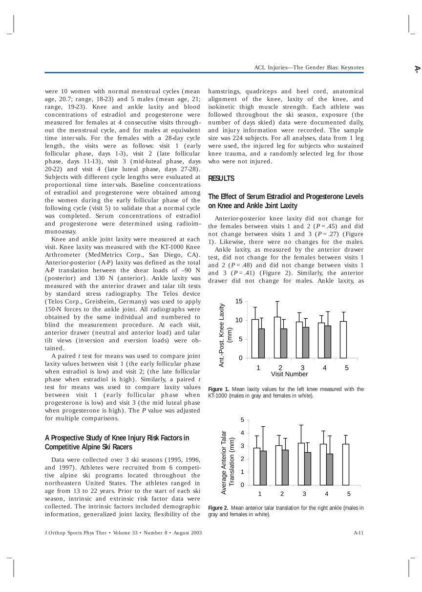
Anterior-posterior knee laxity did not change forthe females between visits 1 and 2 (P = .45) and didnot change between visits 1 and 3 (P = .27) (Figure1). Likewise, there were no changes for the males.
Ankle laxity, as measured by the anterior drawertest, did not change for the females between visits 1and 2 (P = .48) and did not change between visits 1and 3 (P = .41) (Figure 2). Similarly, the anteriordrawer did not change for males. Ankle laxity, as
Figure 1. Mean laxity values for the left knee measured with theKT-1000 (males in gray and females in white).
Figure 2. Mean anterior talar translation for the right ankle (males ingray and females in white).
0
5
10
15
1 2 3 4 5Visit Number
Ant
.-P
ost.
Kne
e La
xity
(m
m)
0
1
2
3
4
5
1 2 3 4 5Ave
rage
Ant
erio
r Ta
lar
Tran
slat
ion
(mm
)
ACL Injuries—The Gender Bias: Keynotes
J Orthop Sports Phys Ther • Volume 33 • Number 8 • August 2003 A-11
A-
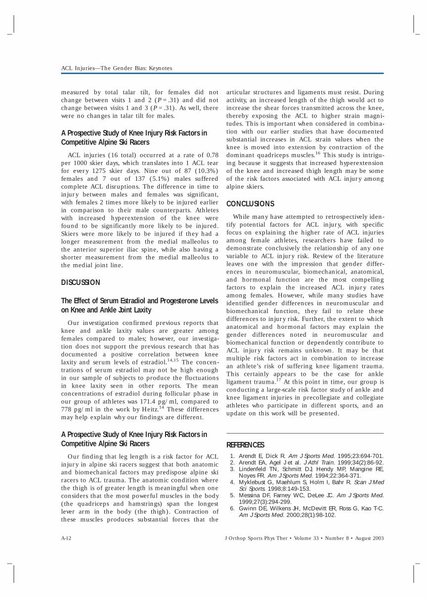
measured by total talar tilt, for females did notchange between visits 1 and 2 (P = .31) and did notchange between visits 1 and 3 (P = .31). As well, therewere no changes in talar tilt for males.
A Prospective Study of Knee Injury Risk Factors inCompetitive Alpine Ski Racers
ACL injuries (16 total) occurred at a rate of 0.78per 1000 skier days, which translates into 1 ACL tearfor every 1275 skier days. Nine out of 87 (10.3%)females and 7 out of 137 (5.1%) males sufferedcomplete ACL disruptions. The difference in time toinjury between males and females was significant,with females 2 times more likely to be injured earlierin comparison to their male counterparts. Athleteswith increased hyperextension of the knee werefound to be significantly more likely to be injured.Skiers were more likely to be injured if they had alonger measurement from the medial malleolus tothe anterior superior iliac spine, while also having ashorter measurement from the medial malleolus tothe medial joint line.
DISCUSSION
The Effect of Serum Estradiol and Progesterone Levelson Knee and Ankle Joint Laxity
Our investigation confirmed previous reports thatknee and ankle laxity values are greater amongfemales compared to males; however, our investiga-tion does not support the previous research that hasdocumented a positive correlation between kneelaxity and serum levels of estradiol.14,15 The concen-trations of serum estradiol may not be high enoughin our sample of subjects to produce the fluctuationsin knee laxity seen in other reports. The meanconcentrations of estradiol during follicular phase inour group of athletes was 171.4 pg/ml, compared to778 pg/ml in the work by Heitz.14 These differencesmay help explain why our findings are different.
A Prospective Study of Knee Injury Risk Factors inCompetitive Alpine Ski Racers
Our finding that leg length is a risk factor for ACLinjury in alpine ski racers suggest that both anatomicand biomechanical factors may predispose alpine skiracers to ACL trauma. The anatomic condition wherethe thigh is of greater length is meaningful when oneconsiders that the most powerful muscles in the body(the quadriceps and hamstrings) span the longestlever arm in the body (the thigh). Contraction ofthese muscles produces substantial forces that the
articular structures and ligaments must resist. Duringactivity, an increased length of the thigh would act toincrease the shear forces transmitted across the knee,thereby exposing the ACL to higher strain magni-tudes. This is important when considered in combina-tion with our earlier studies that have documentedsubstantial increases in ACL strain values when theknee is moved into extension by contraction of thedominant quadriceps muscles.16 This study is intrigu-ing because it suggests that increased hyperextensionof the knee and increased thigh length may be someof the risk factors associated with ACL injury amongalpine skiers.
CONCLUSIONS
While many have attempted to retrospectively iden-tify potential factors for ACL injury, with specificfocus on explaining the higher rate of ACL injuriesamong female athletes, researchers have failed todemonstrate conclusively the relationship of any onevariable to ACL injury risk. Review of the literatureleaves one with the impression that gender differ-ences in neuromuscular, biomechanical, anatomical,and hormonal function are the most compellingfactors to explain the increased ACL injury ratesamong females. However, while many studies haveidentified gender differences in neuromuscular andbiomechanical function, they fail to relate thesedifferences to injury risk. Further, the extent to whichanatomical and hormonal factors may explain thegender differences noted in neuromuscular andbiomechanical function or dependently contribute toACL injury risk remains unknown. It may be thatmultiple risk factors act in combination to increasean athlete’s risk of suffering knee ligament trauma.This certainly appears to be the case for ankleligament trauma.17 At this point in time, our group isconducting a large-scale risk factor study of ankle andknee ligament injuries in precollegiate and collegiateathletes who participate in different sports, and anupdate on this work will be presented.
REFERENCES1. Arendt E, Dick R. Am J Sports Med. 1995;23:694-701.2. Arendt EA, Agel J et al. J Athl Train. 1999;34(2):86-92.3. Lindenfeld TN, Schmitt DJ, Hendy MP, Mangine RE,
Noyes FR. Am J Sports Med. 1994;22:364-371.4. Myklebust G, Maehlum S, Holm I, Bahr R. Scan J Med
Sci Sports. 1998;8:149-153.5. Messina DF, Farney WC, DeLee JC. Am J Sports Med.
1999;27(3):294-299.6. Gwinn DE, Wilkens JH, McDevitt ER, Ross G, Kao T-C.
Am J Sports Med. 2000;28(1):98-102.
ACL Injuries—The Gender Bias: Keynotes
A-12 J Orthop Sports Phys Ther • Volume 33 • Number 8 • August 2003
7. Stevenson H, Webster J, Johnson RJ, Beynnon BD. IowaOrtho J. 1998;18:64-66.
8. Daniel DM, Stone ML, Dobson BE, et al. Am J SportsMed. 1994;22(5), 632-644.
9. Murphy DF, Connolly DAJ, Beynnon BD. British J SportsMed. 2003;37:13-29.
10. Lambson RB, Barnhill BS, Higgins RW. Am J Sport Med.1996;24(2):155-159.
11. LaPrade RF, Burnett QM. Am J Sports Med.1994;22(2):198-203.
12. Orchard J, Seward H, McGivern J, Hood S. Am J SportsMed. 2001;29(2):196-200.
13. Souryal TO, Freeman TR. Am J Sports Med. 1993;21(4),535-539.
14. Heitz NA. J Athl Train. 1999;343(2):144-149.15. Deie M, Sakamaki Y, Sumen Y, Urabe Y, Ikuta Y. Intl
Orthop. 2002;26(3)154-156.16. Beynnon BD, Fleming BC. J Biomech. 1998;31:519-
525.17. Beynnon BD, Renstrom PA, Alosa DM, et al. J Orthop
Res. 2001;19(2):213-220.
KEYNOTE ADDRESS IIBiomechanics of the Anterior CruciateLigamentBraden C. Fleming, PhD1
INTRODUCTION
Many factors have been associated with the destruc-tion of knee joint articular cartilage; however, onlytrauma such as that associated with disruption of theanterior cruciate ligament (ACL) has been shown toinitiate osteoarthritis (OA). The optimal treatment ofan ACL injury remains an enigma, and there isevidence indicating that the current treatment op-tions (ie, ACL reconstruction) will not slow theprogression of OA that occurs following injury. Thus,there is a need to understand how we can preventthese injuries. ACL research has typically focused onthe treatment and diagnosis of these injuries but noton prevention. To understand how to prevent ACLinjuries it is necessary to understand the biomechan-ics of the ACL. We can then utilize this informationto understand better how the ACL can be injuredand how the injuries can be prevented. The goal ofthis presentation is to provide baseline informationabout the biomechanical function of the ACL.
1 McClure Musculoskeletal Research Center, Department ofOrthopaedics and Rehabilitation, College of Medicine, University ofVermont, Burlington, VT.
METHODSOver the last decade, we have developed a tech-
nique to measure ACL strain in human subjects.1
Study participants have been volunteers with normalACLs who were undergoing diagnostic arthroscopyunder local anesthesia, permitting all subjects toretain full control of their muscles. After the routinesurgical procedure was complete, a differential vari-able reluctance transducer (DVRT) was implanted onthe ACL to measure its displacement response undervarious controlled loadings. These displacement datawere used to determine the strain response of theligament using the engineering strain formulation.The focus of this lecture is to review the studiesevaluating the ACL strain response under variousloading conditions: external loading with no muscleactivation,1,4 muscle loading (ie, quadriceps, ham-strings, and gastrocnemius),1,5 weight bearing,4 andrehabilitation exercises.1
RESULTS
ACL strain was measured during various passiveloading conditions: passive flexion-extension motion
ACL Injuries—The Gender Bias: Keynotes
J Orthop Sports Phys Ther • Volume 33 • Number 8 • August 2003 A-13
A-
of the knee (PFE), and under anterior-posterior-directed shear loads, internal-external torques, andvarus-valgus moments applied externally to the tibiarelative to the femur.1,4 It has been argued that theACL is a restraint to all of these loading conditions.Our findings suggest that the ACL remainsunstrained during PFE as the knee is extended from90° to 10° (thigh horizontal). This result was ex-pected because the gravity vector contains a posteri-orly directed component throughout most of thisrange of knee motion. PFE from 10° to full extensionstrained the ACL. Anterior-directed shear loads pro-duced strain values that were greater when the kneewas at 30° as compared to 90°. These data verify thatthe ACL is a primary restraint to anterior tibialtranslation and explain the increased sensitivity of theLachman test compared to the drawer test in detect-ing ACL injuries. We also determined that significantACL strains were produced with the application ofinternal torques. However, the ACL was not strainedwith the application of external torques, varus orvalgus moments up to 10 Nm.
The muscles spanning the knee apply forces andmoments to the knee. As the knee joint position ischanged, the moment arms of the different musclesand joint contact position change. Thus, joint posi-tion and the magnitude of muscle contraction affectACL strain biomechanics. For isometric quadricepscontractions, ACL strain values were dependent onthe knee flexion angle.1 At 30 Nm of extensiontorque, ACL strain values produced at 15° of kneeflexion were significantly greater than those producedat 30°, while no strain was produced at 60° and 90°.For isometric hamstrings contractions, the ACL strainvalues were not dependent on knee flexion angle.Hamstrings contractions did not strain the ACL atany knee angle tested. The ACL strain values pro-duced by simultaneous maximum contraction of thequadriceps and hamstrings muscles were also depen-dent on the knee flexion angle. The strain valuesproduced at 15° and 30° were less than thoseproduced during isolated isometric quadriceps con-tractions but greater than those produced duringisolated isometric hamstrings contractions. The influ-ence of the gastrocnemius is controversial. Becausethe proximal tendon of the gastrocnemius wrapsaround the posterior aspect of the tibial plateau,contraction of the muscle could potentially strain theACL by pushing the tibia anterior when the knee isnear extension. We found that both knee flexionangle and gastrocnemius force affected ACL strains.5
With the knee at 5° and 15° of flexion, contraction ofthe gastrocnemius increased ACL strain relative tothe relaxed state. At the higher knee flexion angles(30° and 45°), the ACL was not strained.
Application of body weight requires the activationof the leg musculature to maintain equilibrium.Previous investigations reported that weight bearingprovides a protective mechanism to the ACL, or ACLgraft, because the articulating condyles are forcedtogether and muscle cocontraction is utilized toincrease contact resistance and joint stiffness.2,8 How-ever, we found a significant increase in ACL strainwith the application of the compressive load pro-duced by body weight as compared to thenonweighted condition with the knee at 20° offlexion.4 These findings suggest that the quadricepsmuscles are dominant in maintaining equilibrium.
We have also evaluated ACL strains during com-monly prescribed rehabilitation activities.1 These ex-ercises included quadriceps-dominated andhamstrings-dominated activities and those that involvecocontraction of these muscle groups. Exercises thatproduced low strain values were those that weredominated by the hamstrings muscle group, incorpo-rated contraction of the quadriceps muscles with theknee flexed at 50° or more, or involved simultaneousquadriceps and hamstrings contraction. Quadricepsdominated activities with the knee between 50° andfull extension produced higher strains. Furthermore,we recently determined that the maximum ACLstrain values produced during squatting, a closed-kinetic-chain exercise thought to be protective of theACL, were similar to those produced during activeextension of the knee, an open-kinetic-chain exercise.The similarity of the strain responses during these 2common rehabilitation exercises calls into questionwhether we should consider exercises to be eithersafe, or unsafe, based on the commonly used closed-and open-kinetic-chain terminology.
DISCUSSION
Seventy percent of ACL injuries are noncontactand the mechanisms remain unknown. The injuriestypically occur when a subject is decelerating, pivot-ing, landing or responding to a perturbation. Thus,they occur in response to different combinations ofloads that are both internally and externally applied.Considering that the knee is located between the 2longest lever arms of the body, it is not surprisingthat ACL injuries are common and that small pertur-bations could produce high loads at the knee, hencethe ACL. Obviously it is not appropriate to use the invivo measurement technique to directly assess injurymechanisms due to patient safety issues. However, itallows us to evaluate the strain response due tosubfailure loadings that can possibly be extrapolated.The strain measurement technique could then beused to determine if these injuries can be prevented
ACL Injuries—The Gender Bias: Keynotes
A-14 J Orthop Sports Phys Ther • Volume 33 • Number 8 • August 2003
using specific techniques (ie, bracing, training). Fu-ture work will be directed at looking at subfailurecombinations of loads and the response of the ACLto perturbations.
CONCLUSIONS
The ACL is a primary restraint to anterior shearrotation and internal rotation of the tibia withrespect to the femur. In our studies, external rotationand varus-valgus angulation did not strain the ACL.However, there are cadaver studies which suggest thatthe ACL is loaded when combinations of these loadsare applied.6,7 Contraction of the quads andgastrocnemius strained the ACL at low knee flexionangles, while contraction of the hamstrings reducedACL strains. Application of a compressive load acrossthe knee joint strained the ACL. Finally, strains up to4% were seen during exercises that simulate activitiesof daily living. These strain values are considerablyless than the failure strain of the ACL and within thetoe region of the stress-strain curve for the ligament.3
ACKNOWLEDGEMENTS
The National Institutes of Health (AR39213;AR40174) and the National Football League Charitiessupported this work. This work was performed incollaboration with Drs. Bruce Beynnon, RobertJohnson, Per Renstrom, Joseph Abate, ClaudeNichols, and Glenn Peura.
REFERENCES1. Beynnon BD. J Biomech. 1998;31:591.2. Beynnon BD. J Orthop Res. 2002;20:332.3. Butler. J Biomech. 1992;25:511.4. Fleming BC. J Biomech. 2001;34:163.5. Fleming BC. J Orthop Res. 2001;19:1178.6. Kanamori. Arthroscopy7. Markolf. J Orthop Res. 1995;13:930.8. Torzilli. Am J Sports Med. 1994;22:105.
ACL Injuries—The Gender Bias: Keynotes
J Orthop Sports Phys Ther • Volume 33 • Number 8 • August 2003 A-15
A-
ACL Injuries—The Gender Bias: Abstracts
I. STRUCTURAL FACTORSCHARACTERISTICS OF ANTERIOR CRUCIATE LIGAMENTINJURIES: PRELIMINARY FINDINGSVan Lunen BL,1 Perrin DH,2 Arnold BL,3 Gieck JH,4 Saliba EN,4
Gansneder BM,4 McCue FC4
1Old Dominion University; 2University of North Carolina at Greens-boro; 3Virginia Commonwealth University; 4University of Virginia.INTRODUCTION
The purpose of this study was to compare several anatomicalreference points and knee laxity assessments for anterior cruciateligament (ACL) injured males and females. Secondly, this studyexamined the anatomical and laxity differences between thoseinjured through a noncontact or contact mechanism. Lastly, thisstudy examined the intratester reliability of several of the measures.METHODS
Thirty subjects with disruption of the ACL who gave informedconsent (10 females, 20 males; age, 25.53 ± 9.54 years; height,176.50 ± 9.39 cm; mass, 83.81 ± 20.70 kg) participated in this study.Subjects were classified as being injured due to contact ornoncontact. Tunnel view radiographs of both knees were taken andthe notch dimensions of popliteal notch width index and half-height notch width index were determined. The subjects weremeasured for quadriceps angle, navicular drop, sagittal kneeextension, tibial fibular varum, tibiofemoral angle, and KT1000measures at 15 and 30 pounds of force. If applicable, the subjectswere asked to place their feet on a premade foot template. Allmeasures were taken bilaterally. Separate 1-way analyses of variancewere performed on each dependent measure. Intraclass correlationcoefficients (ICC2,1) were used to determine intratester reliability.RESULTS
Results indicated that quadriceps angle was significantly greaterfor the males injured through a contact mechanism (F2,29 = 3.987,P = .031) than the males injured by noncontact. The injuredpopliteal notch width index was significantly greater for the contactmales (F2,27 = 6.123, P = .007) than the noncontact males andfemales. The injured half-height notch width index was signifi-cantly greater for the contact males (F2,27 = 3.98, P = .32) than thenoncontact males only. All other analyses of variance were notsignificantly different. Values for intratester reliability ranged from0.57 to 0.94.DISCUSSION
The values of popliteal notch width index support past researchin classifying those individuals with a notch width index of lessthan 0.2000 as being susceptible to noncontact injuries. Furtherresearch should investigate the structural alignment characteristicsin a dynamic mode and the relationship to ACL injury.CONCLUSIONS
Many of the dependent variables examined may not differbetween males and females who are injured through contact ornoncontact mechanisms.
Q ANGLES AND TIBIOFEMORAL ANGLES IN PHYSICALLYACTIVE AND INACTIVE FEMALESLeBlanc SE,1 Livingston LA,1 Mahar SM1
1School of Health and Human Performance, Dalhousie University,Halifax, Nova Scotia, Canada.
INTRODUCTIONFrontal plane quadriceps (Q) angles and tibiofemoral (TF)
angles are often viewed as synonymous measures.1,2 Both havebeen implicated as anatomical risk factors for ACL ligament injuryin females,3,4 yet while similar in appearance, they differ indefinition. The purpose of this study was to examine the relation-ship between the magnitudes of these angles in young adultfemales.METHODS
Physically active (n = 19) and inactive (n = 18) females with amean age of 19.9 ± 1.3 years and no previous history of lower limbinjury participated in the study. The average height and mass ofthe sample were 168.3 ± 5.5 cm and 62.6 ± 7.0 kg, respectively.Markers were applied bilaterally to the ASIS, midpoint of thepatella, and midpoint of the tibial tubercle to define the Q angle.In contrast, TF angles were defined by markers applied to thegreater trochanter, midline of the thigh, midpoint of the tibialtubercle, and the midline of the anterior aspect of the shin. Digitalphotographic images were taken of participants with their feet inself-selected and Romberg (feet side-by-side) stances. Measures ofright and left static Q angles and TF angles were then derived fromthe photos using a manual goniometer. Dependent t tests wereused to examine within-subject differences in Q angles and valgusangles, while differences in the dependent variables by group,stance, and limb were analyzed using univariate repeated-measuresANOVA procedures. Measurement reliabilities (ICC2,1)5 werefound to be excellent (ICC > 0.89).6
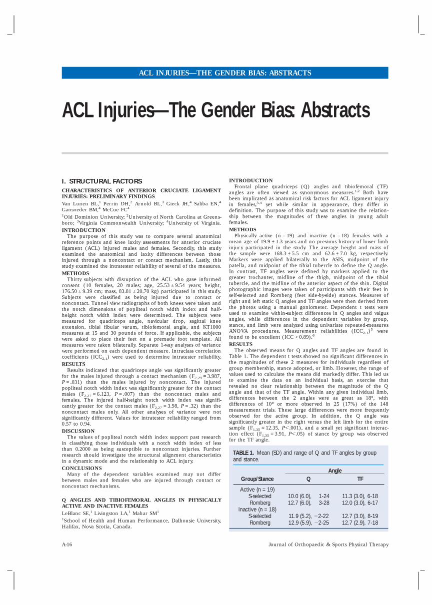
RESULTSThe observed means for Q angles and TF angles are found in
Table 1. The dependent t tests showed no significant differences inthe magnitudes of these 2 measures for individuals regardless ofgroup membership, stance adopted, or limb. However, the range ofvalues used to calculate the means did markedly differ. This led usto examine the data on an individual basis, an exercise thatrevealed no clear relationship between the magnitude of the Qangle and that of the TF angle. Within any given individual limb,differences between the 2 angles were as great as 18°, withdifferences of 10° or more observed in 25 (17%) of the 148measurement trials. These large differences were more frequentlyobserved for the active group. In addition, the Q angle wassignificantly greater in the right versus the left limb for the entiresample (F1,35 = 12.35, P,.001), and a small yet significant interac-tion effect (F1,35 = 3.91, P,.05) of stance by group was observedfor the TF angle.
TABLE 1. Mean (SD) and range of Q and TF angles by groupand stance.
AngleGroup/Stance Q TF
Active (n = 19)S-selected 10.0 (6.0), 1-24 11.3 (3.0), 6-18Romberg 12.7 (6.0), 3-28 12.0 (3.0), 6-17
Inactive (n = 18)S-selected 11.9 (5.2), 22-22 12.7 (3.0), 8-19Romberg 12.9 (5.9), 22-25 12.7 (2.9), 7-18
ACL INJURIES—THE GENDER BIAS: ABSTRACTS
A-16 Journal of Orthopaedic & Sports Physical Therapy
DISCUSSIONThe finding of no statistically significant difference between the
2 measures within individuals should be approached with cautionas our data provide a good example of how the act of pooling datamay mask important findings. The observation of greater differ-ences in magnitude between the Q and TF angles in the activegroup would be expected, given that increases in quadricepsstrength are known to reduce Q angles.7 In contrast, the significantreduction seen in the TF angle for the active group when changingfrom a Romberg to self-selected foot position is small and perhapsclinically insignificant.CONCLUSIONS
The Q angle provides a reasonable estimate of the line of pull ofthe extensor mechanism,8 while by contrast the TF angle describesthe functional adequacy of the bony articulation between thefemur and tibia.9 Given the data presented herein, the 2 shouldnot be seen as synonymous or directly related. Future study ofthese 2 measures, the imbalance between them, and their relation-ship to ACL injury seems warranted.
REFERENCES1. Holloway M. Scientific Am. 2000;11(3):32–37.2. Veenema KR. Physician Sportsmed. 1999;27(8):62.3. Griffin L, et al. J Am Acad Orthop Surg. 2000;8:141-150.4. Huston LJ, et al. Clin Orthop Rel Res. 2000;372:50-63.5. Shrout, et al. Psych Bull. 1979;86:420-428.6. Fleiss J. Reliability of measurement. New York: Wiley, 1986.7. Byl N, et al. J Sport Rehabil. 2000;9:26-34.8. Igbigbi PS, et al. Clin Anat. 2002;15:293-296.9. Schulthies SS, et al. Phys Ther. 1995;75:24-30.
DIFFERENCES IN PEAK KNEE VALGUS ANGLES BETWEENINDIVIDUALS WITH HIGH AND LOW Q ANGLES DURING ASINGLE LIMB SQUATPantano KJ, White SC, Gilchrist LA, Leddy J1Biomechanics Laboratory/Sports Medicine Institute, University atBuffalo, Buffalo, NY.INTRODUCTION
Differences in anatomical alignment between genders, such as agreater quadriceps angle (Q angle), have been suggested as causesof the disparity in anterior cruciate ligament (ACL) injury rates. Alarger Q angle may contribute to increased knee valgus duringmovement and increased stress on the ACL. This study investigated
whether significant differences in peak knee valgus angles exist inhealthy college-aged subjects with a large Q angle, compared tothose with a small Q angle, while performing a single limbsquatting motion. The relationship between Q angle and selectedskeletal measures was also determined.METHODS
Twenty subjects, categorized as having a ‘‘high Q angle’’ ($17°)or a ‘‘low Q angle’’ (#8°) were videotaped during the perfor-mance of a single leg squat. The peak valgus angles for the rightknee were calculated. A t test was used to determine whether therewere differences in peak knee valgus angles between the high andlow Q angle groups (P<.05).RESULTS
There were no significant differences in peak knee valgus anglesexhibited for the high and low Q-angle groups during the singleleg squat (P = .17). Q angle was not associated with pelvic width,tibiofemoral angle, or dynamic knee valgus, but was moderatelycorrelated with the pelvic width/femoral length ratio (r = 0.54).DISCUSSION
The degree of knee valgus exhibited during a single leg squat isnot a function of Q angle and static measures may not bepredictive of knee valgus during dynamic movement.CONCLUSIONS
Further research into gender differences other than Q angle isneeded to explore causes of increased knee valgus during limbloading tasks.
NONCONTACT ACL INJURIES: IMPINGEMENT VERSUS DI-RECT STRETCH
Zhang L-Q, Fung D, Lin F, Makhsous M, Koh JL, Hendrix RW,Nuber GW
Rehabilitation Institute of Chicago and Northwestern University,Chicago, IL.INTRODUCTION
Sudden deceleration, hyperextension, abrupt change in direc-tion, fixed foot, and tibial rotation have been reported as keyelements of noncontact ACL injuries.1,3 However, although tibialinternal and external rotations seem to increase and decrease ACLloading, respectively,4,5 both excessive internal rotation and exter-nal rotation of the tibia seem to be associated with noncontactACL injuries. We hypothesize that tibial external rotation (andabduction) causes ACL impingement against the lateralintercondylar notch wall (especially for knees with a narrow
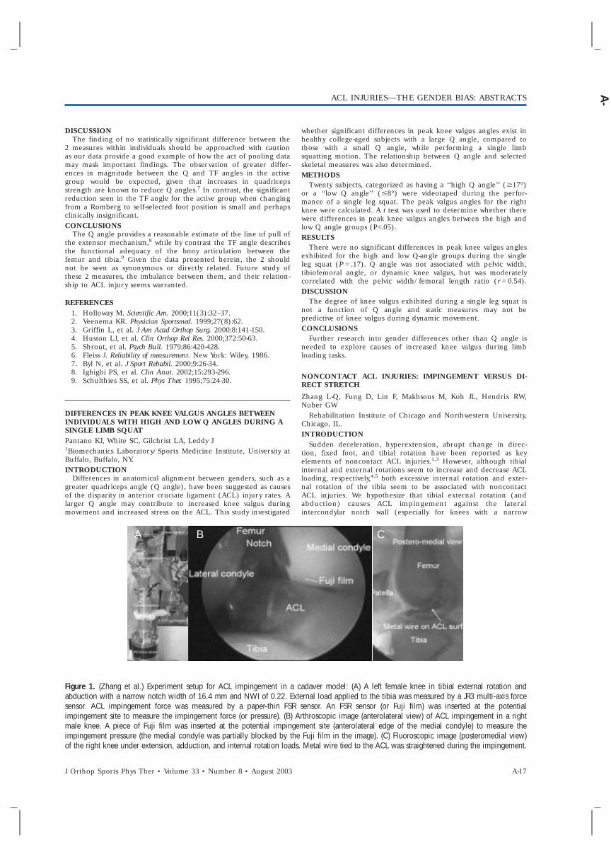
Figure 1. (Zhang et al.) Experiment setup for ACL impingement in a cadaver model: (A) A left female knee in tibial external rotation andabduction with a narrow notch width of 16.4 mm and NWI of 0.22. External load applied to the tibia was measured by a JR3 multi-axis forcesensor. ACL impingement force was measured by a paper-thin FSR sensor. An FSR sensor (or Fuji film) was inserted at the potentialimpingement site to measure the impingement force (or pressure). (B) Arthroscopic image (anterolateral view) of ACL impingement in a rightmale knee. A piece of Fuji film was inserted at the potential impingement site (anterolateral edge of the medial condyle) to measure theimpingement pressure (the medial condyle was partially blocked by the Fuji film in the image). (C) Fluoroscopic image (posteromedial view)of the right knee under extension, adduction, and internal rotation loads. Metal wire tied to the ACL was straightened during the impingement.
A B C
ACL INJURIES—THE GENDER BIAS: ABSTRACTS
J Orthop Sports Phys Ther • Volume 33 • Number 8 • August 2003 A-17
A-
intercondylar notch and certain notch shape), which in turn mayresult in rupture of the ACL. For knees with a regular notch width,ACL may impinge the anterior-lateral edge of the medial condyleduring tibial internal rotation and adduction near full kneeextension. The purpose of the study was to investigate the differentinjury mechanisms by measuring impingement directly and usingfluoroscopy and arthroscopy.METHODS
ACL impingement was investigated using 8 cadaver knee speci-mens (4 female, 4 male). Two of the specimens were fresh frozen,the rest were embalmed. Through an arthroscope or with ACL andintercondylar notch exposed, ACL impingement was inspectedduring manual manipulation of the knee throughout the ROMsabout multiple knee axes. Intercondylar notch width index (NWI)was measured directly from each of the knees.
For the embalmed specimens, the patella was removed. With thefemur fixed to a bench, the tibia was moved passively undermoderate externally applied loads measured by a multi-axis forcesensor (JR3, Inc., Woodland, CA), while the impingement wasdirectly observed through the exposed intercondylar notch andmeasured by force sensitive resistors (FSR, Tekscan, Inc., Boston,MA, or Fuji film, Fuji Photo Film USA, Inc., Carrollton, TX).Six-DOF knee kinematics were measured with a 6-DOF goniometer(Figure 1A).
For the fresh-frozen specimens, a small parapatellar incision wasmade on the specimen to expose the ACL and intercondylarnotch. A paper-thin FSR sensor (or Fuji film) was inserted at thepotential impingement site to record the impingement force(pressure). With the femur fixed to a bench, the knee was movedforcefully throughout its range of motion (ROM) in 3-dimensionalspace to evaluate possible ACL impingement. An arthroscope wasused to inspect the inside of the knee for potential ACL impinge-ment under various loads to the tibia (Figure 1B). A fluoroscopewas also used to evaluate possible ACL impingement. Throughbilateral parapatellar incisions, a flat copper wire (about 3 mmwide and 20 mm long) was tied to the anterior-superior surface ofthe ACL using metal surgical suture. After closing the incisionsusing regular surgical suture, the knee was inspected dynamicallyunder fluoroscope while the knee was moved throughout its3-dimensional ROMs (Figure 1C).RESULTS
NWI (mean ± standard deviation) for the 4 male knees was 0.25± 0.02, for the 4 female knees, 0.19 ± 0.02, and across all the knees,0.22 ± 0.04.
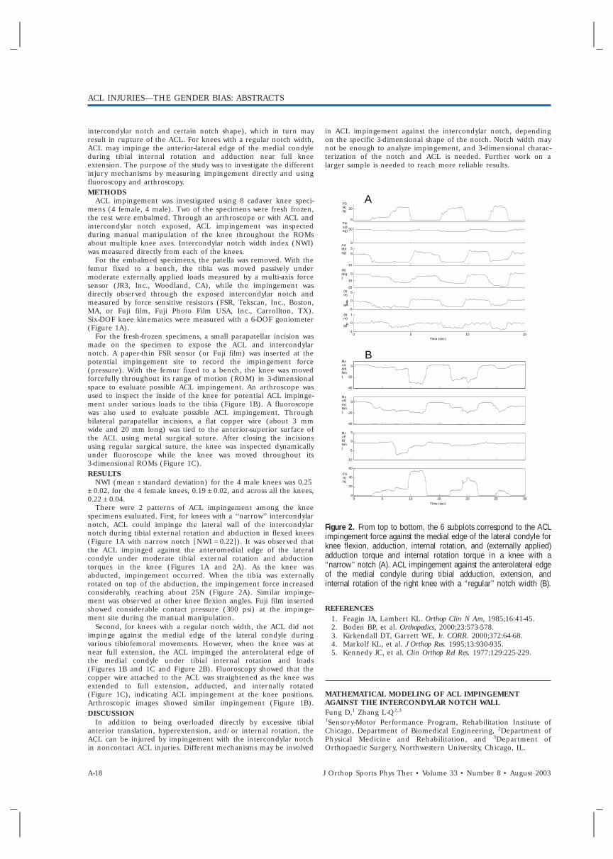
There were 2 patterns of ACL impingement among the kneespecimens evaluated. First, for knees with a ‘‘narrow’’ intercondylarnotch, ACL could impinge the lateral wall of the intercondylarnotch during tibial external rotation and abduction in flexed knees(Figure 1A with narrow notch [NWI = 0.22]). It was observed thatthe ACL impinged against the anteromedial edge of the lateralcondyle under moderate tibial external rotation and abductiontorques in the knee (Figures 1A and 2A). As the knee wasabducted, impingement occurred. When the tibia was externallyrotated on top of the abduction, the impingement force increasedconsiderably, reaching about 25N (Figure 2A). Similar impinge-ment was observed at other knee flexion angles. Fuji film insertedshowed considerable contact pressure (300 psi) at the impinge-ment site during the manual manipulation.
Second, for knees with a regular notch width, the ACL did notimpinge against the medial edge of the lateral condyle duringvarious tibiofemoral movements. However, when the knee was atnear full extension, the ACL impinged the anterolateral edge ofthe medial condyle under tibial internal rotation and loads(Figures 1B and 1C and Figure 2B). Fluoroscopy showed that thecopper wire attached to the ACL was straightened as the knee wasextended to full extension, adducted, and internally rotated(Figure 1C), indicating ACL impingement at the knee positions.Arthroscopic images showed similar impingement (Figure 1B).DISCUSSION
In addition to being overloaded directly by excessive tibialanterior translation, hyperextension, and/or internal rotation, theACL can be injured by impingement with the intercondylar notchin noncontact ACL injuries. Different mechanisms may be involved
in ACL impingement against the intercondylar notch, dependingon the specific 3-dimensional shape of the notch. Notch width maynot be enough to analyze impingement, and 3-dimensional charac-terization of the notch and ACL is needed. Further work on alarger sample is needed to reach more reliable results.
Figure 2. From top to bottom, the 6 subplots correspond to the ACLimpingement force against the medial edge of the lateral condyle forknee flexion, adduction, internal rotation, and (externally applied)adduction torque and internal rotation torque in a knee with a‘‘narrow’’ notch (A). ACL impingement against the anterolateral edgeof the medial condyle during tibial adduction, extension, andinternal rotation of the right knee with a ‘‘regular’’ notch width (B).
REFERENCES1. Feagin JA, Lambert KL. Orthop Clin N Am, 1985;16:41-45.2. Boden BP, et al. Orthopedics, 2000;23:573-578.3. Kirkendall DT, Garrett WE, Jr. CORR. 2000;372:64-68.4. Markolf KL, et al. J Orthop Res. 1995;13:930-935.5. Kennedy JC, et al. Clin Orthop Rel Res. 1977;129:225-229.
MATHEMATICAL MODELING OF ACL IMPINGEMENTAGAINST THE INTERCONDYLAR NOTCH WALLFung D,1 Zhang L-Q2,3
1Sensory-Motor Performance Program, Rehabilitation Institute ofChicago, Department of Biomedical Engineering, 2Department ofPhysical Medicine and Rehabilitation, and 3Department ofOrthopaedic Surgery, Northwestern University, Chicago, IL.
0 20 FS
R(N)
0 50 Fle
x(deg)
-10 0 5
Add(deg)
-20 -10 0 IR(deg)
-5 0 5
M A d d (Nm)
0 5 10 15 -1 0 1
M IR (Nm)
Time (sec)
A
-40 -20
0 Mx=Add(Nm)
-40 -20
0 My=Flex(Nm)
-10 -5 0 5 Mz
=ER(Nm)
0 5 10 15 20 25 30 0 20 40 60
FSR(N)
Time (sec)
B
ACL INJURIES—THE GENDER BIAS: ABSTRACTS
A-18 J Orthop Sports Phys Ther • Volume 33 • Number 8 • August 2003
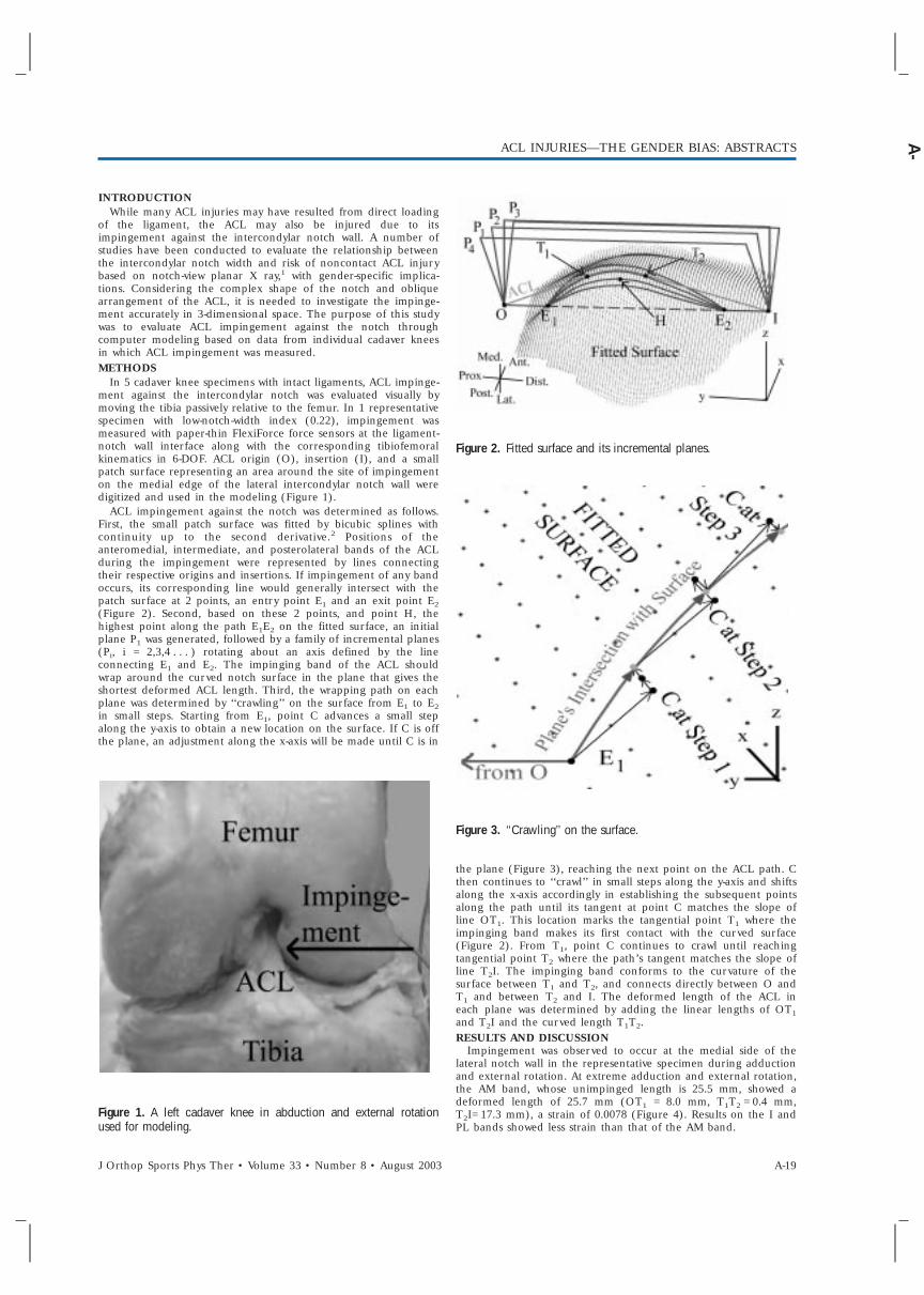
INTRODUCTIONWhile many ACL injuries may have resulted from direct loading
of the ligament, the ACL may also be injured due to itsimpingement against the intercondylar notch wall. A number ofstudies have been conducted to evaluate the relationship betweenthe intercondylar notch width and risk of noncontact ACL injurybased on notch-view planar X ray,1 with gender-specific implica-tions. Considering the complex shape of the notch and obliquearrangement of the ACL, it is needed to investigate the impinge-ment accurately in 3-dimensional space. The purpose of this studywas to evaluate ACL impingement against the notch throughcomputer modeling based on data from individual cadaver kneesin which ACL impingement was measured.METHODS
In 5 cadaver knee specimens with intact ligaments, ACL impinge-ment against the intercondylar notch was evaluated visually bymoving the tibia passively relative to the femur. In 1 representativespecimen with low-notch-width index (0.22), impingement wasmeasured with paper-thin FlexiForce force sensors at the ligament-notch wall interface along with the corresponding tibiofemoralkinematics in 6-DOF. ACL origin (O), insertion (I), and a smallpatch surface representing an area around the site of impingementon the medial edge of the lateral intercondylar notch wall weredigitized and used in the modeling (Figure 1).
ACL impingement against the notch was determined as follows.First, the small patch surface was fitted by bicubic splines withcontinuity up to the second derivative.2 Positions of theanteromedial, intermediate, and posterolateral bands of the ACLduring the impingement were represented by lines connectingtheir respective origins and insertions. If impingement of any bandoccurs, its corresponding line would generally intersect with thepatch surface at 2 points, an entry point E1 and an exit point E2(Figure 2). Second, based on these 2 points, and point H, thehighest point along the path E1E2 on the fitted surface, an initialplane P1 was generated, followed by a family of incremental planes(Pi, i = 2,3,4 . . . ) rotating about an axis defined by the lineconnecting E1 and E2. The impinging band of the ACL shouldwrap around the curved notch surface in the plane that gives theshortest deformed ACL length. Third, the wrapping path on eachplane was determined by ‘‘crawling’’ on the surface from E1 to E2in small steps. Starting from E1, point C advances a small stepalong the y-axis to obtain a new location on the surface. If C is offthe plane, an adjustment along the x-axis will be made until C is in
Figure 1. A left cadaver knee in abduction and external rotationused for modeling.
Figure 2. Fitted surface and its incremental planes.
Figure 3. ‘‘Crawling’’ on the surface.
the plane (Figure 3), reaching the next point on the ACL path. Cthen continues to ‘‘crawl’’ in small steps along the y-axis and shiftsalong the x-axis accordingly in establishing the subsequent pointsalong the path until its tangent at point C matches the slope ofline OT1. This location marks the tangential point T1 where theimpinging band makes its first contact with the curved surface(Figure 2). From T1, point C continues to crawl until reachingtangential point T2 where the path’s tangent matches the slope ofline T2I. The impinging band conforms to the curvature of thesurface between T1 and T2, and connects directly between O andT1 and between T2 and I. The deformed length of the ACL ineach plane was determined by adding the linear lengths of OT1and T2I and the curved length T1T2.RESULTS AND DISCUSSION
Impingement was observed to occur at the medial side of thelateral notch wall in the representative specimen during adductionand external rotation. At extreme adduction and external rotation,the AM band, whose unimpinged length is 25.5 mm, showed adeformed length of 25.7 mm (OT1 = 8.0 mm, T1T2 = 0.4 mm,T2I= 17.3 mm), a strain of 0.0078 (Figure 4). Results on the I andPL bands showed less strain than that of the AM band.
ACL INJURIES—THE GENDER BIAS: ABSTRACTS
J Orthop Sports Phys Ther • Volume 33 • Number 8 • August 2003 A-19
A-
Figure 4. Graphical output of the model showing the impinging AMband.
CONCLUSIONSThe mathematical model provided a useful method to determine
ACL impingement in 3-dimensional space, including its deformedshape, length, and strain as the ligament wraps around the notchsurface. It was demonstrated that ACL impingement on theintercondylar notch wall loaded the ACL considerably. Furtherwork needs to be done to identify the different areas of the notchwall where impingement is likely to occur in relation to notchwidth and gender, and to corroborate with the recorded data fromthe force sensors.
REFERENCES1. Arendt E. In Prevent. Noncontact ACL Injuries. 2001; AAOS,
Rosemont, IL.2. Hayes JG, Halliday J. J. Inst. Maths Applics. 1974;14,89-103.
II. NEUROMUSCULAR FACTORSKNEE JOINT LAXITY AFFECTS MUSCLE ACTIVATION PAT-TERNS AT THE KNEE PRIOR TO AND FOLLOWING AWEIGHT-BEARING PERTURBATIONShultz SJ, Carcia CR, Perrin DHUniversity of North Carolina at Greensboro, Greensboro, NC.INTRODUCTION
Anterior knee joint laxity is greater in females compared tomales and has been implicated as a potential risk factor in ACLinjury. While hormonal and structural differences may contributeto increased anterior knee joint laxity in females, the consequenceof increased joint laxity on neuromuscular and biomechanicalfunction at the knee has received little attention. This studyinvestigated the effects of anterior knee joint laxity on muscletiming and pre- and postperturbation response amplitudes in thegastrocnemius, hamstring, and quadriceps muscles in response to alower extremity perturbation.METHODS
To evoke the reflex response, a lower extremity perturbationdevice produced a forward and either internal rotation (IR) orexternal rotation (ER) of the trunk and femur on the weight-bearing tibia. Preperturbation muscle activity level 50 ms prior tothe perturbation (PreAmp = %MVIC), long latency reflex time (RT= ms), and mean reflex amplitude over 150 ms postperturbation(RAmp = %MVIC) were recorded via surface EMG in the medialand lateral gastrocnemius (MG, LG), hamstring (MH, LH), andquadriceps (MQ, LQ) muscles for both ER and IR. Participantsconsisted of 42 healthy NCAA Division I intercollegiate femaleathletes, comparing 21 with anterior tibial translation (ATT) ,5mm (KT1 = 19.8 ± 1.3 years; 172.4 ± 7.5 cm; 69.3 ± 7.8 kg; 4.3 ± 0.8mm ATT) to 21 with ATT . 7 mm (KT2 = 19.2 ± 1.1 years;171.1 ± 7.3 cm; 69.9 ± 9.3 kg; 9.2 ± 2.0 mm ATT) as measured by astandard knee arthrometer.RESULTS
A mixed-model repeated-measures ANOVA found groups dif-fered in reflex timing by muscle (P = .013), with the LH demon-strating a 16-millisecond delay in KT2 compared to KT1. While a
similar group difference in the onset of the MH was found (KT215 milliseconds.KT1), the post hoc comparison was not signifi-cant. A separate mixed-model repeated-measures ANOVA foundgroups also differed in pre- and postperturbation response ampli-tude by response (PreAmp versus RAmp) (P = .007), response bymuscle (P = .036), and rotation by response by muscle (P= .045).With muscles combined, KT2 demonstrated greater PreAmp (58%)compared to KT1 (47%), but no difference in RAmp (76% versus77%). However, the 3-way interaction revealed this effect wasmuscle specific, with KT2 showing increased PreAmp in the MG(43% versus 23%), LG (61% versus 45%), and LH (65% versus37%), but no difference in the MH (48% versus 43%), MQ (67%versus 72%), or LQ (65% versus 59%). While RAmp was signifi-cantly greater than PreAmp for the MG, LG, MH, and LH for bothgroups, only the LH differed between groups, with KT2 demon-strating 22% greater activation compared to KT1. No differenceswere found between groups or response type for the MQ and LQ.These responses were similar for ER versus IR, with few exceptions.Subtle increases in PreAmp and RAmp were found in KT2 comparedto KT1 for the LQ on IR (PreAmp =66% versus 57%, RAmp = 67%versus 60%), but were not found for ER (PreAmp = 63% versus61%, RAmp = 63% versus 58%). Further, both groups showedgreater RAmp than PreAmp for the MQ for ER (79% versus 66%KT1, 72% versus 64% KT2), but not IR (80% versus 77% KT1,78% versus 71% KT2).DISCUSSION
In summary, participants with increased knee joint laxity demon-strated increased levels of preactivity in the gastrocnemius and LHmuscles during static, single-leg weight bearing prior to a lowerextremity perturbation, then showed increased reflex delays andreflex amplitude of the LH following the perturbation. Thesegroup differences in muscle activation patterns may suggest both aproprioceptive deficit (delayed hamstring reflex) and a learnedcompensatory strategy (increased PreAmp and RAmp) may exist withincreased knee joint laxity. The implication of these findings onknee joint stabilization and injury risk warrants further research.ACKNOWLEDGEMENTS
Funded by a grant from the National Athletic Trainers’ Associa-tion Research and Education Foundation (#999A003). This grantwas originally awarded to the University of Virginia, and latertransferred to the University of North Carolina at Greensboro.
GENDER DIFFERENCES IN HIP AND KNEE KINEMATICS ANDMUSCLE PREACTIVATION DURING SINGLE-LEG LANDINGSSchmitz RJ, Thompson TJ, Riemann BL, Goldfarb AHApplied Neuromechanics Research Laboratory; University of NorthCarolina Greensboro, Greensboro NC.INTRODUCTION
Lower-extremity joint kinematics and the ability of the lower-extremity muscles to act as dynamic stabilizers during periods ofdeceleration have been implicated as potential contributing factorsin the gender bias seen in ACL injury rates. The purpose of thisstudy was to assess lower extremity kinematic patterns and musclepreactivation timing in males and females during 3 differentlanding tasks.METHODS
Thirty recreationally active subjects (15 male [mean ± SD age,22.0 ± 2.8 years; mean ± SD weight, 78.1 ± 14.2 kg; mean ± SDheight, 178.7 ± 8.5 cm]; 15 female [mean ± SD age, 21.3 ± 3.0 years;mean ± SD weight, 57.8 ± 8.0 kg; mean ± SD height, 167.0 ± 7.0cm]) performed 3 single-leg dynamic jump tasks: forward dropjump, lateral jump from ground level, and medial jump fromground level. Height (20% of the subject’s height) and distance(32% of the subject’s height) of each jump were individualized.Three-dimensional joint kinematics of the hip and knee weremeasured by an electromagnetic tracking system during the 5 trialsof each task. Initial joint angles were measured at the time ofground contact. Peak displacement was defined as the differencebetween initial angle and peak angle up to 3 seconds after groundcontact. Time to peak displacement was defined as the periodbetween ground contact and peak displacement. Surface EMGsignals were obtained from the vastus lateralis, biceps femoris, and
ACL INJURIES—THE GENDER BIAS: ABSTRACTS
A-20 J Orthop Sports Phys Ther • Volume 33 • Number 8 • August 2003
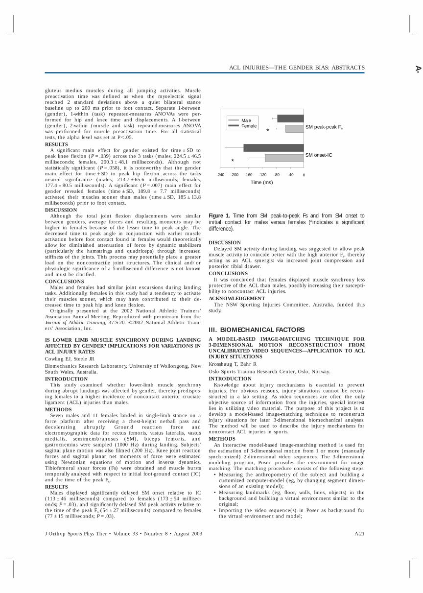
gluteus medius muscles during all jumping activities. Musclepreactivation time was defined as when the myoelectric signalreached 2 standard deviations above a quiet bilateral stancebaseline up to 200 ms prior to foot contact. Separate 1-between(gender), 1-within (task) repeated-measures ANOVAs were per-formed for hip and knee time and displacements. A 1-between(gender), 2-within (muscle and task) repeated-measures ANOVAwas performed for muscle preactivation time. For all statisticaltests, the alpha level was set at P,.05.RESULTS
A significant main effect for gender existed for time ± SD topeak knee flexion (P = .039) across the 3 tasks (males, 224.5 ± 46.5milliseconds; females, 200.3 ± 48.1 milliseconds). Although notstatistically significant (P = .058), it is noteworthy that the gendermain effect for time ± SD to peak hip flexion across the tasksneared significance (males, 213.7 ± 65.6 milliseconds; females,177.4 ± 80.5 milliseconds). A significant (P = .007) main effect forgender revealed females (time ± SD, 189.8 ± 7.7 milliseconds)activated their muscles sooner than males (time ± SD, 185 ± 13.8milliseconds) prior to foot contact.DISCUSSION
Although the total joint flexion displacements were similarbetween genders, average forces and resulting moments may behigher in females because of the lesser time to peak angle. Thedecreased time to peak angle in conjunction with earlier muscleactivation before foot contact found in females would theoreticallyallow for diminished attenuation of force by dynamic stabilizers(particularly the hamstrings and quadriceps) through increasedstiffness of the joints. This process may potentially place a greaterload on the noncontractile joint structures. The clinical and/orphysiologic significance of a 5-millisecond difference is not knownand must be clarified.CONCLUSIONS
Males and females had similar joint excursions during landingtasks. Additionally, females in this study had a tendency to activatetheir muscles sooner, which may have contributed to their de-creased time to peak hip and knee flexion.
Originally presented at the 2002 National Athletic Trainers’Association Annual Meeting. Reproduced with permission from theJournal of Athletic Training, 37:S-20. ©2002 National Athletic Train-ers’ Association, Inc.
IS LOWER LIMB MUSCLE SYNCHRONY DURING LANDINGAFFECTED BY GENDER? IMPLICATIONS FOR VARIATIONS INACL INJURY RATESCowling EJ, Steele JRBiomechanics Research Laboratory, University of Wollongong, NewSouth Wales, Australia.INTRODUCTION
This study examined whether lower-limb muscle synchronyduring abrupt landings was affected by gender, thereby predispos-ing females to a higher incidence of noncontact anterior cruciateligament (ACL) injuries than males.METHODS
Seven males and 11 females landed in single-limb stance on aforce platform after receiving a chest-height netball pass anddecelerating abruptly. Ground reaction force andelectromyographic data for rectus femoris, vastus lateralis, vastusmedialis, semimembranosus (SM), biceps femoris, andgastrocnemius were sampled (1000 Hz) during landing. Subjects’sagittal plane motion was also filmed (200 Hz). Knee joint reactionforces and sagittal planar net moments of force were estimatedusing Newtonian equations of motion and inverse dynamics.Tibiofemoral shear forces (Fs) were obtained and muscle burststemporally analyzed with respect to initial foot-ground contact (IC)and the time of the peak Fs.RESULTS
Males displayed significantly delayed SM onset relative to IC(113 ± 46 milliseconds) compared to females (173 ± 54 millisec-onds; P = .03), and significantly delayed SM peak activity relative tothe time of the peak Fs (54 ± 27 milliseconds) compared to females(77 ± 15 milliseconds; P = .03).
Figure 1. Time from SM peak-to-peak Fs and from SM onset toinitial contact for males versus females (*indicates a significantdifference).
DISCUSSIONDelayed SM activity during landing was suggested to allow peak
muscle activity to coincide better with the high anterior Fs, therebyacting as an ACL synergist via increased joint compression andposterior tibial drawer.CONCLUSIONS
It was concluded that females displayed muscle synchrony lessprotective of the ACL than males, possibly increasing their suscepti-bility to noncontact ACL injuries.ACKNOWLEDGEMENT
The NSW Sporting Injuries Committee, Australia, funded thisstudy.
III. BIOMECHANICAL FACTORSA MODEL-BASED IMAGE-MATCHING TECHNIQUE FOR3-DIMENSIONAL MOTION RECONSTRUCTION FROMUNCALIBRATED VIDEO SEQUENCES—APPLICATION TO ACLINJURY SITUATIONSKrosshaug T, Bahr ROslo Sports Trauma Research Center, Oslo, Norway.INTRODUCTION
Knowledge about injury mechanisms is essential to preventinjuries. For obvious reasons, injury situations cannot be recon-structed in a lab setting. As video sequences are often the onlyobjective source of information from the injuries, special interestlies in utilizing video material. The purpose of this project is todevelop a model-based image-matching technique to reconstructinjury situations for later 3-dimensional biomechanical analyses.The method will be used to describe the injury mechanisms fornoncontact ACL injuries in sports.METHODS
An interactive model-based image-matching method is used forthe estimation of 3-dimensional motion from 1 or more (manuallysynchronized) 2-dimensional video sequences. The 3-dimensionalmodeling program, Poser, provides the environment for imagematching. The matching procedure consists of the following steps:
• Measuring the anthropometry of the subject and building acustomized computer-model (eg, by changing segment dimen-sions of an existing model);
• Measuring landmarks (eg, floor, walls, lines, objects) in thebackground and building a virtual environment similar to theoriginal;
• Importing the video sequence(s) in Poser as background forthe virtual environment and model;
0
Time (ms)
-240 -200 -160 -120 -80 -40
Male Female SM peak-peak Fs
SM onset-IC
*
*
ACL INJURIES—THE GENDER BIAS: ABSTRACTS
J Orthop Sports Phys Ther • Volume 33 • Number 8 • August 2003 A-21
A-
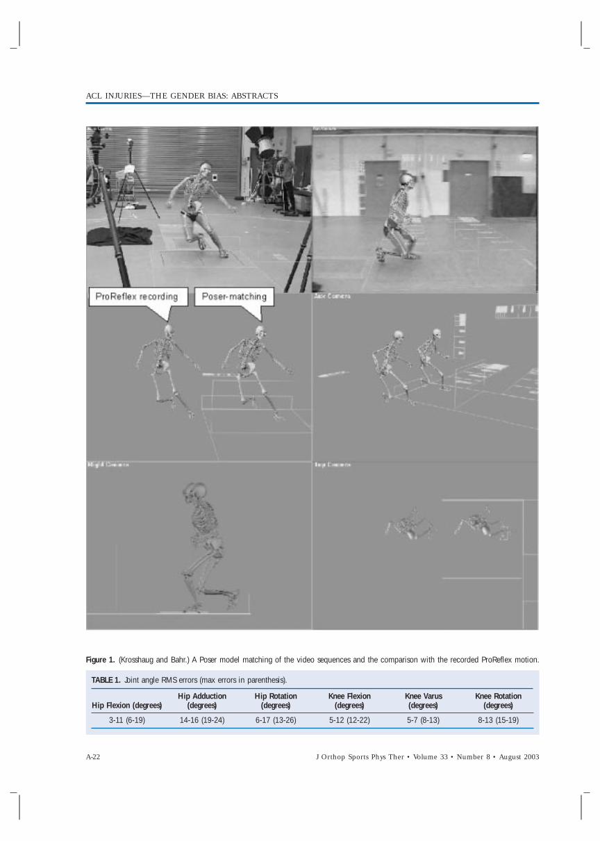
Figure 1. (Krosshaug and Bahr.) A Poser model matching of the video sequences and the comparison with the recorded ProReflex motion.
TABLE 1. Joint angle RMS errors (max errors in parenthesis).
Hip Flexion (degrees)Hip Adduction
(degrees)Hip Rotation
(degrees)Knee Flexion
(degrees)Knee Varus(degrees)
Knee Rotation(degrees)
3-11 (6-19) 14-16 (19-24) 6-17 (13-26) 5-12 (12-22) 5-7 (8-13) 8-13 (15-19)
ACL INJURIES—THE GENDER BIAS: ABSTRACTS
A-22 J Orthop Sports Phys Ther • Volume 33 • Number 8 • August 2003
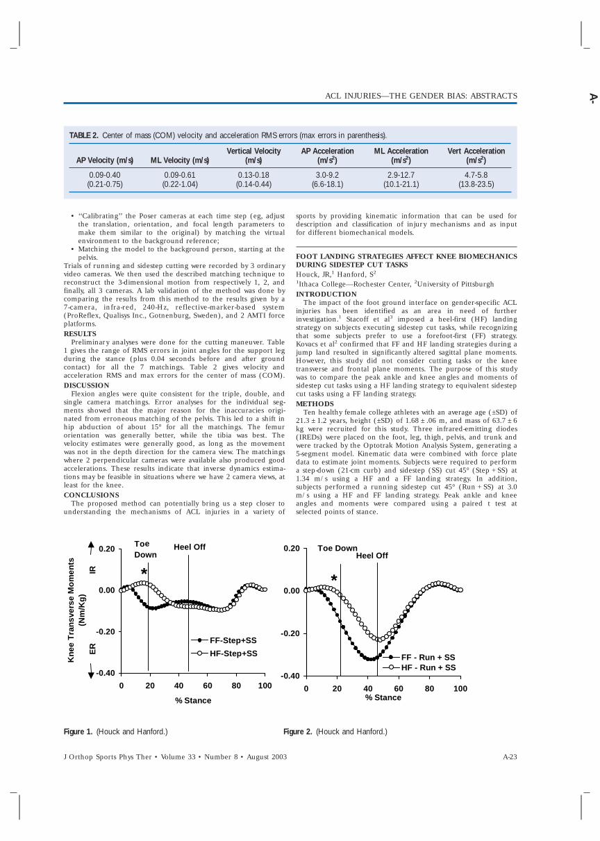
TABLE 2. Center of mass (COM) velocity and acceleration RMS errors (max errors in parenthesis).
AP Velocity (m/s) ML Velocity (m/s)Vertical Velocity
(m/s)AP Acceleration
(m/s2)ML Acceleration
(m/s2)Vert Acceleration
(m/s2)
0.09-0.40(0.21-0.75)
0.09-0.61(0.22-1.04)
0.13-0.18(0.14-0.44)
3.0-9.2(6.6-18.1)
2.9-12.7(10.1-21.1)
4.7-5.8(13.8-23.5)
• ‘‘Calibrating’’ the Poser cameras at each time step (eg, adjustthe translation, orientation, and focal length parameters tomake them similar to the original) by matching the virtualenvironment to the background reference;
• Matching the model to the background person, starting at thepelvis.
Trials of running and sidestep cutting were recorded by 3 ordinaryvideo cameras. We then used the described matching technique toreconstruct the 3-dimensional motion from respectively 1, 2, andfinally, all 3 cameras. A lab validation of the method was done bycomparing the results from this method to the results given by a7-camera, infra-red, 240-Hz, reflective-marker-based system(ProReflex, Qualisys Inc., Gotnenburg, Sweden), and 2 AMTI forceplatforms.RESULTS
Preliminary analyses were done for the cutting maneuver. Table1 gives the range of RMS errors in joint angles for the support legduring the stance (plus 0.04 seconds before and after groundcontact) for all the 7 matchings. Table 2 gives velocity andacceleration RMS and max errors for the center of mass (COM).DISCUSSION
Flexion angles were quite consistent for the triple, double, andsingle camera matchings. Error analyses for the individual seg-ments showed that the major reason for the inaccuracies origi-nated from erroneous matching of the pelvis. This led to a shift inhip abduction of about 15° for all the matchings. The femurorientation was generally better, while the tibia was best. Thevelocity estimates were generally good, as long as the movementwas not in the depth direction for the camera view. The matchingswhere 2 perpendicular cameras were available also produced goodaccelerations. These results indicate that inverse dynamics estima-tions may be feasible in situations where we have 2 camera views, atleast for the knee.CONCLUSIONS
The proposed method can potentially bring us a step closer tounderstanding the mechanisms of ACL injuries in a variety of
sports by providing kinematic information that can be used fordescription and classification of injury mechanisms and as inputfor different biomechanical models.
FOOT LANDING STRATEGIES AFFECT KNEE BIOMECHANICSDURING SIDESTEP CUT TASKSHouck, JR,1 Hanford, S2
1Ithaca College—Rochester Center, 2University of PittsburghINTRODUCTION
The impact of the foot ground interface on gender-specific ACLinjuries has been identified as an area in need of furtherinvestigation.1 Stacoff et al3 imposed a heel-first (HF) landingstrategy on subjects executing sidestep cut tasks, while recognizingthat some subjects prefer to use a forefoot-first (FF) strategy.Kovacs et al2 confirmed that FF and HF landing strategies during ajump land resulted in significantly altered sagittal plane moments.However, this study did not consider cutting tasks or the kneetransverse and frontal plane moments. The purpose of this studywas to compare the peak ankle and knee angles and moments ofsidestep cut tasks using a HF landing strategy to equivalent sidestepcut tasks using a FF landing strategy.METHODS
Ten healthy female college athletes with an average age (±SD) of21.3 ± 1.2 years, height (±SD) of 1.68 ± .06 m, and mass of 63.7 ± 6kg were recruited for this study. Three infrared-emitting diodes(IREDs) were placed on the foot, leg, thigh, pelvis, and trunk andwere tracked by the Optotrak Motion Analysis System, generating a5-segment model. Kinematic data were combined with force platedata to estimate joint moments. Subjects were required to performa step-down (21-cm curb) and sidestep (SS) cut 45° (Step + SS) at1.34 m/s using a HF and a FF landing strategy. In addition,subjects performed a running sidestep cut 45° (Run + SS) at 3.0m/s using a HF and FF landing strategy. Peak ankle and kneeangles and moments were compared using a paired t test atselected points of stance.
Figure 1. (Houck and Hanford.) Figure 2. (Houck and Hanford.)
-0.40
-0.20
0.00
0.20
0 20 40 60 80 100
% Stance
Kne
e T
ran
sver
se M
om
ents
(N
m/K
g)
ER
IR
FF-Step+SS
HF-Step+SS
-0.40
-0.20
0.00
0.20
0 20 40 60 80 100% Stance
FF - Run + SSHF - Run + SS
Toe DownHeel Off
*
Heel OffToDown
*
e
ACL INJURIES—THE GENDER BIAS: ABSTRACTS
J Orthop Sports Phys Ther • Volume 33 • Number 8 • August 2003 A-23
A-
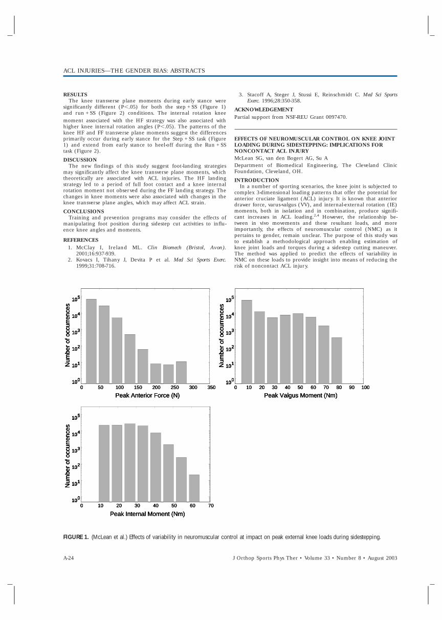
RESULTSThe knee transverse plane moments during early stance were
significantly different (P,.05) for both the step + SS (Figure 1)and run + SS (Figure 2) conditions. The internal rotation kneemoment associated with the HF strategy was also associated withhigher knee internal rotation angles (P,.05). The patterns of theknee HF and FF transverse plane moments suggest the differencesprimarily occur during early stance for the Step + SS task (Figure1) and extend from early stance to heel-off during the Run + SStask (Figure 2).
DISCUSSIONThe new findings of this study suggest foot-landing strategies
may significantly affect the knee transverse plane moments, whichtheoretically are associated with ACL injuries. The HF landingstrategy led to a period of full foot contact and a knee internalrotation moment not observed during the FF landing strategy. Thechanges in knee moments were also associated with changes in theknee transverse plane angles, which may affect ACL strain.
CONCLUSIONSTraining and prevention programs may consider the effects of
manipulating foot position during sidestep cut activities to influ-ence knee angles and moments.
REFERENCES1. McClay I, Ireland ML. Clin Biomech (Bristol, Avon).
2001;16:937-939.2. Kovacs I, Tihany J, Devita P et al. Med Sci Sports Exerc.
1999;31:708-716.
3. Stacoff A, Steger J, Stussi E, Reinschmidt C. Med Sci SportsExerc. 1996;28:350-358.
ACKNOWLEDGEMENTPartial support from NSF-REU Grant 0097470.
EFFECTS OF NEUROMUSCULAR CONTROL ON KNEE JOINTLOADING DURING SIDESTEPPING: IMPLICATIONS FORNONCONTACT ACL INJURYMcLean SG, van den Bogert AG, Su ADepartment of Biomedical Engineering, The Cleveland ClinicFoundation, Cleveland, OH.
INTRODUCTIONIn a number of sporting scenarios, the knee joint is subjected to
complex 3-dimensional loading patterns that offer the potential foranterior cruciate ligament (ACL) injury. It is known that anteriordrawer force, varus-valgus (VV), and internal-external rotation (IE)moments, both in isolation and in combination, produce signifi-cant increases in ACL loading.2,4 However, the relationship be-tween in vivo movements and these resultant loads, and moreimportantly, the effects of neuromuscular control (NMC) as itpertains to gender, remain unclear. The purpose of this study wasto establish a methodological approach enabling estimation ofknee joint loads and torques during a sidestep cutting maneuver.The method was applied to predict the effects of variability inNMC on these loads to provide insight into means of reducing therisk of noncontact ACL injury.
FIGURE 1. (McLean et al.) Effects of variability in neuromuscular control at impact on peak external knee loads during sidestepping.
0 50 100 150 200 250 300 350
Peak Anterior Force (N)
0 10 20 30 40 50 60 70
Peak Internal Moment (Nm)
0 10 20 30 40 50 60 70 80 90 100
Num
ber
of o
ccur
renc
esN
umbe
r of
occ
urre
nces
0
1
2
3
4
5
0
1
2
3
4
5
10
10
10
10
10
10
10
10
10
10
10
10
10
10
10
10
10
10
0
1
2
3
4
5
Num
ber
of o
ccur
renc
es
Peak Valgus Moment (Nm)0 50 100 150 200 250 300 350
Peak Anterior Force (N)
0 10 20 30 40 50 60 70
Peak Internal Moment (Nm)
0 10 20 30 40 50 60 70 80 90 100
Num
ber
of o
ccur
renc
esN
umbe
r of
occ
urre
nces
0
1
2
3
4
5
0
1
2
3
4
5
10
10
10
10
10
10
10
10
10
10
10
10
10
10
10
10
10
10
0
1
2
3
4
5
Num
ber
of o
ccur
renc
es
Num
ber
of o
ccur
renc
esN
umbe
r of
occ
urre
nces
0
1
2
3
4
5
0
1
2
3
4
5
10
10
10
10
10
10
10
10
10
10
10
10
10
10
10
10
10
10
0
1
2
3
4
5
0
1
2
3
4
5
0
1
2
3
4
5
10
10
10
10
10
10
10
10
10
10
10
10
10
10
10
10
10
10
10
10
10
10
10
10
0
1
2
3
4
5
Num
ber
of o
ccur
renc
es
Peak Valgus Moment (Nm)
ACL INJURIES—THE GENDER BIAS: ABSTRACTS
A-24 J Orthop Sports Phys Ther • Volume 33 • Number 8 • August 2003
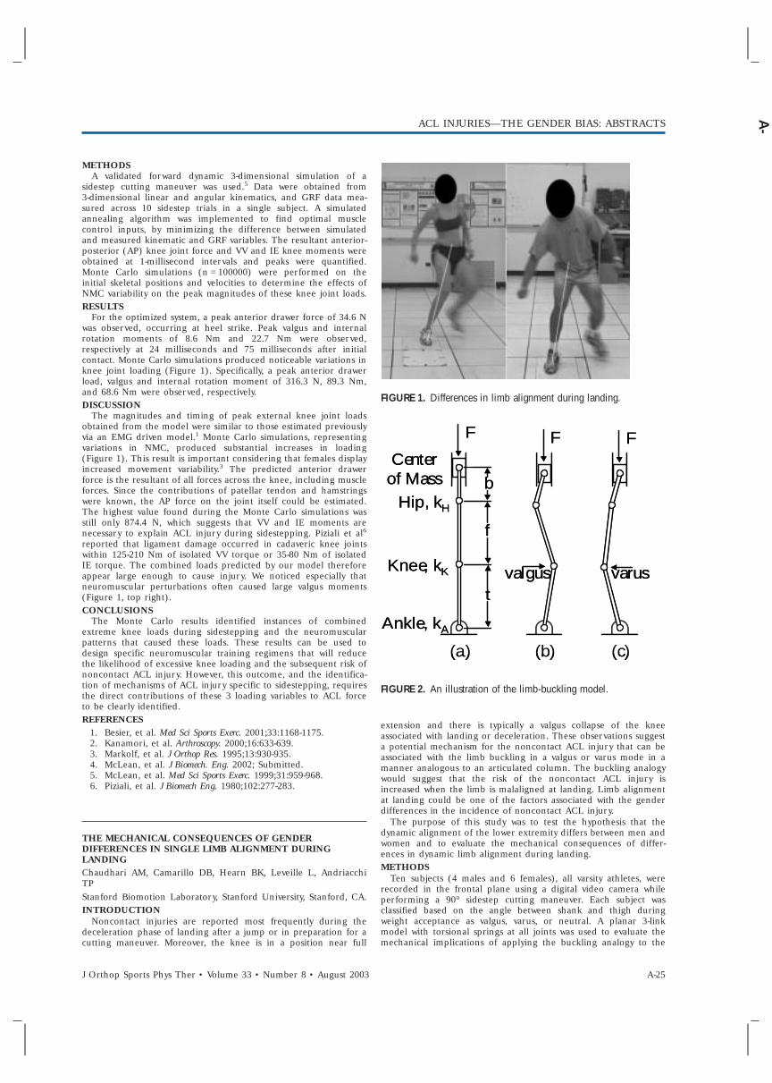
METHODSA validated forward dynamic 3-dimensional simulation of a
sidestep cutting maneuver was used.5 Data were obtained from3-dimensional linear and angular kinematics, and GRF data mea-sured across 10 sidestep trials in a single subject. A simulatedannealing algorithm was implemented to find optimal musclecontrol inputs, by minimizing the difference between simulatedand measured kinematic and GRF variables. The resultant anterior-posterior (AP) knee joint force and VV and IE knee moments wereobtained at 1-millisecond intervals and peaks were quantified.Monte Carlo simulations (n = 100000) were performed on theinitial skeletal positions and velocities to determine the effects ofNMC variability on the peak magnitudes of these knee joint loads.RESULTS
For the optimized system, a peak anterior drawer force of 34.6 Nwas observed, occurring at heel strike. Peak valgus and internalrotation moments of 8.6 Nm and 22.7 Nm were observed,respectively at 24 milliseconds and 75 milliseconds after initialcontact. Monte Carlo simulations produced noticeable variations inknee joint loading (Figure 1). Specifically, a peak anterior drawerload, valgus and internal rotation moment of 316.3 N, 89.3 Nm,and 68.6 Nm were observed, respectively.DISCUSSION
The magnitudes and timing of peak external knee joint loadsobtained from the model were similar to those estimated previouslyvia an EMG driven model.1 Monte Carlo simulations, representingvariations in NMC, produced substantial increases in loading(Figure 1). This result is important considering that females displayincreased movement variability.3 The predicted anterior drawerforce is the resultant of all forces across the knee, including muscleforces. Since the contributions of patellar tendon and hamstringswere known, the AP force on the joint itself could be estimated.The highest value found during the Monte Carlo simulations wasstill only 874.4 N, which suggests that VV and IE moments arenecessary to explain ACL injury during sidestepping. Piziali et al6
reported that ligament damage occurred in cadaveric knee jointswithin 125-210 Nm of isolated VV torque or 35-80 Nm of isolatedIE torque. The combined loads predicted by our model thereforeappear large enough to cause injury. We noticed especially thatneuromuscular perturbations often caused large valgus moments(Figure 1, top right).CONCLUSIONS
The Monte Carlo results identified instances of combinedextreme knee loads during sidestepping and the neuromuscularpatterns that caused these loads. These results can be used todesign specific neuromuscular training regimens that will reducethe likelihood of excessive knee loading and the subsequent risk ofnoncontact ACL injury. However, this outcome, and the identifica-tion of mechanisms of ACL injury specific to sidestepping, requiresthe direct contributions of these 3 loading variables to ACL forceto be clearly identified.REFERENCES
1. Besier, et al. Med Sci Sports Exerc. 2001;33:1168-1175.2. Kanamori, et al. Arthroscopy. 2000;16:633-639.3. Markolf, et al. J Orthop Res. 1995;13:930-935.4. McLean, et al. J Biomech. Eng. 2002; Submitted.5. McLean, et al. Med Sci Sports Exerc. 1999;31:959-968.6. Piziali, et al. J Biomech Eng. 1980;102:277-283.
THE MECHANICAL CONSEQUENCES OF GENDERDIFFERENCES IN SINGLE LIMB ALIGNMENT DURINGLANDINGChaudhari AM, Camarillo DB, Hearn BK, Leveille L, AndriacchiTPStanford Biomotion Laboratory, Stanford University, Stanford, CA.INTRODUCTION
Noncontact injuries are reported most frequently during thedeceleration phase of landing after a jump or in preparation for acutting maneuver. Moreover, the knee is in a position near full
FIGURE 1. Differences in limb alignment during landing.
FIGURE 2. An illustration of the limb-buckling model.
extension and there is typically a valgus collapse of the kneeassociated with landing or deceleration. These observations suggesta potential mechanism for the noncontact ACL injury that can beassociated with the limb buckling in a valgus or varus mode in amanner analogous to an articulated column. The buckling analogywould suggest that the risk of the noncontact ACL injury isincreased when the limb is malaligned at landing. Limb alignmentat landing could be one of the factors associated with the genderdifferences in the incidence of noncontact ACL injury.
The purpose of this study was to test the hypothesis that thedynamic alignment of the lower extremity differs between men andwomen and to evaluate the mechanical consequences of differ-ences in dynamic limb alignment during landing.METHODS
Ten subjects (4 males and 6 females), all varsity athletes, wererecorded in the frontal plane using a digital video camera whileperforming a 90° sidestep cutting maneuver. Each subject wasclassified based on the angle between shank and thigh duringweight acceptance as valgus, varus, or neutral. A planar 3-linkmodel with torsional springs at all joints was used to evaluate themechanical implications of applying the buckling analogy to the
varus
b
f
t
Knee, kK
Ankle, kA
Hip, kH
Center of Mass
F
(a)
F
(b)
valgus
F
(c)
varus
b
f
t
Knee, kK
Ankle, kA
Hip, kH
Center of Mass
F
(a)
F
(b)
valgus
F
(c)
ACL INJURIES—THE GENDER BIAS: ABSTRACTS
J Orthop Sports Phys Ther • Volume 33 • Number 8 • August 2003 A-25
A-
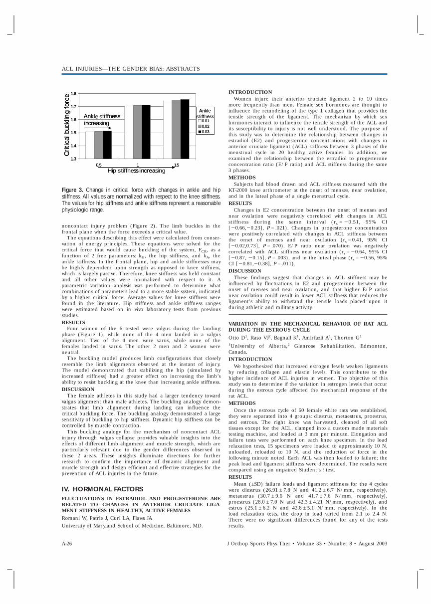
Figure 3. Change in critical force with changes in ankle and hipstiffness. All values are normalized with respect to the knee stiffness.The values for hip stiffness and ankle stiffness represent a reasonablephysiologic range.
noncontact injury problem (Figure 2). The limb buckles in thefrontal plane when the force exceeds a critical value.
The equations describing this effect were calculated from conser-vation of energy principles. These equations were solved for thecritical force that would cause buckling of the system, FCR, as afunction of 2 free parameters: kH, the hip stiffness, and kA, theankle stiffness. In the frontal plane, hip and ankle stiffnesses maybe highly dependent upon strength as opposed to knee stiffness,which is largely passive. Therefore, knee stiffness was held constantand all other values were normalized with respect to it. Aparametric variation analysis was performed to determine whatcombinations of parameters lead to a more stable system, indicatedby a higher critical force. Average values for knee stiffness werefound in the literature. Hip stiffness and ankle stiffness rangeswere estimated based on in vivo laboratory tests from previousstudies.RESULTS
Four women of the 6 tested were valgus during the landingphase (Figure 1), while none of the 4 men landed in a valgusalignment. Two of the 4 men were varus, while none of thefemales landed in varus. The other 2 men and 2 women wereneutral.
The buckling model produces limb configurations that closelyresemble the limb alignments observed at the instant of injury.The model demonstrated that stabilizing the hip (simulated byincreased stiffness) had a greater effect on increasing the limb’sability to resist buckling at the knee than increasing ankle stiffness.DISCUSSION
The female athletes in this study had a larger tendency towardvalgus alignment than male athletes. The buckling analogy demon-strates that limb alignment during landing can influence thecritical buckling force. The buckling analogy demonstrated a largesensitivity of buckling to hip stiffness. Dynamic hip stiffness can becontrolled by muscle contraction.
This buckling analogy for the mechanism of noncontact ACLinjury through valgus collapse provides valuable insights into theeffects of different limb alignment and muscle strength, which areparticularly relevant due to the gender differences observed inthese 2 areas. These insights illuminate directions for furtherresearch to confirm the importance of dynamic alignment andmuscle strength and design efficient and effective strategies for theprevention of ACL injuries in the future.
IV. HORMONAL FACTORSFLUCTUATIONS IN ESTRADIOL AND PROGESTERONE ARERELATED TO CHANGES IN ANTERIOR CRUCIATE LIGA-MENT STIFFNESS IN HEALTHY, ACTIVE FEMALESRomani W, Patrie J, Curl LA, Flaws JAUniversity of Maryland School of Medicine, Baltimore, MD.
INTRODUCTIONWomen injure their anterior cruciate ligament 2 to 10 times
more frequently than men. Female sex hormones are thought toinfluence the remodeling of the type 1 collagen that provides thetensile strength of the ligament. The mechanism by which sexhormones interact to influence the tensile strength of the ACL andits susceptibility to injury is not well understood. The purpose ofthis study was to determine the relationship between changes inestradiol (E2) and progesterone concentrations with changes inanterior cruciate ligament (ACL) stiffness between 3 phases of themenstrual cycle in 20 healthy, active females. In addition, weexamined the relationship between the estradiol to progesteroneconcentration ratio (E/P ratio) and ACL stiffness during the same3 phases.METHODS
Subjects had blood drawn and ACL stiffness measured with theKT-2000 knee arthrometer at the onset of menses, near ovulation,and in the luteal phase of a single menstrual cycle.RESULTS
Changes in E2 concentration between the onset of menses andnear ovulation were negatively correlated with changes in ACLstiffness during the same interval (rs = 20.51, 95% CI[20.66,20.23], P = .021). Changes in progesterone concentrationwere positively correlated with changes in ACL stiffness betweenthe onset of menses and near ovulation (rs = 0.41, 95% CI[20.02,0.73], P = .070). E/P ratio near ovulation was negativelycorrelated with ACL stiffness near ovulation (rs = 20.64, 95% CI[20.87, 20.15], P = .003), and in the luteal phase (rs = 20.56, 95%CI [20.81,20.38], P = .011).DISCUSSION
These findings suggest that changes in ACL stiffness may beinfluenced by fluctuations in E2 and progesterone between theonset of menses and near ovulation, and that higher E/P ratiosnear ovulation could result in lower ACL stiffness that reduces theligament’s ability to withstand the tensile loads placed upon itduring athletic and military activity.
VARIATION IN THE MECHANICAL BEHAVIOR OF RAT ACLDURING THE ESTROUS CYCLE
Otto D1, Raso VJ2, Bagnall K1, Amirfazli A1, Thorton G1
1University of Alberta,2 Glenrose Rehabilitation, Edmonton,Canada.INTRODUCTION
We hypothesized that increased estrogen levels weaken ligamentsby reducing collagen and elastin levels. This contributes to thehigher incidence of ACL injuries in women. The objective of thisstudy was to determine if the variation in estrogen levels that occurduring the estrous cycle affected the mechanical response of therat ACL.METHODS
Once the estrous cycle of 60 female white rats was established,they were separated into 4 groups: diestrus, metaestrus, proestrus,and estrous. The right knee was harvested, cleaned of all softtissues except for the ACL, clamped into a custom made materialstesting machine, and loaded at 3 mm per minute. Elongation andfailure tests were performed on each knee specimen. In the loadrelaxation tests, 15 specimens were loaded to approximately 10 N,unloaded, reloaded to 10 N, and the reduction of force in thefollowing minute noted. Each ACL was then loaded to failure; thepeak load and ligament stiffness were determined. The results werecompared using an unpaired Student’s t test.RESULTS
Mean (±SD) failure loads and ligament stiffness for the 4 cycleswere diestrus (26.91 ± 7.8 N and 41.2 ± 6.7 N/mm, respectively),metaestrus (30.7 ± 9.6 N and 41.7 ± 7.6 N/mm, respectively),proestrus (28.0 ± 7.0 N and 42.3 ± 4.21 N/mm, respectively), andestrus (25.1 ± 6.2 N and 42.8 ± 5.1 N/mm, respectively). In theload relaxation tests, the drop in load varied from 2.1 to 2.4 N.There were no significant differences found for any of the testsresults.
1.3
1.4
1.5
1.6
1.7
1.8
0.5 1 1.5
0.010.020.03
Ankl tiffness increasing
Hip stiffness increasing
Ankle st iffness
Cri
tica
l bu
cklin
g fo
rce
1.3
1.4
1.5
1.6
1.7
1.8
0.5 1 1.5
0.010.020.03
Ankl f ess increa
ss increa
Ankle ff e
Cri
tica
l bu
cklin
g fo
rce
se
ACL INJURIES—THE GENDER BIAS: ABSTRACTS
A-26 J Orthop Sports Phys Ther • Volume 33 • Number 8 • August 2003
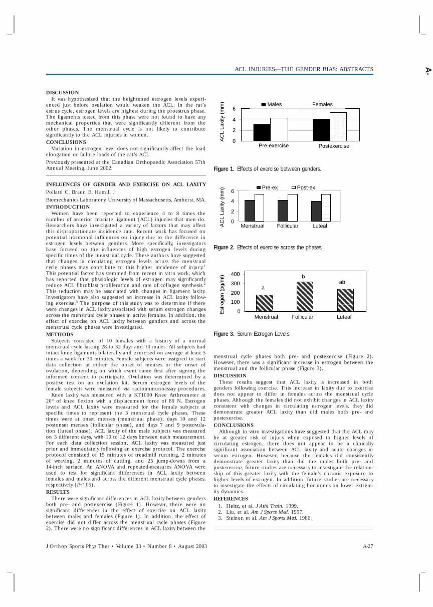
DISCUSSIONIt was hypothesized that the heightened estrogen levels experi-
enced just before ovulation would weaken the ACL. In the rat’sestrus cycle, estrogen levels are highest during the proestrus phase.The ligaments tested from this phase were not found to have anymechanical properties that were significantly different from theother phases. The menstrual cycle is not likely to contributesignificantly to the ACL injuries in women.CONCLUSIONS
Variation in estrogen level does not significantly affect the loadelongation or failure loads of the rat’s ACL.Previously presented at the Canadian Orthopaedic Association 57thAnnual Meeting, June 2002.
INFLUENCES OF GENDER AND EXERCISE ON ACL LAXITYPollard C, Braun B, Hamill JBiomechanics Laboratory, University of Massachusetts, Amherst, MA.INTRODUCTION
Women have been reported to experience 4 to 8 times thenumber of anterior cruciate ligament (ACL) injuries that men do.Researchers have investigated a variety of factors that may affectthis disproportionate incidence rate. Recent work has focused onpotential hormonal influences on injury due to the difference inestrogen levels between genders. More specifically, investigatorshave focused on the influences of high estrogen levels duringspecific times of the menstrual cycle. These authors have suggestedthat changes in circulating estrogen levels across the menstrualcycle phases may contribute to this higher incidence of injury.1
This potential factor has stemmed from recent in vitro work, whichhas reported that physiologic levels of estrogen may significantlyreduce ACL fibroblast proliferation and rate of collagen synthesis.2
This reduction may be associated with changes in ligament laxity.Investigators have also suggested an increase in ACL laxity follow-ing exercise.3 The purpose of this study was to determine if therewere changes in ACL laxity associated with serum estrogen changesacross the menstrual cycle phases in active females. In addition, theeffect of exercise on ACL laxity between genders and across themenstrual cycle phases were investigated.METHODS
Subjects consisted of 10 females with a history of a normalmenstrual cycle lasting 28 to 32 days and 10 males. All subjects hadintact knee ligaments bilaterally and exercised on average at least 5times a week for 30 minutes. Female subjects were assigned to startdata collection at either the onset of menses or the onset ofovulation, depending on which event came first after signing theinformed consent to participate. Ovulation was determined by apositive test on an ovulation kit. Serum estrogen levels of thefemale subjects were measured via radioimmunoassay procedures.
Knee laxity was measured with a KT1000 Knee Arthrometer at20° of knee flexion with a displacement force of 89 N. Estrogenlevels and ACL laxity were measured for the female subjects atspecific times to represent the 3 menstrual cycle phases. Thesetimes were at onset menses (menstrual phase), days 10 and 12postonset menses (follicular phase), and days 7 and 9 postovula-tion (luteal phase). ACL laxity of the male subjects was measuredon 3 different days, with 10 to 12 days between each measurement.For each data collection session, ACL laxity was measured justprior and immediately following an exercise protocol. The exerciseprotocol consisted of 15 minutes of treadmill running, 2 minutesof weaving, 2 minutes of cutting, and 25 jump-downs from a14-inch surface. An ANOVA and repeated-measures ANOVA wereused to test for significant differences in ACL laxity betweenfemales and males and across the different menstrual cycle phases,respectively (P#.05).RESULTS
There were significant differences in ACL laxity between gendersboth pre- and postexercise (Figure 1). However, there were nosignificant differences in the effect of exercise on ACL laxitybetween males and females (Figure 1). In addition, the effect ofexercise did not differ across the menstrual cycle phases (Figure2). There were no significant differences in ACL laxity between the
Figure 1. Effects of exercise between genders.
Figure 2. Effects of exercise across the phases.
Figure 3. Serum Estrogen Levels
menstrual cycle phases both pre- and postexercise (Figure 2).However, there was a significant increase in estrogen between themenstrual and the follicular phase (Figure 3).DISCUSSION
These results suggest that ACL laxity is increased in bothgenders following exercise. This increase in laxity due to exercisedoes not appear to differ in females across the menstrual cyclephases. Although the females did not exhibit changes in ACL laxityconsistent with changes in circulating estrogen levels, they diddemonstrate greater ACL laxity than did males both pre- andpostexercise.CONCLUSIONS
Although in vitro investigations have suggested that the ACL maybe at greater risk of injury when exposed to higher levels ofcirculating estrogen, there does not appear to be a clinicallysignificant association between ACL laxity and acute changes inserum estrogen. However, because the females did consistentlydemonstrate greater laxity than did the males both pre- andpostexercise, future studies are necessary to investigate the relation-ship of this greater laxity with the female’s chronic exposure tohigher levels of estrogen. In addition, future studies are necessaryto investigate the effects of circulating hormones on lower extrem-ity dynamics.REFERENCES
1. Heitz, et al. J Athl Train. 1999.2. Liu, et al. Am J Sports Med. 1997.3. Steiner, et al. Am J Sports Med. 1986.
AC
L La
xity
(m
m)
0 2 4 6 Males Females
Pre-exercise Postexercise
AC
L La
xity
(m
m)
0 2 4 6 Pre-ex Post-ex
Menstrual Follicular LutealE
stro
gen
(pg/
ml)
100
200
300
400
Follicula
0 Menstrual Follicular Luteal
a
b ab
ACL INJURIES—THE GENDER BIAS: ABSTRACTS
J Orthop Sports Phys Ther • Volume 33 • Number 8 • August 2003 A-27
A-
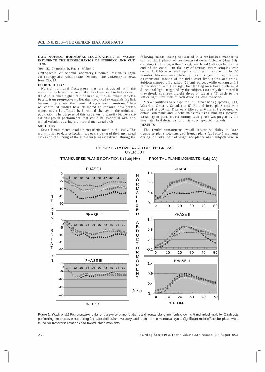
HOW NORMAL HORMONAL FLUCTUATIONS IN WOMENINFLUENCE THE BIOMECHANICS OF STEPPING AND CUT-TINGYack HJ, Chandran R, Rao S, Wilken JOrthopaedic Gait Analysis Laboratory, Graduate Program in Physi-cal Therapy and Rehabilitation Science, The University of Iowa,Iowa City, IA.INTRODUCTION
Normal hormonal fluctuations that are associated with themenstrual cycle are one factor that has been used to help explainthe 2 to 8 times higher rate of knee injuries in female athletes.Results from prospective studies that have tried to establish the linkbetween injury and the menstrual cycle are inconsistent.2 Fewwell-controlled studies have attempted to examine how perfor-mance might be affected by hormonal changes in the uninjuredpopulation. The purpose of this study was to identify biomechani-cal changes in performance that could be associated with hor-monal variations during the normal menstrual cycle.METHODS
Seven female recreational athletes participated in the study. Themonth prior to data collection, subjects monitored their menstrualcycles and the timing of the luteal surge was identified. During the
following month testing was started in a randomized manner tocapture the 3 phases of the menstrual cycle: follicular (days 2-4),ovulatory (LH surge, within 1 day), and luteal (6-8 days before theend of the cycle). On the day of testing, serum samples werecollected. Subjects warmed up by running on a treadmill for 20minutes. Markers were placed on each subject to capture the3-dimensional motion of the right lower limb, pelvis, and trunk.Subjects stepped off a raised (20 cm) walkway while walking at 1.8m per second, with their right foot landing on a force platform. Adirectional light, triggered by the subject, randomly determined ifthey should continue straight ahead or cut at a 45° angle to theleft or right. Five trials of each direction were collected.
Marker positions were captured in 3 dimensions (Optotrak, NDI,Waterloo, Ontario, Canada) at 60 Hz and force plate data werecaptured at 300 Hz. Data were filtered at 6 Hz and processed toobtain kinematic and kinetic measures using KinGait3 software.Variability in performance during each phase was judged by themean standard deviation for 5 trials over specific intervals.RESULTS
The results demonstrate overall greater variability in kneetransverse plane rotations and frontal plane (abductor) momentsduring the initial part of weight acceptance when subjects were in
Figure 1. (Yack et al.) Representative data for transverse plane rotations and frontal plane moments showing 5 individual trials for 2 subjectsperforming the crossover cut during 3 phases (follicular, ovulatory, and luteal) of the menstrual cycle. Significant main effects for phase werefound for transverse rotations and frontal plane moments.
I N T E R N A L
R O T A T I O N
PHASE I
-20 -15 -10 -5 0 0 6 12 18 24 30 36 42 48 54 60
PHASE II
-20 -15 -10 -5 0 0 6 12 18 24 30 36 42 48 54 60
PHASE III
-20 -15 -10 -5 0 0 6 12 18 24 30 36 42 48 54 60
PHASE I
-0 . 1 0.4 0.9 1.4
0 10 20 30 40 50 PHASE II
-0 . 1 0.4 0.9 1.4
0 10 20 30 40 50 PHASE III
-0.1 0.4 0.9 1.4
0 10 20 30 40 50
REPRESENTATIVE DATA FOR THE CROSS-OVER CUT
TRANSVERSE PLANE ROTATIONS (Subj HH) FRONTAL PLANE MOMENTS (Subj JA)
N O RMA L I Z E D A B D U C T O R MOM E N T
(N/kg)
% STRIDE % STRIDE
ACL INJURIES—THE GENDER BIAS: ABSTRACTS
A-28 J Orthop Sports Phys Ther • Volume 33 • Number 8 • August 2003
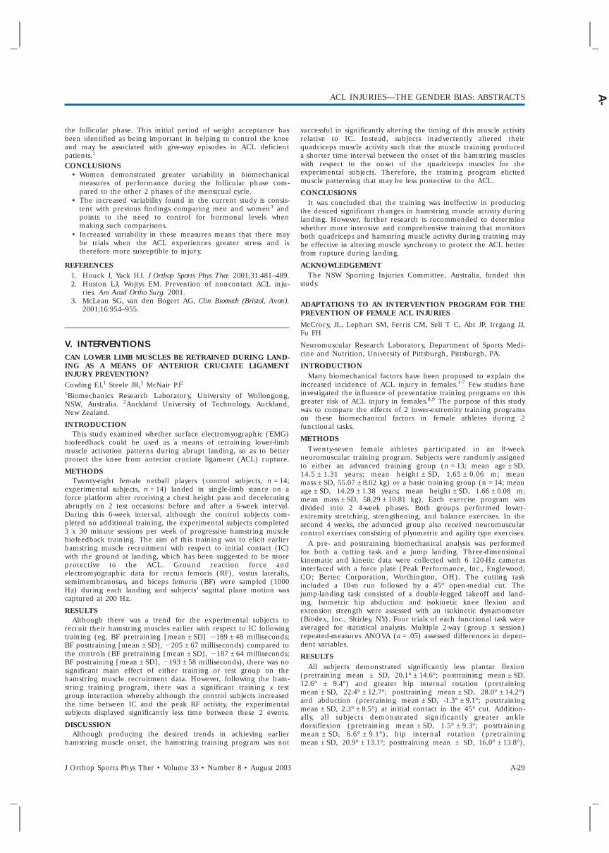
the follicular phase. This initial period of weight acceptance hasbeen identified as being important in helping to control the kneeand may be associated with give-way episodes in ACL deficientpatients.1
CONCLUSIONS• Women demonstrated greater variability in biomechanical
measures of performance during the follicular phase com-pared to the other 2 phases of the menstrual cycle.
• The increased variability found in the current study is consis-tent with previous findings comparing men and women3 andpoints to the need to control for hormonal levels whenmaking such comparisons.
• Increased variability in these measures means that there maybe trials when the ACL experiences greater stress and istherefore more susceptible to injury.
REFERENCES1. Houck J, Yack HJ. J Orthop Sports Phys Ther. 2001;31:481–489.2. Huston LJ, Wojtys EM. Prevention of noncontact ACL inju-
ries. Am Acad Ortho Surg. 2001.3. McLean SG, van den Bogert AG, Clin Biomech (Bristol, Avon).
2001;16:954–955.
V. INTERVENTIONSCAN LOWER LIMB MUSCLES BE RETRAINED DURING LAND-ING AS A MEANS OF ANTERIOR CRUCIATE LIGAMENTINJURY PREVENTION?Cowling EJ,1 Steele JR,1 McNair PJ2
1Biomechanics Research Laboratory, University of Wollongong,NSW, Australia. 2Auckland University of Technology, Auckland,New Zealand.
INTRODUCTIONThis study examined whether surface electromyographic (EMG)
biofeedback could be used as a means of retraining lower-limbmuscle activation patterns during abrupt landing, so as to betterprotect the knee from anterior cruciate ligament (ACL) rupture.
METHODSTwenty-eight female netball players (control subjects, n = 14;
experimental subjects, n = 14) landed in single-limb stance on aforce platform after receiving a chest height pass and deceleratingabruptly on 2 test occasions: before and after a 6-week interval.During this 6-week interval, although the control subjects com-pleted no additional training, the experimental subjects completed3 x 30 minute sessions per week of progressive hamstring musclebiofeedback training. The aim of this training was to elicit earlierhamstring muscle recruitment with respect to initial contact (IC)with the ground at landing, which has been suggested to be moreprotective to the ACL. Ground reaction force andelectromyographic data for rectus femoris (RF), vastus lateralis,semimembranosus, and biceps femoris (BF) were sampled (1000Hz) during each landing and subjects’ sagittal plane motion wascaptured at 200 Hz.
RESULTSAlthough there was a trend for the experimental subjects to
recruit their hamstring muscles earlier with respect to IC followingtraining (eg, BF pretraining [mean ± SD] 2189 ± 48 milliseconds;BF posttraining [mean ± SD], 2205 ± 67 milliseconds) compared tothe controls (BF pretraining [mean ± SD], 2187 ± 64 milliseconds;BF postraining [mean ± SD], 2193 ± 58 milliseconds), there was nosignificant main effect of either training or test group on thehamstring muscle recruitment data. However, following the ham-string training program, there was a significant training x testgroup interaction whereby although the control subjects increasedthe time between IC and the peak RF activity, the experimentalsubjects displayed significantly less time between these 2 events.
DISCUSSIONAlthough producing the desired trends in achieving earlier
hamstring muscle onset, the hamstring training program was not
successful in significantly altering the timing of this muscle activityrelative to IC. Instead, subjects inadvertently altered theirquadriceps muscle activity such that the muscle training produceda shorter time interval between the onset of the hamstring muscleswith respect to the onset of the quadriceps muscles for theexperimental subjects. Therefore, the training program elicitedmuscle patterning that may be less protective to the ACL.
CONCLUSIONSIt was concluded that the training was ineffective in producing
the desired significant changes in hamstring muscle activity duringlanding. However, further research is recommended to determinewhether more intensive and comprehensive training that monitorsboth quadriceps and hamstring muscle activity during training maybe effective in altering muscle synchrony to protect the ACL betterfrom rupture during landing.
ACKNOWLEDGEMENTThe NSW Sporting Injuries Committee, Australia, funded this
study.
ADAPTATIONS TO AN INTERVENTION PROGRAM FOR THEPREVENTION OF FEMALE ACL INJURIES
McCrory, JL, Lephart SM, Ferris CM, Sell T C, Abt JP, Irrgang JJ,Fu FH
Neuromuscular Research Laboratory, Department of Sports Medi-cine and Nutrition, University of Pittsburgh, Pittsburgh, PA.
INTRODUCTIONMany biomechanical factors have been proposed to explain the
increased incidence of ACL injury in females.1-7 Few studies haveinvestigated the influence of preventative training programs on thisgreater risk of ACL injury in females.8,9 The purpose of this studywas to compare the effects of 2 lower-extremity training programson these biomechanical factors in female athletes during 2functional tasks.
METHODSTwenty-seven female athletes participated in an 8-week
neuromuscular training program. Subjects were randomly assignedto either an advanced training group (n = 13; mean age ± SD,14.5 ± 1.31 years; mean height ± SD, 1.65 ± 0.06 m; meanmass ± SD, 55.07 ± 8.02 kg) or a basic training group (n = 14; meanage ± SD, 14.29 ± 1.38 years; mean height ± SD, 1.66 ± 0.08 m;mean mass ± SD, 58.29 ± 10.81 kg). Each exercise program wasdivided into 2 4-week phases. Both groups performed lower-extremity stretching, strengthening, and balance exercises. In thesecond 4 weeks, the advanced group also received neuromuscularcontrol exercises consisting of plyometric and agility type exercises.
A pre- and posttraining biomechanical analysis was performedfor both a cutting task and a jump landing. Three-dimensionalkinematic and kinetic data were collected with 6 120-Hz camerasinterfaced with a force plate (Peak Performance, Inc., Englewood,CO; Bertec Corporation, Worthington, OH). The cutting taskincluded a 10-m run followed by a 45° open-medial cut. Thejump-landing task consisted of a double-legged takeoff and land-ing. Isometric hip abduction and isokinetic knee flexion andextension strength were assessed with an isokinetic dynamometer(Biodex, Inc., Shirley, NY). Four trials of each functional task wereaveraged for statistical analysis. Multiple 2-way (group x session)repeated-measures ANOVA (a = .05) assessed differences in depen-dent variables.
RESULTSAll subjects demonstrated significantly less plantar flexion
(pretraining mean ± SD, 20.1° ± 14.6°; posttraining mean ± SD,12.6° ± 9.4°) and greater hip internal rotation (pretrainingmean ± SD, 22.4° ± 12.7°; posttraining mean ± SD, 28.0° ± 14.2°)and abduction (pretraining mean ± SD, -1.3° ± 9.1°; posttrainingmean ± SD, 2.3° ± 8.5°) at initial contact in the 45° cut. Addition-ally, all subjects demonstrated significantly greater ankledorsiflexion (pretraining mean ± SD, 1.5° ± 9.3°; posttrainingmean ± SD, 6.6° ± 9.1°), hip internal rotation (pretrainingmean ± SD, 20.9° ± 13.1°; posttraining mean ± SD, 16.0° ± 13.8°),
ACL INJURIES—THE GENDER BIAS: ABSTRACTS
J Orthop Sports Phys Ther • Volume 33 • Number 8 • August 2003 A-29
A-
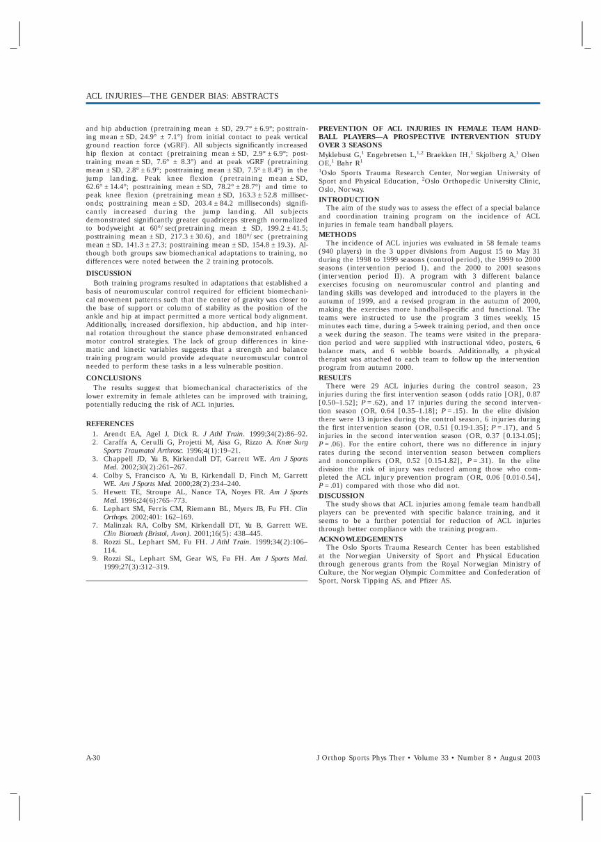
and hip abduction (pretraining mean ± SD, 29.7° ± 6.9°; posttrain-ing mean ± SD, 24.9° ± 7.1°) from initial contact to peak verticalground reaction force (vGRF). All subjects significantly increasedhip flexion at contact (pretraining mean ± SD, 2.9° ± 6.9°; post-training mean ± SD, 7.6° ± 8.3°) and at peak vGRF (pretrainingmean ± SD, 2.8° ± 6.9°; posttraining mean ± SD, 7.5° ± 8.4°) in thejump landing. Peak knee flexion (pretraining mean ± SD,62.6° ± 14.4°; posttraining mean ± SD, 78.2° ± 28.7°) and time topeak knee flexion (pretraining mean ± SD, 163.3 ± 52.8 millisec-onds; posttraining mean ± SD, 203.4 ± 84.2 milliseconds) signifi-cantly increased during the jump landing. All subjectsdemonstrated significantly greater quadriceps strength normalizedto bodyweight at 60°/sec(pretraining mean ± SD, 199.2 ± 41.5;posttraining mean ± SD, 217.3 ± 30.6), and 180°/sec (pretrainingmean ± SD, 141.3 ± 27.3; posttraining mean ± SD, 154.8 ± 19.3). Al-though both groups saw biomechanical adaptations to training, nodifferences were noted between the 2 training protocols.
DISCUSSIONBoth training programs resulted in adaptations that established a
basis of neuromuscular control required for efficient biomechani-cal movement patterns such that the center of gravity was closer tothe base of support or column of stability as the position of theankle and hip at impact permitted a more vertical body alignment.Additionally, increased dorsiflexion, hip abduction, and hip inter-nal rotation throughout the stance phase demonstrated enhancedmotor control strategies. The lack of group differences in kine-matic and kinetic variables suggests that a strength and balancetraining program would provide adequate neuromuscular controlneeded to perform these tasks in a less vulnerable position.
CONCLUSIONSThe results suggest that biomechanical characteristics of the
lower extremity in female athletes can be improved with training,potentially reducing the risk of ACL injuries.
REFERENCES1. Arendt EA, Agel J, Dick R. J Athl Train. 1999;34(2):86–92.2. Caraffa A, Cerulli G, Projetti M, Aisa G, Rizzo A. Knee Surg
Sports Traumatol Arthrosc. 1996;4(1):19–21.3. Chappell JD, Yu B, Kirkendall DT, Garrett WE. Am J Sports
Med. 2002;30(2):261–267.4. Colby S, Francisco A, Yu B, Kirkendall D, Finch M, Garrett
WE. Am J Sports Med. 2000;28(2):234–240.5. Hewett TE, Stroupe AL, Nance TA, Noyes FR. Am J Sports
Med. 1996;24(6):765–773.6. Lephart SM, Ferris CM, Riemann BL, Myers JB, Fu FH. Clin
Orthops. 2002;401: 162–169.7. Malinzak RA, Colby SM, Kirkendall DT, Yu B, Garrett WE.
Clin Biomech (Bristol, Avon). 2001;16(5): 438–445.8. Rozzi SL, Lephart SM, Fu FH. J Athl Train. 1999;34(2):106–
114.9. Rozzi SL, Lephart SM, Gear WS, Fu FH. Am J Sports Med.
1999;27(3):312–319.
PREVENTION OF ACL INJURIES IN FEMALE TEAM HAND-BALL PLAYERS—A PROSPECTIVE INTERVENTION STUDYOVER 3 SEASONSMyklebust G,1 Engebretsen L,1,2 Braekken IH,1 Skjolberg A,1 OlsenOE,1 Bahr R1
1Oslo Sports Trauma Research Center, Norwegian University ofSport and Physical Education, 2Oslo Orthopedic University Clinic,Oslo, Norway.INTRODUCTION
The aim of the study was to assess the effect of a special balanceand coordination training program on the incidence of ACLinjuries in female team handball players.METHODS
The incidence of ACL injuries was evaluated in 58 female teams(940 players) in the 3 upper divisions from August 15 to May 31during the 1998 to 1999 seasons (control period), the 1999 to 2000seasons (intervention period I), and the 2000 to 2001 seasons(intervention period II). A program with 3 different balanceexercises focusing on neuromuscular control and planting andlanding skills was developed and introduced to the players in theautumn of 1999, and a revised program in the autumn of 2000,making the exercises more handball-specific and functional. Theteams were instructed to use the program 3 times weekly, 15minutes each time, during a 5-week training period, and then oncea week during the season. The teams were visited in the prepara-tion period and were supplied with instructional video, posters, 6balance mats, and 6 wobble boards. Additionally, a physicaltherapist was attached to each team to follow up the interventionprogram from autumn 2000.RESULTS
There were 29 ACL injuries during the control season, 23injuries during the first intervention season (odds ratio [OR], 0.87[0.50–1.52]; P = .62), and 17 injuries during the second interven-tion season (OR, 0.64 [0.35–1.18]; P = .15). In the elite divisionthere were 13 injuries during the control season, 6 injuries duringthe first intervention season (OR, 0.51 [0.19-1.35]; P = .17), and 5injuries in the second intervention season (OR, 0.37 [0.13-1.05];P = .06). For the entire cohort, there was no difference in injuryrates during the second intervention season between compliersand noncompliers (OR, 0.52 [0.15-1.82], P = .31). In the elitedivision the risk of injury was reduced among those who com-pleted the ACL injury prevention program (OR, 0.06 [0.01-0.54],P = .01) compared with those who did not.DISCUSSION
The study shows that ACL injuries among female team handballplayers can be prevented with specific balance training, and itseems to be a further potential for reduction of ACL injuriesthrough better compliance with the training program.ACKNOWLEDGEMENTS
The Oslo Sports Trauma Research Center has been establishedat the Norwegian University of Sport and Physical Educationthrough generous grants from the Royal Norwegian Ministry ofCulture, the Norwegian Olympic Committee and Confederation ofSport, Norsk Tipping AS, and Pfizer AS.
ACL INJURIES—THE GENDER BIAS: ABSTRACTS
A-30 J Orthop Sports Phys Ther • Volume 33 • Number 8 • August 2003