Embed Size (px)
Citation preview

APPROPRIATE USE OF ECHOCARDIOGRAPHY
This documen
Board of Trus
The American
cited as follow
AR, Picard M
ASNC/HFSA/
echocardiogra
Appropriate U
American Hea
ure Society of
raphy and I
Cardiovascula
Resonance. J
2010, doi:10.1
ACCF/ASE/AHA/ASNC/HFSA/HRS/SCAI/SCCM/SCCT/SCMR 2011 Appropriate Use Criteria for
Echocardiography
A REPORT OF THE AMERICANCOLLEGE OF CARDIOLOGY FOUNDATION APPROPRIATEUSE CRITERIA TASK FORCE, AMERICAN
SOCIETY OF ECHOCARDIOGRAPHY, AMERICAN HEART ASSOCIATION, AMERICAN SOCIETY OF NUCLEAR CARDIOLOGY,HEART FAILURE SOCIETY OF AMERICA, HEART RHYTHM SOCIETY, SOCIETY FOR CARDIOVASCULAR ANGIOGRAPHY AND
INTERVENTIONS, SOCIETY OF CRITICAL CARE MEDICINE, SOCIETY OF CARDIOVASCULAR COMPUTED TOMOGRAPHY,SOCIETY FOR CARDIOVASCULAR MAGNETIC RESONANCE AMERICAN COLLEGE OF CHEST PHYSICIANS
(J Am Soc Echocardiogr 2011;24:229-67.)
Keywords: ACCF Appropriate Use Criteria, Cardiac imaging, Coronary artery disease, Diagnostic testing,Echocardiography
ECHOCARDIOGRAPHY WRITING GROUP
Pamela S. Douglas, MD, MACC, FAHA, FASE, Chair*Mario J. Garcia, MD, FACC, FACP†David E. Haines, MD, FACC, FHRS‡Wyman W. Lai, MD, MPH, FACC, FASE§Warren J. Manning, MD, FACCk{Ayan R. Patel, MD, FACC#Michael H. Picard, MD, FACC, FASE, FAHA§Donna M. Polk, MD, MPH, FACC, FASE, FASNC**Michael Ragosta, MD, FACC, FSCAI††R. Parker Ward, MD, FACC, FASE, FASNC§Rory B. Weiner, MD**Official American College of Cardiology Foundation
Representative; †Official Society of Cardiovascular ComputedTomography Representative; ‡Official Heart Rhythm SocietyRepresentative; §Official American Society of EchocardiographyRepresentative; kOfficial American Heart AssociationRepresentative; {Official Society for Cardiovascular MagneticResonance Representative; #Official Heart Failure Society ofAmerica Representative; **Official American Society of NuclearCardiology Representative; ††Official Society for CardiovascularAngiography and Interventions Representative.
t was approved by the American College of Cardiology Foundation
tees in November 2010.
College of Cardiology Foundation requests that this document be
s: Douglas PS, Garcia MJ, Haines DE, Lai WW, Manning WJ, Patel
H, Polk DM, Ragosta M, Ward RP, Weiner RB. ACCF/ASE/AHA/
HRS/SCAI/SCCM/SCCT/SCMR 2011 appropriate use criteria for
phy: a report of the American College of Cardiology Foundation
se Criteria Task Force, American Society of Echocardiography,
rt Association, American Society of Nuclear Cardiology, Heart Fail-
America, Heart Rhythm Society, Society for Cardiovascular Angiog-
nterventions, Society of Critical Care Medicine, Society of
r Computed Tomography, and Society for Cardiovascular Magnetic
Am Coll Cardiol 2010: published online before print November 19,
016/j.jacc.2010.11.002.
TECHNICAL PANEL
Steven R. Bailey, MD, FACC, FSCAI, FAHA, ModeratorRory B. Weiner, MD, Writing Group LiaisonPeter Alagona, Jr, MD, FACC*Jeffrey L. Anderson, MD, FACC, FAHA, MACP*kJeanne M. DeCara, MD, FACC, FASE§Rowena J. Dolor, MD, MHSReza Fazel, MD, FACC**John A. Gillespie, MD, FACC‡‡Paul A. Heidenreich, MD, FACC#Luci K. Leykum, MD, MBA, MSCJoseph E. Marine, MD, FACC, FHRS‡Gregory J. Mishkel, MD, FACC, FSCAI, FRCPC††Patricia A. Pellikka, MD, FACC, FAHA, FACP, FASE§Gilbert L. Raff, MD, FACC, FSCCT†Krishnaswami Vijayaraghavan, MD, FACC, FCCP§§Neil J. Weissman, MD, FACC, FAHA*Katherine C. Wu, MD{‡‡Official Health Plan Representative; §§Official American College
of Chest Physicians Representative.
This article is copublished in the Journal of the American College of Cardiology and
the Journal of Cardiovascular Computed Tomography.
Copies: This document is available on the World Wide Web site of the American
College of Cardiology (www.cardiosource.org). For copies of this document,
please contact Elsevier Inc. Reprint Department, fax (212) 633-3820, e-mail
Permissions: Modification, alteration, enhancement, and/or distribution of this
document are not permitted without the express permission of the American
College of Cardiology Foundation. Please contact Elsevier’s permission department
0894-7317/$36.00
Copyright 2011 by the American College of Cardiology.
doi:10.1016/j.echo.2010.12.008
229

230 Douglas et al Journal of the American Society of EchocardiographyMarch 2011
APPROPRIATE USE CRITERIA TASK FORCE
Michael J. Wolk, MD, MACC, ChairSteven R. Bailey, MD, FACC, FSCAI, FAHAPamela S. Douglas, MD, MACC, FAHA, FASERobert C. Hendel, MD, FACC, FAHA, FASNCChristopher M. Kramer, MD, FACC, FAHAJames K. Min, MD, FACCManesh R. Patel, MD, FACCLeslee Shaw, PhD, FACC, FASNCRaymond F. Stainback, MD, FACC, FASEJoseph M. Allen, MA
TABLE OF CONTENTS
ABSTRACT .............................................................................................. 230
PREFACE ...................................................................................................231
1. INTRODUCTION ...................................................................................................232
2. METHODS ................................................................................................................232
3. GENERAL ASSUMPTIONS ...............................................................................233
4. DEFINITIONS ..........................................................................................................233
5. RESULTS OF RATINGS ........................................................................................234
6. ECHOCARDIOGRAPHYAPPROPRIATE USE CRITERIA(BY INDICATION) .................................................................................................235
Table 1: TTE for General Evaluation of Cardiac Structureand Function ...................................................................................235
Table 2: TTE for Cardiovascular Evaluation in anAcute Setting ...................................................................................235
Table 3: TTE for Evaluation of Valvular Function ..................236
Table 4: TTE for Evaluation of Intracardiac andExtracardiac Structures and Chambers ..................................237
Table 5: TTE for Evaluation of Aortic Disease .........................237
Table 6: TTE for Evaluation of Hypertension, HF,or Cardiomyopathy ......................................................................237
Table 7: TTE for Adult Congenital Heart Disease ..................238
Table 8: TEE .........................................................................................239
Table 9: Stress Echocardiography for Detectionof CAD/Risk Assessment: Symptomatic orIschemic Equivalent ......................................................................239
Table 10: Stress Echocardiography for Detectionof CAD/Risk Assessment: Asymptomatic(Without Ischemic Equivalent) .................................................240
Table 11: Stress Echocardiography for Detectionof CAD/Risk Assessment: Asymptomatic (WithoutIschemic Equivalent) in Patient Populations WithDefined Comorbidities ................................................................240
Table 12: Stress Echocardiography FollowingPrior Test Results ............................................................................ 241
Table 13: Stress Echocardiography for RiskAssessment: Perioperative Evaluation forNoncardiac Surgery Without Active CardiacConditions ....................................................................................... 241
Table 14: Stress Echocardiography for Risk Assessment:Within 3 Months of an ACS .....................................................242
Table 15: Stress Echocardiography for Risk Assessment:Postrevascularization (PCI or CABG) ....................................242
Table 16: Stress Echocardiography for Assessmentof Viability/Ischemia .....................................................................242
Table 17: Stress Echocardiography for Hemodynamics(Includes Doppler During Stress) ............................................243
Table 18: Contrast Use in TTE/TEE or StressEchocardiography ..........................................................................243
7. ECHOCARDIOGRAPHYAPPROPRIATE USE CRITERIA(BYAPPROPRIATE USE RATING) ..............................................................244
Table 19. Appropriate Indications(Median Score 7–9) .....................................................................244
Table 20. Uncertain Indications(Median Score 4–6) .....................................................................248
Table 21. Inappropriate Indications(Median Score 1–3) .....................................................................249
8. DISCUSSION ...........................................................................................................253
APPENDIX A: ADDITIONAL ECHOCARDIOGRAPHYDEFINITIONS ........................................................................................ 259
Figure A1. Stepwise Approach to PerioperativeCardiac Assessment ......................................................................259
Table A1: Active Cardiac Conditions for Whichthe Patient Should Undergo Evaluation andTreatment Before Noncardiac Surgery (Class I,Level of Evidence: B) ...................................................................260
Table A2. Perioperative Clinical Risk Factors* .........................260
APPENDIX B: ADDITIONAL METHODS ................................ 260
Relationships With Industry and Other Entities ......................260
Literature Review ...............................................................................260
APPENDIX C: ACCF/ASE/AHA/ASNC/HFSA/HRS/SCAI/SCCM/SCCT/SCMR 2011 APPROPRIATE USE CRITERIAFOR ECHOCARDIOGRAPHY PARTICIPANTS ................ 260
APPENDIX D: ACCF/ASE/AHA/ASNC/HFSA/HRS/SCAI/SCCM/SCCT/SCMR 2011 Appropriate Use Criteria for Echocar-diography Writing Group, Technical Panel, Indication Reviewers,and Task Force–Relationships With Industry and Other Entities(in Alphabetical Order Within Each Group) ................................ 264
REFERENCES ......................................................................................... 266
ABSTRACT
The American College of Cardiology Foundation (ACCF), in partner-ship with the American Society of Echocardiography (ASE) and alongwith key specialty and subspecialty societies, conducted a review of

Abbreviations
ACS = Acute coronarysyndrome
APC = Atrial prematurecontraction
CABG = Coronary artery
bypass grafting surgery
CAD = Coronary artery
disease
CMR = Cardiovascular
magnetic resonance
CRT = Cardiac
resynchronization therapy
CT = Computed tomography
ECG = Electrocardiogram
HF = Heart failure
ICD = Implantablecardioverter-defibrillator
LBBB = Left bundle-branch
block
LV = Left ventricular
MET = Estimated metabolic
equivalents of exercise
MI = Myocardial infarction
PCI = Percutaneous coronaryintervention
RNI = Radionuclide imaging
SPECTMPI = Single-photonemission computed
tomography myocardial
perfusion imaging
STEMI = ST-segment
elevationmyocardial infarction
SVT = Supraventricular
tachycardia
TEE = Transesophagealechocardiogram
TIA = Transient ischemicattack
TIMI = Thrombolysis InMyocardial Infarction
TTE = Transthoracic
echocardiogram
NSTEMI/NSTEMI =Unstable angina/non–ST-
segment elevation myocardial
infarction
VPC = Ventricular prematurecontraction
VT = Ventricular tachycardia
Journal of the American Society of EchocardiographyVolume 24 Number 3
Douglas et al 231
common clinical scenarioswhere echocardiography is fre-quently considered. This docu-ment combines and updates theoriginal transthoracic and transe-sophageal echocardiography ap-propriateness criteria publishedin 2007 (1) and the originalstress echocardiography appro-priateness criteria published in2008 (2). This revision reflectsnew clinical data, reflectschanges in test utilization pat-terns, and clarifies echocardiog-raphy use where omissions orlack of clarity existed in the orig-inal criteria.
The indications (clinical sce-narios) were derived from com-mon applications or anticipateduses, as well as from current clin-ical practice guidelines and re-sults of studies examining theimplementation of the originalappropriate use criteria (AUC).The 202 indications in this docu-ment were developed by a di-verse writing group and scoredby a separate independent tech-nical panel on a scale of 1 to 9,to designate appropriate use(median 7 to 9), uncertain use(median 4 to 6), and inappropri-ate use (median 1 to 3).
Ninety-seven indications wererated as appropriate, 34 wererated as uncertain, and 71 wererated as inappropriate. In general,the use of echocardiography forinitial diagnosis when there isa change in clinical status orwhen the results of the echocar-diogram are anticipated tochange patient managementwere rated appropriate. Routinetesting when there was nochange in clinical status or whenresults of testing were unlikelyto modify management weremore likely to be inappropriatethan appropriate/uncertain.
The AUC for echocardiogra-phy have the potential to impactphysician decision making,healthcare delivery, and reim-bursement policy. Furthermore,recognition of uncertain clinicalscenarios facilitates identificationof areas that would benefit fromfuture research.
PREFACE
In an effort to respond to the need for the rational use of imaging ser-vices in the delivery of high-quality care, the ACCF has undertakena process to determine the appropriate use of cardiovascular imagingfor selected patient indications.
AUC publications reflect an ongoing effort by the ACCF tocritically and systematically create, review, and categorize clinicalsituations where diagnostic tests and procedures are utilized byphysicians caring for patients with cardiovascular diseases. Theprocess is based on current understanding of the technical capa-bilities of the imaging modalities examined. Although impossibleto be entirely comprehensive given the wide diversity of clinicaldisease, the indications are meant to identify common scenariosencompassing the majority of situations encountered in contem-porary practice. Given the breadth of information they convey,the indications do not directly correspond to the NinthRevision of the International Classification of Diseases systemas these codes do not include clinical information, such as symp-tom status.
The ACCF believes that careful blending of a broad range ofclinical experiences and available evidence-based information willhelp guide a more efficient and equitable allocation of healthcareresources in cardiovascular imaging. The ultimate objective ofAUC is to improve patient care and health outcomes in a cost-ef-fective manner, but it is not intended to ignore ambiguity and nu-ance intrinsic to clinical decision making. AUC thus should not beconsidered substitutes for sound clinical judgment and practice ex-perience.
The ACCF AUC process itself is also evolving. In the currentiteration, technical panel members were asked to rate indicationsfor echocardiography in a manner independent and irrespectiveof the prior published ACCF ratings for transthoracic echocardi-ography (TTE) and transesophageal echocardiography (TEE) (1)and stress echocardiography (2) as well as the prior ACCF ratingsfor diagnostic imaging modalities such as cardiac radionuclide im-aging (RNI) (3) and cardiac computed tomography (CT) (4).Given the iterative and evolving nature of the process, readersare counseled that comparison of individual appropriate use rat-ings among modalities rated at different times over the past sev-eral years may not reflect the comparative utility of thedifferent modalities for an indication, as the ratings may varyover time. A comparative evaluation of the appropriate use ofmultiple imaging techniques is currently being undertaken to as-sess the relative strengths of each modality for various clinical sce-narios.
We are grateful to the technical panel and its chair, Steven Bailey,MD, FACC, FSCAI, FAHA, a professional group with a wide rangeof skills and insights, for their thoughtful and thorough deliberationof the merits of echocardiography for various indications. We wouldalso like to thank the 27 individuals who provided a careful review ofthe draft of indications, the parent AUC Task Force ably led byMichael Wolk, MD, MACC, Rory Weiner, MD, and the ACC staff,John C. Lewin, MD, Joseph Allen, Starr Webb, Jenissa Haidari, andLea Binder for their exceptionally skilled support in the generationof this document.
Pamela S. Douglas, MD, MACC, FAHA, FASEChair, Echocardiography Writing GroupMichael J. Wolk, MD, MACCChair, Appropriate Use Criteria Task Force

232 Douglas et al Journal of the American Society of EchocardiographyMarch 2011
1. INTRODUCTION
This report addresses the appropriate use of TTE, TEE, and stressechocardiography. Improvements in cardiovascular imaging tech-nology and an expanding armamentarium of noninvasive diagnos-tic tools and therapeutic options for cardiovascular disease have ledto an increase in cardiovascular imaging. As the field of echocardi-ography continues to advance along with other imaging modalitiesand treatment options, the healthcare community needs to under-stand how to best incorporate this technology into daily clinicalcare.
All prior AUC publications from the ACCF and collaboratingorganizations reflect an ongoing effort to critically and systemat-ically create, review, and categorize the appropriate use of car-diovascular procedures and diagnostic tests. The ACCFrecognizes the importance of revising these criteria in a timelymanner in order to provide the cardiovascular community withthe most accurate indications. Understanding the backgroundand scope of this document are important before interpretingthe rating tables.
This document presents a combination and revision of the 2007ACCF AUC for Transthoracic and TransesophagealEchocardiography (1) and the 2008 ACCF AUC for StressEchocardiography (2). Appropriate echocardiograms are those thatare likely to contribute to improving patients’ clinical outcomes, andimportantly, inappropriate use of echocardiography may be poten-tially harmful to patients and generate unwarranted costs to thehealthcare system.
*Negative consequences include the risks of the procedure (i.e., radiation or
contrast exposure) and the downstream impact of poor test performance such as
delay in diagnosis (false-negatives) or inappropriate diagnosis (false-positives).
2. METHODS
The indications included in this publication cover a wide array ofcardiovascular signs and symptoms as well as clinical judgmentsas to the likelihood of cardiovascular findings. Within each maindisease category, a standardized approach was used to capturethe majority of clinical scenarios without making the list of indica-tions excessive. The approach was to create 5 broad clinical scenar-ios regarding the possible use of echocardiography: 1) for initialdiagnosis; 2) to guide therapy or management, regardless of symp-tom status; 3) to evaluate a change in clinical status or cardiacexam; 4) for early follow-up without change in clinical status;and 5) for late follow-up without change in clinical status. Certainspecific clinical scenarios were addressed with additional focusedindications.
The indications were constructed by experts in echocardiographyand in other fields and were modified on the basis of discussionsamong the task force and feedback from independent reviewersand the technical panel. Wherever possible, indications were mappedto relevant clinical guidelines and key publications/references (OnlineAppendix).
An important focus during the indication revision process was toharmonize the indications across noninvasive modalities, such thatthe wording of the indications are similar with other AUC (3) when-ever it was feasible to do so. New indications as well as indication ta-bles were created, although it remains likely that several clinicalscenarios are not covered by these revised AUC for echocardiogra-phy. Once the revised indications were written, they were reviewedand critiqued by the parent AUC Task Force and by 27 external re-
viewers representing all cardiovascular specialties and primary carebefore being finalized.
A detailed description of the methods used for ranking the se-lected clinical indications is found in a previous publication,‘‘ACCF Proposed Method for Evaluating the Appropriateness ofCardiovascular Imaging’’ (5). Briefly, this process combines evi-dence-based medicine and practice experience by engaging a tech-nical panel in a modified Delphi exercise. Since the original TTE/TEE (1) and stress echocardiography (2) documents and methodspaper (5) were published, several important processes have beenput in place to further enhance the rigor of this process. They in-clude convening a formal writing group with diverse expertise inimaging and clinical care, circulating the indications for external re-view prior to rating by the technical panel, ensuring appropriatebalance of expertise and practice area of the technical panel, devel-opment of a standardized rating package, and establishment of for-mal roles for facilitating panel interaction at the face-to-facemeeting.
The technical panel first rated indications independently. Then, thepanel was convened for a face-to-face meeting for discussion of eachindication. At this meeting, panel members were provided with theirscores and a blinded summary of their peers’ scores. After the meet-ing, panel members were then asked to independently provide theirfinal scores for each indication.
Although panel members were not provided explicit cost informa-tion to help determine their appropriate use ratings, they were askedto implicitly consider cost as an additional factor in their evaluation ofappropriate use. In rating these criteria, the technical panel was askedto assess whether the use of the test for each indication is appropriate,uncertain, or inappropriate, andwas provided the following definitionof appropriate use:
An appropriate imaging study is one in which the expectedincremental information, combinedwith clinical judgment, ex-ceeds the expected negative consequence* by a sufficientlywide margin for a specific indication that the procedure is gen-erally considered acceptable care and a reasonable approachfor the indication.
The technical panel scored each indication as follows:Median Score 7 to 9Appropriate test for specific indication (test is generally acceptable
and is a reasonable approach for the indication).Median Score 4 to 6Uncertain for specific indication (testmay be generally acceptable
and may be a reasonable approach for the indication). Uncertaintyalso implies that more research and/or patient information is neededto classify the indication definitively.
Median Score 1 to 3Inappropriate test for that indication (test is not generally accept-
able and is not a reasonable approach for the indication).The division of these scores into 3 levels of appropriateness is
somewhat arbitrary, and the numeric designations should beviewed as a continuum. Further, there is diversity in clinical opinionfor particular clinical scenarios, such that scores in the intermediatelevel of appropriate use should be labeled ‘‘uncertain,’’ as critical pa-tient or research data may be lacking or discordant. This designa-tion should be a prompt to the field to carry out definitive

Journal of the American Society of EchocardiographyVolume 24 Number 3
Douglas et al 233
research investigations whenever possible. It is anticipated that theAUC reports will continue to be revised as further data are gener-ated and information from the implementation of the criteria is ac-cumulated.
To prevent bias in the scoring process, the technical panel was de-liberately comprised of a minority of specialists in echocardiography.Specialists, although offering important clinical and technical insights,might have a natural tendency to rate the indications within their spe-cialty as more appropriate than nonspecialists. In addition, care wastaken in providing objective, nonbiased information, including guide-lines and key references, to the technical panel.
The level of agreement among panelists as defined by RAND (6)was analyzed based on the BIOMED rule for a panel of 14 to 16members. As such, agreement was defined as an indication where4 or fewer panelists’ ratings fell outside the 3-point region containingthe median score.
Disagreement was defined as where at least 5 panelists’ ratingsfell in both the appropriate and the inappropriate categories. Any in-dication having disagreement was categorized as uncertain regard-less of the final median score. Indications that met neitherdefinition for agreement or disagreement are in a third, unlabeledcategory.
3. GENERAL ASSUMPTIONS
To prevent any inconsistencies in interpretation, specific assumptionswere considered by the writing group in developing the indicationsand by the technical panel when rating the clinical indications forthe appropriate use of inpatient and outpatient adult TTE/TEE andstress echocardiography.
1. ATTE and a TEE examination and report will include the use and interpre-tation of 2-dimensional/M-mode imaging, color flow Doppler, and spectralDoppler as important elements of a comprehensive TTE/TEE (7–9)evaluating relevant cardiac structures and hemodynamics. Stressechocardiography will include rest and stress 2-dimensional imaging ata minimum unless performed for hemodynamics, when Doppler must beincluded (10).
2. All standard echocardiographic techniques for image acquisition, includingstandard rest imaging and stress protocols (10), are available for each indi-cation and have a sensitivity and specificity similar to those found in thepublished literature. Selection for andmonitoring of contrast use is assumedto be in accord with practice guidelines (11).
3. The test is performed and interpreted by qualified individual(s) in a facilitythat is proficient in the echocardiographic technique (12,13).
4. The range of potential indications for echocardiography is quite large,particularly in comparison with other cardiovascular imaging tests.Thus, the indications are, at times, purposefully broad to cover an arrayof cardiovascular signs and symptoms as well as the ordering physician’sbest judgment as to the presence of cardiovascular abnormalities. Addi-tionally, there are likely clinical scenarios that are not covered in this doc-ument.
5. A complete clinical history and physical exam has been completed bya qualified clinician such that the clinical status of the patient can be as-sumed to be valid as stated in the indication (e.g., an asymptomatic patientis truly asymptomatic for the condition in question and that sufficient ques-tioning of the patient has been undertaken).
6. If the reason for a test can be assigned to more than 1 indication, it shouldbe classified under the most appropriate indication.
7. Cost should be considered implicitly in the appropriate use determination.8. For each indication, the rating should reflect whether the echocardiogram is
reasonable for the patient according to the appropriate use definition, not
whether the test is preferred over another modality. It should not be as-sumed that for each indication the decision to perform a diagnostic testhas already beenmade. It also should not consider issues of local availabilityor skill for any modality or attempt in any way to compare 2 tests with eachother.
9. The category of ‘‘uncertain’’ should be used when insufficient clinical dataare available for a definitive categorization or there is substantial disagree-ment regarding the appropriateness of that indication. The designation of‘‘uncertain’’ should not be used as grounds for denial of reimbursement.
10. Indications that describe routine or surveillance echocardiograms im-ply that the test is being considered for a ‘‘periodic’’ evaluation sincea certain period of time has elapsed. The test is not being ordereddue to the anticipation of changing clinical decision making or guidingtherapy.
11. Prosthetic valves and native valves are to be considered together,except where specifically mentioned otherwise in this document. Theseverity of valve stenosis or regurgitation is defined in clinical guide-lines (14,15).
12. In general, it is assumed that TEE is most appropriately used as an adjunctor subsequent test to TTE when indicated, such as when suboptimal TTEimages preclude obtaining a diagnostic study. The indications for whichTEE may reasonably be the test of first choice include, but are not limitedto, the indications presented in Table 8 of this document.
13. Intraoperative TEE is an important use of cardiovascular ultrasound. How-ever, this application is outside the scope of this document and thus is notaddressed here.
14. For all stress imaging, the mode of stress testing is assumed to be exer-cise (e.g., treadmill, bicycle) for patients able to exercise. For patients un-able to exercise, it is assumed that dobutamine is used forechocardiographic stress testing. Any indications requiring a specificmode of stress (e.g., when hemodynamic information is required) arelabeled as such.
15. Doppler hemodynamic assessment during stress echocardiography in-cludes both right and left heart hemodynamics (e.g., valvular gradients,pulmonary artery pressure, mitral regurgitation severity).
16. The indications for the perioperative evaluation for noncardiac surgerywere modeled after the ACCF/AHA guidelines on perioperative cardio-vascular evaluation and care for noncardiac surgery (16). If a patient hassigns/symptoms of suspected cardiac etiology, the clinical scenario shouldbe considered in the symptomatic category (e.g., Indication 1) and not inthe perioperative section.
17. As with other surgeries, the need for coronary artery disease (CAD) assess-ment prior to solid organ transplantation is related to patient and surgicalrisk. In general, solid organ transplantation should be considered in the vas-cular surgery category given that CAD is common in patients with diabetesmellitus who have end-stage renal disease.
4. DEFINITIONS
Definitions of terms used throughout the indication set are listed here.Additional definitions are listed in Appendix A. These definitionswere provided to and discussed with the technical panel prior to rat-ings of indications.
1. Ischemic Equivalent: Chest Pain Syndrome, Anginal Equivalent,or Ischemic ElectrocardiographicAbnormalities:Any constellationof clinical findings that the physician feels is consistent with CAD. Examplesof such findings include, but are not limited to, chest pain, chest tightness,chest burning, shoulder pain, palpitations, jaw pain, new electrocardio-graphic abnormalities, or other symptoms/findings suggestive of CAD.Nonchest pain symptoms (e.g., dyspnea or reduced/worsening effort toler-ance) that are thought to be consistent with CADmay also be considered tobe an ischemic equivalent.
2. Global CAD Risk: It is assumed that clinicians will use current standardmethods of global risk assessment such as those presented in the National
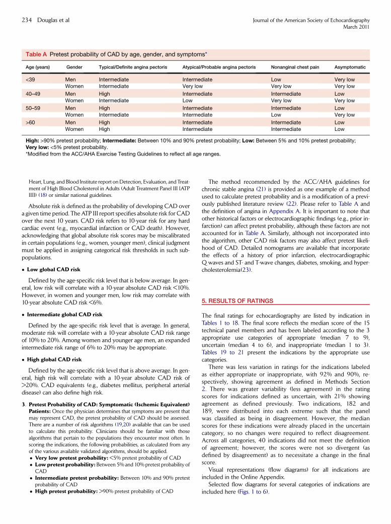
Table A Pretest probability of CAD by age, gender, and symptoms*
Age (years) Gender Typical/Definite angina pectoris Atypical/Probable angina pectoris Nonanginal chest pain Asymptomatic
<39 Men Intermediate Intermediate Low Very lowWomen Intermediate Very low Very low Very low
40–49 Men High Intermediate Intermediate LowWomen Intermediate Low Very low Very low
50–59 Men High Intermediate Intermediate Low
Women Intermediate Intermediate Low Very low
>60 Men High Intermediate Intermediate Low
Women High Intermediate Intermediate Low
High: >90% pretest probability; Intermediate: Between 10% and 90% pretest probability; Low: Between 5% and 10% pretest probability;
Very low: <5% pretest probability.
*Modified from the ACC/AHA Exercise Testing Guidelines to reflect all age ranges.
234 Douglas et al Journal of the American Society of EchocardiographyMarch 2011
Heart, Lung, and Blood Institute report onDetection, Evaluation, and Treat-ment of High Blood Cholesterol in Adults (Adult Treatment Panel III [ATPIII]) (18) or similar national guidelines.
Absolute risk is defined as the probability of developing CAD overa given time period. The ATP III report specifies absolute risk for CADover the next 10 years. CAD risk refers to 10-year risk for any hardcardiac event (e.g., myocardial infarction or CAD death). However,acknowledging that global absolute risk scores may be miscalibratedin certain populations (e.g., women, younger men), clinical judgmentmust be applied in assigning categorical risk thresholds in such sub-populations.
� Low global CAD risk
Defined by the age-specific risk level that is below average. In gen-eral, low risk will correlate with a 10-year absolute CAD risk <10%.However, in women and younger men, low risk may correlate with10-year absolute CAD risk <6%.
� Intermediate global CAD risk
Defined by the age-specific risk level that is average. In general,moderate risk will correlate with a 10-year absolute CAD risk rangeof 10% to 20%. Among women and younger age men, an expandedintermediate risk range of 6% to 20% may be appropriate.
� High global CAD risk
Defined by the age-specific risk level that is above average. In gen-eral, high risk will correlate with a 10-year absolute CAD risk of>20%. CAD equivalents (e.g., diabetes mellitus, peripheral arterialdisease) can also define high risk.
3. Pretest Probability of CAD: Symptomatic (Ischemic Equivalent)Patients: Once the physician determines that symptoms are present thatmay represent CAD, the pretest probability of CAD should be assessed.There are a number of risk algorithms (19,20) available that can be usedto calculate this probability. Clinicians should be familiar with thosealgorithms that pertain to the populations they encounter most often. Inscoring the indications, the following probabilities, as calculated from anyof the various available validated algorithms, should be applied.� Very low pretest probability: <5% pretest probability of CAD� Lowpretest probability: Between 5% and 10%pretest probability ofCAD
� Intermediate pretest probability: Between 10% and 90% pretestprobability of CAD
� High pretest probability: >90% pretest probability of CAD
The method recommended by the ACC/AHA guidelines forchronic stable angina (21) is provided as one example of a methodused to calculate pretest probability and is a modification of a previ-ously published literature review (22). Please refer to Table A andthe definition of angina in Appendix A. It is important to note thatother historical factors or electrocardiographic findings (e.g., prior in-farction) can affect pretest probability, although these factors are notaccounted for in Table A. Similarly, although not incorporated intothe algorithm, other CAD risk factors may also affect pretest likeli-hood of CAD. Detailed nomograms are available that incorporatethe effects of a history of prior infarction, electrocardiographicQ waves and ST- and T-wave changes, diabetes, smoking, and hyper-cholesterolemia(23).
5. RESULTS OF RATINGS
The final ratings for echocardiography are listed by indication inTables 1 to 18. The final score reflects the median score of the 15technical panel members and has been labeled according to the 3appropriate use categories of appropriate (median 7 to 9),uncertain (median 4 to 6), and inappropriate (median 1 to 3).Tables 19 to 21 present the indications by the appropriate usecategories.
There was less variation in ratings for the indications labeledas either appropriate or inappropriate, with 92% and 90%, re-spectively, showing agreement as defined in Methods Section2. There was greater variability (less agreement) in the ratingscores for indications defined as uncertain, with 21% showingagreement as defined previously. Two indications, 182 and189, were distributed into each extreme such that the panelwas classified as being in disagreement. However, the medianscores for these indications were already placed in the uncertaincategory, so no changes were required to reflect disagreement.Across all categories, 40 indications did not meet the definitionof agreement; however, the scores were not so divergent (asdefined by disagreement) as to necessitate a change in the finalscore.
Visual representations (flow diagrams) for all indications areincluded in the Online Appendix.
Selected flow diagrams for several categories of indications areincluded here (Figs. 1 to 6).

Journal of the American Society of EchocardiographyVolume 24 Number 3
Douglas et al 235
6. ECHOCARDIOGRAPHY APPROPRIATE USE CRITERIA (BY
INDICATION)
Table 1 TTE for general evaluation of cardiac structure and function
Indication
Appropriate use
score (1–9)
Suspected Cardiac Etiology—General With TTE1. � Symptoms or conditions potentially related to suspected cardiac etiology including but not limited to
chest pain, shortness of breath, palpitations, TIA, stroke, or peripheral embolic event
A (9)
2. � Prior testing that is concerning for heart disease or structural abnormality including but not limited to
chest X-ray, baseline scout images for stress echocardiogram, ECG, or cardiac biomarkers
A (9)
Arrhythmias With TTE
3. � Infrequent APCs or infrequent VPCs without other evidence of heart disease I (2)
4. � Frequent VPCs or exercise-induced VPCs A (8)
5. � Sustained or nonsustained atrial fibrillation, SVT, or VT A (9)
6. � Asymptomatic isolated sinus bradycardia I (2)
Lightheadedness/Presyncope/Syncope With TTE7. � Clinical symptoms or signs consistent with a cardiac diagnosis known to cause lightheadedness/
presyncope/syncope (including but not limited to aortic stenosis, hypertrophic cardiomyopathy, or HF)A (9)
8. � Lightheadedness/presyncope when there are no other symptoms or signs of cardiovascular disease I (3)9. � Syncope when there are no other symptoms or signs of cardiovascular disease A (7)
Evaluation of Ventricular Function With TTE10. � Initial evaluation of ventricular function (e.g., screening) with no symptoms or signs of cardiovascular disease I (2)
11. � Routine surveillance of ventricular function with known CAD and no change in clinical status or cardiac exam I (3)12. � Evaluation of LV function with prior ventricular function evaluation showing normal function
(e.g., prior echocardiogram, left ventriculogram, CT, SPECT MPI, CMR) in patients in whom
there has been no change in clinical status or cardiac exam
I (1)
Perioperative Evaluation With TTE13. � Routine perioperative evaluation of ventricular function with no symptoms or signs of cardiovascular disease I (2)
14. � Routine perioperative evaluation of cardiac structure and function prior to noncardiac solid organ transplantation U (6)
Pulmonary Hypertension With TTE
15. � Evaluation of suspected pulmonary hypertension including evaluation of right ventricular function and
estimated pulmonary artery pressure
A (9)
16. � Routine surveillance (<1 y) of known pulmonary hypertension without change in clinical status or cardiac exam I (3)
17. � Routine surveillance ($1 y) of known pulmonary hypertension without change in clinical status or cardiac exam A (7)
18. � Re-evaluation of known pulmonary hypertension if change in clinical status or cardiac exam or to guide therapy A (9)
A indicates appropriate; I, inappropriate; U, uncertain.
Table 2 TTE for cardiovascular evaluation in an acute setting
Indication
Appropriate use
score (1–9)
Hypotension or Hemodynamic Instability With TTE19. � Hypotension or hemodynamic instability of uncertain or suspected cardiac etiology A (9)
20. � Assessment of volume status in a critically ill patient U (5)Myocardial Ischemia/Infarction With TTE
21. � Acute chest pain with suspected MI and nondiagnostic ECG when a resting echocardiogramcan be performed during pain
A (9)
22. � Evaluation of a patient without chest pain but with other features of an ischemic equivalent or
laboratory markers indicative of ongoing MI
A (8)
23. � Suspected complication of myocardial ischemia/infarction, including but not limited to acute mitral regurgitation,
ventricular septal defect, free-wall rupture/tamponade, shock, right ventricular involvement, HF, or thrombus
A (9)
Evaluation of Ventricular Function after ACS With TTE
24. � Initial evaluation of ventricular function following ACS A (9)25. � Re-evaluation of ventricular function following ACS during recovery phase when results will guide therapy A (9)
Respiratory Failure With TTE26. � Respiratory failure or hypoxemia of uncertain etiology A (8)
27. � Respiratory failure or hypoxemia when a noncardiac etiology of respiratory failure has been established U (5)
(Continued )

Table 2 (Continued )
Indication
Appropriate use
score (1–9)
Pulmonary Embolism With TTE
28. � Suspected pulmonary embolism in order to establish diagnosis I (2)
29. � Known acute pulmonary embolism to guide therapy (e.g., thrombectomy and thrombolytics) A (8)
30. � Routine surveillance of prior pulmonary embolism with normal right ventricular function
and pulmonary artery systolic pressure
I (1)
31. � Re-evaluation of known pulmonary embolism after thrombolysis or thrombectomy for assessment
of change in right ventricular function and/or pulmonary artery pressure
A (7)
Cardiac Trauma With TTE32. � Severe deceleration injury or chest trauma when valve injury, pericardial effusion, or cardiac injury are
possible or suspected
A (9)
33. � Routine evaluation in the setting of mild chest trauma with no electrocardiographic changes or biomarker elevation I (2)
A indicates appropriate; I, inappropriate; U, uncertain.
Table 3 TTE for evaluation of valvular function
Indication
Appropriate use
score (1–9)
Murmur or Click With TTE34. � Initial evaluation when there is a reasonable suspicion of valvular or structural heart disease A (9)
35. � Initial evaluation when there are no other symptoms or signs of valvular or structural heart disease I (2)36. � Re-evaluation in a patient without valvular disease on prior echocardiogram and no change in
clinical status or cardiac exam
I (1)
37. �Re-evaluation of known valvular heart diseasewith a change in clinical status or cardiac exam or to
guide therapy
A (9)
Native Valvular Stenosis With TTE
38. � Routine surveillance (<3 y) of mild valvular stenosis without a change in clinical status or cardiac
exam
I (3)
39. � Routine surveillance ($3 y) of mild valvular stenosis without a change in clinical status or cardiacexam
A (7)
40. � Routine surveillance (<1 y) of moderate or severe valvular stenosis without a change in clinical
status or cardiac exam
I (3)
41. � Routine surveillance ($1 y) of moderate or severe valvular stenosis without a change in clinical
status or cardiac exam
A (8)
Native Valvular Regurgitation With TTE42. � Routine surveillance of trace valvular regurgitation I (1)
43. � Routine surveillance (<3 y) of mild valvular regurgitation without a change in clinical status or
cardiac exam
I (2)
44. � Routine surveillance ($3 y) of mild valvular regurgitation without a change in clinical status or
cardiac exam
U (4)
45. � Routine surveillance (<1 y) of moderate or severe valvular regurgitation without a change in clinical
status or cardiac exam
U (6)
46. � Routine surveillance ($1 y) of moderate or severe valvular regurgitation without change in clinicalstatus or cardiac exam
A (8)
Prosthetic Valves With TTE47. � Initial postoperative evaluation of prosthetic valve for establishment of baseline A (9)
48. � Routine surveillance (<3 y after valve implantation) of prosthetic valve if no known or suspectedvalve dysfunction
I (3)
49. � Routine surveillance ($3 y after valve implantation) of prosthetic valve if no known or suspected
valve dysfunction
A (7)
50. � Evaluation of prosthetic valve with suspected dysfunction or a change in clinical status or cardiac
exam
A (9)
51. � Re-evaluation of known prosthetic valve dysfunction when it would change management or guide
therapy
A (9)
Infective Endocarditis (Native or Prosthetic Valves) With TTE52. � Initial evaluation of suspected infective endocarditis with positive blood cultures or a new murmur A (9)
53. � Transient fever without evidence of bacteremia or a new murmur I (2)
(Continued )
236 Douglas et al Journal of the American Society of EchocardiographyMarch 2011

Table 3 (Continued )
Indication
Appropriate use
score (1–9)
54. � Transient bacteremia with a pathogen not typically associated with infective endocarditis and/or
a documented nonendovascular source of infection
I (3)
55. �Re-evaluation of infective endocarditis at high risk for progression or complication or with a changein clinical status or cardiac exam
A (9)
56. � Routine surveillance of uncomplicated infective endocarditis when no change in management is
contemplated
I (2)
A indicates appropriate; I, inappropriate; U, uncertain.
Table 4 TTE for evaluation of intracardiac and extracardiac structures and chambers
Indication
Appropriate use
score (1–9)
57. � Suspected cardiac mass A (9)58. � Suspected cardiovascular source of embolus A (9)
59. � Suspected pericardial conditions A (9)60. � Routine surveillance of known small pericardial effusion with no change in clinical status I (2)
61. � Re-evaluation of known pericardial effusion to guide management or therapy A (8)62. � Guidance of percutaneous noncoronary cardiac procedures including but not limited to
pericardiocentesis, septal ablation, or right ventricular biopsy
A (9)
A indicates appropriate; I, inappropriate; U, uncertain.
Table 5 TTE for evaluation of aortic disease
Indication
Appropriate use
score (1–9)
63. � Evaluation of the ascending aorta in the setting of a known or suspected connective tissue
disease or genetic condition that predisposes to aortic aneurysm or dissection (e.g., Marfansyndrome)
A (9)
64. � Re-evaluation of known ascending aortic dilation or history of aortic dissection to establish
a baseline rate of expansion or when the rate of expansion is excessive
A (9)
65. �Re-evaluation of known ascending aortic dilation or history of aortic dissection with a change in
clinical status or cardiac exam or when findings may alter management or therapy
A (9)
66. � Routine re-evaluation for surveillance of known ascending aortic dilation or history of aortic
dissection without a change in clinical status or cardiac exam when findings would not change
management or therapy
I (3)
A indicates appropriate; I, inappropriate; U, uncertain.
Table 6 TTE for evaluation of hypertension, HF, or cardiomyopathy
Indication
Appropriate Use
score (1–9)
Hypertension With TTE67. � Initial evaluation of suspected hypertensive heart disease A (8)
68. � Routine evaluation of systemic hypertension without symptoms or signs of hypertensive heart disease I (3)
69. � Re-evaluation of known hypertensive heart disease without a change in clinical status or cardiac exam U (4)
HF With TTE70. � Initial evaluation of known or suspected HF (systolic or diastolic) based on symptoms, signs, or abnormal
test resultsA (9)
71. � Re-evaluation of known HF (systolic or diastolic) with a change in clinical status or cardiac exam withouta clear precipitating change in medication or diet
A (8)
72. � Re-evaluation of known HF (systolic or diastolic) with a change in clinical status or cardiac exam with
a clear precipitating change in medication or diet
U (4)
73. � Re-evaluation of known HF (systolic or diastolic) to guide therapy A (9)
(Continued )
Journal of the American Society of EchocardiographyVolume 24 Number 3
Douglas et al 237

Table 6 (Continued )
Indication
Appropriate Use
score (1–9)
74. �Routine surveillance (<1 y) of HF (systolic or diastolic) when there is no change in clinical status or cardiac
exam
I (2)
75. �Routine surveillance ($1 y) of HF (systolic or diastolic) when there is no change in clinical status or cardiacexam
U (6)
Device Evaluation (Including Pacemaker, ICD, or CRT) With TTE
76. � Initial evaluation or re-evaluation after revascularization and/or optimal medical therapy to determine
candidacy for device therapy and/or to determine optimal choice of device
A (9)
77. � Initial evaluation for CRT device optimization after implantation U (6)
78. � Known implanted pacing device with symptoms possibly due to device complication or suboptimal
pacing device settings
A (8)
79. � Routine surveillance (<1 y) of implanted device without a change in clinical status or cardiac exam I (1)
80. � Routine surveillance ($1 y) of implanted device without a change in clinical status or cardiac exam I (3)Ventricular Assist Devices and Cardiac Transplantation With TTE
81. � To determine candidacy for ventricular assist device A (9)82. � Optimization of ventricular assist device settings A (7)
83. � Re-evaluation for signs/symptoms suggestive of ventricular assist device-related complications A (9)84. � Monitoring for rejection in a cardiac transplant recipient A (7)
85. � Cardiac structure and function evaluation in a potential heart donor A (9)Cardiomyopathies With TTE
86. � Initial evaluation of known or suspected cardiomyopathy (e.g., restrictive, infiltrative, dilated,
hypertrophic, or genetic cardiomyopathy)
A (9)
87. � Re-evaluation of known cardiomyopathy with a change in clinical status or cardiac exam or to guide
therapy
A (9)
88. � Routine surveillance (<1 y) of known cardiomyopathy without a change in clinical status or cardiac exam I (2)
89. � Routine surveillance ($1 y) of known cardiomyopathy without a change in clinical status or cardiac exam U (5)
90. � Screening evaluation for structure and function in first-degree relatives of a patient with an inherited
cardiomyopathy
A (9)
91. � Baseline and serial re-evaluations in a patient undergoing therapy with cardiotoxic agents A (9)
A indicates appropriate; I, inappropriate; U, uncertain.
Table 7 TTE for adult congenital heart disease
Indication
Appropriate use
score (1–9)
92. � Initial evaluation of known or suspected adult congenital heart disease A (9)
93. � Known adult congenital heart disease with a change in clinical status orcardiac exam
A (9)
94. � Re-evaluation to guide therapy in known adult congenital heart disease A (9)
95. � Routine surveillance (<2 y) of adult congenital heart disease following
complete repair
+ without a residual structural or hemodynamic abnormality+ without a change in clinical status or cardiac exam
I (3)
96. � Routine surveillance ($2 y) of adult congenital heart disease followingcomplete repair
+ without residual structural or hemodynamic abnormality+ without a change in clinical status or cardiac exam
U (6)
97. � Routine surveillance (<1 y) of adult congenital heart disease following
incomplete or palliative repair+ with residual structural or hemodynamic abnormality+ without a change in clinical status or cardiac exam
U (5)
98. � Routine surveillance ($1 y) of adult congenital heart disease following
incomplete or palliative repair
+ with residual structural or hemodynamic abnormality+ without a change in clinical status or cardiac exam
A (8)
A indicates appropriate; I, inappropriate; U, uncertain.
238 Douglas et al Journal of the American Society of EchocardiographyMarch 2011

Table 8 TEE
Indication
Appropriate use
score (1–9)
TEE as Initial or Supplemental Test—General Uses99. � Use of TEE when there is a high likelihood of a nondiagnostic TTE due to patient characteristics or
inadequate visualization of relevant structures
A (8)
100. � Routine use of TEE when a diagnostic TTE is reasonably anticipated to resolve all diagnostic and
management concerns
I (1)
101. � Re-evaluation of prior TEE finding for interval change (e.g., resolution of thrombus after
anticoagulation, resolution of vegetation after antibiotic therapy) when a change in therapy isanticipated
A (8)
102. � Surveillance of prior TEE finding for interval change (e.g., resolution of thrombus after anticoagulation,resolution of vegetation after antibiotic therapy) when no change in therapy is anticipated
I (2)
103. �Guidance during percutaneous noncoronary cardiac interventions including but not limited to closure
device placement, radiofrequency ablation, and percutaneous valve procedures
A (9)
104. � Suspected acute aortic pathology including but not limited to dissection/transsection A (9)
105. � Routine assessment of pulmonary veins in an asymptomatic patient status post pulmonary veinisolation
I (3)
TEE as Initial or Supplemental Test—Valvular Disease106. � Evaluation of valvular structure and function to assess suitability for, and assist in planning of, an
intervention
A (9)
107. � To diagnose infective endocarditis with a low pretest probability (e.g., transient fever, known
alternative source of infection, or negative blood cultures/atypical pathogen for endocarditis)
I (3)
108. � To diagnose infective endocarditis with amoderate or high pretest probability (e.g., staph bacteremia,fungemia, prosthetic heart valve, or intracardiac device)
A (9)
TEE as Initial or Supplemental Test—Embolic Event109. � Evaluation for cardiovascular source of embolus with no identified noncardiac source A (7)
110. � Evaluation for cardiovascular source of embolus with a previously identified noncardiac source U (5)111. � Evaluation for cardiovascular source of embolus with a known cardiac source in which a TEE would
not change management
I (1)
TEE as Initial Test—Atrial Fibrillation/Flutter112. � Evaluation to facilitate clinical decision making with regard to anticoagulation, cardioversion, and/or
radiofrequency ablation
A (9)
113. � Evaluation when a decision has been made to anticoagulate and not to perform cardioversion I (2)
A indicates appropriate; I, inappropriate; U, uncertain.
Table 9 Stress echocardiography for detection of CAD/Risk assessment: Symptomatic or ischemic equivalent
Indication
Appropriate use
score (1–9)
Evaluation of Ischemic Equivalent (Nonacute) With Stress Echocardiography114. � Low pretest probability of CAD
� ECG interpretable and able to exerciseI (3)
115. � Low pretest probability of CAD
� ECG uninterpretable or unable to exercise
A (7)
116. � Intermediate pretest probability of CAD
� ECG interpretable and able to exercise
A (7)
117. � Intermediate pretest probability of CAD
� ECG uninterpretable or unable to exercise
A (9)
118. � High pretest probability of CAD
� Regardless of ECG interpretability and ability to exercise
A (7)
Acute Chest Pain With Stress Echocardiography119. � Possible ACS
� ECG: no ischemic changes or with LBBB or electronically paced ventricular rhythm� Low-risk TIMI score
� Negative troponin levels
A (7)
(Continued )
Journal of the American Society of EchocardiographyVolume 24 Number 3
Douglas et al 239

Table 9 (Continued )
Indication
Appropriate use
score (1–9)
120. � Possible ACS
� ECG: no ischemic changes or with LBBB or electronically paced ventricular rhythm� Low-risk TIMI score
� Peak troponin: borderline, equivocal, minimally elevated
A (7)
121. � Possible ACS
� ECG: no ischemic changes or with LBBB or electronically paced ventricular rhythm� High-risk TIMI score
� Negative troponin levels
A (7)
122. � Possible ACS
� ECG: no ischemic changes or with LBBB or electronically paced ventricular rhythm
� High-risk TIMI score� Peak troponin: borderline, equivocal, minimally elevated
A (7)
123. � Definite ACS I (1)
A indicates appropriate; I, inappropriate; U, uncertain.
Table 10 Stress echocardiography for detection of CAD/Risk assessment: Asymptomatic (without ischemic equivalent)
Indication
Appropriate use
score (1–9)
General Patient Populations With Stress Echocardiography124. � Low global CAD risk I (1)
125. � Intermediate global CAD risk� ECG interpretable
I (2)
126. � Intermediate global CAD risk
� ECG uninterpretable
U (5)
127. � High global CAD risk U (5)
A indicates appropriate; I, inappropriate; U, uncertain.
Table 11 Stress echocardiography for detection of CAD/Risk assessment: Asymptomatic (without ischemic equivalent) in patientpopulations with defined comorbidities
Indication
Appropriate use
score (1–9)
New-Onset or Newly Diagnosed HF or LV Systolic Dysfunction With Stress Echocardiography128. � No prior CAD evaluation and no planned coronary angiography A (7)
Arrhythmias With Stress Echocardiography129. � Sustained VT A (7)
130. � Frequent PVCs, exercise induced VT, or nonsustained VT A (7)
131. � Infrequent PVCs I (3)
132. � New-onset atrial fibrillation U (6)
Syncope With Stress Echocardiography
133. � Low global CAD risk I (3)
134. � Intermediate or high global CAD risk A (7)
Elevated Troponin With Stress Echocardiography135. � Troponin elevation without symptoms or additional evidence of ACS A (7)
A indicates appropriate; I, inappropriate; U, uncertain.
240 Douglas et al Journal of the American Society of EchocardiographyMarch 2011

Table 12 Stress echocardiography following prior test results
Indication
Appropriate use
score (1–9)
Asymptomatic: Prior Evidence of Subclinical Disease With Stress Echocardiography136. � Coronary calcium Agatston score <100 I (2)
137. � Low to intermediate global CAD risk� Coronary calcium Agatston score between 100 and 400
U (5)
138. � High global CAD risk
� Coronary calcium Agatston score between 100 and 400
U (6)
139. � Coronary calcium Agatston score >400 A (7)
140. � Abnormal carotid intimal medial thickness ($0.9 mm and/or the presence of plaque encroaching into the arterial lumen) U (5)
Coronary Angiography (Invasive or Noninvasive) With Stress Echocardiography141. � Coronary artery stenosis of unclear significance A (8)
Asymptomatic or Stable Symptoms With Stress EchocardiographyNormal Prior Stress Imaging Study
142. � Low global CAD risk
� Last stress imaging study <2 y ago
I (1)
143. � Low global CAD risk
� Last stress imaging study $2 y ago
I (2)
144. � Intermediate to high global CAD risk
� Last stress imaging study <2 y ago
I (2)
145. � Intermediate to high global CAD risk
� Last stress imaging study $2 y ago
U (4)
Asymptomatic or Stable Symptoms With Stress Echocardiography Abnormal Coronary Angiographyor Abnormal Prior Stress Study No Prior Revascularization
146. � Known CAD on coronary angiography or prior abnormal stress imaging study
� Last stress imaging study <2 y ago
I (3)
147. � Known CAD on coronary angiography or prior abnormal stress imaging study� Last stress imaging study $2 y ago
U (5)
Treadmill ECG Stress Test With Stress Echocardiography148. � Low-risk treadmill score (e.g., Duke) I (1)
149. � Intermediate-risk treadmill score (e.g., Duke) A (7)150. � High-risk treadmill score (e.g., Duke) A (7)
New or Worsening Symptoms With Stress Echocardiography151. � Abnormal coronary angiography or abnormal prior stress imaging study A (7)
152. � Normal coronary angiography or normal prior stress imaging study U (6)
Prior Noninvasive Evaluation With Stress Echocardiography
153. � Equivocal, borderline, or discordant stress testing where obstructive CAD remains a concern A (8)
A indicates appropriate; I, inappropriate; U, uncertain.
Table 13 Stress echocardiography for risk assessment: Perioperative evaluation for noncardiac surgery without active cardiacconditions
Indication
Appropriate use
score (1–9)
Low-Risk Surgery With Stress Echocardiography154. � Perioperative evaluation for risk assessment I (1)
Intermediate-Risk Surgery With Stress Echocardiography155. � Moderate to good functional capacity ($4 METs) I (3)
156. � No clinical risk factors I (2)
157. � $1 clinical risk factor� Poor or unknown functional capacity (<4 METs)
U (6)
158. � Asymptomatic <1 y post normal catheterization, noninvasive test, or previous revascularization I (1)Vascular Surgery With Stress Echocardiography
159. � Moderate to good functional capacity ($4 METs) I (3)160. � No clinical risk factors I (2)
161. � $1 clinical risk factor� Poor or unknown functional capacity (<4 METs)
A (7)
162. � Asymptomatic <1 y post normal catheterization, noninvasive test, or previous revascularization I (2)
A indicates appropriate; I, inappropriate; U, uncertain.
Journal of the American Society of EchocardiographyVolume 24 Number 3
Douglas et al 241

Table 14 Stress echocardiography for risk assessment: Within 3 months of an ACS
Indication
Appropriate use
score (1–9)
STEMI With Stress Echocardiography163. � Primary PCI with complete revascularization
� No recurrent symptoms
I (2)
164. � Hemodynamically stable, no recurrent chest pain symptoms, or no signs of HF
� To evaluate for inducible ischemia
� No prior coronary angiography since the index event
A (7)
165. � Hemodynamically unstable, signs of cardiogenic shock, or mechanical complications I (1)
UA/NSTEMI With Stress Echocardiography166. � Hemodynamically stable, no recurrent chest pain symptoms, or no signs of HF
� To evaluate for inducible ischemia
� No prior coronary angiography since the index event
A (8)
ACS—Asymptomatic Postrevascularization (PCI or CABG) With Stress Echocardiography167. � Prior to hospital discharge in a patient who has been adequately revascularized I (1)
Cardiac Rehabilitation With Stress Echocardiography168. � Prior to initiation of cardiac rehabilitation (as a stand-alone indication) I (3)
A indicates appropriate; I, inappropriate; U, uncertain.
Table 15 Stress echocardiography for risk assessment: Postrevascularization (PCI or CABG)
Indication
Appropriate use
score (1–9)
Symptomatic With Stress Echocardiography169. � Ischemic equivalent A (8)
Asymptomatic With Stress Echocardiography170. � Incomplete revascularization
� Additional revascularization feasibleA (7)
171. � <5 y after CABG I (2)172. � $5 y after CABG U (6)
173. � <2 y after PCI I (2)174. � $2 y after PCI U (5)
Cardiac Rehabilitation With Stress Echocardiography175. � Prior to initiation of cardiac rehabilitation (as a stand-alone indication) I (3)
A indicates appropriate; I, inappropriate; U, uncertain.
Table 16 Stress echocardiography for assessment of viability/ischemia
Indication
Appropriate use
score (1–9)
Ischemic Cardiomyopathy/Assessment of Viability With Stress Echocardiography176. � Known moderate or severe LV dysfunction
� Patient eligible for revascularization
� Use of dobutamine stress only
A (8)
A indicates appropriate; I, inappropriate; U, uncertain.
242 Douglas et al Journal of the American Society of EchocardiographyMarch 2011

Table 17 Stress echocardiography for hemodynamics (includes doppler during stress)
Indication
Appropriate use
score (1–9)
Chronic Valvular Disease—Asymptomatic With Stress Echocardiography177. � Mild mitral stenosis I (2)
178. � Moderate mitral stenosis U (5)179. � Severe mitral stenosis A (7)
180. � Mild aortic stenosis I (3)181. � Moderate aortic stenosis U (6)
182. � Severe aortic stenosis U (5)183. � Mild mitral regurgitation I (2)
184. � Moderate mitral regurgitation U (5)185. � Severe mitral regurgitation
� LV size and function not meeting surgical criteria
A (7)
186. � Mild aortic regurgitation I (2)
187. � Moderate aortic regurgitation U (5)
188. � Severe aortic regurgitation
� LV size and function not meeting surgical criteria
A (7)
Chronic Valvular Disease—Symptomatic With Stress Echocardiography
189. � Mild mitral stenosis U (5)
190. � Moderate mitral stenosis A (7)
191. � Severe mitral stenosis I (3)192. � Severe aortic stenosis I (1)
193. � Evaluation of equivocal aortic stenosis� Evidence of low cardiac output or LV systolic dysfunction (‘‘low gradient aortic stenosis’’)
� Use of dobutamine only
A (8)
194. � Mild mitral regurgitation U (4)
195. � Moderate mitral regurgitation A (7)
196. � Severe mitral regurgitation
� Severe LV enlargement or LV systolic dysfunction
I (3)
Acute Valvular Disease With Stress Echocardiography197. � Acute moderate or severe mitral or aortic regurgitation I (3)
Pulmonary Hypertension With Stress Echocardiography198. � Suspected pulmonary artery hypertension
� Normal or borderline elevated estimated right ventricular systolic pressure on resting echocardiographic study
U (5)
199. � Routine evaluation of patients with known resting pulmonary hypertension I (3)
200. � Re-evaluation of patient with exercise-induced pulmonary hypertension to evaluate response to therapy U (5)
A indicates appropriate; I, inappropriate; U, uncertain.
Table 18 Contrast use in TTE/TEE or stress echocardiography
Indication
Appropriate use
score (1–9)
201. � Routine use of contrast
� All LV segments visualized on noncontrast images
I (1)
202. � Selective use of contrast� $2 contiguous LV segments are not seen on noncontrast images
A (8)
A indicates appropriate; I, inappropriate; U, uncertain.
Journal of the American Society of EchocardiographyVolume 24 Number 3
Douglas et al 243

244 Douglas et al Journal of the American Society of EchocardiographyMarch 2011
7. ECHOCARDIOGRAPHY APPROPRIATE USE CRITERIA (BY
APPROPRIATE USE RATING)
Table 19 Appropriate indications (median score 7–9)
Indication
Appropriate use
score (1–9)
TTE for General Evaluation of Cardiac Structure and Function Suspected Cardiac Etiology—General1. � Symptoms or conditions potentially related to suspected cardiac etiology including but not
limited to chest pain, shortness of breath, palpitations, TIA, stroke, or peripheral embolic
event
A (9)
2. � Prior testing that is concerning for heart disease or structural abnormality including but not
limited to chest X-ray, baseline scout images for stress echocardiogram, ECG, or cardiac
biomarkers
A (9)
TTE for General Evaluation of Cardiac Structure and Function Arrhythmias
4. � Frequent VPCs or exercise-induced VPCs A (8)5. � Sustained or nonsustained atrial fibrillation, SVT, or VT A (9)
TTE for General Evaluation of Cardiac Structure and Function Lightheadedness/Presyncope/Syncope7. � Clinical symptoms or signs consistent with a cardiac diagnosis known to cause
lightheadedness/presyncope/syncope (including but not limited to aortic stenosis,
hypertrophic cardiomyopathy, or HF)
A (9)
9. � Syncope when there are no other symptoms or signs of cardiovascular disease A (7)
TTE for General Evaluation of Cardiac Structure and Function Pulmonary Hypertension
15. � Evaluation of suspected pulmonary hypertension including evaluation of right ventricular
function and estimated pulmonary artery pressure
A (9)
17. � Routine surveillance ($1 y) of known pulmonary hypertension without change in clinical
status or cardiac exam
A (7)
18. �Re-evaluation of known pulmonary hypertension if change in clinical status or cardiac exam
or to guide therapy
A (9)
TTE for Cardiovascular Evaluation in an Acute Setting Hypotension or Hemodynamic Instability
19. � Hypotension or hemodynamic instability of uncertain or suspected cardiac etiology A (9)
TTE for Cardiovascular Evaluation in an Acute Setting Myocardial Ischemia/Infarction
21. � Acute chest pain with suspected MI and nondiagnostic ECG when a resting
echocardiogram can be performed during pain
A (9)
22. � Evaluation of a patient without chest pain but with other features of an ischemic equivalent
or laboratory markers indicative of ongoing MI
A (8)
23. � Suspected complication of myocardial ischemia/infarction, including but not limited to
acute mitral regurgitation, ventricular septal defect, free-wall rupture/tamponade, shock,
right ventricular involvement, HF, or thrombus
A (9)
TTE for Cardiovascular Evaluation in an Acute Setting Evaluation of Ventricular Function after ACS
24. � Initial evaluation of ventricular function following ACS A (9)25. �Re-evaluation of ventricular function following ACS during recovery phase when results will
guide therapy
A (9)
TTE for Cardiovascular Evaluation in an Acute Setting Respiratory Failure26. � Respiratory failure or hypoxemia of uncertain etiology A (8)
TTE for Cardiovascular Evaluation in an Acute Setting Pulmonary Embolism29. � Known acute pulmonary embolism to guide therapy (e.g., thrombectomy and
thrombolytics)
A (8)
31. � Re-evaluation of known pulmonary embolism after thrombolysis or thrombectomy for
assessment of change in right ventricular function and/or pulmonary artery pressure
A (7)
TTE for Cardiovascular Evaluation in an Acute Setting Cardiac Trauma
32. �Severe deceleration injury or chest traumawhen valve injury, pericardial effusion, or cardiacinjury are possible or suspected
A (9)
TTE for Evaluation of Valvular Function Murmur or Click
34. � Initial evaluation when there is a reasonable suspicion of valvular or structural heart disease A (9)
37. � Re-evaluation of known valvular heart disease with a change in clinical status or cardiac
exam or to guide therapy
A (9)
TTE for Evaluation of Valvular Function Native Valvular Stenosis39. � Routine surveillance ($3 y) of mild valvular stenosis without a change in clinical status or
cardiac examA (7)
(Continued )

Table 19 (Continued )
Indication
Appropriate use
score (1–9)
41. � Routine surveillance ($1 y) of moderate or severe valvular stenosis without a change in
clinical status or cardiac exam
A (8)
46. � Routine surveillance ($1 y) of moderate or severe valvular regurgitation without change in
clinical status or cardiac exam
A (8)
TTE for Evaluation of Valvular Function Prosthetic Valves47. � Initial postoperative evaluation of prosthetic valve for establishment of baseline A (9)
49. � Routine surveillance ($3 y after valve implantation) of prosthetic valve if no known or
suspected valve dysfunction
A (7)
50. � Evaluation of prosthetic valve with suspected dysfunction or a change in clinical status or
cardiac exam
A (9)
51. �Re-evaluation of known prosthetic valve dysfunction when it would changemanagement orguide therapy
A (9)
TTE for Evaluation of Valvular Function Infective Endocarditis (Native or Prosthetic Valves)
52. � Initial evaluation of suspected infective endocarditis with positive blood cultures or a new
murmur
A (9)
55. � Re-evaluation of infective endocarditis at high risk for progression or complication or with
a change in clinical status or cardiac exam
A (9)
TTE for Evaluation of Intracardiac and Extracardiac Structures and Chambers57. � Suspected cardiac mass A (9)
58. � Suspected cardiovascular source of embolus A (9)59. � Suspected pericardial conditions A (9)
61. � Re-evaluation of known pericardial effusion to guide management or therapy A (8)62. � Guidance of percutaneous noncoronary cardiac procedures including but not limited to
pericardiocentesis, septal ablation, or right ventricular biopsy
A (9)
TTE for Evaluation of Aortic Disease63. � Evaluation of the ascending aorta in the setting of a known or suspected connective tissue
disease or genetic condition that predisposes to aortic aneurysm or dissection (e.g.,
Marfan syndrome)
A (9)
64. � Re-evaluation of known ascending aortic dilation or history of aortic dissection to establisha baseline rate of expansion or when the rate of expansion is excessive
A (9)
65. � Re-evaluation of known ascending aortic dilation or history of aortic dissection with
a change in clinical status or cardiac exam or when findings may alter management or
therapy
A (9)
TTE for Evaluation of Hypertension, HF, or Cardiomyopathy Hypertension67. � Initial evaluation of suspected hypertensive heart disease A (8)
TTE for Evaluation of Hypertension, HF, or Cardiomyopathy HF70. � Initial evaluation of known or suspected HF (systolic or diastolic) based on symptoms,
signs, or abnormal test resultsA (9)
71. � Re-evaluation of known HF (systolic or diastolic) with a change in clinical status or cardiac
exam without a clear precipitating change in medication or diet
A (8)
73. � Re-evaluation of known HF (systolic or diastolic) to guide therapy A (9)
TTE for Evaluation of Hypertension, HF, or Cardiomyopathy Device Evaluation (Including Pacemaker, ICD, or CRT)76. � Initial evaluation or re-evaluation after revascularization and/or optimal medical therapy to
determine candidacy for device therapy and/or to determine optimal choice of device
A (9)
78. � Known implanted pacing device with symptoms possibly due to device complication or
suboptimal pacing device settings
A (8)
TTE for Evaluation of Hypertension, HF, or Cardiomyopathy Ventricular Assist Devices and Cardiac Transplantation
81. � To determine candidacy for ventricular assist device A (9)
82. � Optimization of ventricular assist device settings A (7)
83. � Re-evaluation for signs/symptoms suggestive of ventricular assist device-related
complications
A (9)
84. � Monitoring for rejection in a cardiac transplant recipient A (7)
85. � Cardiac structure and function evaluation in a potential heart donor A (9)
TTE for Evaluation of Hypertension, HF, or Cardiomyopathy Cardiomyopathies
86. � Initial evaluation of known or suspected cardiomyopathy (e.g., restrictive, infiltrative,
dilated, hypertrophic, or genetic cardiomyopathy)
A (9)
87. �Re-evaluation of known cardiomyopathy with a change in clinical status or cardiac exam or
to guide therapy
A (9)
(Continued )
Journal of the American Society of EchocardiographyVolume 24 Number 3
Douglas et al 245

Table 19 (Continued )
Indication
Appropriate use
score (1–9)
90. � Screening evaluation for structure and function in first-degree relatives of a patient with an
inherited cardiomyopathy
A (9)
91. � Baseline and serial re-evaluations in a patient undergoing therapy with cardiotoxic agents A (9)
TTE for Adult Congenital Heart Disease92. � Initial evaluation of known or suspected adult congenital heart disease A (9)
93. � Known adult congenital heart disease with a change in clinical status or cardiac exam A (9)94. � Re-evaluation to guide therapy in known adult congenital heart disease A (9)
98. � Routine surveillance ($1 y) of adult congenital heart disease following incomplete orpalliative repair
+ with residual structural or hemodynamic abnormality+ without a change in clinical status or cardiac exam
A (8)
TEE as Initial or Supplemental Test—General Uses
99. � Use of TEE when there is a high likelihood of a nondiagnostic TTE due to patient
characteristics or inadequate visualization of relevant structures
A (8)
101. � Re-evaluation of prior TEE finding for interval change (e.g., resolution of thrombus after
anticoagulation, resolution of vegetation after antibiotic therapy) when a change in therapy
is anticipated
A (8)
103. �Guidance during percutaneous noncoronary cardiac interventions including but not limited
to closure device placement, radiofrequency ablation, and percutaneous valve procedures
A (9)
104. � Suspected acute aortic pathology including but not limited to dissection/transsection A (9)
TEE as Initial or Supplemental Test—Valvular Disease106. � Evaluation of valvular structure and function to assess suitability for, and assist in planning
of, an intervention
A (9)
108. � To diagnose infective endocarditis with a moderate or high pretest probability (e.g., staph
bacteremia, fungemia, prosthetic heart valve, or intracardiac device)
A (9)
TEE as Initial or Supplemental Test—Embolic Event
109. � Evaluation for cardiovascular source of embolus with no identified noncardiac source A (7)
TEE as Initial Test—Atrial Fibrillation/Flutter
112. � Evaluation to facilitate clinical decision making with regards to anticoagulation,
cardioversion, and/or radiofrequency ablation
A (9)
Stress Echocardiography for Detection of CAD/Risk Assessment: Symptomatic or Ischemic Equivalent Evaluationof Ischemic Equivalent (Nonacute)
115. � Low pretest probability of CAD
� ECG uninterpretable or unable to exercise
A (7)
116. � Intermediate pretest probability of CAD
� ECG interpretable and able to exercise
A (7)
117. � Intermediate pretest probability of CAD
� ECG uninterpretable or unable to exercise
A (9)
118. � High pretest probability of CAD
� Regardless of ECG interpretability and ability to exercise
A (7)
Stress Echocardiography for Detection of CAD/Risk Assessment: Symptomatic or Ischemic Equivalent Acute Chest Pain119. � Possible ACS
� ECG: no ischemic changes or with LBBB or electronically paced ventricular rhythm� Low-risk TIMI score
� Negative troponin levels
A (7)
120. � Possible ACS
� ECG: no ischemic changes or with LBBB or electronically paced ventricular rhythm
� Low-risk TIMI score
� Peak troponin: borderline, equivocal, minimally elevated
A (7)
121. � Possible ACS
� ECG: no ischemic changes or with LBBB or electronically paced ventricular rhythm
� High-risk TIMI score� Negative troponin levels
A (7)
122. � Possible ACS� ECG: no ischemic changes or with LBBB or electronically paced ventricular rhythm
� High-risk TIMI score
� Peak troponin: borderline, equivocal, minimally elevated
A (7)
Stress Echocardiography for Detection of CAD/Risk Assessment: Asymptomatic (Without Ischemic Equivalent) in PatientPopulations With Defined Comorbidities New-Onset or Newly Diagnosed HF or LV Systolic Dysfunction
128. � No prior CAD evaluation and no planned coronary angiography A (7)
(Continued )
246 Douglas et al Journal of the American Society of EchocardiographyMarch 2011

Table 19 (Continued )
Indication
Appropriate use
score (1–9)
Stress Echocardiography for Detection of CAD/Risk Assessment: Asymptomatic (Without Ischemic Equivalent) in PatientPopulations With Defined Comorbidities Arrhythmias
129. � Sustained VT A (7)130. � Frequent PVCs, exercise-induced VT, or nonsustained VT A (7)
Stress Echocardiography for Detection of CAD/Risk Assessment: Asymptomatic (Without Ischemic Equivalent) in PatientPopulations With Defined Comorbidities Syncope
134. � Intermediate or high global CAD risk A (7)
Stress Echocardiography for Detection of CAD/Risk Assessment: Asymptomatic (Without Ischemic Equivalent) in PatientPopulations With Defined Comorbidities Elevated Troponin
135. � Troponin elevation without symptoms or additional evidence of ACS A (7)
Stress Echocardiography Following Prior Test Results Asymptomatic: Prior Evidence of Subclinical Disease139. � Coronary calcium Agatston score >400 A (7)
Stress Echocardiography Following Prior Test Results Coronary Angiography (Invasive or Noninvasive)141. � Coronary artery stenosis of unclear significance A (8)
Stress Echocardiography Following Prior Test Results Treadmill ECG Stress Test149. � Intermediate-risk treadmill score (e.g., Duke) A (7)
150. � High-risk treadmill score (e.g., Duke) A (7)
Stress Echocardiography Following Prior Test Results New or Worsening Symptoms
151. � Abnormal coronary angiography or abnormal prior stress imaging study A (7)Stress Echocardiography Following Prior Test Results Prior Noninvasive Evaluation
153. � Equivocal, borderline, or discordant stress testing where obstructive CAD remainsa concern
A (8)
Stress Echocardiography for Risk Assessment: Perioperative Evaluation for Noncardiac Surgery Without Active CardiacConditions Vascular Surgery
161. � $1 clinical risk factor
� Poor or unknown functional capacity (<4 METs)
A (7)
Stress Echocardiography for Risk Assessment: Within 3 Months of an ACS STEMI
164. � Hemodynamically stable, no recurrent chest pain symptoms, or no signs of HF� To evaluate for inducible ischemia
� No prior coronary angiography since the index event
A (7)
Stress Echocardiography for Risk Assessment: Within 3 Months of an ACS UA/NSTEMI
166. � Hemodynamically stable, no recurrent chest pain symptoms, or no signs of HF
� To evaluate for inducible ischemia
� No prior coronary angiography since the index event
A (8)
Stress Echocardiography for Risk Assessment: Postrevascularization (PCI or CABG) Symptomatic
169. � Ischemic equivalent A (8)Stress Echocardiography for Risk Assessment: Postrevascularization (PCI or CABG) Asymptomatic
170. � Incomplete revascularization� Additional revascularization feasible
A (7)
Stress Echocardiography for Assessment of Viability/Ischemia Ischemic Cardiomyopathy/Assessment of Viability
176. � Known moderate or severe LV dysfunction
� Patient eligible for revascularization
� Use of dobutamine stress only
A (8)
Stress Echocardiography for Hemodynamics (Includes Doppler During Stress) Chronic Valvular Disease—Asymptomatic
179. � Severe mitral stenosis A (7)185. � Severe mitral regurgitation
� LV size and function not meeting surgical criteria
A (7)
188. � Severe aortic regurgitation
� LV size and function not meeting surgical criteria
A (7)
Stress Echocardiography for Hemodynamics (Includes Doppler During Stress) Chronic Valvular Disease—Symptomatic
190. � Moderate mitral stenosis A (7)
193. � Evaluation of equivocal aortic stenosis� Evidence of low cardiac output or LV systolic dysfunction (‘‘low gradient aortic stenosis’’)
� Use of dobutamine only
A (8)
195. � Moderate mitral regurgitation A (7)
Contrast Use in TTE/TEE or Stress Echocardiography
202. � Selective use of contrast
� $2 contiguous LV segments are not seen on noncontrast images
A (8)
A indicates appropriate; I, inappropriate; U, uncertain.
Journal of the American Society of EchocardiographyVolume 24 Number 3
Douglas et al 247

Table 20 Uncertain indications (median score 4–6)
Indication
Appropriate use
score (1–9)
TTE for General Evaluation of Cardiac Structure and Function Perioperative Evaluation14. � Routine perioperative evaluation of cardiac structure and function prior to noncardiac solid
organ transplantation
U (6)
TTE for Cardiovascular Evaluation in an Acute Setting Hypotension or Hemodynamic Instability20. � Assessment of volume status in a critically ill patient U (5)
TTE for Cardiovascular Evaluation in an Acute Setting Respiratory Failure27. � Respiratory failure or hypoxemia when a noncardiac etiology of respiratory failure
has been established
U (5)
TTE for Evaluation of Valvular Function Native Valvular Regurgitation44. � Routine surveillance ($3 y) of mild valvular regurgitation without a change in clinical status
or cardiac exam
U (4)
45. � Routine surveillance (<1 y) of moderate or severe valvular regurgitation without a changein clinical status or cardiac exam
U (6)
TTE for Evaluation of Hypertension, HF, or Cardiomyopathy Hypertension
69. � Re-evaluation of known hypertensive heart disease without a change in clinical status
or cardiac exam
U (4)
TTE for Evaluation of Hypertension, HF, or Cardiomyopathy HF
72. � Re-evaluation of known HF (systolic or diastolic) with a change in clinical status or cardiac
exam with a clear precipitating change in medication or diet
U (4)
75. � Routine surveillance ($1 y) of HF (systolic or diastolic) when there is no change in clinicalstatus or cardiac exam
U (6)
TTE for Evaluation of Hypertension, HF, or Cardiomyopathy Device Evaluation(Including Pacemaker, ICD, or CRT)
77. � Initial evaluation for CRT device optimization after implantation U (6)
TTE for Evaluation of Hypertension, HF, or Cardiomyopathy Cardiomyopathies89. � Routine surveillance ($1 y) of known cardiomyopathy without a change in clinical status
or cardiac exam
U (5)
TTE for Adult Congenital Heart Disease96. � Routine surveillance ($2 y) of adult congenital heart disease following complete repair
+ without residual structural or hemodynamic abnormality+ without a change in clinical status or cardiac exam
U (6)
97. � Routine surveillance (<1 y) of adult congenital heart disease following incomplete
or palliative repair
+ with residual structural or hemodynamic abnormality+ without a change in clinical status or cardiac exam
U (5)
TEE as Initial or Supplemental Test—Embolic Event
110. � Evaluation for cardiovascular source of embolus with a previously identified noncardiacsource
U (5)
Stress Echocardiography for Detection of CAD/Risk Assessment: Asymptomatic (Without Ischemic Equivalent)General Patient Populations
126. � Intermediate global CAD risk
� ECG uninterpretable
U (5)
127. � High global CAD risk U (5)
Stress Echocardiography for Detection of CAD/Risk Assessment: Asymptomatic (Without Ischemic Equivalent)in Patient Populations With Defined Comorbidities Arrhythmias
132. � New-onset atrial fibrillation U (6)
Stress Echocardiography Following Prior Test Results Asymptomatic: Prior Evidence of Subclinical Disease137. � Low to intermediate global CAD risk
� Coronary calcium Agatston score between 100 and 400
U (5)
138. � High global CAD risk
� Coronary calcium Agatston score between 100 and 400
U (6)
140. � Abnormal carotid intimal medial thickness ($0.9 mm and/or the presence
of plaque encroaching into the arterial lumen)
U (5)
Stress Echocardiography Following Prior Test Results Asymptomatic or Stable SymptomsNormal Prior Stress Imaging Study
145. � Intermediate to high global CAD risk
� Last stress imaging study $2 y ago
U (4)
(Continued )
248 Douglas et al Journal of the American Society of EchocardiographyMarch 2011
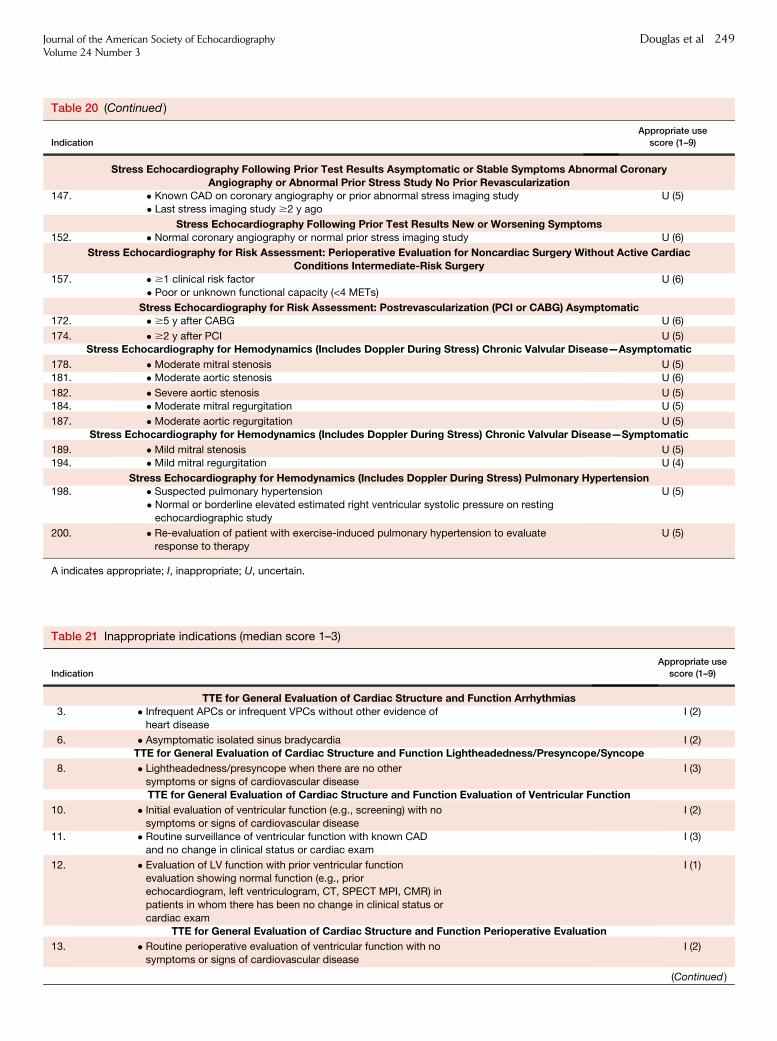
Table 20 (Continued )
Indication
Appropriate use
score (1–9)
Stress Echocardiography Following Prior Test Results Asymptomatic or Stable Symptoms Abnormal CoronaryAngiography or Abnormal Prior Stress Study No Prior Revascularization
147. � Known CAD on coronary angiography or prior abnormal stress imaging study
� Last stress imaging study $2 y ago
U (5)
Stress Echocardiography Following Prior Test Results New or Worsening Symptoms152. � Normal coronary angiography or normal prior stress imaging study U (6)
Stress Echocardiography for Risk Assessment: Perioperative Evaluation for Noncardiac Surgery Without Active CardiacConditions Intermediate-Risk Surgery
157. � $1 clinical risk factor
� Poor or unknown functional capacity (<4 METs)
U (6)
Stress Echocardiography for Risk Assessment: Postrevascularization (PCI or CABG) Asymptomatic172. � $5 y after CABG U (6)
174. � $2 y after PCI U (5)Stress Echocardiography for Hemodynamics (Includes Doppler During Stress) Chronic Valvular Disease—Asymptomatic
178. � Moderate mitral stenosis U (5)181. � Moderate aortic stenosis U (6)
182. � Severe aortic stenosis U (5)184. � Moderate mitral regurgitation U (5)
187. � Moderate aortic regurgitation U (5)
Stress Echocardiography for Hemodynamics (Includes Doppler During Stress) Chronic Valvular Disease—Symptomatic
189. � Mild mitral stenosis U (5)
194. � Mild mitral regurgitation U (4)
Stress Echocardiography for Hemodynamics (Includes Doppler During Stress) Pulmonary Hypertension198. � Suspected pulmonary hypertension
� Normal or borderline elevated estimated right ventricular systolic pressure on resting
echocardiographic study
U (5)
200. � Re-evaluation of patient with exercise-induced pulmonary hypertension to evaluateresponse to therapy
U (5)
A indicates appropriate; I, inappropriate; U, uncertain.
Table 21 Inappropriate indications (median score 1–3)
Indication
Appropriate use
score (1–9)
TTE for General Evaluation of Cardiac Structure and Function Arrhythmias3. � Infrequent APCs or infrequent VPCs without other evidence of
heart disease
I (2)
6. � Asymptomatic isolated sinus bradycardia I (2)
TTE for General Evaluation of Cardiac Structure and Function Lightheadedness/Presyncope/Syncope
8. � Lightheadedness/presyncope when there are no other
symptoms or signs of cardiovascular disease
I (3)
TTE for General Evaluation of Cardiac Structure and Function Evaluation of Ventricular Function
10. � Initial evaluation of ventricular function (e.g., screening) with no
symptoms or signs of cardiovascular disease
I (2)
11. � Routine surveillance of ventricular function with known CAD
and no change in clinical status or cardiac exam
I (3)
12. � Evaluation of LV function with prior ventricular function
evaluation showing normal function (e.g., priorechocardiogram, left ventriculogram, CT, SPECT MPI, CMR) in
patients in whom there has been no change in clinical status or
cardiac exam
I (1)
TTE for General Evaluation of Cardiac Structure and Function Perioperative Evaluation
13. � Routine perioperative evaluation of ventricular function with nosymptoms or signs of cardiovascular disease
I (2)
(Continued )
Journal of the American Society of EchocardiographyVolume 24 Number 3
Douglas et al 249

Table 21 (Continued )
Indication
Appropriate use
score (1–9)
TTE for General Evaluation of Cardiac Structure and Function Pulmonary Hypertension
16. � Routine surveillance (<1 y) of known pulmonary hypertension
without change in clinical status or cardiac exam
I (3)
TTE for Cardiovascular Evaluation in an Acute Setting Pulmonary Embolism
28. � Suspected pulmonary embolism in order to establish diagnosis I (2)30. � Routine surveillance of prior pulmonary embolism with normal
right ventricular function and pulmonary artery systolic
pressure
I (1)
TTE for Cardiovascular Evaluation in an Acute Setting Cardiac Trauma33. � Routine evaluation in the setting of mild chest trauma with no
electrocardiographic changes or biomarker elevation
I (2)
TTE for Evaluation of Valvular Function Murmur or Click35. � Initial evaluation when there are no other symptoms or signs of
valvular or structural heart disease
I (2)
36. � Re-evaluation in a patient without valvular disease on prior
echocardiogram and no change in clinical status or cardiac
exam
I (1)
TTE for Evaluation of Valvular Function Native Valvular Stenosis
38. � Routine surveillance (<3 y) of mild valvular stenosis without
a change in clinical status or cardiac exam
I (3)
40. � Routine surveillance (<1 y) of moderate or severe valvular
stenosis without a change in clinical status or cardiac exam
I (3)
TTE for Evaluation of Valvular Function Native Valvular Regurgitation42. � Routine surveillance of trace valvular regurgitation I (1)
43. � Routine surveillance (<3 y) of mild valvular regurgitation without
a change in clinical status or cardiac exam
I (2)
TTE for Evaluation of Valvular Function Prosthetic Valves
48. � Routine surveillance (<3 y after valve implantation) of prosthetic
valve if no known or suspected valve dysfunction
I (3)
TTE for Evaluation of Valvular Function Infective Endocarditis (Native or Prosthetic Valves)
53. � Transient fever without evidence of bacteremia or a new murmur I (2)
54. � Transient bacteremia with a pathogen not typically associated
with infective endocarditis and/or a documented
nonendovascular source of infection
I (3)
56. � Routine surveillance of uncomplicated infective endocarditis
when no change in management is contemplated
I (2)
TTE for Evaluation of Intracardiac and Extracardiac Structures and Chambers
60. � Routine surveillance of known small pericardial effusion with nochange in clinical status
I (2)
TTE for Evaluation of Aortic Disease
66. � Routine re-evaluation for surveillance of known ascending
aortic dilation or history of aortic dissection without a change in
clinical status or cardiac exam when findings would not changemanagement or therapy
I (3)
TTE for Evaluation of Hypertension, HF, or Cardiomyopathy Hypertension
68. � Routine evaluation of systemic hypertension without symptoms
or signs of hypertensive heart disease
I (3)
TTE for Evaluation of Hypertension, HF, or Cardiomyopathy HF
74. � Routine surveillance (<1 y) of HF (systolic or diastolic) when
there is no change in clinical status or cardiac exam
I (2)
TTE for Evaluation of Hypertension, HF, or Cardiomyopathy DeviceEvaluation (Including Pacemaker, ICD, or CRT)
79. � Routine surveillance (<1 y) of implanted device without
a change in clinical status or cardiac exam
I (1)
80. � Routine surveillance ($1 y) of implanted device without
a change in clinical status or cardiac exam
I (3)
TTE for Evaluation of Hypertension, HF, or Cardiomyopathy Cardiomyopathies
(Continued )
250 Douglas et al Journal of the American Society of EchocardiographyMarch 2011
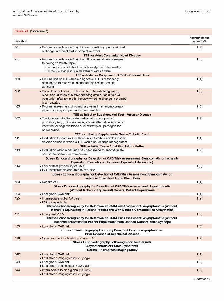
Table 21 (Continued )
Indication
Appropriate use
score (1–9)
88. � Routine surveillance (<1 y) of known cardiomyopathy without
a change in clinical status or cardiac exam
I (2)
TTE for Adult Congenital Heart Disease95. � Routine surveillance (<2 y) of adult congenital heart disease
following complete repair
+ without a residual structural or hemodynamic abnormality+ without a change in clinical status or cardiac exam
I (3)
TEE as Initial or Supplemental Test—General Uses100. � Routine use of TEE when a diagnostic TTE is reasonably
anticipated to resolve all diagnostic and management
concerns
I (1)
102. � Surveillance of prior TEE finding for interval change (e.g.,
resolution of thrombus after anticoagulation, resolution of
vegetation after antibiotic therapy) when no change in therapyis anticipated
I (2)
105. � Routine assessment of pulmonary veins in an asymptomatic
patient status post pulmonary vein isolation
I (3)
TEE as Initial or Supplemental Test—Valvular Disease107. � To diagnose infective endocarditis with a low pretest
probability (e.g., transient fever, known alternative source ofinfection, or negative blood cultures/atypical pathogen for
endocarditis)
I (3)
TEE as Initial or Supplemental Test—Embolic Event111. � Evaluation for cardiovascular source of embolus with a known
cardiac source in which a TEE would not change management
I (1)
TEE as Initial Test—Atrial Fibrillation/Flutter113. � Evaluation when a decision has been made to anticoagulate
and not to perform cardioversionI (2)
Stress Echocardiography for Detection of CAD/Risk Assessment: Symptomatic or IschemicEquivalent Evaluation of Ischemic Equivalent (Nonacute)
114. � Low pretest probability of CAD
� ECG interpretable and able to exercise
I (3)
Stress Echocardiography for Detection of CAD/Risk Assessment: Symptomatic orIschemic Equivalent Acute Chest Pain
123. � Definite ACS I (1)
Stress Echocardiography for Detection of CAD/Risk Assessment: Asymptomatic(Without Ischemic Equivalent) General Patient Populations
124. � Low global CAD risk I (1)
125. � Intermediate global CAD risk
� ECG interpretable
I (2)
Stress Echocardiography for Detection of CAD/Risk Assessment: Asymptomatic (WithoutIschemic Equivalent) in Patient Populations With Defined Comorbidities Arrhythmias
131. � Infrequent PVCs I (3)Stress Echocardiography for Detection of CAD/Risk Assessment: Asymptomatic (Without
Ischemic Equivalent) in Patient Populations With Defined Comorbidities Syncope
133. � Low global CAD risk I (3)
Stress Echocardiography Following Prior Test Results Asymptomatic:Prior Evidence of Subclinical Disease
136. � Coronary calcium Agatston score <100 I (2)
Stress Echocardiography Following Prior Test ResultsAsymptomatic or Stable SymptomsNormal Prior Stress Imaging Study
142. � Low global CAD risk
� Last stress imaging study <2 y ago
I (1)
143. � Low global CAD risk
� Last stress imaging study $2 y ago
I (2)
144. � Intermediate to high global CAD risk
� Last stress imaging study <2 y ago
I (2)
(Continued )
Journal of the American Society of EchocardiographyVolume 24 Number 3
Douglas et al 251
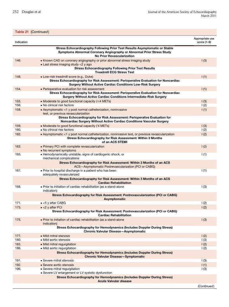
Table 21 (Continued )
Indication
Appropriate use
score (1–9)
Stress Echocardiography Following Prior Test Results Asymptomatic or StableSymptoms Abnormal Coronary Angiography or Abnormal Prior Stress Study
No Prior Revascularization
146. � Known CAD on coronary angiography or prior abnormal stress imaging study
� Last stress imaging study <2 y ago
I (3)
Stress Echocardiography Following Prior Test ResultsTreadmill ECG Stress Test
148. � Low-risk treadmill score (e.g., Duke) I (1)
Stress Echocardiography for Risk Assessment: Perioperative Evaluation for NoncardiacSurgery Without Active Cardiac Conditions Low-Risk Surgery
154. � Perioperative evaluation for risk assessment I (1)Stress Echocardiography for Risk Assessment: Perioperative Evaluation for Noncardiac
Surgery Without Active Cardiac Conditions Intermediate-Risk Surgery
155. � Moderate to good functional capacity ($4 METs) I (3)
156. � No clinical risk factors I (2)
158. � Asymptomatic <1 y post normal catheterization, noninvasive
test, or previous revascularization
I (1)
Stress Echocardiography for Risk Assessment: Perioperative Evaluation forNoncardiac Surgery Without Active Cardiac Conditions Vascular Surgery
159. � Moderate to good functional capacity (’4 METs) I (3)160. � No clinical risk factors I (2)
162. � Asymptomatic <1 y post normal catheterization, noninvasive test, or previous revascularization I (2)
Stress Echocardiography for Risk Assessment: Within 3 Monthsof an ACS STEMI
163. � Primary PCI with complete revascularization
� No recurrent symptoms
I (2)
165. � Hemodynamically unstable, signs of cardiogenic shock, ormechanical complications
I (1)
Stress Echocardiography for Risk Assessment: Within 3 Months of an ACSACS—Asymptomatic Postrevascularization (PCI or CABG)
167. � Prior to hospital discharge in a patient who has been
adequately revascularized
I (1)
Stress Echocardiography for Risk Assessment: Within 3 Months of an ACSCardiac Rehabilitation
168. � Prior to initiation of cardiac rehabilitation (as a stand-alone
indication)
I (3)
Stress Echocardiography for Risk Assessment: Postrevascularization (PCI or CABG)Asymptomatic
171. � <5 y after CABG I (2)
173. � <2 y after PCI I (2)
Stress Echocardiography for Risk Assessment: Postrevascularization (PCI or CABG)Cardiac Rehabilitation
175. � Prior to initiation of cardiac rehabilitation (as a stand-aloneindication)
I (3)
Stress Echocardiography for Hemodynamics (Includes Doppler During Stress)Chronic Valvular Disease—Asymptomatic
177. � Mild mitral stenosis I (2)
180. � Mild aortic stenosis I (3)
183. � Mild mitral regurgitation I (2)
186. � Mild aortic regurgitation I (2)
Stress Echocardiography for Hemodynamics (Includes Doppler During Stress)Chronic Valvular Disease—Symptomatic
191. � Severe mitral stenosis I (3)
192. � Severe aortic stenosis I (1)196. � Severe mitral regurgitation
� Severe LV enlargement or LV systolic dysfunction
I (3)
Stress Echocardiography for Hemodynamics (Includes Doppler During Stress)Acute Valvular disease
(Continued )
252 Douglas et al Journal of the American Society of EchocardiographyMarch 2011

print&
web4C=FPO
Figure 1 Stress echocardiography for detection of CAD/Risk assessment: Symptomatic or ischemic equivalent.
Table 21 (Continued )
Indication
Appropriate use
score (1–9)
197. � Acute moderate or severe mitral or aortic regurgitation I (3)
Stress Echocardiography for Hemodynamics (Includes Doppler During Stress)Pulmonary Hypertension
199. � Routine evaluation of patients with known resting pulmonary
hypertension
I (3)
Contrast Use in TTE/TEE or Stress Echocardiography201. � Routine use of contrast
� All LV segments visualized on noncontrast images
I (1)
A indicates appropriate; I, inappropriate; U, uncertain.
Journal of the American Society of EchocardiographyVolume 24 Number 3
Douglas et al 253
8. DISCUSSION
Appropriate use criteria define patient subgroups where the availablemedical evidence supplemented by expert opinion are combined toassess whether the net benefit or risks of a test or procedure make itreasonable to perform testing (in this document, echocardiography)in a particular clinical situation. The intent of these criteria is to guidethe rational use of a procedure, namely avoidance of either under- orover-utilization, and thereby lead to improved outcomes, more opti-mal healthcare delivery, and justifiable healthcare expenditures.
This document is a revision and combination of the original AUCfor transthoracic and transesophageal echocardiography (1) and stressechocardiography (2). The revision adds insight provided by interimclinical data and standards documents recently published in the liter-ature and clarifies areas in which omissions or lack of clarity existed inthe original criteria. Additionally, since publication of the originalAUC, several studies have assessed the application of these criteriain clinical practice; results from these studies were incorporated intothis revision and will be briefly summarized here.
Implementation Studies
Application of the 2007AUC for TTE has been evaluated at academicmedical centers (22,24–26), in Veterans Affairs (VA) hospitals (27),and in community settings (28,29). Several common themes
deserve emphasis. First, the majority of clinical scenarios for whichTTEs were ordered were captured by AUC indications (11% to 16%of TTEs were unclassified) (24,27). Second, across theimplementation studies, there are remarkably similar rates ofappropriate and inappropriate use of TTE. Among those TTEs withan indication addressed by the AUC (thus removing unclassifiablepatients), the majority were rated as appropriate (87% to 91%) andthe rate of inappropriate TTEs was consistently low (9% to 13%)(24–27). In 1 study of outpatient TTEs (29), the rate of appropriateTTEs was lower (74%), although this may be attributable to a higherproportion of unclassified studies in the outpatient setting, a patternthat has been observed by others (24,26). The presence of a greaterproportion of unclassified TTEs in the outpatient setting might beexpected given that many of the indications in the original AUC (1)specifically address symptoms or a ‘‘change in clinical status.’’
The most common appropriate indications for TTE included initialevaluation of symptoms potentially caused by suspected cardiac etiol-ogy, prior testing concerning for heart disease, evaluation of valvulardisease, and evaluation of a heart failure indication (24) and arerepeated in this revision as Indications 1, 2, 34, and 70.Recommendations for expanding the AUC related to addressing 1)perioperative evaluation (Indications 13 and 14); 2) timing of followup for valvular heart disease (Indications 38 to 41 and 43 to 49); 3) as-sessment for device therapy (Indications 76 to 83); and 4) use in somespecialized care or ‘‘niche’’ programs (e.g., solid organ transplantation)

print&
web4C=FPO
Figure 2 Stress echocardiography for detection of CAD/Risk assessment: Asymptomatic (without ischemic equivalent).
print&
web4C=FPO
Figure 3 Stress echocardiography following prior treadmill ECG, coronary calcium scoring, or carotid intimal medial thickness testresults.
254 Douglas et al Journal of the American Society of EchocardiographyMarch 2011
(Indications 14, 84, and 85), and these scenarios were included in thecurrent document. Finally, more indications reflecting outpatient clini-cal scenarios (e.g., no change in clinical status) were added.
Studies evaluating the application of AUC for TEE had similar re-sults, with the vast majority of classifiable TEEs being ordered for ap-propriate indications (94% to 97%) and a smaller number not beingclassified by the AUC (6% to 9%) (30–32). The fact that the operatoris more intimately involved in the decision to perform TEE may helpto explain the higher appropriate use rate of TEE compared with TTE.
The most common indication for an initial TEE was to guideanticoagulation decisions in patients with atrial fibrillation or flutter(Indications 112 and 113) (30,31). Recommendations for revisionfocused on refinement of the indications for evaluation ofcardiovascular source of embolus (Indications 109 to 111).
Fewer studies have focused on the clinical application of AUC forstress echocardiography (33,34). In 1 study, 19% of stressechocardiograms could not be classified by the AUC (33). Of theechocardiograms that were classified, 66% were for appropriate

print&
web4C=FPO
Figure 4 Stress echocardiography following prior stress imaging or coronary angiogram test results.
print&
web4C=FPO
Figure 5 Stress echocardiography for risk assessment—perioperative evaluation for noncardiac surgery without active cardiac con-ditions.
Journal of the American Society of EchocardiographyVolume 24 Number 3
Douglas et al 255
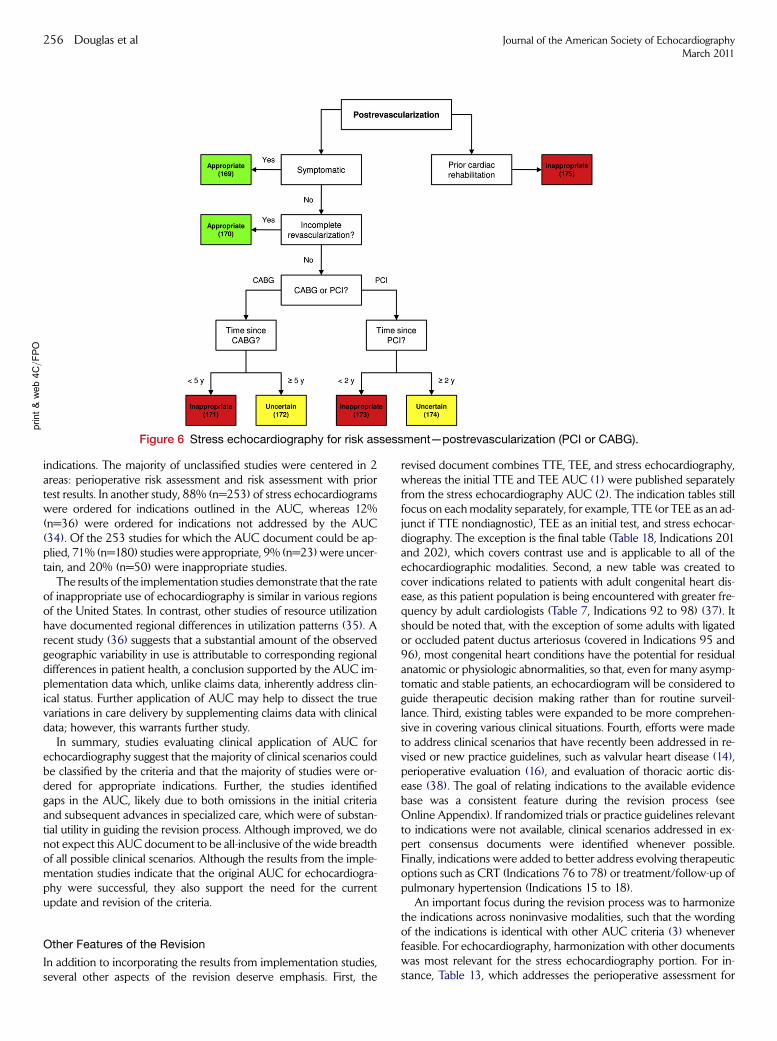
print&web4C=FPO
Figure 6 Stress echocardiography for risk assessment—postrevascularization (PCI or CABG).
256 Douglas et al Journal of the American Society of EchocardiographyMarch 2011
indications. The majority of unclassified studies were centered in 2areas: perioperative risk assessment and risk assessment with priortest results. In another study, 88% (n=253) of stress echocardiogramswere ordered for indications outlined in the AUC, whereas 12%(n=36) were ordered for indications not addressed by the AUC(34). Of the 253 studies for which the AUC document could be ap-plied, 71% (n=180) studies were appropriate, 9% (n=23) were uncer-tain, and 20% (n=50) were inappropriate studies.
The results of the implementation studies demonstrate that the rateof inappropriate use of echocardiography is similar in various regionsof the United States. In contrast, other studies of resource utilizationhave documented regional differences in utilization patterns (35). Arecent study (36) suggests that a substantial amount of the observedgeographic variability in use is attributable to corresponding regionaldifferences in patient health, a conclusion supported by the AUC im-plementation data which, unlike claims data, inherently address clin-ical status. Further application of AUC may help to dissect the truevariations in care delivery by supplementing claims data with clinicaldata; however, this warrants further study.
In summary, studies evaluating clinical application of AUC forechocardiography suggest that the majority of clinical scenarios couldbe classified by the criteria and that the majority of studies were or-dered for appropriate indications. Further, the studies identifiedgaps in the AUC, likely due to both omissions in the initial criteriaand subsequent advances in specialized care, which were of substan-tial utility in guiding the revision process. Although improved, we donot expect this AUC document to be all-inclusive of the wide breadthof all possible clinical scenarios. Although the results from the imple-mentation studies indicate that the original AUC for echocardiogra-phy were successful, they also support the need for the currentupdate and revision of the criteria.
Other Features of the Revision
In addition to incorporating the results from implementation studies,several other aspects of the revision deserve emphasis. First, the
revised document combines TTE, TEE, and stress echocardiography,whereas the initial TTE and TEE AUC (1) were published separatelyfrom the stress echocardiography AUC (2). The indication tables stillfocus on eachmodality separately, for example, TTE (or TEE as an ad-junct if TTE nondiagnostic), TEE as an initial test, and stress echocar-diography. The exception is the final table (Table 18, Indications 201and 202), which covers contrast use and is applicable to all of theechocardiographic modalities. Second, a new table was created tocover indications related to patients with adult congenital heart dis-ease, as this patient population is being encountered with greater fre-quency by adult cardiologists (Table 7, Indications 92 to 98) (37). Itshould be noted that, with the exception of some adults with ligatedor occluded patent ductus arteriosus (covered in Indications 95 and96), most congenital heart conditions have the potential for residualanatomic or physiologic abnormalities, so that, even for many asymp-tomatic and stable patients, an echocardiogram will be considered toguide therapeutic decision making rather than for routine surveil-lance. Third, existing tables were expanded to be more comprehen-sive in covering various clinical situations. Fourth, efforts were madeto address clinical scenarios that have recently been addressed in re-vised or new practice guidelines, such as valvular heart disease (14),perioperative evaluation (16), and evaluation of thoracic aortic dis-ease (38). The goal of relating indications to the available evidencebase was a consistent feature during the revision process (seeOnline Appendix). If randomized trials or practice guidelines relevantto indications were not available, clinical scenarios addressed in ex-pert consensus documents were identified whenever possible.Finally, indications were added to better address evolving therapeuticoptions such as CRT (Indications 76 to 78) or treatment/follow-up ofpulmonary hypertension (Indications 15 to 18).
An important focus during the revision process was to harmonizethe indications across noninvasive modalities, such that the wordingof the indications is identical with other AUC criteria (3) wheneverfeasible. For echocardiography, harmonization with other documentswas most relevant for the stress echocardiography portion. For in-stance, Table 13, which addresses the perioperative assessment for

Journal of the American Society of EchocardiographyVolume 24 Number 3
Douglas et al 257
noncardiac surgery, mirrors Table 4 in the RNI document (3). Thisshould facilitate clinical application of the criteria and assist the pro-cess of future revisions and possibly the development of amultimodal-ity imaging AUC document.
Stress echocardiography tests, like many imaging tests, may pro-vide additional useful information beyond the primary purpose out-lined by the indication. In addition, stress echocardiography doesnot use ionizing radiation. However, the AUC for stress echocardiog-raphy were not developed to quantify the incremental information orother test characteristics beyond addressing the diagnostic need inher-ent in an individual indication.
In ranking indications, panelists were asked to not consider com-parisons to other imaging procedures while completing their rankings.Nevertheless, stress echocardiography and SPECT MPI have similarbodies of evidence to support their use. Therefore, it is not surprisingthat the overwhelming majority of final ratings of stress echocardiog-raphy and stress RNI were concordant for similar clinical indications.However, a small number of the final scores and rating categories re-ported in this document differ from those previously published forstress RNI (3). Specifically, 4 indications (Indications 127, 157, 171,and 172) were rated differently. It is noteworthy that of these 4 indi-cations, 3 also appeared in the first stress echocardiography AUC (2),and all 3 indications were rated similarly in this revision, requiring con-sistency in ratings across the 2 technical panels composed of differentindividuals. The difference in the rating for Indication 127 may havebeen directly affected by publication of the DIAD study (39), whichwas not available at the time of the RNI ratings. Additionally, althoughthe final rankings were different from the RNI ratings, Indications 127and 171 demonstrated agreement within the current echocardiogra-phy technical panel. Therefore, the several indications with ratingsthat differed from RNI may reflect new literature that has becomeavailable since publication of the SPECT appropriateness criteriaand differences in the composition of the 2 panels.
Readers should also note that the categorical summaries tend to ac-centuate differences that sometimes are slight. For example, smallfluctuations in a median rating (e.g., 4 versus 3) will cause an indica-tion to switch appropriateness categories (e.g., from uncertain to inap-propriate). This phenomenon was relevant for Indication 157, whichwas rated as uncertain (median score 6) in this document, while thesame indication in the RNI document (corresponding Indication43) was rated appropriate (median score 7). The most likely reasonfor this is a simple variation in rating by the different panel members,whether because of composition, different levels of clinical experi-ence, publication of additional literature, or different interpretationsof data. The AUC Task Force has carefully examined the issue ofpanel membership and made every effort to ensure similar composi-tion for each panel. The RAND process has documented that the in-terpretation of the literature by different sets of experts can yieldslightly different final ratings (6).
As described in the Methods section, within each main disease cat-egory, a standardized approach was used in order to capture the ma-jority of clinical scenarios without making the list of indicationsexcessive. The approach was to create 5 broad clinical scenarios: 1)for initial diagnosis; 2) to guide therapy or management, regardlessof symptom status; 3) to evaluate a change in clinical status or cardiacexam; 4) for early follow-up without change in clinical status; and 5)for late follow-up without change in clinical status. It should be notedthat many cardiovascular conditions have the potential for residualanatomic or physiologic abnormalities, so that the timing and fol-low-up use of echocardiographic imaging depends on the patient’sclinical status and the magnitude of or risk for residual abnormalities.
Thus, routine surveillance indications for echocardiograms should notapply in those situations in which there has been a change in status orwhere an echocardiogram is being considered to guide therapeuticdecision making. For asymptomatic or stable patients with knownor suspected residual anatomic or physiologic abnormalities, the tim-ing of the follow-up for considering changes in therapy in patientsshould be determined by individual patient factors, and not by thesuggested intervals for routine surveillance studies.
Overall, indications focusing on initial diagnosis, guidance of ther-apy, or evaluation of a change in clinical status were viewed favorablyby the rating panel. Uncertain or inappropriate ratings were morelikely given to early rather than late follow-up, especially for thoseindications when the optimal interval of follow-up for asymptomaticpatients is uncertain. Whenever possible, indications for timing of fol-low-up attempted to follow practice guidelines (14), although formany indications, the most appropriate follow-up interval for asymp-tomatic patients is not well established. For this reason, as well as forclinical expediency, the follow-up interval selected is not meant to berigid but rather to represent an approximate time interval.
Although the overall approach was broad and inclusive, certainspecific clinical scenarios warranted focused indications based on re-sults from the previously mentioned implementation studies.Examples include Indications 71 and 72, which differentiate the re-evaluation of decompensated heart failure when there is no clear pre-cipitating change in medication or diet versus when there is a clearprecipitating factor. In the setting of an obvious change in diet or med-ication, a trial of appropriate medical therapy and monitoring for clin-ical improvement may be justified prior to ordering a repeat imagingtest for assessment of cardiac function (25). As such, Indication 72(clear precipitating change in medication or diet) was rated as uncer-tain, and Indication 71 was rated as appropriate. Another focusedclinical situation is reflected in Indication 76, ‘‘Initial evaluation orre-evaluation after revascularization and/or optimal medical therapyto determine candidacy for device therapy and/or to determine opti-mal choice of device.’’ As per the results of an implementation study(24), this clinical scenario was not well captured in the initial AUCdocument. However, re-evaluation of LVejection fraction after revas-cularization or after a period of medical therapy to determine devicecandidacy represents a standard of care (40) and is a common indica-tion for a TTE. This is now represented by Indication 76, which wasrated as appropriate.
Other specific areas identified by implementation studies as com-mon scenarios and now included are bradycardia (Indication 6) anda new subcategory within TTE for the evaluation of syncope(Indications 7 to 9). Additionally, the sections on valvular heart dis-ease (both resting TTE/TEE and stress echocardiography for hemody-namics) have been expanded in an effort to address a greater numberof clinical scenarios, and closely follow recent guideline recommenda-tions (14).
Despite these extensive revisions and additions, all potential clini-cal scenarios were not covered by the revised AUC for echocardiog-raphy. Additionally, certain recommendations from implementationstudies were considered to represent rare conditions or specializedpractices and were therefore not included in the revised document.If certain clinical situations that are not currently covered are foundto be more frequent than anticipated, they will be incorporatedinto future revisions. This emphasizes the iterative nature of this pro-cess.
Furthermore, there are several general categories that were pur-posefully not addressed. For example, intraoperative use of TEE forcardiac surgery was felt to be beyond the scope of this document.

258 Douglas et al Journal of the American Society of EchocardiographyMarch 2011
More highly specialized echocardiographic techniques, such as 3-di-mensional echocardiography or epicardial imaging, are not addressedin this document. Additionally, as stated in the first paragraph of theAssumptions section, the AUC for TTE, TEE, and stress echocardiog-raphy are for adult patients. Indications for pediatric echocardiogramswere not covered.
New Assumptions and Definition
In addition to adding new clinical indications and clarifying existing in-dications from the original TTE/TEE AUC (1) and stress echocardiog-raphy AUC (2), the writing group also revised and added specificassumptions and definitions. Several general assumptions wereadded. First, the assumption that cost should be implicitly consideredin determining appropriate use of an echocardiogram was added.Second, a new assumption addresses the category of uncertain indica-tions and clarifies that such a rating should not be considered groundsfor withholding reimbursement. Third, a new assumption indicatesthat appropriateness ratings reflect whether a specific test is appropri-ate for a given patient, not whether it is preferred over another modal-ity (e.g., RNI, CT). Thus, the AUC should not be used to provideclinical support for administrative policies regarding test preferences.Finally, an assumption clarifies that routine or surveillance echocar-diograms represent a ‘‘periodic’’ evaluation after a certain period oftime has elapsed, and are not being ordered because of any other clin-ical factors. Other more specific assumptions were also added. Theseinclude consideration of prosthetic and native valves together (unlessotherwise specified) and that use of Doppler for hemodynamics in-cludes assessment of both right and left heart hemodynamics.Furthermore, it is assumed that if a perioperative patient has symp-toms or signs of cardiovascular disease, the study should be classifiedunder a symptomatic indication (e.g., Indication 1), as opposed to anindication in the perioperative category.
Similar to the RNI AUC (3), the writing group revised the definitionof ‘‘chest pain syndrome’’ and adopted the term ‘‘ischemic equivalent,’’which encompasses chest pain syndromes as well as other symptomsand signs that the clinician believes may be attributable to CAD. Thewriting group also adopted the use of global risk assessment when as-sessing risk in asymptomatic patients (41). This revision was supportedby the writing group, technical panel, and external reviewers and is inharmony with the most recent AUC for Cardiac CT (4).
Limitations
The ratings of the indications as appropriate, uncertain, or inappropri-ate are reflective of the body of knowledge at the time the rating pro-cess occurred. It is likely and expected that as science progresses andnew evidence-based guidelines are published, certain indications thatare given 1 ratingmay subsequently be determined to have a differentappropriateness rating in the future. Although this necessarily reflectsthe evolving nature of medical science, it may also introduce apparentdiscrepancies between appropriateness of similar indications for dif-ferent modalities evaluated at different time points. The current evi-dence base and practice guidelines were used to develop theindications whenever available, although for certain indications the lit-erature was limited and clinical expertise played a larger role. This isconsistent with the standard methodology and principles of evi-dence-based medicine as endorsed by the Physician Consortiumfor Performance Improvement (42). Additionally, as mentioned inthe previous text, certain clinical scenarios were intentionally not cov-ered by the indications. When future implementation studies evaluat-
ing this revised AUC for echocardiography are conducted, it maybecome apparent that frequent situations were not covered. As wasthe case for this current revision, results and recommendationsfrom implementation studies will help shape future modifications tothe AUC.
Use of AUC to Improve Care
TheAUC in this report provide an estimate of whether it is reasonableto use echocardiography for a particular clinical scenario, specificallyfor 1 of the 202 indications listed in this document. These criteria areexpected to be useful for clinicians, healthcare facilities, and third-party payers engaged in the delivery of cardiovascular imaging. TheAUC is expected to be valuable across a broad range of situations, in-cluding guiding care of individual patients, educating caregivers, andinforming policy decisions regarding cardiovascular imaging.
AUC represent the first component of the chain of quality domainsfor cardiovascular imaging (43). After ensuring proper test selection,the achievement of quality in imaging includes adherence to bestpractices in image acquisition, image interpretation and results com-munication, as well as incorporation of findings into clinical care.All components are important for optimal patient care, althoughthe development of AUC and their ranking by the technical panelis intended to address only the first quality domain, and assumes nobarriers to other quality standards are being met.
Although these criteria are intended to provide guidance for caredecisions, they cannot serve as substitutes for sound clinical judgmentand practice experience. The writing group recognizes that patientsencountered in clinical practice may not be represented in theseAUC ormay have extenuating features when comparedwith the clin-ical scenarios presented. Additionally, uncertain indications often re-quire individual physician judgment and an in-depth understandingof the patient to better determine the usefulness of a test for a partic-ular scenario. As such, the ranking of an indication as uncertain (4 to6) should not be viewed as limiting the use of echocardiography forsuch patients. It should be emphasized that the technical panel wasinstructed that the ‘‘uncertain’’ designation was still designed to beconsidered as a ‘‘reimbursable’’ category.
These ratings reflect the critical medical literature as well as expertconsensus and are intended to evaluate the appropriate use of specificpatient scenarios to determine overall patterns of care regarding echo-cardiography. In situations where there is substantial variation be-tween the appropriate use rating and what the clinician believes isthe best recommendation for the patient, further considerations or ac-tions, such as a second opinion, may be appropriate. Moreover, it isneither anticipated nor desirable that all physicians or facilities willhave 100% of their echocardiograms deemed appropriate.However, it is desirable, though not realistic, that 0% be inappropri-ate. Related to the overall patterns of care, if the national average ofappropriate and uncertain ratings is 80%, for example, and a physicianor facility has a 40% rate of inappropriate procedures, further exam-ination of the patterns of care may be warranted and helpful. The useof AUC to guide clinical decision making and its impact on patientoutcomes and healthcare quality/efficiency needs to be studied rigor-ously. AUC are also useful as educational tools for both echocardiog-raphy providers and referring physicians. The recently announcedand soon to be implemented incorporation of AUC into echocardiog-raphy laboratory accreditation requirements will encourage their use(44). However, the greatest opportunity to optimize the use of echo-cardiography is in improving individual patient decision making. Thesuccessful application of AUC into clinical practice represents an im-portant area of ongoing quality improvement.

Journal of the American Society of EchocardiographyVolume 24 Number 3
Douglas et al 259
APPENDIX A: ADDITIONAL ECHOCARDIOGRAPHY
DEFINITIONS
1. Angina
� Typical Angina (Definite): Defined as 1) substernal chest pain or dis-comfort that is 2) provoked by exertion or emotional stress and 3) relievedby rest and/or nitroglycerin (45).
� Atypical Angina (Probable): Chest pain or discomfort that lacks 1 ofthe characteristics of definite or typical angina.
� NonanginalChest Pain:Chest pain or discomfort thatmeets 1 or noneof the typical angina characteristics.
2. Acute Coronary Syndrome (ACS)
As defined by the ACC/AHA Guidelines for the Management ofPatients with ST-Elevation Myocardial Infarction: patients with anACS include those whose clinical presentations cover the followingrange of diagnoses: unstable angina, myocardial infarction withoutST-segment elevation (NSTEMI), and myocardial infarction with ST-segment elevation (STEMI) (46).
3. Evaluating Perioperative Risk for Noncardiac Surgery
Method for Determining Perioperative Risk. See Figure A1,‘‘Stepwise Approach to Perioperative Cardiac Assessment,’’ from
Figure A1 Stepwise approach to perioperative cardiac assessmenCardiac evaluation and care algorithm for noncardiac surgery basedcardiac risk factors for patients $50 years of age. HR indicates heaModified from (16).
the ACCF/AHA guidelines on perioperative cardiovascular evalua-tion and care for noncardiac surgery (16). Based on the algorithm,once it is determined that the patient does not require urgent surgery,the clinician should determine the patient’s active cardiac conditions(see Table A1) and/or perioperative risk predictors (see Table A2). Ifany active cardiac conditions and/or major risk predictors are present,Figure A1 suggests consideration of coronary angiography and post-poning or canceling noncardiac surgery. Once perioperative risk pre-dictors are assessed based on the algorithm, then the surgical risk andpatient’s functional status should be used to establish the need fornoninvasive testing.
4. Thrombolysis In Myocardial Infarction (TIMI) Risk Scores
The TIMI risk score (48) is a simple tool composed of 7 (1-point) riskindicators rated on presentation. The composite end points (all-causemortality, new or recurrent MI, or severe recurrent ischemia prompt-ing urgent revascularization within 14 days) increase as the TIMI riskscore increases. The model remained a significant predictor of eventsand test sensitivity and was relatively unaffected/uncompromised bymissing information, such as knowledge of previously documentedcoronary stenosis of $50%. The model’s predictive ability remainedintact with a cutoff of 65 years of age.
The TIMI risk score is determined by the sum of the presence of 7variables at admission; 1 point is given for each of the following
t.on active clinical conditions, known cardiovascular disease, orrt rate; LOE, level of evidence; and MET, metabolic equivalent.

Table A1 Active cardiac conditions for which the patientshould undergo evaluation and treatment before noncardiacsurgery (class I, level of evidence: B)
Condition Examples
Unstable coronary syndromes Unstable or severe angina* (CCS
class III or IV)†
Recent MI‡
Decompensated HF (NYHAfunctional class IV; worsening
or new-onset HF)
Significant arrhythmias High-grade atrioventricular block
Mobitz II atrioventricular block
Third-degree atrioventricularheart block
Symptomatic ventriculararrhythmias
Supraventricular arrhythmias
(including atrial fibrillation) withuncontrolled ventricular rate
(HR >100 bpm at rest)
Symptomatic bradycardia
Newly recognized ventricular
tachycardia
Severe valvular disease Severe aortic stenosis (mean
pressure gradient >40 mm Hg,aortic valve area <1.0 cm2, or
symptomatic)
Symptomatic mitral stenosis(progressive dyspnea on
exertion, extertional
presyncope, or HF)
CCS indicates Canadian Cardiovascular Society; HF, heart failure;
HR, heart rate; MI, myocardial infarction; and NYHA, New York HeartAssociation.
*According to Campeau (47).†May include ‘‘stable’’ angina in patients who are unusually sedentary.‡The American College of Cardiology National Database Library de-
fines recent MI as >7 days but #1 month (within 30 days). Reprinted
from Fleisher et al. (16).
Table A2 Perioperative clinical risk factors*
� History of ischemic heart disease
� History of compensated or prior heart failure
� History if cerebrovascular disease
� Diabetes mellitus (requiring insulin)
� Renal insufficiency (creatinine >2.0)
*As defined by the 2009 ACCF/AHA Focused Update on Periopera-
tive Beta Blockade Incorporated Into the ACC/AHA 2007Guidelines on
Perioperative Cardiovascular Evaluation and Care for Noncardiac Sur-gery (16). Note that these are not standard coronary artery disease risk
factors.
260 Douglas et al Journal of the American Society of EchocardiographyMarch 2011
variables: age $65 years, at least 3 risk factors for CAD, prior coro-nary stenosis of $50%, ST-segment deviation on ECG presentation,at least 2 anginal events in prior 24 hours, use of aspirin in prior 7days, and elevated serum cardiac biomarkers.
Low-Risk TIMI Score: TIMI score <2High-Risk TIMI Score: TIMI score $2
5. ECG–Uninterpretable
Refers to ECGs with resting ST-segment depression ($0.10 mV),complete LBBB, pre-excitation (Wolff-Parkinson-White Syndrome),or paced rhythm.
6. Coronary Angiography
The term coronary angiography refers to invasive cardiac catheteriza-tion or to established noninvasive methods of imaging the coronaryarteries, such as coronary CT angiography.
APPENDIX B: ADDITIONAL METHODS
See the Methods section of the report for a description of panelselection, indication development, scope of indications, and ratingprocess.
Relationships With Industry and Other Entities
A list of all individuals participating in the development and reviewof this document and their institutional and/or organizational affili-ations is presented in Appendix C. The American College ofCardiology Foundation and its partnering organizations rigorouslyavoid any actual, perceived, or potential conflicts of interest thatmight arise as a result of an outside relationship or personal interestof a member of the technical panel. Specifically, all panelists areasked to provide disclosure statements of all relationships that mightbe perceived as real or potential conflicts of interest. These state-ments were reviewed by the Appropriate Use Criteria Task Force,discussed with all members of the technical panel at the face-to-face meeting, and updated and reviewed as necessary. A table ofdisclosures by the technical panel and oversight working groupmember can be found in Appendix D. In addition, to ensure com-plete transparency, complete disclosure information—including rela-tionships not pertinent to this document—is available online asa document supplement.
Literature Review
The technical panel members were asked to refer to the relevant lit-erature provided for each indication table when completing their rat-ings (see Online Appendix).
APPENDIX C: ACCF/ASE/AHA/ASNC/HFSA/HRS/SCAI/
SCCM/SCCT/SCMR 2011
Appropriate Use Criteria for Echocardiography Participants
Echocardiography Writing GroupPamela S. Douglas, MD, MACC, FAHA, FASE–Chair, Appropriate
Use Criteria for Echocardiography, Past President, American Collegeof Cardiology Foundation; Past President American Societyof Echocardiography; and Ursula Geller Professor of Researchin Cardiovascular Diseases, Duke University Medical Center,Durham, NC
Mario J. Garcia, MD, FACC, FACP–Professor of Medicine,Montefiore Medical Center and Albert Einstein College ofMedicine, Bronx, NY

Appendix C ACCF/ASE/AHA/ASNC/HFSA/HRS/SCAI/SCCM/SCCT/SCMR 2011 Appropriate Use Criteria for EchocardiographyWriting Group, Technical Panel, Indication Reviewers, and Task Force–Relationships With Industry and Other Entities(in alphabetical order within each group)
Participant Consultant Speaker
Ownership/
Partnership/
Principal Research
Institutional,
Organizational, or
Other Financial Benefit
Expert
Witness
Echocardiography Appropriate Use Criteria Writing GroupPamela S. Douglas None None None None None None
Mario J. Garcia None None None None None None
David E. Haines None None None None None None
Wyman W. Lai None None None None None None
Warren J. Manning , Lantheus
Medical Imaging
None None , Philips Medical
Systems
None None
Ayan R. Patel None None None None None None
Michael H. Picard None None None , Edwards
Lifesciences
None None
Donna M. Polk None None None None None NoneMichael Ragosta None None None None None None
R. Parker Ward None None None None None NoneRory B. Weiner None None None None None None
Echocardiography Appropriate Use Criteria Technical PanelSteven R. Bailey None None None None None None
Rory B. Weiner None None None None None NonePeter Alagona, Jr None None None None None None
Jeffrey L. Anderson None None None , Toshiba None NoneJeanne M. DeCara None None None None None None
Rowena J. Dolor None None None None None NoneReza Fazel None None None None None None
John A. Gillespie None None None None None NonePaul A. Heidenreich None None None None None None
Luci K. Leykum None None None None None None
Joseph E. Marine None None None None None None
Gregory J. Mishkel None None None None None None
Patricia A. Pellikka None None None None None None
Gilbert L. Raff None None None None None None
Krishnaswami Vijayaraghavan None None None None None None
Neil J. Weissman None None None None None None
Katherine C. Wu None None None None None None
Echocardiography Appropriate Use Criteria Indication ReviewersDrew Baldwin None None None None , Veterans Affairs None
Thomas Behrenbeck None None None None None None
Michael Blaivas None None None None None None
Jeanne M. DeCara None None None None None None
Peter L. Duffy None None None None None None
Kirsten E. Fleischmann None None None None None None
Shawn A. Gregory None None None None None None
Frederick G. Kushner None None None None None None
John Lesser , Vital Images , Siemens Medical
Systems
None None None None
Alexander B. Levitov • Lantheus MedicalImaging
None None None None None
Kapildeo Lotun None None None None None None
John V. Nixon None None None None None NoneDavid T. Porembka None None None None None None
Brian D. Powell None None None None None None
Subha Raman None None None None None None
Gregory S. Thomas None None None None None None
James D. Thomas None None None None None None
(Continued )
Journal of the American Society of EchocardiographyVolume 24 Number 3
Douglas et al 261

Appendix C (Continued )
Participant Consultant Speaker
Ownership/
Partnership/
Principal Research
Institutional,
Organizational, or
Other Financial Benefit
Expert
Witness
Aseem Vashist None None None None None None
Mary N. Walsh None None None None None None
Carole A. Warnes None None None None None None
Joseph N. Wight, Jr None None None None None None
Katherine C. Wu None None None None None None
Echocardiography Appropriate Use Criteria Task Force
Michael J. Wolk None None None None None None
Steven R. Bailey None None None None None None
Pamela S. Douglas None None None None None None
Robert C. Hendel None None None None None None
Christopher M. Kramer None None None None None None
James K. Min None , General Electric
Healthcare
None None None None
Manesh R. Patel • Genzyme None None None None None
Leslee Shaw None None None None None None
Raymond F. Stainback None None None None None None
Joseph M. Allen None None None None None None
This table represents the relevant relationships with industry and other entities that were disclosed by participants at the time of participation. It
does not necessarily reflect relationships with industry at the time of publication. A person is deemed to have a significant interest in a businessif the interest represents ownership of 5% or more of the voting stock or share of the business entity, or ownership of $10 000 or more of the fair
market value of the business entity; or if funds received by the person from the business entity exceed 5% of the person’s gross income for the
previous year. A relationship is considered to be modest if it is less than significant under the preceding definition. Relationships in this table are
modest unless otherwise noted. Names are listed in alphabetical order within each category of review. Participation does not imply endorsement ofthis document.
*Significant relationship.
262 Douglas et al Journal of the American Society of EchocardiographyMarch 2011
David E. Haines, MD, FACC, FHRS–Professor of CardiovascularMedicine, Oakland University, William Beaumont School ofMedicine, Chairman, Department of Cardiovascular Medicine,Beaumont Hospitals, Royal Oak, MI
Wyman W. Lai, MD, MPH, FACC, FASE–Morgan StanleyChildren’s Hospital of NY, Pediatric Cardiology, Director,Noninvasive Cardiac Imaging Laboratory, New York, NY
Warren J. Manning, MD, FACC–Professor of Medicine andRadiology, Harvard Medical School, Beth Israel Deaconess MedicalCenter, Cardiovascular Division, Boston, MA
Ayan R. Patel, MD, FACC–Director, Cardiovascular Imaging &Hemodynamic Laboratory and Physician, Heart Failure & CardiacTransplant Center, Tufts Medical Center; Associate Professor ofMedicine, Tufts University School of Medicine, Boston, MA
Michael H. Picard, MD, FACC, FASE, FAHA–Director,Echocardiography, Massachusetts General Hospital, AssociateProfessor of Medicine, Harvard Medical School, Boston, MA
Donna M. Polk, MD, MPH, FACC, FASE, FASNC–Director,Preventative Cardiology, Hartford Hospital, Hartford, CT
Michael Ragosta, MD, FACC, FSCAI–Professor of Medicine/Cardiology, Director, Cardiac Catheterization Laboratories,Department of Medicine, Division of Cardiovascular Medicine,University of Virginia Health System, Charlottesville, VA
R. Parker Ward, MD, FACC, FASE, FASNC–Associate Professor ofMedicine, Director of Cardiovascular Fellowship Program, Universityof Chicago Medical Center, Chicago, IL
Rory B. Weiner, MD–Clinical and Research Fellow, HarvardMedical School, Massachusetts General Hospital, CardiologyDivision, Boston, MA
Echocardiography Technical Panel
Steven R. Bailey, MD, FACC, FSCAI, FAHA–Moderator of theTechnical Panel, Chair, Division of Cardiology, Professor ofMedicine and Radiology, Janey Briscoe Distinguished Chair,University of Texas Health Sciences Center, San Antonio, TX
Rory B. Weiner, MD–Writing Group Liaison Appropriate UseCriteria for Echocardiography Technical Panel, Clinical andResearch Fellow, Harvard Medical School, Massachusetts GeneralHospital, Cardiology Division, Boston, MA
Peter Alagona, Jr, MD, FACC–Program Director GeneralCardiology, Penn State Heart and Vascular Institute, Hershey, PA
Jeffrey L. Anderson, MD, FACC, FAHA, MACP–Professor ofMedicine, University of Utah, Associate Chief of Cardiology,Intermountain Medical Center, Salt Lake City, UT
Jeanne M. DeCara, MD, FACC, FASE–Associate Professor ofMedicine, University of Chicago Medical Center, Chicago, IL
Rowena J. Dolor, MD, MHS–Assistant Professor, General InternalMedicine, Duke University Medical Center, Durham, NC
Reza Fazel, MD, FACC–Assistant Professor of Medicine, EmoryUniversity, Atlanta, GA
John A. Gillespie, MD, FACC–Clinical Assistant Professor ofFamily Medicine, University of Buffalo, Buffalo, NY
Paul A. Heidenreich, MD, FACC–Assistant Professor of Medicine,Stanford, VA Palo Alto Health Care System, Palo Alto, CA
Luci K. Leykum, MD, MBA, MSc–Associate Professor ofMedicine, South Texas Veterans Health Care System/UT HealthScience Center San Antonio, San Antonio, TX
Joseph E. Marine, MD, FACC, FHRS–Director ofElectrophysiology, Johns Hopkins Bayview Medical Center;

Journal of the American Society of EchocardiographyVolume 24 Number 3
Douglas et al 263
Associate Professor of Medicine, Johns Hopkins University School ofMedicine, Baltimore, MD
Gregory J. Mishkel, MD, FACC, FSCAI, FRCPC–Co-Director,Cardiac Cath Lab, St. John’s Hospital, Director InterventionalCardiology, St. Mary’s Hospital, Prairie Cardiovascular Consultants,Springfield and Decatur, IL
Patricia A. Pellikka, MD, FACC, FAHA, FACP, FASE–Professor ofMedicine, Mayo Clinic College of Medicine, Co-Director,Echocardiography Laboratory, Mayo Clinic, Rochester, MN
Gilbert L. Raff, MD, FACC, FSCCT–Director, Cardiac CT/MRI,William Beaumont Hospitals, Royal Oak, MI
Krishnaswami Vijayaraghavan,MD, FACC, FCCP–Medical Director,CV Research and Education, Scottsdale Healthcare, Scottsdale, AZ
Neil J. Weissman, MD, FACC, FAHA–President, MedStar HealthResearch Institute, Professor of Medicine, Georgetown UniversitySchool of Medicine, Washington, DC
Katherine C. Wu, MD–Associate Professor of Medicine, JohnsHopkins Medical Institutions, Baltimore, MD
External Reviewers of the Appropriate Use CriteriaIndications
Drew Baldwin, MD–Assistant Professor of Clinical Medicine, TulaneUniversity Heart and Vascular Institute, New Orleans, LA
Thomas Behrenbeck, MD, PhD, FCCP–Associate Professor ofMedicine, Mayo Clinic, Rochester, MN
Michael Blaivas, MD, FACEP–Professor of Emergency Medicine,Northside Hospital Forsyth, Atlanta, GA
Robert O. Bonow, MD, MACC–Goldberg DistinguishedProfessor, Chief, Division of Cardiology, Northwestern UniversityFeinberg School of Medicine, Chicago, IL
Jeanne M. DeCara, MD, FACC, FASE–Associate Professor ofMedicine, University of Chicago Medical Center, Chicago, IL
Peter L. Duffy, MD, FACC–Cardiologist, Partner, PinehurstCardiology Consultants, Pinehurst, NC
Kirsten E. Fleischmann, MD, MPH, FACC–Associate Professor ofMedicine, University of California San Francisco School ofMedicine, Division of Cardiology, San Francisco, CA
Shawn A. Gregory, MD–Cardiologist, Massachusetts GeneralHospital, Boston, MA
Scott D. Jerome, DO, FACC, FASNC, FSCCT–Assistant Professorof Medicine, University of Maryland School of Medicine, Director ofAmbulatory Outreach Cardiology, Baltimore, MD
Frederick G. Kushner, MD, FACC–Clinical Professor, TulaneUniversity Medical Center, Medical Director, Heart Clinic ofLouisiana, Marrero, LA
John Lesser, MD, FACC–Director of Cardiovascular CT and MRI,Minneapolis Heart Institute, Minneapolis, MN
Alexander B. Levitov, MD–Professor, Eastern Virginia MedicalSchool, Department of Medicine, Division of Pulmonary andCritical Care Medicine, Norfolk, VA
Kapildeo Lotun, MD–Assistant Professor of Medicine, Chief,Section of Vascular Medicine, Interventional Cardiology, VirginiaCommonwealth University, Division of Cardiology, Richmond, VA
Jennifer H. Mieres, MD, FACC–Associate Professor of Medicine,Hofstra NorthShore-LIJ School of Medicine, Department ofCardiology, Manhasset, NY
Todd D. Miller, MD, FACC–Professor of Medicine, CardiovascularDiseases, Mayo Graduate School of Medicine, Mayo Clinic,Rochester, MN
John V. Nixon, MD, FACC, FAHA–Professor of Medicine, MedicalCollege of Virginia, Virginia Commonwealth University, Director,Echocardiography Laboratories and Heart Station, Richmond, VA
David T. Porembka, MD–Director of PerioperativeEchocardiography, Professor of Anesthesia, Surgery, and InternalMedicine, University of Cincinnati Academic Center, Departmentof Anesthesiology, Cincinnati, OH
Brian D. Powell, MD, FACC–Assistant Professor of Medicine,Mayo Clinic, Rochester, MN
Subha Raman, MD–Associate Professor of Medicine, Ohio StateUniversity, Columbus, OH
Gregory S. Thomas, MD, MPH, FACC–Director, NuclearCardiology, Co-Director, Lipid Clinic, Mission Internal MedicalGroup, Clinical Associate Professor of Medicine, Director ofNuclear Cardiology Training, University of California, Irvine Schoolof Medicine, Dana Point, CA
James D. Thomas, MD, FACC–Director of CardiovascularImaging, Cleveland Clinic Foundation, Cleveland, OH
Aseem Vashist, MD–Cardiologist, Hartford Cardiology Group,Hartford, CT
Mary N. Walsh, MD, FACC–Medical Director, Heart Failure andCardiac Transplantation, St. Vincent Hospital, Clinical AssociateProfessor, Indiana University School of Medicine, Indianapolis, IN
Carole A. Warnes, MD, FACC–Professor of Medicine, MayoClinic, Rochester, MN
Joseph N. Wight, Jr, MD, FACC–Clinical Assistant Professor ofMedicine, University of Vermont School of Medicine, Director ofHeart Failure, Maine Medical Center, Director ofEchocardiography, Maine Cardiology Associates, South Portland, ME
Kim A. Williams, MD, FACC, FAHA, FASNC–Dorothy SusanTimmis Professor and Chair, Division of Cardiology, Professor ofMedicine, Wayne State University School of Medicine, HarperUniversity Hospital, Detroit, MI
Katherine C. Wu, MD–Associate Professor of Medicine, JohnsHopkins Medical Institutions, Baltimore, MD
Appropriate Use Criteria Task Force
Michael J. Wolk, MD, MACC–Chair, Task Force, Past President,American College of Cardiology Foundation and Clinical Professorof Medicine, Weill-Cornell Medical School, New York, NY
Steven R. Bailey, MD, FACC, FSCAI, FAHA–Chair, Division ofCardiology, Professor of Medicine and Radiology, Janey BriscoeDistinguished Chair, University of Texas Health Sciences Center,San Antonio, TX
Pamela S. Douglas, MD, MACC, FAHA, FASE–Past President,American College of Cardiology Foundation; Past PresidentAmerican Society of Echocardiography; and Ursula Geller Professorof Research in Cardiovascular Diseases, Duke University MedicalCenter, Durham, NC
Robert C. Hendel, MD, FACC, FAHA, FASNC–Chair,Appropriate Use Criteria for Radionuclide Imaging WritingGroup–Director of Cardiac Imaging and Outpatient Services,Division of Cardiology, Miami University School of Medicine,Miami, FL
ChristopherM. Kramer, MD, FACC, FAHA–Professor ofMedicineand Radiology and Director, Cardiovascular Imaging Center,University of Virginia Health System, Charlottesville, VA
James K. Min, MD, FACC–Assistant Professor of Medicine,Division of Cardiology, Assistant Professor of Radiology, Weill

264 Douglas et al Journal of the American Society of EchocardiographyMarch 2011
Cornell Medical College, New York Presbyterian Hospital, New York,NY
Manesh R. Patel, MD, FACC–Assistant Professor of Medicine,Division of Cardiology, Duke University Medical Center, Durham,NC
Leslee Shaw, PhD, FACC, FASNC–Professor of Medicine, EmoryUniversity School of Medicine, Atlanta, GA
Raymond F. Stainback, MD, FACC, FASE–Medical Director ofNoninvasive Cardiac Imaging, Texas Heart Institute at St. Luke’sEpiscopal Hospital, Houston, Texas; Clinical Associate Professor ofMedicine, Baylor College of Medicine; President-Elect, IntersocietalCommission for the Accreditation of Echocardiography Laboratories(ICAEL); Hall-Garcia Cardiology Associates, Houston, TX
Joseph M. Allen, MA–Director, TRIP (Translating Research intoPractice), American College of Cardiology Foundation, Washington,DC
Appendix D ACCF/ASE/AHA/ASNC/HFSA/HRS/SCAI/SCCM/SCCWriting Group, Technical Panel, Indication Reviewers, and Task Falphabetical order within each group)
Participant Consultant Speaker
Ow
Partners
Echocardiography Appropriate Use Criteria Writing Group
Pamela S. Douglas None None None
Mario J. Garcia None None None
David E. Haines None None None
Wyman W. Lai None None None
Warren J. Manning �Lantheus MedicalImaging
None None
Ayan R. Patel None None NoneMichael H. Picard None None None
Donna M. Polk None None None
Michael Ragosta None None None
R. Parker Ward None None None
Rory B. Weiner None None None
Echocardiography Appropriate Use Criteria Technical Panel
Steven R. Bailey None None None
Rory B. Weiner None None None
Peter Alagona, Jr None None None
Jeffrey L. AndersonNone None None
Jeanne M. DeCara None None None
Rowena J. Dolor None None None
Reza Fazel None None None
John A. Gillespie None None None
Paul A. HeidenreichNone None None
Luci K. Leykum None None NoneJoseph E. Marine None None None
Gregory J. Mishkel None None NonePatricia A. Pellikka None None None
Gilbert L. Raff None None NoneKrishnaswami
Vijayaraghavan
None None None
Neil J. Weissman None None None
Katherine C. Wu None None None
APPENDIX D: ACCF/ASE/AHA/ASNC/HFSA/HRS/SCAI/
SCCM/SCCT/SCMR 2011 APPROPRIATE USE CRITERIA
FOR ECHOCARDIOGRAPHY WRITING GROUP, TECHNICAL
PANEL, INDICATION REVIEWERS, AND TASK FORCE—
RELATIONSHIPS WITH INDUSTRY AND OTHER ENTITIES (IN
ALPHABETICAL ORDER WITHIN EACH GROUP)
Staff
American College of Cardiology Foundation. John C. Lewin,MD, Chief Executive Officer Joseph M. Allen, MA, Director,TRIP (Translating Research Into Practice)
Starr Webb, MPH, Senior Specialist, Appropriate Use CriteriaJenissa Haidari, MPH, Senior Specialist, Appropriate Use CriteriaLea Binder, MA, Specialist, Appropriate Use CriteriaErinA. Barrett,MPS, Senior Specialist, Science andClinical Policy(17)
T/SCMR 2011 Appropriate Use Criteria for Echocardiographyorce—Relationships With Industry and Other Entities (in
nership/
hip/ Principal Research
Institutional,
Organizational, or Other
Financial Benefit
Expert
Witness
None None None
None None None
None None None
None None None
�Philips MedicalSystems*
None None
None None None�Edwards
Lifesciences
None None
None None None
None None None
None None None
None None None
None None None
None None None
None None None
�Toshiba None None
None None None
None None None
None None None
None None None
None None None
None None NoneNone None None
None None NoneNone None None
None None NoneNone None None
None None None
None None None
(Continued )

Appendix D (Continued )
Participant Consultant Speaker
Ownership/
Partnership/ Principal Research
Institutional,
Organizational, or Other
Financial Benefit
Expert
Witness
Echocardiography Appropriate Use Criteria Indication Reviewers
Drew Baldwin None None None None �Veterans Affairs* None
Thomas
Behrenbeck
None None None None None None
Michael Blaivas None None None None None None
Robert O. Bonow None None None None None NoneJeanne M. DeCara None None None None None None
Peter L. Duffy None None None None None NoneKirsten E.
Fleischmann
None None None None None None
Shawn A. Gregory None None None None None None
Scott D. Jerome None None None None None None
Frederick G.
Kushner
None None None None None None
John Lesser �Vital Images �Siemens MedicalSystems
None None None None
Alexander B.Levitov
�Lantheus MedicalImaging
None None None None None
Kapildeo Lotun None None None None None None
Jennifer H. Mieres None None None None None None
Todd D. Miller None None None None None None
John V. Nixon None None None None None None
David T. PorembkaNone None None None None None
Brian D. Powell None None None None None None
Subha Raman None None None None None None
Gregory S. ThomasNone None None None None None
James D. Thomas None None None None None None
Aseem Vashist None None None None None None
Mary N. Walsh None None None None None None
Carole A. Warnes None None None None None None
Joseph N. Wight, JrNone None None None None None
Kim A. Williams None None None None None None
Katherine C. Wu None None None None None None
Echocardiography Appropriate Use Criteria Task Force
Michael J. Wolk None None None None None None
Steven R. Bailey None None None None None None
Pamela S. Douglas None None None None None None
Robert C. Hendel None None None None None None
Christopher M.Kramer
None None None None None None
James K. Min None �General Electric
Healthcare
None None None None
Manesh R. Patel �Genzyme None None None None None
Leslee Shaw None None None None None None
Raymond F.
Stainback
None None None None None None
Joseph M. Allen None None None None None None
This table represents the relevant relationships with industry and other entities that were disclosed by participants at the time of participation. It
does not necessarily reflect relationships with industry at the time of publication. A person is deemed to have a significant interest in a businessif the interest represents ownership of 5% or more of the voting stock or share of the business entity, or ownership of $10,000 or more of the fair
market value of the business entity; or if funds received by the person from the business entity exceed 5% of the person’s gross income for the
previous year. A relationship is considered to be modest if it is less than significant under the preceding definition. Relationships in this table are
modest unless otherwise noted. Names are listed in alphabetical order within each category of review. Participation does not imply endorsement ofthis document.
*Significant relationship.
Journal of the American Society of EchocardiographyVolume 24 Number 3
Douglas et al 265

266 Douglas et al Journal of the American Society of EchocardiographyMarch 2011
SUPPLEMENTARY DATA
For supplementary data, please see the online version of this article.
REFERENCES
1. Douglas PS, Khandheria B, Stainback RF, et al. ACCF/ASE/ACEP/ASNC/SCAI/SCCT/SCMR 2007 appropriateness criteria for transtho-racic and transesophageal echocardiography: a report of the AmericanCollege of Cardiology Foundation Quality Strategic Directions Commit-tee Appropriateness Criteria Working Group, American Society of Echo-cardiography, American College of Emergency Physicians, AmericanSociety of Nuclear Cardiology, Society for Cardiovascular Angiographyand Interventions, Society of Cardiovascular Computed Tomography,and the Society for Cardiovascular Magnetic Resonance endorsed bythe American College of Chest Physicians and the Society of CriticalCare Medicine. J Am Coll Cardiol 2007;50:187-204.
2. Douglas PS, Khandheria B, Stainback RF, et al. ACCF/ASE/ACEP/AHA/ASNC/SCAI/SCCT/SCMR 2008 appropriateness criteria forstress echocardiography: a report of the American College of CardiologyFoundation Appropriateness Criteria Task Force, American Society ofEchocardiography, American College of Emergency Physicians, Ameri-can Heart Association, American Society of Nuclear Cardiology, Societyfor Cardiovascular Angiography and Interventions, Society of Cardiovas-cular Computed Tomography, and Society for Cardiovascular MagneticResonance endorsed by the Heart Rhythm Society and the Society ofCritical Care Medicine. J Am Coll Cardiol 2008;51:1127-47.
3. Hendel RC, Berman DS, Di Carli MF, et al. ACCF/ASNC/ACR/AHA/ASE/SCCT/SCMR/SNM 2009 appropriate use criteria for cardiac ra-dionuclide imaging: a report of the American College of CardiologyFoundation Appropriate Use Criteria Task Force, the American Societyof Nuclear Cardiology, the American College of Radiology, the Ameri-can Heart Association, the American Society of Echocardiography, theSociety of Cardiovascular Computed Tomography, the Society for Car-diovascular Magnetic Resonance, and the Society of Nuclear Medicine.J Am Coll Cardiol 2009;53:2201-29.
4. Taylor AJ, Cequeira M, Hodgson J, et al. ACCF/SCCT/ACR/AHA/ASE/ASNC/NASCI/SCAI/SCMR 2010 appropriate use criteria for cardiaccomputed tomography: a report of the American College of CardiologyFoundationAppropriateUseCriteria Task Force, the Society ofCardiovas-cular Computed Tomography, the American College of Radiology, theAmerican Heart Association, the American Society of Echocardiography,the American Society of Nuclear Cardiology, the Society for Cardiovascu-lar Angiography and Interventions, and the Society for CardiovascularMagnetic Resonance. J Am Coll Cardiol 2010;56:1864-94.
5. Patel MR, Spertus JA, Brindis RG, et al. ACCF proposedmethod for eval-uating the appropriateness of cardiovascular imaging. J Am Coll Cardiol2005;46:1606-13.
6. Fitch K, Bernstein SJ, Aguilar MD. The RAND/UCLA AppropriatenessMethod User’s Manual. Arlington, Va: RAND; 2001.
7. QuinonesMA,Otto CM, StoddardM, et al. Recommendations for quan-tification of Doppler echocardiography: a report from the DopplerQuantification Task Force of the Nomenclature and Standards Commit-tee of the American Society of Echocardiography. J Am Soc Echocardiogr2002;15:167-84.
8. Thomas JD,AdamsDB,Devries S, et al. Guidelines and recommendationsfor digital echocardiography. J Am Soc Echocardiogr 2005;18:287-97.
9. Lang RM, Bierig M, Devereux RB, et al. Recommendations for chamberquantification: a report from the American Society of Echocardiogra-phy’s Guidelines and Standards Committee and the Chamber Quantifi-cation Writing Group, developed in conjunction with the EuropeanAssociation of Echocardiography, a branch of the European Society ofCardiology. J Am Soc Echocardiogr 2005;18:1440-63.
10. Pellikka PA, Nagueh SF, Elhendy AA, et al. American Society ofEchocardiography recommendations for performance, interpretation,
and application of stress echocardiography. J Am Soc Echocardiogr2007;20:1021-41.
11. Mulvagh SL, Rakowski H, Vannan MA, et al. American Society of Echo-cardiography consensus statement on the clinical applications of ultra-sonic contrast agents in echocardiography. J Am Soc Echocardiogr2008;21:1179-201. quiz 1281.
12. Ryan T, ArmstrongWF, Khandheria BK. Task force 4: training in echocar-diography endorsed by the American Society of Echocardiography. J AmColl Cardiol 2008;51:361-7.
13. Quinones MA, Douglas PS, Foster E, et al. ACC/AHA clinical compe-tence statement on echocardiography: a report of the American Collegeof Cardiology/American Heart Association/American College of Physi-cians-American Society of Internal Medicine Task Force on ClinicalCompetence. J Am Coll Cardiol 2003;41:687-708.
14. Bonow RO, Carabello BA, Chatterjee K, et al. 2008 focused update incor-porated into the ACC/AHA 2006 guidelines for the management of pa-tients with valvular heart disease: a report of the American College ofCardiology/American Heart Association Task Force on Practice Guidelines(Writing Committee to Revise the 1998 Guidelines for the Managementof PatientsWithValvularHeartDisease). JAmCollCardiol 2008;52:e1-142.
15. Zoghbi WA, Chambers JB, Dumesnil JG, et al. Recommendations forevaluation of prosthetic valves with echocardiography and Doppler ul-trasound: a report from the American Society of Echocardiography’sGuidelines and Standards Committee and the Task Force on ProstheticValves, developed in conjunction with the American College of Cardiol-ogy Cardiovascular Imaging Committee, Cardiac Imaging Committee ofthe American Heart Association, the European Association of Echocardi-ography, a registered branch of the European Society of Cardiology, theJapanese Society of Echocardiography and the Canadian Society ofEchocardiography, endorsed by the American College of CardiologyFoundation, American Heart Association, EuropeanAssociation of Echo-cardiography, a registered branch of the European Society of Cardiology,the Japanese Society of Echocardiography, and Canadian Society ofEchocardiography. J Am Soc Echocardiogr 2009;22:975-1014. quiz1082–4.
16. Fleisher LA, Beckman JA, Brown KA, et al. 2009 ACCF/AHA focusedupdate on perioperative beta blockade incorporated into the ACC/AHA 2007 guidelines on perioperative cardiovascular evaluation andcare for noncardiac surgery: a report of the American College of Cardi-ology Foundation/American Heart Association Task Force on PracticeGuidelines. J Am Coll Cardiol 2009;54:e13-118.
17. Plotkin JS, Benitez RM, Kuo PC, et al. Dobutamine stress echocardiogra-phy for preoperative cardiac risk stratification in patients undergoing or-thotopic liver transplantation. Liver Transpl Surg 1998;4:253-7.
18. Executive Summary of the Third Report of the National Cholesterol Ed-ucation Program (NCEP) Expert Panel on Detection, Evaluation, andTreatment of High Blood Cholesterol in Adults (Adult Treatment PanelIII). JAMA 2001;285:2486-97.
19. Pryor DB, Shaw L, McCants CB, et al. Value of the history and physical inidentifying patients at increased risk for coronary artery disease. Ann In-tern Med 1993;118:81-90.
20. Morise AP, Haddad WJ, Beckner D. Development and validation ofa clinical score to estimate the probability of coronary artery disease inmen and women presenting with suspected coronary disease. Am JMed 1997;102:350-6.
20a. Gibbons RJ, Antman EM, Alpert JS, et al. ACC/AHA 2002 guideline up-date for exercise testing: summary article: a report of the American Col-lege of Cardiology/American Heart Association Task Force on PracticeGuidelines (Committee to Update the 1997 Exercise Testing Guidelines).J Am Coll Cardiol 2002;40:1531-40.
21. Gibbons RJ, Abrams J, Chatterjee K, et al. ACC/AHA2002 guideline up-date for the management of patients with chronic stable angina—sum-mary article: a report of the American College of Cardiology/American Heart Association Task Force on Practice Guidelines (Commit-tee on the Management of Patients With Chronic Stable Angina). J AmColl Cardiol 2003;41:159-68.

Journal of the American Society of EchocardiographyVolume 24 Number 3
Douglas et al 267
22. Diamond GA, Forrester JS. Analysis of probability as an aid in the clinicaldiagnosis of coronary-artery disease. N Engl J Med 1979;300:1350-8.
23. Pryor DB, Harrell FE Jr., Lee KL, et al. Estimating the likelihood of signif-icant coronary artery disease. Am J Med 1983;75:771-80.
24. Ward RP, Mansour IN, Lemieux N, et al. Prospective evaluation of theclinical application of the American College of Cardiology Founda-tion/American Society of Echocardiography appropriateness criteriafor transthoracic echocardiography. J Am Coll Cardiol Img 2008;1:663-71.
25. Martin NM, Picard MH. Use and appropriateness of transthoracic echo-cardiography in an academic medical center: a pilot observational study.J Am Soc Echocardiogr 2009;22:48-52.
26. Kirkpatrick JN, Ky B, Rahmouni HW, et al. Application of appropriate-ness criteria in outpatient transthoracic echocardiography. J Am SocEchocardiogr 2009;22:53-9.
27. Willens HJ, Gomez-Marin O, Heldman A, et al. Adherence to appropri-ateness criteria for transthoracic echocardiography: comparisons be-tween a regional Department of Veterans Affairs health care systemand academic practice and between physicians and mid-level providers.J Am Soc Echocardiogr 2009;22:793-9.
28. Ward RP, Krauss D, Mansour IN, et al. Comparison of the clinical appli-cation of the American College of Cardiology Foundation/American So-ciety of Echocardiography appropriateness criteria for outpatienttransthoracic echocardiography in academic and community practicesettings. J Am Soc Echocardiogr 2009;22:1375-81.
29. Rao G, Sajnani N, Kusnetzky LL, Main ML. Appropriate use of transtho-racic echocardiography. Am J Cardiol 2010;105:1640-2.
30. Rao GA, Sajnani NV, Kusnetzky LL, MainML. Appropriate utilization oftransesophageal echocardiography. Am J Cardiol 2009;103:727-9.
31. Mansour IN, Lang RM, Furlong KT, et al. Evaluation of the application ofthe ACCF/ASE appropriateness criteria for transesophageal echocardi-ography in an academic medical center. J Am Soc Echocardiogr 2009;22:517-22.
32. Aggarwal NR, Wuthiwaropas P, Karon BL, et al. Application of the ap-propriateness criteria for echocardiography in an academic medical cen-ter. J Am Soc Echocardiogr 2010;23:267-74.
33. McCully RB, Pellikka PA, Hodge DO, et al. Applicability of appropriate-ness criteria for stress imaging: similarities and differences between stressechocardiography and single-photon emission computed tomographymyocardial perfusion imaging criteria. Circ Cardiovasc Imaging 2009;2:213-8.
34. Mansour IM, Lang RM, Aburuwaida WM, et al. Evaluation of the appli-cation of the ACCF/ASE appropriateness criteria for stress echocardiog-raphy. J Am Soc Echocardiogr 2010;23:1199-204.
35. Sirovich B, Gallagher PM, Wennberg DE, Fisher ES. Discretionary deci-sion making by primary care physicians and the cost of U.S. Healthcare. Health Aff (Millwood) 2008;27:813-23.
36. Zuckerman S, Waidmann T, Berenson R, Hadley J. Clarifying sources ofgeographic differences in Medicare spending. N Engl J Med 2010;363:54-62.
37. Warnes CA, Williams RG, Bashore TM, et al. ACCF/AHA 2008 guide-lines for the management of adults with congenital heart disease: a reportof the American College of Cardiology Foundation/American Heart As-sociation Task Force on Practice Guidelines (Writing Committee to De-velop Guidelines on the Management of Adults With Congenital HeartDisease). J Am Coll Cardiol 2008;52:e1-121.
38. Hiratzka LF, Bakris GL, Beckman JA, et al. 2010 ACCF/AHA/AATS/ACR/ASA/SCA/SCAI/SIR/STS/SVM guidelines for the diagnosis andmanagement of patients with thoracic aortic disease: a report of theAmerican College of Cardiology Foundation/American Heart Associa-tion Task Force on Practice Guidelines, American Association for Tho-racic Surgery, American College of Radiology, American StrokeAssociation, Society of Cardiovascular Anesthesiologists, Society forCardiovascular Angiography and Interventions, Society of InterventionalRadiology, Society of Thoracic Surgeons, and Society for Vascular Med-icine. J Am Coll Cardiol 2010;55. e27-129.
39. Young LH, Wackers FJ, Chyun DA, et al. Cardiac outcomes afterscreening for asymptomatic coronary artery disease in patients withtype 2 diabetes: the DIAD study: a randomized controlled trial.JAMA 2009;301:1547-55.
40. Hunt SA, AbrahamWT, Chin MH, et al. 2009 Focused update incorpo-rated into the ACC/AHA2005 guidelines for the diagnosis andmanage-ment of heart failure in adults: a report of the American College ofCardiology Foundation/American Heart Association Task Force on Prac-tice Guidelines. J Am Coll Cardiol 2009;53:e1-90.
41. Lloyd-Jones DM. Cardiovascular risk prediction: basic concepts, currentstatus, and future directions. Circulation 2010;121:1768-77.
42. Physician Consortium for Performance Improvement (PCPI) PositionStatement. The Evidence Base Required for Measures Development.Chicago, Il: American Medical Association; 2009.
43. Douglas P, Iskandrian AE, Krumholz HM, et al. Achieving quality in car-diovascular imaging: proceedings from the American College of Cardi-ology-Duke University Medical Center Think Tank on Quality inCardiovascular Imaging. J Am Coll Cardiol 2006;48:2141-51.
44. The Intersocietal Accreditation Commission. http://www.intersocietal.org/icael/apply/standards.htm. Accessed June 10, 2010
45. Diamond GA. A clinically relevant classification of chest discomfort.J Am Coll Cardiol 1983;1:574-5.
46. Antman EM, Anbe DT, Armstrong PW, et al. ACC/AHA guidelines forthemanagement of patients with ST-elevationmyocardial infarction—ex-ecutive summary: a report of the American College of Cardiology/American Heart Association Task Force on Practice Guidelines (WritingCommittee to Revise the 1999 Guidelines for the Management of Pa-tients With Acute Myocardial Infarction). J Am Coll Cardiol 2004;44:671-719.
47. Campeau L. Letter: grading of angina pectoris. Circulation 1976;54:522-3.
48. Antman EM, CohenM, Bernink PJ, et al. The TIMI risk score for unstableangina/non-ST elevation MI: a method for prognostication and thera-peutic decision making. JAMA 2000;284:835-42.


![Interpretación y post-procesamiento de imágenes ... · en general (ACCF / ACR / AHA / ASE / ASNC / HRS / NASCI / RSNA / SAIP / SCAI / SCCT / SCMR) [2]. Estas recomendaciones serán](https://img.dokumen.tips/doc/110x75/5f54626f3a7f7d0c09068e81/interpretacin-y-post-procesamiento-de-imgenes-en-general-accf-acr-aha.jpg)