Embed Size (px)
Citation preview
*SCAI Official Representative
†Former Task Force Member during this writing effort
Acknowledgement: The ACC and AHA recognize Dr J. Ward Kennedy for hisdedicated service on developing ACC/AHA guidelines for PTCA and PCI sincetheir inception in 1986 and for his counsel and advice in the preparation of thisguideline.
This document was approved by the American College of CardiologyFoundation Board of Trustees in August 2005, by the American HeartAssociation Science Advisory and Coordinating Committee in August 2005,and by the Society for Cardiovascular Angiography and Interventions Board ofTrustees in August 2005.
When citing this document, the American Heart Association requests that thefollowing citation format be used: Smith SC Jr, Feldman TE, Hirshfeld JW Jr,Jacobs AK, Kern MJ, King SB III, Morrison DA, O’Neill WW, Schaff HV,Whitlow PL, Williams DO. ACC/AHA/SCAI 2005 guideline update for percu-taneous coronary intervention: a report of the American College ofCardiology/American Heart Association Task Force on Practice Guidelines(ACC/AHA/SCAI Writing Committee to Update the 2001 Guidelines for
© 2005 by the American College of Cardiology Foundation and the American Heart Association, Inc.
Percutaneous Coronary Intervention). American Heart Association Web Site.Available at: http://www.americanheart.org.
Copies: This document is available on the World Wide Web sites of theAmerican College of Cardiology (www.acc.org), the American HeartAssociation (www.americanheart.org), and the Society for CardiovascularAngiography and Interventions (www.scai.org). Single copies of this documentare available by calling 1-800-253-4636 or writing the American College ofCardiology Foundation, Resource Center, at 9111 Old Georgetown Road,Bethesda, MD 20814-1699. Ask for reprint number 71-0347. To obtain a copyof the Summary Article published in the January 3, 2006 issue of the Journal ofthe American College of Cardiology, the January 3, 2006 issue of Circulation,and the January 2006 issue of Catheterization and CardiovascularInterventions, ask for reprint number 71-0346. To purchase bulk reprints (spec-ify version and reprint number): Up to 999 copies, call 1-800-611-6083 USonly) or fax 413-665-2671; 1000 or more copies, call 214-706-1789, fax 214-691-6342, or e-mail [email protected].
Permissions: Multiple copies, modification, alteration, enhancement, and/ordistribution of this document are not permitted without the express permissionof the American College of Cardiology Foundation. Please direct requests [email protected].
ACC/AHA/SCAI PRACTICE GUIDELINES—FULL TEXT
ACC/AHA/SCAI 2005 Guideline Update forPercutaneous Coronary InterventionA Report of the American College of Cardiology/American Heart Association TaskForce on Practice Guidelines (ACC/AHA/SCAI Writing Committee to Update the 2001Guidelines for Percutaneous Coronary Intervention)
WRITING COMMITTEE MEMBERSSidney C. Smith, Jr, MD, FACC, FAHA, Chair
TASK FORCE MEMBERSElliott M. Antman, MD, FACC, FAHA, Chair
Sidney C. Smith, Jr., MD, FACC, FAHA, Vice Chair
Ted E. Feldman, MD, FACC, FSCAI*John W. Hirshfeld, Jr, MD, FACC, FSCAI*Alice K. Jacobs, MD, FACC, FAHA, FSCAIMorton J. Kern, MD, FACC, FAHA, FSCAI*Spencer B. King, III, MD, MACC, FSCAI
Douglass A. Morrison, MD, PhD, FACC, FSCAI*William W. O’Neill, MD, FACC, FSCAIHartzell V. Schaff, MD, FACC, FAHAPatrick L. Whitlow, MD, FACC, FAHADavid O. Williams, MD, FACC, FAHA, FSCAI
Cynthia D. Adams, MSN, APRN-BC, FAHAJeffrey L. Anderson, MD, FACC, FAHADavid P. Faxon, MD, FACC, FAHA†Valentin Fuster, MD, PhD, FACC, FAHA, FESC†Jonathan L. Halperin, MD, FACC, FAHALoren F. Hiratzka, MD, FACC, FAHA†
Sharon Ann Hunt, MD, FACC, FAHAAlice K. Jacobs, MD, FACC, FAHARick Nishimura, MD, FACC, FAHAJoseph P. Ornato, MD, FACC, FAHARichard L. Page, MD, FACC, FAHABarbara Riegel, DNSc, RN, FAHA
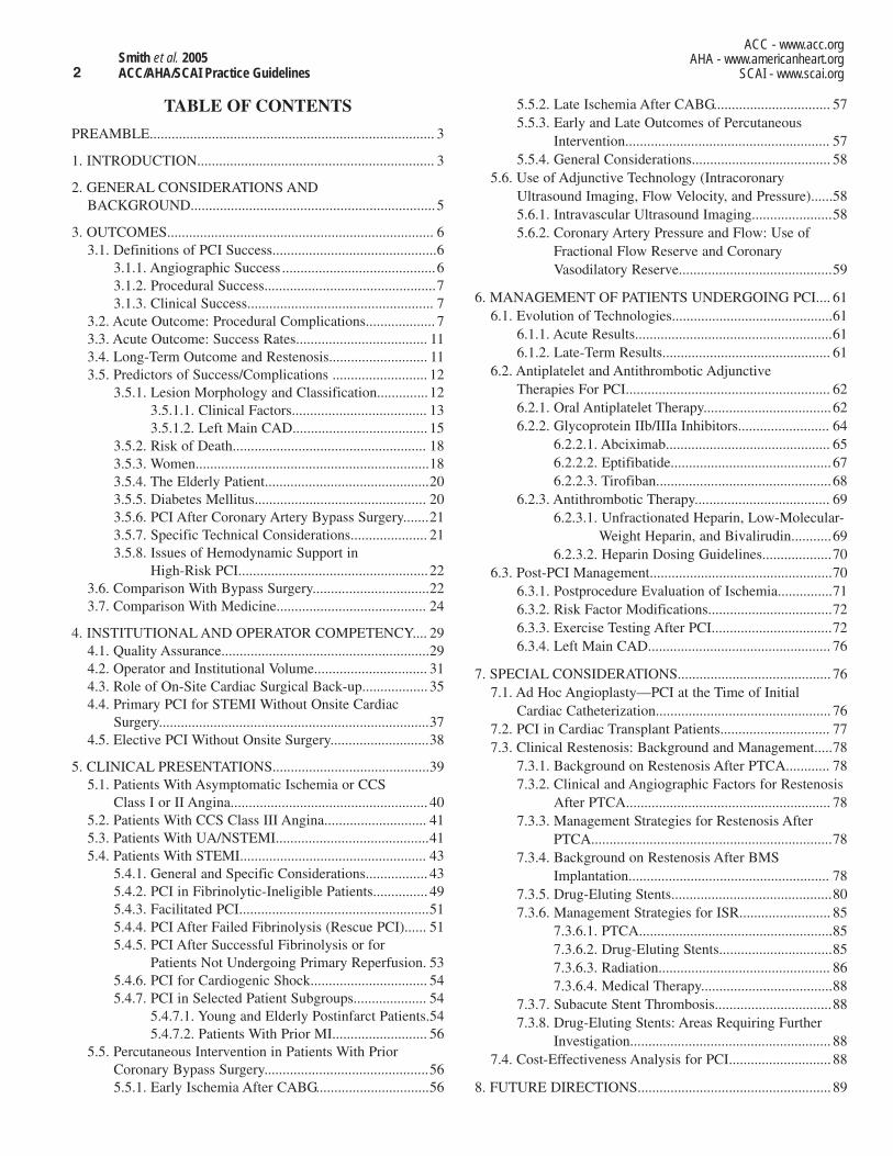
TABLE OF CONTENTS
PREAMBLE.............................................................................. 3
1. INTRODUCTION................................................................. 3
2. GENERAL CONSIDERATIONS ANDBACKGROUND................................................................... 5
3. OUTCOMES......................................................................... 63.1. Definitions of PCI Success.............................................6
3.1.1. Angiographic Success ..........................................63.1.2. Procedural Success...............................................73.1.3. Clinical Success................................................... 7
3.2. Acute Outcome: Procedural Complications................... 73.3. Acute Outcome: Success Rates.................................... 113.4. Long-Term Outcome and Restenosis........................... 113.5. Predictors of Success/Complications .......................... 12
3.5.1. Lesion Morphology and Classification..............123.5.1.1. Clinical Factors..................................... 133.5.1.2. Left Main CAD..................................... 15
3.5.2. Risk of Death..................................................... 183.5.3. Women................................................................183.5.4. The Elderly Patient.............................................203.5.5. Diabetes Mellitus............................................... 203.5.6. PCI After Coronary Artery Bypass Surgery.......213.5.7. Specific Technical Considerations..................... 213.5.8. Issues of Hemodynamic Support in
High-Risk PCI....................................................223.6. Comparison With Bypass Surgery................................223.7. Comparison With Medicine......................................... 24
4. INSTITUTIONAL AND OPERATOR COMPETENCY.... 294.1. Quality Assurance.........................................................294.2. Operator and Institutional Volume............................... 314.3. Role of On-Site Cardiac Surgical Back-up.................. 354.4. Primary PCI for STEMI Without Onsite Cardiac
Surgery..........................................................................374.5. Elective PCI Without Onsite Surgery...........................38
5. CLINICAL PRESENTATIONS...........................................395.1. Patients With Asymptomatic Ischemia or CCS
Class I or II Angina...................................................... 405.2. Patients With CCS Class III Angina............................ 415.3. Patients With UA/NSTEMI..........................................415.4. Patients With STEMI................................................... 43
5.4.1. General and Specific Considerations................. 435.4.2. PCI in Fibrinolytic-Ineligible Patients............... 495.4.3. Facilitated PCI....................................................515.4.4. PCI After Failed Fibrinolysis (Rescue PCI)...... 515.4.5. PCI After Successful Fibrinolysis or for
Patients Not Undergoing Primary Reperfusion. 535.4.6. PCI for Cardiogenic Shock................................ 545.4.7. PCI in Selected Patient Subgroups.................... 54
5.4.7.1. Young and Elderly Postinfarct Patients.545.4.7.2. Patients With Prior MI.......................... 56
5.5. Percutaneous Intervention in Patients With Prior Coronary Bypass Surgery.............................................565.5.1. Early Ischemia After CABG...............................56
2
ACC - www.acc.orgAHA - www.americanheart.org
SCAI - www.scai.orgSmith et al. 2005ACC/AHA/SCAI Practice Guidelines
5.5.2. Late Ischemia After CABG................................ 575.5.3. Early and Late Outcomes of Percutaneous
Intervention........................................................ 575.5.4. General Considerations...................................... 58
5.6. Use of Adjunctive Technology (Intracoronary Ultrasound Imaging, Flow Velocity, and Pressure)......585.6.1. Intravascular Ultrasound Imaging......................585.6.2. Coronary Artery Pressure and Flow: Use of
Fractional Flow Reserve and Coronary Vasodilatory Reserve..........................................59
6. MANAGEMENT OF PATIENTS UNDERGOING PCI.... 616.1. Evolution of Technologies............................................61
6.1.1. Acute Results......................................................616.1.2. Late-Term Results.............................................. 61
6.2. Antiplatelet and Antithrombotic AdjunctiveTherapies For PCI........................................................ 626.2.1. Oral Antiplatelet Therapy...................................626.2.2. Glycoprotein IIb/IIIa Inhibitors......................... 64
6.2.2.1. Abciximab............................................. 656.2.2.2. Eptifibatide............................................676.2.2.3. Tirofiban................................................68
6.2.3. Antithrombotic Therapy..................................... 696.2.3.1. Unfractionated Heparin, Low-Molecular-
Weight Heparin, and Bivalirudin...........696.2.3.2. Heparin Dosing Guidelines...................70
6.3. Post-PCI Management..................................................706.3.1. Postprocedure Evaluation of Ischemia...............716.3.2. Risk Factor Modifications..................................726.3.3. Exercise Testing After PCI.................................726.3.4. Left Main CAD.................................................. 76
7. SPECIAL CONSIDERATIONS.......................................... 767.1. Ad Hoc Angioplasty—PCI at the Time of Initial
Cardiac Catheterization................................................ 767.2. PCI in Cardiac Transplant Patients.............................. 777.3. Clinical Restenosis: Background and Management.....78
7.3.1. Background on Restenosis After PTCA............ 787.3.2. Clinical and Angiographic Factors for Restenosis
After PTCA........................................................ 787.3.3. Management Strategies for Restenosis After
PTCA..................................................................787.3.4. Background on Restenosis After BMS
Implantation....................................................... 787.3.5. Drug-Eluting Stents............................................807.3.6. Management Strategies for ISR......................... 85
7.3.6.1. PTCA.....................................................857.3.6.2. Drug-Eluting Stents...............................857.3.6.3. Radiation............................................... 867.3.6.4. Medical Therapy....................................88
7.3.7. Subacute Stent Thrombosis................................887.3.8. Drug-Eluting Stents: Areas Requiring Further
Investigation....................................................... 887.4. Cost-Effectiveness Analysis for PCI............................ 88
8. FUTURE DIRECTIONS..................................................... 89

3Smith et al. 2005
ACC/AHA/SCAI Practice Guidelines
ACC - www.acc.orgAHA - www.americanheart.orgSCAI - www.scai.org
nosis, management, or prevention of specific diseases orconditions. These guidelines attempt to define practices thatmeet the needs of most patients in most circumstances. Theseguideline recommendations reflect a consensus of expertopinion after a thorough review of the available, current sci-entific evidence and are intended to improve patient care. Ifthese guidelines are used as the basis for regulatory/payerdecisions, the ultimate goal is quality of care and serving thepatient’s best interests. The ultimate judgment regarding careof a particular patient must be made by the healthcareprovider and patient in light of all of the circumstances pre-sented by that patient.
These guidelines were approved for publication by the gov-erning bodies of the ACCF, AHA, and SCAI. The guidelineswill be reviewed annually by the ACC/AHA Task Force onPractice Guidelines and will be considered current unlessthey are revised or withdrawn from distribution. The sum-mary article and recommendations are published in theJanuary 3, 2006 issue of the Journal of the American Collegeof Cardiology, the January 3, 2006 issue of Circulation, andthe January 2006 issue of Catheterization and Cardio-vascular Interventions. The full-text guideline is posted onthe World Wide Web sites of the ACC (www.acc.org), theAHA (www.americanheart.org), and the SCAI(www.scai.org). Copies of the full text and the executivesummary are available from the ACC, AHA, and the SCAI.
Elliott M. Antman, MD, FACC, FAHAChair, ACC/AHA Task Force on Practice Guidelines
1. INTRODUCTION
The ACC/AHA Task Force on Practice Guidelines wasformed to gather information and make recommendationsabout appropriate use of technology for the diagnosis andtreatment of patients with cardiovascular disease.Percutaneous coronary interventions (PCIs) are an importantgroup of technologies in this regard. Although initially limit-ed to balloon angioplasty and termed percutaneous translu-minal coronary angioplasty (PTCA), PCI now includes othernew techniques capable of relieving coronary narrowing.Accordingly, in this document, implantation of intracoronarystents and other catheter-based interventions for treatingcoronary atherosclerosis are considered components of PCI.In this context, PTCA will be used to refer to those studiesusing only balloon angioplasty, whereas PCI will refer to thebroader group of percutaneous techniques. These new tech-nologies have impacted the effectiveness and safety profileinitially established for balloon angioplasty. Moreover, addi-tional experience has been gained in the use of adjunctivepharmacological treatment with glycoprotein (GP) IIb/IIIareceptor antagonists and the use of bivalirudin, thienopy-ridines, and drug-eluting stents (DES). In addition, sincepublication of the guidelines in 2001, greater experience inthe performance of PCI in patients with acute coronary syn-dromes and in community hospital settings has been gained.In view of these developments, an update of these guidelines
Appendix 1. Relationships With Industry: Writing Committee...........................................................91
Appendix 2. Relationships With Industry: Peer Reviewers............................................................92
References................................................................................93
PREAMBLE
It is important that the medical profession play a significantrole in critically evaluating the use of diagnostic proceduresand therapies as they are introduced and tested in the detec-tion, management, or prevention of disease states. Rigorousand expert analysis of the available data documenting rela-tive benefits and risks of those procedures and therapies canproduce helpful guidelines that improve the effectiveness ofcare, optimize patient outcomes, and favorably affect theoverall cost of care by focusing resources on the most effec-tive strategies.
The American College of Cardiology (ACC) and theAmerican Heart Association (AHA) have jointly engaged inthe production of such guidelines in the area of cardiovascu-lar disease since 1980. This effort is directed by theACC/AHA Task Force on Practice Guidelines, whose chargeis to develop and revise practice guidelines for important car-diovascular diseases and procedures. The Task Force ispleased to have this guideline cosponsored by the Society forCardiovascular Angiography and Interventions (SCAI).Experts in the subject under consideration have been select-ed from all three organizations to examine subject-specificdata and write guidelines. The process includes additionalrepresentatives from other medical practitioner and specialtygroups where appropriate. Writing groups are specificallycharged to perform a formal literature review, weigh thestrength of evidence for or against a particular treatment orprocedure, and include estimates of expected health out-comes where data exist. Patient-specific modifiers, comor-bidities, and issues of patient preference that might influencethe choice of particular tests or therapies are considered, aswell as frequency of follow-up and cost-effectiveness. Whenavailable, information from studies on cost will be consid-ered; however, review of data on efficacy and clinical out-comes will be the primary basis for preparing recommenda-tions in these guidelines.
The ACC/AHA Task Force on Practice Guidelines makesevery effort to avoid any actual, potential, or perceived con-flicts of interest that might arise as a result of an outside rela-tionship or personal interest of a member of the writingpanel. Specifically, all members of the writing panel areasked to provide disclosure statements of all such relation-ships that might be perceived as real or potential conflicts ofinterest. These statements are reviewed by the parent taskforce, reported orally to all members of the writing panel ateach meeting, and updated and reviewed by the writing com-mittee as changes occur.
The practice guidelines produced are intended to assisthealthcare providers in clinical decision making by describ-ing a range of generally acceptable approaches for the diag-

is warranted. This document reflects the opinion of theACC/AHA/SCAI writing committee charged with updatingthe 2001 guidelines for PCI (1).
Several issues relevant to the Writing Committee’s processand the interpretation of the guidelines have been noted pre-viously and are worthy of restatement. First, PCI is a tech-nique that has been continually refined and modified; hence,continued, periodic guideline revision is anticipated. Second,these guidelines are to be viewed as broad recommendationsto aid in the appropriate application of PCI. Under uniquecircumstances, exceptions may exist. These guidelines areintended to complement, not replace, sound medical judg-ment and knowledge. They are intended for operators whopossess the cognitive and technical skills for performing PCIand assume that facilities and resources required to properlyperform PCI are available. As in the past, the indications arecategorized as class I, II, or III on the basis of a multifactor-ial assessment of risk and expected efficacy viewed in thecontext of current knowledge and the relative strength of thisknowledge.
These classes summarize the recommendations for proce-dures or treatments as follows:
Class I: Conditions for which there is evidence forand/or general agreement that a given proce-dure or treatment is beneficial, useful, andeffective.
Class II: Conditions for which there is conflicting evi-dence and/or a divergence of opinion aboutthe usefulness/efficacy of a procedure ortreatment.
Class IIa: Weight of evidence/opinion is infavor of usefulness/efficacy.
Class IIb: Usefulness/efficacy is less wellestablished by evidence/opinion.
Class III: Conditions for which there is evidence and/orgeneral agreement that a procedure/treat-ment is not useful/effective and in some casesmay be harmful.
In addition, the weight of evidence in support of the rec-ommendation is listed as follows:
• Level of Evidence A: Data derived from multiple random-ized clinical trials or meta-analyses.
• Level of Evidence B: Data derived from a single random-ized trial or nonrandomized studies.
• Level of Evidence C: Only consensus opinion of experts,case studies, or standard-of-care.
A recommendation with level of evidence B or C does notimply that the recommendation is weak. Many importantclinical questions addressed in the guidelines do not lendthemselves to clinical trials. Even though randomized trials
4
ACC - www.acc.orgAHA - www.americanheart.org
SCAI - www.scai.orgSmith et al. 2005ACC/AHA/SCAI Practice Guidelines
are not available, there may be a very clear clinical consen-sus that a particular test or therapy is useful and effective.
In instances where recommendations of class III, level ofevidence C, occur, it is recognized that the bases of these rec-ommendations are opinion and the consensus of the writinggroup. In this setting, it is not unreasonable for clinical trialsto be conducted to further investigate the validity of this con-sensus opinion. The schema for classification of recommen-dations and level of evidence is summarized in Table 1,which also illustrates how the grading system provides anestimate of the size of the treatment effect and an estimate ofthe certainty of the treatment effect.
The committee conducted comprehensive searching of thescientific and medical literature on PCI, with special empha-sis on randomized controlled trials and meta-analyses pub-lished since 2001. In addition to broad-based searching onPCI, specific targeted searches were performed on the fol-lowing subtopics: catheter-based intervention, stents (drug-eluting and bare-metal), cardiac biomarkers (e.g., creatinekinase and troponins), pharmacological therapy (aspirin,thienopyridines, GP IIb/IIIa inhibitors, heparin, and directthrombin inhibitors), special populations (women, patientswith diabetes, elderly), coronary artery bypass grafting(CABG), high-risk PCI, quality, outcomes, volume, left mainPCI (protected and unprotected), distal embolic protection,intravascular ultrasound (IVUS), fractional flow reserve(FFR), vascular closure, and secondary prevention/risk fac-tor modification. The complete list of keywords is beyondthe scope of this section. The committee reviewed all com-piled reports from computerized searches and conductedadditional searching by hand. Literature citations were gen-erally restricted to published manuscripts appearing in jour-nals listed in Index Medicus. Because of the scope andimportance of certain ongoing clinical trials and other emerg-ing information, published abstracts were cited when theywere the only published information available. Additionally,the Committee reviewed and incorporated recommendationsand/or text from published ACC/AHA or SCAI documents tomaintain consistency, as appropriate.
Initially, this document describes the background informa-tion that forms the foundation for specific recommendations.Topics fundamental to coronary intervention are reviewed,followed by separate discussions relating to unique technicaland operational issues. This format is designed to enhancethe usefulness of this document for the assessment and careof patients with coronary artery disease (CAD). Formal rec-ommendations for the use of PCI according to clinical pres-entation are included in Section 5. A clear distinction isdrawn between the emergency use of PCI for patients withST-segment elevation myocardial infarction (STEMI),termed “primary PCI,” and all other procedures, which areincluded under the term “elective PCI” (see Section 4.2 forfurther discussion).
This committee includes cardiologists with and withoutinvolvement in interventional procedures, and a cardiac sur-geon. This document was reviewed by 2 official reviewersnominated by ACC; 2 official reviewers nominated by AHA;

5Smith et al. 2005
ACC/AHA/SCAI Practice Guidelines
ACC - www.acc.orgAHA - www.americanheart.orgSCAI - www.scai.org
Tabl
e 1.
App
lyin
g C
lass
ific
atio
n of
Rec
omm
enda
tions
and
Lev
el o
f E
vide
nce
“Siz
e of
Tre
atm
ent
Eff
ect”
*Dat
a av
aila
ble
from
clin
ical
tri
als
or r
egis
trie
s ab
out
the
usef
ulne
ss/e
ffic
acy
in d
iffe
rent
sub
-pop
ulat
ions
, suc
h as
gen
der,
age,
his
tory
of
diab
etes
, his
tory
of
prio
r M
I, h
isto
ry o
f he
art
failu
re, a
nd p
rior
asp
irin
use
. Are
com
men
datio
nw
ith L
evel
of
Evi
denc
e B
or
C d
oes
not
impl
y th
at t
he r
ecom
men
datio
n is
wea
k. M
any
impo
rtan
t cl
inic
al q
uest
ions
add
ress
ed i
n th
e gu
idel
ines
do
not
lend
the
mse
lves
to
clin
ical
tri
als.
Eve
n th
ough
ran
dom
ized
tri
als
are
not
avai
labl
e,th
ere
may
be
a ve
ry c
lear
clin
ical
con
sens
us th
at a
par
ticul
ar te
st o
r th
erap
y is
use
ful o
r ef
fect
ive.
†In
2003
, the
AC
C/A
HA
Task
For
ce o
n Pr
actic
e G
uide
lines
dev
elop
ed a
list
of
sugg
este
d ph
rase
s to
use
whe
n w
ritin
g re
com
men
datio
ns. A
ll re
com
men
datio
ns in
this
gui
delin
e ha
ve b
een
wri
tten
in f
ull s
ente
nces
that
exp
ress
a c
ompl
ete
thou
ght,
such
tha
t a
reco
mm
enda
tion,
eve
n if
sep
arat
ed a
nd p
rese
nted
apa
rt f
rom
the
res
t of
the
doc
umen
t (i
nclu
ding
hea
ding
s ab
ove
sets
of
reco
mm
enda
tions
), w
ould
stil
l co
nvey
the
ful
l in
tent
of
the
reco
mm
enda
tion.
It
is h
oped
tha
tth
is w
ill in
crea
se r
eade
rs’c
ompr
ehen
sion
of
the
guid
elin
es a
nd w
ill a
llow
que
ries
at t
he in
divi
dual
rec
omm
enda
tion
leve
l.

6
ACC - www.acc.orgAHA - www.americanheart.org
SCAI - www.scai.orgSmith et al. 2005ACC/AHA/SCAI Practice Guidelines
The value of coronary angioplasty was further defined bycomparing its results to those of alternative methods of treat-ment. Randomized clinical trials have assessed the outcomesof patients treated by a strategy of initial angioplasty to oneof medical therapy alone or to coronary artery bypass surgery(10-14). The results of these trials have clarified the utility ofangioplasty in terms of effectiveness, complications, andpatient selection. The technique of coronary angioplasty hasalso been expanded by the development of devices thatreplace or serve as adjuncts to the balloon catheter. These“new devices” have been evaluated and have had a variableimpact in enhancing the immediate- and long-term efficacyand safety of coronary angioplasty. The following section ofthis report expands on this background and describes thepractice of PCI as it is applied today.
Advances in coronary-based interventions, especially theuse of bare-metal stents (BMS) and drug-eluting stents(DES), have improved the efficacy and safety profile of per-cutaneous revascularization observed for patients undergo-ing PTCA. For example, stents reduce both the acute risk ofmajor complications and late-term restenosis. The success ofnew coronary devices in meeting these goals is reflected inpart by the rapid transition from the use of PTCA alone (lessthan 30%) to the high use of PCI with stenting, which wasgreater than 70% by the late 1990s (Figure 1) (15).Atherectomy devices and stenting, associated with improvedacute angiographic and clinical outcomes compared withPTCA alone in specific subsets, continue to be applied to awider patient domain that includes multivessel disease andcomplex coronary anatomy. However, strong evidence (levelA data from multiple randomized clinical trials) is primarilyavailable for stenting over PTCA in selected patients under-going single-vessel PCI.
2 official reviewers nominated by SCAI; 1 official reviewerfrom the ACC/AHA Task Force on Practice Guidelines; and8 content reviewers, including members from the AHACommittee on Diagnostic and Interventional CardiacCatheterization and the ACCF Cardiac Catheterization andIntervention Committee.
2. GENERAL CONSIDERATIONS ANDBACKGROUND
Coronary angioplasty was first introduced by AndreasGruentzig in 1977 (2) as a nonsurgical method for coronaryarterial revascularization. Fundamentally, the techniqueinvolved advancing a balloon-tipped catheter to an area ofcoronary narrowing, inflating the balloon, and then removingthe catheter after deflation. Early reports demonstrated thatballoon angioplasty could reduce the severity of coronarystenosis and diminish or eliminate objective and subjectivemanifestations of ischemia (3-5). Although angioplasty wasclearly feasible and effective, the scope of coronary diseaseto be treated was quite narrow. Also, because angioplastycould result in sudden arterial occlusion and subsequentmyocardial infarction (MI), immediate access to coronarybypass surgery was essential (6). With experience and time,however, the cognitive and technical aspects as much as theequipment used to perform angioplasty became morerefined. Observational reports of large numbers of patientsconfirmed that coronary angioplasty could be applied tobroad groups of coronary patients with higher rates of suc-cess and lower rates of complications than seen in initialexperiences (7,8). More than 1 000 000 PCI procedures areperformed yearly in the United States (9), and it has beenestimated that nearly 2 000 000 procedures are performedannually worldwide.
Figure 1. Frequency of device use in the SCAI registry. Source data from Laskey et al. Catheter Cardiovasc Interv 2000;49:19-22(15).
7Smith et al. 2005
ACC/AHA/SCAI Practice Guidelines
ACC - www.acc.orgAHA - www.americanheart.orgSCAI - www.scai.org
The range of non-balloon revascularization technologyapproved by the Food and Drug Administration (FDA) foruse in native and/or graft coronary arteries includes balloonexpandable stents, DES, extraction atherectomy, directionalcoronary atherectomy, rotational atherectomy, rheolyticthrombectomy catheter, proximal and distal embolic protec-tion devices, excimer laser coronary atherectomy, and localradiation devices to reduce in-stent restenosis (ISR) (16,17).A variety of devices are under investigation, including newdesigns of balloon or self-expanding stents and mechanicalthrombectomy devices. This guideline update will focus onthe FDA-approved balloon-related and non-balloon coronaryrevascularization devices.
3. OUTCOMES
The outcomes of PCI are measured in terms of success andcomplications and are related to the mechanisms of theemployed devices, as well as the clinical and anatomicpatient-related factors. Complications can be divided into 2categories: (a) those common to all arterial catheterizationprocedures and (b) those related to the specific technologyused for the coronary procedure. Specific definitions of suc-cess and complications exist, and where appropriate, the def-initions used herein are consistent with the ACC-NationalCardiovascular Data Registry (NCDR®) CatheterizationLaboratory Module version 3.0 (18). The committee recom-mends such standards whenever feasible in order to accom-modate the common database for the assessment of out-comes. With increased operator experience, new technology,and adjunctive pharmacotherapy, the overall success andcomplication rates of angioplasty have improved.
3.1. Definitions of PCI Success
The success of a PCI procedure may be defined by angio-graphic, procedural, and clinical criteria.
3.1.1. Angiographic Success
A successful PCI produces substantial enlargement of thelumen at the target site. The consensus definition before thewidespread use of stents was the achievement of a minimumstenosis diameter reduction to less than 50% in the presenceof grade 3 Thrombolysis In Myocardial Infarction (TIMI)flow (assessed by angiography) (1). However, with theadvent of advanced adjunct technology, including coronarystents, a minimum stenosis diameter reduction to less than20% has been the clinical benchmark of an optimal angio-graphic result. Frequently, there is a disparity between thevisual assessment and computer-aided quantitative stenosismeasurement (19,20), and, thus, the determination of successmay be problematic when success rates are self-reported.
3.1.2. Procedural Success
A successful PCI should achieve angiographic success with-out major clinical complications (e.g., death, MI, emergencycoronary artery bypass surgery) during hospitalization (1,3).
Although the occurrence of emergency coronary arterybypass surgery and death are easily identified end points, thedefinition of procedure-related MI has been debated. Thedevelopment of Q waves in addition to a threshold value ofcreatine kinase (CK) elevation has been commonly used.Most agree that the definition of MI as put forth by theACC/European Society of Cardiology document on the rede-finition of MI (21) should be the accepted standard.However, the clinical significance and definition of cardiacbiomarker elevations in the absence of Q waves remains thesubject of investigation and debate (21a). Several reportshave identified non–Q-wave MIs with CK-MB elevations 3to 5 times the upper limit of normal as having clinical sig-nificance (22,23). One report suggests that a greater than 5times increase in CK-MB is associated with worsened out-come (24). Thus, this degree of increase in CK-MB withoutQ waves is considered by most to qualify as an associatedcomplication of PCI. Troponin T or I elevation occurs fre-quently after PCI. The timing of the peak elevation after PCIis unclear (25). Minor elevations do not appear to have prog-nostic value, whereas marked (greater than 5 times) eleva-tions are associated with worsened 1-year outcome (Table 2)(26-40). Troponin T or I elevation occurs more frequentlythan CK-MB increase after PCI (34).
3.1.3. Clinical Success
In the short term, a clinically successful PCI includesanatomic and procedural success with relief of signs and/orsymptoms of myocardial ischemia after the patient recoversfrom the procedure. The long-term clinical success requiresthat the short-term clinical success remain durable and thatthe patient have persistent relief of signs and symptoms ofmyocardial ischemia for more than 6 months after the proce-dure. Restenosis is the principal cause of lack of long-termclinical success when a short-term clinical success has beenachieved. Restenosis is not considered a complication butrather an associated response to vascular injury. The inci-dence of clinically important restenosis may be judged by thefrequency with which subsequent revascularization proce-dures are performed on target vessels after the index proce-dure.
3.2. Acute Outcome: Procedural Complications
Class IAll patients who have signs or symptoms suggestive ofMI during or after PCI and those with complicatedprocedures should have CK-MB and troponin I or Tmeasured after the procedure. (Level of Evidence: B)
Class IIaRoutine measurement of cardiac biomarkers (CK-MB and/or troponin I or T) in all patients undergoingPCI is reasonable 8 to 12 hours after the procedure.(Level of Evidence: C)

8
ACC - www.acc.orgAHA - www.americanheart.org
SCAI - www.scai.orgSmith et al. 2005ACC/AHA/SCAI Practice Guidelines
Tabl
e 2.
Inci
denc
e of
Tro
poni
n E
leva
tions
Aft
er P
ercu
tane
ous
Cor
onar
y In
terv
entio
n in
the
Publ
ishe
d L
itera
ture
Fir
st A
utho
rof
%
P
osit
ive
Pro
gnos
tic
Stud
y (R
efer
ence
)n
Mar
ker
Pos
itiv
eD
efin
itio
nIn
form
atio
n
Hun
t (29
)22
Tro
poni
n I
0G
reat
er th
an
N/A
6 ng
per
mL
Rav
kild
e (3
0)23
Tro
poni
n T
13G
reat
er th
an
N/A
0.12
ng
per
mL
Kar
im (
31)
25T
ropo
nin
T44
Gre
ater
than
N
/A0.
2 ng
per
mL
La
Vec
chia
(32
)19
(St
ent)
Tro
poni
n T
37 c
TnI
; N
/AN
/A25
(ba
lloon
PC
I)an
d21
cT
nTtr
opon
in I
14 c
TnI
; 0
cTnT
Joha
nsen
(33
)75
Tro
poni
n T
28G
reat
er th
an
N/A
0.1
ng p
er m
L
Shyu
(34
)59
(St
ent)
Tro
poni
n T
29G
reat
er th
an
Sign
ific
antly
hig
her
inci
denc
e of
ele
vate
d cT
nTin
pat
ient
s 61
(ba
lloon
PC
I)13
0.1
ng p
er m
Lun
derg
oing
ste
ntin
g th
an a
ngio
plas
ty a
lone
.
Ber
tinch
ant (
35)
105
Tro
poni
n I
22G
reat
er th
an
No
diff
eren
ce in
inci
denc
e of
rec
urre
nt a
ngin
a, M
I, c
ardi
ac d
eath
, 0.
1 ng
per
mL
or R
R a
fter
12
mon
ths
betw
een
patie
nts
posi
tive
or n
egat
ive
for
cTnI
. Ste
ntin
g no
t ass
ocia
ted
with
mor
e m
inor
m
yoca
rdia
l dam
age
than
ang
iopl
asty
.
Gar
barz
(36
)10
9T
ropo
nin
I27
Gre
ater
than
N
o as
soci
atio
n be
twee
n po
st-P
CI
cTnI
and
adv
erse
isch
emic
eve
nts.
0.3
ng p
er m
L
Fuch
s (3
7)11
29T
ropo
nin
I31
Gre
ater
than
cT
nTle
vels
gre
ater
than
3×
nor
mal
lim
it as
soci
ated
0.
15 n
g pe
r m
Lw
ith in
crea
sed
risk
of
maj
or in
-hos
pita
l com
plic
atio
ns,
but n
o as
soci
atio
n w
ith a
dver
se in
term
edia
te-t
erm
(8
mon
ths)
clin
ical
ou
tcom
es.
Can
tor
(26)
481
Tro
poni
n I
48 o
vera
ll;
Gre
ater
than
Si
gnif
ican
tly h
ighe
r 90
-day
rat
es o
f M
I an
d th
e co
mpo
site
of
MI
26 a
fter
1.
5 ng
per
mL
or d
eath
in p
atie
nts
with
pos
itive
cT
nI.
excl
udin
gpo
sitiv
e or
un
know
n pr
e-PC
I cT
nI
Wu
(38)
98T
ropo
nin
T26
Gre
ater
than
A
t a m
ean
of 7
7 m
onth
s fo
llow
-up,
no
incr
ease
in r
isk
of m
ajor
adv
erse
0.
1 ng
per
mL
even
ts d
etec
ted
in r
elat
ion
to p
ost-
PCI
cTnT
elev
atio
n.
Kiz
er (
27)
212
Tro
poni
n T
40 p
ositi
ve p
rior
G
reat
er th
an o
rPr
e-PC
I cT
nTel
evat
ion
was
sig
nifi
cant
ly r
elat
ed to
eve
nt-f
ree
surv
ival
to
PC
I; 1
8
equa
l to
duri
ng 6
-yea
r fo
llow
-up;
in b
asel
ine
nega
tive
patie
nts,
pos
itive
cT
nTof
bas
elin
e0.
1 ng
per
mL
was
the
only
inde
pend
ent p
redi
ctor
of
maj
or a
dver
se e
vent
s at
1 y
ear;
ne
gativ
e po
st-P
CI
elev
atio
ns o
f cT
nTgr
eate
r th
an o
r eq
ual t
o 5×
nor
mal
was
the
wer
e po
sitiv
est
rong
est l
ong-
term
pre
dict
or o
f m
ajor
adv
erse
eve
nts
at 6
yea
rs.
post
-PC
I
Ric
ciar
di (
39)
286
Tro
poni
n I
13.6
Gre
ater
than
cT
nI e
leva
tions
gre
ater
than
3-f
old
care
pre
dict
ive
of f
utur
e m
ajor
2.
3 ng
per
mL
adve
rse
card
iac
even
ts (
MA
CE
). I
ncre
ased
inci
denc
e of
MA
CE
is
acc
ount
ed f
or b
y hi
gher
rat
e of
ear
ly R
R a
nd n
ot la
te c
ardi
ac e
vent
s.
Kin
i (40
)28
73T
ropo
nin
I38
.9G
reat
er th
an
Nei
ther
cT
nI p
eak
elev
atio
ns n
or a
ny s
ubgr
oup
pred
icte
d m
id-t
erm
2
ng p
er m
Lm
orta
lity
in lo
w-
to m
ediu
m-r
isk
patie
nts.
cTnI
indi
cate
s ca
rdia
c tr
opon
in I
; cT
nT, c
ardi
ac tr
opon
in T
; N/A
, not
app
licab
le; P
CI,
per
cuta
neou
s co
rona
ry in
terv
entio
n; a
nd R
R, r
epea
t rev
ascu
lari
zatio
n.

9Smith et al. 2005
ACC/AHA/SCAI Practice Guidelines
ACC - www.acc.orgAHA - www.americanheart.orgSCAI - www.scai.org
Another study indicated that more extensive stent expan-sion resulted in CK release but did not increase adverse car-diac events (59). Accordingly, it is important to acknowledgethat the significance of mild biomarker rises after clinicallysuccessful PCI should be distinguished from situationswherein patients experience an unequivocal “clinical” infarc-tion manifested by chest pain and diagnostic ECG findings(60).
Routine measurement of CK-MB is advocated by some(21) and actually mandated by certain healthcare systems. Inthis regard, the current Committee supports the recommen-dations of the 2001 Guidelines and recommends that allpatients who have signs or symptoms suggestive of MI dur-ing or after PCI and those with complicated proceduresshould have CK-MB and troponin I or T measured after theprocedure. In addition, the Committee recommends that rou-tine measurement of cardiac biomarkers (CK-MB and/or tro-ponin I or T) in every patient undergoing PCI is reasonable 8to 12 h after the procedure. In such patients, a new CK-MBor troponin I or T rise greater than 5 times the upper limit ofnormal would constitute a clinically significant periproce-dural MI.
The need to perform emergency coronary artery bypasssurgery (CABG) has been considered as a potential compli-cation of PCI. Typically, CABG is performed as a rescuerevascularization procedure to treat acute ischemia or infarc-tion resulting from PCI-induced acute coronary occlusion. Inthe era of balloon angioplasty, the rate of emergency CABGwas 3.7% (49). In a more contemporary time period, with theavailability of stents, the reported rate was 0.4% among asimilar cohort of patients.
Various definitions have been proposed for stroke. A com-mon feature to definitions has been a loss of neurologic func-tion of vascular cause that lasts more than 24 h. More recent-ly, attention has been directed to refining the definition oftransient ischemic attack (TIA), which indirectly broadensthat of stroke (61). The time-based definition of a TIA is asudden, focal neurologic deficit that lasts less than 24 h thatis of presumed vascular origin and confined to an area of thebrain or eye perfused by a specific artery. The new definitionof TIA is a brief episode of neurologic dysfunction caused bybrain or retinal ischemia, with clinical symptoms typicallylasting less than 1 hour and without evidence of infarction.Presence of cerebral infarction by imaging techniques con-stitutes evidence of stroke regardless of the duration ofsymptoms.
Bleeding is a complication of increasing concern with themore frequent use of potent antithrombin and antiplateletagents. A frequently used definition for bleeding developedby the TIMI group includes classification as major, moder-ate, or minor. Major bleeding is defined as intracranial,intraocular, or retroperitoneal hemorrhage or any hemor-rhage requiring a transfusion or surgical intervention or thatresults in a hematocrit decrease of greater than 15% or hemo-globin decrease of greater than 5 g per dL (62). Episodes ofhemorrhage of lesser magnitude would fall into the moder-
Complications associated with PCI are similar to thoseresulting from diagnostic cardiac catheterization, but theirprevalence is more frequent. Complications have been cate-gorized as major (death, MI, and stroke) or minor (transientischemic attack, access site complications, renal insufficien-cy, or adverse reactions to radiographic contrast). Additionalspecific complications include intracoronary thrombosis,coronary perforation, tamponade, and arrhythmias.
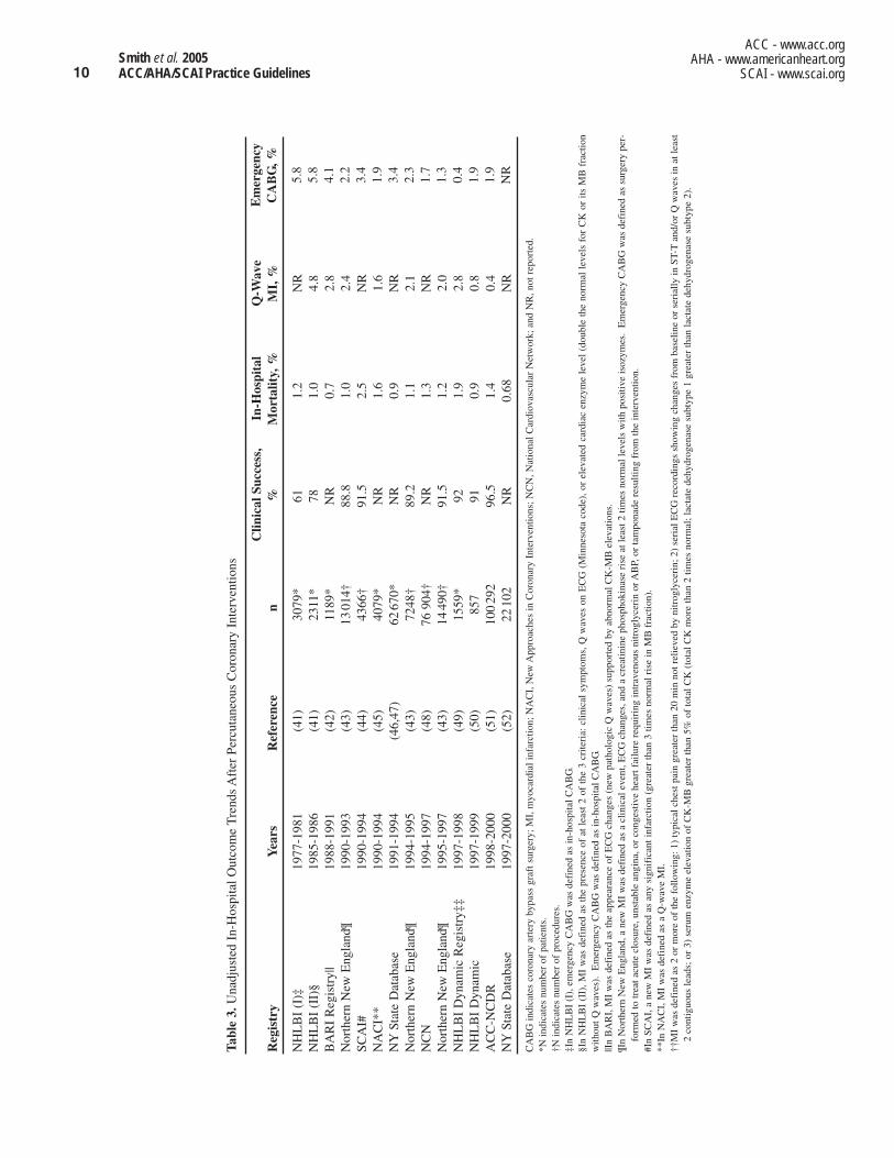
Reported rates for death after diagnostic catheterizationrange from 0.08% to 0.14%, whereas analyses of large reg-istries indicate overall unadjusted in-hospital rates for PCI of0.4% to 1.9% (Table 3) (41-52). This range is greatly influ-enced by the clinical indication for which PCI is performed,with the highest mortality rates occurring among patientswith STEMI and cardiogenic shock. Death in such patientsmay not be a direct result of the PCI procedure but rather aconsequence of the patient’s underlying illness. For example,in a combined analysis of PCI as primary reperfusion thera-py for STEMI, the short-term mortality rate was 7% (53).Even after exclusion of patients with cardiogenic shock, in-hospital mortality was 5%.
Myocardial infarction can be a direct result of PCI, mostcommonly due to abrupt coronary occlusion or intracoronaryembolization of obstructive debris. Determining and com-paring the incidence of MI after PCI is difficult because thedefinition of MI as a result of PCI is controversial. The con-ventional definition requires 2 of the following: a) prolongedchest discomfort or its equivalent; b) development of patho-logic Q waves; and c) rise in serum cardiac biomarkers abovea critical level. Rates of periprocedural MI using this defini-tion have ranged from 0.4% to 4.9%. Using a consistent def-inition for MI, the incidence of this complication hasdeclined approximately 50% with the routine use of intra-coronary stents (21,21a,50).
More recently, an isolated rise and fall in either CK-MB ortroponin is considered to be a marker of myocardial necrosis(21). The relationship between cardiac biomarker elevationand myocardial cell death and evidence of subendocardialinfarction on magnetic resonance imaging (MRI) supportthis position (54,55). Furthermore, large rises in cardiac bio-markers are associated with an increased risk for late death(26,56,57). Whether death in such patients is a consequenceof the myonecrosis or a marker of patients who are atincreased risk for death because of more advanced coronarydisease is unclear. Complicating our understanding of theimplications of this definition is the very frequently observedmild to modest elevation of serum CK-MB among patientswith apparently uncomplicated PCI. When troponin is meas-ured after PCI, more than 70% of patients exhibit elevatedvalues after an otherwise successful intervention (58). Suchpatients may have no symptoms or electrocardiographic(ECG) abnormalities to suggest ischemia yet are “enzymepositive.” One study has suggested a postprocedural increasein troponin T of 5 times normal is predictive for adverseevents at 6 years. The long-term prognostic significance ofsmaller postprocedural troponin T elevations awaits furtherinvestigation (27) (Table 2) (26-40).
10
ACC - www.acc.orgAHA - www.americanheart.org
SCAI - www.scai.orgSmith et al. 2005ACC/AHA/SCAI Practice Guidelines
Tabl
e 3.
Una
djus
ted
In-H
ospi
tal O
utco
me
Tre
nds
Aft
er P
ercu
tane
ous
Cor
onar
y In
terv
entio
ns
Clin
ical
Suc
cess
, In
-Hos
pita
l Q
-Wav
e R
egis
try
Yea
rsR
efer
ence
n%
Mor
talit
y, %
MI,
%
NH
LB
I (I
)‡19
77-1
981
(41)
3079
*61
1.2
NR
NH
LB
I (I
I)§
1985
-198
6(4
1)23
11*
781.
04.
8B
AR
I R
egis
try|
|19
88-1
991
(42)
1189
*N
R0.
72.
8N
orth
ern
New
Eng
land
¶19
90-1
993
(43)
1301
4†88
.81.
02.
4SC
AI#
1990
-199
4(4
4)43
66†
91.5
2.5
NR
NA
CI*
*19
90-1
994
(45)
40
79*
NR
1.6
1.6
NY
Stat
e D
atab
ase
1991
-199
4(4
6,47
)62
670*
NR
0.9
NR
Nor
ther
n N
ew E
ngla
nd¶
1994
-199
5(4
3)72
48†
89.2
1.1
2.1
NC
N19
94-1
997
(48)
76 9
04†
NR
1.3
NR
Nor
ther
n N
ew E
ngla
nd¶
1995
-199
7(4
3)14
490†
91.5
1.2
2.0
NH
LB
I D
ynam
ic R
egis
try‡
‡19
97-1
998
(49)
1559
*92
1.9
2.8
NH
LB
I D
ynam
ic19
97-1
999
(50)
857
910.
90.
8A
CC
-NC
DR
1998
-200
0(5
1)10
029
296
.51.
40.
4N
YSt
ate
Dat
abas
e19
97-2
000
(52)
2210
2N
R0.
68N
R
CA
BG
indi
cate
s co
rona
ry a
rter
y by
pass
gra
ft s
urge
ry; M
I, m
yoca
rdia
l inf
arct
ion;
NA
CI,
New
App
roac
hes
in C
oron
ary
Inte
rven
tions
; NC
N, N
atio
nal C
ardi
ovas
cula
r N
etw
ork;
and
NR
, not
rep
orte
d.*N
indi
cate
s nu
mbe
r of
pat
ient
s.†N
indi
cate
s nu
mbe
r of
pro
cedu
res.
‡In
NH
LB
I (I
), e
mer
genc
y C
AB
G w
as d
efin
ed a
s in
-hos
pita
l CA
BG
.§I
n N
HL
BI
(II)
, MI
was
def
ined
as
the
pres
ence
of
at l
east
2 o
f th
e 3
crite
ria:
clin
ical
sym
ptom
s, Q
wav
es o
n E
CG
(M
inne
sota
cod
e), o
r el
evat
ed c
ardi
ac e
nzym
e le
vel
(dou
ble
the
norm
al l
evel
s fo
r C
K o
r its
MB
fra
ctio
nw
ithou
t Q w
aves
). E
mer
genc
y C
AB
G w
as d
efin
ed a
s in
-hos
pita
l CA
BG
.||I
n B
AR
I, M
I w
as d
efin
ed a
s th
e ap
pear
ance
of
EC
G c
hang
es (
new
pat
holo
gic
Q w
aves
) su
ppor
ted
by a
bnor
mal
CK
-MB
ele
vatio
ns.
¶In
Nor
ther
n N
ew E
ngla
nd, a
new
MI
was
def
ined
as
a cl
inic
al e
vent
, EC
G c
hang
es, a
nd a
cre
atin
ine
phos
phok
inas
e ri
se a
t lea
st 2
tim
es n
orm
al le
vels
with
pos
itive
isoz
ymes
. E
mer
genc
y C
AB
G w
as d
efin
ed a
s su
rger
y pe
r-fo
rmed
to tr
eat a
cute
clo
sure
, uns
tabl
e an
gina
, or
cong
estiv
e he
art f
ailu
re r
equi
ring
intr
aven
ous
nitr
ogly
ceri
n or
AB
P, o
r ta
mpo
nade
res
ultin
g fr
om th
e in
terv
entio
n.#I
n SC
AI,
a n
ew M
I w
as d
efin
ed a
s an
y si
gnif
ican
t inf
arct
ion
(gre
ater
than
3 ti
mes
nor
mal
ris
e in
MB
fra
ctio
n).
**In
NA
CI,
MI
was
def
ined
as
a Q
-wav
e M
I.††
MI
was
def
ined
as
2 or
mor
e of
the
follo
win
g: 1
) ty
pica
l che
st p
ain
grea
ter
than
20
min
not
rel
ieve
d by
nitr
ogly
ceri
n; 2
) se
rial
EC
G r
ecor
ding
s sh
owin
g ch
ange
s fr
om b
asel
ine
or s
eria
lly in
ST-
Tan
d/or
Q w
aves
in a
t lea
st2
cont
iguo
us le
ads;
or
3) s
erum
enz
yme
elev
atio
n of
CK
-MB
gre
ater
than
5%
of
tota
l CK
(to
tal C
K m
ore
than
2 ti
mes
nor
mal
; lac
tate
deh
ydro
gena
se s
ubty
pe 1
gre
ater
than
lact
ate
dehy
drog
enas
e su
btyp
e 2)
.
Em
erge
ncy
CA
BG
, %
5.8
5.8
4.1
2.2
3.4
1.9
3.4
2.3
1.7
1.3
0.4
1.9
1.9
NR

11Smith et al. 2005
ACC/AHA/SCAI Practice Guidelines
ACC - www.acc.orgAHA - www.americanheart.orgSCAI - www.scai.org
In addition to multivessel disease, other clinical factorsadversely impact late mortality. In randomized patients withtreated diabetes undergoing PTCA in BARI, the 5-year sur-vival was 65.5%, and the cardiac mortality rate was 20.6%compared with 5.8% in patients without treated diabetes(67), although among eligible but not randomized diabeticpatients treated with PTCA, the 5-year cardiac mortality ratewas 7.5% (68). In the 1985-1986 NHLBI PTCA Registry, 4-year survival was significantly lower in women (89.2%) thanin men (93.4%) (69). In addition, although LV dysfunctionwas not associated with an increase in in-hospital mortalityor nonfatal MI in patients undergoing PTCA in the same reg-istry, it was an independent predictor of a higher long-termmortality (70).
A major determinant of event-free survival after coronaryintervention is the incidence of restenosis, which had, untilthe development of stents, remained fairly constant despitemultiple pharmacologic and mechanical approaches to limitthis process (Table 4) (71-95). The incidence of restenosisafter coronary intervention varies depending on the defini-tion, i.e., whether clinical or angiographic restenosis or tar-get-vessel revascularization is measured (96). Data frommultiple randomized clinical trials and prospective registriessuggest that DES incorporating either rapamycin or paclitax-el with a timed-release polymer are associated with a reduc-tion in restenosis rates to less than 10% across a wide spec-trum of clinical and angiographic subsets.
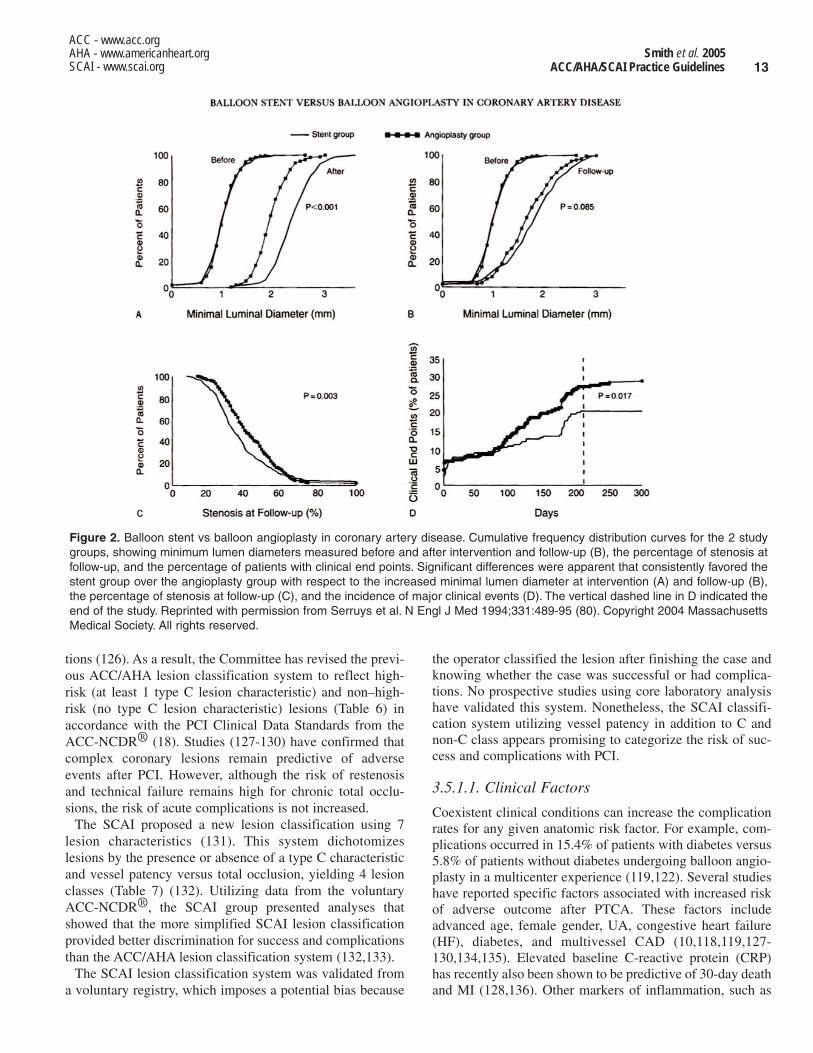
The pathogenesis of the response to mechanical coronaryinjury is thought to relate to a combination of growth factorstimulation, smooth muscle cell migration and proliferation,organization of thrombus, platelet deposition, and elasticrecoil (97,98). In addition, change in vessel size (or lack ofcompensatory enlargement) has been implicated (99). It hasbeen suggested that attempts to reduce restenosis have failedin part because of lack of recognition of the importance ofthis factor (100). Although numerous definitions of resteno-sis have been proposed, greater than 50% diameter stenosisat follow-up angiography has been most frequently usedbecause it was thought to correlate best with maximal flowand therefore ischemia. However, it is now recognized thatthe response to arterial injury is a continuous rather than adichotomous process, occurring to some degree in allpatients (101). Therefore, cumulative frequency distributionsof the continuous variables of minimal lumen diameter orpercent diameter stenosis are frequently used to evaluaterestenosis in large patient populations (102) (Figure 2) (80).
Although multiple clinical factors (diabetes, unstable angi-na [UA]/NSTEMI, STEMI, and prior restenosis) (103,104),angiographic factors (proximal left anterior descendingartery [LAD], small vessel diameters, total occlusion, longlesion length, and saphenous vein grafts [SVGs]) (105), andprocedural factors (higher postprocedure percent diameterstenosis, smaller minimal lumen diameter, and smaller acutegain) (102) have been associated with an increased incidenceof restenosis, the ability to integrate these factors and predictthe risk of restenosis in individual patients after the proce-dure remains difficult. The most promising potential
ate/minor categories. A listing of other bleeding classifica-tions has been developed for use by the ACC-NCDR® (18).
3.3. Acute Outcome: Success Rates
Success has been described on both a lesion and patientbasis. In early studies of PTCA, lesion success is defined asan absolute 20% reduction in lesion severity with final steno-sis less than 50%. When describing the results of multipleattempted lesions, success is classified as either partial (somebut not all attempted lesions successfully treated) or total(each attempted lesion successfully treated). Procedural suc-cess is defined as the achievement of either partial or totalangiographic success without death, MI, or emergencyCABG (49).
Reported rates of angiographic success now range between82% and 98% depending on the device used and the types oflesions attempted. Formal comparisons demonstrate that suc-cess rates are now higher (91% to 92%) in the era of newtechnology, which includes stents and contemporary drugtherapies, than in the era of conventional balloon angioplas-ty (72% to 74%) (49). The types of lesions attempted strong-ly influence success rates. The chance of dilating a chronictotal occlusion averages 65%, and specific clinical andanatomic factors have been identified that affect this rate(63). Quite different are the success rates for total occlusionsassociated with STEMI. Success rates over 90% can beexpected in this subgroup (64).
With an increase in angiographic success rates and adecline in periprocedural MI and the need for emergencyCABG, procedural success rates have risen from a range of80% to 85% to a range of 90% to 95% (Table 3) (41-52).
3.4. Long-Term Outcome and Restenosis
Although improvements in technology, such as stents, haveresulted in an improved acute outcome of the procedure, theimpact of these changes on long-term (5 to 10 years) out-come may be less dramatic because factors such as advancedage, reduced left ventricular (LV) function, and progressionof complex multivessel disease in patients currently under-going PCI may have a more important influence. In addition,available data on long-term outcome are mostly limited topatients undergoing PTCA. Ten-year follow-up of the initialcohort of patients treated with PTCA revealed an 89.5% sur-vival rate (95% in patients with single-vessel disease, 81% inpatients with multivessel disease) (65). In patients undergo-ing PTCA within the 1985-1986 National Heart, Lung, andBlood Institute (NHLBI) PTCA Registry (66), 5-year sur-vival was 92.9% for patients with single-vessel disease,88.5% for those with 2-vessel disease, and 86.5% for thosewith 3-vessel disease. In patients with multivessel diseaseundergoing PTCA in the Bypass AngioplastyRevascularization Investigation (BARI) (10), 5-year survivalwas 86.3%, and infarct-free survival was 78.7%.Specifically, 5-year survival was 84.7% in patients with 3-vessel disease and 87.6% in patients with 2-vessel disease.

12
ACC - www.acc.orgAHA - www.americanheart.org
SCAI - www.scai.orgSmith et al. 2005ACC/AHA/SCAI Practice Guidelines
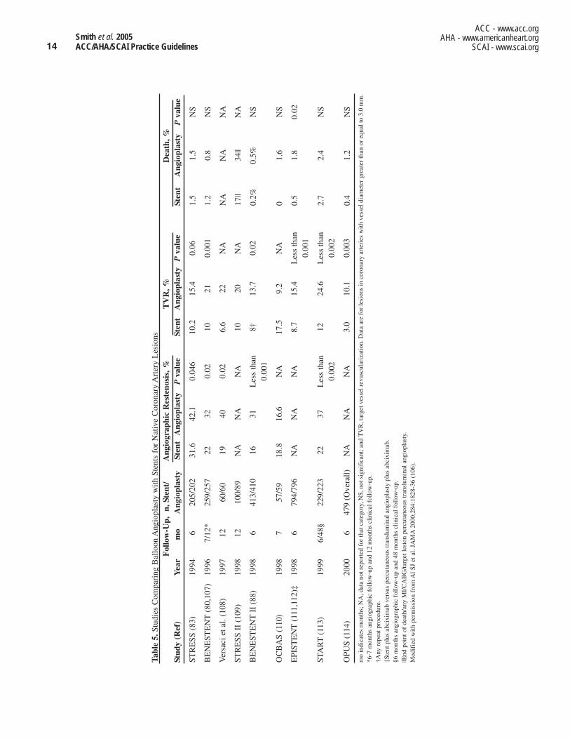
approaches to favorably impact the restenosis process arestents and, more recently, DES and catheter-based radiation.More than 6300 patients have been studied in 12 randomizedclinical trials to assess the efficacy of PTCA versus stents toreduce restenosis (Table 5) (80,83,88,106-114).
The pivotal BENESTENT (BElgian NEtherlands STENTstudy) (80) and STRESS (STent REStenosis Study) trials(83) documented that stents significantly reduce angiograph-ic restenosis compared with balloon angioplasty (BENES-TENT: 22% vs 32%; STRESS: 32% vs 42%, respectively).These results were further corroborated in the BENESTENTII trial, in which the angiographic restenosis rate was reducedby almost half (from 31% to 16% in patients treated with bal-loon angioplasty versus heparin-coated stents, respectively)(88).
In addition, randomized studies in patients with ISR haveshown that both intracoronary gamma and beta radiation sig-nificantly reduced the rate of subsequent angiographic andclinical restenosis by 30% to 50% (92,115-117). Late suba-cute thrombosis was observed in some of these series (117),but this syndrome has resolved with judicious use of stentsand extended adjunct antiplatelet therapy with ticlopidine or
clopidogrel. The development of DES has significantlyreduced the rate of ISR (see Section 7.3.6 for full discus-sion).
3.5. Predictors of Success/Complications
3.5.1. Lesion Morphology and Classification
Target lesion anatomic factors related to adverse outcomeshave been widely examined. Lesion morphology andabsolute stenosis severity were identified as the prominentpredictors of immediate outcome during PTCA in theprestent era (118,119). Abrupt vessel closure, due primarilyto thrombus or dissection, was reported in 3% to 8% ofpatients and was associated with certain lesion characteris-tics (120-122). The risk of PTCA in the prestent era relativeto anatomic subsets has been identified in previous NHLBIPTCA Registry data (7) and by the ACC/AHA Task Force onPractice Guidelines (1,123). The lesion classification basedon severity of characteristics proposed in the past (123-125)has been principally altered using the present PCI tech-niques, which capitalize on the ability of stents to manageinitial and subsequent complications of coronary interven-
Table 4. Selected Trials of Pharmacological and Mechanical Approaches to Limit Restenosis
Restenosis Rate, %Study Year Reference n Agent Placebo or Control Agent
Schwartz 1988 (71) 376 Aspirin and dipyridamole 39 38Ellis 1989 (72) 416 Heparin 37 41Pepine 1990 (73) 915 Methylprednisolone 39 40CARPORT 1991 (74) 649 Vapiprost 19 21O’Keefe 1992 (75) 197 Colchicine 22 22MERCATOR 1992 (76) 735 Cilazapril 28 28CAVEAT* 1993 (77) 500 DCA versus PTCA 57 50CCAT 1993 (78) 136 DCA versus PTCA 43 46Serruys 1993 (79) 658 Ketanserin 32 32BENESTENT* 1994 (80) 520 Stent versus PTCA 32 22ERA 1994 (81) 458 Enoxaparin 51 52Leaf 1994 (82) 551 Fish oil 46 52STRESS* 1994 (83) 410 Stent versus PTCA 42 32Weintraub 1994 (84) 404 Lovastatin 42 39BOAT* 1998 (85) 492 DCA versus PTCA 40 31Wantanabe* 1996 (86) 118 Probucol 40 20Tardif* 1997 (87) 317 Probucol 39 21BENESTENT II* 1998 (88) 823 Stent versus PTCA 31 17TREAT* 1999 (89) 255 Tranilast 39 18PRESTO* 2000 (90) 192 DCA and Tranilast 26 11ARTIST* 2002 (91) 298 Rotablation (in-stent) 51 65
versus PTCASTART* 2002 (92) 476 Radiation (in-stent) 45 29SIRIUS* 2003 (93) 1058 Sirolimus-coated stent versus 36 9
bare stentTAXUS-IV* 2004 (94) 1314 Paclitaxel-coated stent versus 27 8
bare stentRESCUT 2004 (95) 428 Cutting balloon (in-stent) 31 30
versus PTCA
DCA indicates directional coronary atherectomy; n, number of patients; and PTCA, percutaneous transluminal coronary angioplasty.*P less than 0.05.
13Smith et al. 2005
ACC/AHA/SCAI Practice Guidelines
ACC - www.acc.orgAHA - www.americanheart.orgSCAI - www.scai.org
Figure 2. Balloon stent vs balloon angioplasty in coronary artery disease. Cumulative frequency distribution curves for the 2 studygroups, showing minimum lumen diameters measured before and after intervention and follow-up (B), the percentage of stenosis atfollow-up, and the percentage of patients with clinical end points. Significant differences were apparent that consistently favored thestent group over the angioplasty group with respect to the increased minimal lumen diameter at intervention (A) and follow-up (B),the percentage of stenosis at follow-up (C), and the incidence of major clinical events (D). The vertical dashed line in D indicated theend of the study. Reprinted with permission from Serruys et al. N Engl J Med 1994;331:489-95 (80). Copyright 2004 MassachusettsMedical Society. All rights reserved.
the operator classified the lesion after finishing the case andknowing whether the case was successful or had complica-tions. No prospective studies using core laboratory analysishave validated this system. Nonetheless, the SCAI classifi-cation system utilizing vessel patency in addition to C andnon-C class appears promising to categorize the risk of suc-cess and complications with PCI.
3.5.1.1. Clinical Factors
Coexistent clinical conditions can increase the complicationrates for any given anatomic risk factor. For example, com-plications occurred in 15.4% of patients with diabetes versus5.8% of patients without diabetes undergoing balloon angio-plasty in a multicenter experience (119,122). Several studieshave reported specific factors associated with increased riskof adverse outcome after PTCA. These factors includeadvanced age, female gender, UA, congestive heart failure(HF), diabetes, and multivessel CAD (10,118,119,127-130,134,135). Elevated baseline C-reactive protein (CRP)has recently also been shown to be predictive of 30-day deathand MI (128,136). Other markers of inflammation, such as
tions (126). As a result, the Committee has revised the previ-ous ACC/AHA lesion classification system to reflect high-risk (at least 1 type C lesion characteristic) and non–high-risk (no type C lesion characteristic) lesions (Table 6) inaccordance with the PCI Clinical Data Standards from theACC-NCDR® (18). Studies (127-130) have confirmed thatcomplex coronary lesions remain predictive of adverseevents after PCI. However, although the risk of restenosisand technical failure remains high for chronic total occlu-sions, the risk of acute complications is not increased.
The SCAI proposed a new lesion classification using 7lesion characteristics (131). This system dichotomizeslesions by the presence or absence of a type C characteristicand vessel patency versus total occlusion, yielding 4 lesionclasses (Table 7) (132). Utilizing data from the voluntaryACC-NCDR®, the SCAI group presented analyses thatshowed that the more simplified SCAI lesion classificationprovided better discrimination for success and complicationsthan the ACC/AHA lesion classification system (132,133).
The SCAI lesion classification system was validated froma voluntary registry, which imposes a potential bias because
14
ACC - www.acc.orgAHA - www.americanheart.org
SCAI - www.scai.orgSmith et al. 2005ACC/AHA/SCAI Practice Guidelines
Tabl
e 5.
Stud
ies
Com
pari
ng B
allo
on A
ngio
plas
ty w
ith S
tent
s fo
r N
ativ
e C
oron
ary
Art
ery
Les
ions
Fol
low
-Up,
n, S
tent
/ A
ngio
grap
hic
Res
teno
sis,
%T
VR
, %St
udy
(Ref
)Y
ear
mo
Ang
iopl
asty
Sten
tA
ngio
plas
tyP
valu
eSt
ent
Ang
iopl
asty
Pva
lue
STR
ESS
(83
)19
946
205/
202
31.6
42.1
0.04
610
.215
.40.
06
BE
NE
STE
NT
(80,
107)
1996
7/12
*25
9/25
722
320.
0210
210.
001
Ver
saci
et a
l. (1
08)
1997
1260
/60
1940
0.02
6.6
22N
A
STR
ESS
II
(109
)19
9812
100/
89N
AN
AN
A10
20N
A
BE
NE
STE
NT
II (
88)
1998
641
3/41
016
31L
ess
than
8†13
.70.
020.
001
OC
BA
S (1
10)
1998
757
/59
18.8
16.6
NA
17.5
9.2
NA
EPI
STE
NT
(111
,112
)‡19
986
794/
796
NA
NA
NA
8.7
15.4
Les
s th
an
0.00
1
STA
RT
(113
)19
996/
48§
229/
223
2237
Les
s th
an12
24.6
Les
s th
an
0.00
20.
002
OPU
S (1
14)
2000
647
9 (O
vera
ll)N
AN
AN
A3.
010
.10.
003
mo
indi
cate
s m
onth
s; N
A, d
ata
not r
epor
ted
for
that
cat
egor
y, N
S, n
ot s
igni
fica
nt; a
nd T
VR
, tar
get v
esse
l rev
ascu
lari
zatio
n. D
ata
are
for
lesi
ons
in c
oron
ary
arte
ries
with
ves
sel d
iam
eter
gre
ater
than
or
equa
l to
3.0
mm
.*6
-7 m
onth
s an
giog
raph
ic f
ollo
w-u
p an
d 12
mon
ths
clin
ical
fol
low
-up.
†Any
rep
eat p
roce
dure
.‡S
tent
plu
s ab
cixi
mab
ver
sus
perc
utan
eous
tran
slum
inal
ang
iopl
asty
plu
s ab
cixi
mab
.§6
mon
ths
angi
ogra
phic
fol
low
-up
and
48 m
onth
s cl
inic
al f
ollo
w-u
p.||E
nd p
oint
of
deat
h/an
y M
I/C
AB
G/ta
rget
lesi
on p
ercu
tane
ous
tran
slum
inal
ang
iopl
asty
.M
odif
ied
with
per
mis
sion
fro
m A
l SJ
et a
l. JA
MA
2000
;284
:182
8-36
(10
6).
Dea
th, %
Sten
tA
ngio
plas
tyP
valu
e
1.5
1.5
NS
1.2
0.8
NS
NA
NA
NA
17||
34||
NA
0.2%
0.5%
NS
01.
6N
S
0.5
1.8
0.02
2.7
2.4
NS
0.4
1.2
NS

15Smith et al. 2005
ACC/AHA/SCAI Practice Guidelines
ACC - www.acc.orgAHA - www.americanheart.orgSCAI - www.scai.org
Table 6. Lesion Classification System
Descriptions of a High-Risk Lesion (Type C Lesion)Diffuse (length greater than 2 cm)Excessive tortuosity of proximal segmentExtremely angulated segments, greater than 90°Total occlusions more than 3 months old and/or bridging collaterals*Inability to protect major side branchesDegenerated vein grafts with friable lesions*
*The high risk with these criteria is for technical failure and increased restenosis, not for acute complications.
Table 7. SCAI Lesion Classification System: Characteristics of ClassI-IV Lesions
Type I lesions (highest success expected, lowest risk)(1) Does not meet criteria for C lesion(2) Patent
Type II lesions(1) Meets any of these criteria for ACC/AHA C lesion
Diffuse (greater than 2 cm length)Excessive tortuosity of proximal segmentExtremely angulated segments, greater than 90°Inability to protect major side branchesDegenerated vein grafts with friable lesions
(2) Patent
Type III lesions(1) Does not meet criteria for C lesion(2) Occluded
Type IV lesions(1) Meets any of the criteria for ACC/AHA C lesion
Diffuse (greater than 2 cm length)Excessive tortuosity of proximal segmentExtremely angulated segments, greater than 90°Inability to protect major side branchesDegenerated vein grafts with friable lesionsOccluded for more than 3 months
(2) Occluded
Reprinted from Krone et al. Evaluation of the American College of Cardiology/AmericanHeart Association and the Society for Coronary Angiography and Interventions lesion clas-sification system in the current “stent era” of coronary interventions (from the ACC-National Cardiovascular Data Registry®). Am J Cardiol 2003;92:394 (Appendix B) (132).
interleukin-6 and other cytokines, have also been shown tobe predictive of outcome (137). The BARI trial found thatpatients with diabetes and multivessel CAD had an increasedperiprocedural risk of ischemic complications and increased5-year mortality compared with patients without diabetes orpatients with diabetes undergoing bypass surgery using inter-nal mammary artery (IMA) grafts (10,42). Patients withimpaired renal function, especially those with diabetes, are atincreased risk for contrast nephropathy (138) and increased30-day and 1-year mortality (139,140). Renal insufficiencyis a strong predictor of outcome in both primary and electivePCI (141-143). Increased risk for death or severe compro-mise in LV function may occur in association with a compli-cation involving a vessel that also supplies collateral flow toviable myocardium. Certain variables were used to prospec-tively identify patients at risk for significant cardiovascularcompromise during PTCA (144,145). These resulted in a
composite 4-variable scoring system, prospectively validatedto be both sensitive and specific in predicting cardiovascularcollapse for failed PTCA, which includes: 1) percentage ofmyocardium at risk (e.g., greater than 50% viable myocardi-um at risk and LV ejection fraction of less than 25%), 2) pre-angioplasty percent diameter stenosis, 3) multivessel CAD,and 4) diffuse disease in the dilated segment (124) or a highmyocardial jeopardy score (125). Patients with higher pre-procedural jeopardy scores were shown to have a greaterlikelihood of cardiovascular collapse when abrupt vessel clo-sure occurred during PTCA (144).
3.5.1.2. Left Main CAD
CABG has long been considered the “gold standard” forrevascularization of lesions in the unprotected left main(ULM) coronary artery (146). With the advent of newer tech-nology utilizing BMS and DES, experience has been gainedin performing PCI in ULM coronary artery lesions. Somestudies have demonstrated that stenting of the ULM is feasi-ble and appears to be a promising strategy in selectedpatients (147-152). Patients treated for ULM disease havevaried from those presenting with stable angina to those withMI and shock. However, despite the feasibility and high pro-cedural success rate of ULM PCI in the pre-DES era, thereare reports of an unacceptably high incidence of long-termadverse events (153-155). This may be attributed to theinclusion of high-risk patients, such as those not consideredgood surgical candidates. The experience with BMS forULM PCI in the multicenter ULTIMA (Unprotected LeftMain Trunk Intervention Multicenter Assessment) registrysuggested a high early mortality (2% per month among hos-pital survivors over the first 6 months after hospital dis-charge), and careful surveillance with coronary angiographywas recommended (153) (see Section 6.3.4). Patients pre-senting with MI, ULM occlusion, and cardiogenic shockhave lower successful PCI rates (69.7% vs 100%, P equals0.040), higher in-hospital mortality (71.4% vs 10%, P equals0.0008), and higher 1-year mortality rates (P equals 0.0064)than stable MI patients regardless of performance of primaryPCI with stents (155).
More recently, published studies of left main PCI usingDES have reported 6-month or 1-year death rates rangingfrom 0% to 14% (Table 8) (147-150,152-161). Furthermore,ISR appears to be improved with the use of DES versusBMS. One of the larger studies performed to date showedthat the 6-month angiographic restenosis rate was signifi-

16
ACC - www.acc.orgAHA - www.americanheart.org
SCAI - www.scai.orgSmith et al. 2005ACC/AHA/SCAI Practice Guidelines
Tabl
e 8.
Publ
ishe
d T
rial
s an
d Se
lect
ed R
egis
try
Exp
erie
nces
of
PCI
for
Unp
rote
cted
Lef
t Mai
n C
oron
ary
Art
ery
Sten
osis
Fir
st A
utho
r,
In-H
ospi
tal
Mor
talit
y, %
Y
ear
(Ref
eren
ce)
Dev
ice
Use
dn
Mor
talit
y, %
(Fol
low
-Up
Per
iod)
Res
teno
sis,
%T
VR
, %C
omm
ents
Elli
s 19
94-1
996
50%
BM
S10
720
.666
.0 p
lus
or
20.8
N/A
Surv
ival
to h
ospi
tal d
isch
arge
31%
in a
cute
MI
patie
nts;
(1
47)
min
us 4
.7 (
9 m
o)in
ele
ctiv
e pa
tient
s, in
-hos
pita
l mor
talit
y 5.
9% in
goo
d ca
ndid
ates
for
CA
BG
, 30.
4% in
poo
r C
AB
G c
andi
date
s;
in-h
ospi
tal s
urvi
val s
tron
gly
corr
elat
ed w
ith L
VE
F
Silv
estr
i 199
3-19
98
100%
BM
S14
09%
hig
h
2% h
igh
CA
BG
ris
k;
2317
.4G
ood
imm
edia
te r
esul
ts o
f PC
I in
UL
M
(148
)C
AB
G r
isk;
2.6%
low
CA
BG
sten
osis
, esp
ecia
lly in
goo
d C
AB
G c
andi
date
s0%
low
ri
sk (
6 m
o)C
AB
G r
isk
Bla
ck 1
994-
1998
10
0% B
MS
924.
310
.8 (
7.3
plus
or
N/A
7.3
PCI
to U
LM
app
ears
bet
ter
in c
andi
date
s fo
r C
AB
G th
an(1
49)
min
us 5
.8 m
o)in
pat
ient
s in
who
m C
AB
G is
con
trai
ndic
ated
; tre
ndto
war
d ca
rdia
c m
orta
lity
with
3-v
esse
l dis
ease
and
low
LV
EF;
low
fin
al s
tent
lum
en d
iam
eter
onl
y si
gnif
ican
t pr
edic
tor
of c
ardi
ac m
orta
lity
ULT
IMA
1993
-199
8 68
.8%
BM
S27
913
.724
.2 (
1 y)
N/A
33.6
*46
% o
f pa
tient
s w
ere
deem
ed in
oper
able
or
at h
igh
risk
(1
53)
for
CA
BG
; in
patie
nts
less
than
65
y ol
d w
ith L
VE
F gr
eate
r th
an 3
0% a
nd n
o sh
ock,
0%
per
ipro
cedu
ral
deat
hs a
nd 1
-yea
r m
orta
lity
3.4%
; 2%
per
mon
th d
eath
rate
am
ong
hosp
ital s
urvi
vors
ove
r 6
mon
ths
afte
r di
s-ch
arge
; car
eful
sur
veill
ance
with
cor
onar
y an
giog
raph
y re
com
men
ded
Park
199
5-20
00
100%
BM
S12
70
3.1
(25.
5 pl
us o
r19
11.8
Ele
ctiv
e st
entin
g in
pat
ient
s w
ith n
orm
al L
VE
F; I
VU
S (1
50)
min
us 1
6.7
mo)
may
opt
imiz
e im
med
iate
res
ults
; sig
nifi
cant
ly lo
wer
re
sten
osis
rat
e w
ith d
ebul
king
bef
ore
sten
ting
Taka
gi 1
993-
2001
96
% B
MS
670
16.4
(31
plu
s or
31
.423
.9H
igh
rate
of
rest
enos
is a
nd R
R; 1
1.9%
car
diac
mor
talit
y;
(154
)m
inus
23
mo)
sign
ific
antly
hig
her
card
iac
mor
talit
y in
pat
ient
s w
ith
Pars
onne
t sco
re g
reat
er th
an 1
5 at
3 y
ears
Park
199
5-20
00
100%
BM
S27
00
7.4
(32.
3 pl
us o
r 21
.116
.7†
Goo
d ov
eral
l lon
g-te
rm (
3-ye
ar)
surv
ival
in s
elec
ted
(152
)m
inus
18.
5 m
o)pa
tient
s w
ith n
orm
al L
VE
F; c
ombi
ned
CA
D a
nd p
ost-
proc
edur
al lu
men
dia
met
er s
igni
fica
nt p
redi
ctor
s of
M
AC
E
Saka
i 199
2-20
00
65%
to 7
4% B
MS
3871
.4%
with
sho
ck;
71.4
% w
ith s
hock
;N
/AN
/APa
tient
s w
ith a
cute
MI
due
to U
LM
ste
nosi
s an
d sh
ock
(155
)10
% w
ithou
t sho
ck20
% w
ithou
t ha
ve p
oor
surv
ival
reg
ardl
ess
of p
erfo
rman
ce o
f PC
I sh
ock
(1 y
)w
ith s
tent
s
Con
tinu
ed o
n ne
xt p
age
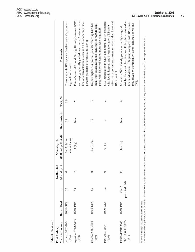
17Smith et al. 2005
ACC/AHA/SCAI Practice Guidelines
ACC - www.acc.orgAHA - www.americanheart.orgSCAI - www.scai.org
Tabl
e 8.
Con
tinu
ed
Fir
st A
utho
r,
In-H
ospi
tal
Mor
talit
y, %
Y
ear
(Ref
eren
ce)
Dev
ice
Use
dn
Mor
talit
y, %
(Fol
low
-Up
Per
iod)
Res
teno
sis,
%T
VR
, %C
omm
ents
de L
ezo
2002
-200
4 10
0% S
ES
520
0 (1
2 pl
us o
r 3.
81.
9T
reat
men
t with
SE
S ap
pear
s fe
asib
le a
nd s
afe;
pro
mis
-(1
56)
min
us 4
mo)
ing
mid
term
res
ults
Ago
ston
i 200
2-20
03
100%
DE
S58
25
(1 y
)N
/A7
Rat
e of
eve
nts
did
not d
iffe
r si
gnif
ican
tly b
etw
een
IVU
S (1
58)
and
angi
ogra
phic
ally
gui
ded
proc
edur
es; A
nato
mic
loca
-tio
n of
ath
eros
cler
otic
dis
ease
in U
LM
art
ery
only
inde
-pe
nden
t pre
dict
or o
f ev
ents
at f
ollo
w-u
p
Chi
effo
200
2-20
04
100%
DE
S85
03.
5 (6
mo)
1919
Des
pite
hig
her
risk
pro
file
, pat
ient
s re
ceiv
ing
DE
S ha
d (1
59)
sign
ific
ant a
dvan
tage
in th
e in
cide
nce
of M
AC
E c
om-
pare
d w
ith h
isto
rica
l con
trol
gro
up r
ecei
ving
BM
S
Park
200
3-20
04
100%
SE
S10
20
0 (1
y)
72
SES
impl
anta
tion
in U
LM
and
nor
mal
LV
EF
asso
ciat
ed
(160
)w
ith lo
w in
-hos
pita
l and
1-y
ear
mor
talit
y; S
ES
mor
e ef
fect
ive
in p
reve
ntin
g in
-ste
nt r
este
nosi
s th
an h
isto
rica
l B
MS
cont
rol
RE
SEA
RC
H/ 2
003
100%
DE
S95
(15
11
14 (
1 y)
N/A
6M
ore
than
50%
of
stud
y po
pula
tion
at h
igh
surg
ical
T-
SEA
RC
H 2
002-
prot
ecte
d L
M)
risk
by
Pars
onne
t cla
ssif
icat
ion;
47%
rel
ativ
e ri
sk r
educ
-(1
61)
tion
in M
AC
E in
DE
S gr
oup
com
pare
d w
ith B
MS
con-
trol
, dri
ven
by s
igni
fica
ntly
low
er in
cide
nce
of M
I an
d T
VR
*1-Y
ear
estim
ate;
†32
.3 p
lus
or m
inus
18.
5 m
o.
n in
dica
tes
num
ber
of p
atie
nts;
LV
EF,
LV
ejec
tion
frac
tion;
MA
CE
, maj
or a
dver
se c
ardi
ac e
vent
s; R
R, r
epea
t rev
ascu
lari
zatio
n; S
ES,
sir
olim
us-e
lutin
g st
ent;
TV
R, t
arge
t-ve
ssel
rev
ascu
lari
zatio
n; a
nd U
LM
, unp
rote
cted
left
mai
n.

18
ACC - www.acc.orgAHA - www.americanheart.org
SCAI - www.scai.orgSmith et al. 2005ACC/AHA/SCAI Practice Guidelines
3.5.3. Women
An estimated 33% of the more than 1 million PCIs per-formed in the United States annually are in women. The needfor more data concerning outcomes from PCI in women hasled the AHA to issue a scientific statement summarizingavailable studies (169). Compared with men, women under-going PCI are older and have a higher incidence of hyper-tension, diabetes mellitus, hypercholesterolemia, and comor-bid disease (69,170-174). Women also have more UA and ahigher functional class of stable angina (CanadianCardiovascular Society [CCS] class III and IV) for a givenextent of disease (175). Yet, despite the higher-risk profile inwomen, the extent of epicardial coronary disease is similar to(or less than) that in men. In addition, although women pre-senting for revascularization have less multivessel diseaseand better LV systolic function, the incidence of HF is high-er in women than in men. The reason for this gender paradoxis unclear, but it has been postulated that women have morediastolic dysfunction than men (176).
Early reports of patients undergoing PTCA revealed alower procedural success rate in women (172); however,subsequent studies have noted similar angiographic outcomeand incidence of MI and emergency CABG in women andmen (69). Although reports have been inconsistent, in sever-al large-scale registries, in-hospital mortality is significantlyhigher in women (177), and an independent effect of genderon acute mortality after PTCA persists after adjustments forthe baseline higher-risk profile in women (69,178). The rea-son for the increase in mortality is unknown, but small ves-sel size (179) and hypertensive heart disease in women havebeen thought to play a role. Although a few studies havenoted that gender is not an independent predictor of mortali-ty when body surface area (a surrogate for vessel size) isaccounted for (171), the impact of body size on outcome hasnot been thoroughly evaluated. The higher incidence of vas-cular complications, coronary dissection, and perforation inwomen undergoing coronary intervention has been attributedto the smaller vasculature in women than in men. In addition,diagnostic IVUS studies have not detected any gender-spe-cific differences in plaque morphology or luminal dimen-sions once differences in body surface area were corrected,which suggests that differences in vessel size account forsome of the apparent early and late outcome differences pre-viously noted in women (180). It has also been postulatedthat the volume shifts and periods of transient ischemia dur-ing PTCA are less well tolerated by the hypertrophied ven-tricle in women, and HF has shown to be an independent pre-dictor of mortality in both women and men undergoingPTCA (181).
Women continue to have increased bleeding and vascularcomplications compared with men, but these rates havedecreased with the use of smaller sheath sizes and earlysheath removal, weight-adjusted heparin dosing, and lessaggressive anticoagulation regimens (169). Use of IIb/IIIaplatelet receptor antagonists during PCI is not associatedwith an increased risk of major bleeding in women
cantly lower in the ULM group receiving DES than in thosewho received BMS (7.0% vs 30.3%, P less than 0.001)(160). The lower rate of restenosis of DES compared withBMS has been confirmed in other studies of ULM PCI (159).
There have been some attempts to predict success of ULMPCI using customary risk factors such as age, renal failure,coronary calcification, and location of the lesion in the leftmain coronary artery. In general, younger patients with pre-served LV function, noncalcified coronary arteries, and com-plete delivery of stent, fare better. Maintenance ofantiplatelet therapy after the procedure is critical, as is theimplementation of secondary prevention therapies. Carefulpostprocedure surveillance with coronary angiography isneeded to prevent fatal MI or sudden death that may be asso-ciated with ISR with a large area of myocardium in jeopardy;however, the frequency and best method of follow-up areunknown (162). One study’s authors from the BMS era sug-gested routine surveillance angiography at 2 and 4 monthsafter PCI (153). Others advocate routine stress testing or car-diac catheterization at 3 and 6 months even in asymptomaticpatients (148,150). Studies from the DES era have reportedperforming routine angiography 4 to 8 months after PCI orearlier if clinically indicated by symptoms or documentedmyocardial ischemia (159,160). Other issues that remain tobe resolved are technical issues (e.g., optimal bifurcationstenting technique, stent size), degree of revascularizationnecessary, cost-effectiveness, and the selection of patientsbest suited for DES.
In conclusion, CABG using IMA grafting is the “gold stan-dard” for treatment of ULM disease and has proven benefiton long-term outcomes. The use of DES has shown encour-aging short-term outcomes, but long-term follow-up is need-ed. Nevertheless, the use of PCI for patients with significantULM stenosis who are candidates for revascularization butnot suitable for CABG can improve cardiovascular outcomesand is a reasonable revascularization strategy in carefullyselected patients. Recommendations for ULM PCI in specif-ic angina subsets can be found in Sections 5.1, 5.2, 5.3, and5.4 and in Section 6.3.4 for post-PCI follow-up.
3.5.2. Risk of Death
In the majority of patients undergoing elective PCI, death asa result of PCI is directly related to the occurrence of coro-nary artery occlusion and is most frequently associated withpronounced LV failure (144,145). The clinical and angio-graphic variables associated with increased mortality includeadvanced age, female gender, diabetes, prior MI, multivesseldisease, left main or equivalent coronary disease, a large areaof myocardium at risk, pre-existing impairment of LV orrenal function, post-PCI worsening of renal function, andcollateral vessels supplying significant areas of myocardiumthat originate distal to the segment to be dilated(10,118,120,122,134,135,138,139,140,144,163-167). Peri-procedural stroke also increases in-hospital and 1-year mor-tality (168). PCI in the setting of STEMI is associated with asignificantly higher death rate than is seen in elective PCI.

19Smith et al. 2005
ACC/AHA/SCAI Practice Guidelines
ACC - www.acc.orgAHA - www.americanheart.orgSCAI - www.scai.org
(182,183), and the direct thrombin inhibitor bivalirudin dur-ing elective PCI appears to reduce the risk of bleeding (com-bined major and moderate bleeding) in both women and mencompared with unfractionated heparin (184).
Improved outcomes have been reported in more recentlytreated women undergoing both PTCA and PCI, despite thefact that the women are older and have more complex diseasethan women treated previously (Table 9) (69,170,185-189).In fact, in the 1993-1994 NHLBI PTCA Registry (open towomen only), procedural success was higher and major com-plications lower than in women treated in the 1985-1986 reg-istry (190). Additionally, in patients undergoing PTCA inBARI, in-hospital mortality, MI, emergency coronary arterybypass surgery rates, and 5-year mortality were similar inwomen and men, although women had a higher incidence ofperiprocedural HF and pulmonary edema (188).
The widespread use of stents and adjunctive pharmacolog-ic therapy has improved outcomes in patients undergoingcontemporary PCI (80,83,112,191-202). Early studies ofdrug-eluting stents in small vessels (less than or equal to 2.75mm), of particular importance in women, report favorablelong-term results in both women and men (203). The hopethat stents would eliminate the difference in outcomesbetween women and men has not been realized. Gender dif-ferences in mortality have persisted for patients treated withstents both in the setting of acute and nonacute MI (204). Ina meta-analysis of invasive versus conservative therapy ofpatients with UA/NSTEMI, men demonstrated a clear sur-vival advantage using routine invasive therapy with GPIIb/IIIa inhibitors and intracoronary stents; however, usingsimilar therapy, the results for women were not significantlyimproved (205), although it has been shown that the benefitsof an invasive strategy have been limited to high-risk women(206).
In women with STEMI, the relative benefit of primary PCIcompared with fibrinolytic therapy is similar to that in men,but there is a larger absolute benefit in women owing to theirhigher event rate (207). In patients with shock complicatingacute MI, the benefit of revascularization is similar inwomen and men (208).
In general, the risks and benefits of adjunctive pharma-cotherapy in women are similar to those in men, although anincreased rate of minor bleeding has been reported in womentreated with abciximab (183). When IIb/IIIa platelet receptorantagonists are used with unfractionated heparin, a lowerdose of the latter should be considered to decrease the risk ofbleeding in women (Table 9) (69,170,185-189).
Few gender-specific data are available on the outcomes ofother percutaneous coronary devices. Although directionalcoronary atherectomy has been associated with lower proce-dural success and higher bleeding complications in women(209), similar benefit to that in men has been reported fromembolic protection devices used in saphenous vein PCI (210)and from vascular brachytherapy (169).
Tabl
e 9.
Gen
der-
Spec
ific
Mor
talit
y R
isk
Fol
low
-Up
Mor
talit
y, M
en
Stud
yY
ears
Ref
eren
ceW
omen
Ver
sus
Men
(n)
(yea
rs)
Dev
ice
Ver
sus
Wom
en (
%)
May
o C
linic
1979
-199
0(1
86)
3027
(82
4 vs
220
3)5.
5PT
CA
27 v
ersu
s 22
Em
ory
Uni
vers
ity19
80-1
991
(187
)10
785
(284
5 vs
794
0)5
PTC
A8
vers
us 5
NH
LB
I PT
CA
Reg
istr
y19
85-1
986
(69)
2136
(54
6 vs
159
0)4
PTC
A6.
6 ve
rsus
10.
8
BA
RI
1988
-199
4(1
88)
1829
(48
9 vs
134
0)5
PTC
A12
.8 v
ersu
s 12
.0
NA
CI
1990
-199
4(1
89)
2855
(97
5 vs
188
0)1
PCI
5.7
vers
us 5
.9
Nor
ther
n N
ew E
ngla
nd19
94-1
999
(170
)33
666
(10
997
vs 2
2|68
9)In
-hos
pita
lPC
I1.
21 v
ersu
s 1.
06
NH
LB
I D
ynam
ic R
egis
try
1997
-199
8(1
85)
2524
(89
5 vs
162
9)1
PCI
4.3
vers
us 6
.5
CI
indi
cate
s co
nfid
ence
inte
rval
; NS,
not
sig
nifi
cant
; n, n
umbe
r of
pat
ient
s; O
R, o
dds
ratio
; PC
I, p
ercu
tane
ous
coro
nary
inte
rven
tion;
and
PC
TA, p
ercu
tane
ous
tran
slum
inal
cor
onar
y an
giop
last
y. F
or e
xpan
sion
of
stud
y na
mes
, see
cor
resp
ondi
ng r
efer
ence
.*E
xpre
ssed
as
rela
tive
risk
(R
R).
Adj
uste
d O
R
P(9
5% C
I)
0.06
0.94
(0.
76 to
1.1
5)
0.00
021.
08 (
0.84
to 1
.39)
0.00
11.
20 (
0.84
to 1
.73)
NS
0.60
(0.
43 to
0.8
4)
NS
NS
0.09
61.
24 (
0.96
to 1
.60)
0.02
21.
26*
(0.8
5 to
1.8
7)
20
ACC - www.acc.orgAHA - www.americanheart.org
SCAI - www.scai.orgSmith et al. 2005ACC/AHA/SCAI Practice Guidelines
(18.0% vs 6.7%), and 3-year (21.6% vs 9.6%) mortality ratesthan nondiabetic patients (226). Patients with diabetes with afirst MI who were randomly assigned to the early invasivestrategy faired worse than those managed conservatively(42-day mortality: death or MI, or death alone, 14.8% vs4.2%; P less than 0.001) (227). An early catheterization andintervention strategy after fibrinolysis was of little benefit inthese patients with diabetes. Although adjusted in-hospitalmortality was not different in diabetic and nondiabeticpatients, data from the NHLBI registry showed that at 1 year,adjusted mortality and repeat revascularization were signifi-cantly higher in diabetics (228). Thus, routine catheterizationand PCI in this patient subgroup should be based on clinicalneed and ischemic risk stratification.
Stenting decreases the need for target-vessel revasculariza-tion procedures in diabetic patients compared with PTCA(229). The efficacy of stenting with GP IIb/IIIa inhibitorswas assessed in the diabetic population compared with thosewithout diabetes in a substudy of the EPISTENT (Evaluationof IIb/IIIa Platelet Inhibitor for Stenting) trial (230). Onehundred seventy-three diabetic patients were randomized tostent/placebo combination, 162 patients to stent/abciximabcombination, and 156 patients to PTCA/abciximab combina-tion. For the composite end point of death, MI, or target-ves-sel revascularization, the rates were as follows: 25%, 23%,and 13% for the stent/placebo, PTCA/abciximab, andstent/abciximab groups (P equals 0.005). Irrespective ofrevascularization strategy, abciximab significantly reduced6-month death and MI rate in patients with diabetes for allstrategies. Likewise, 6-month target-vessel revascularizationwas reduced in the stent/abciximab group approach. One-year mortality for diabetics was 4.1% for the stent/placebogroup and 1.2% for the stent/abciximab group. Although thisdifference was not significant, the combination of stentingand abciximab among diabetics resulted in a significantreduction in 6-month rates of death and target-vessel revas-cularization compared with stent/placebo or PTCA/abcix-imab therapy (230). Similar results in 1-year target-vesselrevascularization and mortality have been reported withabciximab and the small-molecule GP IIb/IIIa inhibitortirofiban (231). (See Section 6.2.2 Glycoprotein IIb/IIIaInhibitors.) The BARI trial, in which stents and abciximabwere not used, showed that survival was better for patientswith treated diabetes undergoing CABG with an arterial con-duit than for those undergoing PTCA (232). The benefit ofCABG in patients with diabetes may be related to lessenedmortality after subsequent Q-wave MI among patients withdiabetes. In the BARI trial, the benefit of bypass surgery indiabetic patients was greater in those patients with moreextensive disease (e.g., more than 4 lesions). This advantagewas largely due to a lower mortality for subsequent MI(233).
Since the BARI trial was completed, several studies haveassessed the use of PCI with stenting versus CABG inpatients with multivessel disease. Patients with diabetes wereassessed specifically in studies from the ARTS (ArterialRevascularization Therapies Study) and AWESOME (Angina
3.5.4. The Elderly Patient
Age greater than 75 years is one of the major clinical vari-ables associated with increased risk of complications (211-214). In the elderly population, the morphologic and clinicalvariables are compounded by advanced years, with the veryelderly having the highest risk of adverse outcomes (215). Inoctogenarians, although feasibility has been established formost interventional procedures, the risks associated withboth percutaneous and nonpercutaneous revascularizationare increased (216-218). Octogenarians undergoing PCI havea higher incidence of prior MI, lower LV ejection fraction,and more frequent HF (219,220). In the stent era, proceduralsuccess and restenosis rates are comparable to those fornonoctogenarians, albeit with higher incidences beingreported for in-hospital and long-term mortality and for vas-cular and bleeding complications (221). A multicenter studycompared an early invasive strategy versus an early conser-vative strategy in 2220 patients hospitalized for UA/NSTE-MI. Among patients 65 years or older, the early invasivestrategy conferred a 4.8% absolute risk reduction (39%Relative Risk Reduction [RRR]) in death or MI at 6 months.In a post hoc analysis, patients aged 75 years or older expe-rienced a 10.8% reduction (56% RRR) in 6-month death orMI with an early invasive strategy. However, there was a sig-nificant major bleeding rate in patients aged 75 years or olderassigned to an invasive versus a conservative strategy(16.6% vs 6.5%, P equals 0.009) (222). For patients enrolledin the Controlled Abciximab and Device Investigation toLower Late Angioplasty Complications (CADILLAC) trialof PCI for STEMI using routine stenting versus balloonangioplasty, with or without abciximab administration inboth revascularization strategies, 1-year mortality increasedexponentially for each decile of age after 65 years (1.6% forpatients less than 55 years, 2.1% for 55 to 65 years, 7.1% for65 to 75 years, 11% for greater than 75 years; P less than0.0001). The incidence of stroke and major bleeding was alsoincreased in the elderly at 1 year. Abciximab administrationdid not confer a benefit in elderly patients but was deemedsafe. In contrast, routine stent implantation in elderly patientsreduced 1-year rates of ischemic target-vessel revasculariza-tion (7.0% vs 17.6%, P less than 0.0001) and subacute or latethrombosis (0% vs 2.2%, P equals 0.005) compared with bal-loon angioplasty. The authors acknowledged that additionalrisks/benefits of stent or IIb/IIIa inhibitor use in elderlypatients with STEMI might have become evident had morepatients been enrolled in this study (223). Thus, with rareexception (primary PCI for cardiogenic shock in patientsgreater than 75 years of age), a separate category has notbeen created in these guidelines for the elderly (224).However, their higher incidence of comorbidities and risk forbleeding complications should be taken into account whenconsidering the need for PCI (218,225).
3.5.5. Diabetes Mellitus
In the TIMI-IIB study of MI, patients with diabetes mellitushad significantly higher 6-week (11.6% vs 4.7%), 1-year

21Smith et al. 2005
ACC/AHA/SCAI Practice Guidelines
ACC - www.acc.orgAHA - www.americanheart.orgSCAI - www.scai.org
embolic protection devices (Percusurge and GuideWire)(253-255) are associated with promising results (254,255)(see Section 5.5.2, Late Ischemia After CABG). The nativevessels should be treated with PCI if feasible. Patients witholder and/or severely diseased SVGs may benefit from elec-tive repeat CABG surgery rather than PCI (256,257).
In some circumstances, PCI of a protected left main coro-nary artery stenosis with a patent and functional LAD or leftcircumflex coronary conduit can be considered. PCI shouldbe recognized as a palliative procedure with the potential todelay the ultimate application of repeat CABG surgery.
3.5.7. Specific Technical Considerations
Certain outcomes of PCI may be specifically related to thetechnology utilized for coronary recanalization.Periprocedural CK-MB elevation appears to occur more fre-quently after use of ablative technology such as rotational ordirectional atherectomy (23,77,85,243,258). Antecedent UAappears to be a clinical predictor of slow flow and peripro-cedural infarction after ablative technologies (259), anddirect platelet activation has been demonstrated to occur withboth directional and rotational atherectomy (260). In supportof the premise that platelets play a pathophysiologic role inperiprocedural MI are observations that the presence andmagnitude of CK-MB elevation after ablative technologiescan be reduced to levels observed after PTCA by the admin-istration of prophylactic platelet GP IIb/IIIa receptor block-ade (261,262).
Coronary perforation may occur more commonly after theuse of atheroablative devices, including rotational, direction-al, or extraction atherectomy, and excimer laser coronaryangioplasty. However, the incidence of perforation has beenreported variably to be 0.10% to 1.14% with balloon angio-plasty, 0.25% to 0.70% with directional coronary atherecto-my, 0.0% to 1.3% with rotational atherectomy, 1.3% to 2.1%with extraction atherectomy, and 1.9% to 2.0% after excimerlaser coronary angioplasty (263,264). Coronary perforationcomplicates PCI more frequently in the elderly and inwomen. Although 20% of perforations may be secondary tothe coronary guidewire, most are related to the specific tech-nology used. Perforation is usually (80% to 90%) evident atthe time of the interventional procedure and should be a pri-mary consideration in the differential diagnosis for cardiactamponade manifest within 24 h of the procedure. Perfor-ations may be classified on the basis of angiographic appear-ance as type I (extraluminal crater without extravasation),type II (pericardial and myocardial blush without contrast jet
With Extremely Serious Operative Mortality Evaluation)groups. Glycoprotein IIb/IIIa inhibitors were used in approx-imately 11% of AWESOME PCI patients and were not incor-porated into the ARTS protocol. At 3 years of follow-up, thesurvival rates of the diabetic subsets treated with CABG andPCI were not significantly different in either ARTS or AWE-SOME. Repeat revascularization was higher with PCI in thesubsets of patients with diabetes in both trials.
Randomized trials, meta-analysis of trials, and epidemio-logical studies have shown the superiority of DES over BMSin terms of reducing late repeat revascularization (234-236).There are, as yet, inadequate data from which to infer impacton long-term survival after PCI for patients with diabetes.The sum effect of DES and GP IIb/IIIa inhibitors will beassessed against contemporary CABG in multivessel-diseasepatients with diabetes in the upcoming National Institutes ofHealth (NIH)–sponsored Future RevascularizationEvaluation in Patients With Diabetes Mellitus: OptimalManagement of Multivessel Disease (FREEDOM trial)(237). A discussion about the selection of patients with dia-betes for surgical revascularization or PCI may be found inSection 3.6, Comparison With Bypass Surgery. Preliminarydata suggest late outcomes in diabetic patients after PCI aresimilar to nondiabetics if the hemoglobin A1C can be main-tained less than 7.0% (238). Management of other risk fac-tors, particularly lipid abnormalities, in patients with dia-betes has also been shown to have a very significant effect onlong-term outcome (239-242). These observations empha-size the importance of diabetes management and secondaryprevention therapies after PCI.
3.5.6. PCI After Coronary Artery Bypass Surgery
Although speculated to be at higher risk, patients having PCIof native vessels after prior coronary bypass surgery have, inrecent years, nearly equivalent interventional outcomes andcomplication rates compared with patients having similarinterventions without prior surgery. For PCI of SVG, studiesindicate that the rate of successful angioplasty exceeds 90%,the death rate is less than 1.2%, and the rate of Q-wave MI isless than 2.5% (Table 10) (243-248). The incidence ofnon–Q-wave MI may be higher than that associated withnative coronary arteries (249-251).
In consideration of PCI for SVG, the age of the SVG andduration and severity of myocardial ischemia should betaken into consideration. Use of GP IIb/IIIa blockers has notbeen shown to improve results of angioplasty in vein grafts(252). However, preliminary studies of 2 different distal
Table 10. Probability of Success, Complications, and Restenosis After Balloon Angioplasty or Stenting in Patients After Coronary Bypass Surgery
Conduit Site Reference Success Rate Death Rate MI Rate* Restenosis Rate†
Saphenous vein graft (243-246) Greater than 92% Less than 2% 15% 20% to 35%
Internal mammary artery (247) 97% Less than 1% 12.5% 7% anastomotic, 25% ostial site
Left main (248) 95% Less than 2% 10% 25%
MI indicates myocardial infarction.*Greater than 3 times normal CK-MB on serial determinations after intervention..†Restenosis measured as target-vessel revascularization.

22
ACC - www.acc.orgAHA - www.americanheart.org
SCAI - www.scai.orgSmith et al. 2005ACC/AHA/SCAI Practice Guidelines
extravasation), and type III (extravasation through a frank [1mm] perforation) (263). In the absence of extravasation (typeIII), the majority of perforations may be effectively managedwithout urgent surgical intervention. Type III perforationshave been successfully managed nonoperatively with peri-cardiocentesis, reversal of anticoagulation, and either pro-longed perfusion balloon inflation at the site of perforation ordeployment of a covered stent. Perforations caused byatheroablative devices usually require surgical repair.
3.5.8. Issues of Hemodynamic Support in High-Risk PCI
Controversy exists about the ability to predict hemodynamiccompromise during PCI. Hemodynamic compromise,defined as a decrease in systolic blood pressure to anabsolute level less than 90 mm Hg during balloon inflation,was often associated with LV ejection fraction less than 35%,greater than 50% of myocardium at risk, and PTCA per-formed on the last remaining vessel (120,163).
Early feasibility studies of high-risk PTCA using percuta-neous cardiopulmonary support (CPS) indicated thatalthough initial likelihood of success was high, vascular mor-bidity was also high, with an incidence of 43% (265,266).However, no study has published data to validate commonlyused high-risk categorizations.
Elective high-risk PCI can be performed safely withoutintra-aortic balloon pump (IABP) or CPS in most circum-stances. Emergency high-risk PCI such as primary PCI forSTEMI can usually be performed without IABP or CPS. CPSfor high-risk PCI should be reserved only for patients at theextreme end of the spectrum of hemodynamic compromise,such as those patients with extremely depressed LV functionand patients in cardiogenic shock. However, in patients withborderline hemodynamics, ongoing ischemia, or cardiogenicshock, insertion of an intra-aortic balloon just before coro-nary instrumentation has been associated with improved out-comes (267,268). Furthermore, it is reasonable to obtain vas-cular access in the contralateral femoral artery before theprocedure in patients in whom the risk of hemodynamiccompromise is high, thereby facilitating intra-aortic ballooninsertion, if necessary.
For high-risk patients, clinical and anatomic variablesinfluencing complications and outcome should be assessedbefore the performance of PCI to determine procedural risk,the risk of abrupt vessel closure, and potential for cardiovas-cular collapse. In patients having a higher-risk profile (suchas those with LV dysfunction, single patent vessel or ULM,degenerated SVG, or high thrombus burden in the obstructedvessel), consideration of alternative therapies, particularlycoronary bypass surgery, formalized surgical standby, orperiprocedural hemodynamic support should be addressedbefore proceeding with PCI. Several small retrospectivestudies have evaluated the use of elective balloon pump sup-port before high-risk PCI. These studies generally reveal suc-cessful reperfusion by PCI, with improved procedural or in-hospital morbidity and mortality (267,269,270). An alterna-
tive approach is to use standby IABP, which results in slight-ly greater complications for patients undergoing standbyIABP than for those in whom the IABP was in place beforethe procedure (271). Available data for the use of IABP inhigh-risk patients involve retrospective analyses of relative-ly small numbers of patients; therefore, no formal recom-mendations are suggested. The decision to proceed withIABP before PCI remains a clinical judgment made by thephysician based on the high-risk characteristics of coronaryanatomy and overall status of the patient.
3.6. Comparison With Bypass Surgery
The major advantage of PCI is its relative ease of use andavoidance of general anesthesia, thoracotomy, extracorpore-al circulation, central nervous system complications, andprolonged convalescence. Repeat PCI can be performedmore easily than repeat bypass surgery, and revascularizationcan be achieved more quickly in emergency situations. Thedisadvantages of PCI are early restenosis and the inability torelieve many totally occluded arteries and/or those vesselswith extensive atherosclerotic disease.
Coronary artery bypass surgery has the advantages ofgreater durability (graft patency rates exceeding 90% at 10years with arterial conduits) (272) and more complete revas-cularization regardless of the morphology of the obstructingatherosclerotic lesion. Generally speaking, the greater theextent of coronary atherosclerosis and its diffuseness, themore compelling the choice of coronary artery bypass sur-gery, particularly if LV function is depressed. Patients with alesser extent of disease and localized lesions are good candi-dates for endovascular approaches.
PTCA and coronary artery bypass surgery have been com-pared in many nonrandomized and randomized studies.Whereas randomized controlled trials are the only way tocompletely eliminate bias between comparative therapies,large prospective registries can best extend observations tobroad segments of the population who might be excludedfrom randomized trials. Through risk-adjustment methodolo-gies, large groups of patients can be evaluated between ther-apies to attempt to eliminate the impact of baseline differ-ences. A number of registries have compared coronarybypass graft surgery with PCI (52). New York State man-dates a registry of all patients undergoing PCI and CABGthat is monitored by audit and provides survival data on allNew York State residents. Patients with multivessel diseasetreated between January 1, 1997, and December 31, 2000,were followed up for 3 years (52). During this period whenstent utilization was common, the adjusted hazard ratiofavored surgery for all subsets of multivessel diseasepatients. The surgical advantage was greatest for patientswith 3-vessel disease with involvement of the proximal LADand least for patients with 2-vessel disease without anteriordescending involvement. One important factor differentiat-ing the techniques was significantly more complete revascu-larization in the surgery group. By identifying trends such as
23Smith et al. 2005
ACC/AHA/SCAI Practice Guidelines
ACC - www.acc.orgAHA - www.americanheart.orgSCAI - www.scai.org
low-up results indicated that freedom from combined cardiacevents was significantly greater for bypass surgery than forthe PTCA group (77% vs 47%; P less than 0.001). However,there were no differences in overall and cardiac mortality orin the frequency of MI between the 2 groups. Patients whohad bypass surgery were more frequently free of angina(79% vs 57%) and had fewer additional reinterventions(6.3% vs 37%) than patients who had PTCA. This study indi-cated that freedom from combined cardiac events at 3-yearfollow-up was greater in bypass patients than in those whohad PTCA and that the PTCA group had a higher incidenceof recurrence of angina and need for repeat procedures.Cumulative cost at 3 years was greater for surgery than forthe PTCA group.
In the ARTS trial, the first trial to compare stenting withsurgery, there was no significant difference in mortalitybetween PCI and surgical groups at 1 and 3 years (281,282).The main difference compared with previous PTCA andCABG trials was an approximate 50% reduction in the needfor repeat revascularization in a group randomized to PCIwith stent placement (281).
Similar results were reported by the Stent or Surgery (SoS)trial. In this trial, 988 patients with multivessel disease wererandomized to PCI (78% received stents) or CABG. At amedian follow-up of 2 years, 21% of the PCI group requiredrepeat revascularization compared with 6% of the CABGgroup (hazard ratio 3.85, 95% confidence interval [CI] 2.56-5.79, P less than 0.0001). The incidence of death or Q-waveMI was similar in both groups (hazard ratio 0.95, 95% CI0.63-1.42, P equals 0.80). Mortality was higher in the PCIgroup, but this was influenced by a particularly low surgicalmortality and a high rate of noncardiovascular deaths in thePCI group (283).
The ERACI II study randomized 450 patients with multi-vessel disease (91% UA) to PCI or CABG. At a mean follow-up of 18.5 months, survival was 96.9% in PCI group versus92.5% in the CABG group (P less than 0.017). Freedomfrom MI was also better in the PCI group than in the CABGgroup (97.7% vs 93.4%, P less than 0.017). Similar to otherstudies, the need for repeat revascularization was higher inthe PCI group (16.8% vs 4.8%, P less than 0.002) (284).
In the AWESOME study, 454 patients with medicallyrefractory myocardial ischemia and high-risk features foradverse outcomes with surgery were randomized to eitherPCI (54% received stents) or CABG. High-risk featuresincluded: prior open heart surgery, age greater than 70 years,LV ejection fraction less than 0.35, MI within 7 days, orIABP required. Comparable survival was observed betweenthe PCI and CABG groups at 3 years (80% vs 79%), withmore frequent repeat revascularization in the PCI group.Additionally, survival free of UA in the PCI group was with-in 90% of that in the CABG group (285).
Direct comparison of initial strategies of PCI or CABG inpatients with multivessel coronary disease is possible onlyby randomized trials because of selection criteria of patientsfor PCI. There have been 5 large (more than 300 patients)randomized trials of PTCA versus CABG and 2 smaller stud-
these, registries can provide important insight for clinicalimprovement.
The most accurate comparisons of outcomes are best madefrom prospective randomized trials of patients suitable foreither treatment. Although results of these trials provide use-ful information for selection of therapy in several patientsubgroups, prior studies of PTCA may not reflect outcome ofcurrent PCI practice, which includes frequent use of stentsand antiplatelet drugs. Similarly, many previous studies ofCABG may not reflect outcome of current surgical practice,in which arterial conduits are used whenever practicable.Beating heart bypass operations are also employed for select-ed patients with single-vessel disease with reduced morbidi-ty (273). In addition, patients are selected for PCI (with orwithout stenting) because of certain lesion characteristics,and these anatomic criteria are not required for CABG.
Randomized trials also must be interpreted carefully. It isunethical to withhold subsequent PCI or CABG frompatients solely because they fail an earlier treatment; thus,comparative prospective studies can only compare initialstrategies of revascularization. This critically important pointis frequently overlooked by those who claim that a random-ized study proves equally good outcome of one method ofrevascularization over the other.
Despite these limitations, some generalizations can bemade from comparative trials of PTCA and CABG. First, formost patients with single-vessel disease, late survival is sim-ilar with either revascularization strategy, and this might beexpected given the generally good prognosis of most patientswith single-vessel disease managed medically (274-276).
Two prospective clinical trials have evaluated PTCA andCABG for revascularization of isolated disease of the LAD.Investigators in the Medicine, Angioplasty or Surgery Study(MASS) used a combined end point of cardiac death, MI, orrefractory angina requiring repeat revascularization by sur-gery; at 3 years of follow-up, this combined end pointoccurred in 24% of PTCA patients, 17% of medical patients,and 3% of surgical patients (277). Importantly, there was nodifference in overall survival in the 3 groups. In theLausanne trial of 134 patients with isolated LAD diseasetreated by either PTCA (68 patients) or bypass with an IMA,survival was similar in the 2 groups, and 94% of PTCApatients and 95% of CABG patients were free of limitingsymptoms (278). However, patients in the PTCA group tookmore antianginal drugs than surgical patients, and at medianfollow-up of 2.5 years, 86% of CABG-treated versus 43% ofPTCA-treated patients were free from late events (P less than0.01); this difference was primarily due to restenosis (32%)requiring subsequent CABG (16%) or PTCA (15%). Neitherof the 2 aforementioned trials included stenting, a techniquethat would be expected to reduce rates of early restenosis byas much as 50% in appropriately selected lesions(108,279,280).
In a similar manner, the 3-year follow-up of the Argentinerandomized trial of PTCA versus CABG multivessel disease(ERACI study) (279) demonstrated that in patients random-ized to PTCA or bypass surgery, the 1-, 3-, and 5-year fol-

24
ACC - www.acc.orgAHA - www.americanheart.org
SCAI - www.scai.orgSmith et al. 2005ACC/AHA/SCAI Practice Guidelines
236) (see also Section 7.3.5 on DES). At this writing, no pub-lished studies are available comparing PCI with DES toCABG; thus, the impact of contemporary therapy with DEScompared with CABG requires further evaluation. TheARTS II study compared outcomes for 600 surgically treat-ed patients in ARTS II with 600 similar patients prospective-ly treated with multistent, sirolimus-eluting stent (SES)implantation [P.W. Serruys, oral presentation, AmericanCollege of Cardiology Scientific Session, Orlando, Fla,March 2005]. Preliminary data from that study showed alower rate of perioperative MI for the stent group. The sur-gery group still had fewer repeat revascularization proce-dures; however, the difference was markedly attenuatedcompared with the ARTS I BMS group. Furthermore, med-ical management of atherosclerosis, both before and afterrevascularization, has continued to evolve with the increaseduse of beta-blockers, inhibitors of the renin-angiotensin-aldosterone system, and lipid-lowering agents. Otherchanges in patient management that may influence theseconclusions are the use of GP IIb/IIIa inhibitors, as men-tioned above, and the use of direct thrombin inhibitors dur-ing PCI, the more frequent use of IMA grafts, and the emer-gence of less invasive surgical approaches. It is likely thatduring the progress of their disease, many patients will ben-efit from a combined application of percutaneous and surgi-cal techniques, taking advantage of the low morbidity of per-cutaneous methods and the established long-term benefit ofsurgical revascularization with arterial conduits.Recommendations for revascularization in various patientsubsets are presented in Section 5.
3.7. Comparison With Medicine
There has been a considerable effort made to evaluate the rel-ative effectiveness of bypass surgery compared with PCI forcoronary artery revascularization. In contrast to this, very lit-tle effort has been directed toward comparing medical thera-py with PCI for the management of stable and UA. Severalrandomized trials are currently available comparing PCI withthe medical management of angina (Table 12) (289,296-302). Most trials comparing PCI with medical therapy haveutilized PTCA, not stents, in comparison with medical ther-apy, and no major trials are available comparing DES withmedical therapy. The ACME (Angioplasty Compared toMedicine) investigators randomized 212 patients with sin-gle-vessel disease, stable angina pectoris, and ischemia ontreadmill testing to PTCA or medical therapy. This trialdemonstrated superior control of symptoms and better exer-cise capacity in patients managed with PTCA than in thosegiven medical therapy. Death and MI were infrequent occur-rences, and their incidence was similar in both groups. TheVeterans Administration ACME trial investigators providedlong-term results in an additional 101 randomized patientswith double-vessel disease not previously reported (300) thatindicated that patients randomized to medical therapy orPTCA had similar improvement in exercise duration, free-dom from angina, and improvement in quality of life at the
ies and 5 large trials of PCI using stents versus CABG (10-12,279,281,283-289). Characteristics of the studies are sum-marized in Table 11 (11,12,279,282-290). These trialsdemonstrate that in appropriately selected patients with mul-tivessel coronary disease, an initial strategy of standard PCIwith BMS yields similar overall outcomes (e.g., death, MI)to initial revascularization with coronary artery bypass.
An important exception to the conclusion of the relativesafety of PCI in multivessel disease is the subgroup ofpatients with treated diabetes mellitus. In BARI, the onlytrial with a sufficiently large patient enrollment to examinesurvival alone, the data showed that among treated diabeticpatients assigned to PTCA, 7-year survival was 55.7% com-pared with 76.4% for patients having CABG (P equals0.0011); the improved outcome with CABG was due toreduced cardiac mortality (5.8% vs 20.6%, P equals 0.0003),which was confined to those receiving at least 1 IMA graft(10,67,290). There was no mortality difference at 7 years inthe remainder of the patients, those without diabetes andpatients with diabetes not undergoing medical treatment(290). Better survival of diabetic patients with multivesseldisease treated initially with CABG has also been observedin a large retrospective study from Emory (291) and in the 8-year results of Emory Angioplasty Surgery Trial (EAST)(292). In the BARI trial, the benefit of bypass surgery in dia-betic patients was greater in those patients with more exten-sive disease (e.g., more than 4 lesions). This advantage waslargely due to a lower mortality for subsequent MI (233,293).As compelling as these reports may be, it is of interest thattreated diabetic patients enrolled in the BARI registry did notshow a similar advantage for CABG over PCI, which sug-gests that physician judgment in the selection of diabeticpatients for PCI may be an important factor (42,68).
Patients with diabetes have been evaluated specifically instudies from the ARTS and AWESOME groups, whichincluded the use of stents (294,295). GP IIb/IIIa inhibitorswere used in approximately 11% of AWESOME PCI patientsand were not incorporated into the ARTS protocol. After 3years of follow-up, the survival rates of the diabetic subsetstreated with CABG and PCI were not significantly differentin either ARTS or AWESOME. Repeat revascularization washigher with PCI in the subsets of patients with diabetes inboth trials. The sum effect of DES and GP IIb/IIIa inhibitorswill be assessed against contemporary CABG in multivesseldisease patients with diabetes in the upcoming NIH-spon-sored FREEDOM trial.
Overall, 6 trials have been published comparing PCI usingstents with CABG in single-vessel or multivessel disease.Both revascularization techniques relieve angina. In aggre-gate, these trials have not shown a difference between CABGand PCI in terms of mortality or procedural MI among thepopulations studied, which have mostly included low-riskpatients. Stents appear to have narrowed the late repeatrevascularization difference that favored CABG in the bal-loon era. Randomized trials, meta-analysis of trials, and epi-demiological studies have shown the superiority of DES overBMS in terms of reducing late repeat revascularization (234-
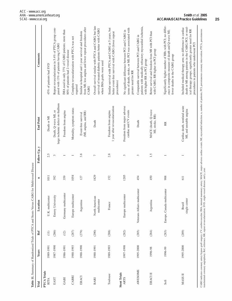
25Smith et al. 2005
ACC/AHA/SCAI Practice Guidelines
ACC - www.acc.orgAHA - www.americanheart.orgSCAI - www.scai.org
Tabl
e 11
.Sum
mar
y of
Ran
dom
ized
Tri
als
of P
TC
Aan
d St
ents
Ver
sus
CA
BG
for
Mul
tives
sel D
isea
se
Tri
alY
ears
Ref
Loc
atio
nn
Fol
low
-Up,
yE
nd P
oint
PT
CA
Tri
als
RIT
A19
89-1
991
(11)
U.K
. mul
ticen
ter
1011
2.5
Dea
th o
r M
I
EA
ST19
87-1
990
(286
)E
mor
y U
nive
rsity
392
3D
eath
, Q-w
ave
MI,
or
larg
e is
chem
ic d
efec
t on
thal
lium
GA
BI
1986
-199
1(1
2)G
erm
any
mul
ticen
ter
359
1Fr
eedo
m f
rom
ang
ina
CA
BR
I19
88-1
993
(287
)E
urop
e m
ultic
ente
r10
541
Mor
talit
y, s
ympt
om s
tatu
s
ER
AC
I19
88-1
990
(279
)A
rgen
tina
127
3.8
Eve
nt-f
ree
surv
ival
(M
I, a
ngin
a, a
nd R
R)
BA
RI
1988
-199
1(2
90)
Nor
th A
mer
ican
18
297
Dea
thm
ultic
ente
r
Toul
ouse
1989
-199
3(2
88)
Fran
ce15
22.
8Fr
eedo
m f
rom
ang
ina
1 ye
ar a
fter
rev
ascu
lari
zatio
n
Sten
t T
rial
sA
RT
S19
97-1
998
(282
)E
urop
e m
ultic
ente
r12
053
Free
dom
fro
m m
ajor
adv
erse
ca
rdia
c an
d C
Vev
ents
AW
ESO
ME
1995
-200
0(2
85)
Vet
eran
s A
ffai
rs m
ultic
ente
r45
43
Dea
th
ER
AC
I II
1996
-98
(284
) A
rgen
tina
450
1.5
MA
CE
(de
ath,
Q-w
ave
MI,
str
oke,
RR
)
SoS
1996
-99
(283
)E
urop
e, C
anad
a m
ultic
ente
r98
81
RR
MA
SS I
I19
95-2
000
(289
)B
razi
l61
11
Car
diac
dea
th, n
onfa
tal a
cute
si
ngle
cen
ter
MI,
and
uns
tabl
e an
gina
CA
BG
indi
cate
s co
rona
ry a
rter
y by
pass
gra
ft s
urge
ry; C
V, c
ereb
rova
scul
ar; I
MA
, int
erna
l mam
mar
y ar
tery
; MA
CE
, maj
or a
dver
se c
ardi
ac e
vent
s; M
I, m
yoca
rdia
l inf
arct
ion;
n, n
umbe
r of
pat
ient
s; P
CI,
per
cuta
neou
s co
rona
ry in
terv
entio
n; P
TC
A, p
ercu
tane
ous
tran
slum
inal
cor
onar
y an
giop
last
y; R
ef, r
efer
ence
; RR
, rep
eat r
evas
cula
riza
tion;
SV
D, s
ingl
e-ve
ssel
dis
ease
; and
y, y
ear.
Com
men
ts
45%
of
patie
nts
had
SVD
Rep
eat r
evas
cula
riza
tion
in 5
.4%
of
PTC
Agr
oup
com
-pa
red
with
13%
of
patie
nts
havi
ng C
AB
G
IMA
used
in o
nly
37%
of
CA
BG
pat
ient
s; m
ore
than
80%
of
patie
nts
had
2-ve
ssel
dis
ease
Com
plet
e re
vasc
ular
izat
ion
with
PT
CA
was
not
requ
ired
Sim
ilar
in-h
ospi
tal a
nd 1
-yea
r su
rviv
al a
nd f
reed
omfr
om M
I; le
ss a
ngin
a an
d fe
wer
rep
eat p
roce
dure
s af
ter
CA
BG
Ove
rall
surv
ival
sim
ilar
with
PT
CA
and
CA
BG
, but
late
surv
ival
of
trea
ted
diab
etic
pat
ient
s be
tter
with
CA
BG
whe
n IM
Agr
afts
wer
e us
ed
Sim
ilar
surv
ival
with
PT
CA
and
CA
BG
at 5
yea
rs, b
utbe
tter
even
t-fr
ee s
urvi
val w
ith C
AB
G (
few
er r
epea
tpr
oced
ures
)
No
sign
ific
ant d
iffe
renc
e be
twee
n PC
I an
d C
AB
G in
term
s of
dea
th, s
trok
e, o
r M
I; P
CI
was
ass
ocia
ted
with
grea
ter
need
for
RR
Com
para
ble
surv
ival
bet
wee
n PC
I an
d C
AB
G in
patie
nts
with
med
ical
ly r
efra
ctor
y m
yoca
rdia
l isc
hem
ia,
with
hig
her
RR
in P
CI
grou
p
Bet
ter
surv
ival
and
fre
edom
fro
m M
I w
ith P
CI
than
with
CA
BG
; RR
hig
her
in P
CI
grou
p
Sign
ific
antly
hig
her
num
ber
of R
Rs
with
PC
I; n
o di
ffer
-en
ce in
com
posi
te m
easu
re o
f de
ath
and
Q-w
ave
MI;
few
er d
eath
s in
the
CA
BG
gro
up
Incl
uded
med
ical
ther
apy
arm
; no
diff
eren
ce in
car
diac
deat
h or
MI
amon
g pa
tient
s in
the
CA
BG
, PC
I, o
r m
ed-
ical
ther
apy
grou
ps; s
igni
fica
ntly
gre
ater
nee
d fo
r R
Rpr
oced
ures
in p
atie
nts
who
und
erw
ent P
CI

26
ACC - www.acc.orgAHA - www.americanheart.org
SCAI - www.scai.orgSmith et al. 2005ACC/AHA/SCAI Practice Guidelines
Tabl
e 12
.PC
I C
ompa
riso
n W
ith M
edic
al T
hera
py Pat
ient
R
esul
tsSt
udy
Yea
rR
efn
Pop
ulat
ion
Tre
atm
ent
Fol
low
-Up
PC
IM
edic
al T
hera
pySi
gnif
ican
ce (
P)
AC
ME
1992
(296
)21
2Pa
tient
s w
ith
Med
ical
ther
apy
6 m
o64
% le
ss a
ngin
a46
% le
ss a
ngin
aL
ess
than
0.0
1si
ngle
-vs
vess
el d
isea
seba
lloon
ang
iopl
asty
VA
AC
ME
1997
(300
)32
8Pa
tient
s w
ith
Med
ical
ther
apy
3 y
63%
less
ang
ina
48%
less
ang
ina
0.02
docu
men
ted
vsch
roni
c ba
lloon
ang
iopl
asty
stab
le a
ngin
a
227
sing
le-
vess
el d
isea
se
101
doub
le-
vess
el d
isea
se
AC
IP19
97(3
01)
558
Patie
nts
with
A
ngin
a-gu
ided
2
y4.
7% d
eath
or
MI
8.8%
dea
th o
r M
I L
ess
than
0.0
1do
cum
ente
d C
AD
dr
ug th
erap
yfo
r is
chem
ia-g
uide
d an
d as
ympt
omat
ic
vs a
ngin
a-
drug
ther
apy
isch
emia
plus
isch
emia
guid
ed d
rug
12.1
% d
eath
or
MI
183
angi
na-
ther
apy
vs
for
angi
na-g
uide
dgu
ided
re
vasc
ular
izat
ion
drug
ther
apy
drug
ther
apy
183
angi
na-
plus
is
chem
ia-g
uide
d dr
ug th
erap
y
192
reva
scul
ariz
atio
n by
PT
CA
or C
AB
G
AV
ER
T19
99(2
98)
341
Patie
nts
with
sta
ble
Med
ical
ther
apy
18 m
o21
% is
chem
ic
13%
isch
emic
0.04
8; 0
.045
C
AD
, nor
mal
w
ith
even
tsev
ents
need
ed f
or
LVfu
nctio
n, a
nd
ator
vast
atin
sign
ific
ance
an
gina
cla
ss I
/II;
vs
PT
CA
due
to in
teri
m
patie
nts
requ
ired
anal
ysis
to c
ompl
ete
4 m
inon
Bru
ce p
roto
col
Con
tinu
ed o
n ne
xt p
age
Com
men
ts
The
PT
CA
grou
p ha
d le
ss a
ngin
a, b
ette
r ex
erci
se p
er-
form
ance
, and
mor
e im
prov
emen
t in
qual
ity-o
f-lif
esc
ores
but
had
mor
e co
mpl
icat
ions
(em
erge
ncy
bypa
ss2
patie
nts,
MI
in 5
, and
rep
eat P
TC
Ain
16)
Am
ong
patie
nts
with
sin
gle-
vess
el d
isea
se, t
he P
TC
Agr
oup
had
less
ang
ina,
bet
ter
exer
cise
per
form
ance
, and
mor
e im
prov
emen
t in
qual
ity-o
f-lif
e sc
ores
40%
of
patie
nts
had
prev
ious
MI,
23%
had
pri
or P
TC
Aor
CA
BG
, and
38%
had
trip
le-v
esse
l dis
ease
Onl
y 2
deat
hs a
mon
g 34
1 pa
tient
s in
18
mon
ths;
sig
nif-
ican
t im
prov
emen
t in
angi
na in
pat
ient
s tr
eate
d w
ithPT
CA
com
pare
d w
ith m
edic
al th
erap
y

27Smith et al. 2005
ACC/AHA/SCAI Practice Guidelines
ACC - www.acc.orgAHA - www.americanheart.orgSCAI - www.scai.org
Tabl
e 12
.Con
tinu
ed
Pat
ient
R
esul
tsSt
udy
Yea
rR
efn
Pop
ulat
ion
Tre
atm
ent
Fol
low
-Up
PC
IM
edic
al T
hera
pySi
gnif
ican
ce (
P)
RIT
A-2
2003
(299
)10
1853
% w
ith
Med
ical
ther
apy
7 y
14.5
%
12.3
% d
eath
0.
21C
lass
II
angi
navs
deat
h or
MI
or M
I47
% w
ith p
rior
ang
ina
ballo
on
7% w
ith tr
iple
-an
giop
last
yve
ssel
dis
ease
RIT
A-3
2002
(302
)18
10Su
spec
ted
card
iac
Med
ical
ther
apy
and
1 y
7.6%
dea
th
8.3%
dea
thN
Sch
est p
ain
at r
est
eith
er e
arly
inva
sive
or M
Ior
MI
with
doc
umen
ted
or c
onse
rvat
ive
CA
D (
at le
ast 1
of
(s
elec
tivel
y in
vasi
ve)
the
follo
win
g:tr
eatm
ent s
trat
egy;
EC
G c
hang
es,
both
gro
ups
rece
ived
pa
thol
ogic
al
enox
apar
in in
add
ition
Q w
aves
, pre
viou
sto
sta
ndar
d ar
teri
ogra
m)
med
ical
ther
apy
MA
SS-I
I20
04(2
89)
611
Stab
le a
ngin
a,
Med
ical
ther
apy,
1
y4.
5% d
eath
1.5%
dea
thN
Sm
ultiv
esse
l dis
ease
, C
AB
G, o
r PC
Ipr
eser
ved
LVfu
nctio
n
n in
dica
tes
num
ber
of p
atie
nts;
CA
BG
, cor
onar
y ar
tery
byp
ass
graf
t; C
AD
, cor
onar
y ar
tery
dis
ease
; MI,
myo
card
ial i
nfar
ctio
n; P
TC
A, p
ercu
tane
ous
tran
slum
inal
cor
onar
y an
giop
last
y; a
nd P
CI,
per
cuta
neou
s co
rona
ry in
terv
en-
tion.
Com
men
ts
PTC
Agr
oup
had
incr
ease
d ra
tes
of d
eath
and
MI
but
had
6.4%
less
cla
ss I
I an
gina
or
wor
se a
t 5 y
ears
and
long
er e
xerc
ise
trea
dmill
tim
e at
3 y
ears
Sim
ilar
resu
lts f
or d
eath
or
MI
betw
een
trea
tmen
tgr
oups
; sig
nifi
cant
dif
fere
nce
in p
rim
ary
end
poin
t(d
eath
, MI,
ref
ract
ory
angi
na)
due
to h
alvi
ng o
f re
frac
-to
ry a
ngin
a in
inte
rven
tion
grou
p
Agg
ress
ive
med
ical
man
agem
ent f
or m
ultiv
esse
l dis
-ea
se h
as lo
w in
cide
nce
of e
arly
eve
nts,
incl
udin
g de
ath
and
Q-w
ave
MI,
but
is in
feri
or to
PC
I (a
nd C
AB
G)
for
cont
rol o
f an
gina

28
ACC - www.acc.orgAHA - www.americanheart.org
SCAI - www.scai.orgSmith et al. 2005ACC/AHA/SCAI Practice Guidelines
The Atorvastatin Versus Revascularization Treatment(AVERT) trial (298) randomly assigned 341 patients withstable CAD, normal LV function, and class I and/or II angi-na to PTCA or medical therapy with 80 mg of atorvastatindaily (mean low-density lipoprotein cholesterol equals 77mg per dL). At 18 months of follow-up, 13% of the medical-ly treated group had ischemic events compared with 21% ofthe PTCA group (P equals 0.048). Angina relief was greaterin those treated with PTCA. Although not statistically differ-ent when adjusted for interim analysis, these data suggestthat in low-risk patients with stable CAD, aggressive lipid-lowering therapy can be as effective as PTCA in reducingischemic events.
During the MASS-II trial (289), 611 patients with stableangina, multivessel disease, and preserved LV function wererandomized to 3 treatment groups: medical therapy, CABG,or PCI (medical therapy n equals 203, CABG n equals 203,and PCI n equals 205). One-year survival was similar in the3 groups at 98.5%, 96.0%, and 95.6%, respectively. At 1 yearof follow-up, a Q-wave MI had occurred in 2% of CABGpatients, 8% of the PCI patients, and 3% of the medical ther-apy patients. By 1 year, additional revascularization proce-dures were performed in 8.3% of medical therapy patients,13.3% of PCI patients, and only 0.5% of CABG patients.More patients were free of angina at 1 year in the CABG andPCI groups (88% and 79%, respectively) than in the medicaltherapy groups, in which only 46% were free of angina. Thissmall contemporary trial utilizing aggressive medical man-agement demonstrated that medical therapy for multivesseldisease has a low incidence of early events including deathand Q-wave MI but is inferior to PCI and CABG for the con-trol of angina.
Given the limited data available from randomized trialscomparing medical therapy with PCI, it seems prudent toconsider medical therapy for the initial management of mostpatients with CCS classification class I and II stable anginaand to reserve PCI and CABG for those patients with moresevere symptoms and ischemia. The symptomatic patientwho wishes to remain physically active, regardless of age,will usually require PCI or CABG to accomplish this.
The Clinical Outcomes Utilization Revascularization andAggressive Drug Evaluation (COURAGE) trial wasdesigned to compare intensive medical therapy with PCI plusintensive medical therapy. Enrollment has been completed,and results are expected to be available in the next few years.This trial will provide further valuable information about therelative merits of medical treatment plus PCI versus medicaltreatment alone and will also give us a detailed assessment ofoutcomes relative to quality of life and economic cost (303).The Bypass Angioplasty Revascularization trial in patientswith diabetes (BARI 2d) was designed to compare revascu-larization in addition to aggressive medical therapy inpatients with diabetes compared with aggressive medicaltherapy alone. Enrollment was completed in the first quarterof 2005.
Patients with UA and NSTEMI have been randomized tomedical therapy or PCI in the FRagmin and Fast
time of 6-month follow-up. Thus, these patients with double-vessel PTCA did not demonstrate superior control of theirsymptoms as compared with medical therapy, as was experi-enced by the ACME patients with single-vessel disease. Thissmall study suggests that PTCA is less effective in control-ling symptoms in patients with double-vessel disease andstable angina than in those with single-vessel disease.
The Randomized Intervention Treatment of Angina(RITA)-2 investigators randomized 1018 patients with stableangina to PTCA or conservative (medical) therapy(297,299). Patients who had inadequate control of theirsymptoms with optimal medical therapy were allowed tocross over to myocardial revascularization. The combinedend point of the trial was all-cause mortality and nonfatal MI.The 504 PTCA and 514 medically treated patients were fol-lowed up for a mean of 7 years. Death due to all causesoccurred in 43 (8.5%) of the PTCA patients and 43 (8.4%) ofthe medical patients. Of the 86 deaths, only 8 were due toheart disease. Angina improved in both groups, but there wasa 16.5% absolute excess of grade 2 or worse angina in themedical group at 3 months after randomization (P less than0.001). These differences in angina narrowed over time, withthe PTCA group always having less angina than the med-ically treated patients. Thus, RITA-2 demonstrated thatPTCA results in better control of symptoms of ischemia andimproves exercise capacity compared with medical therapybut is associated with a higher combined end point of deathand periprocedural MI. It is important to remember thatalthough the patients in this trial were asymptomatic or hadonly mild angina, 62% of them had multivessel CAD, and34% had significant disease in the proximal segment of theLAD (301). Thus, most of these patients had severe anatom-ic CAD.
The Asymptomatic Cardiac Ischemia Pilot (ACIP) studyprovides additional information comparing medical therapywith PTCA or CABG revascularization in patients with doc-umented CAD and asymptomatic ischemia by both stresstesting and ambulatory ECG monitoring (301). This trial ran-domized 558 patients suitable for revascularization by PTCAor CABG to 3 treatment strategies: angina-guided drug ther-apy (n equals 183), angina- plus ischemia-guided drug ther-apy (n equals 183), and revascularization by PTCA or CABGsurgery (n equals 192). Of the 192 patients who were ran-domized to revascularization, 102 were selected for PTCAand 90 for CABG. At 2 years of follow-up, death or MI hadoccurred in 4.7% of the revascularization patients comparedwith 8.8% of the ischemia-guided group and 12.1% of theangina-guided group (P less than 0.01). Because a large por-tion of the patients underwent CABG surgery instead ofPTCA to achieve complete revascularization, it is not appro-priate to directly compare these results with RITA-2.Nonetheless, the ACIP study suggests that outcomes ofrevascularization with CABG surgery and PTCA are veryfavorable compared with medical therapy in patients withasymptomatic ischemia with or without mild angina. Itshould be emphasized that aggressive lipid-lowering therapywas not widely employed in either treatment arm of ACIP.

29Smith et al. 2005
ACC/AHA/SCAI Practice Guidelines
ACC - www.acc.orgAHA - www.americanheart.orgSCAI - www.scai.org
Revascularisation during InStability in Coronary artery dis-ease (FRISC) II and Treat Angina with Aggrastat and deter-mine the Cost of Therapy with an Invasive or ConservativeStrategy (TACTICS) TIMI 18 trials, as well as in RITA-3.These trials utilizing stenting as the primary therapy havefavored the invasive approach (206,302,304). They are dis-cussed in Section 5.3.
4. INSTITUTIONAL AND OPERATORCOMPETENCY
4.1. Quality Assurance
Class I1. An institution that performs PCI should establish an
ongoing mechanism for valid peer review of its quali-ty and outcomes. Review should be conducted both atthe level of the entire program and at the level of theindividual practitioner. Quality-assessment reviewsshould take risk adjustment, statistical power, andnational benchmark statistics into consideration.Quality-assessment reviews should include both tabu-lation of adverse event rates for comparison withbenchmark values and case review of complicatedprocedures and some uncomplicated procedures.(Level of Evidence: C)
2. An institution that performs PCI should participate ina recognized PCI data registry for the purpose ofbenchmarking its outcomes against current nationalnorms. (Level of Evidence: C)
Definition of Quality in PCI
Satisfactory quality in PCI may be defined as the appropriateselection of patients for the procedure and the achievementof risk-adjusted outcomes that are comparable to nationalbenchmark standards in terms of procedure success andadverse event rates. To achieve optimal quality and outcomesin PCI, it is necessary that both the physician operator andthe supporting institution be appropriately skilled and expe-rienced.
Institutional Quality-Assurance Requirement
PCI is a demanding, technically complex procedure. Thepotential exists for substantial quality variation among bothoperators and institutions.
In the United States, responsibility for quality assurance isvested in the healthcare institution, which is responsible tothe public to ensure that patient care conducted under itsjurisdiction is of acceptable quality. Thus, the institution hasthe responsibility to monitor its PCI program’s quality withrespect to process, appropriateness, and outcomes in order toidentify and correct any circumstances in which quality fallsbelow accepted norms. Quality-assessment review should beconducted both at the level of the entire program and at thelevel of the individual practitioner.
Each institution that performs PCI must establish an ongo-ing mechanism for valid peer review of its quality and out-comes. The program should provide an opportunity for inter-
ventionalists, as well as for physicians who do not performangioplasty but are knowledgeable about it, to review itsoverall results on a regular basis. The review process shouldtabulate the results achieved both by individual physicianoperators and by the overall program and compare them withnational benchmark standards with appropriate risk adjust-ment. Valid quality assessment requires that the institutionmaintain meticulous records that include the patient demo-graphic and clinical characteristics necessary to assessappropriateness and to conduct risk adjustment.
Role of Risk Adjustment in Assessing Quality
A raw adverse event rate that is not appropriately risk adjust-ed has little meaning. Data compiled from large registries ofprocedures performed in recent years have generated multi-variate risk-adjustment models for adverse event rates forPCI in the current era. Six multivariate models of the risk ofmortality after PCI have been published (43,47,305-308).
Although these models differ somewhat, they are consis-tent in identifying acute MI, shock, and age as importantrisk-stratification variables for mortality. The ACC-NCDRreported a univariate mortality rate of 0.5% for patientsundergoing elective PCI, a mortality rate of 5.1% for patientsundergoing primary PCI within 6 h of the onset of STEMI,and a mortality rate of 28% for patients undergoing PCI forcardiogenic shock (305). Thus, it is clear that to assess PCImortality rates, patients should be stratified by whether theyare undergoing elective PCI, primary PCI for acute STEMIwithout shock, or primary PCI for STEMI with shock.
Challenges in Determining Quality
As discussed above, given the complexity of case selectionand procedure conduct, quality is difficult to measure in PCIand is not determined solely by adverse event rates evenwhen properly adjusted for risk. Accurate assessment ofquality becomes more problematic for low-volume operatorsand institutions, because absolute event rates are expected tobe small. Thus, particularly in low-volume circumstances,quality may be better assessed by an intensive case reviewprocess conducted by recognized experts who can properlyjudge all of the facets of the conduct of a case. Case reviewalso has merit in high-volume situations, because it can iden-tify subtleties of case selection and procedure conduct thatmay not be reflected in pooled statistical data.
Requirement for Institutional Resources and Support
A high-quality PCI program requires appropriately trainedand experienced skilled physician operators. However, theoperator does not work in a vacuum. An operator needs awell-maintained, high-quality cardiac catheterization facilityto practice effectively. In addition, the operator depends on amultidisciplinary institutional infrastructure for support andresponse to emergencies. Thus, to provide quality PCI serv-ices, the institution must ensure that its catheterization facil-ity is properly equipped and managed and that all of its nec-essary support services are of high quality and are readilyavailable.
30
ACC - www.acc.orgAHA - www.americanheart.org
SCAI - www.scai.orgSmith et al. 2005ACC/AHA/SCAI Practice Guidelines
tional techniques such as rotational atherectomy, balloonvalvuloplasty, and closure of patent foramen ovale and atrialseptal defect.
It is recommended that an interventional cardiology opera-tor be certified by the American Board of Internal Medicinein interventional cardiology. Ideally, board certification ininterventional cardiology should be required for credential-ing. The American Board of Internal Medicine certifyingexamination in interventional cardiology has been adminis-tered annually since 1999. As of the 2004 administration, atotal of 4718 individuals have been certified.
Privilege Renewal
Criteria for practitioner privilege renewal should be based onboth activity level and outcomes. The assessment processshould ascertain whether a physician operator’s activity levelis sufficient to maintain competence. In addition, the assess-ment process should assess the appropriateness of the opera-tor’s case selection and compare the operator’s risk-adjustedoutcomes with established national benchmark standards(310). This is discussed in depth in Section 4.2. Currentbenchmark standards for mortality, complication rates, andrisk adjustment will be subject to future revision as proce-dure technique is refined and newer data emerge. It is impor-tant that institutions assist with these efforts by participatingin active database efforts to track clinical and proceduralinformation for individual operators and their institutions.
Outcome Data Tabulation and Reporting
Institutions performing PCI should gather data needed tomonitor their outcomes and should submit their data to anational registry for benchmarking purposes. Institutionsshould conduct meticulous record keeping that details thecases performed-patient demographics and comorbidities,
The Quality-Assessment Process
Quality assessment is a complex process that includes morethan mere tabulation of success and complication rates.Components of quality in coronary interventional proceduresinclude appropriateness of case selection, quality of proce-dure execution, proper response to intraprocedural problems,accurate assessment of procedure outcome, and appropriate-ness of postprocedure management. It is important to con-sider each of these parameters when conducting a quality-assessment review. A quality program performs appropriate-ly selected procedures that achieve risk-adjusted outcomes,in terms of procedure success and complication rates, that arecomparable to national benchmark standards. Patient charac-teristics that determine appropriateness are discussed else-where in this document. Multivariate models that predict riskhave been published previously (43,47,305-308).
It is accepted that quality-assurance monitoring is best con-ducted through the peer review process despite the politicalchallenges associated with colleagues evaluating each other.There has been a considerable controversy surroundingefforts to define standards, criteria, and methodologies forconducting quality assessment. There are many challenges toconducting this process in a fair and valid manner.
The cornerstone of quality-assurance monitoring is theassessment of procedural outcomes in terms of success andadverse event rates. Other components of quality-assurancemonitoring include establishment of criteria for assessingprocedure appropriateness and application of proper riskadjustment to interpret adverse event rates. Because adverseevents should be rare, a valid estimate of a properly risk-adjusted adverse event rate generally requires tabulation ofthe results of a large number of procedures. This adds anadditional challenge to the valid assessment of low-volumeoperators and institutions. The responsible supervisingauthority should monitor the issues outlined in Table 13(309).
Initial Physician Operator Credentialing Criteria
The institutional credentialing committee should documentthat an interventionalist wishing to initiate practice meets theestablished training criteria, including those of the ACC TaskForce on Training in Cardiac Catheterization andInterventional Cardiology (310-312). The ACC TrainingStatement (312) for coronary invasive training requires a 3-year comprehensive cardiac program with 12 months oftraining in diagnostic catheterization, during which thetrainee performs 300 diagnostic catheterizations, with at least200 of those as the primary operator. Interventional trainingrequires a fourth year of fellowship, during which the traineeshould perform more than 250 but not more than 600 inter-ventional procedures (312). The physician’s training pro-gram director should certify that the candidate has complet-ed the program and has achieved the necessary competenceto perform coronary interventional procedures independent-ly. The certification should also include whether the candi-date has achieved requisite competence in related interven-
Table 13. Key Components of a Quality-Assurance Program
Clinical proficiencyGeneral indications/contraindicationsInstitutional and individual operator complication rates, mortalityand emergency CABG
Institutional and operator procedure volumesTraining and qualifications of support staff
Equipment maintenance and managementQuality of laboratory facility [See ACC/SCAI Expert ConsensusDocument on Catheterization Laboratory Standards (309)]
Quality improvement processEstablishment of an active concurrent database to track clinical andprocedural information and patient outcomes for individual opera-tors and the institution. The ACC-NCDR® or other databases arestrongly recommended for this purpose
Radiation safetyEducational program in the diagnostic use of X-rayPatient and operator exposure
ACC indicates American College of Cardiology; CABG, coronary artery bypass graft sur-gery; NCDR®, National Cardiovascular Data Registry; and SCAI, Society forCardiovascular Angiography and Interventions.

31Smith et al. 2005
ACC/AHA/SCAI Practice Guidelines
ACC - www.acc.orgAHA - www.americanheart.orgSCAI - www.scai.org
Class IIbThe benefit of primary PCI for STEMI patients eligi-ble for fibrinolysis when performed by an operatorwho performs fewer than 75 procedures per year (orfewer than 11 PCIs for STEMI per year) is not wellestablished. (Level of Evidence: C)
Class IIIIt is not recommended that elective PCI be performedby low-volume operators (fewer than 75 proceduresper year) at low-volume centers (200 to 400) with orwithout onsite cardiac surgery (310,312). An institu-tion with a volume of fewer than 200 procedures peryear, unless in a region that is underserved because ofgeography, should carefully consider whether itshould continue to offer this service. (Level ofEvidence: B)
Operator and Institution Volume-OutcomeRelationships
Threshold activity level standards for institutions and opera-tors have been particularly controversial. Such standards are
cardiovascular characteristics including type of presentation,coronary anatomy, ventricular function, procedures per-formed, and periprocedural complications. These data arenecessary to permit appropriate risk adjustment. Institutionsshould carefully monitor their risk-adjusted outcomes at thelevel of the institution and of the individual operators andshould ascertain that their outcomes fall within nationalnorms. One example is the ACCF CathKit®, a tool that pro-vides templates and guidance for the quality assessmentprocess.
This Writing Committee agrees with the ACC Task Forcerecommendations for the Assessment and Maintenance ofProficiency in Coronary Interventional Procedures (310).Institutions and healthcare providers performing PCI shouldmeet the standards outlined in Table 14 (309,310,312) and inSection 4.2.
4.2. Operator and Institutional Volume
Class I1. Elective PCI should be performed by operators with
acceptable annual volume (at least 75 procedures) athigh-volume centers (more than 400 procedures) withonsite cardiac surgery (310,312). (Level of Evidence:B)
2. Elective PCI should be performed by operators andinstitutions whose historical and current risk-adjust-ed outcomes statistics are comparable to those report-ed in contemporary national data registries. (Level ofEvidence: C)
3. Primary PCI for STEMI should be performed byexperienced operators who perform more than 75elective PCI procedures per year and, ideally, at least11 PCI procedures for STEMI per year. Ideally, theseprocedures should be performed in institutions thatperform more than 400 elective PCIs per year andmore than 36 primary PCI procedures for STEMI peryear. (Level of Evidence B)
Class IIa1. It is reasonable that operators with acceptable volume
(at least 75 PCI procedures per year) perform PCI atlow-volume centers (200 to 400 PCI procedures peryear) with onsite cardiac surgery (310,312). (Level ofEvidence: B)
2. It is reasonable that low-volume operators (fewer than75 PCI procedures per year) perform PCI at high-vol-ume centers (more than 400 PCI procedures per year)with onsite cardiac surgery (310,312). Ideally, opera-tors with an annual procedure volume less than 75should only work at institutions with an activity levelof more than 600 procedures per year. Operators whoperform fewer than 75 procedures per year shoulddevelop a defined mentoring relationship with a high-ly experienced operator who has an annual procedur-al volume of at least 150 procedures per year. (Level ofEvidence: B)
Table 14. Considerations for the Assessment and Maintenance ofProficiency in Coronary Interventional Procedures
InstitutionsQuality-assessment monitoring of privileges and risk-stratified out-comes
Provide support for a quality-assurance staff person (e.g., nurse) tomonitor complications
Minimal institutional performance activity of 200 interventions peryear, with ideal minimum of 400 interventions per year
Interventional program director who has a career experience ofmore than 500 PCI procedures and who is board certified by theABIM in interventional cardiology
Facility and equipment requirements to provide high-resolution flu-oroscopy and digital video processing
Experienced support staff to respond to emergencies (see Section4.3, Role of On-Site Cardiac Surgical Backup for discussion)
Establishment of a mentoring program for operators who performfewer than 75 procedures per year by individuals who perform atleast 150 procedures per year
PhysiciansProcedural volume of 75 per year or more
Continuation of privileges based on outcome benchmark rates, withconsideration of not granting privileges to operators who exceedadjusted case mix benchmark complication rates for a 2-year peri-od
Ongoing quality assessment comparing results with current bench-marks, with risk stratification of complication rates
Board certification by ABIM in interventional cardiology
ABIM indicates American Board of Internal Medicine; and PCI, percutaneous coronaryintervention.
32
ACC - www.acc.orgAHA - www.americanheart.org
SCAI - www.scai.orgSmith et al. 2005ACC/AHA/SCAI Practice Guidelines
Other studies have also supported the relationship of com-plication rate to procedural volume (47,306,314). Althoughsome investigators have suggested that low procedure vol-ume does not contribute to poor outcomes (44,309), thesestudies are small in number and underpowered for analysis(318).
Progress in technique and instrumentation has reducedabsolute complication rates, which makes the proceduresafer and more effective. This has fueled the opinion that thevolume-outcome relationship has weakened, justifying advo-cacy that PCI be diffused to smaller-volume institutions andlower-volume operators. Although it is possible to considerearlier studies anachronistic because of the lack of availabil-ity of coronary stents and other adjunctive therapies, studiesbased on data sets accumulated in the stent era continue toshow a volume-outcome relationship (albeit with lowerabsolute event rates).
Brown evaluated the outcomes of PCI at all hospitals inCalifornia in 1997 (324). Mortality for PCI in which a stentwas used was 1.5% in hospitals performing fewer than 400procedures per year compared with 1.1% in hospitals per-forming more than 400 procedures per year. The rate ofemergent CABG was 1.2% in hospitals performing fewerthan 400 procedures per year compared with 0.8% in hospi-tals that performed more than 400 procedures per year.
Moscucci et al. studied the outcomes of 18 504 consecutivePCIs performed at 14 hospitals in Michigan in 2002 (325).Operator volume was divided in quintiles (1-33, 34-89, 90-139, 140-206, and 207-582 procedures per year). The pri-mary end point was a composite of MACE including death,CABG, stroke or TIA, MI, and repeat PCI at the same siteduring the index hospitalization. The unadjusted MACE ratewas significantly higher in quintiles 1 and 2 of operator vol-ume than in quintile 5 (7.38% and 6.13% vs 4.15%, P equals0.002 and P equals 0.0001, respectively). A similar trend wasobserved for in-hospital death. After adjustment for comor-bidities, patients treated by low-volume operators had a 63%increased odds of MACE (adjusted odds ratio [OR] 1.63,95% CI 1.29-2.06, P less than 0.0001 for quintile 1; adjustedOR 1.63, 95% CI 1.34-1.90, P less than 0.0001 for quintile 2vs quintile 5) but not of in-hospital death. Overall, high-vol-ume operators had better outcomes than low-volume opera-tors in both low-risk and high-risk patients (325).
Distinction Between Elective PCI and Primary PCI for STEMI
Elective PCI and primary PCI for STEMI are different,although related, disciplines. Experience in elective PCItranslates only partially to experience with primary PCI forSTEMI. Throughout this guideline, a distinction is drawnbetween primary PCI, which is performed under emergencycircumstances, and all other PCI procedures, which areincluded under the term “elective.” The volume-outcomerelationship exists for both elective procedures and primaryangioplasty for STEMI (326-328) but has important differ-ences. Available data indicate that the best results are
derived from the principle that proper skill maintenancerequires a requisite activity level. It is logical that both athreshold experience and an ongoing activity level are nec-essary to achieve and maintain requisite proficiency in PCI.
Standards originally formulated for the earliest versions ofthese guidelines were based on opinion (Level of Evidence:C) drawing on the well-documented existence of volume-outcome relationships for other complex surgical procedures.Initially, a panel of experts identified a threshold activitylevel of 75 procedures per year as necessary for maintenanceof competence in PCI (313). Subsequent studies of PCI con-tinue to identify annual procedural volume both at the pro-gram level and at the operator level as strongly correlatedwith complication rates. Most studies’ findings are consistentwith the operator threshold of 75 procedures per year (47,306,309,314-320).
Most studies of the PCI volume-outcome relationship focuson mortality and emergent bypass surgery as quality-deter-mining outcome variables. These variables, while important,encompass only a portion of the overall quality determinantsfor PCI.
McGrath et al. examined volume outcome relationshipsusing procedures performed on 167 208 Medicare recipientsin 1997 (321). Procedures performed by low-volume physi-cians (fewer than 30 Medicare procedures per year) had agreater emergency CABG rate (2.25%) than procedures per-formed by high-volume physicians (more than 60 Medicareprocedures per year; 1.55%, P less than 0.001). An increased30-day mortality rate was found for low-volume programs(fewer than 80 Medicare procedures per year) versus high-volume programs (more than 160 Medicare procedures peryear; 4.29% vs 3.15%, P less than 0.001).
Kimmel et al., using data from the SCAI, found that aninverse relationship existed between the number of angio-plasty procedures performed at a hospital and the rate ofmajor complications (315). These results were risk stratifiedand independent of the patient-risk profile. Significantlyfewer complications occurred in laboratories that performedat least 400 angioplasty procedures per year.
Jollis et al. found that low-volume hospitals were associat-ed with higher rates of emergency coronary artery bypasssurgery and death (316). Improved outcomes were identifiedwith a threshold volume of 75 Medicare angioplasties perphysician and 200 Medicare angioplasty procedures per hos-pital. Using a 35% to 50% ratio of Medicare patients, thethreshold value was 150 to 200 angioplasty procedures percardiologist and 400 to 600 angioplasty procedures per insti-tution (322).
Epstein et al., using an administrative data set, analyzedrisk-adjusted mortality in 362748 admissions to 1000 UnitedStates hospitals between 1997 and 2000 during which a PCIwas performed (323). They found a consistent trend ofdecreasing risk-adjusted mortality with increasing hospitalvolume. The differences between groups were small, andthere was considerable heterogeneity within groups, whichindicates that hospital volume is not the sole determinant ofoutcome.
33Smith et al. 2005
ACC/AHA/SCAI Practice Guidelines
ACC - www.acc.orgAHA - www.americanheart.orgSCAI - www.scai.org
obtained by operators who are highly experienced both inelective PCI and in primary PCI for STEMI who work ininstitutions that have established an active program for per-forming primary PCI for STEMI.
Operator experience in elective PCI is not sufficient to con-fer expertise in primary PCI for STEMI. This finding is notsurprising, because there are aspects of procedure conductthat are unique to primary PCI for STEMI.
Vakili et al., analyzing primary PCI procedures for STEMIperformed in New York State, found no relationship betweenphysician total angioplasty procedure volume and mortalityafter primary PCI for STEMI but did find an associationbetween an operator’s primary PCI activity level and the out-come of primary PCI for STEMI that was independent of theoperator’s experience in elective PCI (328,329). Low-vol-ume physicians, who performed 1 to 10 primary PCI proce-dures per year, had an unadjusted mortality rate of 7.1%compared with 3.8% for physicians who performed 11 ormore primary PCI procedures per year.
Magid et al. analyzed the National Registry of MyocardialInfarction (NRMI) database and grouped acute-care hospi-tals by volume tertiles of primary PCI for STEMI procedures(327). They found a reduction in risk-adjusted mortality withincreasing hospital volume of PCI: low volume (fewer than16 procedures), 6.2%; intermediate volume (17 to 48 proce-dures), 4.5%; and high volume (more than 49 procedures),3.4% (327). Canon et al. analyzed or reviewed 20 080 con-secutive patients with STEMI in the NRMI-2 database (330).A multivariate model was used to show that overall adjustedmortality was lower as volume increased, with the greatestreduction in mortality occurring at hospitals performingmore than 3 angioplasties per month (330). Different studiesidentified different cutpoints. The relationship between thestudies is graded, and the individual cutpoints are artifacts ofanalysis methodology.
Vakili et al. found a doubling of mortality in STEMIpatients who underwent PCI in hospitals that performedfewer than 400 total PCI procedures per year compared withhospitals that performed more than 400 (8.1% vs 4.3%)(329). Furthermore, they found that high-volume hospitalsthat performed more than 56 primary PCI procedures peryear had a nonsignificant trend toward a lower crude mortal-ity rate (4.0% vs 5.8%), with a multivariate OR for mortali-ty of 0.53 (0.29 to 1.1). The best outcomes were achieved byhigh-volume physicians working in high-volume hospitals(crude mortality rate 3.7% compared with 7.1% for low-vol-ume physicians in low-volume hospitals; adjusted relativerisk 0.51, 95% CI 0.26 to 0.99).
Canto et al. (331) also found a graded relationship betweenhospital volume and mortality after PCI for STEMI. Thehighest quartile of hospital volume performed more than 33primary PCIs for STEMI per year and achieved a 28% reduc-tion in mortality compared with the lowest-volume hospitals.
Appropriateness of Activity Levels as a Surrogate for Quality
The documented relationships between activity level andoutcome are statistical associations, and activity level is nota surrogate for quality. The heterogeneity within hospitalvolume groups found by Epstein et al. (323) validates thatactivity level is an incomplete surrogate for quality. An activ-ity level above a threshold value does not guarantee goodquality, and an activity level below a threshold value may notnecessarily indicate lower quality. Thus, high-volume opera-tors and institutions are not necessarily of uniformly highquality, and low-volume operators and institutions are not,by definition, poor.
However, an activity level below a threshold necessarilyraises the question of whether an operator or institution hassufficient ongoing experience to maintain expertise andskills. In particular, it is plausible that an operator or institu-tion that is below a threshold activity level cannot accrue thenecessary ongoing experience to perform complex proce-dures skillfully, to acquire experience with new techniquesand devices, and to respond effectively to adverse and emer-gency situations. The emergency response consideration isparticularly relevant, because the likelihood of a seriouscomplication cannot be predicted from patient baseline char-acteristics.
Quality Assessed by Outcomes: Statistical PowerConsiderations and the Importance of Case Reviews
The quality of both institutions and operators should ulti-mately be judged through the quality-assessment process asoutlined in Section 4.1. Because expected adverse event ratesare low, a large number of procedures are required to achievethe requisite statistical power to assign an interpretable con-fidence interval to an operator’s or a program’s adverse eventrate estimate. Furthermore, adverse event rates cannot beinterpreted without appropriate risk adjustment.
The first approximation in assessing an operator’s or a pro-gram’s quality is to compare the actual adverse event rate toan expected rate as predicted by an accepted risk-predictionmodel (ACC-NCDR® model or Dynamic Registry model).Calculation of an expected adverse event rate can be con-ducted by entering the characteristics of the group of patientstreated into the model. The model yields an expected adverseevent rate with confidence intervals that can then be com-pared with the actual event rate. Interpretation of the expect-ed adverse event rate is complex because of the precision ofthe estimate. An arbitrary criterion will need to be applied todetermine whether a particular actual adverse event rate is anoutlier when compared with the expected event rate. Forexample, 50% of operators may be expected to have anadverse event rate above the expected value purely bychance. Thus, merely being above the predicted mean valuedoes not automatically identify an operator or a program asan outlier.
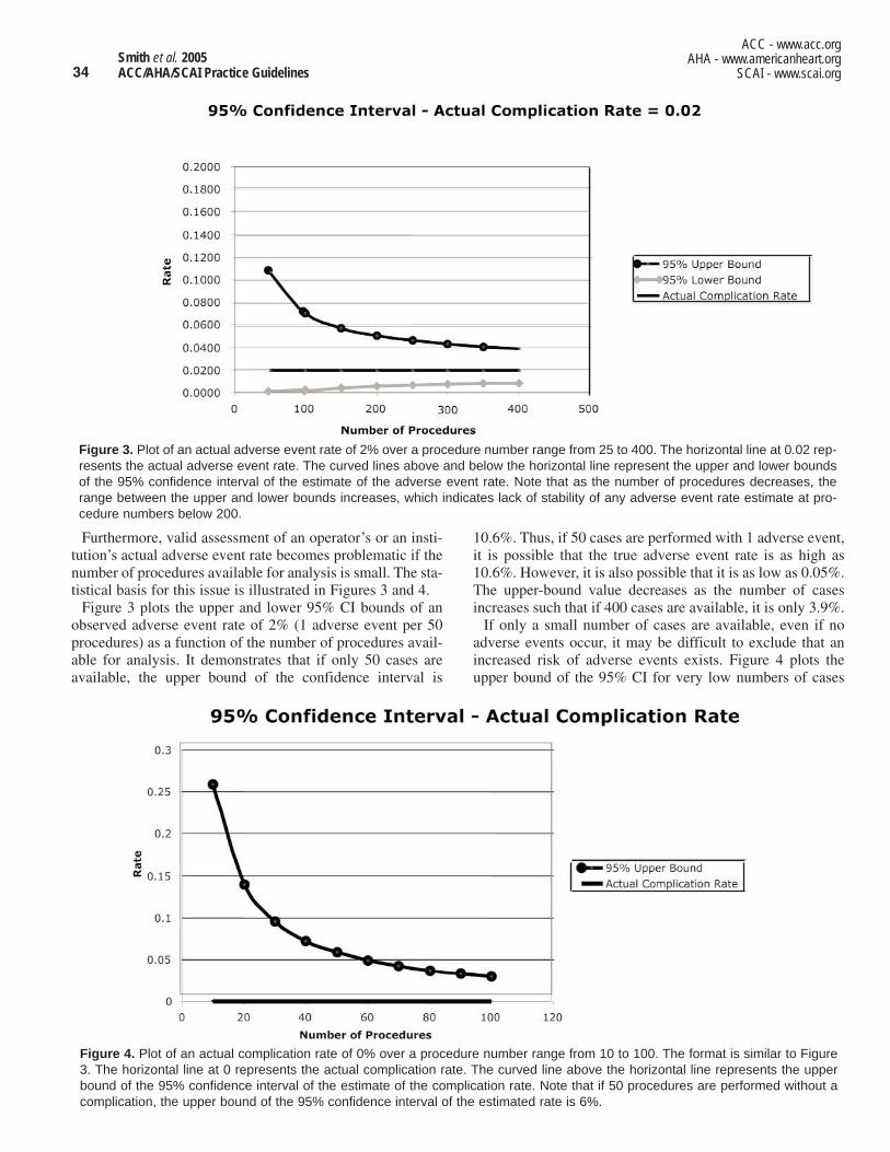
34
ACC - www.acc.orgAHA - www.americanheart.org
SCAI - www.scai.orgSmith et al. 2005ACC/AHA/SCAI Practice Guidelines
10.6%. Thus, if 50 cases are performed with 1 adverse event,it is possible that the true adverse event rate is as high as10.6%. However, it is also possible that it is as low as 0.05%.The upper-bound value decreases as the number of casesincreases such that if 400 cases are available, it is only 3.9%.
If only a small number of cases are available, even if noadverse events occur, it may be difficult to exclude that anincreased risk of adverse events exists. Figure 4 plots theupper bound of the 95% CI for very low numbers of cases
Furthermore, valid assessment of an operator’s or an insti-tution’s actual adverse event rate becomes problematic if thenumber of procedures available for analysis is small. The sta-tistical basis for this issue is illustrated in Figures 3 and 4.
Figure 3 plots the upper and lower 95% CI bounds of anobserved adverse event rate of 2% (1 adverse event per 50procedures) as a function of the number of procedures avail-able for analysis. It demonstrates that if only 50 cases areavailable, the upper bound of the confidence interval is
Figure 4. Plot of an actual complication rate of 0% over a procedure number range from 10 to 100. The format is similar to Figure3. The horizontal line at 0 represents the actual complication rate. The curved line above the horizontal line represents the upperbound of the 95% confidence interval of the estimate of the complication rate. Note that if 50 procedures are performed without acomplication, the upper bound of the 95% confidence interval of the estimated rate is 6%.
Figure 3. Plot of an actual adverse event rate of 2% over a procedure number range from 25 to 400. The horizontal line at 0.02 rep-resents the actual adverse event rate. The curved lines above and below the horizontal line represent the upper and lower boundsof the 95% confidence interval of the estimate of the adverse event rate. Note that as the number of procedures decreases, therange between the upper and lower bounds increases, which indicates lack of stability of any adverse event rate estimate at pro-cedure numbers below 200.

35Smith et al. 2005
ACC/AHA/SCAI Practice Guidelines
ACC - www.acc.orgAHA - www.americanheart.orgSCAI - www.scai.org
performed with a zero adverse event rate. It demonstratesthat if 10 consecutive cases are performed without a compli-cation, the upper bound of the 95% CI is 25%. If 50 casesare performed without an adverse event, the upper bound is5.8%.
Thus, although it is likely that certain low-volume opera-tors and institutions perform procedures with acceptablequality, satisfactory quality is difficult to prove unless a suf-ficient number of procedures are compiled for analysis. Thequality-assessment process must take the above issues intoconsideration. This means that it is essential that institutionand operator outcomes be tracked over sufficiently longperiods of time to assemble a sufficient number of proce-dures to permit a satisfactory analysis.
In addition, mere tabulation of adverse event rates, evenwith appropriate risk adjustment, is inadequate to judgeoperator or program quality. Such tabulations do not addressnumerous other quality issues, in particular, appropriateness.Thus, the quality-assessment process should also conductdetailed reviews both of cases that have adverse outcomes(to determine the cause(s) of the adverse event) and ofuncomplicated cases (to judge case selection appropriate-ness and procedure execution quality). These reviews shouldbe conducted by recognized experienced interventionalistsdrawn either from within the institution or externally if arequisite number of appropriately qualified, unconflictedindividuals are not available.
Role of Low-Volume PCI Programs
There is an ongoing debate as to whether PCI servicesshould be diffused widely to be available in most healthcareinstitutions or whether the service should be regionalizedand concentrated in specialized high-volume centers. Giventhe widespread availability of sophisticated interventional/surgical programs in the United States, it is difficult todemonstrate a need for additional low-volume programs toperform elective angioplasty except in underserved areasthat are geographically distant from major centers. At thiswriting, outcome data that link activity level to outcomesindicate that the development of small cardiovascular surgi-cal programs to support angioplasty is a poor use ofresources that will likely lead to suboptimal results (320). Ingeneral, the proliferation of small angioplasty or small sur-gical programs to support such angioplasty programs is notneeded to improve patient access to PCI services and wouldappear not to be in the interest of fostering optimal quality;thus, it should be discouraged. An exception to this principleshould be when geographic considerations become impor-tant determinants of patient access.
These data support the conclusion that not every cardiolo-gist desiring to perform PCI should perform these proce-dures, and not every hospital that would like to have aninterventional program should start one (322). This caveat isparticularly true where high-volume programs and operatorsare already nearby.
The Writing Committee, therefore, recommends that elec-tive PCI be performed by higher-volume operators (75 casesper year) with advanced technical skills (e.g., subspecialtycertification) at institutions with fully equipped interven-tional laboratories and an experienced support staff. Thissetting is optimally a high-volume center (more than 400cases per year) with an onsite cardiovascular surgical pro-gram (332).
It is recommended that primary PCI for STEMI be per-formed by higher-volume operators experienced in bothelective PCI and primary PCI for STEMI with ongoingactivity levels of more than 75 elective PCI procedures peryear and, ideally, annual PCI for STEMI activity levels of atleast 11 per year. It is clear that an effective PCI for STEMIprogram, irrespective of whether cardiac surgery is availableonsite, requires appropriate physician operator expertise,appropriate institutional commitment, and the achievementof the requisite utilization levels. The nursing and technicalcatheterization laboratory staff must be experienced in han-dling acutely ill patients, must be skilled in all aspects ofinterventional equipment, and must participate in a 24-hours-per-day, 365-days-per-year call schedule. Ideally,these procedures should be performed in institutions thatperform more than 400 elective PCIs per year and more than36 primary PCIs for STEMI per year and that achieve risk-adjusted outcomes that are comparable to national bench-mark standards.
The Writing Committee cannot recommend angioplasty bylow-volume operators (fewer than 75 cases per year) work-ing in low-volume institutions (200 to 400 cases per year)with or without onsite surgical coverage. As noted earlier,ongoing investigational experience and clinical data aremandatory if these recommendations are to be modified.Any change in this recommendation awaits further dataassessing the safety and outcomes for patients treated in var-ious settings.
4.3. Role of Onsite Cardiac Surgical Back-Up
Class I1. Elective PCI should be performed by operators with
acceptable annual volume (at least 75 procedures peryear) at high-volume centers (more than 400 proce-dures annually) that provide immediately availableonsite emergency cardiac surgical services. (Level ofEvidence: B)
2. Primary PCI for patients with STEMI should be per-formed in facilities with onsite cardiac surgery. (Levelof Evidence: B)
Class IIIElective PCI should not be performed at institutionsthat do not provide onsite cardiac surgery. (Level ofEvidence: C)*
*Several centers have reported satisfactory results based on carefulcase selection with well-defined arrangements for immediate trans-
36
ACC - www.acc.orgAHA - www.americanheart.org
SCAI - www.scai.orgSmith et al. 2005ACC/AHA/SCAI Practice Guidelines
Many emergency surgery patients did not receive a coronarystent, which indicates that either a stent delivery was not fea-sible or a stent would not solve the problem that requiredsurgical intervention. Data from the ACC-NCDR® indicatethat PCI programs staffed by highly experienced practition-ers still experience a 0.4% likelihood of a patient requiringemergency cardiac surgery for a complication that developedduring a procedure. Roughly half of patients who requireemergency surgery are severely hemodynamically unstableat the time of transfer to the operating room. Furthermore,analyses of series of patients requiring emergency cardiacsurgery indicate that patient baseline characteristics do notpredict the risk of the need for emergency surgery (305,342).
It has been argued that a well-planned strategy to providerapid transfer to a surgical center in the event of a complica-tion is tantamount to providing onsite surgical backup sup-port. Such strategies are unrealistic because they are logisti-cally difficult to achieve and require that a critically illpatient be transported outside of a hospital environment, pos-sibly without a physician in attendance. Furthermore, if aninstitution without cardiac surgery is sufficiently close to onethat provides surgery to permit sufficiently timely transfer,there is little justification for not transferring the patient elec-tively in the first place.
Although individual programs have reported successfulresults, the national experience with PCI programs at institu-tions that do not offer onsite cardiac surgery has been lesssatisfactory. Wennberg et al. (356) analyzed the Medicaredatabase for a 2-year period from 1999 to 2001 (when stentsand IIb/IIIa inhibitors were in widespread use). They identi-fied 178 hospitals without onsite cardiac surgical facilitiesand 943 hospitals with onsite cardiac surgery that performedPCI during that period. After adjusting for baseline differ-ences, they found similar mortality rates in patients whounderwent primary PCI for STEMI. However, for the largernonprimary/rescue PCI population, mortality was higher inhospitals without onsite cardiac surgery (adjusted OR 1.38;95% CI 1.14 to 1.67; P equals 0.001). This increase in mor-tality was primarily confined to hospitals that performed 50or fewer Medicare PCIs per year. This experience is consis-tent with the concept that expansion of PCI services outsideof large, full-service centers creates small, low-volume pro-grams with inadequate infrastructure that are not able to per-form PCI at the same level of sophistication and quality thata larger institution can.
This Writing Committee concludes that performance ofelective PCI in a setting without immediately availableonsite cardiac surgery potentially compromises patient safe-ty and is not recommended. Although the frequency of PCIcomplications for which the outcome is favorably affected byprompt surgery is small, it is nonetheless finite.Consequently, performance of PCI in such a setting exposesthe patient to a small but very real additional and medicallyunnecessary risk. In addition, an institution without an estab-lished cardiac surgery program is likely to be a low-volumeinstitution less able to offer as high quality PCI service as alarger, full-service institution. Therefore, at this time, the
fer to a surgical program (333-337,348-353). A small, but real frac-tion of patients undergoing elective PCI will experience a life-threat-ening complication that could be managed with the immediate onsiteavailability of cardiac surgical support but cannot be managed effec-tively by urgent transfer. Wennberg, et al., found higher mortality inthe Medicare database for patients undergoing elective PCI in insti-tutions without onsite cardiac surgery (356). This recommendationmay be subject to revision as clinical data and experience increase.
The purpose of cardiac surgical backup for PCI is to pro-vide emergent hemodynamic support and revascularizationto salvage complications that cannot be addressed bycatheter-based techniques. PCI can be complicated by life-threatening hemodynamic and ischemic emergencies thatcan be addressed only by the availability of emergency car-diac surgery. The role of onsite cardiac surgical backup is 2-fold: onsite cardiac surgical backup provides prompt avail-ability of cardiac surgical support in the event of a hemody-namic or ischemic emergency, and onsite cardiac surgicalbackup is a surrogate for an institution’s overall capability toprovide a highly experienced and promptly available team torespond to a catheterization laboratory emergency.
Cardiac surgical backup for PCI has evolved from a formalsurgical standby in the 1980s to an informal arrangement offirst-available operating room and, in some cases, off-sitesurgical backup (44,333-337). With the advent of intracoro-nary stenting, there has been a decrease in the need for emer-gency CABG ranging between 0.4% and 2% (49,305,338-342). Not surprisingly, emergency CABG surgery for apatient with an occluded or dissected coronary artery is asso-ciated with a higher mortality than elective surgery (146,343-347). Emergency procedures are also associated with highrates of perioperative infarction and less frequent use of arte-rial conduits. Complex CAD intervention, hemodynamicinstability, and prolonged time to reperfusion are contribut-ing factors to the increased risk of emergency bypass surgery.
Technical improvements in PCI instruments and techniquehave led to the concept that the requirement for emergencycardiac surgery is sufficiently rare that PCI can be performedsafely without onsite surgery. This has led to the develop-ment of elective angioplasty programs without onsite surgi-cal coverage. Several centers have reported satisfactoryresults based on careful case selection with well-definedarrangements for immediate transfer to a surgical program(333-337,348-352). These studies of angioplasty withoutonsite surgical coverage have not identified significant dif-ferences in the outcomes, which recalls the infrequent rate ofcomplications (353). Despite many reported successfulangioplasty series without onsite surgical backup and a verylow percentage of need for off-site surgery in failed angio-plasty, some clinicians have expressed concern (354,355)about the appropriateness of elective angioplasty in centerswithout onsite surgical coverage.
Even with current interventional techniques, life-threaten-ing complications requiring surgical intervention still occur.Such complications include left main coronary dissection,spiral coronary trunk dissection, and coronary perforation.
37Smith et al. 2005
ACC/AHA/SCAI Practice Guidelines
ACC - www.acc.orgAHA - www.americanheart.orgSCAI - www.scai.org
raised the question of whether primary PCI should be per-formed at institutions with diagnostic cardiac catheterizationlaboratories that do not perform elective PCI or have onsitecardiac surgery. For this reason, the establishment of PCIprograms at institutions without onsite cardiovascular sur-gery has been promoted as necessary to maintain quality ofcare (333-335,367-376).
PCI in the early phase of a STEMI requires a cognitiveknowledge base and technical skill set that is somewhat dif-ferent from that required to perform elective PCI. PrimaryPCI for STEMI can be technically difficult and requires evenmore skill and experience than routine PCI in the stablepatient. The linkage between experience in performing elec-tive PCI and primary PCI is incomplete (328). A successfulprimary PCI program requires an experienced operator andan experienced laboratory technical staff accustomed tomanaging critically ill patients (377). In addition, it is neces-sary to have available a broad range of catheters, guidewires,stents, and other devices (e.g., IABP) that are required toachieve results in an acutely ill patient (Table 15) (368).
Observational data from large, multi-institutional data setshave demonstrated that patients with STEMI who are treatedwith primary PCI performed by interventionalists with limit-ed experience at institutions with low volume experienceoutcomes comparable to those achieved by fibrinolytic ther-apy (331). Thus, the benefits of primary PCI for STEMIrequire the infrastructure of a well-organized program withrequisite experience and capabilities. In the absence of suchcapabilities, either onsite fibrinolytic therapy or transfer to acenter that routinely performs complex PCI will often be amore effective and efficient course of action (123). TheDanish Myocardial Infarction Study (DANAMI-2) demon-strated superior results in patients with STEMI who wereurgently transferred to an experienced PCI center comparedwith those for whom fibrinolytic therapy was administeredlocally. In addition, the results in patients emergently trans-ferred for primary PCI were comparable to those achieved inpatients receiving primary PCI who initially presented to thePCI center institution (378). Nonetheless, fibrinolysisremains an acceptable form of therapy (379) and is likelypreferable to acute PCI by an inexperienced team (62,379).
There are important institutional considerations in creatingan effective program of primary PCI for STEMI. An institu-tion must commit its catheterization facility to be capable ofa 24-hours-per-day, 7-days-per-week rapid response to apatient presenting with STEMI. In addition, the institution’scatheterization facility staff must be sufficiently trained andexperienced in the management of the seriously ill patientwith STEMI. In general, this means that the institution bestpositioned to provide effective PCI for STEMI is the institu-tion with an active high-quality elective PCI program.
It has been demonstrated that institutions without an elec-tive PCI program that care for a large number of patientswith STEMI can create high-quality programs of PCI forSTEMI. These programs require the 24-hours-per-day, 7-days-per-week availability of experienced interventionalistsand an institutional commitment to invest in the physical and
Writing Committee continues to support the recommenda-tion that elective PCI should not be performed in facilitieswithout onsite cardiac surgery. Mere convenience should notreplace safety and efficacy in establishing an elective PCIprogram without onsite surgery. As with many dynamic areasin interventional cardiology, these recommendations may besubject to revision as clinical data and experience increase.
4.4. Primary PCI for STEMI Without Onsite Cardiac Surgery
Class IIbPrimary PCI for patients with STEMI might be con-sidered in hospitals without onsite cardiac surgery,provided that appropriate planning for programdevelopment has been accomplished, including appro-priately experienced physician operators (more than75 total PCIs and, ideally, at least 11 primary PCIs peryear for STEMI), an experienced catheterization teamon a 24 hours per day, 7 days per week call schedule,and a well-equipped catheterization laboratory withdigital imaging equipment, a full array of interven-tional equipment, and intra-aortic balloon pumpcapability, and provided that there is a proven planfor rapid transport to a cardiac surgery operatingroom in a nearby hospital with appropriate hemody-namic support capability for transfer. The procedureshould be limited to patients with STEMI or MI withnew or presumably new left bundle-branch block onECG and should be performed in a timely fashion(goal of balloon inflation within 90 minutes of presen-tation) by persons skilled in the procedure (at least 75PCIs per year) and at hospitals performing a mini-mum of 36 primary PCI procedures per year. (Level ofEvidence: B)
Class IIIPrimary PCI should not be performed in hospitalswithout onsite cardiac surgery and without a provenplan for rapid transport to a cardiac surgery operat-ing room in a nearby hospital or without appropriatehemodynamic support capability for transfer. (Levelof Evidence: C)
Fibrinolytic trials in STEMI have demonstrated that earlyreperfusion saves myocardium and reduces mortality (357-360). Randomized trials comparing primary PCI for STEMIhave shown that primary PCI performed by a highly experi-enced team achieves superior results. Primary PCI, com-pared with fibrinolytic therapy, has achieved modest reduc-tions in overall mortality, but its overall benefit is chieflyleveraged by a reduction in early recurrent ischemic events(361-364).
In patients who have a contraindication to fibrinolytic ther-apy, or when there are complications such as cardiogenicshock, catheter-based therapy may limit infarct size(365,366). Thus, the potential overall superiority and greaterapplicability of primary PCI for the treatment of STEMI has
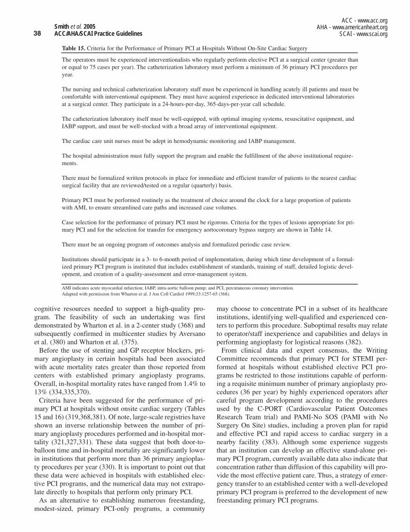
38
ACC - www.acc.orgAHA - www.americanheart.org
SCAI - www.scai.orgSmith et al. 2005ACC/AHA/SCAI Practice Guidelines
may choose to concentrate PCI in a subset of its healthcareinstitutions, identifying well-qualified and experienced cen-ters to perform this procedure. Suboptimal results may relateto operator/staff inexperience and capabilities and delays inperforming angioplasty for logistical reasons (382).
From clinical data and expert consensus, the WritingCommittee recommends that primary PCI for STEMI per-formed at hospitals without established elective PCI pro-grams be restricted to those institutions capable of perform-ing a requisite minimum number of primary angioplasty pro-cedures (36 per year) by highly experienced operators aftercareful program development according to the proceduresused by the C-PORT (Cardiovascular Patient OutcomesResearch Team trial) and PAMI-No SOS (PAMI with NoSurgery On Site) studies, including a proven plan for rapidand effective PCI and rapid access to cardiac surgery in anearby facility (383). Although some experience suggeststhat an institution can develop an effective stand-alone pri-mary PCI program, currently available data also indicate thatconcentration rather than diffusion of this capability will pro-vide the most effective patient care. Thus, a strategy of emer-gency transfer to an established center with a well-developedprimary PCI program is preferred to the development of newfreestanding primary PCI programs.
cognitive resources needed to support a high-quality pro-gram. The feasibility of such an undertaking was firstdemonstrated by Wharton et al. in a 2-center study (368) andsubsequently confirmed in multicenter studies by Aversanoet al. (380) and Wharton et al. (375).
Before the use of stenting and GP receptor blockers, pri-mary angioplasty in certain hospitals had been associatedwith acute mortality rates greater than those reported fromcenters with established primary angioplasty programs.Overall, in-hospital mortality rates have ranged from 1.4% to13% (334,335,370).
Criteria have been suggested for the performance of pri-mary PCI at hospitals without onsite cardiac surgery (Tables15 and 16) (319,368,381). Of note, large-scale registries haveshown an inverse relationship between the number of pri-mary angioplasty procedures performed and in-hospital mor-tality (321,327,331). These data suggest that both door-to-balloon time and in-hospital mortality are significantly lowerin institutions that perform more than 36 primary angioplas-ty procedures per year (330). It is important to point out thatthese data were achieved in hospitals with established elec-tive PCI programs, and the numerical data may not extrapo-late directly to hospitals that perform only primary PCI.
As an alternative to establishing numerous freestanding,modest-sized, primary PCI-only programs, a community
Table 15. Criteria for the Performance of Primary PCI at Hospitals Without On-Site Cardiac Surgery
The operators must be experienced interventionalists who regularly perform elective PCI at a surgical center (greater thanor equal to 75 cases per year). The catheterization laboratory must perform a minimum of 36 primary PCI procedures peryear.
The nursing and technical catheterization laboratory staff must be experienced in handling acutely ill patients and must becomfortable with interventional equipment. They must have acquired experience in dedicated interventional laboratoriesat a surgical center. They participate in a 24-hours-per-day, 365-days-per-year call schedule.
The catheterization laboratory itself must be well-equipped, with optimal imaging systems, resuscitative equipment, andIABP support, and must be well-stocked with a broad array of interventional equipment.
The cardiac care unit nurses must be adept in hemodynamic monitoring and IABP management.
The hospital administration must fully support the program and enable the fulfillment of the above institutional require-ments.
There must be formalized written protocols in place for immediate and efficient transfer of patients to the nearest cardiacsurgical facility that are reviewed/tested on a regular (quarterly) basis.
Primary PCI must be performed routinely as the treatment of choice around the clock for a large proportion of patientswith AMI, to ensure streamlined care paths and increased case volumes.
Case selection for the performance of primary PCI must be rigorous. Criteria for the types of lesions appropriate for pri-mary PCI and for the selection for transfer for emergency aortocoronary bypass surgery are shown in Table 14.
There must be an ongoing program of outcomes analysis and formalized periodic case review.
Institutions should participate in a 3- to 6-month period of implementation, during which time development of a formal-ized primary PCI program is instituted that includes establishment of standards, training of staff, detailed logistic devel-opment, and creation of a quality-assessment and error-management system.
AMI indicates acute myocardial infarction; IABP, intra-aortic balloon pump; and PCI, percutaneous coronary intervention. Adapted with permission from Wharton et al. J Am Coll Cardiol 1999;33:1257-65 (368).

39Smith et al. 2005
ACC/AHA/SCAI Practice Guidelines
ACC - www.acc.orgAHA - www.americanheart.orgSCAI - www.scai.org
not been reduced to negligible levels. A small but valid frac-tion of patients undergoing elective PCI will experience alife-threatening complication that could be managed with theimmediate onsite availability of cardiac surgical support butcannot be managed effectively by urgent transfer. Lotfi et al.reported the experience of a large, high-quality coronaryinterventional center (384). Of 6582 PCI procedures per-formed between 1996 and 2000, 45 (0.7%) required emer-gency cardiac surgery. Of the 45 patients, 11 (0.2%) requiredtruly emergent surgery because they were too unstable to tol-erate an interhospital transfer. Thus, under the best of cir-cumstances 1 in 500 patients undergoing elective PCI willexperience a life-threatening complication that can be sal-vaged by immediate access to onsite cardiac surgery. As pre-viously noted, Section 4.4, Wennberg, et al., found highermortality in the Medicare database for patients undergoingelective PCI in institutions without onsite cardiac surgery(356). Furthermore, the availability of onsite cardiac surgeryis a surrogate for overall program size and capability, as wellas for the availability of many other experienced supportservices.
Caution is warranted before an unrestricted policy for PCIin hospitals without appropriate facilities is endorsed.Several outstanding and critically important clinical issues,such as timely management of ischemic complications, ade-quacy of specialized postinterventional care, logistics formanaging cardiac surgical or vascular complications andoperator/laboratory volumes, and accreditation, must beaddressed. Mere convenience should not replace safety andefficacy in the establishment of an elective PCI programwithout onsite surgery.
At this time, the Writing Committee, therefore, continuesto support the recommendation that elective PCI should notbe performed in facilities without onsite cardiac surgery. Aswith many dynamic areas in interventional cardiology, theserecommendations may be subject to revision as clinical dataand experience increase.
5. CLINICAL PRESENTATIONS
A broad spectrum of clinical presentations exists whereinpatients may be considered candidates for PCI, ranging fromasymptomatic to severely symptomatic or unstable, withvariable degrees of jeopardized myocardium. In this guide-line, the CCS classification system for grading angina pec-toris is used to summarize the severity of angina, as shownbelow (Table 17) (385).
Each time a patient is considered for revascularization, thepotential risk and benefits of the particular procedure underconsideration must be weighed against alternative therapies(Table 18). When PCI is considered, the benefits and risks ofsurgical revascularization and medical therapy alwaysdeserve thoughtful discussion with the patient and family.The initial simplicity and associated low morbidity of PCIcompared with surgical therapy is always attractive, but thepatient and family must understand the limitations inherentin current PCI procedures, including a realistic presentation
4.5. Elective PCI Without Onsite Surgery
Class IIIElective PCI should not be performed at institutionsthat do not provide onsite cardiac surgery. (Level ofEvidence: C)*
*Several centers have reported satisfactory results based on carefulcase selection with well-defined arrangements for immediate transferto a surgical program (333-337,348-353). A small, but real fraction ofpatients undergoing elective PCI will experience a life-threateningcomplication that could be managed with the immediate onsite avail-ability of cardiac surgical support but cannot be managed effectivelyby urgent transfer. Wennberg et al., found higher mortality in theMedicare database for patients undergoing elective PCI in institu-tions without onsite cardiac surgery (356). This recommendationmay be subject to revision as clinical data and experience increase.
Technical improvements in interventional cardiology haveled to the development of elective angioplasty programswithout onsite surgical coverage. Several centers havereported satisfactory results based on careful case selectionwith well-defined arrangements for immediate transfer to asurgical program (333-337,348-353). The studies of angio-plasty without onsite surgical coverage have not identifiedsignificant differences in outcomes, which recalls the infre-quent rate of complications (353). Despite many reportedsuccessful angioplasty series without onsite surgical backupand a very low percentage need for off-site surgery in failedangioplasty, some clinicians have expressed concern(354,355) about the appropriateness of elective angioplastyin centers without onsite surgical coverage. Life-threateningcomplications of elective PCI are, fortunately, rare but have
Table 16. Patient Selection for Primary PCI and EmergencyAortocoronary Bypass at Hospitals Without On-Site Cardiac Surgery
Avoid intervention in hemodynamically stable patients with:Significant (greater than or equal to 60%) stenosis of an unprotectedleft main coronary artery upstream from an acute occlusion in theleft coronary system that might be disrupted by the angioplastycatheter
Extremely long or angulated infarct-related lesions with TIMI grade3 flow
Infarct-related lesions with TIMI grade 3 flow in stable patients with3-vessel disease (319, 381)
Infarct-related lesions of small or secondary vessels
Hemodynamically significant lesions in other than the infarct artery
Transfer for emergency aortocoronary bypass surgery patientswith:
High-grade residual left main or multivessel coronary disease andclinical or hemodynamic instability present after primary PCI ofoccluded vessels, preferably with IABP support.
IABP indicates intra-aortic balloon pump; PCI, percutaneous coronary intervention; andTIMI, Thrombolysis In Myocardial Infarction.
Adapted with permission from Wharton et al. J Am Coll Cardiol 1999;33:1257-65 (368).

40
ACC - www.acc.orgAHA - www.americanheart.org
SCAI - www.scai.orgSmith et al. 2005ACC/AHA/SCAI Practice Guidelines
Class IIb1. The effectiveness of PCI for patients with asympto-
matic ischemia or CCS class I or II angina who have2- or 3-vessel disease with significant proximal LADCAD who are otherwise eligible for CABG with 1arterial conduit and who have treated diabetes orabnormal LV function is not well established. (Level ofEvidence: B)
2. PCI might be considered for patients with asympto-matic ischemia or CCS class I or II angina with non-proximal LAD CAD that subtends a moderate area ofviable myocardium and demonstrates ischemia onnoninvasive testing. (Level of Evidence: C)
Class IIIPCI is not recommended in patients with asympto-matic ischemia or CCS class I or II angina who do notmeet the criteria as listed under the class II recom-mendations or who have 1 or more of the following: a. Only a small area of viable myocardium at risk
(Level of Evidence: C)b. No objective evidence of ischemia. (Level of
Evidence: C)c. Lesions that have a low likelihood of successful
dilatation. (Level of Evidence: C)d. Mild symptoms that are unlikely to be due to
myocardial ischemia. (Level of Evidence: C)e. Factors associated with increased risk of morbidity
or mortality. (Level of Evidence: C)f. Left main disease and eligibility for CABG. (Level
of Evidence: C)g. Insignificant disease (less than 50% coronary
stenosis). (Level of Evidence: C)
In the previous ACC/AHA guidelines for PCI, specific rec-ommendations were made separately for patients with sin-gle-vessel or multivessel disease (1,123). The current tech-niques of PCI have matured to the point at which, in patientswith favorable anatomy, the competent practitioner can per-form either single-vessel or multivessel PCI with low riskand with a high likelihood of initial success. For this reason,in this update of the guidelines, recommendations have beenmade largely based on the patient’s clinical condition, spe-
of the likelihood of restenosis and the potential for incom-plete revascularization compared with CABG surgery. Inpatients with CAD who are asymptomatic or have only mildsymptoms, the potential benefit of antianginal drug therapyalong with an aggressive program of risk reduction must alsobe understood by the patient before a revascularization pro-cedure is performed. In those clinical settings in which PCIis recommended without evidence that it will reduce cardio-vascular mortality but in which it does hold a promise toreduce symptoms, a class IIa or IIb classification has beenchosen, which indicates a role for patient preference.
5.1. Patients With Asymptomatic Ischemia or CCS Class I or II Angina
Class IIa1. PCI is reasonable in patients with asymptomatic
ischemia or CCS class I or II angina and with 1 ormore significant lesions in 1 or 2 coronary arteriessuitable for PCI with a high likelihood of success anda low risk of morbidity and mortality. The vessels tobe dilated must subtend a moderate to large area ofviable myocardium or be associated with a moderateto severe degree of ischemia on noninvasive testing.(Level of Evidence: B)
2. PCI is reasonable for patients with asymptomaticischemia or CCS class I or II angina, and recurrentstenosis after PCI with a large area of viablemyocardium or high-risk criteria on noninvasive test-ing. (Level of Evidence: C)
3. Use of PCI is reasonable in patients with asympto-matic ischemia or CCS class I or II angina with sig-nificant left main CAD (greater than 50% diameterstenosis) who are candidates for revascularization butare not eligible for CABG. (Level of Evidence: B)
Table 17. Grading of Angina Pectoris According to CCSClassification
Class Description of Stage
I “Ordinary physical activity does not cause…angina,” such as walking or climbing stairs. Angina occurs with strenuous,rapid, or prolonged exertion at work or recreation.
II “Slight limitation of ordinary activity.” Angina occurs onwalking or climbing stairs rapidly; walking uphill; walkingor stair climbing after meals; in cold, in wind, or under emo-tional stress; or only during the few hours after awaking.Angina occurs on walking more than 2 blocks on the leveland climbing more than 1 flight of ordinary stairs at a nor-mal pace and under normal conditions.
III “Marked limitations of ordinary physical activity.” Anginaoccurs on walking 1 to 2 blocks on the level and climbing1flight of stairs under normal conditions and at a normal pace.
IV “Inability to carry on any physical activity without discom-fort—anginal symptoms may be present at rest.”
CCS indicates Canadian Cardiovascular Society.Adapted with permission from Campeau. Circulation 1976;54:522-3 (385).
Table 18. Provider Checklist: Key Areas for Consideration
Patients at High RiskAssess key clinical and anatomic variables Consider alternative therapies such as CABG in consultation with
the patientEnsure that formalized surgical standby is availableEnsure periprocedural hemodynamic support is available
Patients at Low RiskAssess key clinical and anatomic variablesConsider alternative therapies such as medical therapy in consulta-
tion with the patient
CABG indicates coronary artery bypass graft surgery.

41Smith et al. 2005
ACC/AHA/SCAI Practice Guidelines
ACC - www.acc.orgAHA - www.americanheart.orgSCAI - www.scai.org
cific coronary lesion morphology and anatomy, LV function,and associated medical conditions, and less emphasis hasbeen placed on the number of lesions or vessels requiringPCI. The CCS classification of angina (I to IV) is used todefine the severity of symptoms. The categories described inthis section refer to an initial PCI procedure in a patient with-out prior CABG surgery. The randomized trials comparingPCI and medical therapy have been discussed (Table 12)(11,12,279,282-290).
The Writing Committee recognizes that the majority ofpatients with CCS class I or II angina should be treated med-ically. The published ACIP study (301) casts some doubt onthe wisdom of medical management for those higher-riskpatients who are asymptomatic or have mild angina but haveobjective evidence by both treadmill testing and ambulatorymonitoring of significant myocardial ischemia and CAD. Inaddition, a substantial portion of the middle-aged and older-age populations in the United States remain physicallyactive, participating in sports, such as tennis and skiing, orperforming regular and vigorous physical exercise, such asjogging, have CAD. For such individuals with moderate orsevere ischemia and few symptoms, revascularization withPCI or CABG surgery may reduce their risk of serious orfatal cardiac events (301). For this reason, patients in this cat-egory of higher-risk asymptomatic ischemia or mild symp-toms and severe anatomic CAD are placed in class IIa or IIbrecommendations. PCI may be considered if there is a highlikelihood of success and a low risk of morbidity or mortali-ty. The judgment of the experienced physician is deemedvaluable in assessing the extent of ischemia.
5.2. Patients With CCS Class III Angina
Class IIa1. It is reasonable that PCI be performed in patients
with CCS class III angina and single-vessel or multi-vessel CAD who are undergoing medical therapy andwho have 1 or more significant lesions in 1 or morecoronary arteries suitable for PCI with a high likeli-hood of success and low risk of morbidity or mortali-ty. (Level of Evidence: B)
2. It is reasonable that PCI be performed in patientswith CCS class III angina with single-vessel or multi-vessel CAD who are undergoing medical therapy withfocal saphenous vein graft lesions or multiple stenoseswho are poor candidates for reoperative surgery.(Level of Evidence: C)
3. Use of PCI is reasonable in patients with CCS class IIIangina with significant left main CAD (greater than50% diameter stenosis) who are candidates for revas-cularization but are not eligible for CABG. (Level ofEvidence: B)
Class IIb1. PCI may be considered in patients with CCS class III
angina with single-vessel or multivessel CAD who areundergoing medical therapy and who have 1 or morelesions to be dilated with a reduced likelihood of suc-
cess. (Level of Evidence: B)2. PCI may be considered in patients with CCS class III
angina and no evidence of ischemia on noninvasivetesting or who are undergoing medical therapy andhave 2- or 3-vessel CAD with significant proximalLAD CAD and treated diabetes or abnormal LV func-tion. (Level of Evidence: B)
Class IIIPCI is not recommended for patients with CCS classIII angina with single-vessel or multivessel CAD, noevidence of myocardial injury or ischemia on objec-tive testing, and no trial of medical therapy, or whohave 1 of the following:a. Only a small area of myocardium at risk. (Level of
Evidence: C)b. All lesions or the culprit lesion to be dilated with
morphology that conveys a low likelihood of suc-cess. (Level of Evidence: C)
c. A high risk of procedure-related morbidity or mor-tality. (Level of Evidence: C)
d. Insignificant disease (less than 50% coronarystenosis). (Level of Evidence: C)
e. Significant left main CAD and candidacy forCABG. (Level of Evidence: C)
The primary benefit of PCI among patients with CCS classIII angina and single-vessel or multivessel CAD resides inthe relief of symptoms, which may be accomplished withmedical therapy. However, many patients with moderate orsevere stable angina do not respond adequately to medicaltherapy and often have significant coronary artery stenosesthat are suitable for revascularization with CABG surgery orPCI. In addition, a proportion of these patients have reducedLV systolic function, which places them in a group that isknown to have improved survival with CABG surgery andpossibly with revascularization by PCI (386-389). In patientswithout diabetes with 1- or 2-vessel disease in whom angio-plasty of 1 or more lesions has a high likelihood of initialsuccess, PCI is the preferred approach. In a minority of suchpatients, CABG surgery may be preferred, particularly forthose in whom the LAD can be revascularized with the IMAor in those with left main coronary disease. (See Section3.5.1.2 on left main CAD.)
5.3. Patients With UA/NSTEMI
Class IAn early invasive PCI strategy is indicated forpatients with UA/NSTEMI who have no seriouscomorbidity and coronary lesions amenable to PCI.Patients must have any of the following high-risk fea-tures:a. Recurrent ischemia despite intensive anti-ischemic
therapy. (Level of Evidence: A)b. Elevated troponin level. (Level of Evidence: A)c. New ST-segment depression. (Level of Evidence: A)d. HF symptoms or new or worsening MR. (Level of
42
ACC - www.acc.orgAHA - www.americanheart.org
SCAI - www.scai.orgSmith et al. 2005ACC/AHA/SCAI Practice Guidelines
e. Significant left main CAD and candidacy forCABG. (Level of Evidence: B)
Clinical investigations have evaluated the use of routinecatheterization and PCI for patients with UA or NSTEMI andhave yielded inconsistent results. TIMI-IIIB was the firsttrial to compare strategies of routine catheterization andrevascularization in addition to medical therapy and selectiveuse of aggressive treatment. In TIMI-IIIB, there was no dif-ference in the incidence of death or recurrent MI at 1 yearbetween the 2 strategies, but patients treated by the aggres-sive strategy experienced less angina and repeat hospitaliza-tions for ischemia and required fewer medications (390). Inthe VANQWISH trial (Veterans Affairs Non–Q-WaveInfarction Strategies in Hospital) performed by the USVeterans Administration, no difference in death or death andMI was observed between the 2 strategies at late follow-up,but the minority of patients in the aggressive strategyreceived revascularization, and the mortality rate for thosehaving CABG was high (391). The FRISC II trial comparedmedical and revascularization approaches among patientsafter 6 days of low-molecular-weight heparin therapy beforea decision regarding PCI (304). Those randomized to theconservative therapy only underwent PCI if they had at least3 mm of ST-segment depression on stress testing. Comparedwith prior studies, patients assigned to the aggressive strate-gy in FRISC II experienced a 22% reduction (P equals0.031) in the incidence of death or MI at 6 months (9.4%)compared with conservatively treated patients (12.1%). Inaddition, there was a significant decrease in the MI rate aloneand a nonsignificantly lower mortality rate in the treatedgroup (1.9% vs 2.9%; P equals 0.10). Symptoms of anginaand hospital readmission were decreased 50% by the inva-sive strategy. These findings were supported by long-termfollow-up from the FRISC II study that indicated that low-molecular-weight heparin and early intervention lowered therisk of death, MI, and revascularization in unstable coronarysyndromes, at least during the first month of therapy. Earlyprotective therapy could be used to reduce the risk of lateevents in patients waiting for definitive PCI (392). This treat-ment benefit was most pronounced for high-risk patients.The FRISC II trial (304) results support the use of catheteri-zation and revascularization for selected patients with anacute coronary syndrome. The TACTICS trial randomized2220 patients to an early invasive strategy in which cardiaccatheterization and revascularization were performed 4 to 48h after randomization or to a conservative strategy in whichrevascularization was reserved for those patients who devel-oped recurrent ischemia after medical stabilization (393). Allpatients were treated with aspirin, heparin, beta-blockers,cholesterol-lowering therapy, and tirofiban. The primary endpoint, a composite of death, MI, and rehospitalization forworsening chest pain by 6 months, was lower in patientsassigned to the invasive strategy (15.9% vs 19.4% in patientsassigned to conservative therapy; P equals 0.0025). The rateof death or MI was also significantly reduced at 6 months inthe invasive strategy arm (7.3% vs 9.5% in patients assigned
Evidence: A)e. Depressed LV systolic function. (Level of Evidence:
A)f. Hemodynamic instability. (Level of Evidence: A)g. Sustained ventricular tachycardia. (Level of
Evidence: A)h. PCI within 6 months. (Level of Evidence: A)i. Prior CABG. (Level of Evidence: A)
Class IIa1. It is reasonable that PCI be performed in patients
with UA/NSTEMI and single-vessel or multivesselCAD who are undergoing medical therapy with focalsaphenous vein graft lesions or multiple stenoses whoare poor candidates for reoperative surgery. (Level ofEvidence: C)
2. In the absence of high-risk features associated withUA/NSTEMI, it is reasonable to perform PCI inpatients with amenable lesions and no contraindica-tion for PCI with either an early invasive or earlyconservative strategy. (Level of Evidence: B)
3. Use of PCI is reasonable in patients with UA/NSTE-MI with significant left main CAD (greater than 50%diameter stenosis) who are candidates for revascular-ization but are not eligible for CABG. (Level ofEvidence: B)
Class IIb1. In the absence of high-risk features associated with
UA/NSTEMI, PCI may be considered in patients withsingle-vessel or multivessel CAD who are undergoingmedical therapy and who have 1 or more lesions to bedilated with reduced likelihood of success. (Level ofEvidence: B)
2. PCI may be considered in patients with UA/NSTEMIwho are undergoing medical therapy who have 2- or3-vessel disease, significant proximal LAD CAD, andtreated diabetes or abnormal LV function. (Level ofEvidence: B)
Class IIIIn the absence of high-risk features associated withUA/NSTEMI, PCI is not recommended for patientswith UA/NSTEMI who have single-vessel or multi-vessel CAD and no trial of medical therapy, or whohave 1 or more of the following: a. Only a small area of myocardium at risk. (Level of
Evidence: C)b. All lesions or the culprit lesion to be dilated with
morphology that conveys a low likelihood of suc-cess. (Level of Evidence: C)
c. A high risk of procedure-related morbidity ormortality. (Level of Evidence: C)
d. Insignificant disease (less than 50% coronarystenosis). (Level of Evidence: C)
43Smith et al. 2005
ACC/AHA/SCAI Practice Guidelines
ACC - www.acc.orgAHA - www.americanheart.orgSCAI - www.scai.org
shock, unless further support is futile because of thepatient’s wishes or contraindications/unsuitability forfurther invasive care. (Level of Evidence: A)
3. Primary PCI should be performed in patients withsevere congestive heart failure and/or pulmonaryedema (Killip class 3) and onset of symptoms within12 hours. The medical contact-to-balloon or door-to-balloon time should be as short as possible (i.e., goalwithin 90 minutes). (Level of Evidence: B)
Class IIa1. Primary PCI is reasonable for selected patients 75
years or older with ST elevation or left bundle-branchblock or who develop shock within 36 hours of MI andare suitable for revascularization that can be per-formed within 18 hours of shock. Patients with goodprior functional status who are suitable for revascu-larization and agree to invasive care may be selectedfor such an invasive strategy. (Level of Evidence: B)
2. It is reasonable to perform primary PCI for patientswith onset of symptoms within the prior 12 to 24hours and 1 or more of the following: a. Severe congestive heart failure (Level of Evidence:
C)b. Hemodynamic or electrical instability (Level of
Evidence: C)c. Evidence of persistent ischemia (Level of Evidence:
C)
Class IIbThe benefit of primary PCI for STEMI patients eligi-ble for fibrinolysis when performed by an operatorwho performs fewer than 75 PCI procedures per year(or fewer than 11 PCIs for STEMI per year) is not wellestablished. (Level of Evidence: C)
Class III1. Elective PCI should not be performed in a non-
infarct-related artery at the time of primary PCI ofthe infarct related artery in patients without hemody-namic compromise. (Level of Evidence: C)
2. Primary PCI should not be performed in asympto-matic patients more than 12 hours after onset ofSTEMI who are hemodynamically and electricallystable. (Level of Evidence: C)
Acute STEMI results from a severe and sudden cessationof myocardial blood flow, most commonly due to athero-sclerotic-thrombotic occlusion of a major epicardial coro-nary artery. PCI is a very effective method for re-establishingcoronary perfusion and is suitable for 90% of patients.Considerable data support the use of PCI for patients withSTEMI (53,364,398). Reported rates of achieving TIMI 3flow, the goal of reperfusion therapy, range from 70% to 90%(399). Late follow-up angiography demonstrates that 87% ofinfarct arteries remain patent (400). Although most studies ofprimary PCI have been in patients who are eligible to receivefibrinolytic therapy, considerable experience supports the
to conservative therapy; P less than 0.05) (393). The TIMI-TACTICS group (394) has proposed a new risk stratification.The early invasive strategy was particularly effective forpatients at moderate to high risk. The greater benefits derivedfrom PCI in the TACTICS and FRISC trials compared withthe TIMI III and VANQWISH trials can be explained in partby the use of stents and GP-receptor blockers and lowerperiprocedural complications in the TACTICS and FRISC IItrials. In several studies published to date, the use of routineinvasive therapy in patients with UA/NSTEMI, accompaniedby IIb/IIIa receptor antagonists, has been shown to improvesurvival (205,302,393,395-397). New trials such as RITA-3(302) further demonstrate the safety and effectiveness of anearly invasive strategy.
It is recognized by the Committee that the assessment ofrisk of unsuccessful PCI or serious morbidity or mortalitymust always be made with consideration of the alternativetherapies available for the patient, including more intensiveor prolonged medical therapy or surgical revascularization(Table 19) (302,304,390,391,393), especially in patients withUA/NSTEMI.
When CABG surgery is a poor option because of high riskdue to special considerations or other organ system disease,patients otherwise in class IIb may be appropriately managedwith PCI. Under these special circumstances, formal surgicalconsultation is recommended.
5.4. Patients With STEMI
5.4.1. General and Specific Considerations
Class IGeneral considerations:
1. If immediately available, primary PCI should be per-formed in patients with STEMI (including true poste-rior MI) or MI with new or presumably new left bun-dle-branch block who can undergo PCI of the infarctartery within 12 hours of symptom onset, if per-formed in a timely fashion (balloon inflation goalwithin 90 minutes of presentation) by persons skilledin the procedure (individuals who perform more than75 PCI procedures per year, ideally at least 11 PCIsper year for STEMI). The procedure should be sup-ported by experienced personnel in an appropriatelaboratory environment (one that performs more than200 PCI procedures per year, of which at least 36 areprimary PCI for STEMI, and that has cardiac sur-gery capability). (Level of Evidence: A) Primary PCIshould be performed as quickly as possible, with agoal of a medical contact-to-balloon or door-to-bal-loon time within 90 minutes. (Level of Evidence: B)
Specific Considerations:
2. Primary PCI should be performed for patients lessthan 75 years old with ST elevation or presumablynew left bundle-branch block who develop shockwithin 36 hours of MI and are suitable for revascular-ization that can be performed within 18 hours of

Tabl
e 19
.In
vasi
ve V
ersu
s C
onse
rvat
ive
Stra
tegi
es in
Uns
tabl
e A
ngin
a Pa
tient
s
Pat
ient
St
udy
Yea
rsR
efn
Pop
ulat
ion
Tre
atm
ent
Fol
low
-Up
TIM
I-II
IB19
89-1
992
(390
)14
73Pa
tient
s 21
to 7
6 M
edic
al th
erap
y 6
wk
year
s of
age
(t
PAvs
pla
cebo
) pr
esen
ting
with
in
and
earl
y24
h o
f is
chem
icin
vasi
ve o
rdi
scom
fort
at r
est
cons
erva
tive
cons
iste
nt w
ith
stra
tegy
unst
able
ang
ina
or
1 y
non–
Q-w
ave
MI
VA
NQ
WIS
H19
93-1
996
(391
)92
0Pa
tient
s w
ith a
n In
vasi
ve v
s A
vera
ge 2
3 m
oev
olvi
ng M
Ico
nser
vativ
e
FRIS
C I
I19
96-1
998
(304
)24
57Pa
tient
’s is
chem
ic
Ear
ly in
vasi
ve6
mo
sym
ptom
s in
pre
viou
s th
erap
y or
48 h
ours
acc
ompa
nied
no
ninv
asiv
e by
EC
G c
hang
es o
r tr
eatm
ent
elev
ated
mar
kers
stra
tegy
. Pa
tient
s al
so
rece
ived
da
ltepa
rin
or p
lace
bo f
or
3 m
onth
s
TAC
TIC
S-19
97-1
999
(393
)22
20U
Aan
d N
STE
MI
Med
ical
ther
apy
6 m
oT
IMI
18w
ith E
CG
cha
nges
, (a
spir
in, h
epar
in,
elev
ated
leve
ls o
f tir
ofib
an)
and
card
iac
biom
arke
rs,
eith
er e
arly
a
hist
ory
of C
AD
, in
vasi
ve o
ror
all
3 fi
ndin
gsco
nser
vativ
e (s
elec
tivel
y in
vasi
ve)
trea
tmen
t st
rate
gy
RIT
A-3
1997
-200
1(3
02)
1810
Susp
ecte
d ca
rdia
c M
edic
al th
erap
y1
ych
est p
ain
at r
est
and
eith
er e
arly
w
ith d
ocum
ente
d in
vasi
ve o
r ev
iden
ce o
f C
AD
co
nser
vativ
e(a
t lea
st 1
of
the
(sel
ectiv
ely
follo
win
g: E
CG
in
vasi
ve)
chan
ges,
pat
holo
gica
l tr
eatm
ent
Q w
aves
, pre
viou
s st
rate
gy; b
oth
arte
riog
ram
)gr
oups
rec
eive
d en
oxap
arin
in
addi
tion
tost
anda
rd
med
ical
th
erap
y
CA
BG
indi
cate
s co
rona
ry a
rter
y by
pass
gra
ft s
urge
ry; C
AD
, cor
onar
y ar
tery
dis
ease
; EC
G, e
lect
roca
rdio
grap
hy; E
TT,
exe
rcis
e tr
eadm
ill te
st; M
I, m
yoca
rdia
l inf
arct
ion;
mo,
mon
th; n
, num
ber;
NS,
not
sig
nifi
cant
; NST
EM
I, n
on–S
T-el
evat
ion
myo
card
ial i
nfar
ctio
n; P
CI,
per
cuta
neou
s co
rona
ry in
ter-
vent
ion;
Ref
, ref
eren
ce; t
PA, a
ltepl
ase;
UA
, uns
tabl
e an
gina
; wk,
wee
k; a
nd y
, yea
r.
Res
ults
Med
ical
P
CI
The
rapy
P
16.2
%18
.1%
NS
com
bine
d co
mbi
ned
prim
ary
prim
ary
end
poin
tsen
d po
ints
12.4
%10
.6%
32.9
%30
.3%
0.35
deat
h de
ath
and
MI
and
MI
9.4%
12
.1%
0.
031
deat
h de
ath
or M
Ior
MI
7.3%
9.
5%
Les
s th
ande
ath
deat
h 0.
05or
MI
or M
I
7.6%
8.
3%N
Sde
ath
deat
h or
MI
or M
I
Com
men
ts
Alth
ough
no
diff
eren
ce w
as f
ound
in c
ombi
ned
prim
ary
end
poin
ts (
deat
h, M
I, p
ositi
ve E
TT
),th
e ea
rly
inva
sive
str
ateg
y w
as a
ssoc
iate
d w
ithsh
orte
r ho
spita
l sta
y an
d lo
wer
inci
denc
e of
reho
spita
lizat
ion
Few
er p
atie
nts
trea
ted
cons
erva
tivel
y ha
d de
ath
plus
MI
or d
eath
at h
ospi
tal d
isch
arge
at 1
mon
th a
nd a
t 1 y
ear;
the
inva
sive
gro
up h
ad a
high
er C
AB
G m
orta
lity
rate
(11
.6%
vs
3.4%
)
Inva
sive
str
ateg
y w
as a
ssoc
iate
d w
ith 5
0%lo
wer
rec
urre
nt a
ngin
a an
d ho
spita
l rea
dmis
sion
rate
s
Sign
ific
ant 2
2% r
elat
ive
risk
red
uctio
n in
com
-po
site
end
poi
nt o
f de
ath,
non
fata
l MI,
and
reho
spita
lizat
ion
Sim
ilar
resu
lts f
or d
eath
or
MI
betw
een
trea
t-m
ent g
roup
s; s
igni
fica
nt d
iffe
renc
e in
pri
mar
yen
d po
int (
deat
h, M
I, r
efra
ctor
y an
gina
) du
e to
halv
ing
of r
efra
ctor
y an
gina
in th
e in
terv
entio
ngr
oup

45Smith et al. 2005
ACC/AHA/SCAI Practice Guidelines
ACC - www.acc.orgAHA - www.americanheart.orgSCAI - www.scai.org
each 30-minute delay from symptom onset to balloon infla-tion, P equals 0.04) (424).
The enthusiasm for primary PCI has led to the concept ofemergency interhospital transfer for catheter-based reperfu-sion rather than fibrinolytic therapy in the initial hospital(425-427). Five randomized trials have enrolled 2466patients with favorable results for PCI versus fibrinolytictherapy (378,381,407,412). Mortality was reduced with PCI(6.8% vs 9.6%, relative risk 0.69, 95% CI 0.51 to 0.0.92, Pequals 0.01), as was the combined end point of death, nonfa-tal reinfarction, and stroke (8.5% vs 15.5%, relative risk0.51, 95% CI 0.39 to 0.65, P less than 0.0001). Importantly,mean time to treatment was delayed only 44 min in thesestudies (Figure 8) (378,415). In contrast, first hospital door-to-balloon time, as recorded in 1346 patients undergoinghospital transfer before PCI in NRMI-4, was 185 min in theUnited States in 2002 (Figure 9) (428). Emergency transportin Europe is centrally organized and more efficient than inthe United States (Table 20) (378,381,407,412,415). Delaysin door-to-balloon time versus door-to-needle time of morethan 60 min because of interhospital transfer might negatethe potential mortality benefit of transfer for primary PCIover immediate intravenous fibrinolysis demonstrated inthese trials (421). However, transfer of patients to PCI-capa-ble centers should be accomplished when fibrinolytic thera-py is contraindicated or unsuccessful, when cardiogenicshock ensues, when the anticipated delay is less than 60 min,or when symptoms have been present for more than 2 to 3 h(410,415). To achieve optimal results, a systems approachfor rapid triage and transfer must be established. Time fromfirst hospital door to balloon inflation in the second hospitalshould be as short as possible, with a goal within 90 min.Significant reductions in door-to-balloon times might beachieved by transporting patients directly to PCI centers,rather than transporting them to the nearest hospital, if inter-hospital transfer will subsequently be required to obtain pri-mary PCI. Central to the success of all of the acute reperfu-sion strategies is a well-developed process of triage, as dis-cussed in the STEMI guidelines (332,429).
Primary PCI with stenting has been compared with fibri-nolytic therapy in 12 randomized clinical trials(366,378,380,381,407-412,415,430). These investigationsdemonstrate that PCI-treated patients experience lower mor-tality rates (5.9% vs 7.7%, OR 0.75, 95% CI 0.60 to 0.94, Pequals 0.013), fewer reinfarctions (1.6% vs 5.1%, OR 0.31,95% CI 0.21 to 0.44, P equals 0.0001), and fewer hemor-rhagic strokes than those treated by fibrinolysis (53).Compared with PTCA, intracoronary stents achieve a betterimmediate angiographic result with a larger arterial lumen,less reclosure of the infarct-related artery, and fewer subse-quent ischemic events (431-433). Primary stenting has beencompared with primary angioplasty in 9 studies (64,106,433-440) (Table 21). There were no differences in mortality(3.0% vs 2.8%) or reinfarction (1.8% vs 2.1%) rates.However, subsequent target-vessel revascularization rateswere lower with stenting (440).
value of PCI for patients who may not be suitable for fibri-nolytic therapy owing to an increased risk of bleeding (401).
Primary PCI has been compared with fibrinolytic therapyin 23 randomized clinical trials (361-363,378,380,381,402-415), including the SHOCK (SHould we emergently revas-cularize Occluded Coronaries in cardiogenic shocK?) trial(366). The recommendations for primary PCI in patientswith cardiogenic shock are discussed and summarized sepa-rately in Section 5.4.6. These investigations consistentlydemonstrate that PCI-treated patients experience lowershort-term mortality rates (7.0% vs 9.0%, relative risk 0.73,96% CI 0.62 to 0.86, P equals 0.0002, and 5.0% vs 7.0%, rel-ative risk 0.70, 95% CI 0.58 to 0.85, P equals 0.003 exclud-ing the SHOCK trial), fewer nonfatal reinfarctions (3.0% vs7.0%, relative risk 0.35, 95% CI 0.27 to 0.45, P equals0.0003), and fewer hemorrhagic strokes (0.05% vs 1.0%, rel-ative risk 0.05, 95% CI 0.006 to 0.35, P equals 0.0001) thanthose treated by fibrinolysis (53), albeit with an increasedrisk of bleeding (7.0% vs 5.0%, RR 1.3, 95% CI 1.02 to 1.65,P equals 0.032). These results have been achieved in medicalcenters and by providers experienced in the performance ofprimary PCI and under circumstances in which angioplastycan be performed promptly after patient presentation. Themagnitude of the treatment differences for death, nonfatalreinfarction, and stroke vary depending on whether PCI iscompared with streptokinase or a fibrin-specific lytic. Theshort- and long-term outcomes of patients with STEMI treat-ed by fibrinolysis versus PCI and the numbers of patientswho need to be treated to prevent 1 event or to cause a harm-ful complication when PCI is selected instead of fibrinolysisas the reperfusion strategy are shown in Figure 5(53,416,417).
Time from symptom onset to reperfusion is an importantpredictor of patient outcome. Two studies (330,418) havereported increasing mortality rates with increasing door-to-balloon times. Other studies have shown better LV functionand fewer complications when reperfusion occurs before PCI(419,420). An analysis of the randomized controlled trialscomparing fibrinolysis with primary PCI suggests that themortality benefit with PCI exists when treatment is delayedby no more than 60 min (Figure 6) (421). Mortality increas-es significantly with each 15-minute delay in the timebetween arrival and restoration of TIMI-3 flow (door-to–TIMI-3 flow time), which further underscores the impor-tance of timely reperfusion in patients who undergo primaryPCI (422). Given that the door-to-needle time goal is 30 min,this Writing Committee joins the Task Force on theManagement of Acute Myocardial Infarction of theEuropean Society of Cardiology (423) and the ACC/AHASTEMI Guidelines Writing Committee (332) in lowering thedoor-to-balloon time goal from 120 to 90 min in an attemptto maximize the benefits for reperfusion by PCI (Figure 7)(418). Importantly, after adjustment for baseline characteris-tics, time from symptom onset to balloon inflation is signifi-cantly correlated with 1-year mortality in patients undergo-ing primary PCI for STEMI (relative risk equals 1.08 for

46
ACC - www.acc.orgAHA - www.americanheart.org
SCAI - www.scai.orgSmith et al. 2005ACC/AHA/SCAI Practice Guidelines
Figure 5. Percutaneous coronary intervention vs fibrinolysis for STEMI. The short-term (4 to 6 weeks; top left) and long-term (topright) outcomes for the various end points shown are plotted for STEMI patients randomized to PCI or fibrinolysis for reperfusion in23 trials (n=7739). Based on the frequency of events for each end point in the 2 treatment groups, the number needed to treat (NNT)or number needed to harm (NNH) is shown for the short-term (bottom left) and long-term (bottom right) outcomes. Modified with per-mission from Elsevier (Keeley et al. The Lancet, 2003, 361, 13-20). Note: The magnitude of the treatment differences for death, non-fatal reinfarction, and stroke vary depending on whether PCI is compared with streptokinase or a fibrin-specific lytic. For example,when primary PCI is compared with alteplase (tPA) and the SHOCK trial is excluded, the mortality rate is 5.5% vs 6.7% (OR 0.81,95% CI 0.64 to 1.03, P equals 0.081). Source: Melandri. Circulation 2003;108:e162. CVA indicates cerebrovascular accident; Hem.Stroke, hemorrhagic stroke; MI, myocardial infarction; PCI, percutaneous coronary intervention; PTCA, percutaneous transluminalcoronary angioplasty; Rec. Isch, recurrent ischemia; ReMI, recurrent MI; and STEMI, ST-elevation myocardial infarction.
Preliminary reports suggest that compared with conven-tional BMS, DES are not associated with increased risk whenused for primary PCI in patients with STEMI. Postprocedurevessel patency, biomarker release, and the incidence of short-term adverse events were similar in patients receiving SES orBMS. Thirty-day event rates of death, reinfarction, or revas-cularization were 7.5% versus 10.4%, respectively (P equals0.4) (441).
Furthermore, the impact of IIb/IIIa platelet receptor antag-onists in the setting of primary PCI has undergone consider-able evaluation. In a randomized trial of stents plus abcix-imab compared with fibrinolysis plus abciximab in patientswith STEMI, myocardial salvage and salvage index meas-ured by technetium-99m sestamibi scintigraphy was signifi-cantly greater in the stent group (430). In a similar study
comparing primary PCI with stent plus abciximab to fibri-nolysis with alteplase, infarct size was smaller and the cumu-lative incidence of death, reinfarction, or stroke at 6 monthssignificantly lower in the primary PCI group (411).
However, results of studies comparing primary PCI withstents with or without IIb/IIIa platelet receptor antagonistshave been less consistent. In the CADILLAC trial, a com-posite of death, reinfarction, disabling stroke, and ischemia-driven target-vessel revascularization was similar in patientstreated with stents with or without abciximab (64). Yet, in asimilar randomized comparison of stent plus abciximab ver-sus stent alone in patients with STEMI (ADMIRAL trial;Abciximab before Direct angioplasty and stenting inMyocardial Infarction Regarding Acute and Long-term fol-low-up), a composite of death, reinfarction, or urgent target-

47Smith et al. 2005
ACC/AHA/SCAI Practice Guidelines
ACC - www.acc.orgAHA - www.americanheart.orgSCAI - www.scai.org
Figure 7. Relationship between 30-day mortality and time from study enrollment to first balloon inflation. Patients assigned to angio-plasty in whom angioplasty was not performed are also shown. n indicates number of patients; and PTCA, percutaneous translumi-nal coronary angioplasty. Reprinted with permission from Berger et al. Circulation 1999;100:14-20 (418).
Figure 6. PCI versus fibrinolysis with fibrin-specific agents: is timing (almost) everything? PCI indicates percutaneous coronary inter-vention. Modified with permission from Nallamothu and Bates. Am J Cardiol 2003;92:824-6 (421).
vessel revascularization at 30 days occurred significantlyless often in the abciximab group than in the control group(6.0% vs 14.6%, P equals 0.01), a difference that was sus-tained at 6 months of follow-up (442). The less favorablecomparable clinical outcomes in patients treated with abcix-imab in the CADILLAC trial compared with those in theADMIRAL trial have been attributed to the earlier adminis-tration of abciximab in the latter trial. The results of a pooledanalysis of these 2 trials plus 3 similar trials (RAPPORT,
ISAR-2, and ACE) (443-445) suggest that early (before coro-nary angiography) administration of abciximab will be asso-ciated with the most favorable clinical outcomes (446).
PCI appears to have its greatest mortality benefit in high-risk patients. In patients with cardiogenic shock, an absolute9% reduction in 30-day mortality with mechanical revascu-larization instead of immediate medical stabilization wasreported in the SHOCK trial (366) (see Section 5.4.6, PCI forCardiogenic Shock). In NRMI-II, patients with HF had a

48
ACC - www.acc.orgAHA - www.americanheart.org
SCAI - www.scai.orgSmith et al. 2005ACC/AHA/SCAI Practice Guidelines
delays in reperfusion and performance by low-volume orpoor-outcome operators/centers do not occur. The physi-cians, nursing, and technical catheterization laboratory staffmust be experienced in handling acutely ill patients, must beskilled in all aspects of interventional equipment and proce-dures, and must participate in a 24-hours-per-day, 365-days-per-year call schedule. Interventional cardiologists and cen-ters should strive for 1) balloon dilation within 90 min ofadmission and diagnosis of STEMI (452); 2) TIMI 2 to 3flow attained in more than 90% of patients; 3) emergencyCABG rate less than 2% among all patients undergoing theprocedure; 4) actual performance of PCI in 85% of patientsbrought to the laboratory; and 5) a risk-adjusted in-hospitalmortality rate less than 7% in patients without cardiogenic
33% relative risk reduction with primary PCI compared witha 9% relative risk reduction with fibrinolytic therapy (447-449). Primary PCI in patients with anterior STEMI reducesmortality compared with fibrinolytic therapy, but there is nodifference in patients with nonanterior STEMI (450,451).
Despite the evidence supporting primary PCI in the treat-ment of STEMI, there is serious concern that a routine poli-cy of primary PCI for patients with STEMI will result inunacceptable delays in achieving reperfusion in a substantialnumber of patients and less than optimal outcomes if per-formed by less experienced operators. The mean time delayfor PCI instead of fibrinolysis in the randomized trials wasonly 40 min (364). Strict performance criteria must be man-dated for primary angioplasty programs so that excessive
Figure 8. Comparison of elapsed time to fibrinolysis versus primary PCI. Time is presented as a continuous variable in minutes onthe horizontal axis. For DANAMI-2, times reflect components of delay from symptom onset to randomization (vertical bar) and arefurther separated according to whether patients presented at community referral hospitals or those equipped for primary PCI. Forthose patients randomized to PCI at a referral hospital, the 3 components of delay after randomization are related to duration of stayat referral hospital, time for transport to PCI hospital, and delay from arrival at PCI hospital to balloon inflation. Lysis indicates fibri-nolysis; PCI, percutaneous coronary intervention; Rand, randomization; SK, streptokinase; and Transp, transportation. Top graphreprinted with permission from Anderson et al. N Engl J Med 2003;349:733-42 (378). Copyright 2003 Massachusetts Medical Society.All rights reserved. Bottom graph reprinted from Widimsky et al. Eur Heart J 2003;24:94-104 (415) with permission from theEuropean Society of Cardiology.
(378)
(415)
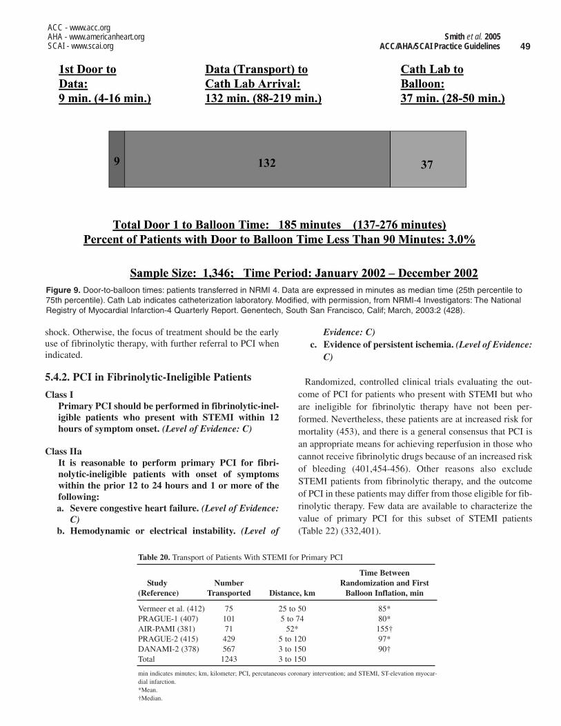
49Smith et al. 2005
ACC/AHA/SCAI Practice Guidelines
ACC - www.acc.orgAHA - www.americanheart.orgSCAI - www.scai.org
shock. Otherwise, the focus of treatment should be the earlyuse of fibrinolytic therapy, with further referral to PCI whenindicated.
5.4.2. PCI in Fibrinolytic-Ineligible Patients
Class IPrimary PCI should be performed in fibrinolytic-inel-igible patients who present with STEMI within 12hours of symptom onset. (Level of Evidence: C)
Class IIaIt is reasonable to perform primary PCI for fibri-nolytic-ineligible patients with onset of symptomswithin the prior 12 to 24 hours and 1 or more of thefollowing: a. Severe congestive heart failure. (Level of Evidence:
C)b. Hemodynamic or electrical instability. (Level of
Evidence: C)c. Evidence of persistent ischemia. (Level of Evidence:
C)
Randomized, controlled clinical trials evaluating the out-come of PCI for patients who present with STEMI but whoare ineligible for fibrinolytic therapy have not been per-formed. Nevertheless, these patients are at increased risk formortality (453), and there is a general consensus that PCI isan appropriate means for achieving reperfusion in those whocannot receive fibrinolytic drugs because of an increased riskof bleeding (401,454-456). Other reasons also excludeSTEMI patients from fibrinolytic therapy, and the outcomeof PCI in these patients may differ from those eligible for fib-rinolytic therapy. Few data are available to characterize thevalue of primary PCI for this subset of STEMI patients(Table 22) (332,401).
Table 20. Transport of Patients With STEMI for Primary PCI
Time Between Study Number Randomization and First
(Reference) Transported Distance, km Balloon Inflation, min
Vermeer et al. (412) 75 25 to 50 85*PRAGUE-1 (407) 101 5 to 74 80*AIR-PAMI (381) 71 52* 155†PRAGUE-2 (415) 429 5 to 120 97*DANAMI-2 (378) 567 3 to 150 90†Total 1243 3 to 150
min indicates minutes; km, kilometer; PCI, percutaneous coronary intervention; and STEMI, ST-elevation myocar-dial infarction.*Mean. †Median.
Figure 9. Door-to-balloon times: patients transferred in NRMI 4. Data are expressed in minutes as median time (25th percentile to75th percentile). Cath Lab indicates catheterization laboratory. Modified, with permission, from NRMI-4 Investigators: The NationalRegistry of Myocardial Infarction-4 Quarterly Report. Genentech, South San Francisco, Calif; March, 2003:2 (428).

50
ACC - www.acc.orgAHA - www.americanheart.org
SCAI - www.scai.orgSmith et al. 2005ACC/AHA/SCAI Practice Guidelines
Tabl
e 21
.St
udie
s C
ompa
ring
PT
CA
with
Ste
nts
in A
cute
Myo
card
ial I
nfar
ctio
n
Ear
ly E
vent
s (0
-30
days
), %
Fol
low
-Up
n, S
tent
/Su
cces
s A
nySt
udy
Yea
r(m
o)P
TC
A(%
)C
ross
over
Dea
thR
einf
arct
ion
TV
RE
vent
FRE
SCO
(43
4)19
986
75/7
599
*N
A0/
01.
3/2.
61.
3/12
3/15
GR
AM
I (4
38)
1998
1252
/52
98/9
4.2
25*
3.8/
7.6
0/7.
60/
5.7
3.8/
19.2
Sury
apra
nata
20
014
112/
115
98/9
62/
132/
31/
4N
AN
Aet
al.
(43
9)
PAST
A(4
36)
1999
1267
/69
99/9
71/
103/
73/
46/
136/
19
Sten
t-PA
MI
(433
)19
996
452/
448
89.4
/92.
71.
5/15
3.5/
1.8
0.4/
1.1
1.8/
3.8
4.6/
5.8
STE
NT
IM-2
(43
5)20
0012
101/
110
95/9
4.5
3/36
.41/
04/
3.6
5/5.
45/
5.4
PSA
AM
I (4
37)
2001
710
plus
44
/44
NA
1/27
2/5
0/2
0/9
5/11
or m
inus
282
days
CA
DIL
LA
Ca
(64)
2002
651
2/51
894
.5/9
4.7
16†
2.2/
2.5
1.0/
0.8
3.2/
5.6
5.7/
8.3
CA
DIL
LA
Cb
(64)
2002
652
4/52
896
.9/9
6.1
14‡
2.7/
1.1
0.8/
0.8
1.6/
3.4
4.4/
4.8
n in
dica
tes
num
ber
of p
atie
nts;
NA
, dat
a no
t gat
here
d fo
r th
at c
ateg
ory;
and
TV
R, t
arge
t-ve
ssel
rev
ascu
lari
zatio
n. A
ll da
ta a
re p
rese
nted
as
valu
es f
or s
tent
/PT
CA
grou
ps.
CA
DIL
LA
Ca
= S
tent
alo
ne a
nd P
TC
Aal
one
arm
s.C
AD
ILL
AC
b =
Ste
nt p
lus
abci
xim
ab a
nd P
TC
Apl
us a
bcix
imab
arm
s.*S
ucce
ss r
ate
of 9
9% b
efor
e ra
ndom
izat
ion.
†Val
ues
for
cros
sove
rs f
rom
PT
CA
to s
tent
trea
tmen
t.‡V
alue
s fo
r cr
osso
vers
fro
m P
TC
Aor
ste
ntin
g al
one
to c
ombi
natio
n w
ith a
bcix
imab
trea
tmen
t.§7
-mon
th f
ollo
w-u
p; n
= 6
36; i
ndep
ende
nt o
f ab
cixi
mab
use
.M
odif
ied
from
Al S
J et
al.
JAM
A20
00;2
84:1
828-
36 (
106)
.
Lat
e E
vent
s (C
umul
ativ
e) %
Any
Res
teno
sis
Dea
thR
einf
arct
ion
TV
RE
vent
17/4
31/
01/
37/
2513
/32
NA
NA
NA
14/2
117
/35
NA
3/3
1/9
13/3
416
/38
17/3
7.5
5/9
NA
NA
22/4
9
20.3
/33.
54.
2/2.
72.
4/2.
27.
7/1.
712
.6/2
0.1
25.3
/39.
63/
1.9
4.0/
5.5
17.8
/28.
212
.9/2
0.0
24/6
19/
182/
916
/34
23/4
3
22.2
/40.
8§3.
0/4.
51.
6/1.
88.
3/15
.711
.5/2
0.0
NA
4.2/
2.5
2.2/
2.7
5.2/
13.8
10.2
/16.
5
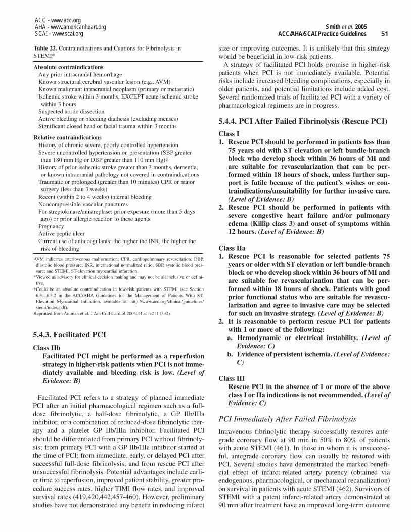
51Smith et al. 2005
ACC/AHA/SCAI Practice Guidelines
ACC - www.acc.orgAHA - www.americanheart.orgSCAI - www.scai.org
5.4.3. Facilitated PCI
Class IIbFacilitated PCI might be performed as a reperfusionstrategy in higher-risk patients when PCI is not imme-diately available and bleeding risk is low. (Level ofEvidence: B)
Facilitated PCI refers to a strategy of planned immediatePCI after an initial pharmacological regimen such as a full-dose fibrinolytic, a half-dose fibrinolytic, a GP IIb/IIIainhibitor, or a combination of reduced-dose fibrinolytic ther-apy and a platelet GP IIb/IIIa inhibitor. Facilitated PCIshould be differentiated from primary PCI without fibrinoly-sis; from primary PCI with a GP IIb/IIIa inhibitor started atthe time of PCI; from immediate, early, or delayed PCI aftersuccessful full-dose fibrinolysis; and from rescue PCI afterunsuccessful fibrinolysis. Potential advantages include earli-er time to reperfusion, improved patient stability, greater pro-cedure success rates, higher TIMI flow rates, and improvedsurvival rates (419,420,442,457-460). However, preliminarystudies have not demonstrated any benefit in reducing infarct
size or improving outcomes. It is unlikely that this strategywould be beneficial in low-risk patients.
A strategy of facilitated PCI holds promise in higher-riskpatients when PCI is not immediately available. Potentialrisks include increased bleeding complications, especially inolder patients, and potential limitations include added cost.Several randomized trials of facilitated PCI with a variety ofpharmacological regimens are in progress.
5.4.4. PCI After Failed Fibrinolysis (Rescue PCI)
Class I1. Rescue PCI should be performed in patients less than
75 years old with ST elevation or left bundle-branchblock who develop shock within 36 hours of MI andare suitable for revascularization that can be per-formed within 18 hours of shock, unless further sup-port is futile because of the patient’s wishes or con-traindications/unsuitability for further invasive care.(Level of Evidence: B)
2. Rescue PCI should be performed in patients withsevere congestive heart failure and/or pulmonaryedema (Killip class 3) and onset of symptoms within12 hours. (Level of Evidence: B)
Class IIa1. Rescue PCI is reasonable for selected patients 75
years or older with ST elevation or left bundle-branchblock or who develop shock within 36 hours of MI andare suitable for revascularization that can be per-formed within 18 hours of shock. Patients with goodprior functional status who are suitable for revascu-larization and agree to invasive care may be selectedfor such an invasive strategy. (Level of Evidence: B)
2. It is reasonable to perform rescue PCI for patientswith 1 or more of the following: a. Hemodynamic or electrical instability. (Level of
Evidence: C)b. Evidence of persistent ischemia. (Level of Evidence:
C)
Class IIIRescue PCI in the absence of 1 or more of the aboveclass I or IIa indications is not recommended. (Level ofEvidence: C)
PCI Immediately After Failed Fibrinolysis
Intravenous fibrinolytic therapy successfully restores ante-grade coronary flow at 90 min in 50% to 80% of patientswith acute STEMI (461). In those in whom it is unsuccess-ful, antegrade coronary flow can usually be restored withPCI. Several studies have demonstrated the marked benefi-cial effect of infarct-related artery patency (obtained viaendogenous, pharmacological, or mechanical recanalization)on survival in patients with acute STEMI (462). Survivors ofSTEMI with a patent infarct-related artery demonstrated at90 min after treatment have an improved long-term outcome
Table 22. Contraindications and Cautions for Fibrinolysis inSTEMI*
Absolute contraindicationsAny prior intracranial hemorrhageKnown structural cerebral vascular lesion (e.g., AVM)Known malignant intracranial neoplasm (primary or metastatic)Ischemic stroke within 3 months, EXCEPT acute ischemic strokewithin 3 hours
Suspected aortic dissectionActive bleeding or bleeding diathesis (excluding menses)Significant closed head or facial trauma within 3 months
Relative contraindicationsHistory of chronic severe, poorly controlled hypertension Severe uncontrolled hypertension on presentation (SBP greaterthan 180 mm Hg or DBP greater than 110 mm Hg)†
History of prior ischemic stroke greater than 3 months, dementia,or known intracranial pathology not covered in contraindications
Traumatic or prolonged (greater than 10 minutes) CPR or majorsurgery (less than 3 weeks)
Recent (within 2 to 4 weeks) internal bleeding Noncompressible vascular punctures For streptokinase/anistreplase: prior exposure (more than 5 daysago) or prior allergic reaction to these agents
PregnancyActive peptic ulcer Current use of anticoagulants: the higher the INR, the higher therisk of bleeding
AVM indicates arteriovenous malformation; CPR, cardiopulmonary resuscitation; DBP,diastolic blood pressure; INR, international normalized ratio; SBP, systolic blood pres-sure; and STEMI, ST-elevation myocardial infarction.
*Viewed as advisory for clinical decision making and may not be all inclusive or defini-tive.
†Could be an absolute contraindication in low-risk patients with STEMI (see Section6.3.1.6.3.2 in the ACC/AHA Guidelines for the Management of Patients With ST-Elevation Myocardial Infarction, available at: http://www.acc.org/clinical/guidelines/stemi/index.pdf).
Reprinted from Antman et al. J Am Coll Cardiol 2004;44:e1-e211 (332).

52
ACC - www.acc.orgAHA - www.americanheart.org
SCAI - www.scai.orgSmith et al. 2005ACC/AHA/SCAI Practice Guidelines
coronary reperfusion occurs over the subsequent h after fib-rinolytic therapy in many patients. Although infarct-relatedartery patency is achieved in only 50% to 85% of patients 90min after fibrinolytic therapy, it rises to 90% by 24 h (473).Such “late” reperfusion may improve survival without therisk of invasive procedures coupled with fibrinolytic therapy.Confounding the issue, both fibrinolysis and PCI may suc-cessfully restore flow in the epicardial artery but fail toimprove microvascular perfusion.
Hours to Days After Failed Fibrinolysis
Patency of the infarct-related artery is an important predictorof mortality in survivors of STEMI (462,463,474).Compared with those with a patent infarct artery, survivorsof STEMI with a persistently occluded artery after fibrinoly-sis, PCI, or no reperfusion therapy have 1) increased LVdilatation (475), 2) a greater incidence of spontaneous andinducible ventricular arrhythmias (476), and 3) a poorerprognosis (477). On the basis of observational and experi-mental data, it has been hypothesized that infarct arterypatency may favorably influence LV remodeling and electri-cal stability, even if accomplished at a time when salvage ofischemic myocardium is unlikely (i.e., more than 12 h todays after coronary artery occlusion). Five small randomizedtrials, which enrolled a total of 562 patients, have directlytested the hypothesis that mechanical opening of persistenttotal occlusions late after MI will improve long-term LVremodeling and clinical outcomes (the late open arteryhypothesis). Most studies enrolled a combination of patients,including those who had failed fibrinolysis and those whohad not received reperfusion therapy (478-480), with a rangefrom almost no fibrinolytic therapy (481) to fibrinolytic ther-apy in nearly all patients (482). There was wide variation inthe effect of routine PCI compared with medical therapy onlyon LV size and function. Most studies showed no significantdifferences between the treatment groups (478,479). Onesingle-center study of 83 patients with LAD occlusionsreported improved LV volumes and clinical outcomes (com-posite of HF, MI, and death) at 6 months in the PCI group(481). In contrast, a multicenter study of 66 patients withLAD occlusions reported significantly worse LV remodel-ing, with progressive LV dilation at 1 year and more clinicalevents in the PCI group than in those assigned to optimalmedical therapy alone (482). The latter included very highrates of beta-blocker and angiotensin converting enzymeinhibitor use. The largest multicenter study, DECOPI(DEsobstruction COronaire en Post-Infarctus), enrolled 212patients and reported no difference in the primary end point,the composite of death, ventricular tachycardia, and MI at 6months (483). Stents were used in 80% of patients in the PCIgroup, and GP IIb/IIIa antagonists were used in 9%. Thestudy reached less than one third of the target sample sizeand was severely underpowered, as were all the other stud-ies, to assess clinical events.
Selection of patients for revascularization based on viabil-ity testing has gained a great deal of investigational support,i.e., delayed enhancement or low-dose dobutamine cardiac
compared with those with an occluded infarct-related artery,even when LV systolic function is similar (463,464). TheREACT trial (Rapid Early Action for Coronary Treatment)was a randomized trial comparing medical therapy, immedi-ate PCI, or repeat fibrinolytic in patients previously treatedwith fibrinolytic therapy. Preliminary data at 30 days demon-strated a significant advantage to rescue PCI [A.H.Gershlick, oral presentation, American Heart AssociationScientific Sessions, New Orleans, LA, November 2004.]
Rescue (also known as salvage) PCI is defined as PCI with-in 12 h after failed fibrinolysis for patients with continuingmyocardial ischemia. Rescue PCI has resulted in higher ratesof early infarct-artery patency, improved regional infarct-zone wall motion, and greater freedom from adverse in-hos-pital clinical events compared with a deferred PCI strategy ormedical therapy (465). The Randomized Evaluation ofRescue PCI with Combined Utilization Endpoints (RES-CUE) trial demonstrated a reduction in rates of in-hospitaldeath and combined death and HF that was maintained up to1 year after study entry for patients presenting with anteriorwall STEMI who failed fibrinolytic therapy when performeda mean of 8 h after symptom onset (466,467). Improvementin TIMI grade flow from 2 to 3 may offer additional clinicalbenefit. Similar data are not available for patients withnonanterior STEMI.
A major problem in adopting a strategy of rescue PCI liesin the limitation of accurate identification of patients inwhom fibrinolytic therapy has not restored antegrade coro-nary flow. Unless unsuccessful fibrinolysis is recognized andcorrected quickly (within 3 to 6 h of onset of symptoms), sal-vage of ischemic myocardium is unlikely. Unfortunately,clinical markers of reperfusion, such as relief of ischemic-type chest discomfort, partial resolution of ST-segment ele-vation, and reperfusion arrhythmias, have limited predictivevalue in identifying failure of fibrinolysis (468). Immediatecatheterization of all patients after fibrinolytic therapy toidentify those with an occluded infarct-related artery in aprior era in which the practice of PCI was less mature failedto show a significant benefit and was associated with bleed-ing complications. However, there was no specific studyusing stents and current pharmacotherapy. This strategy isbeing re-evaluated in clinical trials testing facilitated PCI inthe contemporary PCI setting.
Even in the patient with documented failure of fibrinolysis,rescue PCI has limitations. First, because extensive myocar-dial necrosis occurs when coronary occlusion has been pres-ent for more than 3 h (469), PCI may not salvage a substan-tial amount of myocardium, considering the time delay asso-ciated with presentation of the patient to the hospital afteronset of symptoms, infusion of the fibrinolytic agent, recog-nition of failed fibrinolysis, and subsequent initiation of PCI.Second, rescue PCI fails to reestablish antegrade coronaryflow in about 10% of patients, and reocclusion of the infarct-related artery occurs in as many as 20% of the remainder(470), although GP IIb/IIIa inhibitors and stent implantationmay have improved these results. Third, unsuccessful rescuePCI is associated with a high mortality (471,472). Finally,
53Smith et al. 2005
ACC/AHA/SCAI Practice Guidelines
ACC - www.acc.orgAHA - www.americanheart.orgSCAI - www.scai.org
tine PCI immediately after fibrinolysis may increase thechance for vascular complications at the catheterizationaccess site and hemorrhage into the infarct-related vesselwall (491).
Hours to Days After Successful Fibrinolysis
It was initially suggested that elective PCI of the stenoticinfarct-related artery hours to days after fibrinolysis mightallow sufficient time for development of a more stable hemo-static milieu at the site of previous thrombotic occlusion. Inthis setting, PCI would be safer and more effective in reduc-ing the incidence of reocclusion and improving survival.Two large randomized, prospective trials from an earlier PCIera tested this hypothesis, with both concluding that 1) thereare fewer complications if PCI is delayed for several daysafter fibrinolytic therapy, and 2) routine PCI in the absenceof spontaneous or provocable ischemia does not improve LVfunction or survival (226,489,490,492). Thus, in unselectedpatients receiving fibrinolytic therapy, PCI of the stenoticinfarct-related artery in the absence of evidence of recurrentischemia within 48 h did not appear to be beneficial.
Great improvements in equipment, operator experience,and adjunctive pharmacotherapy have increased PCI successrates and decreased complications. More recently, the inva-sive strategy for patients with NSTEMI has been given aclass I recommendation by the ACC/AHA 2002 GuidelineUpdate for the Management of Patients With UA/NSTEMI(493). Patients with STEMI are increasingly being treatedsimilarly as an extension of this approach. Although 7 pub-lished reports (474,480,494-498) support this strategy, ran-domized studies similar to those in NSTEMI are needed.
One study supports the policy of performing catheteriza-tion and subsequent revascularization for patients who dohave spontaneous or inducible angina after STEMI. TheDANAMI trial (499) randomly assigned 1008 survivors of afirst acute MI treated with fibrinolytic therapy within 12 h ofonset of symptoms to catheterization and subsequent revas-cularization or standard medical therapy if they showed evi-dence of spontaneous or inducible angina. Those who under-went revascularization had less UA and fewer nonfatal MIsduring a 2.5-year period of follow-up than those patients ran-domly assigned to medical treatment only (18% and 5.6% vs30% and 10.5%, respectively). Among 500 patients undergo-ing fibrinolysis for STEMI, the GRACIA-1 (randomizedtrial comparing stenting within 24 hours of thrombolysis ver-sus ischemia-guided approach to thrombolysed acutemyocardial infarction with ST elevation) trial compared astrategy of angiography and intervention within 6 to 24 h offibrinolysis to an ischemia-guided conservative approach forintervention. Eighty percent of patients assigned to angiogra-phy and intervention underwent stenting of the culprit arterycompared with 20% in the ischemia-guided group. At 1 year,patients in the invasive group had a lower frequency of theprimary end point (death, reinfarction, or revascularization;9% vs 21%, P equals 0.008), and they tended to have areduced rate of death or reinfarction (7% vs 12%, P equals
MRI assessment. If viability is shown, outcomes are excel-lent, whereas if transmural MI is present, it is not, and revas-cularization is not recommended (484-486).
There are no convincing data to support the routine use oflate adjuvant PCI days after failed fibrinolysis or for patientswho do not receive reperfusion therapy. Nevertheless, this isbeing done in some STEMI patients as an extension of theinvasive strategy for NSTEMI patients. The Occluded ArteryTrial (OAT) is currently randomizing patients to test whetherroutine PCI days to weeks after MI improves long-term clin-ical outcomes in asymptomatic high-risk patients with anoccluded infarct-related artery (487).
5.4.5. PCI After Successful Fibrinolysis or forPatients Not Undergoing Primary Reperfusion
Class I1. In patients whose anatomy is suitable, PCI should be
performed when there is objective evidence of recur-rent MI. (Level of Evidence: C)
2. In patients whose anatomy is suitable, PCI should beperformed for moderate or severe spontaneous orprovocable myocardial ischemia during recoveryfrom STEMI. (Level of Evidence: B)
3. In patients whose anatomy is suitable, PCI should beperformed for cardiogenic shock or hemodynamicinstability. (Level of Evidence: B)
Class IIa1. It is reasonable to perform routine PCI in patients
with LV ejection fraction less than or equal to 0.40,HF, or serious ventricular arrhythmias. (Level ofEvidence: C)
2. It is reasonable to perform PCI when there is docu-mented clinical heart failure during the acute episode,even though subsequent evaluation shows preservedLV function (LV ejection fraction greater than 0.40).(Level of Evidence: C)
Class IIbPCI might be considered as part of an invasive strate-gy after fibrinolytic therapy. (Level of Evidence: C)
PCI Immediately After Successful Fibrinolysis
In early studies, asymptomatic patients undergoing routinePCI of the stenotic infarct-related artery immediately aftersuccessful fibrinolysis showed no benefit with regard to sal-vage of jeopardized myocardium or prevention of reinfarc-tion or death. In addition, in some studies, this approach wasassociated with an increased incidence of adverse events,including bleeding, recurrent ischemia, emergency CABGsurgery, and death (488-491). However, these studies havenot been repeated in the modern interventional era withimproved equipment, improved antiplatelet and anticoagu-lant therapy, and coronary stents. Notwithstanding this, rou-

54
ACC - www.acc.orgAHA - www.americanheart.org
SCAI - www.scai.orgSmith et al. 2005ACC/AHA/SCAI Practice Guidelines
reduction in 30-day mortality (P less than 0.02), whereasthere was no apparent benefit for the small cohort (n equals56) of patients more than 75 years old. These data stronglysupport the approach that patients younger than 75 yearswith STEMI complicated by cardiogenic shock shouldundergo emergency revascularization and support measures.Three registries (504-506) have demonstrated a marked sur-vival benefit for elderly patients who are clinically selectedfor revascularization (approximately 1 of 5 patients), so agealone should not disqualify a patient for early revasculariza-tion (see Section 3.5.9).
Several additional discussions elsewhere in this guidelineare important to consider in these patients. Intra-aortic bal-loon pump support or ventricular assist devices can stabilizehemodynamics so that revascularization procedures can beperformed (see Section 3.5.8). Post hoc analyses (507-509)have suggested that GP IIb/IIIa inhibitors may reduce mor-tality, but the studies are limited by lower than expected mor-tality rates, larger than expected mortality reduction, andsmall sample sizes. Although PCI in a noninfarct artery is notrecommended in stable patients, it can be beneficial inhemodynamically compromised patients if the stenotic arteryperfuses a large area of myocardium and the procedure canbe performed efficiently. In patients with significant leftmain disease or severe 3-vessel disease and without rightventricular infarction or major comorbidities such as renalinsufficiency or severe pulmonary disease, CABG can beconsidered as the revascularization strategy (Figure 10)(510).
5.4.7. PCI in Selected Patient Subgroups
5.4.7.1. Young and Elderly Postinfarct Patients
Although not supported by randomized trials, routine cardiaccatheterization after fibrinolytic therapy for STEMI has beena frequently performed strategy in all age groups. Youngpatients (less than 50 years old) often undergo cardiaccatheterization after fibrinolytic therapy owing to a “per-ceived need” to define coronary anatomy and thus establishpsychological as well as clinical outcomes. In contrast, olderpatients (greater than 75 years of age) have higher in-hospi-tal and long-term mortality rates and enhanced clinical out-comes when treated with primary PCI (511-514). In addition,patients thought to be candidates for implantable cardiovert-er-defibrillator placement and those with small infarcts mayundergo cardiac catheterization for further evaluation afterSTEMI.
In a secondary analysis of the TIMI-IIB study that com-pared angiographic findings and clinical outcomes among841 young (aged less than 50 years) and 859 older (aged 65to 70 years) patients randomly assigned to an invasive orconservative post-lytic management strategy (515), theyounger patients assigned to the invasive strategy common-ly had insignificant (i.e., less than 60% diameter stenosis)and single-vessel CAD. Severe 3-vessel or left main coro-nary disease findings were infrequent (3-vessel incidence,4%; left main, 0%). Fatal and nonfatal MI and death through-
0.07). In the angiography and intervention group, 81% hadTIMI-3 flow before PCI was performed (494).
Days to Weeks After Successful Fibrinolysis
Continued thrombus lysis and remodeling of the infarctartery stenosis occur over the days to weeks after successfulfibrinolysis, which makes the underlying residual coronarystenosis more stable and less prone to rethrombosis and reoc-clusion. Thus, a delay in performing PCI for days to weeksafter fibrinolysis might improve survival, even though earli-er routine PCI does not. To date, there have not been ade-quately sized trials to evaluate this treatment strategy. Twoolder, small, randomized trials (488,500) demonstrated simi-lar LV function, rates of reinfarction, and mortality inpatients randomized to PCI or conservative therapy.
5.4.6. PCI for Cardiogenic Shock
Class IPrimary PCI is recommended for patients less than 75years old with ST elevation or left bundle-branchblock who develop shock within 36 hours of MI andare suitable for revascularization that can be per-formed within 18 hours of shock, unless further sup-port is futile because of the patient’s wishes or con-traindications/unsuitability for further invasive care.(Level of Evidence: A)
Class IIaPrimary PCI is reasonable for selected patients 75years or older with ST elevation or left bundle-branchblock who develop shock within 36 hours of MI andare suitable for revascularization that can be per-formed within 18 hours of shock. Patients with goodprior functional status who are suitable for revascu-larization and agree to invasive care may be selectedfor such an invasive strategy. (Level of Evidence: B)
Observational studies support the value of PCI for patientswho develop cardiogenic shock in the early hours of MI. Forpatients who do not have mechanical causes of shock, suchas acute mitral regurgitation or septal or free wall rupture,mortality among those having PCI is lower than among thosetreated by medical means (366). However, undergoing car-diac catheterization alone, with or without PCI, is associatedwith a lower mortality because of patient selection bias(501).
Two randomized clinical trials (366,502) have further clar-ified the role of emergency revascularization in STEMI com-plicated by cardiogenic shock. Both showed a statisticallyinsignificant, but clinically important, absolute 9% reductionin 30-day mortality. In the SHOCK trial (366), the survivalcurves continued to progressively diverge such that at 6months and 1 year, there was a significant mortality reduc-tion with emergency revascularization (53% vs 66%, P lessthan 0.03) (503). The prespecified subgroup analysis ofpatients less than 75 years old showed an absolute 15%
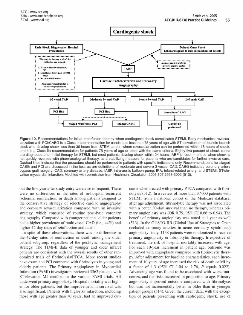
55Smith et al. 2005
ACC/AHA/SCAI Practice Guidelines
ACC - www.acc.orgAHA - www.americanheart.orgSCAI - www.scai.org
come when treated with primary PTCA compared with fibri-nolysis (512). In a review of more than 37 000 patients withSTEMI from a national cohort of the Medicare database,after age adjustment, fibrinolytic therapy was not associatedwith a better 30-day survival than no therapy, whereas pri-mary angioplasty was (OR 0.79, 95% CI 0.66 to 0.94). Thebenefit of primary angioplasty was noted at 1 year as well(513). In the GUSTO-IIB (Global Use of Strategies to Openoccluded coronary arteries in acute coronary syndromes)angioplasty study, 1138 patients were randomized to receiveprimary angioplasty or fibrinolytic therapy. Irrespective oftreatment, the risk of hospital mortality increased with age.For each 10-year increment in patient age, outcome wasimproved with angioplasty compared with fibrinolytic thera-py. After adjustment for baseline characteristics, each incre-ment of 10 years of age increased the risk of death or MI bya factor of 1.3 (95% CI 1.04 to 1.76, P equals 0.022).Advancing age was found to be associated with worse out-comes, and the risks increased in proportion to age. Primaryangioplasty improved outcome compared with fibrinolysisbut was not incrementally better in older than in youngerpatient groups (514). Given the current data, with the excep-tion of patients presenting with cardiogenic shock, use of
out the first year after study entry were also infrequent. Therewere no differences in the rates of in-hospital recurrentischemia, reinfarction, or death among patients assigned tothe conservative strategy of selective cardiac angiographyand coronary revascularization compared with an invasivestrategy, which consisted of routine post-lytic coronaryangiography. Compared with younger patients, older patientshad a higher prevalence of multivessel CAD (i.e., 44%) andhigher 42-day rates of reinfarction and death.
In spite of these observations, there was no difference inthe 42-day rates of reinfarction or death among the olderpatient subgroup, regardless of the post-lytic managementstrategy. The TIMI-II data of younger and older infarctpatients are consistent with the overall results of other ran-domized trials of fibrinolysis/PTCA. More recent studieshave examined PCI compared with fibrinolysis in young andelderly patients. The Primary Angioplasty in MyocardialInfarction (PAMI) investigators reviewed 3362 patients withST-elevation MI enrolled in the various PAMI trials. Allunderwent primary angioplasty. Hospital mortality was high-er for older patients, but the improvement in survival wasalso significant. Patients with high-risk infarction, includingthose with age greater than 70 years, had an improved out-
Figure 10. Recommendations for initial reperfusion therapy when cardiogenic shock complicates STEMI. Early mechanical revascu-larization with PCI/CABG is a Class I recommendation for candidates less than 75 years of age with ST elevation or left bundle-branchblock who develop shock less than 36 hours from STEMI and in whom revascularization can be performed within 18 hours of shock,and it is a Class IIa recommendation for patients 75 years of age or older with the same criteria. Eighty-five percent of shock casesare diagnosed after initial therapy for STEMI, but most patients develop shock within 24 hours. IABP is recommended when shock isnot quickly reversed with pharmacological therapy, as a stabilizing measure for patients who are candidates for further invasive care.Dashed lines indicate that the procedure should be performed in patients with specific indications only. Recommendations for stagedCABG and PCI are discussed in the text, as are definitions of moderate and severe 3-vessel CAD. CABG indicates coronary arterybypass graft surgery; CAD, coronary artery disease; IABP, intra-aortic balloon pump; IRA, infarct-related artery; and STEMI, ST-ele-vation myocardial infarction. Modified with permission from Hochman. Circulation 2003;107:2998-3002 (510).

56
ACC - www.acc.orgAHA - www.americanheart.org
SCAI - www.scai.orgSmith et al. 2005ACC/AHA/SCAI Practice Guidelines
evidence of ischemia should be obtained.) (Level ofEvidence: B)
3. PCI is reasonable in patients with diseased vein graftsmore than 3 years after CABG. (Level of Evidence: B)
4. PCI is reasonable when technically feasible in patientswith a patent left internal mammary artery graft whohave clinically significant obstructions in other ves-sels. (Level of Evidence: C)
Class III1. PCI is not recommended in patients with prior CABG
for chronic total vein graft occlusions. (Level ofEvidence: B)
2. PCI is not recommended in patients who have multi-ple target lesions with prior CABG and who have mul-tivessel disease, failure of multiple SVGs, andimpaired LV function unless repeat CABG posesexcessive risk due to severe comorbid conditions.(Level of Evidence: B)
5.5.1. Early Ischemia After CABG
Recurrent ischemia early (less than 30 days) postoperativelyusually reflects graft failure, often secondary to thrombosis(522-524), and may occur in both saphenous vein and arteri-al graft conduits (525). Incomplete revascularization andunbypassed native vessel stenoses or stenoses distal to abypass graft anastomosis may also precipitate recurrentischemia. Urgent coronary angiography is indicated to definethe anatomic cause of ischemia and to determine the bestcourse of therapy. Emergency PCI of a focal graft stenosis(venous or arterial) or recanalization of an acute graft throm-bosis may successfully relieve ischemia in the majority ofpatients. Balloon dilatation across suture lines has beenaccomplished safely within days of surgery (526-528).Intracoronary fibrinolytic therapy should be administeredwith caution during the first week postoperatively (529-532),and if required, residual thrombus may be “targeted” in lowdoses through a local drug delivery system. Conversely,mechanical thrombectomy with newer catheter technologiesmay be effective without the attendant risk of fibrinolysis(533). Adjunctive therapy with abciximab for percutaneousintervention during the first week after bypass surgery hasbeen limited but intuitively may pose less risk for hemor-rhage than fibrinolysis. Because flow in vein graft conduitsis pressure dependent, IABP support should be considered inthe context of systemic hypotension or severe LV dysfunc-tion. If feasible, PCI of both bypass graft and native vesseloffending stenoses should be attempted, particularly if intra-coronary stents can be deployed successfully.
When ischemia occurs 1 to 12 months after surgery, thecause is usually perianastomotic graft stenosis. Distal anas-tomotic stenoses (both arterial and venous) respond well toballoon dilation alone and have a more favorable long-termprognosis than stenoses involving the midshaft or proximalvein graft anastomosis (250,251,534-537). Midshaft veingraft stenoses occurring during this time frame are usually
PCI should be determined by clinical need without specialconsideration of age.
5.4.7.2. Patients With Prior MI
A prior MI is an independent predictor of death, reinfarction,and need for urgent coronary bypass surgery (516). In fibri-nolytic trials, 14% to 20% of enrolled patients had a historyof prior MI (490,517,518), and patients with prior MI havehigher rates of reinfarction after fibrinolytic therapy (519).
In the TIMI-II study, patients with a history of prior MI hada higher 42-day mortality (8.8% vs 4.3%; P less than 0.001),higher prevalence of multivessel CAD (60% vs 28%; P lessthan 0.001), and a lower LV ejection fraction (42% vs 48%;P less than 0.001) than patients with a first MI (520). Amongpatients assigned to the conservative post-lytic strategy,those with a prior MI had a significantly higher 42-day mor-tality than patients with a first MI (11.5% vs 3.5%; P lessthan 0.001), whereas in the invasive strategy, the mortalityoutcome was essentially the same in the 2 patient groups.Mortality tended to be lower among patients with a prior MIundergoing the invasive versus the conservative strategy, abenefit that persisted up to 1 year after study entry (492).
In a registry involving almost 12 000 patients with acutecoronary syndromes, with and without ST-segment eleva-tion, a history of prior MI was noted in almost one third ofpatients. There was no significant increase in relative risk forhospital mortality in this group (521). Some analyses of pre-dictors of mortality for angioplasty after fibrinolytic therapyhave found relatively little importance for prior infarctionand emphasized the much greater importance of time delaysin achieving reperfusion among patients with failed fibrinol-ysis (418).
Given the above findings and current practice, PCI shouldbe based on clinical need. The presence of prior MI placesthe patient in a higher-risk subset and should be consideredin the PCI decision.
5.5. Percutaneous Intervention in Patients WithPrior Coronary Bypass Surgery
Class I1. When technically feasible, PCI should be performed
in patients with early ischemia (usually within 30days) after CABG. (Level of Evidence: B)
2. It is recommended that distal embolic protectiondevices be used when technically feasible in patientsundergoing PCI to saphenous vein grafts. (Level ofEvidence: B)
Class IIa1. PCI is reasonable in patients with ischemia that
occurs 1 to 3 years after CABG and who have pre-served LV function with discrete lesions in graft con-duits. (Level of Evidence: B)
2. PCI is reasonable in patients with disabling anginasecondary to new disease in a native coronary circula-tion after CABG. (If angina is not typical, objective
57Smith et al. 2005
ACC/AHA/SCAI Practice Guidelines
ACC - www.acc.orgAHA - www.americanheart.orgSCAI - www.scai.org
stent deployment (244,245,562-569) than with balloonangioplasty of SVG stenoses, long-term prognosis remainsguarded, and late recurrent ischemic events may be due toboth restenosis of the target lesion and diffuse vein graft dis-ease (570-572). Final patency after PCI is greater for distalSVG lesions than for ostial or mid-SVG lesions (535), andstenosis location appears to be a better determinant of finalpatency than graft age or the type of interventional deviceused.
Percutaneous intervention for chronic vein graft occlusionhas been problematic. Balloon angioplasty alone has beenassociated with high complication rates and low rates of sus-tained patency (572). Although prolonged intragraft infusionof fibrinolytic therapy was reported to successfully recanal-ize 69% of a selected group of patients with chronic SVGocclusion of less than 6 months’ duration, long-term patencyrates with or without adjunctive stent deployment were low(573-575). In addition, prolonged fibrinolytic therapy hasbeen associated with thromboembolic MI (576-579),intracranial hemorrhage (580), and intramyocardial hemor-rhage (581), as well as vascular access-site complications.Favorable results have been obtained with both local “target-ed” and more prolonged infusion of fibrinolytic agents fornonocclusive intragraft thrombus (582,583). Fibrinolyticcatheter-based systems appear to successfully treat SVGthrombosis as well as or better than fibrinolytic agents (584).
5.5.3. Early and Late Outcomes of Percutaneous Intervention
Before the general availability of coronary stenting, overallangioplasty procedural success rates exceeded 90%, andadverse outcomes of emergency repeat coronary bypass sur-gery (2.3%) and death (0.8%) were infrequent as reported incombined series of over 2000 patients with prior bypass sur-gery undergoing percutaneous intervention (250,585-597).These results are comparable to those achieved in patientswithout prior bypass surgery, an observation confirmed byNHLBI registry data (7). The most common complicationsobserved in this population are NSTEMI and atheroem-bolism, particularly after SVG intervention (538,598).
Patients with prior bypass surgery who undergo successfulPCI have a long-term outcome that is dependent on patientage, the degree of LV dysfunction, and the presence of mul-tivessel coronary atherosclerosis. The best long-term resultsare observed after recanalization of distal anastomoticstenoses occurring within 1 year of operation. Angioplasty ofdistal anastomotic stenoses involving IMA grafts has beenassociated with similar, favorable long-term patency rates(543,544). Conversely, event-free survival is less favorableafter angioplasty of totally occluded SVGs, ostial vein graftstenoses, or grafts with diffuse or multicentric disease (570-572). Coexistent multisystem disease, the presence of whichmay have prompted the choice of a percutaneous revascular-ization strategy, may also influence long-term outcomes inthis population.
due to intimal hyperplasia. Restenosis may be less frequentand event-free survival may be enhanced after angioplasty ofSVGs dilated within 6 months of surgery compared withgrafts of older age. The immediate results of PCI in midshaftostial or distal anastomotic vein graft stenoses may beenhanced by coronary stent deployment (537,538). Ablativetechnologies such as directional atherectomy or excimerlaser coronary angioplasty may facilitate angioplasty andstent deployment in patients with aorto-ostial vein graftstenoses (539,540).
Stenoses in the midportion or origin of the IMA graft areuncommon but respond to balloon dilation (541,542) withstent deployment as feasible. Long-term follow-up ofpatients after IMA angioplasty has demonstrated sustainedbenefit and relief of ischemia in the majority of patients(543,544). Balloon angioplasty with or without stent deploy-ment can be performed successfully in patients with distalanastomotic stenoses involving the gastroepiploic arterybypass graft and in patients with free radial artery bypassgrafts (545). Percutaneous intervention has also been effec-tive in relieving ischemia for patients with stenosis of thesubclavian artery proximal to the origin of a patent left IMAbypass graft (546,547).
5.5.2. Late Ischemia After CABG
Ischemia occurring more than 1 year postoperatively usuallyreflects the development of new stenoses in graft conduitsand/or native vessels that may be amenable to PCI (548). At3 years or more after SVG implantation, atheroscleroticplaque is frequently evident and is often progressive. Theselesions may be friable and often have associated thrombusformation, which may contribute to the occurrence of slowflow, distal embolization, and periprocedural MI afterattempted percutaneous intervention (56). Slow flow occursmore frequently in grafts that have diffuse atheroscleroticinvolvement, angiographically demonstrable thrombus, andirregular or ulcerative lesion surfaces, and with long lesionsthat have large plaque volume (549,550). Although a reducedincidence of distal embolization has been reported after theuse of the extraction atherectomy catheter to recanalizestenoses in older vein graft conduits (551-555), embolizationmay still complicate adjunctive balloon dilation. Distalembolic protection devices have significantly reduced theoccurrence of complications of embolization in SVGs andshould be used when possible (254,255). Slow flow withsigns and symptoms of myocardial ischemia may be amelio-rated by the intragraft administration of agents such asadenosine, diltiazem, nitroprusside, and verapamil (549,556-559). The adjunctive administration of abciximab duringvein graft intervention was evaluated in a meta-analysis of 5studies that demonstrated no improvement in outcomes afterPCI and in the absence of distal protection and was associat-ed with a high incidence of death and nonfatal ischemicevents (252).
Although postprocedural minimum lumen diameter is larg-er after directional coronary atherectomy (243,560,561) or
58
ACC - www.acc.orgAHA - www.americanheart.org
SCAI - www.scai.orgSmith et al. 2005ACC/AHA/SCAI Practice Guidelines
a. Assessment of the adequacy of deployment of coro-nary stents, including the extent of stent appositionand determination of the minimum luminal diame-ter within the stent. (Level of Evidence: B)
b. Determination of the mechanism of stent restenosis(inadequate expansion versus neointimal prolifera-tion) and to enable selection of appropriate thera-py (vascular brachytherapy versus repeat balloonexpansion). (Level of Evidence: B)
c. Evaluation of coronary obstruction at a locationdifficult to image by angiography in a patient witha suspected flow-limiting stenosis. (Level ofEvidence: C)
d. Assessment of a suboptimal angiographic resultafter PCI. (Level of Evidence: C)
e. Establishment of the presence and distribution ofcoronary calcium in patients for whom adjunctiverotational atherectomy is contemplated. (Level ofEvidence: C)
f. Determination of plaque location and circumferen-tial distribution for guidance of directional coro-nary atherectomy. (Level of Evidence: B)
Class IIbIVUS may be considered for the following: a. Determination of the extent of atherosclerosis in
patients with characteristic anginal symptoms anda positive functional study with no focal stenoses ormild CAD on angiography. (Level of Evidence: C)
b. Preinterventional assessment of lesional character-istics and vessel dimensions as a means to select anoptimal revascularization device. (Level ofEvidence: C)
c. Diagnosis of coronary disease after cardiac trans-plantation. (Level of Evidence: C)
Class IIIIVUS is not recommended when the angiographicdiagnosis is clear and no interventional treatment isplanned. (Level of Evidence: C)
IVUS imaging provides a tomographic 360-degree sagittalscan of the vessel from the lumen through the media to thevessel wall. IVUS measurements of arterial dimensions(minimal and maximal diameters, cross-sectional area, andplaque area) complement and enhance angiographic infor-mation. IVUS has been used to refine device selectionthrough plaque characterization (e.g., calcified) and arterysizing. IVUS has contributed to the understanding of themechanisms of coronary angioplasty in general and specifi-cally to the advancement of coronary stenting without long-term anticoagulation (612-617). In a large observationalstudy, IVUS-guided angioplasty resulted in a decreased finalresidual plaque area from 51% to 34%, despite a final angio-graphic percent stenosis of 0% (612). IVUS-facilitated stentdeployment was associated with a subacute thrombosis rateof 0.3% without systemic anticoagulation, although
5.5.4. General Considerations
Aged, diffuse, friable, and degenerative SVG disease in theabsence of a patent arterial conduit to the LAD represents aprime consideration for repeat surgical revascularization. Incontrast, the presence of a patent arterial conduit to the LADfavors a percutaneous interventional approach to other ves-sels (599). The overall risk of repeat operation, especially thepresence of comorbidities such as concomitant cerebrovas-cular, renal, or pulmonary disease and the potential for jeop-ardizing patent, nondiseased bypass conduits, must be con-sidered carefully. Isolated, friable stenoses in vein grafts maybe approached with primary stenting or the combination ofextraction atherectomy and stenting in an attempt to reducethe likelihood of distal embolization. Distal embolic protec-tion devices have reduced the occurrence of complications ofembolization significantly and should be used when possible(254,255) (see Sections 5.5.2 and 6.1.1).
Another therapeutic option for patients with prior coronarybypass surgery that has become available is grafting with theIMA through a “minimally invasive” surgical approach(273,600-604). This strategy, which avoids both the risk ofcardiopulmonary bypass (stroke or coagulopathy) and repeatmedian sternotomy, may be particularly applicable topatients with chronic native-vessel LAD coronary occlusionand friable atherosclerotic disease that involves a prior SVGto this vessel. The role of combining a minimally invasivesurgical approach with PCI requires further study (605,606).
In general, patients with multivessel disease, failure ofmultiple SVGs, and moderately impaired LV function derivethe greatest benefit from the durability provided by surgicalrevascularization with arterial conduits. Regardless of repeatrevascularization strategy, risk factor modification with ces-sation of smoking (607,608) and lipid-lowering therapy(609,610) should be implemented in patients with priorCABG surgery. An aggressive lipid-lowering strategy thattargets a low-density lipoprotein cholesterol level substan-tially less than 100 mg per dL (optional therapeutic target forlow-density lipoprotein cholesterol less than 70 mg per dL invery-high-risk patients) (611) can be effective in reducingrecurrent ischemic events and the need for subsequent revas-cularization procedures (610).
5.6. Use of Adjunctive Technology (Intracoronary Ultrasound Imaging, Flow Velocity, and Pressure)
The limitations of coronary angiography for diagnostic andinterventional procedures can be reduced by the use ofadjunctive technology such as intracoronary ultrasoundimaging, flow velocity, and pressure. Information obtainedfrom the adjunctive modalities of intravascular imaging andphysiology can improve PCI methods and outcomes.
5.6.1. Intravascular Ultrasound Imaging
Class IIaIVUS is reasonable for the following:

59Smith et al. 2005
ACC/AHA/SCAI Practice Guidelines
ACC - www.acc.orgAHA - www.americanheart.orgSCAI - www.scai.org
tal studies to evaluate the efficacy of systemic and locallydelivered antirestenotic therapies (624-628) or in clinicalresearch trials to assess the effect of therapies for dyslipi-demia on vascular wall and plaque structure.
5.6.2. Coronary Artery Pressure and Flow: Use of Fractional Flow Reserve and CoronaryVasodilatory Reserve
Class IIaIt is reasonable to use intracoronary physiologicmeasurements (Doppler ultrasound, fractional flowreserve) in the assessment of the effects of intermedi-ate coronary stenoses (30% to 70% luminal narrow-ing) in patients with anginal symptoms. Coronarypressure or Doppler velocimetry may also be useful asan alternative to performing noninvasive functionaltesting (e.g., when the functional study is absent orambiguous) to determine whether an intervention iswarranted. (Level of Evidence: B)
Class IIb1. Intracoronary physiologic measurements may be con-
sidered for the evaluation of the success of PCI inrestoring flow reserve and to predict the risk ofrestenosis. (Level of Evidence: C)
2. Intracoronary physiologic measurements may be con-sidered for the evaluation of patients with anginalsymptoms without an apparent angiographic culpritlesion. (Level of Evidence: C)
Class IIIRoutine assessment with intracoronary physiologicmeasurements such as Doppler ultrasound or frac-tional flow reserve to assess the severity of angio-graphic disease in patients with a positive, unequivo-cal noninvasive functional study is not recommended.(Level of Evidence: C)
Historically, translesional pressure gradients were used asend points for early interventional cardiology procedures.The use of a translesional pressure gradient measured at restwas abandoned because of difficult technique and improvedangiographic imaging. Pijls et al. (545) introduced the con-cept of fractional flow reserve (FFR) of the myocardium, theratio of distal coronary pressure to aortic pressure measuredduring maximal hyperemia, which correlates with the frac-tion of normal blood flow through the stenotic artery(629,630). The coronary pressure measuring technique is rel-atively simple, especially with pressure guidewires, amethod superior to small catheters. The normal FFR valuefor all vessels under all hemodynamic conditions, regardlessof the status of microcirculation, is 1.0. FFR values less than0.75 are associated with abnormal stress tests (631). Unlikecoronary flow velocity reserve (CVR), the FFR is relativelyindependent of microcirculatory disturbances. FFR does notuse measurements in a reference vessel and is thought to beepicardial lesion-specific. FFR provides no information on
antiplatelet agents are still required for stenting (612). In theplacement of coronary stents, because radiographic contrastmaterial can be located between stent struts and the vascularwall, an angiographic appearance of a large lumen may existwhen the stent has not been fully deployed. IVUS documentsfull apposition of stent struts to the vessel wall (612).
IVUS is not necessary for all stent procedures. The resultsof the French Stent Registry study of 2900 patients treatedwithout warfarin and without IVUS reported a subacute clo-sure rate of 1.8% (618). In the Stent AnticoagulationRegimen Study (STARS) (619), a subacute closure rate of0.6% in patients having optimal stent implantation supportsthe approach that IVUS does not appear to be required rou-tinely in all stent implantations. However, the use of IVUS toevaluate results in high-risk procedures (e.g., those patientswith multiple stents, impaired TIMI grade flow or coronaryflow reserve, and marginal angiographic appearance)appears warranted.
The long-term outcomes when adjunctive IVUS is used arecurrently under study. In the Multicenter Ultrasound Stent InCoronaries (MUSIC) trial of 161 patients (620), which eval-uated optimal stent expansion (defined as complete apposi-tion of the stent over its length) with symmetrical expansion(defined as a ratio of minimum to maximum luminal diame-ter greater than 0.7) and minimal luminal area (comparedwith greater than 80% of the reference area), the subacuteclosure rate was 1.3% with monotherapy of aspirin. Theangiographic restenosis rate was less than 10% when stentcross-sectional areas were greater than 9.0 mm2.
Fitzgerald et al. reported that the degree of stent expansionas measured by IVUS directly correlates to clinical outcomesin the CRUISE (Can Routine Ultrasound Influence StentExpansion) study (621). This multicenter study compared270 patients with IVUS-guided stent implantation withIVUS-documented, but not guided, stent implantation in 229patients. At 9-month follow-up, there was no difference inrates of death or MI, but the target-lesion revascularizationrate was substantially lower in the IVUS-guided group (8.5%vs 15.3%; P equals 0.019). These data suggest that ultra-sound guidance of stent implantation may result in moreeffective stent expansion than angiographic guidance aloneand subsequently reduce the need for late target-lesion revas-cularization.
In several instances, IVUS has been useful in determiningthe reason for reduced efficacy of new technology. In theRESCUT (REStenosis CUTting balloon evaluation) trialcomparing cutting balloon with PTCA for ISR, IVUS exam-inations showed that there was stent underexpansion when acutting balloon was used at low pressure compared withhigh-pressure balloons (91).
IVUS has also identified complications of PCI that requirefurther therapy. Postprocedure hematomas that were notidentifiable by angiography were identified by IVUS (622).
Stent underexpansion was also shown to be common indiabetic patients assessed with angiography. This can berevealed by IVUS so that further expansion of the stent canbe accomplished (623). IVUS increasingly is also being usedto measure the volume of intimal hyperplasia in experimen-

60
ACC - www.acc.orgAHA - www.americanheart.org
SCAI - www.scai.orgSmith et al. 2005ACC/AHA/SCAI Practice Guidelines
the microcirculation or on the absolute magnitude of thechange in coronary flow.
On the other hand, CVR is the ratio of hyperemic to basalflow and reflects flow resistance through the epicardialartery and the microvascular bed. CVR less than 2.0 is posi-tively correlated to abnormal stress perfusion imaging (632-634). In some cases, the uncertainty as to whether theimpaired flow reserve is due to the target stenosis or to anabnormal microcirculation may be reduced by use of relativecoronary flow reserve (rCVR, which is equal to CVR of thetarget vessel divided by CVR of the reference vessel). Frompreliminary studies, rCVR greater than 0.8 may have prog-nostic values similar to those of negative stress testing (635).There is a correlation between rCVR and pressure-derivedFFR (629,635). An abnormal CVR indicates that the stenosisin the epicardial artery is significant when the microcircula-tion is normal. For coronary lesion assessment, the bestmeasurement appears to be FFR.
CVR measurement of less than 2 after stent placement wasan independent predictor of target-vessel revascularization.CVR after PCI in DEBATE-2 (Doppler Endpoints BalloonAngioplasty Trial Europe) also predicted early MACE due tomicrocirculatory disturbances (636). However, because ofthe complexity in the interpretation of CVR, pressure-derived FFR is the preferred measurement for lesion assess-ment and outcome of PCI. Coronary physiologic measure-ments associated with major clinical outcomes are supportedby numerous studies (Table 23) (632,637).
Strong correlations exist between myocardial stress testingand FFR or CVR (633,638-649). An FFR of less than 0.75identified physiologically significant stenoses associatedwith inducible myocardial ischemia with high sensitivity(88%), specificity (100%), positive predicted value (100%),and overall accuracy (93%). An abnormal CVR (less than2.0) corresponded to reversible myocardial perfusion imag-ing defects with high sensitivity (86% to 92%), specificity(89% to 100%), predictive accuracy (89% to 96%), and pos-itive and negative predictive values (84% to 100% and 77%to 95%, respectively).
The clinical outcomes of deferring coronary interventionfor intermediate stenoses with normal physiology areremarkably consistent, with clinical event rates of less than10% over a 2-year follow-up period (639,647-651). Bech etal. (649) studied 325 patients with intermediate coronarystenosis without documented myocardial ischemia and ran-domly assigned those with FFR greater than 0.75 to a defer-ral group of 91 patients or a performance group of 90patients. PTCA was performed as planned in 144 patientswith FFR less than 0.75. At clinical follow-up of 1, 3, 6, 12,and 24 months, event-free survival was similar between thedeferral and performance groups (92% vs 89% at 12 monthsand 89% vs 83% at 24 months). However, these rates weresignificantly lower in the reference (PTCA) group (80% at12 months and 78% at 24 months). The percentage ofpatients free from angina was similar between the deferraland the performance group at 12 and 24 months, but therewas a significantly higher incidence of angina in the refer-
Tabl
e 23
.Cat
hete
r-B
ased
Ana
tom
ic a
nd P
hysi
olog
ical
Cri
teri
a A
ssoc
iate
d W
ith C
linic
al O
utco
mes
App
licat
ion
IVU
SC
VR
rCV
RF
FR
Isch
emia
det
ectio
nL
ess
than
3 to
4 m
m2
Les
s th
an 2
.0L
ess
than
0.8
Les
s th
an 0
.75
Def
erre
d an
giop
last
yN
AG
reat
er th
an 2
.0N
AG
reat
er th
an 0
.75
End
poi
nt o
f st
entin
gG
reat
er th
an 9
mm
2G
reat
er th
an 0
.94
Gre
ater
than
80%
ref
eren
ce
(dep
endi
ng o
n di
ffus
e ar
ea, f
ull a
ppos
ition
(de
pend
ing
on
dise
ase
in p
ersi
sten
t seg
men
t)ve
ssel
siz
e an
d vo
lum
e pl
us
mor
phol
ogy
of p
laqu
e in
ta
rget
-ves
sel s
egm
ent)
CV
R in
dica
tes
coro
nary
flo
w v
eloc
ity r
eser
ve; F
FR, f
ract
iona
l flo
w r
eser
ve; I
VU
S, in
trav
ascu
lar
ultr
asou
nd; N
A, n
ot a
pplic
able
; and
rC
VR
, rel
ativ
e co
rona
ry f
low
vel
ocity
.M
odif
ied
with
per
mis
sion
fro
m K
ern.
Cir
cula
tion
2000
;101
:134
4-51
(63
7).
61Smith et al. 2005
ACC/AHA/SCAI Practice Guidelines
ACC - www.acc.orgAHA - www.americanheart.orgSCAI - www.scai.org
Beta-radiation systems have been used most widely, result-ing in an approximately 50% reduction in the need for rein-tervention over the 9 months after the procedure (92,659).In-stent restenosis is now significantly less than in prioryears, but even with drug-eluting stenting, the problem stillexists. Early observation of the use of DES to treat ISR hasshown mixed results. Studies are currently under way com-paring placement of DES to brachytherapy for ISR. Resultsof those trials are not available at this time.
6.1.1. Acute Results
Class IIt is recommended that distal embolic protectiondevices be used when technically feasible in patientsundergoing PCI to saphenous vein grafts. (Level ofEvidence: B)
Historically, one of the important limitations of balloonangioplasty has been its high rate of abrupt closure (4% to7%) and less than optimal acute angiographic result (30%residual diameter stenosis, with frequent evidence of dissec-tions). Significant reductions in acute complication rates forPTCA have resulted from the wide use of stenting, which hasbeen shown to reduce abrupt closure and periproceduralemergency surgery rates. Improved acute outcomes in termsof reduced target-lesion residual diameter stenosis have alsobeen seen with the use of coronary stents, directional coro-nary atherectomy, and other adjunctive therapies. TheGuardWire distal protection device, as studied in the SAFER(Saphenous vein graft Angioplasty Free of EmboliRandomized) trial, has reduced the incidence of MI inpatients treated for SVG lesions (255), and the FilterWirewas shown not to be inferior to the GuardWire in the FIRE(FilterWire EX Randomized Evaluation) trial (254) (seeSection 5.5.2.). However, “embolic” protection devices havenot shown a similar benefit in the setting of primary PCI forSTEMI, as noted in the EMERALD (Enhanced MyocardialEfficacy and Recovery by Aspiration of Liberated Debris)trial (GuardWire), in which distal protection did not conveysignificant benefit (661). Thus, the use of distal embolic pro-tection devices for STEMI patients undergoing PCI requiresfurther evaluation (253,661).
6.1.2. Late-Term Results
PCI devices, especially coronary stents, offer the possibilityof lower restenosis than with PTCA in the native coronarycirculation. Lower restenosis rates have been demonstratedfor balloon-expandable stents in large (3 mm) native coro-nary arteries (80,83) and in saphenous vein lesions (562).
The use of stents in smaller arteries has shown mixedresults. Use of stenting in the treatment of chronic totalocclusions has been superior to balloon angioplasty alone(652,653). The use of vascular brachytherapy has beenshown to reduce restenosis rates and improve clinical out-comes in patients with ISR (92,658,660).
Directional coronary atherectomy, when applied aggres-sively, produces a larger lumen and has been associated with
ence group (67% vs 50% at 12 months and 80% vs 50% at24 months). These data indicated that in patients with coro-nary stenosis without evidence of ischemia, coronary pres-sure–derived FFR identifies those patients who will benefitfrom PCI as well as those who will not.
FFR after stenting predicts adverse cardiac events at fol-low-up. Pijls et al. (648) examined 750 patients with post-procedural FFR and related these findings to MACE at 6months. In 76 patients (10.2%), 1 adverse event occurred.Five patients died, 19 experienced MI, and 52 underwent atleast 1 repeat target-vessel revascularization. Fractional flowreserve immediately after stenting was an independent vari-able related to all types of events. In 36% of patients, FFRnormalized (greater than 0.95) with an event rate of 5%. In32% of patients with poststenting FFR between 0.90 and0.95, the event rate was 6%. In the remaining 32% with FFRless than 0.90, event rates were 20%. In 6% of patients withFFR less than 0.80, the event rate was 30% (Table 23) (637).FFR after stenting is a strong predictor of outcome at 6months. These data suggest that both edge stent subnormal-ization and diffuse disease are associated with worse long-term outcome.
6. MANAGEMENT OF PATIENTSUNDERGOING PCI
6.1. Evolution of Technologies
The introduction of coronary stents and other devices hasbroadened the scope of patients who can be approached byPCI beyond those who could be safely treated by PTCAalone. Coronary stenting has become the dominant final ther-apy in patients undergoing PCI. The NHLBI registry, whichcollects sampling of unselected patients from 15 medium- tolarge-volume institutions, shows increasing use of stentingover the past 5 years. In the most recent wave of this registry,83.6% of PCI patients received stents, and stents were placedin 79.4% of all lesions treated. Stenting has been more suc-cessful than balloon angioplasty in mid-sized coronarylesions, chronic total occlusions (652,653), and SVGs (562).Directional coronary atherectomy has been used successful-ly in proximal anterior descending lesions and bifurcationlesions (638). Rotational atherectomy successfully treats cal-cific and diffusely diseased coronary vessels (654) and ostialstenoses (655,656). Excimer laser has been used to treat dif-fuse disease (657). Vascular brachytherapy has been success-ful in treating restenosis occurring within stents(92,658,659). Other adjunctive therapies for ISR have shownmixed results. The cutting balloon has been used successful-ly; however, a recent trial did not show superiority for thecutting balloon compared with the normal balloon (95).Rotary ablation, excimer laser, and restenting have also beenused for ISR; however, there are no data to indicate that thesemethods are better than balloon angioplasty.
Intracoronary brachytherapy with both gamma and betaradiation sources has been effective in treatment of ISR, andboth radiation sources were approved by the FDA as therapyapproved specifically for ISR (92,658-660).

62
ACC - www.acc.orgAHA - www.americanheart.org
SCAI - www.scai.orgSmith et al. 2005ACC/AHA/SCAI Practice Guidelines
Evidence: C)4. It is reasonable that patients undergoing brachyther-
apy be given daily clopidogrel 75 mg indefinitely anddaily aspirin 75 to 325 mg indefinitely unless there issignificant risk for bleeding. (Level of Evidence: C)
Class IIbIn patients in whom subacute thrombosis may be cat-astrophic or lethal (unprotected left main, bifurcatingleft main, or last patent coronary vessel), plateletaggregation studies may be considered and the dose ofclopidogrel increased to 150 mg per day if less than50% inhibition of platelet aggregation is demonstrat-ed. (Level of Evidence: C)
Aspirin reduces the frequency of ischemic complicationsafter PCI. Although the minimum effective aspirin dosage inthe setting of PCI has not been established, for those patientsnot already taking chronic aspirin therapy (75 to 162 mg perday), an empiric dose of aspirin (300 to 325 mg ) given atleast 2 h and preferably 24 h before the PCI procedure is gen-erally recommended (665-668). Although other antiplateletagents have antiplatelet effects similar to aspirin (669), onlythe thienopyridine derivatives (670) ticlopidine and clopido-grel have been used routinely as alternative antiplateletagents in aspirin-sensitive patients during coronary angio-plasty. Glycoprotein IIb/IIIa antagonists might also be sub-stituted for aspirin before PCI. However, aspirin desensitiza-tion can be performed safely in selected patients (671,672).A strategy of pretreatment with clopidogrel in patients whohave not already had their coronary anatomy defined is con-troversial, because patients who undergo CABG within 5 to7 days of clopidogrel treatment have an increased risk ofbleeding (665,673).
Clopidogrel and ticlopidine have similar side effects, whichinclude gastrointestinal distress (20%), cutaneous rashes(4.8% to 15%), and abnormal liver function tests (674).Severe neutropenia has been reported to occur in approxi-mately 1% of patients taking ticlopidine (674,675). Rare(less than 1:1000) but fatal episodes of thrombotic thrombo-cytopenic purpura have also been reported (676-678).Patients receiving ticlopidine should be monitored for theoccurrence of this untoward sequela. A shorter duration (10to 14 days) of ticlopidine therapy may reduce untoward sideeffects of therapy while maintaining therapeutic efficacy(679). For these reasons, clopidogrel has become the pre-ferred thienopyridine for patients undergoing PCI. Availabledata show that approximately 4% to 30% of patients treatedwith conventional doses of clopidogrel do not display ade-quate platelet response (680). Preliminary data suggests thatclopidogrel “nonresponders” may be at higher risk forthrombotic events. Thus, in patients in whom stent thrombo-sis may be catastrophic or lethal (ULM, bifurcating leftmain, and last patent coronary vessel), platelet aggregationstudies may be considered and the dose of clopidogrelincreased to 150 mg per day if less than 50% inhibition ofplatelet aggregation is demonstrated.
a lower angiographic restenosis rate (85). Despite theimprovement in acute results seen for rotational atherectomyand excimer laser, there is no evidence that these devicesimprove late outcomes in lesions that can be safely treatedwith balloon angioplasty or stenting alone (662-664).
6.2. Antiplatelet and Antithrombotic AdjunctiveTherapies for PCI
6.2.1. Oral Antiplatelet Therapy
Class I1. Patients already taking daily chronic aspirin therapy
should take 75 to 325 mg of aspirin before the PCIprocedure is performed. (Level of Evidence: A)
2. Patients not already taking daily chronic aspirin ther-apy should be given 300 to 325 mg of aspirin at least 2hours and preferably 24 hours before the PCI proce-dure is performed. (Level of Evidence: C)
3. After the PCI procedure, in patients with neitheraspirin resistance, allergy, nor increased risk of bleed-ing, aspirin 325 mg daily should be given for at least 1month after bare-metal stent implantation, 3 monthsafter sirolimus-eluting stent implantation, and 6months after paclitaxel-eluting stent implantation,after which daily chronic aspirin use should be con-tinued indefinitely at a dose of 75 to 162 mg. (Level ofEvidence: B)
4. A loading dose of clopidogrel should be administeredbefore PCI is performed. (Level of Evidence: A) Anoral loading dose of 300 mg, administered at least 6hours before the procedure, has the best establishedevidence of efficacy. (Level of Evidence: B)
5. In patients who have undergone PCI, clopidogrel 75mg daily should be given for at least 1 month afterbare-metal stent implantation (unless the patient is atincreased risk of bleeding; then it should be given fora minimum of 2 weeks), 3 months after sirolimus stentimplantation, and 6 months after paclitaxel stentimplantation, and ideally up to 12 months in patientswho are not at high risk of bleeding. (Level ofEvidence: B)
Class IIa1. If clopidogrel is given at the time of procedure, sup-
plementation with GP IIb/IIIa receptor antagonistscan be beneficial to facilitate earlier platelet inhibitionthan with clopidogrel alone. (Level of Evidence: B)
2. For patients with an absolute contraindication toaspirin, it is reasonable to give a 300-mg loading doseof clopidogrel, administered at least 6 hours beforePCI, and/or GP IIb/IIIa antagonists, administered atthe time of PCI. (Level of Evidence: C)
3. When a loading dose of clopidogrel is administered, aregimen of greater than 300 mg is reasonable toachieve higher levels of antiplatelet activity more rap-idly, but the efficacy and safety compared with a 300-mg loading dose are less established. (Level of

63Smith et al. 2005
ACC/AHA/SCAI Practice Guidelines
ACC - www.acc.orgAHA - www.americanheart.orgSCAI - www.scai.org
Before the advent of potent combination antiplatelet thera-py in recent years, enthusiasm for stenting during MI (withor without ST elevation) or UA was tempered by the suddenand often unpredictable occurrence of subacute stent throm-bosis, which developed in 3.5% to 8.6% of stent-treatedpatients (80,83,681,682). Anatomic factors (e.g., underdila-tion of the stent, proximal and distal dissections, poor inflowor outflow obstruction, less than 3-mm vessel diameter) werebelieved to predispose some patients to the occurrence ofsubacute stent thrombosis (612,683,684). With the advance-ments in PCI technology and adjunctive antiplatelet therapy(aspirin plus thienopyridine) after PCI, the incidence of stentthrombosis is now approximately 1% (685,686). The poten-tial risk of stent occlusion should be considered when dis-continuation of antiplatelet therapy is contemplated inpatients undergoing stent implantation (687,688).
The efficacy of combination antiplatelet therapy in patientsundergoing urgent and elective stent implantation has beenshown by the Intracoronary Stenting and AntithromboticRegimen (ISAR) trial of 517 patients treated with BMS forMI, suboptimal angioplasty, or other high-risk clinical andanatomic features. Patients were randomly assigned to treat-ment with aspirin plus ticlopidine or aspirin, intravenousheparin, and phenprocoumon after successful stent place-ment (689). The primary end point of cardiac death, MI,coronary bypass surgery, or repeat angioplasty occurred in1.5% of patients assigned to antiplatelet therapy and 6.2% ofthose assigned to anticoagulant therapy (relative risk 0.25;95% CI 0.06 to 0.77) (689).
In the STARS trial (619), the efficacy of aspirin (325 mgdaily), the combination of aspirin (325 mg daily) plus ticlo-pidine (500 mg daily for 1 month), and aspirin (325 mgdaily) plus warfarin on ischemic end points at 30 days in1653 in low-risk patients after optimal BMS placementdemonstrated more adverse events in patients not receivingticlopidine as part of the therapeutic regimen. The primary30-day composite end point of death, target-lesion revascu-larization, subacute thrombosis, or MI was 3.6% in patientsassigned to aspirin only, 2.7% in those assigned to aspirinplus warfarin, and 0.5% in those assigned to aspirin plusticlopidine (aspirin plus ticlopidine vs aspirin alone, P lessthan 0.001; aspirin plus ticlopidine vs aspirin plus warfarin,P equals 0.014) (619). Pretreatment with ticlopidine withouta loading dose for more than 72 h may allow more effectiveinhibition of platelet activation than shorter durations of ther-apy (691,692).
In the CURE (Clopidogrel in Unstable angina to preventRecurrent Events) trial, the effects of clopidogrel in additionto aspirin were tested in 12 562 patients with non–ST-eleva-tion acute coronary syndromes with either positive biomark-ers of myocardial injury or new ECG changes (665). Thepatients were randomized to receive an immediate 300-mgloading dose of clopidogrel in the emergency room followedby 75 mg a day for 1 year or to a matching placebo.
The primary end points of MI, stroke, and cardiovasculardeath from randomization to 1 year were reported. There wasa 20% RRR in the primary outcome of MI, stroke, or cardio-
vascular death, a highly significant result at 12 months inpatients treated with clopidogrel. The most pronounced ben-efit was observed in the reduction of MIs, with the largestreductions of 40% in Q-wave or ST-elevation MI, also sta-tistically significant. In parallel with the reduction in largeMI was a 43% reduction in the use of fibrinolytic therapyafter randomization and an 18% reduction in radiologicallyconfirmed HF, both of which reached statistical significance.In PCI CLARITY, patients treated with fibrinolysis forSTEMI who underwent PCI 2 to 8 days after receiving a 300mg loading dose of clopidogrel, had reduced incidence ofCV death or ischemic complications when compared to thosereceiving 300 mg clopidogrel immediately prior to PCI(665a). In another trial (ISAR-REACT [Intracoronary Stent-ing and Antithrombotic Regimen-Rapid Early Action forCoronary Treatment]), a higher loading dose of clopidogrel(600 mg) was used before elective, low-risk stent procedureswith favorable results compared with routine abciximabadministration (693). However, the sample size was such thatit may have been underpowered to show a benefit of abcix-imab administration in low-risk populations.
After PCI with BMS implantation, short term (at least 1month) clopidogrel therapy in addition to aspirin leads togreater protection from thrombotic complications thanaspirin alone. The benefits of long-term treatment withclopidogrel after PCI and the benefit of initiating pretreat-ment with clopidogrel with a preprocedural loading dose inaddition to aspirin therapy were tested in CREDO(Clopidogrel for the Reduction of Events DuringObservation), a randomized, double-blind, controlled trial ofearly and sustained dual oral antiplatelet therapy after PCI(666). In this trial of 2116 patients undergoing PCI from 99North American centers, the patients received either a 300-mg loading dose of clopidogrel (n equals 1053) or placebo(no loading dose; n equals 1063) 3 to 24 h before PCI. Allpatients thereafter received clopidogrel 75 mg daily throughday 28. For the following 12 months, patients in the loadingdose group received clopidogrel and those in the controlgroup received placebo. All patients received aspirin (325mg per day through day 28, 81 to 325 mg daily thereafter)throughout the study. At 1 year, long-term clopidogrel thera-py was associated with a 27% RRR in the combined risk ofdeath, MI, or stroke for an absolute reduction of 3% (Pequals 0.02). Clopidogrel pretreatment did not significantlyreduce MACE at 28 days. However, in a prespecified sub-group analysis, the patients who received clopidogrel at least6 h before PCI had a RRR of 39% (P equals 0.051) for thecombined end point compared with no reduction with treat-ment less than 6 h before PCI. Major bleeding risk at 1 yearincreased but not significantly (8.8% with clopidogrel vs6.7% with placebo, P equals 0.07). These data suggest thatafter PCI, long-term clopidogrel therapy (1 year) significant-ly reduced the risk of adverse ischemic events. A 300-mgloading dose of clopidogrel given at least 3 h before the pro-cedure did not reduce events at 28 days, but longer intervalsbetween the loading dose and PCI appeared to be associatedwith a highly favorable trend toward reduced events.
64
ACC - www.acc.orgAHA - www.americanheart.org
SCAI - www.scai.orgSmith et al. 2005ACC/AHA/SCAI Practice Guidelines
than 3 times the upper limit of normal), or target-vesselrevascularization up to 30 days after the procedure occurredin 4% of patients in the 600-mg loading dose group and 12%in the 300-mg loading dose group (P equals 0.041) owing toa reduction in periprocedural MI. This was a small study ofrelatively low-risk patients, with only a few patients receiv-ing IIb/IIIa inhibitors. Thus, whether the results would alsoapply to higher-risk patients taking IIb/IIIa blockers isunknown (695). Some insights may be derived from theCLEAR PLATELETS study (Clopidogrel Loading WithEptifibatide to Arrest the Reactivity of Platelets), which eval-uated a 300-mg and 600-mg clopidogrel loading dose with orwithout eptifibatide in 120 patients undergoing electivestenting procedures. Clopidogrel was administered immedi-ately after stenting. Aggregometry and flow cytometry wereused to assess platelet reactivity. The authors concluded thata strategy of eptifibatide administration was associated withsuperior platelet inhibition and lower cardiac biomarkerrelease than high-dose (600 mg) or standard-dose (300 mg)clopidogrel at the time of PCI (696). Further study is neededto determine the relationship of platelet reactivity to clinicaloutcomes such as bleeding, myocardial necrosis, and stentthrombosis, which could not be derived from this small,pharmacodynamic study.
Continuation of combination treatment with aspirin andclopidogrel after PCI appears to reduce rates of cardiovascu-lar ischemic events (666,667,697,698). On the basis of ran-domized clinical trial protocols, aspirin 325 mg daily shouldbe given for at least 1 month after BMS implantation (unlessthere is a risk of bleeding, in which case it should be givenfor 2 weeks), 3 months after SES implantation, and 6 monthsafter paclitaxel-eluting stent (PES) implantation, after whichdaily chronic aspirin should be continued indefinitely at adose of 75 to 162 mg. Likewise, clopidogrel 75 mg dailyshould be given for at least 1 month after BMS implantation,3 months after SES implantation, and 6 months after PESimplantation and ideally up to 12 months in patients who arenot at high risk of bleeding. To reduce the incidence of bleed-ing complications associated with dual antiplatelet therapy,lower-dose aspirin (75 to 162 mg daily) is recommended forlong-term therapy (665).
6.2.2. Glycoprotein IIb/IIIa Inhibitors
Class IIn patients with UA/NSTEMI undergoing PCI with-out clopidogrel administration, a GP IIb/IIIa inhibitor(abciximab, eptifibatide, or tirofiban) should beadministered. (Level of Evidence: A)*
Class IIa1. In patients with UA/NSTEMI undergoing PCI with
clopidogrel administration, it is reasonable to admin-ister a GP IIb/IIIa inhibitor (abciximab, eptifibatide,or tirofiban). (Level of Evidence: B)*
2. In patients with STEMI undergoing PCI, it is reason-able to administer abciximab as early as possible.
Importantly, the CREDO trial did not have a control groupthat was given a loading dose at the time of the procedure.
The effects of pretreatment with clopidogrel and aspirinfollowed by long-term therapy in patients undergoing PCIwas also evaluated in the PCI CURE study (667). The PCICURE study examined 2658 patients with non–ST-elevationacute coronary syndromes undergoing PCI assigned random-ly to double-blind treatment with clopidogrel (n equals 1313)or placebo (n equals 1345). The patients were pretreated withaspirin and the study drug for 6 days before PCI during ini-tial hospital admission and for 10 days overall. After PCI,80% of patients in both groups received open-label clopido-grel for 4 weeks, after which the study drug was restarted fora mean of 8 months. Fifty-nine patients (4.5% in the clopi-dogrel group) experienced the primary end point of cardio-vascular death, MI, or urgent target-lesion revascularizationwithin 30 days compared with 6.4% in the placebo group (Pequals 0.03). Long-term clopidogrel administration after PCIconferred a lower rate of cardiovascular death, MI, or anyrevascularization (P equals 0.03) and cardiovascular death orMI (P equals 0.047). Including events before and after PCI,there was an overall reduction of 31% in cardiovasculardeath and MI (P equals 0.002).
The use of clopidogrel in patients with diabetes had espe-cially favorable results. The CAPRIE (Clopidogrel versusAspirin in Patients at Risk of Ischemic Events) trial showeda 9% RRR that favored clopidogrel versus aspirin for car-diovascular events. A subgroup analysis in patients who hadprior cardiac surgery and who had been randomized to clopi-dogrel revealed a significant reduction in risk of MI, stroke,and cardiovascular death compared with those taking aspirin.In patients with diabetes in the CAPRIE substudy, the bene-fit of clopidogrel appeared larger compared with aspirinalone, especially in those who required insulin (694).Tabulation of the number of adverse events prevented per1000 patients treated for 1 year with clopidogrel comparedwith aspirin revealed 9 events prevented in the patients with-out diabetes, with 21 events in all patients with diabetes and38 in insulin-requiring patients with diabetes.
Further trials are needed to identify the optimum loadingdose and timing of clopidogrel administration before PCI. Astrategy of administering a 300-mg loading dose 6 h beforePCI has the best established evidence of efficacy (666).Higher loading doses increase the magnitude and speed ofinhibition of platelet aggregation; however, no large-scalerandomized trials have been conducted to date comparing theefficacy and safety of different loading doses of clopidogrel.Furthermore, an important consideration in the decision forpretreatment is the increased risk of bleeding in patientsmanaged with CABG. The ARMYDA-2 trial (Antiplatelettherapy for the Reduction of MYocardial Damage duringAngioplasty) is a randomized, prospective, double-blindstudy of patients with stable angina or UA/NSTEMI andindications for coronary angiography. In this trial, 126patients were randomized to a 600-mg loading dose and 129patients to a 300-mg loading dose 4 to 8 h before PCI. Theprimary end point of death, MI (defined as CK-MB greater

65Smith et al. 2005
ACC/AHA/SCAI Practice Guidelines
ACC - www.acc.orgAHA - www.americanheart.orgSCAI - www.scai.org
(Level of Evidence: B)3. In patients undergoing elective PCI with stent place-
ment, it is reasonable to administer a GP IIb/IIIainhibitor (abciximab, eptifibatide, or tirofiban). (Levelof Evidence: B)
Class IIbIn patients with STEMI undergoing PCI, treatmentwith eptifibatide or tirofiban may be considered.(Level of Evidence: C)
*It is acceptable to administer the GP IIb/IIIa inhibitor before perform-ance of the diagnostic angiogram (“upstream treatment”) or just beforePCI (“in-lab treatment”).
Aspirin is only a partial inhibitor of platelet aggregation(699,700), because it affects only cyclooxygenase, therebypreventing the formation of thromboxane A2. Functionallyactive GP IIb/IIIa receptors aggregate platelets through fibrinbound at the receptor sites. These receptors are activated bya variety of agonists, including thromboxane A2, serotonin,adenosine diphosphate, and collagen, among others. Thebinding of fibrinogen and other adhesive proteins to adjacentplatelets by means of the GP IIb/IIIa receptor serves as the“final common pathway” of platelet-thrombus formation andcan be effectively attenuated by GP IIb/IIIa antagonists.These agents have reduced the frequency of ischemic com-plications after coronary angioplasty. Individual studies eval-uating the impact of intravenous GP IIb/IIIa receptor antag-onists on survival for patients undergoing PCI have not hadadequate power to examine a difference in mortality. Twometa-analyses of GP IIb/IIIa trials (abciximab, eptifibatide,and tirofiban) have been performed to examine this potentialbenefit. In 1 meta-analysis involving 12 trials of 20 186patients, overall 30-day mortality was significantly reducedby GP IIb/IIIa inhibition, although no individual trial showeda mortality benefit. At 6 months, the survival benefit was notsignificant. The trials included in this analysis encompasseda range of patient characteristics (e.g., UA, NSTEMI, andSTEMI), therapeutic regimens (e.g., elective PCI, primaryPCI), and adjunctive drugs. In another meta-analysis, whichinvolved 19 trials of 20 137 patients, 30-day and 6-monthmortality were both significantly reduced for those receivingIIb/IIIa receptor antagonists (Tables 24a and 24b)(64,111,112,191,195,198,200,201,442,443,701-717). Thus,patients undergoing PCI can expect a lower 30-day mortali-ty when GP IIb/IIIa therapy is utilized. The RRR appears tobe similar in trials of patients with or without acute MI andfor trials using stents or another PCI as the intended primaryprocedure. Similar reductions in nonfatal MI are seen inassociation with the use of GP IIb/IIIa receptor antagonists.
There is no consistent evidence that the GP IIb/IIIainhibitors reduce the frequency of late restenosis in patientswithout diabetes. In EPISTENT, patients with diabetes whoreceived abciximab therapy in conjunction with stent deploy-ment had a 51% reduction in target-vessel revascularizationat 6 months (230,718). This trial is the only one that has
shown a reduction in target-vessel revascularization in thediabetic group.
A long-term mortality benefit of abciximab in patients withdiabetes undergoing PCI was demonstrated in a pooledanalysis of 3 trials (EPIC, EPILOG, and EPISTENT; 4.5% vs2.5%, P equals 0.03) (718). A meta-analysis showed that the30-day mortality benefit in patients with diabetes in the set-ting of UA/NSTEMI was greater in patients undergoing PCI(719).
In a meta-analysis of invasive versus conservative therapyof patients with UA/NSTEMI, men demonstrated a clear sur-vival advantage with routine invasive therapy with GPIIb/IIIa inhibitors and intracoronary stents; however, withsimilar therapy, the results for women were not improvedsignificantly (205).
On the basis of the numerous trials to date, intravenous GPIIb/IIIa receptor inhibitors should be considered in patientsundergoing PCI, particularly those with UA/NSTEMI orwith other clinical characteristics of high risk (Table 25).Detailed discussion of the trials applicable to UA/NSTEMIand STEMI patients can be found in the respectiveACC/AHA guidelines (332,493).
6.2.2.1. Abciximab
Trials of GP IIb/IIIa inhibitors have utilized different defini-tions for adjuncting end points. These should be consideredwhen the results are evaluated.
The clinical safety and efficacy of abciximab have beenevaluated extensively in many randomized trials of patientswith acute coronary syndromes with and without high-riskclinical features. These studies include EPIC (Evaluation of7E3 for the Prevention of Ischemic Complications) (704),EPILOG (Evaluation of Percutaneous transluminal coronaryangioplasty to Improve Long-term Outcome with abciximabGP IIb/IIIA blockade) (705), and EPISTENT (Evaluation ofPlatelet IIb/IIIa Inhibition in STENTing) (111). Despite earlyproblems with excessive bleeding when weight-adjustedheparin dosing was not employed, abciximab was superior toplacebo in all settings for reducing MACE.
ISAR-REACT randomly compared abciximab (n equals1079) versus placebo (n equals 1080) in low-risk PCIpatients pretreated with high-dose clopidogrel (600 mg oral-ly) 2 h before the procedure, then with 75 mg BID for 3 daysfollowed by 75 mg per day for 3 months (693). At 30 days,there was no difference between the groups. Thus, in thattrial of low-risk patients having elective PCI, there was nobenefit to the use of abciximab in patients receiving high-dose pretreatment with clopidogrel. The sample size wassuch that it may have been underpowered to show a benefitin low-risk populations (693).
Heeschen et al. (192), for the CAPTURE (Chimeric c7E3AntiPlatelet Therapy in Unstable angina REfractory to stan-dard treatment ) investigators, demonstrated that troponin T,but not C-reactive protein, was predictive of cardiac risk dur-ing the initial 72-h period in the treatment of UA patientswith standard therapy or with abciximab. Hamm et al. (193),
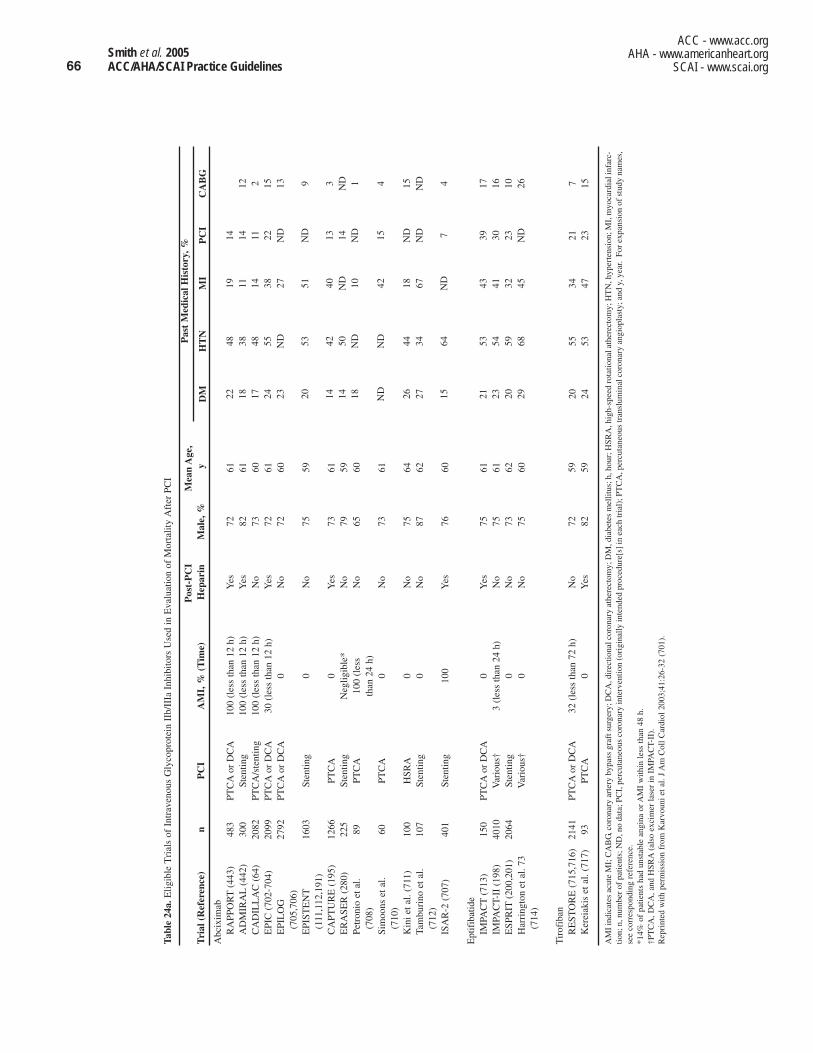
66
ACC - www.acc.orgAHA - www.americanheart.org
SCAI - www.scai.orgSmith et al. 2005ACC/AHA/SCAI Practice Guidelines
Tabl
e 24
a.E
ligib
le T
rial
s of
Int
rave
nous
Gly
copr
otei
n II
b/II
Ia I
nhib
itors
Use
d in
Eva
luat
ion
of M
orta
lity
Aft
er P
CI
Pos
t-P
CI
Mea
n A
ge,
Tri
al (
Ref
eren
ce)
nP
CI
AM
I, %
(T
ime)
Hep
arin
Mal
e, %
y
Abc
ixim
abR
APP
OR
T(4
43)
483
PTC
Aor
DC
A10
0 (l
ess
than
12
h)Y
es72
61A
DM
IRA
L(4
42)
300
Sten
ting
100
(les
s th
an 1
2 h)
Yes
8261
CA
DIL
LA
C (
64)
2082
PTC
A/s
tent
ing
100
(les
s th
an 1
2 h)
No
7360
EPI
C (
702-
704)
2099
PTC
Aor
DC
A30
(le
ss th
an 1
2 h)
Yes
7261
EPI
LO
G
2792
PTC
Aor
DC
A0
No
7260
(705
,706
)E
PIST
EN
T16
03St
entin
g0
No
7559
(111
,112
,191
)C
APT
UR
E (
195)
1266
PTC
A0
Yes
7361
ER
ASE
R (
280)
225
Sten
ting
Neg
ligib
le*
No
7959
Petr
onio
et a
l. 89
PTC
A10
0 (l
ess
No
6560
(708
)th
an 2
4 h)
Sim
oons
et a
l. 60
PTC
A0
No
7361
(710
)K
ini e
t al.
(711
)10
0H
SRA
0N
o75
64Ta
mbu
rino
et a
l. 10
7St
entin
g0
No
8762
(712
)IS
AR
-2 (
707)
401
Sten
ting
100
Yes
7660
Ept
ifib
atid
eIM
PAC
T(7
13)
150
PTC
Aor
DC
A0
Yes
7561
IMPA
CT-
II (
198)
4010
Var
ious
†3
(les
s th
an 2
4 h)
No
7561
ESP
RIT
(200
,201
)20
64St
entin
g0
No
7362
Har
ring
ton
et a
l. 73
Var
ious
†0
No
7560
(714
)
Tir
ofib
anR
EST
OR
E (
715,
716)
2141
PTC
Aor
DC
A32
(le
ss th
an 7
2 h)
No
7259
Ker
eiak
is e
t al.
(717
)93
PTC
A0
Yes
8259
AM
I in
dica
tes
acut
e M
I; C
AB
G, c
oron
ary
arte
ry b
ypas
s gr
aft s
urge
ry; D
CA
, dir
ectio
nal c
oron
ary
athe
rect
omy;
DM
, dia
bete
s m
ellit
us; h
, hou
r; H
SRA
, hig
h-sp
eed
rota
tiona
l ath
erec
tom
y; H
TN
, hyp
erte
nsio
n; M
I, m
yoca
rdia
l inf
arc-
tion;
n, n
umbe
r of
pat
ient
s; N
D, n
o da
ta; P
CI,
per
cuta
neou
s co
rona
ry in
terv
entio
n (o
rigi
nally
inte
nded
pro
cedu
re[s
] in
eac
h tr
ial)
; PT
CA
, per
cuta
neou
s tr
ansl
umin
al c
oron
ary
angi
opla
sty;
and
y, y
ear.
For
exp
ansi
on o
f st
udy
nam
es,
see
corr
espo
ndin
g re
fere
nce.
*14%
of
patie
nts
had
unst
able
ang
ina
or A
MI
with
in le
ss th
an 4
8 h.
†PT
CA
, DC
A, a
nd H
SRA
(als
o ex
cim
er la
ser
in I
MPA
CT-
II).
Rep
rint
ed w
ith p
erm
issi
on f
rom
Kar
voun
i et a
l. J
Am
Col
l Car
diol
200
3;41
:26-
32 (
701)
.
Pas
t M
edic
al H
isto
ry, %
DM
HT
NM
IP
CI
CA
BG
2248
1914
1838
1114
1217
4814
112
2455
3822
1523
ND
27N
D13
2053
51N
D9
1442
4013
314
50N
D14
ND
18N
D10
ND
1
ND
ND
4215
4
2644
18N
D15
2734
67N
DN
D
1564
ND
74
2153
4339
1723
5441
3016
2059
3223
1029
6845
ND
26
2055
3421
724
5347
2315

67Smith et al. 2005
ACC/AHA/SCAI Practice Guidelines
ACC - www.acc.orgAHA - www.americanheart.orgSCAI - www.scai.org
Readministration Registry (R3), there were no cases of ana-phylaxis or other allergic manifestations whether or notHACA was present, and HACA was not predictive of anyother measure of complication or success. From the R3study, HACA has been shown to be an IgG (not IgE)immunoglobulin that does not neutralize abciximab. Themore worrisome clinical phenomenon associated with read-ministration is the potential for increased rates of thrombo-cytopenia. In the 500-patient R3, a 4.4% incidence in throm-bocytopenia (to a platelet count of less than 100 × 109 perliter) was observed, with half of the patients developingacute profound thrombocytopenia (to a platelet count of lessthan 20 × 109 per liter). This potential complication shouldalways be monitored when treating a patient with abciximab(194-197). Abciximab readministration poses greater riskwithin 2 weeks of original abciximab dose.
6.2.2.2. Eptifibatide
The clinical utility of eptifibatide, a short-acting cyclic hep-tapeptide that also inhibits the GP IIb/IIIa receptor, was eval-uated in the Integrilin to Manage Platelet Aggregation toprevent Coronary Thrombosis-II (IMPACT-II) trial, a dou-ble-blind, randomized, placebo-controlled multicenter trialthat enrolled 4010 patients undergoing coronary angioplasty(198). Patients were assigned to treatment with aspirin,heparin and placebo, aspirin, heparin, and eptifibatide bolus(135 mcg per kg) followed by a low-dose eptifibatide infu-
for the CAPTURE investigators, also reported that amongthe 1265 patients with UA enrolled in the CAPTURE trial,troponin T and CK-MB from 890 patients correlated withsubsequent 6-month adverse cardiac risk. In patients withoutelevated troponin T levels, there was no benefit of treatmentwith respect to the relative risk of death or MI at 6 months(OR 1.26, CI 95% 0.74 to 2.31; P equals 0.47). Serum tro-ponin T level, which is considered to be a surrogate markerfor thrombus formation, identified a high-risk subgroup ofpatients with refractory UA suitable for coronary interven-tion who would particularly benefit from antiplatelet treat-ment with abciximab (192).
One putative limitation of abciximab is the potential forimmune-mediated hypersensitivity reactions after subse-quent readministration. Thrombocytopenia after readminis-tration occurs in 3.5% to 6.3% of patients, which is similar tothe rate of occurrence in patients receiving abciximab for thefirst time. Therefore, the absence of thrombocytopenia aftera first abciximab exposure does not guarantee protectionagainst its occurrence upon re-exposure. Moreover, theprevalence of severe thrombocytopenia (2.8%) and profoundthrombocytopenia (2.0%) is greater with readministrationthan the incidence observed after first-time administration(1.0% and 0.4% for severe and profound thrombocytopenia,respectively) (202). With the first administration, humanantichimeric antibodies (HACA) form in approximately 6%of patients (702). The implications of HACA, however, areunclear. Among 500 patients enrolled in the ReoPro
Table 24b. Subgroup Analyses for Mortality After PCI in Trials of Glycoprotein IIb/IIIa Inhibitors (See Table 24a for eligi-ble trials)
30 Days 6 MonthsNo. of Studies RR (95% CI) No. of Studies RR (95% CI) 20 (20 137)* 0.69 (0.53 to 0.90)* 14 (15 651)* 0.79 (0.64 to 0.97)*
PopulationAMI 6 (3355) 0.69 (0.45 to 1.05) 6 (3355) 0.76 (0.55 to 1.05)
Mixed 2 (4240) 0.95 (0.54 to 1.68) 2 (4240) 0.97 (0.65 to 1.44)
Non-AMI 12 (12 542) 0.59 (0.39 to 0.89) 6 (8056) 0.71 (0.49 to 1.03)
ProcedureStent 7 (5736) 0.69 (0.43 to 1.09) 7 (5736) 0.70 (0.49 to 1.01)
Other 13 (14 401) 0.70 (0.51 to 0.96) 7 (9915) 0.84 (0.65 to 1.09)
PostprocedureHeparin 7 (4791) 0.72 (0.47 to 1.09) 5 (4548) 0.83 (0.60 to 1.13)
No heparin 13 (15 346) 0.68 (0.49 to 0.95) 9 (11 103) 0.77 (0.58 to 1.01)
AgentAbciximab 14 (11 606) 0.69 (0.51 to 0.94) 12 (11 446) 0.77 (0.61 to 0.96)
Tirofiban 2 (2234) 1.05 (0.42 to 2.61) 1 (2141) 1.27 (0.65 to 2.48)
Eptifibatide 4 (6297) 0.60 (0.33 to 1.06) 1 (2064) 0.56 (0.24 to 1.34)
AMI indicates acute myocardial infarction; CI, confidence interval; No., number; and RR, risk ratio (fixed effects). There was no statistically significant heterogeneity in any case, and random effects estimates were similar (data not shown).*Refers to all patients.Reprinted with permission from Karvouni et al. J Am Coll Cardiol 2003;41:6-32 (701).
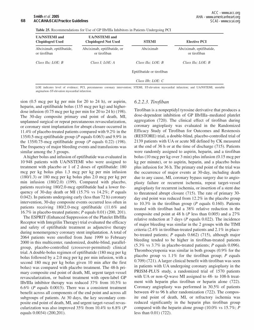
68
ACC - www.acc.orgAHA - www.americanheart.org
SCAI - www.scai.orgSmith et al. 2005ACC/AHA/SCAI Practice Guidelines
sion (0.5 mcg per kg per min for 20 to 24 h), or aspirin,heparin, and eptifibatide bolus (135 mcg per kg) and higher-dose infusion (0.75 mcg per kg per min for 20 to 24 h) (198).The 30-day composite primary end point of death, MI,unplanned surgical or repeat percutaneous revascularization,or coronary stent implantation for abrupt closure occurred in11.4% of placebo-treated patients compared with 9.2% in the135/0.5-mcg eptifibatide group (P equals 0.063) and 9.9% inthe 135/0.75-mcg eptifibatide group (P equals 0.22) (198).The frequency of major bleeding events and transfusions wassimilar among the 3 groups.
A higher bolus and infusion of eptifibatide was evaluated in10 948 patients with UA/NSTEMI who were assigned totreatment with placebo or 1 of 2 doses of eptifibatide: 180mcg per kg bolus plus 1.3 mcg per kg per min infusion(180/1.3) or 180 mcg per kg bolus plus 2.0 mcg per kg permin infusion (180/2.0) (199). Compared with placebo,patients receiving 180/2.0-mcg eptifibatide had a lower fre-quency of 30-day death or MI (15.7% vs 14.2%; P equals0.042). In patients undergoing early (less than 72 h) coronaryintervention, 30-day composite events occurred less often inpatients receiving 180/2.0-mcg eptifibatide (11.6% and16.7% in placebo-treated patients; P equals 0.01) (200, 201).
The ESPRIT (Enhanced Suppression of the Platelet IIb/IIIaReceptor with Integrilin Therapy) trial evaluated the efficacyand safety of eptifibatide treatment as adjunctive therapyduring nonemergency coronary stent implantation. A total of2064 patients were enrolled from June 1999 to February2000 in this multicenter, randomized, double-blind, parallel-group, placebo-controlled (crossover-permitted) clinicaltrial. A double-bolus regimen of eptifibatide (180 mcg per kgbolus followed by a 2.0 mcg per kg per min infusion, with asecond 180 mcg per kg bolus given 10 min after the firstbolus) was compared with placebo treatment. The 48-h pri-mary composite end point of death, MI, urgent target-vesselrevascularization, or bailout treatment with open-label GPIIb/IIIa inhibitor therapy was reduced 37% from 10.5% to6.6% (P equals 0.0015). There was a consistent treatmentbenefit across all components of the end point and across allsubgroups of patients. At 30 days, the key secondary com-posite end point of death, MI, and urgent target-vessel revas-cularization was also improved 35% from 10.4% to 6.8% (Pequals 0.0034) (200,201).
6.2.2.3. Tirofiban
Tirofiban is a nonpeptidyl tyrosine derivative that produces adose-dependent inhibition of GP IIb/IIIa–mediated plateletaggregation (720). The clinical effect of tirofiban duringcoronary angioplasty was evaluated in the RandomizedEfficacy Study of Tirofiban for Outcomes and Restenosis(RESTORE) trial, a double-blind, placebo-controlled trial of2139 patients with UA or acute MI defined by CK measuredat the end of 36 h or at the time of discharge (715). Patientswere randomly assigned to aspirin, heparin, and a tirofibanbolus (10 mcg per kg over 3 min) plus infusion (0.15 mcg perkg per minute), or to aspirin, heparin, and a placebo bolusplus infusion for 36 h. The primary end point of the trial wasthe occurrence of major events at 30-day, including deathdue to any cause, MI, coronary bypass surgery due to angio-plasty failure or recurrent ischemia, repeat target-vesselangioplasty for recurrent ischemia, or insertion of a stent dueto threatened abrupt closure (715). The rate of primary 30-day end point was reduced from 12.2% in the placebo groupto 10.3% in the tirofiban group (P equals 0.160). Patientstreated with tirofiban had a 38% relative reduction in thecomposite end point at 48 h (P less than 0.005) and a 27%relative reduction at 7 days (P equals 0.022). The incidenceof major bleeding was similar in the 2 groups with the TIMIcriteria (2.4% in tirofiban-treated patients and 2.1% in place-bo-treated patients; P equals 0.662) (715), although majorbleeding tended to be higher in tirofiban-treated patients(5.3% vs 3.7% in placebo-treated patients; P equals 0.096).Thrombocytopenia was similar in both groups (0.9% for theplacebo group vs 1.1% for the tirofiban group; P equals0.709) (721). A larger clinical benefit with tirofiban was seenin patients with UA undergoing coronary angioplasty in thePRISM-PLUS study, a randomized trial of 1570 patientswith UA or non–Q-wave MI assigned to 48- to 108-h treat-ment with heparin plus tirofiban or heparin alone (722).Coronary angioplasty was performed in 30.5% of patientsbetween 49 to 96 h after randomization (722). The compos-ite end point of death, MI, or refractory ischemia wasreduced significantly in the heparin plus tirofiban groupcompared with the heparin alone group (10.0% vs 15.7%; Pless than 0.01) (722).
Table 25. Recommendations for Use of GP IIb/IIIa Inhibitors in Patients Undergoing PCI
UA/NSTEMI and UA/NSTEMI and Clopidogrel Used Clopidogrel Not Used STEMI Elective PCI
Abciximab, eptifibatide, Abciximab, eptifibatide, or Abciximab Abciximab, eptifibatide,or tirofiban or tirofiban or tirofiban
Class IIa; LOE: B Class I; LOE: A Class IIa; LOE: B Class IIa; LOE: B
Eptifibatide or tirofiban
Class IIb; LOE: C
LOE indicates level of evidence; PCI, percutaneous coronary intervention; STEMI, ST-elevation myocardial infarction; and UA/NSTEMI, unstableangina/non–ST-elevation myocardial infarction.

69Smith et al. 2005
ACC/AHA/SCAI Practice Guidelines
ACC - www.acc.orgAHA - www.americanheart.orgSCAI - www.scai.org
400 to 600 s) of periprocedural anticoagulation are associat-ed with an increased risk for bleeding complications (736).
The safety of low-dose heparin during coronary angioplas-ty has also been shown in a recent study. Fatal complications(0.3%), emergency bypass surgery (1.7%), MI (3.3%), orrepeat angioplasty within 48 h (0.7%) were uncommon afteran empiric bolus of heparin 5000 U at the beginning of theprocedure (618). In a smaller randomized study of 400patients assigned to fixed-dose heparin (15 000 internationalunits [IU]) or weight-adjusted heparin (100 IU per kg), therewere no differences in procedural success or bleeding com-plications between the 2 groups (737), although use of theweight-adjusted heparin resulted in earlier sheath removaland more rapid transfer to a step-down unit (737). Anotheradvantage of weight-adjusted heparin dosing is that “over-shooting” of the ACT value can be avoided.
Two analyses of ACT and PCI-related complications havenot detected any relationship between ACT level andischemic complications, which suggests that the degree ofanticoagulation during PCI may be less of a factor in deter-mining complications than in the earlier era of balloon angio-plasty (738,739). The results of these limited studies suggestthat heparin is an intraprocedural component for PCI, despitedosing uncertainties and an unpredictable therapeuticresponse with the unfractionated preparation. It appears thatweight-adjusted heparin dosing may provide a clinicallysuperior anticoagulation method to fixed heparin dosing,although definitive studies are lacking.
Routine use of unfractionated heparin after an uncompli-cated coronary angioplasty is no longer recommended(72,740-743) and may be associated with more frequentbleeding events (72,740), particularly when platelet GPIIb/IIIa inhibitors are used (72,740). Subcutaneous adminis-tration of unfractionated heparin (741) may provide a saferand less costly means of extending antithrombin therapy thanintravenous unfractionated heparin if there are clinical rea-sons to continue anticoagulation, such as residual thrombusor significant residual dissections.
In the SYNERGY (Superior Yield of the New strategy ofEnoxaparin, Revascularization and Glycoprotein IIb/IIIaInhibitors) study, patients with NSTEMI were randomized totreatment with either unfractionated heparin or subcuta-neously administered enoxaparin. In patients who underwentPCI within 8 h of the last subcutaneous dose, no additionalanticoagulation was administered. In those patients whounderwent PCI 8 to 12 h after the last subcutaneous dose, anadditional intravenous dose of enoxaparin 0.3 mg per kg wasadministered at the time of PCI. The rates of major ischemiccomplications in those patients undergoing PCI were similarbetween those treated with unfractionated heparin and thosetreated with enoxaparin (744). Bleeding was observed to behigher in those patients who “crossed over” from one antico-agulant to the other. Some of these crossover patients werethose who received a different anticoagulant than what theyhad been randomized to and had received before. On thebasis of this observation, it appears prudent to not give anadditional anticoagulant to patients who are receiving one
6.2.3. Antithrombotic Therapy
6.2.3.1. Unfractionated Heparin, Low-Molecular-Weight Heparin, and Bivalirudin
Class I1. Unfractionated heparin should be administered to
patients undergoing PCI. (Level of Evidence: C)2. For patients with heparin-induced thrombocytopenia,
it is recommended that bivalirudin or argatroban beused to replace heparin. (Level of Evidence: B)
Class IIa1. It is reasonable to use bivalirudin as an alternative to
unfractionated heparin and glycoprotein IIb/IIIaantagonists in low-risk patients undergoing electivePCI. (Level of Evidence: B)
2. Low-molecular-weight heparin is a reasonable alter-native to unfractionated heparin in patients withUA/NSTEMI undergoing PCI. (Level of Evidence: B)
Class IIbLow-molecular-weight heparin may be considered asan alternative to unfractionated heparin in patientswith STEMI undergoing PCI. (Level of Evidence: B)
Intravenous unfractionated heparin prevents clot formationat the site of arterial injury (723) and on coronary guidewiresand catheters used for coronary angioplasty (724). Althoughthe intensity of anticoagulation with unfractionated heparinis generally determined with activated partial thromboplastintimes, these values are less useful for monitoring anticoagu-lation during coronary angioplasty, because higher levels ofanticoagulation are needed than can be discriminated withthe activated partial thromboplastin time alone. Instead, theactivated clotting time (ACT) has been more useful to followheparin therapy during coronary angioplasty (725). TheHemochron and HemoTec devices are commonly used tomeasure ACT values during coronary angioplasty (725-727).The Hemochron ACT generally exceeds the HemoTec ACTby 30 to 50 s, although considerable measurement variabili-ty exists.
Empiric recommendations regarding heparin dosage dur-ing coronary angioplasty have been proposed (728,729), butACT levels after a fixed dose of unfractionated heparin mayvary substantially due to differences in body size (730), con-comitant use of other medications, including intravenousnitroglycerin (731,732), and in the presence of acute coro-nary syndromes that increase heparin resistance.
The relationship between the level of the ACT and devel-opment of ischemic complications during coronary angio-plasty has been controversial. Whereas some studies haveidentified an inverse relationship between the initial ACTand the risk of ischemic events (733,734), others found eitherno relationship or a direct relationship between the degree ofanticoagulation and occurrence of complications (735). It isgenerally believed that very high levels (ACTs greater than

70
ACC - www.acc.orgAHA - www.americanheart.org
SCAI - www.scai.orgSmith et al. 2005ACC/AHA/SCAI Practice Guidelines
to achieve a target ACT of 200 s with either the HemoTec orHemochron device. The currently recommended target ACTfor eptifibatide and tirofiban is less than 300 s during coro-nary angioplasty. Postprocedural heparin infusions are notrecommended during GP IIb/IIIa therapy (748-750).
Transitioning of patients with acute coronary syndromeswho have been treated with enoxaparin from the medicalfloor to the cardiac catheterization laboratory is based onpharmacokinetic data, clinical experience, and expert opin-ion. In patients who received the last subcutaneously admin-istered dose of enoxaparin within 8 h, no additional antico-agulant therapy is needed before PCI is performed. Inpatients who received the last subcutaneously administereddose of enoxaparin between 8 and 12 h before PCI, an addi-tional 0.3 mg per kg dose of enoxaparin should be adminis-tered intravenously before PCI (whether or not the patient isto be treated with a GP IIb/IIIa inhibitor). Alternatively, inthe latter group of patients, supplemental anticoagulationwith unfractionated heparin can be used. Unfractionatedheparin 50 U per kg (with a target ACT of 200 to 250 s) maybe administered in those patients to be treated with a GPIIb/IIIa inhibitor; 60 U per kg unfractionated heparin (with atarget ACT of 250 to 300 s) may be administered in thosepatients who are not concomitantly treated with a GP IIb/IIIainhibitor. A higher risk of bleeding may result if patientscross over between different anticoagulant therapies duringthe index admission.
Low-molecular-weight heparins have little effect on meas-urements of ACT. Therefore, the ACT should not be used asa guide to anticoagulation therapy in patients currently beingtreated with a low-molecular-weight heparin. Sheathremoval when followed by manual groin compression maybe performed 4 h after the last intravenous dose of enoxa-parin or 6 to 8 h after the last subcutaneous dose of enoxa-parin (751,752).
6.3. Post-PCI Management
After PCI, in-hospital care should focus on monitoring thepatient for recurrent myocardial ischemia, achieving hemo-stasis at the catheter insertion site, detecting and preventingcontrast-induced renal failure, and monitoring results of thevascular closure device, if used (753). Attention should alsobe directed toward implementing appropriate secondary ath-erosclerosis prevention programs. The patient should under-stand and adhere to recommended medical therapies andbehavior modifications known to reduce subsequent morbid-ity and mortality from coronary heart disease.
Most patients can be safely discharged from the hospitalwithin the next calendar day after an uncomplicated electivePCI. Special skilled nursing units have been developed bymany institutions to facilitate post-PCI management.Specific protocols for sheath removal, continuation of anti-coagulation or antiplatelet therapies, and observation forrecurrent myocardial ischemia/infarction and contrast-induced renal failure are of particular assistance in ensuringappropriate outcomes during this period. Pilot studies sug-gest that selected patients may be discharged on the same day
form of anticoagulant (e.g., not to give unfractionatedheparin to those who have received subcutaneous enoxaparinwithin the last 12 h and not to give intravenous enoxaparin tothose receiving intravenous heparin).
The safety and efficacy of low-molecular-weight heparintherapy in patients undergoing PCI has been evaluated. In allbut 1 of these studies, the agent studied has been enoxaparin.These studies have found bleeding and ischemic complica-tion rates to be low and comparable to those observed in PCIpatients who had been treated with unfractionated heparin.
In those patients who have received subcutaneous enoxa-parin for the treatment of NSTEMI and are to undergo PCIwithin 8 h of the last subcutaneous dose, no additional anti-coagulant should be administered. In those who undergo PCI8 to 12 h after the last subcutaneous dose, an additional intra-venous dose of 0.3 mg per kg should be administered imme-diately before device activation.
Bivalirudin, a hirudin analog, is a direct thrombin inhibitor.It has been tested against heparin and a GP IIb/IIIa inhibitorin the REPLACE-2 trial of patients undergoing PCI withouthigh-risk features. The primary end point at 30 days includ-ed major bleeding plus the usual end points of death, MI, andurgent revascularization. These events occurred in 9.2% ofthe bivalirudin group and 10% of the group given unfrac-tionated heparin plus GP IIb/IIIa inhibitors (nonsignificant).The secondary end point was freedom from death, MI, andurgent revascularization and occurred in 7.6% of thebivalirudin group and 7.1% of the group given unfractionat-ed heparin plus GP IIb/IIIa inhibitors (also nonsignificant),but bleeding (combined major and moderate bleeding) wassignificantly reduced in the bivalirudin group (from 7.1% to2.4%, P less than 0.001). Although a small, nonsignificantincrease in periprocedural NSTEMI was seen in thebivalirudin-treated patients, by 1 year mortality was not sig-nificantly increased in the bivalirudin group (1.89% vs 2.46%) (744a). These results established that bivalirudin is notsuperior to standard therapy, but it appears to be a reasonablealternative in non-high-risk patients (745). Bivalirudin is agood anticoagulant for use in patients with heparin-inducedthrombocytopenia and those with renal failure (746).Argatroban is also an effective therapy for heparin-inducedthrombocytopenia (747). More data are needed to establishits use for patients with STEMI, NSTEMI, and diabetes.
6.2.3.2. Heparin Dosing Guidelines
In those patients who do not receive GP IIb/IIIa inhibitors,sufficient unfractionated heparin should be given duringcoronary angioplasty to achieve an ACT of 250 to 300 s withthe HemoTec device and 300 to 350 s (200,201) with theHemochron device. A weight-adjusted bolus heparin (70 to100 IU per kg) can be used to avoid excess anticoagulation.If the target values for ACT are not achieved after a bolus ofheparin, additional heparin boluses (2000 to 5000 IU) can begiven. Early sheath removal should be performed when theACT falls to less than 150 to 180 s.
The unfractionated heparin bolus should be reduced to 50to 70 IU per kg when GP IIb/IIIa inhibitors are given in order

71Smith et al. 2005
ACC/AHA/SCAI Practice Guidelines
ACC - www.acc.orgAHA - www.americanheart.orgSCAI - www.scai.org
degree to which these technologies reduce length of hospitalstay and cost remains to be determined (764,770-772).
Patients with pre-existing renal insufficiency, diabetes, anddehydration are at higher risk and should be monitored forcontrast-induced nephropathy, generally defined as anincrease of greater than 25% or greater than 0.5 mg per dL inserum creatinine that occurs within 48 h after PCI. In addi-tion, those patients receiving higher contrast loads or a sec-ond contrast load within 72 h and those undergoing IABPplacement should have renal function assessed. A risk scorebased on 8 variables (hypotension, IABP, HF, chronic renalinsufficiency, diabetes, age more than 75 years, anemia, andcontrast volume) has been developed to assist in the identifi-cation of patients at risk for contrast-induced nephropathyafter PCI (773). Whenever possible, nephrotoxic drugs (cer-tain antibiotics, nonsteroidal anti-inflammatory agents, andcyclosporine) and metformin (especially in those with pre-existing renal dysfunction) should be withheld for 24 hbefore PCI is performed, and consideration should be givento withholding angiotensin converting enzyme inhibitors andangiotensin receptor blockers on the day of the procedure(774-776). Although data on the prevention of contrast-induced nephropathy are inconclusive, several measuresincluding preprocedural and postprocedural hydration, use oflow and iso-osmolar contrast agents, and pretreatment withacetylcysteine or sodium bicarbonate may be helpful inreducing the incidence of contrast-induced nephropathyamong higher-risk patients (774,775,777).
6.3.1. Postprocedure Evaluation of Ischemia
After PCI, chest pain may occur in as many as 50% ofpatients. ECG evidence of ischemia identifies those with sig-nificant risk for acute vessel closure (6,118,119,778-780).When angina pectoris or ischemic ECG changes occur afterPCI, the decision to proceed with further interventional pro-cedures, CABG surgery, or medical therapy should be indi-vidualized on the basis of factors such as hemodynamic sta-bility, amount of myocardium at risk, and the likelihood thatthe treatment will be successful.
A 12-lead ECG should be obtained before and soon afterPCI and again if symptoms should occur. Angina-like symp-toms with ECG changes will assist in determining the needfor repeat angiography and for additional therapy.
As discussed in Section 6.2.2, coronary stents and plateletGP receptor inhibitors have reduced the incidence of acuteclosure significantly. Factors that correlate with a poor out-come after acute coronary closure include age greater than70 years, large ischemic burden, presentation with acutecoronary syndromes, and LV ejection fraction less than 30%(778-780).
Elevated levels of CK or the MB subfraction (CK-MB) orECG abnormalities are reported to occur in 5% to 30% ofpatients after PCI (23). The mechanisms associated with CKrelease include side-branch occlusion, distal embolization,intimal dissection, and coronary spasm (781). A more fre-quent requirement for revascularization procedures and a
after PCI (754,755) especially when the procedure is per-formed by the percutaneous radial or brachial approach(756). However, confirmation by larger studies is necessarybefore widespread endorsement of this strategy.
In the prior setting of aggressive systemic anticoagulation,vascular complications may occur in as many as 14% ofpatients after PCI, but those requiring surgical repair occur in3.5% (736) of patients, although lower rates of vascular com-plications can now be expected with reduced anticoagulationand smaller sheath sizes (757-762). Major factors associatedwith vascular complications include use of fibrinolytic orplatelet inhibitor therapy, coexisting peripheral vascular dis-ease, female gender, prolonged heparin use with delayedsheath removal, and older age (736,758,760-764). Althoughmost bleeding complications at the vascular access site areobvious and readily managed, physicians and nurses shouldremain alert for retroperitoneal hematoma, the signs andsymptoms of which may include hypotension, markedsuprainguinal tenderness, and severe back or lower-quadrantabdominal pain (765). Post-PCI hematocrit should be moni-tored for a decrease greater than absolute 5% to 6%.Computed tomography can confirm the diagnosis ofretroperitoneal hematoma, and more than 80% of patientscan be treated conservatively with transfusions without sur-gery (764). Pseudoaneurysms may be treated effectively withultrasound-directed compression in the majority of patientswho are not bleeding and do not require continued anticoag-ulation (763,766,767). Arteriovenous fistulas, generallyoccurring late after a procedure, are detected by a continuousmurmur over the puncture site and, in rare cases, may beassociated with high-output failure. In general, repeat use ofthe access site should be avoided because of the possibilityof making the fistula larger, accessing the vein when attempt-ing to access the artery, and increasing potential issues withhemostasis. Both pseudoaneurysm and arteriovenous fistulacan occur secondary to cannulation of the superficial ratherthan the common femoral artery (768). Arterial compressionsystems and percutaneous vascular closure devices reducethe incidence of vascular complications (753,756). A meta-analysis involving 37 000 patients undergoing diagnosticcoronary arteriography and PCI compared manual compres-sion with 3 closure devices (VasoSeal™, AngioSeal™, andPerClose™). No difference was seen in access-related com-plications between manual compression, PerClose™, andAngioSeal™; however, there were more complications asso-ciated with VasoSeal™ than with manual compression. Thecomplications evaluated included pseudoaneurysm requiringultrasound-guided compression or surgical repair; arteriove-nous fistula; retroperitoneal hematoma causing hemodynam-ic compromise and necessitating surgery, blood transfusion,prolonged hospitalization, and/or death; femoral arterythrombosis (vessel occlusion requiring surgery or thrombol-ysis); surgical vascular repair; access-site infection necessi-tating treatment with antibiotics or surgical drainage; andblood transfusion. The study was performed with early gen-erations of devices. Potential benefits of newer adjunctivetherapies are not well established (753,769). However, the
72
ACC - www.acc.orgAHA - www.americanheart.org
SCAI - www.scai.orgSmith et al. 2005ACC/AHA/SCAI Practice Guidelines
Depending on the risk factors and contraindications pres-ent, advice should include antithrombotic therapy (aspirinand/or clopidogrel or ticlopidine), control of hypertension,diabetic management, aggressive control of serum lipids,maintenance of a low-density lipoprotein cholesterol levelsubstantially below 100 mg per dL (optional therapeutic tar-get less than 70 mg per dL in very-high-risk patients [611]),abstinence from tobacco use, weight control, regular exer-cise, beta-blocker use, and inhibition of the renin-angiotensin-aldosterone system as recommended in theACC/AHA Guidelines for the Management of Patients WithST-Elevation Myocardial Infarction (Table 26) (332,783).Given the natural history and pathophysiology of CADamong patients undergoing PCI, the clinically indicated sec-ondary prevention measures (Table 26) (332,783), whichusually include aspirin, statin therapy, beta-blockers, andinhibitors of the renin-angiotensin-aldosterone system,should be continued indefinitely except in those patientsintolerant to these agents (242,783-792). Patients shouldreceive instructions on participation in cardiac rehabilitationand the timing of return to full activities, be informed to con-tact their physician or seek immediate medical attention ifsymptoms recur, and have made plans for a follow-up visit toassess compliance with secondary prevention therapies.
6.3.3. Exercise Testing After PCI
The published ACC/AHA practice guidelines for exercisetesting (793) provide an excellent summary of the availableinformation on exercise testing after PTCA. Althoughrestenosis remains the major limitation of PCI, symptom sta-tus is an unreliable index to development of restenosis, with25% of asymptomatic patients documented as havingischemia on exercise testing (794). (See Section 5.1, PatientsWith Asymptomatic Ischemia or CCS Class I or II Anginafor further information.)
To identify restenosis rather than predict the probability ofits occurrence, patients may be tested later (3 to 6 monthsafter PCI). Table 27 reviews the predictive value of exercisetesting for restenosis (794-802). Variability is attributed pre-dominantly to differences in the populations studied and cri-teria for restenosis.
Because myocardial ischemia, whether painful or silent,worsens prognosis (803), some authorities have advocatedroutine testing. However, the ACC/AHA practice guidelinesfor exercise testing favor selective evaluation in patients con-sidered to be at particularly high risk (e.g., patients withdecreased LV function, multivessel CAD, proximal LADdisease, previous sudden death, diabetes mellitus, left maindisease, hazardous occupations, and suboptimal PCI results)(793). The exercise ECG is an insensitive predictor ofrestenosis, with sensitivities ranging from 40% to 55%, sig-nificantly less than those obtainable with single photon emis-sion computed tomography (SPECT) (804,805) or exerciseechocardiography (806-808). This lower sensitivity of theexercise ECG and its inability to localize disease limit itsusefulness in patient management both before and after PCI(797,804,809). For these reasons, stress imaging is preferred
higher risk of death or subsequent MI are associated withelevated cardiac biomarkers, increasing as a continuousfunction with no obvious threshold effect. Both acute andchronic complications are more common among patientswith elevated cardiac biomarkers. Even in patients with low-level elevations of CK-MB in whom the in-hospital risk islow, the intermediate- and long-term risks are also increased.Postprocedural increases in CK and CK-MB are not specificfor a particular technique and have been reported after bal-loon angioplasty, directional and rotablator atherectomy,excimer laser angioplasty, and stent placement. Kong et al.(782) found that increased levels of CK are a significantindependent predictor of cardiac mortality and subsequentMI (56). Cardiac mortality after elective PCI was signifi-cantly higher for patients with high (more than 3.0 times nor-mal) and intermediate (1.5 to 3.0 times normal) CK com-pared with those with low CK (more than 1.0 but less than1.5 times normal) elevations and control patients (P equals0.007). (See Section 3.2, Acute Outcome: ProceduralComplications.)
CK and CK-MB measurements should be obtained inpatients with suspected ischemia (prolonged chest pain, side-branch occlusion, recurrent ischemia, or hemodynamic insta-bility) during PCI. Ideally, the European Society ofCardiology and the ACC recommend that small infarcts mayand should be detected by serial blood sampling and analysisbefore and after the procedure (6 to 8 h before and 24 h after,respectively) (21). In patients in whom a clinically drivenCK-MB determination is made, a CK-MB index increase ofmore than 5 times the upper limit of normal should be treat-ed as signifying an MI, and the patient should be referred forfurther observation. The results of CK-MB should be con-sidered for the discharge management strategies for thesepatients.
The troponin isoforms I and T have a high level of sensi-tivity and specificity for the diagnosis of acute MI. TroponinT or I elevation occurs frequently after PCI. The timing ofthe peak elevation after PCI is unclear (25). Minor elevationsdo not appear to have prognostic value, whereas marked(more than 5 times) elevations are associated with worsened1-year outcome (26,27).
6.3.2. Risk Factor Modifications
All patients should be instructed about necessary behaviorand risk factor modification, and the appropriate medicaltherapies should be initiated for the secondary prevention ofatherosclerosis before the patient leaves the hospital. Theinterventional cardiologist should emphasize the importanceof these measures directly to the patient, because failure todo so may suggest that secondary prevention therapies arenot necessary. The interventional cardiologist should interactwith the primary care physician to ensure that the necessarysecondary prevention therapies initiated during hospitaliza-tion are maintained by patients after discharge from the hos-pital. Secondary prevention measures are an essential part oflong-term therapy because they can reduce future morbidityand mortality associated with the atherosclerotic process.

73Smith et al. 2005
ACC/AHA/SCAI Practice Guidelines
ACC - www.acc.orgAHA - www.americanheart.orgSCAI - www.scai.org
Table 26. Comprehensive Risk Reduction for Patients With Coronary and Other Vascular Disease After PCI
Goals Intervention Recommendations
Smoking: Ask about tobacco status at every visit. Strongly encourage patient and family toGoal stop smoking and to avoid environmental tobacco smoke. Assess the tobaccoComplete cessation. No exposure to user’s willingness to quit. Assist by counseling and developing a plan for quitting.environmental tobacco smoke Arrange follow-up, referral to special programs, orpharmacological therapy
(including nicotine replacement and buproprion). Urge avoidance of exposure toenvironmental tobacco smoke at work and home.
Blood pressure control:GoalLess than 140 over 90 mm Hg orless than 130 over 80 mm Hg if chronic kidney disease or diabetes is present
Lipid management:(TG less than 200 mg per dL)
Primary goalLDL-C substantially less than 100 mg per dL (optional target less than 70 mg per dL for very-high-risk patients)¶
Lipid management: If TG is greater than or equal to 150 mg per dL or HDL-C is less than (TG 200 mg per dL or greater) 40 mg per dL:
• Emphasize weight management and physical activity. Advise smoking cessation.
Primary goal If TG is 200-499 mg per dL: Non–HDL-C* substantially less • After LDL-C–lowering therapy†**, consider adding fibrate or niacin‡.than 130 mg per dL
If TG is greater than or equal to 500 mg per dL:• Consider fibrate or niacin‡ before LDL-C–lowering therapy.†**• Consider omega-3 fatty acids as adjunct for high TG.
Physical activity: Assess risk, preferably with exercise test, to guide prescription.Minimum goal Encourage minimum of 30 to 60 minutes of activity, preferably daily or at30 minutes 5 days per week; least 5 times weekly (brisk walking, jogging, cycling, or other aerobic optimal daily activity) supplemented by an increase in daily lifestyle activities (e.g., walking
breaks at work, gardening, household work). Encourage resistance training 2 days per week.Cardiac rehabilitation programs are recommended, particularly for those patients with multiple modifiable risk factors and/or those moderate- to high-risk patients in whom supervised exercise training is warranted.
Continued on next page
If blood pressure is 120 over 80 mm Hg or greater:·
• Initiate or maintain lifestyle modification (weight control, increasedphysical activity, alcohol moderation, moderate sodium restriction,and emphasis on fruits, vegetables, and low-fat dairy products) in all patients.
If blood pressure is 140 over 90 mm Hg or greater (or 130 over 80 mm Hg orgreater for individuals with chronic kidney disease or diabetes):
• Add blood pressure medication, emphasizing the use of beta-blockers andinhibitors of the renin-angiotensin-aldosterone system.
Start dietary therapy in all patients (less than 7% of total calories as saturated fat and lessthan 200 mg of cholesterol per day). Promote physical activity and weight management.Encourage increased consumption of omega-3 fatty acids in fish# or 1 g per day omega-3fatty acids from supplements for risk reduction (for treatment of elevated TG, higherdoses are usually necessary for risk reduction).
Assess fasting lipid profile in all patients, preferably within 24 h of an acute event. Forpatients hospitalized, initiate lipid-lowering medication as recommended below beforedischarge according to the following guide:
LDL-C less than 100 mg per dL LDL-C greater than or equal to (baseline or on-treatment): 100 mg per dL (baseline or on-
treatment):• Statins preferred to lower LDL-C. • Initiate or intensify LDL-
C–lowering therapy with drug treatment. May require combination therapy with standard-dose ezetimide, bile acid sequestrant, or niacin‡.

74
ACC - www.acc.orgAHA - www.americanheart.org
SCAI - www.scai.orgSmith et al. 2005ACC/AHA/SCAI Practice Guidelines
Table 26. Continued
Goals Intervention Recommendations
Weight management: Calculate BMI and measure waist circumference as part of evaluation. Monitor Goal response of BMI and waist circumference to therapy.BMI 18.5 to 24.9 kg per m2
Start weight management and physical activity as appropriate. Desirable BMI Waist circumference: range: 18.5 to 24.9 kg per m2.Women: Less than 35 inchesMen: Less than 40 inches If waist circumference is 35 inches or greater in women or 40 inches or greater
in men, initiate lifestyle changes and consider treatment strategies for metabolic syndrome.
Diabetes management: Appropriate glucose-lowering therapy to achieve near-normal fasting plasma Goal glucose as indicated by HbA1c.HbA1c less than 7%
Treatment of other risk factors (e.g., physical activity, weight management, blood pressure cholesterol management).
Antiplatelet agents/ anticoagulants: For all post-PCI stented patients, aspirin 325 mg daily should be given for at least1 month after bare metal stent implantation, 3 months after sirolimus stent, and 6months after paclitaxel stent, after which daily chronic aspirin†† (75 to 162 mg perday) should be continued indefinitely in all patients if not contraindicated.
For post-PCI stented patients, clopidogrel 75 mg per day should be given for at least 1 month after bare metal stent implantation, 3 months after sirolimus stent, and 6 months after paclitaxel stent, after which clopidogrel should ideally be con-tinued up to 12 months in all stented patients who are not at high risk of bleeding. Use warfarin in combination with clopidogrel and low-dose aspirin with great caution and when INR is carefully regulated (2.0 to 3.0).
Manage warfarin to INR 2.5 to 3.5 for post-MI patients when clinically indicated or for those not able to take aspirin or clopidogrel.
Renin-angiotensin- Consider ACE inhibitors for all CHD patients indefinitely; start early after MI in aldosterone system blockers: stable high-risk patients (anterior MI, previous MI, Killip class greater than or equal
to II [S3 gallop, rales, radiographic HF]).
Continue indefinitely in for all patients with LV dysfunction (ejection fraction less than or equal to 0.40) or symptoms of heart failure.
Use as needed to manage blood pressure or consider for chronic therapy in all other patients.
Use angiotensin receptor blockers in post-STEMI patients who are intolerant of ACEinhibitors and who have either clinical or radiological signs of heart failure or LVEFless than 0.40.
Aldosterone blockade in post-STEMI patients without significant renal dysfunction§or hyperkalemia¦ who are already receiving therapeutic doses of an ACE inhibitor, have an LVEF less than or equal to 0.40, and have either diabetes or heart failure.
Beta-blockers: Start in all post-MI and acute patients (arrhythmia, LV dysfunction, inducibleischemia). Continue for a minimum of 6 months; continue indefinitely in patients with STEMI. Observe usual contraindications. Use as needed to manage angina, rhythm, or blood pressure in all other patients.
ACE indicates angiotensin-converting enzyme; BMI, body mass index; HF, congestive heart failure; CHD, coronary heart disease; HDL-C, high-density lipoprotein cholesterol; INR, interna-tional normalized ratio; LDL-C, low-density lipoprotein cholesterol; LV, left ventricular; LVEF, left ventricular ejection fraction; MI, myocardial infarction; PCI, percutaneous coronary inter-vention; STEMI, ST-elevation myocardial infarction; and TG, triglyceride.
*Non–HDL-C equals total cholesterol minus HDL cholesterol.†Treat to a goal of non–HDL-C substantially less than 130 mg per dL.‡Dietary supplement niacin must not be used as a substitute for prescription niacin.§Creatinine should be less than or equal to 2.5 mg per dL in men and less than or equal to 2.0 mg per dL in women.¦Potassium should be less than or equal to 5.0 mEq per liter.¶Patients with acute coronary syndromes and other very-high-risk patients (e.g., established CHD plus multiple major risk factors [especially diabetes] or severe and poorly controlled risk fac-
tors [especially continued cigarette smoking and/or metabolic syndrome]) should be considered for optional LDL-C goal less than 70 mg per dL.#Pregnant and lactating women should limit their intake of fish to minimize exposure to methylmercury.**The use of resin is relatively contraindicated when TGs are greater than 200 mg per dL.††Some recommend avoiding regular use of ibuprofen, which may limit the cardioprotective effects of aspirin. Use of cyclo-oxygenase-2 inhibitors may be associated with increased incidence
of cardiovascular events.
Modified with permission from Smith et al. Circulation 2001;104:1577-9 (810) (undergoing update with anticipated release in 2005) and Antman et al. J Am Coll Cardiol 2004;44:e1-e211 (362).
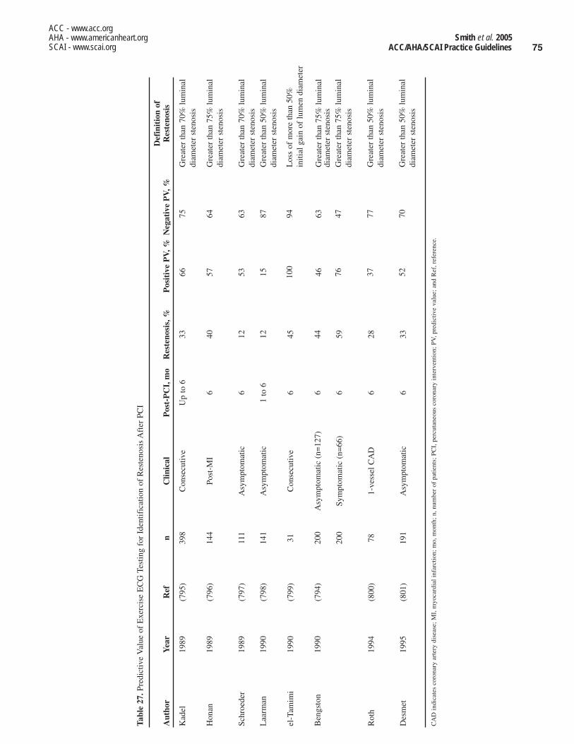
75Smith et al. 2005
ACC/AHA/SCAI Practice Guidelines
ACC - www.acc.orgAHA - www.americanheart.orgSCAI - www.scai.org
Tabl
e 27
.Pre
dict
ive
Val
ue o
f E
xerc
ise
EC
G T
estin
g fo
r Id
entif
icat
ion
of R
este
nosi
s A
fter
PC
I
Aut
hor
Yea
rR
efn
Clin
ical
Pos
t-P
CI,
mo
Res
teno
sis,
%
Kad
el19
89(7
95)
398
Con
secu
tive
Up
to 6
33
Hon
an19
89(7
96)
144
Post
-MI
640
Schr
oede
r19
89(7
97)
111
Asy
mpt
omat
ic6
12
Laa
rman
1990
(798
)14
1A
sym
ptom
atic
1 to
612
el-T
amim
i19
90(7
99)
31C
onse
cutiv
e6
45
Ben
gsto
n19
90(7
94)
200
Asy
mpt
omat
ic (
n=12
7)6
44
200
Sym
ptom
atic
(n=
66)
659
Rot
h19
94(8
00)
781-
vess
el C
AD
628
Des
met
1995
(801
)19
1A
sym
ptom
atic
633
CA
D in
dica
tes
coro
nary
art
ery
dise
ase;
MI,
myo
card
ial i
nfar
ctio
n; m
o, m
onth
; n, n
umbe
r of
pat
ient
s; P
CI,
per
cuta
neou
s co
rona
ry in
terv
entio
n; P
V, p
redi
ctiv
e va
lue;
and
Ref
, ref
eren
ce.
Def
init
ion
of
Pos
itiv
e P
V, %
Neg
ativ
e P
V, %
Res
teno
sis
6675
Gre
ater
than
70%
lum
inal
di
amet
er s
teno
sis
5764
Gre
ater
than
75%
lum
inal
di
amet
er s
teno
sis
5363
Gre
ater
than
70%
lum
inal
di
amet
er s
teno
sis
1587
Gre
ater
than
50%
lum
inal
di
amet
er s
teno
sis
100
94L
oss
of m
ore
than
50%
in
itial
gai
n of
lum
en d
iam
eter
4663
Gre
ater
than
75%
lum
inal
di
amet
er s
teno
sis
7647
Gre
ater
than
75%
lum
inal
di
amet
er s
teno
sis
3777
Gre
ater
than
50%
lum
inal
di
amet
er s
teno
sis
5270
Gre
ater
than
50%
lum
inal
di
amet
er s
teno
sis

76
ACC - www.acc.orgAHA - www.americanheart.org
SCAI - www.scai.orgSmith et al. 2005ACC/AHA/SCAI Practice Guidelines
surveillance angiography at 2 to 3 months for all patientsafter ULM PCI. Studies from the DES era have reported theperformance of routine angiography 4 to 8 months after PCIor earlier if clinically indicated by symptoms or documentedmyocardial ischemia (159,160).
7. SPECIAL CONSIDERATIONS
7.1. Ad Hoc Angioplasty—PCI at the Time of Initial Cardiac Catheterization
Ad hoc coronary intervention is defined as PCI performed atthe same time as diagnostic cardiac catheterization. Duringthe past several years, in an effort to reduce hospital lengthof stay and potentially reduce costs, PCI has increasinglybeen performed immediately after the diagnostic coronaryangiographic procedure (811), with reported incidence rang-ing from 52% to 83% (812-814). The indications for diag-nostic catheterization and coronary angiography in differentcatheterization laboratory settings are discussed in theACC/SCAI Expert Consensus Document on CatheterizationLaboratory Standards (Table 28) (309,815).
Ad hoc angioplasty has several inherent advantages. Itexpedites patient care, avoids a second invasive procedurewith its associated risks and recognized morbidity, andreduces total X-ray exposure and therefore cost, but only insettings in which intrinsic risks are low (813). However, adhoc intervention is associated with a higher procedural con-trast use and should be avoided in situations where excessive
to evaluate symptomatic patients after PCI. If the patient’sexertional capacity is significantly limited, coronary angiog-raphy may be more efficacious to evaluate symptoms of typ-ical angina. Exercise testing after discharge is helpful foractivity counseling and exercise training as part of cardiacrehabilitation. Neither exercise testing nor radionuclideimaging is indicated for the routine, periodic monitoring ofasymptomatic patients after PCI without specific indications.
6.3.4. Left Main CAD
Class IIaIt is reasonable that patients undergoing PCI tounprotected left main coronary obstructions be fol-lowed up with coronary angiography between 2 and 6months after PCI. (Level of Evidence: C)
Careful postprocedure surveillance with coronary angiog-raphy is needed to prevent fatal MI or sudden death that maybe associated with ISR with a large area of myocardium injeopardy; however, the frequency and best method of follow-up is unknown (162). Early experience in the ULTIMA reg-istry using BMS for ULM lesions suggested a high earlymortality (2% per month) after PCI, which led the study’sauthors to suggest routine surveillance angiography at 2 and4 months after PCI (153). Others advocate routine stress test-ing or cardiac catheterization at 3 and 6 months even inasymptomatic patients (148,150). In view of these observa-tions and suggestions, the Committee recommends routine
Table 28. Exclusion Criteria for Invasive Cardiac Procedures in Settings Without Full-Support Services
Type of Location Patient Diagnostic Procedures Therapeutic Procedures
Hospitals Adult Age greater than 75 years All valvuloplasty procedures, complex adult NYHA class III or IV heart failure congenital heart disease diagnostic or therapeuticAcute, intermediate, or high-risk procedures
ischemic syndromesRecent MI with postinfarction ischemia Diagnostic pericardiocentesis when the effusion is Pulmonary edema thought to be caused small or moderate in size and there is no
by ischemia tamponadeMarkedly abnormal noninvasive test
indicating a high likelihood of left main or Elective coronary interventionsevere multivessel coronary disease
Known left main coronary artery diseaseSevere valvular dysfunction, especially in the
setting of depressed LV performance
Pediatric No procedures approved No procedures approved
Freestanding Adult All of the above plus high-risk patients by laboratories virtue of comorbid conditions, including need
for anticoagulation, poorly controlled hyper-tension or diabetes, contrast allergy, or renal insufficiency
Pediatric No procedures approved No procedures approved
LV indicates left ventricular, MI, myocardial infarction; and NYHA, New York Heart Association. Modified from Bashore et al. J Am Coll Cardiol 2001;37:2170-214.
77Smith et al. 2005
ACC/AHA/SCAI Practice Guidelines
ACC - www.acc.orgAHA - www.americanheart.orgSCAI - www.scai.org
contrast has been used and when adequate pretreatment withaspirin or antiplatelet agents has not been achieved (814).
In contrast to ad hoc angioplasty, a staged approach alsohas several advantages. It allows ample time to review theangiogram and plan the procedural strategy; discuss therisks, benefits, and alternatives with the patient and family;and obtain consultation from cardiothoracic surgical col-leagues. It is far more difficult to adequately inform thepatient of risks, benefits, and alternatives without knowledgeof the anatomy and the extent of coronary disease. A stagedapproach also allows for optimal hydration and pretreatmentwith oral antiplatelet agents. Explicit and clear informed con-sent, especially for ad hoc PCI, should be discussed by theinterventional cardiologist with the patient and family.
Studies evaluating the outcome of patients undergoing adhoc coronary intervention have reported that informedpatients with suitable anatomy have a shorter hospital stay,less radiation exposure, and lower costs without an increasein procedural complications compared with patients under-going a staged approach (812,813,816,817). In a multicentercohort study of 35 700 patients undergoing elective coronaryangioplasty between 1992 and 1995, the risk of a major com-plication (MI, emergency CABG, or death) from combined(“ad hoc”) versus staged procedures was 2% and 1.6%,respectively. After adjustment for clinical and angiographicdifferences between groups, the risk from combined proce-dures was not significantly different. However, patients withmultivessel disease, women, patients older than 65 years, andpatients undergoing multilesion coronary angioplasty were atincreased risk of an adverse outcome (818). In an analysis ofpatients in the New York State PCI Registry, in-hospital mor-tality was similar in patients undergoing ad hoc and stagedprocedures, although patients with HF had a significantlylower mortality when undergoing staged procedures. Thesestudies suggest that it is safe to perform PCI after diagnosticcatheterization in selected patients (819).
Ad hoc coronary intervention is particularly suitable forpatients with clinical evidence of restenosis 6 to 12 monthsafter the initial procedure (820), patients undergoing primaryangioplasty for MI, and patients with refractory UA in needof urgent revascularization (821). Before the procedure,these patients should be treated with aspirin and clopidogrel(822) only when PCI with stent placement is highly likely,and they should give appropriate informed consent for antic-ipated PCI. Ad hoc PCI should be performed only in a well-informed patient, particularly in the setting of single-vesseldisease without morphologic features predictive of anadverse outcome, when it is clear that this treatment strategyis the best alternative. However, ad hoc percutaneous revas-cularization should not be performed in patients in whom theangiographic findings are unanticipated or in whom the indi-cation, suitability, or preference for percutaneous revascular-ization is unclear (823). Patient safety should be the para-mount consideration when ad hoc intervention is being con-sidered. This Committee endorses the recommendationsfrom the SCAI that ad hoc PCI be individualized and not bea standard or required strategy for all patients (824). The
Writing Committee encourages future studies to further eval-uate the outcomes associated with ad hoc angioplasty and itscost effectiveness.
7.2. PCI in Cardiac Transplant Patients
Allograft atherosclerosis and vasculopathy are the maincause of death in cardiac transplant recipients. Because nomedical therapy is known to prevent graft atherosclerosis,and retransplantation is associated with decreased survival,palliative therapy with PCI has been proposed and performed(825). No single medical center has performed PCI in manypatients, and thus, the responses and outcomes of a largecohort are unavailable for review. However, pooled informa-tion from 11 medical centers retrospectively analyzingresults of coronary angioplasty in cardiac transplant patientshas been reported (826). These investigators concluded thatalthough high procedural success can be achieved and PCImay be applied in a selected cardiac transplant populationwith success and complication rates comparable to the rou-tine patient population, it remains unknown whether PCIprolongs allograft survival.
Coronary stenting in cardiac allograft vascular disease hasbeen performed in small numbers of patients with favorableresults (827). Heublein et al. (828) compared angioplasty andstenting in 27 patients who received 48 stents, 5.7 plus orminus 2.9 years after heart transplantation. Coronary angio-plasty resulted in a minimal increase in luminal dimensionscompared with stenting (2.04 plus or minus 0.36 mm forangioplasty vs 2.53 plus or minus 0.38 mm for stenting).There were no stent thromboses or bleeding complications.At a mean follow-up period of 8 plus or minus 5 months(range 2 weeks to 23 months), all patients were clinicallyevent-free. Six of 24 stented vessels in 16 patients hadrestenosis greater than 50% by ultrasound or angiography 6months after the procedure. These somewhat disappointingresults highlight the need for a better understanding of themechanism of graft vasculopathy and the development ofrefined, specific PCI-related therapies with better outcomes.The largest reported experience of PCI in cardiac transplantrecipients to date showed that PCI with stents is effective inrelieving focal stenoses in patients with allograft coronarydisease (829). Between 1990 and 2000, 62 patients (1.5 to 15years after transplant) underwent 151 procedures that result-ed in PCI of 219 lesions. Periprocedural mortality was low at2% (4 of 151 procedures). Two-year freedom from allograftcoronary disease death or graft loss was 74% for 1-vesseldisease at first PCI, 75% for 2-vessel disease, and 27% for 3-vessel disease (P equals 0.009). There were no incidences ofacute stent thrombosis. Freedom from repeat PCI of the samevessel ranged from 75% at 6 months to 57% at 4 years.Freedom from restenosis ranged from 95% at 1 month to57% at 6 months. Multivariate predictors of freedom fromrestenosis were the use of stents, higher antiproliferativeimmunosuppressant dose, and an era effect (e.g., proceduraladvances and widespread use of periprocedural GP IIb/IIIainhibitors and thienopyridines, among others). Long-term

78
ACC - www.acc.orgAHA - www.americanheart.org
SCAI - www.scai.orgSmith et al. 2005ACC/AHA/SCAI Practice Guidelines
Long-term patency of the initial target lesion may beachieved with repeated balloon dilatations. In a series of1455 de novo lesions treated with PTCA, angiographicrestenosis requiring repeat PTCA developed in 32% (842).Late patency was achieved in 93% of lesions with up to 3PTCA procedures. Only 23 lesions (1.6%) required 4 ormore procedures (842).
Although atheroablation devices have been developed in anattempt to lower the second restenosis risk in patients, nonehas shown an incremental benefit over PTCA. In a study of1569 patients who underwent excimer laser coronary angio-plasty for restenotic (n equals 620 patients) or de novo (nequals 949) lesions (843), procedural success was higher inrestenotic patients (92% vs 88% in de novo patients; P lessthan 0.001), although clinical recurrence was high in bothgroups (49% in restenotic patients and 44% in de novopatients, P equals NS) (843).
Stent placement is superior to PTCA for the treatment ofrestenotic lesions. In the REstenosis STent (REST) Study(844), a randomized clinical trial, late clinical and angio-graphic outcomes were compared in 351 patients undergoingeither PTCA or Palmaz-Schatz stent placement for restenot-ic lesions. Stent-treated patients had lower rates of target-lesion revascularization (10% vs 32% in balloon-treatedpatients) and restenosis (18% vs 32% in balloon-treatedpatients; P equals 0.03) (844).
Given these findings, it is recommended that patients whodevelop restenosis after an initially successful PTCA be con-sidered for repeat PCI with stent placement. Factors that mayinfluence this decision include the technical difficulty of theinitial procedure, the potential for the lesion to be treatedsuccessfully with a stent, and the severity and extent of therestenotic process. If restenosis presents as a much longerlesion than was originally present, additional procedures mayaggravate rather than relieve coronary narrowing. If repeatintervention is performed, treatment with a stent appears tobe preferred. Each time restenosis recurs, considerationshould be given to alternate methods of revascularization,particularly CABG surgery, as well as continued medicaltherapy. Patients who have angiographic evidence of resteno-sis but no symptoms or evidence for ischemia may be able tocontinue with medical therapy alone. It is recommended thatpatients who develop restenosis after PTCA or atheroablativedevice therapy plus PTCA be candidates for repeat coronaryintervention with intracoronary stents if anatomy is appro-priate. Patients who have no signs or symptoms of ischemiaand who have intermediate (50%) stenoses at the time ofclinical follow-up may not require PCI and, especially wherethe anatomy is complex, may be followed up for evidence ofischemia rather than subjected to PCI.
7.3.4. Background on Restenosis After BMSImplantation
Although coronary stents have been shown to reduce the fre-quency of restenosis compared with conventional balloonangioplasty, lumen renarrowing due to intimal hyperplasiawithin the stent may develop in 17% to 32% of patients
survival effects remain under examination (Table 29)(826,830-833).
7.3. Clinical Restenosis: Background andManagement
7.3.1. Background on Restenosis After PTCA
Angiographic restenosis after PTCA has been reported tooccur in 32% to 40% of patients within 6 months after theprocedure (80,85). Initial procedural success rates afterPTCA of restenotic lesions appear similar to those afterPTCA for de novo lesions. The risk for repeat angiographicrestenosis after repeat PTCA for a single episode of resteno-sis also appears similar to the restenosis risk for de novolesions (834,835). The risk of recurrent symptoms progres-sively increases with the number of restenosis episodes,approaching 50% to 53% for patients undergoing a fourthPTCA for a third episode of restenosis (836,837).
7.3.2. Clinical and Angiographic Factors forRestenosis After PTCA
A number of factors are associated with lesion recurrenceamong patients undergoing a second PTCA for restenosis.These factors include an interval less than 60 to 90 daysbetween the initial angioplasty and the treatment of resteno-sis (834-838), LAD lesion location (837), multivessel versussingle-vessel redilations (838), the presence of diabetes mel-litus (834,838), hypertension (834), UA (834), need for high-er (7 atm) balloon inflation pressures (835), and multiple (3)balloon inflations (835,836). Of these, the most importantfactor is the time between the initial and subsequent PTCA(839). In a series of 423 patients, restenosis was more com-mon in those having repeat angioplasty less than 3 monthsafter a first angioplasty than in patients undergoing later redi-lation (56% vs 37%, P equals 0.007) (839).
Some studies have suggested that lesions become longerand more severe after repeat PTCA of restenotic lesions(840,841). In a serial angiographic study, the mean stenosislength before the initial angioplasty was 7.0 mm butincreased to 8.7 mm at the time of the repeat procedure (anincrease of more than 1.7 mm, 95% CI 0.6 to 2.8 mm, P lessthan 0.01) (841). A history of restenosis may also predict therisk for subsequent restenosis after PTCA of a new lesion(104). Multivariate analysis identified that prior restenosis (Pless than 0.02, OR equals 3.4), LAD location of stenosis (Pless than 0.04, OR equals 3.0), and severity of stenosis beforePTCA (P less than 0.02, OR equals 1.8) were independentlyassociated with restenosis after PTCA (104).
7.3.3. Management Strategies for Restenosis After PTCA
Class IIaIt is reasonable to consider that patients who developrestenosis after PTCA or PTCA with atheroablativedevices are candidates for repeat coronary interven-tion with intracoronary stents if anatomic factors areappropriate. (Level of Evidence: B)
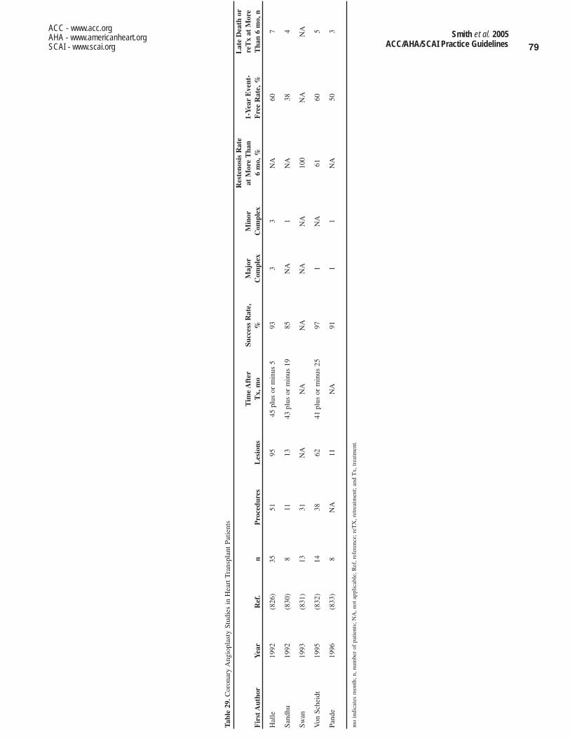
79Smith et al. 2005
ACC/AHA/SCAI Practice Guidelines
ACC - www.acc.orgAHA - www.americanheart.orgSCAI - www.scai.org
Tabl
e 29
.Cor
onar
y A
ngio
plas
ty S
tudi
es in
Hea
rt T
rans
plan
t Pat
ient
s
Tim
e A
fter
Succ
ess
Rat
e,
Maj
orF
irst
Aut
hor
Yea
rR
ef.
nP
roce
dure
sL
esio
nsT
x, m
o%
Com
plex
Hal
le19
92(8
26)
3551
9545
plu
s or
min
us 5
933
Sand
hu19
92(8
30)
811
1343
plu
s or
min
us 1
985
NA
Swan
1993
(831
)13
31N
AN
AN
AN
A
Von
Sch
eidt
1995
(832
)14
3862
41 p
lus
or m
inus
25
971
Pand
e19
96(8
33)
8N
A11
NA
911
Res
teno
sis
Rat
e L
ate
Dea
th o
rM
inor
at M
ore
Tha
n1-
Yea
rE
vent
-re
Tx
at M
ore
Com
plex
6 m
o, %
Fre
e R
ate,
%T
han
6 m
o, n
3N
A60
7
1N
A38
4
NA
100
NA
NA
NA
6160
5
1N
A50
3
mo
indi
cate
s m
onth
; n, n
umbe
r of
pat
ient
s; N
A, n
ot a
pplic
able
; Ref
, ref
eren
ce; r
eTX
, ret
reat
men
t; an
d T
x, tr
eatm
ent.

80
ACC - www.acc.orgAHA - www.americanheart.org
SCAI - www.scai.orgSmith et al. 2005ACC/AHA/SCAI Practice Guidelines
New coronary devices, including directional (857,858),rotational (859,860), extraction (861-865), and pullback(866) atherectomy, a cutting balloon, and excimer laser-assisted angioplasty, have also been used for stent restenosisbefore balloon dilation. Although some comparative registryseries have suggested an improved angiographic outcomeassociated with the use of these ablative devices, no long-term studies demonstrating clinical advantage have beencompleted.
When a significant residual stenosis exists after conven-tional PTCA of stent restenosis fails to achieve an optimallumen diameter, additional stents have been used to improvethe initial angiographic result (867-869). Although prelimi-nary results of clinical trials failed to demonstrate a benefitusing routine BMS placement for the treatment of stentrestenosis, favorable results have been shown with DES (seeSection 7.3.5 for further discussion) (116,870,871).
Acute platelet inhibition with abciximab does not reduceISR, as demonstrated in the ERASER (Evaluation of ReoProAnd Stenting to Eliminate Restenosis) study (280). In a studyof 225 patients randomly allocated to placebo or abciximabbefore intervention, 215 patients received a stent and thestudy drug. Of the 191 patients who returned for follow-upmore than 4 months after evaluation, there was no differencebetween tissue volume as measured by IVUS between theplacebo and treatment groups. Lack of abciximab benefitwas confirmed by quantitative angiography. The investiga-tors concluded that potent platelet inhibition with abciximabas administered in the ERASER study did not reduce ISR.
Since the last (2001) revision of the ACC/AHA PCI guide-line, the proportional use of stents in percutaneous interven-tions has continued to increase. In part, this derives from ran-domized trial data suggesting that routine stenting is moreeffective than provisional stenting (636,872-874). In addi-tion, stents are being used in a much wider spectrum of coro-nary and even graft anatomies (875). Accordingly, ISR hasbecome increasingly important. Because stents prevent elas-tic recoil and late negative remodeling, the predominantmechanism of ISR is neointimal hyperplasia due to smoothmuscle cell proliferation and extracellular matrix production.Two of the biggest changes since the 2001 revision havebeen the expanding databases of 1) brachytherapy to treatISR and 2) DES to try to prevent ISR.
7.3.5. Drug-Eluting Stents
Class IA drug-eluting stent (DES) should be considered as analternative to the bare-metal stent in subsets ofpatients in whom trial data suggest efficacy. (Level ofEvidence: A)
Class IIbA DES may be considered for use in anatomic settingsin which the usefulness, effectiveness, and safety havenot been fully documented in published trials. (Levelof Evidence: C)
(80,83,845). A number of factors have been associated withthe propensity to develop stent restenosis, including smallvessel size (846), smaller postprocedure minimum lumendiameter (847), higher residual percent diameter stenosis(848), lesions located in the LAD (83), stent length, and thepresence of diabetes mellitus (721,841,842,844,846-848).
Stent restenosis may occur within the stent, due to intimalhyperplasia, or at the stent margins, due to both intimalhyperplasia and arterial remodeling (849). A serial IVUSstudy performed in 115 lesions treated with the Palmaz-Schatz stent demonstrated that tissue growth was uniformlydistributed throughout the stent at follow-up study, with aslightly higher tendency for neointimal tissue accumulationat the central articulation (850). The stent lumen tended to besmallest at the articulation site, presumably owing to tissueprolapse between the stent struts. For multiple stents, therewas no difference in the postintervention or follow-up lumenwhen overlapped stents were compared with nonoverlappedstents (850). In another series of patients treated with thePalmaz-Schatz stent, 77 (26%) of 301 stent margins wererestenotic at follow-up (more than 50% late lumen loss)(849). The dominant periprocedural predictor of stent marginrestenosis was the plaque burden of the contiguous referencesegment (849).
Balloon angioplasty has been used frequently to treatpatients with stent restenosis (851-853). The mechanism oflumen improvement after balloon angioplasty for stentrestenosis relates to further stent expansion (851) and extru-sion of the tissue through the stent struts (851-854). In anIVUS study of 64 restenotic Palmaz-Schatz stents, 56% plusor minus 28% of the lumen enlargement was the result ofadditional stent expansion and 44% plus or minus 28% wasthe result of a decrease in neointimal tissue (851). Despitethe use of high-pressure balloon dilation, a relatively highresidual stenosis (18% plus or minus 12%) remained aftertreatment with balloon angioplasty.
The outcome after balloon angioplasty has been variable,depending, in part, on the size of the stented segment andlength of the stent restenosis (855). In a consecutive series of124 patients presenting with stent restenosis successfullytreated with repeat percutaneous intervention, clinical fol-low-up was obtained at 27.4 plus or minus 14.7 months(855). Recurrent clinical events occurred in 25 patients(20%), including death (2%), MI (1%), and target-vesselrevascularization (11%) (855). Cumulative event-free sur-vival at 12 and 24 months was 86.2% and 80.7%, respec-tively (855).
A number of factors have been related to the frequency ofclinical recurrence after balloon angioplasty for stentrestenosis (855), which include repeat intervention in SVGs,multivessel disease, low ejection fraction, and a 3-monthinterval between stent implantation and repeat intervention.One preliminary report has shown target-lesion revascular-ization was related to the length of the stent restenosis, rang-ing from 10% for focal stent stenosis to 25% for intrastentrestenosis, 50% for diffuse stent restenosis, and 80% for stenttotal occlusions (856).

81Smith et al. 2005
ACC/AHA/SCAI Practice Guidelines
ACC - www.acc.orgAHA - www.americanheart.orgSCAI - www.scai.org
ed stents, the long-term outcome depends on the response toboth stent and coating. For DES, the long-term healingresponse depends on the response to the polymer and thetherapeutic agent, as well as the stent. As evidenced by thetrials of gold coating and the preliminary experiences (reg-istry) with the QuaDS stent, actinomycin, and batimastat,some combinations are potentially even more proliferative,inflammatory, or thrombogenic than BMS.
Peer-reviewed publications of human DES implantation,including consecutive case series and randomized trials, areavailable for 3 polymer-based, drug-eluting, balloon-expand-able stent systems (Table 30) (236,441,624,628,697,698,879-888): the antiproliferative, antimigratory, anti-inflam-matory macrolide antibiotic rapamycin (sirolimus) affixed toa stent (Bx Velocity); the 7-hexanoyltaxol (QP2)-elutingpolymer stent system (QuaDS); and the microtubuleinhibitor paclitaxel (TAXUS) affixed to a stent. Each of thesesystems had undergone rigorous testing in animal modelsthat demonstrated an intact endothelial layer and significantreductions in neointimal hyperplasia and inflammation.
The first reported series of 45 patients who underwent SESimplantation in either Sao Paulo, Brazil, or Rotterdam,Holland, demonstrated the virtual absence of intimal hyper-plasia at 4 months (889). Subsequent studies of the samecohort at 1 and 2 years continued to document sustained sup-pression of neointimal hyperplasia as detected with bothIVUS and quantitative angiography (628,879). In theRandomized Study With the Sirolimus-Eluting Bx VelocityBalloon-Expandable Stent (RAVEL) trial, 238 patients wererandomly allocated between BMS and SES (624). At 6months the binary restenosis rate was 26% for the bare metalgroup versus 0% for the DES group, and there were no sub-acute stent thromboses with a 2-month dual antiplatelet reg-imen. In 1 year of follow-up, the bare metal group had a 29%rate of MACE versus 5.8% for the sirolimus-eluting group;this difference was driven entirely by target-vessel revascu-larization.
A 3-year follow-up of the RAVEL trial (890), involving114 patients from the SES arm and 113 in the BMS arm, doc-umented target-vessel revascularization in 11.4% of the SESgroup compared with 33.6% of the BMS group. These datasupport the long-term durability of SES in reducing repeatrevascularization compared with BMS.
The SIRIUS (Sirolimus-Eluting Balloon Expandable Stentin the Treatment of Patients With De Novo Native CoronaryArtery Lesions) investigators reported 1058 patients ran-domly allocated at 1 of 53 centers between BMS and SES(93). This cohort included diabetic patients (26%) and some-what longer lesions (mean 14.4 mm) and smaller-diametervessels (mean 2.8 mm) than the RAVEL population. Again,the sirolimus-eluting group had lower MACE at 270 daysthan the BMS group (7.1% vs 18.9%), which was driven bylower rates of target-vessel revascularization (4.1% vs16.6%). Both quantitative angiography and IVUS were usedto document that the mechanism for this salutary effect wasdecreased neointimal hyperplasia. SIRIUS was the pivotal
All PCI creates injury to the vessel wall, specifically tearsor dissection. Larger devices and higher pressures are asso-ciated with tears at deeper levels of the vessel wall (media oreven adventitia, as opposed to intima and plaque boundaryonly). All injuries tend to heal; specifically, injury to the ves-sel wall is associated with re-establishment of an intactendothelial layer. Failure to re-establish an intact, functionalendothelial layer is likely to be associated with continuedrisk of arterial thrombosis and an abnormal balance betweenvasoconstrictive and vasodilatory mechanisms. In general,deeper injury is associated with more proliferative healing(876-878). The demonstration, by quantitative angiography,that late lumen diameter after balloon angioplasty follows a“normal” or Gaussian distribution supports the concept thatrestenosis is an exaggerated healing response rather than adistinct biologic process, which occurs in a minority of indi-viduals.
For balloon angioplasty, the healing response includes, onthe macroscopic level, negative (narrowing) and positive(dilatation) remodeling, elastic recoil, and neointimal hyper-plasia. Because stents block elastic recoil and negativeremodeling, ISR is predominantly due to neointimal hyper-plasia. Neointimal hyperplasia is the name given to a com-plex process of multifactorial causation, which leads to ves-sel lumen encroachment. The causes of neointimal hyperpla-sia appear to include, but are not limited to, the followinginflammatory response involving cells and molecular media-tors; growth factors and cytokines; release of mediators andupregulation of signaling systems that stimulate cellularmigration and proliferation; activation, adherence, andaggregation of platelets; and thrombosis with release of clot-ting factors. Neointimal hyperplasia may be distinct fromatherosclerosis and negative remodeling, but it shares manyof the same causative factors. Accordingly, investigators andclinicians are inclined to try many of the same antithrombot-ic, antiplatelet, anti-inflammatory, and antiproliferativeagents to try to modify atherosclerosis, neointimal hyperpla-sia, and negative remodeling. Additionally, many therapeuticagents affect multiple mechanisms.
To date, no systemically administered therapeutic agent hasconsistently reduced restenosis after balloon angioplasty orplacement of BMS. Stents have reduced restenosis relative toballoon angioplasty (albeit with increased late loss due toincreased neointimal proliferation), and locally deliveredradiation (brachytherapy) has reduced ISR. Taken together,these observations and the early success of sirolimus- andpaclitaxel-eluting stents have supported the paradigm ofblocking elastic recoil and negative remodeling with amechanical stent and inhibiting neointimal hyperplasia witha locally delivered (higher concentration than can beachieved systemically) antiproliferative and anti-inflamma-tory agent.
Local delivery of a therapeutic agent with stents has taken2 forms: simple coating of the stent and adherence of thetherapeutic agent to a polymer, which allows for sustainedrelease over time. Diffusion of the therapeutic agent into thetissues and into blood is an additional complexity. For coat-
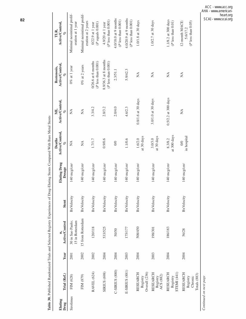
82
ACC - www.acc.orgAHA - www.american-
heart.orgSCAI - www.scai.org
Tabl
e 30
.Pub
lishe
d R
ando
miz
ed T
rial
s an
d Se
lect
ed R
egis
try
Exp
erie
nces
of
Dru
g-E
lutin
g St
ents
Com
pare
d W
ith B
are
Met
al S
tent
s
Elu
ting
n,
E
luti
ng D
rug
Dru
gT
rial
(R
ef.)
Yea
rA
ctiv
e/C
ontr
olSt
ent
Dos
age
Siro
limus
FIM
(62
8)20
0130
in S
ao P
aulo
;B
xVel
ocity
140
mcg
/cm
2
15 in
Rot
terd
am
FIM
(87
9)20
0215
fro
m R
otte
rdam
BxV
eloc
ity14
0 m
cg/c
m2
RA
VE
L(6
24)
2002
120/
118
BxV
eloc
ity14
0 m
cg/c
m2
SIR
IUS
(698
)20
0453
3/52
5B
xVel
ocity
140
mcg
/cm
2
C-S
IRIU
S (8
80)
2004
50/5
0B
xVel
ocity
140
mcg
/cm
2
E-S
IRIU
S (8
81)
2003
175/
177
BxV
eloc
ity14
0 m
cg/c
m2
RE
SEA
RC
H
2004
508/
450
BxV
eloc
ity14
0 m
cg/c
m2
Reg
istr
y O
vera
ll (2
36)
RE
SEA
RC
H
2003
198/
301
BxV
eloc
ity14
0 m
cg/c
m2
Reg
istr
yA
CS
(882
)
RE
SEA
RC
H
2004
186/
183
BxV
eloc
ity14
0 m
cg/c
m2
Reg
istr
yST
EM
I (4
41)
RE
SEA
RC
H
2004
56/2
8B
xVel
ocity
140
mcg
/cm
2
Reg
istr
yC
hron
icTo
tals
(88
3)
Con
tinu
ed o
n ne
xt p
age
Dea
ths
MI,
Res
teno
sis,
TL
R,
Act
ive/
Con
trol
, A
ctiv
e/C
ontr
ol,
Act
ive/
Con
trol
,A
ctiv
e/C
ontr
ol,
%%
%%
NA
NA
0% a
t 1 y
ear
Min
imal
neo
intim
al p
rolif
-er
atio
n at
1 y
ear
NA
NA
0% a
t 2 y
ears
Min
imal
neo
intim
al p
rolif
-er
atio
n at
2 y
ears
1.7/
1.7
3.3/
4.2
0/26
.6 a
t 6 m
onth
s0/
22.9
at 1
yea
r(P
less
than
0.0
01)
(Peq
uals
0.0
01)
0.9/
0.6
2.8/
3.2
8.9/
36.3
at 8
mon
ths
4.9/
20 a
t 1 y
ear
(Ple
ss th
an 0
.001
)(P
less
than
0.0
01)
0/0
2.0/
4.0
2.3/
51.1
4.0/
18.0
at 9
mon
ths
(Ple
ss th
an 0
.001
)
1.1/
0.6
4.6/
2.3
5.9/
42.3
4.0/
20.9
at 9
mon
ths
(Ple
ss th
an 0
.001
)
1.6/
2.0
0.8/
1.6
at 3
0 da
ysN
A1.
0/1.
8 at
30
days
at 3
0 da
ys
3.0/
3.0
3.0/
1.0
at 3
0 da
ysN
A1.
0/2.
7 at
30
days
at 3
0 da
ys
8.3/
8.2
0.5/
2.2
at 3
00 d
ays
NA
1.1/
8.2
at 3
00 d
ays
at 3
00 d
ays
(Ple
ss th
an 0
.01)
0/0
NA
NA
12-m
onth
MA
CE
:in
hos
pita
l5.
6/17
.2(P
less
than
0.0
5)
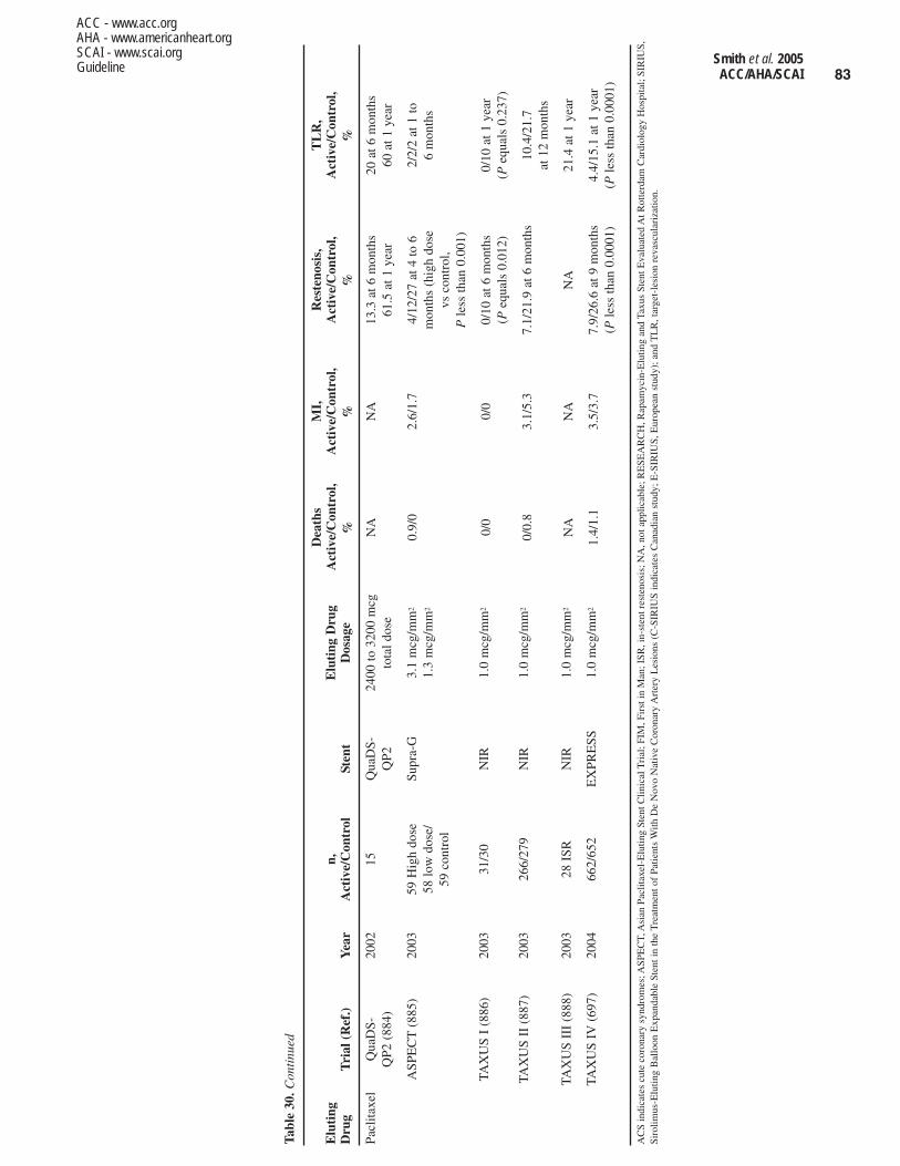
83Smith et al. 2005ACC/AHA/SCAI
ACC - www.acc.orgAHA - www.americanheart.orgSCAI - www.scai.orgGuideline
Tabl
e 30
.Con
tinu
ed
Elu
ting
n,
E
luti
ng D
rug
Dru
gT
rial
(R
ef.)
Yea
rA
ctiv
e/C
ontr
olSt
ent
Dos
age
Pacl
itaxe
lQ
uaD
S-20
0215
Qua
DS-
2400
to 3
200
mcg
Q
P2 (
884)
QP2
tota
l dos
e
ASP
EC
T(8
85)
2003
59 H
igh
dose
Supr
a-G
3.1
mcg
/mm
2
58 lo
w d
ose/
1.3
mcg
/mm
2
59 c
ontr
ol
TAX
US
I (8
86)
2003
31/3
0N
IR1.
0 m
cg/m
m2
TAX
US
II (
887)
2003
266/
279
NIR
1.0
mcg
/mm
2
TAX
US
III
(888
)20
0328
ISR
NIR
1.0
mcg
/mm
2
TAX
US
IV(6
97)
2004
662/
652
EX
PRE
SS1.
0 m
cg/m
m2
AC
S in
dica
tes
cute
cor
onar
y sy
ndro
mes
; ASP
EC
T, A
sian
Pac
litax
el-E
lutin
g St
ent C
linic
al T
rial
; FIM
, Fir
st in
Man
; ISR
, in-
sten
t res
teno
sis;
NA
, not
app
licab
le; R
ESE
AR
CH
, Rap
amyc
in-E
lutin
g an
d Ta
xus
Sten
t Eva
luat
ed A
t Rot
terd
am C
ardi
olog
y H
ospi
tal;
SIR
IUS,
Siro
limus
-Elu
ting
Bal
loon
Exp
anda
ble
Sten
t in
the
Tre
atm
ent o
f Pa
tient
s W
ith D
e N
ovo
Nat
ive
Cor
onar
y A
rter
y L
esio
ns (
C-S
IRIU
S in
dica
tes
Can
adia
n st
udy;
E-S
IRIU
S, E
urop
ean
stud
y); a
nd T
LR
, tar
get-
lesi
on r
evas
cula
riza
tion.
Dea
ths
MI,
Res
teno
sis,
TL
R,
Act
ive/
Con
trol
, A
ctiv
e/C
ontr
ol,
Act
ive/
Con
trol
,A
ctiv
e/C
ontr
ol,
%%
%%
NA
NA
13.3
at 6
mon
ths
20 a
t 6 m
onth
s61
.5 a
t 1 y
ear
60 a
t 1 y
ear
0.9/
02.
6/1.
74/
12/2
7 at
4 to
6
2/2/
2 at
1 to
m
onth
s (h
igh
dose
6
mon
ths
vs c
ontr
ol,
Ple
ss th
an 0
.001
)
0/0
0/0
0/10
at 6
mon
ths
0/10
at 1
yea
r(P
equa
ls 0
.012
)(P
equa
ls 0
.237
)
0/0.
83.
1/5.
37.
1/21
.9 a
t 6 m
onth
s10
.4/2
1.7
at 1
2 m
onth
s
NA
NA
NA
21.4
at 1
yea
r
1.4/
1.1
3.5/
3.7
7.9/
26.6
at 9
mon
ths
4.4/
15.1
at 1
yea
r(P
less
than
0.0
001)
(Ple
ss th
an 0
.000
1)

84
ACC - www.acc.orgAHA - www.americanheart.org
SCAI - www.scai.orgSmith et al. 2005ACC/AHA/SCAI Practice Guidelines
• Most of the follow-up is still relatively short-term (1 yearor less)
• Comparison of the 2 FDA-released systems is needed andshould provide clinically useful information. One suchtrial that supplies information in this regard is ISAR-DESIRE (Intracoronary Stenting and AngiographicResults: Drug-Eluting Stents for In-Stent Restenosis),which is discussed in Section 7.3.6.2.
• Preliminary results from randomized trials (REALITY,SIRTAX) comparing SES and PES (CYPHER versusTAXUS) have not shown large differences in clinical out-comes [M.C. Morice, oral presentation, American Collegeof Cardiology Scientific Session, Orlando, Fla, March2005; S. Windecker, oral presentation, American Collegeof Cardiology Scientific Session, Orlando, Fla, March2005].
• Mandated angiographic follow-up applied in trials hasincreased the reintervention rate, and therefore, the differ-ence between DES and BMS in clinical practice may beless.
The major trials of SES and PES, which were the basis forFDA approval, involved patients with stable or unstableischemia with documented coronary artery narrowing of51% to 99%, which stenoses were, for the most part, between2.75 and 3.5 mm in diameter and 15 to 30 mm in length.Specific clinical exclusions from these landmark trialsincluded the following: MI within 48 h; LV ejection fractionless than 0.25; previous or planned use of brachytherapy;previous PCI of the same lesion; coexisting medical condi-tions likely to limit life expectancy; contraindications toaspirin, thienopyridines, or stent substances; and severe renalor hematologic comorbidity. Specific angiographic exclu-sions from these landmark trials included the following:ostial lesions; bifurcation lesions; ULM lesion; SVG lesion;severe calcification; angiographic thrombus; severe tortuosi-ty; and occluded vessel. Babapulle and coworkers have per-formed a Bayesian meta-analysis of randomized clinical tri-als of DES that incorporates the results of RAVEL, SIRIUS,C-SIRIUS, and E-SIRIUS regarding rapamycin-elutingstents and TAXUS, I, II, and IV regarding PES (234, 880,881). These authors also included 4 trials of a nonpolymericformulation of paclitaxel, which has not been shown to beeffective in reducing restenosis or target-lesion or target-ves-sel revascularization and which has not been released forcommercial use (234).
In an effort to extend the application of DES to most of theother clinical angiographic subsets, unstudied in these land-mark trials, Serruys and coworkers at ThoraxcenterRotterdam, the Netherlands, established the RESEARCHRegistry (882). In this experience, the rapamycin-elutingstent has been used as the default strategy since it becameavailable, and consecutive prospective cases of particularclinical and angiographic subsets have been compared with
trial for FDA release of the rapamycin, polymer, Bx Velocitysystem.
Subsequent studies from the RESEARCH (Rapamycin-Eluting and TAXUS Stent Evaluated At RotterdamCardiology Hospital) registry experience at Thoraxcenter,Rotterdam, Netherlands, have documented the short-termsafety of using these SES systems in patients with acutecoronary syndromes, including STEMI (882,891). An addi-tional small registry experience from the Rotterdam groupsuggests the potential applicability of the sirolimus DES sys-tem to ISR. A consecutive case series of 368 patients with735 lesions for which 841 SES were implanted documentedonly 11 cases of restenosis (greater than 50% diameter), andall of those occurred in a focal pattern (892). The operatorsin that series, which included longer lesions (mean length oflesion 17.48 plus or minus 12.19 mm) and more complexanatomic subsets, learned from earlier studies of DES edgelesions to fully cover diseased segments (mean stent length27.59 plus or minus 14.02 mm) (892).
TAXUS-I was the first feasibility and safety study of thepaclitaxel, polymer, NIR stent system. There were 61patients randomly allocated between a BMS and DES. At 12months, the MACE rate was 3% (1 event) in the TAXUSgroup and 10% in the BMS group (4 events in 3 patients),and there were no subacute stent thromboses. Although thesedifferences were not statistically significantly different, thecontinuous outcome of minimal lumen diameter was signifi-cantly better in the TAXUS group (886).
The ASPECT trial was a 3-center prospective, randomizedtrial of 177 patients with short (less than 15 mm), favorable(2.25 to 3.5 mm diameter) native vessel lesions who wererandomly allocated between bare-metal Cook Supra-G stentsand stents bonded with 1 of 2 doses of paclitaxel (885).Interpretation of this trial was complicated by the use of 3different antiplatelet regimens. Binary restenosis was 4% inthe high dose of paclitaxel, 12% in the low dose of paclitax-el, and 27% in the BMS arm. Subsequent mechanistic stud-ies with IVUS documented that the paclitaxel-coated stentsreduced neointimal hyperplasia (893).
TAXUS-IV was a prospective, randomized clinical trial ofthe slow-release; polymer-based paclitaxel-NIR stent systemconducted at 73 US centers (94). A total of 1314 patientswith native coronary lesions 10 to 28 mm in length and 2.5to 3.75 mm in diameter were randomly allocated betweenBMS and the paclitaxel polymer system. At 9 months, angio-graphic restenosis was reduced from 26.6% to 7.9% withDES, albeit there were no differences in death, MI, or suba-cute stent thrombosis (0.6% and 0.8%, respectively). It is pri-marily on the basis of TAXUS-IV that the FDA released thepaclitaxel, polymer NIR stent system. TAXUS-III was a reg-istry study that demonstrated the potential efficacy of thisDES system for ISR (888).
There is considerable promise and excitement surroundingthe release of DES; nevertheless, important reservationsremain, including the following:
85Smith et al. 2005
ACC/AHA/SCAI Practice Guidelines
ACC - www.acc.orgAHA - www.americanheart.orgSCAI - www.scai.org
relates to further stent expansion (851) and extrusion of thetissue through the stent struts (851-854). In an IVUS study of64 restenotic Palmaz-Schatz stents, 56% plus or minus 28%of the lumen enlargement was the result of additional stentexpansion, and 44% plus or minus 28% was the result of adecrease in neointimal tissue (851). Despite the use of high-pressure balloon dilation, a relatively high residual stenosis(18% plus or minus 12%) remained after treatment withPTCA.
The outcome after PTCA has been variable, depending inpart on the size of the stented segment and length of the stentrestenosis (855). In a consecutive series of 124 patients pre-senting with stent restenosis successfully treated with repeatPTCA, clinical follow-up was obtained at 27.4 plus or minus14.7 months (855). Recurrent clinical events occurred in 25patients (20%), including death (2%), MI (1%), and target-vessel revascularization (11%) (855). Cumulative event-freesurvival at 12 and 24 months was 86.2% and 80.7%, respec-tively (855).
A number of factors have been related to the frequency ofclinical recurrence after PTCA for ISR (855), which includerepeat intervention in SVGs, multivessel disease, low ejec-tion fraction, and a 3-month interval between stent implanta-tion and repeat intervention. One report involving 245patients receiving BMS in the pre-DES era has categorizedISR into 4 classifications: focal, diffuse intrastent, diffuseproliferative, and total occlusion. Pattern I contains 4 types(A-D). Patterns II through IV are defined according to geo-graphic position of ISR in relation to the previously implant-ed stent. Target-lesion revascularization was related to thelength of the ISR, ranging from 10% for focal in-stent steno-sis (class I) to 25% for intrastent restenosis (class II), 50%for diffuse proliferative ISR (class III), and 80% for ISR withtotal occlusion (class IV) (856).
A broad array of catheter-based technologies, includingdirectional (857,858), rotational (859,860), extraction (861-865), and pullback (866) atherectomy, a cutting balloon, andexcimer laser-assisted angioplasty, have been used to treatISR in association with PTCA. Although some comparativeregistry series have suggested an improved angiographic out-come associated with the use of these ablative devices, nolong-term studies demonstrating clinical advantage havebeen completed.
When a significant residual stenosis exists after conven-tional PTCA for ISR, PCI with stenting has been used toimprove the initial angiographic result (867-869). Althoughpreliminary results of clinical trials failed to demonstrate abenefit using routine BMS placement for the treatment ofISR, favorable results have been shown with DES, as sum-marized in the following section (116,870,871).
7.3.6.2. Drug-Eluting Stents
Class IIaIt is reasonable to perform repeat PCI for ISR with aDES or a new DES for patients who develop ISR ifanatomic factors are appropriate. (Level of Evidence:B)
the immediately prior experience at the same institution withthe particular subset (882).
Patients with acute coronary syndromes were consideredfor SES in a RESEARCH registry comparison of 198 con-secutive patients receiving the SES versus a prior consecu-tive case series of 301 patients with acute coronary syn-dromes treated with BMS (882). Major adverse cardiacevents including death (3.0% vs 3.0%), nonfatal MI (3.0% vs1.0%), and target-vessel revascularization (1.0% vs 2.7%)were comparable for SES and BMS (total 6.1% vs 6.6%).Lemos and colleagues reported a series of 186 consecutiveSTEMI patients treated with an SES and compared thatgroup with 183 patients treated with a BMS in terms of bothshort- and long-term outcomes (441). MACE at 30 days(7.5% vs 10.4%) and stent thrombosis (0% vs 1.6%) werenot significantly different for SES compared with BMSpatients. By 300 days, both target-vessel revascularization(1.1% vs 8.2%) and MACE (9.4% vs 17.0%) were reducedwith SES versus BMS (441). A quantitative study reportedby Saia et al demonstrated a binary restenosis rate of 0% at 6months in a subset of 96 STEMI patients, which document-ed reduced late loss to a degree comparable to what was seenin the landmark trials of more stable patients with less com-plex anatomy (891).
Hoye and coauthors compared the outcomes of a consecu-tive case cohort of 56 patients with chronic total occlusionstreated with SES and compared them with a prior consecu-tive case series of 28 patients with chronic total occlusionstreated with BMS (883). By 12 months, MACE was 96.4%with SES and 82.1% with BMS (P less than 0.05 by log-ranktesting) (883). A consecutive case series of 19 patients with21 lesions of old SVGs treated with SES had 0% early revas-cularization and a 1-year (average) MACE of 84% (894).Unlike the other RESEARCH Registry series, this reportincluded no historic control group.
7.3.6. Management Strategies for ISR
Since the last (2001) revision of the ACC/AHA PCI guide-line, the proportional use of stents in percutaneous interven-tions has continued to increase. In part, this derives from ran-domized trial data suggesting that routine stenting is moreeffective than provisional stenting (636,872-874). In addi-tion, stents are being used in a much wider spectrum of coro-nary and even graft anatomies (875). Accordingly, ISR hasbecome increasingly important. Because stents prevent elas-tic recoil and late negative remodeling, the predominantmechanism of ISR is neointimal hyperplasia due to smoothmuscle cell proliferation and extracellular matrix production.Two of the biggest changes since the 2001 revision havebeen the expanding databases evaluating the use of 1) DESto prevent ISR and 2) brachytherapy to treat ISR (discussedin Sections 7.3.6.2 and 7.3.6.3, respectively).
7.3.6.1. PTCA
PTCA has been used to treat patients with ISR (851-853).The mechanism of lumen improvement after PTCA for ISR

86
ACC - www.acc.orgAHA - www.americanheart.org
SCAI - www.scai.orgSmith et al. 2005ACC/AHA/SCAI Practice Guidelines
with ISR (898-900). In the 2001 revision of the guideline, theinitial results of the SCRIPPS trial (Scripps CoronaryRadiation to Inhibit Proliferation Post Stenting) were sum-marized (117). Since then, the 5-year results of the SCRIPPScohort have been published (901). The Ir-192–treatedpatients continued to demonstrate improved event-free sur-vival (freedom from death, MI, or target-lesion revascular-ization) compared with placebo (61.5% vs 34.5%; P equals0.02) (900). As shown in Table 31, this composite end pointderived from improvements in each of the 3 component endpoints (92,116,117,658-660,901-903).
A number of reports of the GAMMA-1 trial have been pub-lished since the 2001 revision (622,658,904,905). The initialreport of 9 months of follow-up demonstrated a statisticallysignificant reduction in target-lesion revascularization withIr-192 (42% vs 24 %; P less than 0.01). Death and MI wereinsignificantly higher with radiation.
The WRIST (Washington Radiation for In-Stent RestenosisTrial) investigators randomized 130 patients (65/65) withISR between placebo and 15 Gy Ir-192 (116). The SVG-WRIST investigators randomized 120 patients (60/60)between placebo and Ir-192 for the treatment of ISR in SVGs(902). Again, the brachytherapy-treated cohort had lowerrates of binary restenosis (21% vs 44%; P equals 0.005) andtarget-lesion revascularization (17% vs 57%; P less than0.001).
Among the specific limitations of gamma radiation are theneed for long treatment times and the high radiation expo-sure, which necessitate special shielding and removal of stafffrom the treatment room during dwell times (906). Beta radi-ation, in the form of electrons or particulate energy, has alsodemonstrated effectiveness in randomized trials of ISR,despite its more limited tissue penetration (92,659,660).Taken together, these data support the effectiveness of radia-tion in reducing restenosis after treatment of ISR. Furtherinvestigation into the causes of late stent thrombosis (907)have led to recommendations that 1) new stents not beimplanted at the time of brachytherapy unless necessary and2) antiplatelet therapy with both aspirin and a thienopyridinebe continued for at least 6 to 12 months after brachytherapy(898-900,906).
Brachytherapy dosing for ISR is prescribed so as to achieveadequate radiation in the vessel wall to block cellular prolif-eration. The manufacturer’s recommended dosing for thebeta radiation source is 18.4 Gy at 2 mm from the sourcecenter for vessels from 2.7 to 3.35 mm in diameter and 23 Gyfor vessels 3.35 to 4.0 mm in diameter. It is also recom-mended that radiation be delivered over the entire segmentinjured by balloon dilation and that at least a 5-mm marginbe allowed on each side of the injured segment (908).
To date, the following potential limitations have beenobserved with the use of brachytherapy to treat ISR: edgestenoses or geographic miss; acute thrombosis; late throm-bosis and occlusion (up to 14%); persistent dissections; latestent malapposition; increased plaque burden outside thestent; IVUS echolucent areas or black holes (898-900,906);
In-stent restenosis represents a clinical challenge of greatinterest for DES technology. Sousa and coworkers treated 25consecutive cases of ISR with SES (870). They demonstrat-ed minimal intimal hyperplasia and no delayed malapposi-tion by intracoronary ultrasound (870). Clinically, theyreported a remarkable 0 repeat revascularizations, stentthromboses, or deaths (870). Degertekin and colleaguesreported a group of 26 consecutive ISR patients treated withSES (871). They also used 3-dimensional ultrasound to doc-ument minimal neointimal formation by 4 months (871).This more complex cohort included 1 patient with transplantvasculopathy and 4 with prior brachytherapy, which provid-ed support not only for the effectiveness of SES but also ofthe need for prolonged antiplatelet therapy and risk factormodification in patients with diffuse coronary disease (871).Saia and coworkers reported a series of 12 patients with ISRrefractory to brachytherapy who received SES; 4 of 10patients who were followed up long-term developed resteno-sis (895).
The ISAR-DESIRE trial compared the use of balloonangioplasty SES and PES treatment of ISR in 300 patients(896). Angiography at 6 months in 92% of the patients (nequals 275) demonstrated angiographic restenosis in 44.6%(41 of 92) of the balloon-alone group; 14.3% (13 of 91) ofthe SES group; and 21.7% (20 of 92) of the PES group. BothDES were superior to balloon alone, reducing the incidenceof target-vessel revascularization (33% for balloon alone vs8% for SES and 19% for PES). There was a trend towardsuperiority of SES over PES in angiographic restenosis thatwas marginally significant (P equals 0.19) and significancefor target-vessel revascularization (P equals 0.02). Thesedata support the use of either approved DES for the treatmentof ISR over a BMS. Additional data for DES and ISR havebeen reported in the TReatment Of Patients with an In-STENT REstenotic Coronary Artery Lesion (TROPICAL)Study, which assessed outcomes in 155 patients with ISRreceiving an SES. In-lesion late loss of 0.08 plus or minus0.49 and a binary restenosis rate of 9.7% was reported at 6-month follow-up, with a reintervention rate of 7.4% (897).Furthermore, preliminary data suggest that in patients receiv-ing SES for ISR (TROPICAL group), late lesion loss andbinary restenosis at 6 months were significantly reducedcompared with a historical group receiving brachytherapyfor ISR in the GAMMA I and II studies [F.J. Neumann, oralpresentation, EuroPCR, Paris, France, May 2004]. Thepotential benefit of SES compared with brachytherapyremains to be delineated in ongoing randomized trials.
7.3.6.3. Radiation
Class IIaBrachytherapy can be useful as a safe and effectivetreatment for ISR. (Level of Evidence: A)
Both gamma energy (photons), and beta energy (electrons)have been used in randomized clinical trials and prospectiveregistries to reduce the neointimal proliferation associated

87Smith et al. 2005
ACC/AHA/SCAI Practice Guidelines
ACC - www.acc.orgAHA - www.americanheart.orgSCAI - www.scai.org
Tabl
e 31
.Ran
dom
ized
Clin
ical
Tri
als
of B
rach
ythe
rapy
for
In-
Sten
t Res
teno
sis
Fol
low
-Up,
nA
ngio
grap
hic/
Res
teno
sis,
Tri
al (
Ref
.)Y
ear
XR
T/P
lace
boC
linic
alSo
urce
XR
T/P
lace
bo
SCR
IPPS
(11
7)19
9726
/29
6 m
oG
amm
a17
%/5
4%12
mo
IR-1
92(P
equa
ls 0
.01)
PRE
VE
NT
(660
)20
0080
/25
6 m
oB
eta
22%
/50%
12 m
oP-
32(P
equa
ls 0
.018
)
WR
IST
(116
)20
0065
/65
6 m
oG
amm
a22
%/6
0%12
mo
IR-1
92(P
equa
ls 0
.000
1)
GA
MM
A-O
NE
(65
8)20
0113
1/12
16
mo
Gam
ma
32%
/55%
9 m
oIR
-192
(Peq
uals
0.0
1)
INH
IBIT
(659
)20
0216
6/16
69
mo
Bet
a26
%/5
2%29
0 d
P-32
(Ple
ss th
an 0
.000
1)
SCR
IPPS
(90
1)20
0226
/29
5 y
Gam
ma
NA
IR-1
92
STA
RT
(92)
2002
244/
232
8 m
oB
eta
29%
/45%
Sr-9
0/Y
-90
(Peq
uals
0.0
01)
SVG
-WR
IST
(902
)20
0260
/60
6 m
oG
amm
a21
%/4
4%12
mo
IR-1
92(P
equa
ls 0
.005
)
Lon
g W
RIS
T(9
03)
2003
60/6
06
mo
Gam
ma
45%
/73%
12 m
oIR
-192
(Ple
ss th
an 0
.05)
n in
dica
tes
num
ber
of p
atie
nts;
NA
, not
app
licab
le; N
S, n
ot s
igni
fica
nt; M
I, m
yoca
rdia
l inf
arct
ion;
TL
R, t
arge
t-le
sion
rev
ascu
lari
zatio
n; a
nd X
RT,
rad
iatio
n th
erap
y.
TL
R, %
MI,
Dea
th,
XR
T/P
lace
boX
RT
/Pla
cebo
XR
T/P
lace
bo
12%
/45%
4%/0
%0%
/3 %
(Peq
uals
0.0
1)(P
equa
ls N
S)(P
equa
ls N
S)
6%/2
4%10
%/4
%1%
/0%
(Ple
ss th
an 0
.05)
(Peq
uals
NS)
(Peq
uals
NS)
3%/6
3%9%
/9%
6%/6
%(P
less
than
0.0
01)
(Peq
uals
NS)
(Peq
uals
NS)
24%
/42%
9.9%
/4.1
%3.
1%/0
.8%
(Ple
ss th
an 0
.01)
(Peq
uals
0.0
9)(P
equa
ls 0
.17)
8%/2
6%3%
/3%
3%/2
%(P
less
than
0.0
001)
(Peq
uals
NS)
(Peq
uals
NS)
23%
/48%
4%/1
0%19
%/3
1%(P
equa
ls 0
.05)
(Peq
uals
NS)
(Peq
uals
NS)
14%
/25%
1.7%
/3.3
%1.
3%/0
.5%
(Ple
ss th
an 0
.001
)(P
equa
ls 0
.364
)(P
equa
ls 0
.625
)
17%
/57%
2%/3
%7%
/7%
(Ple
ss th
an 0
.001
)(P
equa
ls N
S)(P
equa
ls N
S)
39%
/62%
16.9
%/1
8.3%
6.8%
/1.7
%(P
less
than
0.0
5)(P
equa
ls N
S)(P
equa
ls N
S)
88
ACC - www.acc.orgAHA - www.americanheart.org
SCAI - www.scai.orgSmith et al. 2005ACC/AHA/SCAI Practice Guidelines
ized comparative trials of DES versus BMS: acute coronarysyndromes; STEMI, chronic total occlusions; SVGs; andISR. Most of the data currently available regarding the use ofDES for ostial lesions, bifurcation lesions, ULM arteries, andextremely long segments are in the form of uncontrolled casereports or series. Nonetheless, given the promising results inreducing late target-lesion and target-vessel revasculariza-tion in nearly every group, it is to be expected that registryand randomized trial data will continue to accumulate at arapid pace.
7.4. Cost-Effectiveness Analysis for PCI
Among all diseases worldwide, ischemic heart disease cur-rently ranks fifth in disability burden and is projected to rankfirst by the year 2020 (911). As healthcare delivery systemsin countries with established economic markets continue toincorporate new and expensive technologies, the costs ofmedical care have seemingly escalated beyond the revenuehistorically allotted to health care. Given limited healthcareresources, a cost-effectiveness analysis is appropriate toevaluate percutaneous coronary revascularization strategies(912). The results of cost-effectiveness analyses for anycomparable treatment are reported in terms of the incremen-tal cost per unit of health gained, such as 1 year of life adjust-ed to perfect health (quality-adjusted life year, QALY) com-pared with the standard of care (913). By modeling differenttreatments, different patient subsets, and different levels ofdisease, a series of cost-effectiveness ratios may be con-structed to show the tradeoffs associated with choosingamong competing interventions.
Although there is no established cost-effectiveness ratiothreshold, cost-effectiveness ratios of less than $20 000 perQALY (such as seen in the treatment of severe diastolichypertension or with cholesterol lowering in patients withischemic heart disease) are considered highly favorable andconsistent with well-accepted therapies. Incremental cost-effectiveness ratios that range between $20 000 and $60 000per QALY may be viewed as reasonably cost-effective andthus acceptable in most countries, whereas ratios greater than$60000 to $80000 may be considered too expensive for mosthealthcare systems. The Committee defines useful and effi-cacious treatments, in terms of cost-effectiveness, as treat-ments with acceptable or favorable cost-effectiveness ratios.Cost-effectiveness analysis is not by itself sufficient to incor-porate all factors necessary for medical decision making onan individual patient basis, nor is it sufficient to dictate thebroad allocation of societal resources for health care. Rather,cost-effectiveness analysis aims to serve mainly as an aid tomedical decision making on the basis of comparison withother evaluated therapies.
The results of cost-effectiveness analysis in the field of per-cutaneous revascularization for ischemic heart disease havebeen derived from decision models that incorporate litera-ture-based procedure-related morbidity and mortality, coro-nary disease–related mortality, and estimates of the benefit ofselected revascularization procedures. When available,
and very late catch-up phenomenon (in studies with morethan 1 year of follow-up).
7.3.6.4. Medical Therapy
Acute platelet inhibition with abciximab does not reduceISR, as demonstrated in the ERASER study (280). In a studyof 225 patients randomly allocated to placebo or abciximabbefore intervention, 215 patients received a stent and thestudy drug. Of the 191 patients who returned for follow-upmore than 4 months after evaluation, there was no differencebetween tissue volume, as measured by IVUS, between theplacebo and treatment groups. Lack of abciximab benefitwas confirmed by quantitative angiography. The investiga-tors concluded that potent platelet inhibition with abciximabas administered in the ERASER study did not reduce ISR. Inthe Oral Sirolimus to Inhibit Recurrent In-Stent Restenosis(OSIRUS) trial, 300 patients were randomly assigned toreceive a cumulative loading dose of either placebo (0 mg),usual-dose (8 mg) oral sirolimus, or high-dose (24 mg) oralsirolimus 2 days before and the day of repeat PCI, followedby maintenance therapy of 2 mg per day for 7 days (909).Restenosis was significantly reduced from 42.2% to 36.8%and 22.1% in the placebo, usual-dose, and high-dose groups,respectively (P equals 0.005). The need for target-vesselrevascularization was reduced from 25.5% to 24.2% and15.2%, respectively, although this was not statistically sig-nificant (P equals 0.08). Blood concentration of oralsirolimus was significantly correlated with late lumen loss atfollow-up (P less than 0.001). The investigators concludedthat oral adjunctive sirolimus treatment for treatment of ISRresulted in significant improvement in the angiographicparameters of restenosis. Further elucidation of optimal dos-ing, need for pretreatment, and duration of oral sirolimus, aswell as long-term follow-up, are needed.
7.3.7. Subacute Stent Thrombosis
The issues of subacute stent thrombosis and technical issueswith the PES balloon-delivery system were early causes forconcern (910). After many more data have been accumulat-ed, as exemplified by the above-cited registry data, theredoes not appear to be an increased incidence of early throm-bosis with either SES or PES. As reported in the FDA edito-rial (910), Boston Scientific has recalled a number ofTAXUS stent systems because of reports of balloon deflationor retrieval problems and is working closely with the FDA tomonitor the situation.
7.3.8. Drug-Eluting Stents: Areas RequiringFurther Investigation
Both small vessels (less than 2.75 mm) and long lesions(greater than 18 mm) have been included in the C-SIRIUSand E-SIRIUS trials (880,881). In addition, there are increas-ing numbers of patients entered into prospective registriesand compared retrospectively with the following clinical andangiographic subsets, which were not included in random-
89Smith et al. 2005
ACC/AHA/SCAI Practice Guidelines
ACC - www.acc.orgAHA - www.americanheart.orgSCAI - www.scai.org
The use of DES is affecting the cost-effectiveness of PCI.In the SIRIUS trial (93), there were 21 fewer repeat revascu-larization procedures per 100 patients treated with thesirolimus stent. Although the DES group’s hospital costswere $2800 more, much of that was negated in follow-up bythe high reintervention rate in the BMS group (920).However, the number of repeat procedures in such trials withroutine angiographic follow-up is inflated compared withregistries of BMS, which suggests only 6 to 7 repeat proce-dures are avoided by routinely using DES (882). The ulti-mate cost effectiveness of drug-eluting stenting will dependon the cost of the stents, how many are implanted per patient,and how many repeat procedures are avoided.
Because cost-effectiveness analysis research is new in thefield of PCI, its results are limited. The Committee under-scores the need for cost containment and careful decisionmaking regarding the use of PCI strategies.
8. FUTURE DIRECTIONS
The field of coronary intervention has expanded dramatical-ly over the past decade and will continue to evolve over thenext several years. New directions will focus on strategiesthat will further improve procedural safety, reduce the recur-rence rate after PCI, and expand the procedure to more com-plex anatomic subsets. Clinical acceptance of these tech-nologies will be based on demonstration of safety and effica-cy over conventional therapies in randomized clinical stud-ies. Several novel strategies are summarized below.
Because the widespread use of stent implantation has less-ened the risk of need for emergency bypass, future clinicalresearch will focus on remaining obstacles that decrease pro-cedural success or increase risk. Chronic total occlusionremains a stubborn problem. New devices such as theFrontrunner catheter and new ultrastiff guidewires showsome promise in improving procedural success; however,new approaches are needed.
Degenerated vein graft disease remains a high-risk subset.The SAFER trial (255) has demonstrated that distal protec-tion with a balloon occlusive device with intraproceduralaspiration decreases procedural risk. Similarly, a number ofdistal filter devices are undergoing active testing (254). Theresults of one such multicenter trial comparing a filter-basedcatheter with a balloon occlusive and aspiration deviceshowed similar results for MACE at 30 days (254). In spiteof these approaches, these procedures are still associatedwith MACE event rates of 8% to10%. More research is stillneeded in this area. The use of distal protection devices insettings other than degenerative vein graft disease requiresfurther study. For example, initial studies in primary PCIsuggested a benefit with the FilterWire™; however, subse-quent trials with the GuideWire have failed to show any ben-efit, instead showing poor outcomes in this setting. Thus, fur-ther research is needed before this technology is adopted foruse beyond degenerative vein graft disease.
Dramatic advances have been made in the treatment ofrestenosis. Although the oral agents tranilast (921) and folic
results from randomized trials (levels of evidence A and B)are used to estimate the outcomes of each decision treebranch within the decision-analytical model, for example,using data estimating the restenosis rate after uncomplicatedcoronary stenting of a single, simple lesion. Cost-effective-ness analyses have been used to compare medical therapywith PTCA with CABG (914), balloon angioplasty withcoronary stenting (915,916), and routine coronary angiogra-phy after acute MI with symptom-driven coronary angiogra-phy (917).
In patients with severe angina, normal LV function, andsingle-vessel disease of the LAD, the cost-effectiveness ratiofor PTCA, directional coronary atherectomy, or coronarystenting that can be expected to provide a more than 90%success rate with a less than 3% major acute complicationrate is very favorable (less than $20 000 per QALY) com-pared with medical therapy (914). The rating also applies topatients with symptomatic angina or documented ischemiaand 2-vessel coronary disease, in whom percutaneous coro-nary revascularization can be expected to provide a morethan 90% success rate with a less than 3% major acute com-plication rate. In patients with 3-vessel coronary disease whohave comorbidities that increase the operative risk for CABGsurgery, PCI that is believed to be safe and feasible is rea-sonably acceptable ($20 000 to $60 000 per QALY). Inpatients in the post-MI setting, a strategy of routine,non–symptom-driven coronary angiography and PCI per-formed for critical (greater than 70% diameter stenosis) cul-prit coronary lesions amenable to balloon angioplasty orstenting has been proposed to be reasonably cost-effective inmany subgroups (917).
In patients with symptomatic angina or documentedischemia and 3-vessel coronary disease, for which bypasssurgery can be expected to provide full revascularization andan acute complication rate of less than 5%, the cost-effec-tiveness of PCI is not well established. Although PTCA for2- and 3-vessel coronary disease appears to be as safe as butinitially less expensive than CABG surgery, the costs ofPTCA converge toward the higher costs of bypass surgeryafter 3 to 5 years (918,919). Thus, whereas PTCA or CABGsurgery has been shown to be cost-effective compared withmedical therapy, there is no evidence for incremental cost-effectiveness of PTCA over bypass surgery for 2- or 3-vesselcoronary disease in patients who are considered good candi-dates for both procedures. For patients with 1- or 2-vesselcoronary disease who are asymptomatic or have only mildangina, without documented left main disease, the estimatedcost-effectiveness ratios for PCI are greater than $80 000 perQALY compared with medical therapy and are thus consid-ered less favorable.
The initial mean cost of angioplasty was 65% that of sur-gery, but the need for repeat interventions increased medicalexpenses so that after 5 years, the total medical cost of PTCAwas 95% that of surgery ($56 225 vs $58 889), a significantdifference of $2664 (P equals 0.047). Compared with CABG,PTCA appeared less costly for patients with 2-vessel diseasebut not for patients with 3-vessel disease.

90
ACC - www.acc.orgAHA - www.americanheart.org
SCAI - www.scai.orgSmith et al. 2005ACC/AHA/SCAI Practice Guidelines
rial infusion of marrow-derived stem cells (926) and directinjection of skeletal muscle–derived myoblasts (927) formyogenesis. Studies to date were performed in patients withsevere angina; thus, it is uncertain how this technology mightapply to other subsets of patients with coronary disease (e.g.,acute coronary syndromes, ischemic cardiomyopathy), andrigorous, blinded evaluation of these approaches must occur.
STAFFAmerican College of CardiologyChristine W. McEntee, Chief Executive Officer
Katherine D. Doermann, Senior Specialist, Practice Guidelines
Kristina N. Petrie, MS, Associate Director, Evidence-BasedMedicine
Dawn R. Phoubandith, MSW, Associate Director, PracticeGuidelines
Peg Christian, Librarian, Knowledge Management
American Heart AssociationM. Cass Wheeler, Chief Executive Officer
Rose Marie Robertson, MD, FACC, FAHA, Chief Science Officer
Kathryn A. Taubert, PhD, FAHA, Senior Science Advisor
Society for Cardiovascular Angiography and Interventions
Norm Linsky, Executive Director
Wayne Powell, Senior Director, Advocacy and Guidelines
acid have proven unsuccessful, other catheter-based strate-gies have dramatically decreased restenosis risk.Brachytherapy (for ISR), rapamycin-eluting stents, and PEShave been extremely effective. Subgroups such as diffuseISR and insulin-dependent diabetes mellitus will require fur-ther study. Other therapies, such as photodynamic therapy,cryotherapy, and therapeutic ultrasound, remain interestingbut unproven approaches to treat restenosis.
In patients with refractory angina who have no vessels suit-ed for revascularization, a number of new therapies are beingtested. Enhanced external counterpulsation appears todecrease symptoms (922). Treatment with fibroblast growthfactor by an intracoronary approach also shows promise(923). Percutaneous laser transmyocardial revascularizationhas shown mixed results. The PACIFIC trial (Potential ClassImprovement From Intramyocardial Channels) putativelydemonstrated some benefit of percutaneous laser transmy-ocardial revascularization, but the major limitation of thatstudy was that it was not placebo-controlled; thus, after itsfailure to address potential concerns, general consensusattributes the results in PACIFIC to a placebo effect. Also, inPACIFIC, diverse outcomes tended to be higher with lasertherapy (924). Although the randomized, double-blindBELIEF trial (Blinded Evaluation of Laser PMRIntervention Electively For angina pectoris) of 82 patientsappeared to show some benefit of percutaneous laser trans-myocardial revascularization versus sham procedure onangina class and quality-of-life measures, the results wereinconclusive given the small size of the study (925). To date,data are insufficient for FDA approval of percutaneous lasertherapy. A new frontier has been opened with the intra-arte-

91Smith et al. 2005
ACC/AHA/SCAI Practice Guidelines
ACC - www.acc.orgAHA - www.americanheart.orgSCAI - www.scai.org
APPENDIX 1. ACC/AHA/SCAI Committee to Update 2002 Guidelines for Percutaneous Coronary Intervention—Relationships withIndustry
Committee Speakers Bureau/ Stock Member Research Grant Honoraria Ownership Consultant
Dr. Ted E. Feldman Abbot Boston Scientific None NoneBoston Scientific
CardiaEV3
EvalveGuidant
Dr. John W. Hirshfeld, Jr None None None None
Dr. Alice K. Jacobs None None None Wyeth
Dr. Morton J. Kern None None None None
Dr. Spencer B. King, III Guidant BMS-Sanofi None MedtronicGuidant Novoste
Dr. Douglass A. Morrison None None None None
Dr. William W. O’Neill None None None None
Dr. Hartzell V. Schaff None None None None
Dr. Sidney C. Smith, Jr Merck Bayer Johnson & Johnson Bristol-Myers Medtronic Squibb
Eli LillyPfizer
Sanofi-Aventis
Dr. Patrick L. Whitlow Abbot None Medtronic NoneCordis, Inc
Fox Hollow TechnologiesLumend, Inc
Dr. David O. Williams None None None None
This table represents the relevant relationships of authors with industry that were reported orally at the initial writing committee meeting in July 2002 and updated in con-junction with all meetings and conference calls of the writing committee. It does not reflect any actual or potential relationships at the time of publication.

APPENDIX 2. External Peer Reviewers for the ACC/AHA/SCAI 2005 Guideline Update for Percutaneous Coronary Intervention*
Peer Reviewer Research Speakers Bureau/ Stock Consultant/Name† Representation Grant Honoraria Ownership Advisory Board
Dr. Michael Cowley Official Reviewer – None None None NoneSCAI
Dr. David Faxon Offical Reviewer – None None None NoneACC/AHA Task Forceon Practice Guidelines
Dr. Roxana Mehran Official Reviewer – None The Medicines Co. None NoneSCAI
Dr. E. Magnus Ohman Official Reviewer – Berlex None Inovise Medical NoneAHA Bristol-Myers Squibb Medtronic
Millennium Response MedicalSchering-Plough
Dr. Richard Pomerantz Official Reviewer – Aventis Amgen MedacorpACCF Board of Johnson & Johnson
Governors PfizerSchering-Plough
Dr. Robert D. Safian Official Reviewer – None None None NoneAHA
Dr. W. Douglas Weaver Official Reviewer – None None None NoneACCF Board of
Trustees
Dr. Jeffrey L. Anderson Content Reviewer – Sanofi/Bristol-Myers Merck None MerckIndividual Review Squibb Sanofi/Bristol-Myers
Novartis Squibb
Dr. Elliott M. Antman Content Reviewer – Aventis None None AventisIndividual Review Bayer
BiositeBoehringer-MannheimBristol-Myers Squibb
British BiotechCentor
Cor/MillenniumCorvasDade
GenentechLilly
MerckPfizerSunol
Dr. Larry S. Dean Content Reviewer – None None None NoneACCF Cardiac
Catheterization andIntervention Committee
Dr. Tommaso Gori Content Reviewer – None None None NoneAHA Diagnostic and
Interventional CardiacCatheterization
Committee
Dr. Sharon A. Hunt Content Reviewer – None None None NoneIndividual Review
Dr. Lloyd Klein Content Reviewer – None None None NoneAHA Diagnostic and
Interventional CardiacCatheterization
Committee
Dr. Glenn Levine Content Reviewer None Aventis None NoneAHA Diagnostic and
Interventional CardiacCatheterization
Committee
Dr. Joseph P. Ornato Content Reviewer – Genentech None None Bristol-Myers SquibbIndividual Review Genentech
This table represents the relevant relationships of peer reviewers with industry to this topic that were disclosed at the time of peer review of this guideline. It does not necessarily reflect relationships withindustry at the time of publication.
*Participation in the peer review process does not imply endorsement of the document.†Names are listed in alphabetical order within each category of review.

93Smith et al. 2005
ACC/AHA/SCAI Practice Guidelines
ACC - www.acc.orgAHA - www.americanheart.orgSCAI - www.scai.org
ic thrombectomy with intracoronary urokinase for coronary andvein graft thrombus (the Vein Graft AngioJet Study [VeGAS 2]).Am J Cardiol 2002;89:326-30.
17. Liu F, Haude M, Ge J, Eick B, Baumgart D, Erbel R.Recanalization of totally occluded saphenous vein bypass graftswith rheolytic thrombectomy device AngioJet catheter. JIntervent Cardiol 1998;11:49-53.
18. American College of Cardiology-National Cardiovascular DataRegistry Version 3.0. 2004;
19. Detre KM, Wright E, Murphy ML, Takaro T. Observer agreementin evaluating coronary angiograms. Circulation 1975;52:979-86.
20. Brown BG, Bolson EL, Dodge HT. Quantitative computer tech-niques for analyzing coronary arteriograms. Prog Cardiovasc Dis1986;28:403-18.
21. Alpert JS, Thygesen K, Antman E, Bassand JP. Myocardialinfarction redefined—a consensus document of The JointEuropean Society of Cardiology/American College of CardiologyCommittee for the redefinition of myocardial infarction. J AmColl Cardiol 2000;36:959-69.
21a. Luepker RV, Apple FS, Christenson RH, et al. Case definitions foracute coronary heart disease in epidemiology and clinicalresearch studies: a statement from the AHA Council onEpidemiology and Prevention; AHA Statistics Committee; WorldHeart Federation Council on Epidemiology and Prevention; theEuropean Society of Cardiology Working Group onEpidemiology and Prevention; Centers for Disease Control andPrevention; and the National Heart, Lung, and Blood Institute.Circulation 2003;108:2543-9
22. Abdelmeguid AE, Topol EJ. The myth of the myocardial ‘infarct-let’ during percutaneous coronary revascularization procedures.Circulation 1996;94:3369-75.
23. Califf RM, Abdelmeguid AE, Kuntz RE, et al. Myonecrosis afterrevascularization procedures. J Am Coll Cardiol 1998;31:241-51.
24. Ellis SG, Chew D, Chan A, Whitlow PL, Schneider JP, Topol EJ.Death following creatine kinase-MB elevation after coronaryintervention: identification of an early risk period: importance ofcreatine kinase-MB level, completeness of revascularization, ven-tricular function, and probable benefit of statin therapy.Circulation 2002;106:1205-10.
25. Miller WL, Garratt KN, Burritt MF, Reeder GS, Jaffe AS. Timingof peak troponin T and creatine kinase-MB elevations after per-cutaneous coronary intervention. Chest 2004;125:275-80.
26. Cantor WJ, Newby LK, Christenson RH, et al. Prognostic signif-icance of elevated troponin I after percutaneous coronary inter-vention. J Am Coll Cardiol 2002;39:1738-44.
27. Kizer JR, Muttrej MR, Matthai WH, et al. Role of cardiac troponinT in the long-term risk stratification of patients undergoing percu-taneous coronary intervention. Eur Heart J 2003;24:1314-22.
28. Newby LK, Alpert JS, Ohman EM, Thygesen K, Califf RM.Changing the diagnosis of acute myocardial infarction: implica-tions for practice and clinical investigations. Am Heart J2002;144:957-80.
29. Hunt AC, Chow SL, Shiu MF, Chilton DC, Cummins B,Cummins P. Release of creatine kinase-MB and cardiac specifictroponin-I following percutaneous transluminal coronary angio-plasty. Eur Heart J 1991;12:690-3.
30. Ravkilde J, Nissen H, Mickley H, Andersen PE, Thayssen P,Horder M. Cardiac troponin T and CK-MB mass release aftervisually successful percutaneous transluminal coronary angio-plasty in stable angina pectoris. Am Heart J 1994;127:13-20.
31. Karim MA, Shinn MS, Oskarsson H, Windle J, Deligonul U.Significance of cardiac troponin T release after percutaneous
References1. Smith SC, Jr., Dove JT, Jacobs AK, et al. ACC/AHA guidelines of
percutaneous coronary interventions (revision of the 1993 PTCAguidelines)--executive summary: a report of the American Collegeof Cardiology/American Heart Association Task Force on PracticeGuidelines (committee to revise the 1993 guidelines for percuta-neous transluminal coronary angioplasty). J Am Coll Cardiol2001;37:2215-39.
2. Gruntzig AR, Senning A, Siegenthaler WE. Nonoperative dilata-tion of coronary-artery stenosis: percutaneous transluminal coro-nary angioplasty. N Engl J Med 1979;301:61-8.
3. Kent KM, Bentivoglio LG, Block PC, et al. Percutaneous translu-minal coronary angioplasty: report from the Registry of theNational Heart, Lung, and Blood Institute. Am J Cardiol1982;49:2011-20.
4. Williams DO, Riley RS, Singh AK, Most AS. Restoration of nor-mal coronary hemodynamics and myocardial metabolism afterpercutaneous transluminal coronary angioplasty. Circulation1980;62:653-6.
5. Miller DD, Verani MS. Current status of myocardial perfusionimaging after percutaneous transluminal coronary angioplasty. JAm Coll Cardiol 1994;24:260-6.
6. Detre KM, Holmes DRJ, Holubkov R, et al. Incidence and conse-quences of periprocedural occlusion: the 1985-1986 NationalHeart, Lung, and Blood Institute Percutaneous TransluminalCoronary Angioplasty Registry. Circulation 1990;82:739-50.
7. Detre KM, Holubkov R, Kelsey S, et al. Percutaneous translumi-nal coronary angioplasty in 1985-1986 and 1977-1981: theNational Heart, Lung, and Blood Institute Registry. N Engl J Med1988;318:265-70.
8. O'Keefe JHJr, Rutherford BD, McConahay DR, et.al. Multivesselcoronary angioplasty from 1980 to 1989: procedural results andlong-term outcome. J Am Coll Cardiol 1990;16:1097-102.
9. American Heart Association. 2004 Heart and Stroke StatisticalUpdate. 2003;
10. Comparison of coronary bypass surgery with angioplasty inpatients with multivessel disease: the Bypass AngioplastyRevascularization Investigation (BARI) Investigators. [publishederratum appears in N Engl J Med 1997 Jan 9;336(2):147]. N EnglJ Med 1996;335:217-25.
11. Coronary angioplasty versus coronary artery bypass surgery: theRandomized Intervention Treatment of Angina (RITA) trial.Lancet 1993;341:573-80.
12. Hamm CW, Reimers J, Ischinger T, Rupprecht HJ, Berger J,Bleifeld W. A randomized study of coronary angioplasty com-pared with bypass surgery in patients with symptomatic multi-vessel coronary disease: German Angioplasty Bypass SurgeryInvestigation (GABI). N Engl J Med 1994;331:1037-43.
13. Rodriguez A, Boullon F, Perez-Balino N, et.al. Argentine ran-domized trial of percutaneous transluminal coronary angioplastyversus coronary artery bypass surgery in multivessel disease(ERACI): in-hospital results and 1-year follow-up. J Am CollCardiol 1993;22:1060-7.
14. Pocock SJ, Henderson RA, Riockards AF, et.al. Meta-analysis ofrandomized trials comparing coronary angioplasty with bypasssurgery. Lancet 1995;346:1184-9.
15. Laskey WK, Kimmel S, Krone RJ. Contemporary trends in coro-nary intervention: a report from the Registry of the Society forCardiac Angiography and Interventions. Catheter CardiovascInterv 2000;49:19-22.
16. Kuntz RE, Baim DS, Cohen DJ, et al. A trial comparing rheolyt-

94
ACC - www.acc.orgAHA - www.americanheart.org
SCAI - www.scai.orgSmith et al. 2005ACC/AHA/SCAI Practice Guidelines
intervention in the current era compared with 1985-1986: theNational Heart, Lung, and Blood Institute Registries. Circulation2000;102:2945-51.
50. Srinivas VS, Brooks MM, Detre KM, et al. Contemporary percu-taneous coronary intervention versus balloon angioplasty for mul-tivessel coronary artery disease: a comparison of the NationalHeart, Lung and Blood Institute Dynamic Registry and theBypass Angioplasty Revascularization Investigation (BARI)study. Circulation 2002;106:1627-33.
51. Anderson HV, Shaw RE, Brindis RG, et al. A contemporaryoverview of percutaneous coronary interventions. The AmericanCollege of Cardiology-National Cardiovascular Data Registry(ACC-NCDR®). J Am Coll Cardiol 2002;39:1096-103.
52. Hannan EL, Racz MJ, Walford G, et al. Long-term outcomes ofcoronary-artery bypass grafting versus stent implantation. N EnglJ Med 2005;352:2174-83.
53. Keeley EC, Boura JA, Grines CL. Primary angioplasty versusintravenous thrombolytic therapy for acute myocardial infarction:a quantitative review of 23 randomised trials. Lancet 2003;361:13-20.
54. Ricciardi MJ, Wu E, Davidson CJ, et al. Visualization of discretemicroinfarction after percutaneous coronary intervention associ-ated with mild creatine kinase-MB elevation. Circulation2001;103:2780-3.
55. Selvanayagam JB, Porto I, Channon K, et al. Troponin elevationafter percutaneous coronary intervention directly represents theextent of irreversible myocardial injury: insights from cardiovas-cular magnetic resonance imaging. Circulation 2005;111:1027-32.
56. Hong MK, Mehran R, Dangas G, et al. Creatine kinase-MBenzyme elevation following successful saphenous vein graftintervention is associated with late mortality. Circulation1999;100:2400-5.
57. Stone GW, Mehran R, Dangas G, Lansky AJ, Kornowski R, LeonMB. Differential impact on survival of electrocardiographic Q-wave versus enzymatic myocardial infarction after percutaneousintervention: a device-specific analysis of 7147 patients.Circulation 2001;104:642-7.
58. Bonz AW, Lengenfelder B, Strotmann J, et al. Effect of addition-al temporary glycoprotein IIb/IIIa receptor inhibition on troponinrelease in elective percutaneous coronary interventions after pre-treatment with aspirin and clopidogrel (TOPSTAR trial). J AmColl Cardiol 2002;40:662-8.
59. Iakovou I, Mintz GS, Dangas G, et al. Increased CK-MB releaseis a “trade-off” for optimal stent implantation: an intravascularultrasound study. J Am Coll Cardiol 2003;42:1900-5.
60. Williams DO. A twist in our understanding of enzyme elevationafter coronary intervention. J Am Coll Cardiol 2003;42:1906-8.
61. Albers GW, Caplan LR, Easton JD, et al. Transient ischemicattack--proposal for a new definition. N Engl J Med2002;347:1713-6.
62. Antman EM, Giugliano RP, Gibson CM, et al. Abciximab facili-tates the rate and extent of thrombolysis: results of the thrombol-ysis in myocardial infarction (TIMI) 14 trial. The TIMI 14Investigators. Circulation 1999;99:2720-32.
63. Noguchi T, Miyazaki MS, Morii I, Daikoku S, Goto Y, Nonogi H.Percutaneous transluminal coronary angioplasty of chronic totalocclusions. Determinants of primary success and long-term clini-cal outcome. Catheter Cardiovasc Interv 2000;49:258-64.
64. Stone GW, Grines CL, Cox DA, et al. Comparison of angioplas-ty with stenting, with or without abciximab, in acute myocardialinfarction. N Engl J Med 2002;346:957-66.
65. King SB, III. Ten-year completed follow-up of percutaneoustransluminal coronary angioplasty: the early Zurich experience. J
transluminal coronary angioplasty. Am J Cardiol 1995;76:521-3.32. La VL, Bedogni F, Finocchi G, et al. Troponin T, troponin I and
creatine kinase-MB mass after elective coronary stenting. CoronArtery Dis 1996;7:535-40.
33. Johansen O, Brekke M, Stromme JH, et al. Myocardial damageduring percutaneous transluminal coronary angioplasty as evi-denced by troponin T measurements. Eur Heart J 1998;19:112-7.
34. Shyu KG, Kuan PL, Cheng JJ, Hung CR. Cardiac troponin T, cre-atine kinase, and its isoform release after successful percutaneoustransluminal coronary angioplasty with or without stenting. AmHeart J 1998;135:862-7.
35. Bertinchant JP, Polge A, Ledermann B, et al. Relation of minorcardiac troponin I elevation to late cardiac events after uncompli-cated elective successful percutaneous transluminal coronaryangioplasty for angina pectoris. Am J Cardiol 1999;84:51-7.
36. Garbarz E, Iung B, Lefevre G, et al. Frequency and prognosticvalue of cardiac troponin I elevation after coronary stenting. AmJ Cardiol 1999;84:515-8.
37. Fuchs S, Kornowski R, Mehran R, et al. Prognostic value of car-diac troponin-I levels following catheter-based coronary inter-ventions. Am J Cardiol 2000;85:1077-82.
38. Wu AH, Boden WE, McKay RG. Long-term follow-up of patientswith increased cardiac troponin concentrations following percuta-neous coronary intervention. Am J Cardiol 2002;89:1300-2.
39. Ricciardi MJ, Davidson CJ, Gubernikoff G, et al. Troponin I ele-vation and cardiac events after percutaneous coronary interven-tion. Am Heart J 2003;145:522-8.
40. Kini AS, Lee P, Marmur JD, et al. Correlation of postpercuta-neous coronary intervention creatine kinase-MB and troponin Ielevation in predicting mid-term mortality. Am J Cardiol2004;93:18-23.
41. King SB, Yeh W, Holubkov R, et al. Balloon angioplasty versusnew device intervention: clinical outcomes. A comparison of theNHLBI PTCA and NACI registries. J Am Coll Cardiol1998;31:558-66.
42. Feit F, Brooks MM, Sopko G, et al. Long-term clinical outcomein the Bypass Angioplasty Revascularization InvestigationRegistry: comparison with the randomized trial. BARIInvestigators. Circulation 2000;101:2795-802.
43. McGrath PD, Malenka DJ, Wennberg DE, et al. Changing out-comes in percutaneous coronary interventions: a study of 34,752procedures in northern New England, 1990 to 1997. NorthernNew England Cardiovascular Disease Study Group. J Am CollCardiol 1999;34:674-80.
44. Grassman ED, Johnson SA, Krone RJ. Predictors of success andmajor complications for primary percutaneous transluminal coro-nary angioplasty in acute myocardial infarction: an analysis ofthe 1990 to 1994 Society for Cardiac Angiography andInterventions registries. J Am Coll Cardiol 1997;30:201-8.
45. Marks DS, Mensah GA, Kennard ED, Detre K, Holmes DRJ.Race, baseline characteristics, and clinical outcomes after coro-nary intervention: The New Approaches in CoronaryInterventions (NACI) registry. Am Heart J 2000;140:162-9.
46. Holmes DRJ, Berger PB, Garratt KN, et al. Application of theNew York State PTCA mortality model in patients undergoingstent implantation. Circulation 2000;102:517-22.
47. Hannan EL, Racz M, Ryan TJ, et al. Coronary angioplasty vol-ume-outcome relationships for hospitals and cardiologists. JAMA1997;277:892-8.
48. Peterson ED, Lansky AJ, Anstrom KJ, et al. Evolving trends ininterventional device use and outcomes: results from the NationalCardiovascular Network Database. Am Heart J 2000;139:198-207.
49. Williams DO, Holubkov R, Yeh W, et al. Percutaneous coronary
95Smith et al. 2005
ACC/AHA/SCAI Practice Guidelines
ACC - www.acc.orgAHA - www.americanheart.orgSCAI - www.scai.org
loon-expandable-stent implantation with balloon angioplasty inpatients with coronary artery disease: Benestent Study Group. NEngl J Med 1994;331:489-95.
81. Faxon DP, Spiro TE, Minor S, et al. Low molecular weightheparin in prevention of restenosis after angioplasty: results ofEnoxaparin Restenosis (ERA) Trial. Circulation 1994;90:908-14.
82. Leaf A, Jorgensen MB, Jacobs AK, et al. Do fish oils preventrestenosis after coronary angioplasty? Circulation 1994;90:2248-57.
83. Fischman DL, Leon MB, Baim DS, et al. A randomized compar-ison of coronary-stent placement and balloon angioplasty in thetreatment of coronary artery disease: Stent Restenosis StudyInvestigators. N Engl J Med 1994;331:496-501.
84. Weintraub WS, Boccuzzi SJ, Klein JL, et al. Lack of effect oflovastatin on restenosis after coronary angioplasty. LovastatinRestenosis Trial Study Group. N Engl J Med 1994;331:1331-7.
85. Baim DS, Cutlip DE, Sharma SK, et al. Final results of theBalloon vs Optimal Atherectomy Trial (BOAT). Circulation1998;97:322-31.
86. Watanabe K, Sekiya M, Ikeda S, Miyagawa M, Hashida K.Preventive effects of probucol on restenosis after percutaneoustransluminal coronary angioplasty. Am Heart J 1996;132:23-9.
87. Tardif JC, Cote G, Lesperance J, et al. Probucol and multivitaminsin the prevention of restenosis after coronary angioplasty.Multivitamins and Probucol Study Group. N Engl J Med1997;337:365-72.
88. Serruys PW, van Hout B, Bonnier H, et al. Randomised compar-ison of implantation of heparin-coated stents with balloon angio-plasty in selected patients with coronary artery disease (BenestentII) [published erratum appears in Lancet 1998 Oct31;352(9138):1478]. Lancet 1998;352:673-81.
89. Tamai H, Katoh O, Suzuki S, et al. Impact of tranilast on resteno-sis after coronary angioplasty: tranilast restenosis followingangioplasty trial (TREAT). Am Heart J 1999;138:968-75.
90. Holmes D, Fitzgerald P, Goldberg S, et al. The PRESTO(Prevention of restenosis with tranilast and its outcomes) proto-col: a double-blind, placebo-controlled trial. Am Heart J2000;139:23-31.
91. vom DJ, Dietz U, Haager PK, et al. Rotational atherectomy doesnot reduce recurrent in-stent restenosis: results of the angioplastyversus rotational atherectomy for treatment of diffuse in-stentrestenosis trial (ARTIST). Circulation 2002;105:583-8.
92. Popma JJ, Suntharalingam M, Lansky AJ, et al. Randomized trialof 90Sr/90Y beta-radiation versus placebo control for treatmentof in-stent restenosis. Circulation 2002;106:1090-6.
93. Moses JW, Leon MB, Popma JJ, et al. Sirolimus-eluting stentsversus standard stents in patients with stenosis in a native coro-nary artery. N Engl J Med 2003;349:1315-23.
94. Stone GW, Ellis SG, Cox DA, et al. A polymer-based, paclitaxel-eluting stent in patients with coronary artery disease. N Engl JMed 2004;350:221-31.
95. Albiero R, Silber S, Di MC, et al. Cutting balloon versus conven-tional balloon angioplasty for the treatment of in-stent restenosis:results of the restenosis cutting balloon evaluation trial (RES-CUT). J Am Coll Cardiol 2004;43:943-9.
96. Popma JJ, Califf RM, Topol EJ. Clinical trials of restenosis aftercoronary angioplasty. Circulation 1991;84:1426-36.
97. Haudenschild CC. Pathobiology of restenosis after angioplasty.Am J Med 1993;94:40S-4S.
98. Currier JW, Haudenschild C, Faxon DP. Pathophysiology ofrestenosis: clinical implications. In: Ischinger T, Gohlke H, edi-tors. Strategies in Primary and Secondary Prevention of CoronaryArtery Disease. W. Zuckschwerdt Verlag, 1992:181-92.
99. Mintz GS, Popma JJ, Pichard AD, et al. Arterial remodeling after
Am Coll Cardiol 1993;22:353-60.66. Holmes DR, Jr., Kip KE, Kelsey SF, Detre KM, Rosen AD. Cause
of death analysis in the NHLBI PTCA Registry: results and con-siderations for evaluating long-term survival after coronary inter-ventions. J Am Coll Cardiol 1997;30:881-7.
67. Influence of diabetes on 5-year mortality and morbidity in a ran-domized trial comparing CABG and PTCA in patients with mul-tivessel disease: the Bypass Angioplasty RevascularizationInvestigation (BARI). Circulation 1997;96:1761-9.
68. Detre KM, Guo P, Holubkov R, et al. Coronary revascularizationin diabetic patients: a comparison of the randomized and obser-vational components of the Bypass AngioplastyRevascularization Investigation (BARI). Circulation 1999;99:633-40.
69. Kelsey SF, James M, Holubkov AL, Holubkov R, Cowley MJ,Detre KM. Results of percutaneous transluminal coronary angio-plasty in women: 1985-1986 National Heart, Lung, and BloodInstitute’s Coronary Angioplasty Registry. Circulation1993;87:720-7.
70. Holmes DRJ, Detre KM, Williams DO, et al. Long-term outcomeof patients with depressed left ventricular function undergoingpercutaneous transluminal coronary angioplasty: the NHLBIPTCA Registry. Circulation 1993;87:21-9.
71. Schwartz L, Bourassa MG, Lesperance J, et al. Aspirin and dipyri-damole in the prevention of restenosis after percutaneous translu-minal coronary angioplasty. N Engl J Med 1988;318:1714-9.
72. Ellis SG, Roubin GS, Wilentz J, Douglas JSJ, King SB, III. Effectof 18- to 24-hour heparin administration for prevention ofrestenosis after uncomplicated coronary angioplasty. Am Heart J1989;117:777-82.
73. Pepine CJ, Hirshfeld JW, MacDonald RG, et al. A controlled trialof corticosteroids to prevent restenosis after coronary angioplas-ty: M-HEART Group. Circulation 1990;81:1753-61.
74. Serruys PW, Rutsch W, Heyndrickx GR, et al. Prevention ofrestenosis after percutaneous transluminal coronary angioplastywith thromboxane A2-receptor blockade: a randomized, double-blind, placebo-controlled trial. Coronary Artery RestenosisPrevention on Repeated Thromboxane-Antagonism Study (CAR-PORT). Circulation 1991;84:1568-80.
75. O’Keefe JH, Jr., McCallister BD, Bateman TM, Kuhnlein DL,Ligon RW, Hartzler GO. Ineffectiveness of colchicine for the pre-vention of restenosis after coronary angioplasty. J Am CollCardiol 1992;19:1597-600.
76. Does the new angiotensin converting enzyme inhibitor cilazaprilprevent restenosis after percutaneous transluminal coronaryangioplasty? Results of the MERCATOR study: a multicenter,randomized, double-blind placebo-controlled trial. MulticenterEuropean Research Trial with Cilazapril after Angioplasty toPrevent Transluminal Coronary Obstruction and Restenosis(MERCATOR) Study Group. Circulation 1992;86:100-10.
77. Topol EJ, Leya F, Pinkerton CA, et al. A comparison of direc-tional atherectomy with coronary angioplasty in patients withcoronary artery disease: the CAVEAT Study Group. N Engl J Med1993;329:221-7.
78. Adelman AG, Cohen EA, Kimball BP, et al. A comparison ofdirectional atherectomy with balloon angioplasty for lesions ofthe left anterior descending coronary artery. N Engl J Med1993;329: 228-33.
79. Serruys PW, Klein W, Tijssen JP, et al. Evaluation of ketanserin inthe prevention of restenosis after percutaneous transluminal coro-nary angioplasty: a multicenter randomized double-blind place-bo-controlled trial. Circulation 1993;88:1588-601.
80. Serruys PW, de Jaegere P, Kiemeneij F, et al. A comparison of bal-

96
ACC - www.acc.orgAHA - www.americanheart.org
SCAI - www.scai.orgSmith et al. 2005ACC/AHA/SCAI Practice Guidelines
119. Ellis SG, Vandormael MG, Cowley MJ, et al. Coronary morpho-logic and clinical determinants of procedural outcome with angio-plasty for multivessel coronary disease: implications for patientselection. Multivessel Angioplasty Prognosis Study Group.Circulation 1990;82:1193-202.
120. Hartzler GO, Rutherford BD, McConahay DR, Johnson WL,Giorgi LV. “High-risk” percutaneous transluminal coronaryangioplasty. Am J Cardiol 1988;61:33G-7G.
121. Gaul G, Hollman J, Simpfendorfer C, Franco I. Acute occlusionin multiple lesion coronary angioplasty: frequency and manage-ment. J Am Coll Cardiol 1989;13:283-8.
122. Ellis SG, Roubin GS, King SB, III, et al. In-hospital cardiac mor-tality after acute closure after coronary angioplasty: analysis ofrisk factors from 8,207 procedures. J Am Coll Cardiol 1988;11:211-6.
123. Guidelines for percutaneous transluminal coronary angioplasty: areport of the American College of Cardiology/American HeartAssociation Task Force on Assessment of Diagnostic andTherapeutic Cardiovascular Procedures (Committee onPercutaneous Transluminal Coronary Angioplasty). J Am CollCardiol 1993;22:2033-54.
124. de Feyter PJ, van den Brand M, Laarman GJ, et al. Acute coro-nary artery occlusion during and after percutaneous transluminalcoronary angioplasty: frequency, prediction, clinical course,management, and follow-up [published erratum appears inCirculation 1991 Jul;84(1):446]. Circulation 1991;83:927-36.
125. Myler RK, Shaw RE, Stertzer SH, et al. Lesion morphology andcoronary angioplasty: current experience and analysis. J Am CollCardiol 1992;19:1641-52.
126. Kastrati A, Schomig A, Elezi S, et al. Prognostic value of themodified American College of Cardiology/American Heart Asso-ciation stenosis morphology classification for long-term angio-graphic and clinical outcome after coronary stent placement.Circulation 1999;100:1285-90.
127. Budde T, Haude M, Hopp HW, et al. A prognostic computermodel to individually predict post-procedural complications ininterventional cardiology: the INTERVENT Project. Eur Heart J1999;20:354-63.
128. Chew DP, Bhatt DL, Robbins MA, et al. Incremental prognosticvalue of elevated baseline C-reactive protein among establishedmarkers of risk in percutaneous coronary intervention. Circulation2001;104:992-7.
129. Harrell L, Schunkert H, Palacios IF. Risk predictors in patientsscheduled for percutaneous coronary revascularization. CatheterCardiovasc Interv 1999;48:253-60.
130. Singh M, Lennon RJ, Holmes DR, Jr., Bell MR, Rihal CS.Correlates of procedural complications and a simple integer riskscore for percutaneous coronary intervention. J Am Coll Cardiol2002;40:387-93.
131. Zaacks SM, Allen JE, Calvin JE, et al. Value of the AmericanCollege of Cardiology/American Heart Association stenosis mor-phology classification for coronary interventions in the late1990s. Am J Cardiol 1998;82:43-9.
132. Krone RJ, Shaw RE, Klein LW, et al. Evaluation of the AmericanCollege of Cardiology/American Heart Association and theSociety for Coronary Angiography and Interventions lesion clas-sification system in the current “stent era” of coronary interven-tions (from the ACC-National Cardiovascular Data Registry). AmJ Cardiol 2003;92:389-94.
133. Krone RJ, Laskey WK, Johnson C, et al. A simplified lesion clas-sification for predicting success and complications of coronaryangioplasty. Registry Committee of the Society for CardiacAngiography and Intervention. Am J Cardiol 2000;85:1179-84.
coronary angioplasty: a serial intravascular ultrasound study.Circulation 1996;94:35-43.
100. Currier JW, Faxon DP. Restenosis after percutaneous transluminalcoronary angioplasty: have we been aiming at the wrong target? JAm Coll Cardiol 1995;25:516-20.
101. Kuntz RE, Baim DS. Defining coronary restenosis: newer clini-cal and angiographic paradigms. Circulation 1993;88:1310-23.
102. Kuntz RE, Gibson CM, Nobuyoshi M, Baim DS. Generalizedmodel of restenosis after conventional balloon angioplasty, stent-ing and directional atherectomy. J Am Coll Cardiol 1993;21:15-25.
103. Blackshear JL, O’Callaghan WG, Califf RM. Medical approachesto prevention of restenosis after coronary angioplasty. J Am CollCardiol 1987;9:834-48.
104. Bresee SJ, Jacobs AK, Garber GR, et al. Prior restenosis predictsrestenosis after coronary angioplasty of a new significant narrow-ing. Am J Cardiol 1991;68:1158-62.
105. Hirshfeld JWJ, Schwartz JS, Jugo R, et al. Restenosis after coro-nary angioplasty: a multivariate statistical model to relate lesionand procedure variables to restenosis. The M-HEARTInvestigators. J Am Coll Cardiol 1991;18:647-56.
106. Al SJ, Berger PB, Holmes DR, Jr. Coronary artery stents. JAMA2000;284:1828-36.
107. Continued benefit of coronary stenting versus balloon angioplas-ty: one-year clinical follow-up of Benestent trial. Benestent StudyGroup. J Am Coll Cardiol 1996;27:255-61.
108. Versaci F, Gaspardone A, Tomai F, Crea F, Chiariello L, GioffrePA. A comparison of coronary-artery stenting with angioplasty forisolated stenosis of the proximal left anterior descending coronaryartery. N Engl J Med 1997;336:817-22.
109. George CJ, Baim DS, Brinker JA, et al. One-year follow-up of theStent Restenosis (STRESS I) Study. Am J Cardiol 1998;81:860-5.
110. Rodriguez A, Ayala F, Bernardi V, et al. Optimal coronary balloonangioplasty with provisional stenting versus primary stent(OCBAS): immediate and long-term follow-up results. J Am CollCardiol 1998;32:1351-7.
111. Randomised placebo-controlled and balloon-angioplasty-con-trolled trial to assess safety of coronary stenting with use ofplatelet glycoprotein-IIb/IIIa blockade. The EPISTENTInvestigators. Evaluation of Platelet IIb/IIIa Inhibitor for Stenting.Lancet 1998;352:87-92.
112. Lincoff AM, Califf RM, Moliterno DJ, et al. Complementary clini-cal benefits of coronary-artery stenting and blockade of platelet gly-coprotein IIb/IIIa receptors. Evaluation of Platelet IIb/IIIa Inhibitionin Stenting Investigators. N Engl J Med 1999;341:319-27.
113. Betriu A, Masotti M, Serra A, et al. Randomized comparison ofcoronary stent implantation and balloon angioplasty in the treat-ment of de novo coronary artery lesions (START): a four-year fol-low-up. J Am Coll Cardiol 1999;34:1498-506.
114. Weaver WD, Reisman MA, Griffin JJ, et al. Optimum percuta-neous transluminal coronary angioplasty compared with routinestent strategy trial (OPUS-1): a randomised trial. Lancet2000;355:2199-203.
115. Deleted in press. See Ref 658.116. Waksman R, White RL, Chan RC, et al. Intracoronary gamma-
radiation therapy after angioplasty inhibits recurrence in patientswith in-stent restenosis. Circulation 2000 May 9;101 (18 ):2165 -71 101:2165-71.
117. Teirstein PS, Massullo V, Jani S, et al. Catheter-based radiothera-py to inhibit restenosis after coronary stenting. N Engl J Med1997;336:1697-703.
118. Ellis SG, Roubin GS, King SB, III, et al. Angiographic and clini-cal predictors of acute closure after native vessel coronary angio-plasty. Circulation 1988;77:372-9.

97Smith et al. 2005
ACC/AHA/SCAI Practice Guidelines
ACC - www.acc.orgAHA - www.americanheart.orgSCAI - www.scai.org
ed left main coronary artery stenosis: effect of debulking beforestenting and intravascular ultrasound guidance. J Am Coll Cardiol2001;38:1054-60.
151. Kelley MP, Klugherz BD, Hashemi SM, et al. One-year clinicaloutcomes of protected and unprotected left main coronary arterystenting. Eur Heart J 2003;24:1554-9.
152. Park SJ, Park SW, Hong MK, et al. Long-term (three-year) out-comes after stenting of unprotected left main coronary arterystenosis in patients with normal left ventricular function. Am JCardiol 2003;91:12-6.
153. Tan WA, Tamai H, Park SJ, et al. Long-term clinical outcomesafter unprotected left main trunk percutaneous revascularizationin 279 patients. Circulation 2001;104:1609-14.
154. Takagi T, Stankovic G, Finci L, et al. Results and long-term pre-dictors of adverse clinical events after elective percutaneous inter-ventions on unprotected left main coronary artery. Circulation2002;106:698-702.
155. Sakai K, Nakagawa Y, Kimura T, et al. Primary angioplasty ofunprotected left main coronary artery for acute anterolateralmyocardial infarction. J Invasive Cardiol 2004;16:621-5.
156. de Lezo JS, Medina A, Pan M, et al. Rapamycin-eluting stents forthe treatment of unprotected left main coronary disease. Am HeartJ 2004;148:481-5.
157. Arampatzis CA, Lemos PA, Hoye A, et al. Elective sirolimus-eluting stent implantation for left main coronary artery disease:six-month angiographic follow-up and 1-year clinical outcome.Catheter Cardiovasc Interv 2004;62:292-6.
158. Agostoni P, Valgimigli M, Van Mieghem CA, et al. Comparisonof early outcome of percutaneous coronary intervention forunprotected left main coronary artery disease in the drug-elutingstent era with versus without intravascular ultrasonic guidance.Am J Cardiol 2005;95:644-7.
159. Chieffo A, Stankovic G, Bonizzoni E, et al. Early and mid-termresults of drug-eluting stent implantation in unprotected left main.Circulation 2005;111:791-5.
160. Park SJ, Kim YH, Lee BK, et al. Sirolimus-eluting stent implan-tation for unprotected left main coronary artery stenosis: compar-ison with bare metal stent implantation. J Am Coll Cardiol2005;45:351-6.
161. Valgimigli M, Van Mieghem CA, Ong AT, et al. Short- and long-term clinical outcome after drug-eluting stent implantation for thepercutaneous treatment of left main coronary artery disease:insights from the Rapamycin-Eluting and Taxus Stent EvaluatedAt Rotterdam Cardiology Hospital registries (RESEARCH and T-SEARCH). Circulation 2005;111:1383-9.
162. Sadeghi HM, O’Neill WW, Grines CL. Percutaneous interventionof unprotected left main coronary artery. J Interv Cardiol2003;16:281-8.
163. Bergelson BA, Jacobs AK, Cupples LA, et al. Prediction of riskfor hemodynamic compromise during percutaneous transluminalcoronary angioplasty. Am J Cardiol 1992;70:1540-5.
164. Califf RM, Phillips HR3, Hindman MC, et al. Prognostic value of acoronary artery jeopardy score. J Am Coll Cardiol 1985;5:1055-63.
165. Holmes DRJ, Holubkov R, Vlietstra RE, et al. Comparison of com-plications during percutaneous transluminal coronary angioplastyfrom 1977 to 1981 and from 1985 to 1986: the National Heart,Lung, and Blood Institute Percutaneous Transluminal CoronaryAngioplasty Registry. J Am Coll Cardiol 1988;12:1149-55.
166. Sinclair IN, McCabe CH, Sipperly ME, Baim DS. Predictors,therapeutic options and long-term outcome of abrupt reclosure.Am J Cardiol 1988;61:61G-6G.
167. Anderson RD, Ohman EM, Holmes DR, Jr, et al. Prognostic value
134. Tan KH, Sulke N, Taub N, Sowton E. Clinical and lesion morpho-logic determinants of coronary angioplasty success and complica-tions: current experience. J Am Coll Cardiol 1995;25:855-65.
135. Kimmel SE, Berlin JA, Strom BL, Laskey WK. Development andvalidation of simplified predictive index for major complicationsin contemporary percutaneous transluminal coronary angioplastypractice: the Registry Committee of the Society for CardiacAngiography and Interventions. J Am Coll Cardiol 1995;26:931-8.
136. Buffon A, Liuzzo G, Biasucci LM, et al. Preprocedural serum lev-els of C-reactive protein predict early complications and laterestenosis after coronary angioplasty. J Am Coll Cardiol1999;34:1512-21.
137. Mueller C, Neumann FJ, Perruchoud AP, Buettner HJ. Renalfunction and long term mortality after unstable angina/non-STsegment elevation myocardial infarction treated very early andpredominantly with percutaneous coronary intervention. Heart2004;90:902-7.
138. Taliercio CP, Vlietstra RE, Fisher LD, Burnett JC. Risks for renaldysfunction with cardiac angiography. Ann Intern Med1986;104:501-4.
139. Rihal CS, Textor SC, Grill DE, et al. Incidence and prognosticimportance of acute renal failure after percutaneous coronaryintervention. Circulation 2002;105:2259-64.
140. Gruberg L, Mintz GS, Mehran R, et al. The prognostic implica-tions of further renal function deterioration within 48 h of inter-ventional coronary procedures in patients with pre-existent chron-ic renal insufficiency. J Am Coll Cardiol 2000;36:1542-8.
141. Best PJ, Lennon R, Ting HH, et al. The impact of renal insuffi-ciency on clinical outcomes in patients undergoing percutaneouscoronary interventions. J Am Coll Cardiol 2002;39:1113-9.
142. Naidu SS, Selzer F, Jacobs A, et al. Renal insufficiency is an inde-pendent predictor of mortality after percutaneous coronary inter-vention. Am J Cardiol 2003;92:1160-4.
143. Sadeghi HM, Stone GW, Grines CL, et al. Impact of renal insuf-ficiency in patients undergoing primary angioplasty for acutemyocardial infarction. Circulation 2003;108:2769-75.
144. Ellis SG, Myler RK, King SB, III, et al. Causes and correlates ofdeath after unsupported coronary angioplasty: implications foruse of angioplasty and advanced support techniques in high-risksettings. Am J Cardiol 1991;68:1447-51.
145. Block P, Peterson E, Krone R, et al. Identification of variablesneeded to risk adjust outcome of coronary interventions: evi-dence-based guidelines for efficient data collection. J Am CollCardiol 1998;32:275-82.
146. Eagle KA, Guyton RA, Davidoff R, et al. ACC/AHA 2004 guide-line update for coronary artery bypass graft surgery: summaryarticle. A report of the American College of Cardiology/AmericanHeart Association Task Force on Practice Guidelines (Committeeto Update the 1999 Guidelines for Coronary Artery Bypass GraftSurgery). J Am Coll Cardiol 2004;44:1146-310.
147. Ellis SG, Tamai H, Nobuyoshi M, et al. Contemporary percuta-neous treatment of unprotected left main coronary stenoses: initialresults from a multicenter registry analysis 1994-1996.Circulation 1997;96:3867-72.
148. Silvestri M, Barragan P, Sainsous J, et al. Unprotected left maincoronary artery stenting: immediate and medium-term outcomesof 140 elective procedures. J Am Coll Cardiol 2000;35:1543-50.
149. Black A, Cortina R, Bossi I, Choussat R, Fajadet J, Marco J.Unprotected left main coronary artery stenting: correlates ofmidterm survival and impact of patient selection. J Am CollCardiol 2001;37:832-8.
150. Park SJ, Hong MK, Lee CW, et al. Elective stenting of unprotect-

98
ACC - www.acc.orgAHA - www.americanheart.org
SCAI - www.scai.orgSmith et al. 2005ACC/AHA/SCAI Practice Guidelines
pooled analysis from EPIC, EPILOG and EPISTENT trials.Evaluation of 7E3 for the Prevention of Ischemic Complications.Evaluation in Percutaneous Transluminal Coronary Angioplastyto Improve Long-Term Outcome with Abciximab GP IIb/IIIablockade. Evaluation of Platelet IIb/IIIa Inhibitor for Stent. J AmColl Cardiol 2000;36:381-6.
184. Lincoff AM, Bittl JA, Harrington RA, et al. Bivalirudin and pro-visional glycoprotein IIb/IIIa blockade compared with heparinand planned glycoprotein IIb/IIIa blockade during percutaneouscoronary intervention: REPLACE-2 randomized trial. JAMA2003;289:853-63.
185. Jacobs AK, Johnston JM, Haviland A, et al. Improved outcomesfor women undergoing contemporary percutaneous coronaryintervention: a report from the National Heart, Lung, and BloodInstitute Dynamic registry. J Am Coll Cardiol 2002;39:1608-14.
186. Bell MR, Grill DE, Garratt KN, Berger PB, Gersh BJ, HolmesDR, Jr. Long-term outcome of women compared with men aftersuccessful coronary angioplasty. Circulation 1995;91:2876-81.
187. Weintraub WS, Wenger NK, Kosinski AS, et al. Percutaneoustransluminal coronary angioplasty in women compared with men.J Am Coll Cardiol 1994;24:81-90.
188. Jacobs AK, Kelsey SF, Brooks MM, et al. Better outcome forwomen compared with men undergoing coronary revasculariza-tion: a report from the bypass angioplasty revascularization inves-tigation (BARI). Circulation 1998;98:1279-85.
189. Robertson T, Kennard ED, Mehta S, et al. Influence of gender onin-hospital clinical and angiographic outcomes and on one-yearfollow-up in the New Approaches to Coronary Intervention(NACI) registry. Am J Cardiol 1997;80:26K-39K.
190. Jacobs AK, Kelsey SF, Yeh W, et al. Documentation of decline inmorbidity in women undergoing coronary angioplasty: a reportfrom the 1993-94 NHLBI Percutaneous Transluminal CoronaryAngioplasty Registry. National Heart, Lung, and Blood Institute.Am J Cardiol 1997;80:979-84.
191. Topol EJ, Mark DB, Lincoff AM, et al. Outcomes at 1 year andeconomic implications of platelet glycoprotein IIb/IIIa blockadein patients undergoing coronary stenting: results from a multicen-tre randomised trial. EPISTENT Investigators. Evaluation ofPlatelet IIb/IIIa Inhibitor for Stenting. Lancet 1999;354:2019-24.
192. Heeschen C, Hamm CW, Bruemmer J, Simoons ML. Predictivevalue of C-reactive protein and troponin T in patients with unsta-ble angina: a comparative analysis. CAPTURE Investigators.Chimeric c7E3 AntiPlatelet Therapy in Unstable anginaREfractory to standard treatment trial. J Am Coll Cardiol2000;35:1535-42.
193. Hamm CW, Heeschen C, Goldmann B, et al. Benefit of abcix-imab in patients with refractory unstable angina in relation toserum troponin T levels. c7E3 Fab Antiplatelet Therapy inUnstable Refractory Angina (CAPTURE) Study Investigators[published erratum appears in N Engl J Med 1999 Aug12;341(7):548]. N Engl J Med 1999;340:1623-9.
194. Madan M, Kereiakes DJ, Hermiller JB, et al. Efficacy of abcix-imab readministration in coronary intervention. Am J Cardiol2000 Feb 15 ;85(4):435-40.
195. Randomised placebo-controlled trial of abciximab before andduring coronary intervention in refractory unstable angina: theCAPTURE Study [see comments] [published erratum appears inLancet 1997 Sep 6;350(9079):744]. Lancet 1997;349:1429-35.
196. Bhatt DL, Lincoff AM, Kereiakes DJ, et al. Reduction in the needfor unplanned stenting with the use of platelet glycoproteinIIb/IIIa blockade in percutaneous coronary intervention. Am JCardiol 1998;82:1105-6, A6.
of congestive heart failure history in patients undergoing percuta-neous coronary interventions. J Am Coll Cardiol 1998;32:936-41.
168. Fuchs S, Stabile E, Kinnaird TD, et al. Stroke complicating per-cutaneous coronary interventions: incidence, predictors, andprognostic implications. Circulation 2002;106:86-91.
169. Lansky AJ, Hochman JS, Ward PA, et al. Percutaneous coronaryintervention and adjunctive pharmacotherapy in women: a state-ment for healthcare professionals from the American HeartAssociation. Circulation 2005;111:940-53.
170. Malenka DJ, Wennberg DE, Quinton HA, et al. Gender-relatedchanges in the practice and outcomes of percutaneous coronaryinterventions in Northern New England from 1994 to 1999. J AmColl Cardiol 2002;40:2092-101.
171. Bell MR, Holmes DRJ, Berger PB, Garratt KN, Bailey KR, GershBJ. The changing in-hospital mortality of women undergoing per-cutaneous transluminal coronary angioplasty. JAMA 1993;269:2091-5.
172. Cowley MJ, Mullin SM, Kelsey SF, et al. Sex differences in earlyand long-term results of coronary angioplasty in the NHLBIPTCA Registry. Circulation 1985;71:90-7.
173. McEniery PT, Hollman J, Knezinek V, et al. Comparative safetyand efficacy of percutaneous transluminal coronary angioplasty inmen and in women. Cathet Cardiovasc Diagn 1987;13:364-71.
174. Kahn JK, Rutherford BD, McConahay DR, et al. Comparison ofprocedural results and risks of coronary angioplasty in men andwomen for conditions other than acute myocardial infarction. AmJ Cardiol 1992;69:1241-2.
175. Davis KB, Chaitman B, Ryan T, Bittner V, Kennedy JW.Comparison of 15-year survival for men and women after initialmedical or surgical treatment for coronary artery disease: a CASSregistry study. Coronary Artery Surgery Study. J Am Coll Cardiol1995;25:1000-9.
176. Mendes LA, Davidoff R, Cupples LA, Ryan TJ, Jacobs AK.Congestive heart failure in patients with coronary artery disease:the gender paradox. Am Heart J 1997;134:207-12.
177. Welty FK, Lewis SM, Kowalker W, Shubrooks SJ, Jr. Reasons forhigher in-hospital mortality >24 hours after percutaneous translu-minal coronary angioplasty in women compared with men. Am JCardiol 2001;88:473-7.
178. Malenka DJ, O’Connor GT, Quinton H, et al. Differences in out-comes between women and men associated with percutaneoustransluminal coronary angioplasty. A regional prospective studyof 13,061 procedures. Northern New England CardiovascularDisease Study Group. Circulation 1996;94:II99-104.
179. Cantor WJ, Miller JM, Hellkamp AS, et al. Role of target vesselsize and body surface area on outcomes after percutaneous coro-nary interventions in women. Am Heart J 2002;144:297-302.
180. Kornowski R, Lansky AJ, Mintz GS, et al. Comparison of menversus women in cross-sectional area luminal narrowing, quanti-ty of plaque, presence of calcium in plaque, and lumen location incoronary arteries by intravascular ultrasound in patients with sta-ble angina pectoris. Am J Cardiol 1997;79:1601-5.
181. Greenberg MA, Mueller HS. Why the excess mortality in womenafter PTCA? Circulation 1993;87:1030-2.
182. Lansky AJ, Pietras C, Costa RA, et al. Gender differences in out-comes after primary angioplasty versus primary stenting with andwithout abciximab for acute myocardial infarction: results of theControlled Abciximab and Device Investigation to Lower LateAngioplasty Complications (CADILLAC) trial. Circulation2005;111:1611-8.
183. Cho L, Topol EJ, Balog C, et al. Clinical benefit of glycoproteinIIb/IIIa blockade with Abciximab is independent of gender:

99Smith et al. 2005
ACC/AHA/SCAI Practice Guidelines
ACC - www.acc.orgAHA - www.americanheart.orgSCAI - www.scai.org
of hemorrhagic stroke in patients <75 years versus > or =75 yearsof age among patients receiving glycoprotein IIb/IIIa inhibitorsduring percutaneous coronary interventions. Am J Cardiol2004;93:346-9.
214. Assali AR, Moustapha A, Sdringola S, et al. The dilemma of suc-cess: percutaneous coronary interventions in patients > or = 75years of age-successful but associated with higher vascular com-plications and cardiac mortality. Catheter Cardiovasc Interv2003;59:195-9.
215. Taddei CF, Weintraub WS, Douglas JS Jr, et al. Influence of ageon outcome after percutaneous transluminal coronary angioplasty.Am J Cardiol 1999;84:245-51.
216. Tsai TP, Chaux A, Kass RM, Gray RJ, Matloff JM. Aortocoronarybypass surgery in septuagenarians and octogenarians. CardiovascSurg (Torino) 1989;30:364-8.
217. Holt GW, Sugrue DD, Bresnahan JF, et al. Results of percuta-neous transluminal coronary angioplasty for unstable angina pec-toris in patients 70 years of age and older. Am J Cardiol 1988;61:994-7.
218. Cohen HA, Williams DO, Holmes DR, Jr., et al. Impact of age onprocedural and 1-year outcome in percutaneous transluminalcoronary angioplasty: a report from the NHLBI DynamicRegistry. Am Heart J 2003;146:513-9.
219. Simpfendorfer C, Raymond R, Schraider J, et al. Early- and long-term results of pecutaneous transluminal coronary angioplasty inpatients 70 years of age and older with angina pectoris. Am JCardiol 1988;62:959-61.
220. Klein LW, Block P, Brindis RG, et al. Percutaneous coronaryinterventions in octogenarians in the American College ofCardiology-National Cardiovascular Data Registry: developmentof a nomogram predictive of in-hospital mortality. J Am CollCardiol 2002;40:394-402.
221. Chauhan MS, Kuntz RE, Ho KL, et al. Coronary artery stentingin the aged. J Am Coll Cardiol 2001;37:856-62.
222. Bach RG, Cannon CP, Weintraub WS, et al. The effect of routine,early invasive management on outcome for elderly patients withnon-ST-segment elevation acute coronary syndromes. Ann InternMed 2004;141:186-95.
223. Guagliumi G, Stone GW, Cox DA, et al. Outcome in elderlypatients undergoing primary coronary intervention for acutemyocardial infarction: results from the Controlled Abciximab andDevice Investigation to Lower Late Angioplasty Complications(CADILLAC) trial. Circulation 2004;110:1598-604.
224. Trial of invasive versus medical therapy in elderly patients withchronic symptomatic coronary-artery disease (TIME): a ran-domised trial. Lancet 2001;358:951-7.
225. Batchelor WB, Anstrom KJ, Muhlbaier LH, et al. Contemporaryoutcome trends in the elderly undergoing percutaneous coronaryinterventions: results in 7,472 octogenarians. NationalCardiovascular Network Collaboration. J Am Coll Cardiol2000;36:723-30.
226. Terrin ML, Williams DO, Kleiman NS, et al. Two- and three-yearresults of the Thrombolysis in Myocardial Infarction (TIMI)Phase II clinical trial. J Am Coll Cardiol 1993;22:1763-72.
227. Aguirre FV, Younis LT, Chaitman BR, et al. Early and 1-year clin-ical outcome of patients’ evolving non-Q-wave versus Q-wavemyocardial infarction after thrombolysis: results from the TIMIII Study. Circulation 1995;91:2541-8.
228. Laskey WK, Selzer F, Vlachos HA, et al. Comparison of in-hos-pital and one-year outcomes in patients with and without diabetesmellitus undergoing percutaneous catheter intervention (from theNational Heart, Lung, and Blood Institute Dynamic Registry). Am
197. Tcheng JE, Kereiakes DJ, Braden GA, et al. Safety of abciximabretreatment: final clinical report of the ReoPro readministrationregistry (R3). Circulation 1998;98 (Suppl):I-17.
198. Randomised placebo-controlled trial of effect of eptifibatide oncomplications of percutaneous coronary intervention: IMPACT-II. Integrilin to Minimise Platelet Aggregation and CoronaryThrombosis-II. Lancet 1997;349:1422-8.
199. Inhibition of platelet glycoprotein IIb/IIIa with eptifibatide inpatients with acute coronary syndromes. The PURSUIT TrialInvestigators. Platelet Glycoprotein IIb/IIIa in Unstable Angina:Receptor Suppression Using Integrilin Therapy. N Engl J Med1998;339:436-43.
200. Novel dosing regimen of eptifibatide in planned coronary stentimplantation (ESPRIT): a randomised, placebo-controlled trial.Lancet 2000;356:2037-44.
201. O’shea JC, Madan M, Cantor WJ, et al. Design and methodologyof the ESPRIT trial: evaluating a novel dosing regimen eptifi-batide in percutaneous coronary intervention. Am Heart J2000;140:834-9.
202. Dery JP, Braden GA, Lincoff AM, et al. Final results of theReoPro readministration registry. Am J Cardiol 2004;93:979-84.
203. Ardissino D, Cavallini C, Bramucci E, et al. Sirolimus-eluting vsuncoated stents for prevention of restenosis in small coronaryarteries: a randomized trial. JAMA 2004;292:2727-34.
204. Watanabe CT, Maynard C, Ritchie JL. Comparison of short-termoutcomes following coronary artery stenting in men versuswomen. Am J Cardiol 2001;88:848-52.
205. Bavry AA, Kumbhani DJ, Quiroz R, Ramchandani SR, KenchaiahS, Antman EM. Invasive therapy along with glycoprotein IIb/IIIainhibitors and intracoronary stents improves survival in non-ST-segment elevation acute coronary syndromes: a meta-analysis andreview of the literature. Am J Cardiol 2004;93:830-5.
206. Wiviott SD, Cannon CP, Morrow DA, et al. Differential expres-sion of cardiac biomarkers by gender in patients with unstableangina/non-ST-elevation myocardial infarction: a TACTICS-TIMI18 (Treat Angina with Aggrastat and determine Cost of Therapywith an Invasive or Conservative Strategy-Thrombolysis InMyocardial Infarction 18) substudy. Circulation 2004;109:580-6.
207. Tamis-Holland JE, Palazzo A, Stebbins AL, et al. Benefits of directangioplasty for women and men with acute myocardial infarction:results of the Global Use of Strategies to Open Occluded Arteriesin Acute Coronary Syndromes Angioplasty (GUSTO II-B)Angioplasty Substudy. Am Heart J 2004;147:133-9.
208. Wong SC, Sleeper LA, Monrad ES, et al. Absence of gender dif-ferences in clinical outcomes in patients with cardiogenic shockcomplicating acute myocardial infarction. A report from theSHOCK Trial Registry. J Am Coll Cardiol 2001;38:1395-401.
209. Movsowitz HD, Emmi RP, Manginas A, et al. Directional coro-nary atherectomy in women compared with men. Clin Cardiol1994;17:597-602.
210. Ahmed JM, Dangas G, Lansky AJ, et al. Influence of gender onearly and one-year clinical outcomes after saphenous vein graftstenting. Am J Cardiol 2001;87:401-5.
211. Thompson RC, Holmes DR Jr, Gersh BJ, Mock MB, Bailey KR.Percutaneous transluminal coronary angioplasty in the elderly:early and long-term results. J Am Coll Cardiol 1991;17:1245-50.
212. Wennberg DE, Makenka DJ, Sengupta A, et al. Percutaneoustransluminal coronary angioplasty in the elderly: epidemiology,clinical risk factors, and in-hospital outcomes. The Northern NewEngland Cardiovascular Disease Study Group. Am Heart J1999;137:639-45.
213. Iakovou I, Dangas G, Mintz GS, et al. Comparison of frequency

100
ACC - www.acc.orgAHA - www.americanheart.org
SCAI - www.scai.orgSmith et al. 2005ACC/AHA/SCAI Practice Guidelines
endoluminal stenting and directional atherectomy. Am J Cardiol1992;70:161-7.
245. Wong SC, Baim DS, Schatz RA, et al. Immediate results and lateoutcomes after stent implantation in saphenous vein graft lesions:the multicenter U.S. Palmaz-Schatz stent experience. ThePalmaz-Schatz Stent Study Group. J Am Coll Cardiol 1995;26:704-12.
246. Brener SJ, Ellis SG, Apperson-Hansen C, Leon MB, Topol EJ.Comparison of stenting and balloon angioplasty for narrowings inaortocoronary saphenous vein conduits in place for more than fiveyears. Am J Cardiol 1997;79:13-8.
247. Gruberg L, Dangas G, Mehran R, et al. Percutaneous revascular-ization of the internal mammary artery graft: short- and long-termoutcomes. J Am Coll Cardiol 2000;35:944-8.
248. Lopez JJ, Ho KK, Stoler RC, et al. Percutaneous treatment of pro-tected and unprotected left main coronary stenoses with newdevices: immediate angiographic results and intermediate-termfollow-up. J Am Coll Cardiol 1997;29:345-52.
249. Dorros G, Lewin RF, Mathiak LM. Coronary angioplasty inpatients with prior coronary artery bypass surgery: all prior coro-nary artery bypass surgery patients and patients more than 5 yearsafter coronary bypass surgery. Cardiol Clin 1989;7:791-803.
250. Plokker HW, Meester BH, Serruys PW. The Dutch experience inpercutaneous transluminal angioplasty of narrowed saphenousveins used for aortocoronary arterial bypass. Am J Cardiol1991;67:361-6.
251. Webb JG, Myler RK, Shaw RE, et al. Coronary angioplasty aftercoronary bypass surgery: initial results and late outcome in 422patients. J Am Coll Cardiol 1990;16:812-20.
252. Roffi M, Mukherjee D, Chew DP, et al. Lack of benefit fromintravenous platelet glycoprotein IIb/IIIa receptor inhibition asadjunctive treatment for percutaneous interventions of aortocoro-nary bypass grafts: a pooled analysis of five randomized clinicaltrials. Circulation 2002;106:3063-7.
253. Limbruno U, Micheli A, De CM, et al. Mechanical prevention ofdistal embolization during primary angioplasty: safety, feasibility,and impact on myocardial reperfusion. Circulation 2003;108:171-6.
254. Stone GW, Rogers C, Hermiller J, et al. Randomized comparisonof distal protection with a filter-based catheter and a balloonocclusion and aspiration system during percutaneous interventionof diseased saphenous vein aorto-coronary bypass grafts.Circulation 2003;108:548-53.
255. Baim DS, Wahr D, George B, et al. Randomized trial of a distalembolic protection device during percutaneous intervention ofsaphenous vein aorto-coronary bypass grafts. Circulation2002;105:1285-90.
256. Mathew V, Clavell AL, Lennon RJ, Grill DE, Holmes DRJ.Percutaneous coronary interventions in patients with prior coro-nary artery bypass graft surgery: Changes in patient characteris-tics and outcome during two decades. Am J Med 2000;108:127.
257. Bhatt DL, Topol E. Percutaneous coronary intervention forpatients with prior bypass surgery: Therapy in Evolution. Am JMed 2000;108:176.
258. Teirstein PS, Warth DC, Haq N, et al. High speed rotational coro-nary atherectomy for patients with diffuse coronary artery disease.J Am Coll Cardiol 1991;18:1694-701.
259. Sharma SK, Dangas G, Mehran R, et al. Risk factors for the devel-opment of slow flow during rotational coronary atherectomy. AmJ Cardiol 1997;80:219-22.
260. Dehmer GJ, Nichols TC, Bode AP, et al. Assessment of plateletactivation by coronary sinus blood sampling during balloonangioplasty and directional coronary atherectomy. Am J Cardiol
J Cardiol 2002;90:1062-7.229. Gaxiola E, Vlietsra R, Browne KF, et al. Is the outcome of coro-
nary stenting worse in elderly patients? J Interven Cardiol1998;11:37-40.
230. Marso SP, Lincoff AM, Ellis SG, et al. Optimizing the percutaneousinterventional outcomes for patients with diabetes mellitus: resultsof the EPISTENT (Evaluation of platelet IIb/IIIa inhibitor for stent-ing trial) diabetic substudy. Circulation 1999;100:2477-84.
231. Roffi M, Moliterno DJ, Meier B, et al. Impact of different plateletglycoprotein IIb/IIIa receptor inhibitors among diabetic patientsundergoing percutaneous coronary intervention: : Do Tirofibanand ReoPro Give Similar Efficacy Outcomes Trial (TARGET) 1-year follow-up. Circulation 2002;105:2730-6.
232. Brooks RC, Detre KM. Clinical trials of revascularization thera-py in diabetics. Curr Opin Cardiol 2000;15:287-92.
233. Detre KM, Lombardero MS, Brooks MM, et al. The effect of pre-vious coronary-artery bypass surgery on the prognosis of patientswith diabetes who have acute myocardial infarction. BypassAngioplasty Revascularization Investigation Investigators. NEngl J Med 2000;342:989-97.
234. Babapulle MN, Joseph L, Belisle P, Brophy JM, Eisenberg MJ. Ahierarchical Bayesian meta-analysis of randomised clinical trialsof drug-eluting stents. Lancet 2004;364:583-91.
235. Schofer J, Schluter M. Are drug-eluting stents a panacea forpatients with coronary heart disease? Lancet 2004;364:558-9.
236. Lemos PA, Serruys PW, van Domburg RT, et al. Unrestricted uti-lization of sirolimus-eluting stents compared with conventionalbare stent implantation in the “real world”: the Rapamycin-Eluting Stent Evaluated At Rotterdam Cardiology Hospital(RESEARCH) registry. Circulation 2004;109:190-5.
237. Future Revascularization Evaluation in Patients with DiabetesMellitus: Optimal Management of Multivessel Disease. NationalLibrary of Medicine. Available at http://www.clinicaltrials.gov/ct/gui/show/NCT00086450,.Last update 6-30-2005.
238. Corpus RA, George PB, House JA, et al. Optimal glycemic con-trol is associated with a lower rate of target vessel revasculariza-tion in treated type II diabetic patients undergoing elective percu-taneous coronary intervention. J Am Coll Cardiol 2004;43:8-14.
239. Pyorala K, Pedersen TR, Kjekshus J, Faergeman O, Olsson AG,Thorgeirsson G. Cholesterol lowering with simvastatin improvesprognosis of diabetic patients with coronary heart disease. A sub-group analysis of the Scandinavian Simvastatin Survival Study(4S). Diabetes Care 1997;20:614-20.
240. Prevention of cardiovascular events and death with pravastatin inpatients with coronary heart disease and a broad range of initialcholesterol levels. The Long-Term Intervention with Pravastatinin Ischaemic Disease (LIPID) Study Group. N Engl J Med1998;339:1349-57.
241. Goldberg RB, Mellies MJ, Sacks FM, et al. Cardiovascular eventsand their reduction with pravastatin in diabetic and glucose-intol-erant myocardial infarction survivors with average cholesterol lev-els: subgroup analyses in the cholesterol and recurrent events(CARE) trial. The Care Investigators. Circulation 1998;98:2513-9.
242. MRC/BHF Heart Protection Study of cholesterol lowering withsimvastatin in 20,536 high-risk individuals: a randomised place-bo-controlled trial. Lancet 2002;360:7-22.
243. Holmes DRJ, Topol EJ, Califf RM, et al. A multicenter, random-ized trial of coronary angioplasty versus directional atherectomyfor patients with saphenous vein bypass graft lesions: CAVEAT-IIInvestigators. Circulation 1995;91:1966-74.
244. Pomerantz RM, Kuntz RE, Carrozza JP, et al. Acute and long-term outcome of narrowed saphenous venous grafts treated by

101Smith et al. 2005
ACC/AHA/SCAI Practice Guidelines
ACC - www.acc.orgAHA - www.americanheart.orgSCAI - www.scai.org
J Am Coll Cardiol 1995;26:1600-5.278. Goy JJ, Eeckhout E, Burnand B, et al. Coronary angioplasty ver-
sus left internal mammary artery grafting for isolated proximal leftanterior descending artery stenosis. Lancet 1994;343:1449-53.
279. Rodriguez A, Mele E, Peyregne E, et al. Three-year follow-up of theArgentine Randomized Trial of Percutaneous TransluminalCoronary Angioplasty Versus Coronary Artery Bypass Surgery inMultivessel Disease (ERACI). J Am Coll Cardiol 1996;27:1178-84.
280. Acute platelet inhibition with abciximab does not reduce in-stentrestenosis (ERASER study). The ERASER Investigators.Circulation 99 A.D.;100:799-806.
281. Serruys PW, Unger F, Sousa JE, et al. Comparison of coronary-artery bypass surgery and stenting for the treatment of multives-sel disease. N Engl J Med 2001;344:1117-24.
282. Legrand VM, Serruys PW, Unger F, et al. Three-year outcomeafter coronary stenting versus bypass surgery for the treatment ofmultivessel disease. Circulation 2004;109:1114-20.
283. Coronary artery bypass surgery versus percutaneous coronaryintervention with stent implantation in patients with multivesselcoronary artery disease (the Stent or Surgery trial): a randomisedcontrolled trial. Lancet 2002;360:965-70.
284. Rodriguez A, Bernardi V, Navia J, et al. Argentine RandomizedStudy: Coronary Angioplasty with Stenting versus CoronaryBypass Surgery in patients with Multiple-Vessel Disease (ERACIII): 30-day and one-year follow-up results. ERACI IIInvestigators. J Am Coll Cardiol 2001;37:51-8.
285. Morrison DA, Sethi G, Sacks J, et al. Percutaneous coronary inter-vention versus coronary artery bypass graft surgery for patientswith medically refractory myocardial ischemia and risk factors foradverse outcomes with bypass: a multicenter, randomized trial.Investigators of the Department of Veterans Affairs CooperativeStudy #385, the Angina With Extremely Serious OperativeMortality Evaluation (AWESOME). J Am Coll Cardiol2001;38:143-9.
286. King SB, III, Lembo NJ, Weintraub WS, et al. A randomized trialcomparing coronary angioplasty with coronary bypass surgery:Emory Angioplasty versus Surgery Trial (EAST). N Engl J Med1994;331:1044-50.
287. First-year results of CABRI (Coronary Angioplasty versusBypass Revascularisation Investigation): CABRI TrialParticipants. Lancet 1995;346:1179-84.
288. Carrie D, Elbaz M, Puel J, et al. Five-year outcome after coronaryangioplasty versus bypass surgery in multivessel coronary arterydisease: results from the French Monocentric Study. Circulation1997;96:II-1-6.
289. Hueb W, Soares PR, Gersh BJ, et al. The medicine, angioplasty,or surgery study (MASS-II): a randomized, controlled clinicaltrial of three therapeutic strategies for multivessel coronary arterydisease: one-year results. J Am Coll Cardiol 2004;43:1743-51.
290. Seven-year outcome in the Bypass Angioplasty RevascularizationInvestigation (BARI) by treatment and diabetic status. J Am CollCardiol 2000;35:1122-9.
291. Weintraub WS, Stein B, Kosinski A, et al. Outcome of coronarybypass surgery versus coronary angioplasty in diabetic patientswith multivessel coronary artery disease. J Am Coll Cardiol1998;31:10-9.
292. King SB, III, Kosinski AS, Guyton RA, Lembo NJ, WeintraubWS. Eight-year mortality in the Emory Angioplasty versusSurgery Trial (EAST). J Am Coll Cardiol 2000;35:1116-21.
293. Rozenman Y, Sapoznikov D, Mosseri M, et al. Long-term angio-graphic follow-up of coronary balloon angioplasty in patientswith diabetes mellitus: a clue to the explanation of the results of
1997;80:871-7.261. Koch KC, vom DJ, Kleinhans E, et al. Influence of a platelet
GPIIb/IIIa receptor antagonist on myocardial hypoperfusion dur-ing rotational atherectomy as assessed by myocardial Tc-99m ses-tamibi scintigraphy. J Am Coll Cardiol 1999;33:998-1004.
262. Bhatt DL, Lincoff AM, Califf RM, et al. The benefit of abciximabin percutaneous coronary revascularization is not device-specific.Am J Cardiol 2000;85:1060-4.
263. Ellis SG, Ajluni S, Arnold AZ, et al. Increased coronary perfora-tion in the new device era: incidence, classification, management,and outcome. Circulation 1994;90:2725-30.
264. Ajluni SC, Glazier S, Blankenship L, O’Neill WW, Safian RD.Perforations after percutaneous coronary interventions: clinical,angiographic, and therapeutic observations. Cathet CardiovascDiagn 1994;32:206-12.
265. Vogel RA, Tommaso CL, Gundry SR. Initial experience withcoronary angioplasty and aortic valvuloplasty using electivesemipercutaneous cardiopulmonary support. Am J Cardiol1988;62:811-3.
266. Shawl FA, Domanski MJ, Hernandez TJ, Punja S. Emergencypercutaneous cardiopulmonary bypass support in cardiogenicshock from acute myocardial infarction. Am J Cardiol1989;64:967-70.
267. Kreidieh I, Davies DW, Lim R, et al. High-risk coronary angio-plasty with elective intra-aortic balloon pump support. Int JCardiol 1992;35:147-52.
268. Anwar A, Mooney MR, Stertzer SH, et al. Intra-aortic ballooncounter pulsation support for elective coronary angioplasty in thesetting of poor left ventricular function: a two center experience.J Invas Cardiol 1990;2:175-80.
269. Voudris V, Marco J, Morice MC, Fajadet J, Royer T. “High-risk”percutaneous transluminal coronary angioplasty with preventiveintra-aortic balloon counterpulsation. Cathet Cardiovasc Diagn1990;19:160-4.
270. Kahn JK, Rutherford BD, McConahay DR, Johnson WL, GiorgiLV, Hartzler GO. Supported “high risk” coronary angioplastyusing intraaortic balloon pump counterpulsation. J Am CollCardiol 1990;15:1151-5.
271. Briguori C, Sarais C, Pagnotta P, et al. Elective versus provision-al intra-aortic balloon pumping in high-risk percutaneous translu-minal coronary angioplasty. Am Heart J 2003;145:700-7.
272. Loop FD, Lytle BW, Cosgrove DM, et al. Influence of the inter-nal-mammary-artery graft on 10-year survival and other cardiacevents. N Engl J Med 1986;314:1-6.
273. Kshettry VR, Flavin TF, Emery RW, Nicoloff DM, Arom KV,Petersen RJ. Does multivessel, off-pump coronary artery bypassreduce postoperative morbidity? Ann Thorac Surg 2000;69:1725-30.
274. Alderman EL, Bourassa MG, Cohen LS, et al. Ten-year follow-upof survival and myocardial infarction in the randomized CoronaryArtery Surgery Study. Circulation 1990;82:1629-46.
275. Eighteen-year follow-up in the Veterans Affairs CooperativeStudy of Coronary Artery Bypass Surgery for stable angina: theVA Coronary Artery Bypass Surgery Cooperative Study Group.Circulation 1992;86:121-30.
276. Danchin N, Brengard A, Ethevenot G, et al. Ten year follow-up ofpatients with single vessel coronary artery disease that was suit-able for percutaneous transluminal coronary angioplasty. Br HeartJ 1988;59:275-9.
277. Hueb WA, Bellotti G, de Oliveira SA, et al. The Medicine,Angioplasty or Surgery Study (MASS): a prospective, random-ized trial of medical therapy, balloon angioplasty or bypass sur-gery for single proximal left anterior descending artery stenoses.

102
ACC - www.acc.orgAHA - www.americanheart.org
SCAI - www.scai.orgSmith et al. 2005ACC/AHA/SCAI Practice Guidelines
308. Holmes DR, Selzer F, Johnston JM, et al. Modeling and risk pre-diction in the current era of interventional cardiology: a reportfrom the National Heart, Lung, and Blood Institute DynamicRegistry. Circulation 2003;107:1871-6.
309. Bashore TM, Bates ER, Berger PB, et al. American College ofCardiology/Society for Cardiac Angiography and InterventionsClinical Expert Consensus Document on cardiac catheterizationlaboratory standards: a report of the American College ofCardiology Task Force on Clinical Expert Consensus Documents.J Am Coll Cardiol 2001;37:2170-214.
310. Hirshfeld JW, Jr., Ellis SG, Faxon DP, et al. ACC clinical compe-tence statement: recommendations for the assessment and mainte-nance of proficiency in coronary interventional procedures. J AmColl Cardiol 1998;31:722-43.
311. Pepine CJ, Babb JD, Brinker JA, et al. Guidelines for training inadult cardiovascular medicine. Core Cardiology TrainingSymposium (COCATS): Task Force 3: training in cardiaccatheterization and interventional cardiology. J Am Coll Cardiol1995;25:14-6.
312. Hirshfeld JWJ, Banas JSJ, Brundage BH, et al. American Collegeof Cardiology training statement on recommendations for thestructure of an optimal adult interventional cardiology trainingprogram: a report of the American College of Cardiology taskforce on clinical expert consensus documents. J Am Coll Cardiol1999;34:2141-7.
313. Guidelines for percutaneous transluminal coronary angioplasty: areport of the American College of Cardiology/American HeartAssociation Task Force on Assessment of Diagnostic andTherapeutic Cardiovascular Procedures (Subcommittee onPercutaneous Transluminal Coronary Angioplasty). J Am CollCardiol 1988;12:529-45.
314. Shook TL, Sun GW, Burstein S, Eisenhauer AC, Matthews RV.Comparison of percutaneous transluminal coronary angioplastyoutcome and hospital costs for low-volume and high-volumeoperators. Am J Cardiol 1996;77:331-6.
315. Kimmel SE, Berlin JA, Laskey WK. The relationship betweencoronary angioplasty procedure volume and major complications.JAMA 1995;274:1137-42.
316. Jollis JG, Peterson ED, Nelson CL, et al. Relationship betweenphysician and hospital coronary angioplasty volume and outcomein elderly patients. Circulation 1997;95:2485-91.
317. Ho V. Evolution of the volume-outcome relation for hospitals per-forming coronary angioplasty. Circulation 2000;101:1806-11.
318. Hamad N, Pichard AD, Lyle HR, Lindsay J, Jr. Results of percu-taneous transluminal coronary angioplasty by multiple, relativelylow frequency operators: 1986-1987 experience. Am J Cardiol1988;61:1229-31.
319. O’Neill WW, Brodie BR, Ivanhoe R, et al. Primary coronaryangioplasty for acute myocardial infarction (the PrimaryAngioplasty Registry). Am J Cardiol 1994;73:627–34.
319a.Hannan EL, Wu C, Wallford G, et al. Volume-outcome relation-ships for percutaneous coronary interventions in the stent era.Circulation 2005;112:1171-79.
320. Hannan EL, Kilburn HJ, Bernard H, O’Donnell JF, Lukacik G,Shields EP. Coronary artery bypass surgery: the relationshipbetween inhospital mortality rate and surgical volume after con-trolling for clinical risk factors. Med Care 1991;29:1094-107.
321. McGrath PD, Wennberg DE, Dickens JD, et al. Relation betweenoperator and hospital volume and outcomes following percuta-neous coronary interventions in the era of the coronary stent.JAMA 2000;284:3139-44.
322. Ryan TJ. The critical question of procedure volume minimums
the BARI study. Balloon Angioplasty RevascularizationInvestigation. J Am Coll Cardiol 1997;30:1420-5.
294. Abizaid A, Costa MA, Centemero M, et al. Clinical and econom-ic impact of diabetes mellitus on percutaneous and surgical treat-ment of multivessel coronary disease patients: insights from theArterial Revascularization Therapy Study (ARTS) trial.Circulation 2001;104:533-8.
295. Sedlis SP, Morrison DA, Lorin JD, et al. Percutaneous coronaryintervention versus coronary bypass graft surgery for diabeticpatients with unstable angina and risk factors for adverse out-comes with bypass: outcome of diabetic patients in the AWE-SOME randomized trial and registry. J Am Coll Cardiol2002;40:1555-66.
296. Parisi AF, Folland ED, Hartigan P. A comparison of angioplastywith medical therapy in the treatment of single-vessel coronaryartery disease. Veterans Affairs ACME Investigators. N Engl JMed 1992;326:10-6.
297. Coronary angioplasty versus medical therapy for angina: the sec-ond Randomised Intervention Treatment of Angina (RITA-2)trial. RITA-2 trial participants. Lancet 1997;350:461-8.
298. Pitt B, Waters D, Brown WV, et al. Agressive lipid-lowering ther-apy compared with angioplasty in stable coronary artery disease.Atorvastatin versus Revascularization Treatment Investigators. NEngl J Med 1999;341:70-6.
299. Henderson RA, Pocock SJ, Clayton TC, et al. Seven-year out-come in the RITA-2 trial: coronary angioplasty versus medicaltherapy. J Am Coll Cardiol 2003;42:1161-70.
300. Folland ED, Hartigan PM, Parisi AF. Percutaneous transluminalcoronary angioplasty versus medical therapy for stable anginapectoris: outcomes for patients with double-vessel versus single-vessel coronary artery disease in a Veterans Affairs Cooperativerandomized trial. Veterans Affairs ACME Investigators. J AmColl Cardiol 1997;29:1505-11.
301. Davies RF, Goldberg AD, Forman S, et al. Asymptomatic CardiacIschemia Pilot (ACIP) study two-year follow-up: outcomes ofpatients randomized to initial strategies of medical therapy versusrevascularization. Circulation 1997;95:2037-43.
302. Fox KA, Poole-Wilson PA, Henderson RA, et al. Interventionalversus conservative treatment for patients with unstable angina ornon-ST-elevation myocardial infarction: the British HeartFoundation RITA 3 randomised trial. Randomized InterventionTrial of unstable Angina. Lancet 2002;360:743-51.
303. Dakik HA, Kleiman NS, Farmer JA, et al. Intensive medical ther-apy versus coronary angioplasty for suppression of myocardialischemia in survivors of acute myocardial infarction: a prospec-tive, randomized pilot study. Circulation 1998;98:2017-23.
304. Invasive compared with non-invasive treatment in unstable coro-nary-artery disease: FRISC II prospective randomised multicentrestudy. FRagmin and Fast Revascularisation during InStability inCoronary artery disease Investigators. Lancet 1999;354:708-15.
305. Shaw RE, Anderson HV, Brindis RG, et al. Development of a riskadjustment mortality model using the American College ofCardiology-National Cardiovascular Data Registry (ACC-NCDR) experience: 1998-2000. J Am Coll Cardiol 2002;39:1104-12.
306. Ellis SG, Weintraub W, Holmes D, Shaw R, Block PC, King SB,III. Relation of operator volume and experience to procedural out-come of percutaneous coronary revascularization at hospitals withhigh interventional volumes. Circulation 1997;95:2479-84.
307. Moscucci M, Kline-Rogers E, Share D, et al. Simple bedsideadditive tool for prediction of in-hospital mortality after percuta-neous coronary interventions. Circulation 2001;104:263-8.

103Smith et al. 2005
ACC/AHA/SCAI Practice Guidelines
ACC - www.acc.orgAHA - www.americanheart.orgSCAI - www.scai.org
plasty complications after the introduction of coronary stents:results from a consecutive series of 2242 patients. Am Heart J1996;132:503-7.
340. Stauffer JC, Eeckhout E, Vogt P, Kappenberger L, Goy JJ. Stand-by versus stent-by during percutaneous transluminal coronaryangioplasty. Am Heart J 1995;130:21-6.
341. Andreasen JJ, Mortensen PE, Andersen LI, et al. Emergencycoronary artery bypass surgery after failed percutaneous translu-minal coronary angioplasty. Scand Cardiovasc J 2000;34:242-6.
342. Shubrooks SJ Jr, Nesto RW, Leeman D, et al. Urgent coronarybypass surgery for failed percutaneous coronary intervention in thestent era: Is backup still necessary? Am Heart J 2001;142:190-6.
343. Berger PB, Stensrud PE, Daly RC, et al. Time to reperfusion andother procedural characteristics of emergency coronary arterybypass surgery after unsuccessful coronary angioplasty. Am JCardiol 1995;76:565-9.
344. Beyersdorf F, Mitrev Z, Sarai K, et al. Changing patterns ofpatients undergoing emergency surgical revascularization foracute coronary occlusion: importance of myocardial protectiontechniques. J Thorac Cardiovasc Surg 1993;106:137-48.
345. Nollert G, Amend J, Detter C, Reichart B. Coronary artery bypassgrafting after failed coronary angioplasty: risk factors and long-term results. Thorac Cardiovasc Surg 1995;43:35-9.
346. Lazar HL, Faxon DP, Paone G, et al. Changing profiles of failedcoronary angioplasty patients: impact on surgical results. AnnThorac Surg 1992;53:269-73.
347. Talley JD, Weintraub WS, Roubin GS, et al. Failed elective per-cutaneous transluminal coronary angioplasty requiring coronaryartery bypass surgery: in-hospital and late clinical outcome at 5years. Circulation 1990;82:1203-13.
348. Loubeyre C, Morice MC, Berzin B, et al. Emergency coronaryartery bypass surgery following coronary angioplasty and stent-ing: results of a French multicenter registry. Catheter CardiovascInterv 1999;47:441-8.
349. Vogt A, Bonzel T, Harmjanz D, et al. PTCA registry of Germancommunity hospitals. Arbeitsgemeinschaft Leitender Kardio-logischer Krankenhausarzte (ALKK) Study Group [see com-ments]. Eur Heart J 1997;18:1110-4.
350. Dellavalle A, Steffenino G, Ribichini F, Russo P, Uslenghi E.Elective coronary angioplasty with and without surgical standby:clinical and angiographic criteria for the selection of patients.Coron Artery Dis 1995;6:513-20.
351. Richardson SG, Morton P, Murtagh JG, O’Keeffe DB, Murphy P,Scott ME. Management of acute coronary occlusion during per-cutaneous transluminal coronary angioplasty: experience of com-plications in a hospital without on site facilities for cardiac sur-gery. BMJ 1990;300:355-8.
352. Reifart N, Preusler W, Schwarz F, et al. A large center experienceof coronary angioplasty without on-site surgical standby. In:Topol EJ, Serruys PW, editors. Current Review of InterventionalCardiology. Philadelphia: Current Medicine, 1995:296-303.
353. Yusuf S, Wittes J, Probstfield J, Tyroler HA. Analysis and inter-pretation of treatment effects in subgroups of patients in random-ized clinical trials. JAMA 1991;266:93-8.
354. Loop FD, Whitlow PL. “Beauty is in the eye ....” Am J Cardiol1992;70:1608-9.
355. Kent KM. Interventional cardiology: 1990s. Am J Cardiol1992;70:1607-8.
356. Wennberg DE, Lucas FL, Siewers AE, Kellett MA, Malenka DJ.Outcomes of percutaneous coronary interventions performed atcenters without and with onsite coronary artery bypass graft sur-gery. JAMA 2004;292:1961-8.
for coronary angioplasty. JAMA 1995;274:1169-70.323. Epstein AJ, Rathore SS, Volpp KG, Krumholz HM. Hospital per-
cutaneous coronary intervention volume and patient mortality,1998 to 2000: does the evidence support current procedure vol-ume minimums? J Am Coll Cardiol 2004;43:1755-62.
324. Brown DL. Analysis of the institutional volume-outcome rela-tions for balloon angioplasty and stenting in the stent era inCalifornia. Am Heart J 2003;146:1071-6.
325. Moscucci M, Share D, Smith D, et al. Relationship BetweenOperator Volume and Adverse Outcome in ContemporaryPercutaneous Coronary Intervention Practice: An Analysis of aQuality-Controlled Multicenter Percutaneous CoronaryIntervention Clinical Database. J Am Coll Cardiol 2005;46:625-32.
326. Every NR, Maynard C, Schulman K, Ritchie JL. The associationbetween institutional primary angioplasty procedure volume andoutcome in elderly Americans. J Invasive Cardiol 2000;12:303-8.
327. Magid DJ, Calonge BN, Rumsfeld JS, et al. Relation betweenhospital primary angioplasty volume and mortality for patientswith acute MI treated with primary angioplasty vs thrombolytictherapy. JAMA 2000;284:3131-8.
328. Vakili BA, Kaplan R, Brown DL. Volume-outcome relation forphysicians and hospitals performing angioplasty for acutemyocardial infarction in New York state. Circulation 2001;104:2171-6.
329. Vakili BA, Brown DL. Relation of total annual coronary angio-plasty volume of physicians and hospitals on outcomes of primaryangioplasty for acute myocardial infarction (data from the 1995Coronary Angioplasty Reporting System of the New York StateDepartment of Health). Am J Cardiol 2003;91:726-8.
330. Cannon CP, Gibson CM, Lambrew CT, et al. Relationship ofsymptom-onset-to-balloon time and door-to-balloon time withmortality in patients undergoing angioplasty for acute myocardialinfarction. JAMA 2000;283:2941-7.
331. Canto JG, Every NR, Magid DJ, et al. The volume of primaryangioplasty procedures and survival after acute myocardial infarc-tion: National Registry of Myocardial Infarction 2 Investigators.N Engl J Med 2000;342:1573-80.
332. Antman EM, Anbe DT, Armstrong PW, et al. ACC/AHA guide-lines for the management of patients with ST-elevation myocar-dial infarction: a report of the American College ofCardiology/American Heart Association Task Force on PracticeGuidelines (Committee to Revise the 1999 Guidelines for theManagement of patients with acute myocardial infarction). J AmColl Cardiol 2004;44:E1-E211.
333. Klinke WP, Hui W. Percutaneous transluminal coronary angio-plasty without on-site surgical facilities. Am J Cardiol1992;70:1520-5.
334. Meier B, Urban P, Dorsaz PA, Favre J. Surgical standby for coro-nary balloon angioplasty. JAMA 1992;268:741-5.
335. Iniguez A, Macaya C, Hernandez R, et al. Comparison of resultsof percutaneous transluminal coronary angioplasty with and with-out selective requirement of surgical standby. Am J Cardiol1992;69:1161-5.
336. Surgical cover for percutaneous transluminal coronary angio-plasty: the Council of the British Cardiovascular InterventionSociety. Br Heart J 1992;68:339-41.
337. Sowton E, de Bono D, Gribbin B, O’Keeffe B, et al. Coronaryangioplasty in the United Kingdom. Br Heart J 1991;66:325-31.
338. Hasdai D, Berger PB, Bell MR, Rihal CS, Garratt KN, HolmesDR Jr. The changing face of coronary interventional practice: theMayo Clinic experience. Arch Intern Med 1997;157:677-82.
339. Altmann DB, Racz M, Battleman DS, et al. Reduction in angio-

104
ACC - www.acc.orgAHA - www.americanheart.org
SCAI - www.scai.orgSmith et al. 2005ACC/AHA/SCAI Practice Guidelines
372. Smyth DW, Richards AM, Elliott JM. Direct Angioplasty forMyocardial Infarction: One-Year Experience in a Center withSurgical Back-up 220 Miles Away. J Invasive Cardiol1997;9:324-32.
373. Weaver WD, Parsons LS, Every NR. Primary coronary angio-plasty in hospitals with and without surgery backup. J InvasCardiol 1995;7:34F-9F.
374. Meier B. Surgical standby for percutaneous transluminal coro-nary angioplasty. In: Topol EJ, ed. Textbook of InterventionalCardiology. Phildelphia: W.B. Saunders Company, 1999:466-74.
375. Wharton TP Jr, Grines LL, Turco MA, et al. Primary angioplastyin acute myocardial infarction at hospitals with no surgery on-site(the PAMI-No SOS study) versus transfer to surgical centers forprimary angioplasty. J Am Coll Cardiol 2004;43:1943-50.
376. Brodie BR. Primary percutaneous coronary intervention at hospi-tals without on-site cardiac surgery: expanding the use of mechan-ical reperfusion for acute myocardial infarction. J Am CollCardiol 2004;43:1951-3.
377. Vogel JH. Angioplasty in the patient with an evolving myocardialinfarction: with and without surgical backup. Clin Cardiol1992;15:880-2.
378. Andersen HR, Nielsen TT, Rasmussen K, et al. A comparison ofcoronary angioplasty with fibrinolytic therapy in acute myocar-dial infarction. N Engl J Med 2003;349:733-42.
379. Lange RA, Hillis LD. Should thrombolysis or primary angioplas-ty be the treatment of choice for acute myocardial infarction?Thrombolysis—the preferred treatment. N Engl J Med1996;335:1311-2; discussion 1316-7.
380. Aversano T, Aversano LT, Passamani E, et al. Thrombolytic ther-apy vs primary percutaneous coronary intervention for myocar-dial infarction in patients presenting to hospitals without on-sitecardiac surgery: a randomized controlled trial. JAMA2002;287:1943-51.
381. Grines CL, Westerhausen DR Jr, Grines LL, et al. A randomizedtrial of transfer for primary angioplasty versus on-site thromboly-sis in patients with high-risk myocardial infarction: the AirPrimary Angioplasty in Myocardial Infarction study. J Am CollCardiol 2002;39:1713-9.
382. Caputo RP, Ho KK, Stoler RC, et al. Effect of continuous qualityimprovement analysis on the delivery of primary percutaneoustransluminal coronary angioplasty for acute myocardial infarc-tion. Am J Cardiol 1997;79:1159-64.
383. Ryan TJ, Antman EM, Brooks NH, et al. ACC/AHA guidelinesfor the management of patients with acute myocardial infarction:a report of the American College of Cardiology/American HeartAssociation Task Force on Practice Guidelines (Committee onManagement of Acute Myocardial Infarction). J Am Coll Cardiol1999;34:890-911.
384. Lotfi M, Mackie K, Dzavik V, Seidelin PH. Impact of delays tocardiac surgery after failed angioplasty and stenting. J Am CollCardiol 2004;43:337-42.
385. Campeau L. Letter: Grading of angina pectoris. Circulation1976;54:522-3.
386. Coronary artery surgery study (CASS): a randomized trial ofcoronary artery bypass surgery. Quality of life in patients ran-domly assigned to treatment groups. Circulation 1983;68:951-60.
387. Eleven-year survival in the Veterans Administration randomizedtrial of coronary bypass surgery for stable angina: the VeteransAdministration Coronary Artery Bypass Surgery CooperativeStudy Group. N Engl J Med 1984;311:1333-9.
388. Varnauskas E. Survival, myocardial infarction, and employmentstatus in a prospective randomized study of coronary bypass sur-
357. Effectiveness of intravenous thrombolytic treatment in acutemyocardial infarction: Gruppo Italiano per lo Studio dellaStreptochinasi nell’Infarto Miocardico (GISSI). Lancet1986;1:397-402.
358. Randomised trial of intravenous streptokinase, oral aspirin, both,or neither among 17,187 cases of suspected acute myocardialinfarction: ISIS-2. ISIS-2 (Second International Study of InfarctSurvival) Collaborative Group. Lancet 1988;2:349-60.
359. Weaver WD, Cerqueira M, Hallstrom AP, et al. Prehospital-initi-ated vs hospital-initiated thrombolytic therapy: the MyocardialInfarction Triage and Intervention Trial. JAMA 1993;270:1211-6.
360. Stone GW, Brodie BR, Griffin JJ, et al. Role of cardiac surgery inthe hospital phase management of patients treated with primaryangioplasty for acute myocardial infarction. Am J Cardiol 2000Jun 1 ;85(11):1292-6.
361. Grines CL, Browne KF, Marco J, et al. A comparison of immedi-ate angioplasty with thrombolytic therapy for acute myocardialinfarction. The Primary Angioplasty in Myocardial InfarctionStudy Group [see comments]. N Engl J Med 1993;328:673-9.
362. Zijlstra F, de Boer MJ, Hoorntje JC, Reiffers S, Reiber JH,Suryapranata H. A comparison of immediate coronary angioplas-ty with intravenous streptokinase in acute myocardial infarction[see comments]. N Engl J Med 1993;328:680-4.
363. Gibbons RJ, Holmes DR, Reeder GS, Bailey KR, HopfenspirgerMR, Gersh BJ. Immediate angioplasty compared with the admin-istration of a thrombolytic agent followed by conservative treat-ment for myocardial infarction. The Mayo Coronary Care Unitand Catheterization Laboratory Groups. N Engl J Med 1993;328:685-91.
364. Weaver WD, Simes RJ, Betriu A, et al. Comparison of primarycoronary angioplasty and intravenous thrombolytic therapy foracute myocardial infarction: a quantitative review. JAMA1997;278:2093-8.
365. Moosvi AR, Khaja F, Villanueva L, Gheorghiade M, Douthat L,Goldstein S. Early revascularization improves survival in cardio-genic shock complicating acute myocardial infarction. J Am CollCardiol 1992;19:907-14.
366. Hochman JS, Sleeper LA, Webb JG, et al. Early revascularizationin acute myocardial infarction complicated by cardiogenic shock.SHOCK Investigators. Should We Emergently RevascularizeOccluded Coronaries for Cardiogenic Shock. N Engl J Med1999;341:625-34.
367. Vogel JH. Changing trends for surgical standby in patients under-going percutaneous transluminal coronary angioplasty. Am JCardiol 1992;69:25F-32F.
368. Wharton TPJ, McNamara NS, Fedele FA, Jacobs MI, GladstoneAR, Funk EJ. Primary angioplasty for the treatment of acutemyocardial infarction: experience at two community hospitalswithout cardiac surgery. J Am Coll Cardiol 1999;33:1257-65.
369. Weaver WD, Litwin PE, Martin JS. Use of direct angioplasty fortreatment of patients with acute myocardial infarction in hospitalswith and without on-site cardiac surgery: the MyocardialInfarction, Triage, and Intervention Project Investigators.Circulation 1993;88:2067-75.
370. Iannone LA, Anderson SM, Phillips SJ. Coronary angioplasty foracute myocardial infarction in a hospital without cardiac surgery.Tex Heart Inst J 1993;20:99-104.
371. Brush JEJ, Thompson S, Ciuffo AA, et al. RetrospectiveComparison of a Strategy of Primary Coronary Angioplasty ver-sus Intravenous Thrombolytic Therapy for Acute MyocardialInfarction in a Community Hospital without Cardiac SurgicalBackup. J Invasive Cardiol 1996;8:91-8.

105Smith et al. 2005
ACC/AHA/SCAI Practice Guidelines
ACC - www.acc.orgAHA - www.americanheart.orgSCAI - www.scai.org
402. A clinical trial comparing primary coronary angioplasty with tis-sue plasminogen activator for acute myocardial infarction. TheGlobal Use of Strategies to Open Occluded Coronary Arteries inAcute Coronary Syndromes (GUSTO IIb) Angioplasty SubstudyInvestigators [published erratum appears in N Engl J Med 1997Jul 24;337(4):287]. N Engl J Med 1997;336: 1621-8.
403. Ribeiro EE, Silva LA, Carneiro R, et al. Randomized trial ofdirect coronary angioplasty versus intravenous streptokinase inacute myocardial infarction. J Am Coll Cardiol 1993;22:376-80.
404. Grinfeld L, Berrocal D Bellardi J. Fibrinolytics versus primaryangioplasty in acute myocardial infarction (FAP): a randmoziedtrial in a community hospital in Argentina. J Am Coll Cardiol1996;27 (suppl):A222.
405. Akhras F, Ousa AA Swan G et al. Primary coronary angioplastyor intravenous thrombolysis for patients with acute myocardialinfarction? Acute and late follow-up results in a new cardiac unit.J Am Coll Cardiol 1997;29:A235-A236.
406. Zijlstra F, Beukema WP, van’t Hof AW, et al. Randomized com-parison of primary coronary angioplasty with thrombolytic thera-py in low risk patients with acute myocardial infarction. J AmColl Cardiol 1997;29:908-12.
407. Widimsky P, Groch L, Zelizko M, Aschermann M, Bednar F,Suryapranata H. Multicentre randomized trial comparing trans-port to primary angioplasty vs immediate thrombolysis vs com-bined strategy for patients with acute myocardial infarction pre-senting to a community hospital without a catheterization labora-tory. The PRAGUE study. Eur Heart J 2000;21:823-31.
408. de Boer MJ, Ottervanger JP, van’t Hof AW, Hoorntje JC,Suryapranata H, Zijlstra F. Reperfusion therapy in elderly patientswith acute myocardial infarction: a randomized comparison ofprimary angioplasty and thrombolytic therapy. J Am Coll Cardiol2002;39:1723-8.
409. Le May MR, Labinaz M, Davies RF, et al. Stenting versus throm-bolysis in acute myocardial infarction trial (STAT). J Am CollCardiol 2001;37:985-91.
410. Bonnefoy E, Lapostolle F, Leizorovicz A, et al. Primary angio-plasty versus prehospital fibrinolysis in acute myocardial infarc-tion: a randomised study. Lancet 2002;360:825-9.
411. Schomig A, Kastrati A, Dirschinger J, et al. Coronary stentingplus platelet glycoprotein IIb/IIIa blockade compared with tissueplasminogen activator in acute myocardial infarction. Stent versusThrombolysis for Occluded Coronary Arteries in Patients withAcute Myocardial Infarction Study Investigators. N Engl J Med2000;343:385-91.
412. Vermeer F, Oude Ophuis AJ, Van de Berg EJ, et al. Prospectiverandomised comparison between thrombolysis, rescue PTCA, andprimary PTCA in patients with extensive myocardial infarctionadmitted to a hospital without PTCA facilities: a safety and feasi-bility study. Heart 1999;82:426-31.
413. Ribichini F, Steffenino G, Dellavalle A, et al. Comparison ofthrombolytic therapy and primary coronary angioplasty with lib-eral stenting for inferior myocardial infarction with precordial ST-segment depression: immediate and long-term results of a ran-domized study. J Am Coll Cardiol 1998;32:1687-94.
414. Garcia E, Elizaga J, Perez-Castellano N, et al. Primary angioplas-ty versus systemic thrombolysis in anterior myocardial infarction.J Am Coll Cardiol 1999;33:605-11.
415. Widimsky P, Budesinsky T, Vorac D, et al. Long distance trans-port for primary angioplasty vs immediate thrombolysis in acutemyocardial infarction. Final results of the randomized nationalmulticentre trial--PRAGUE-2. Eur Heart J 2003;24:94-104.
416. Grines CL, Serruys P, O’Neill WW. Fibrinolytic therapy: is it a
gery. Circulation 1985;72:V90-101.389. Yusuf S, Zucker D, Peduzzi P, et al. Effect of coronary artery
bypass graft surgery on survival: overview of 10-year results fromrandomised trials by the Coronary Artery Bypass Graft SurgeryTrialists Collaboration [published erratum appears in Lancet 1994Nov 19;344(8934):1446]. Lancet 1994;344:563-70.
390. Anderson HV, Cannon CP, Stone PH, et al. One-year results of theThrombolysis in Myocardial Infarction (TIMI) IIIB clinical trial.A randomized comparison of tissue-type plasminogen activatorversus placebo and early invasive versus early conservative strate-gies in unstable angina and non-Q wave myocardial infarction. JAm Coll Cardiol 1995;26:1643-50.
391. Boden WE, O’Rourke RA, Crawford MH, et al. Outcomes inpatients with acute non-Q-wave myocardial infarction randomlyassigned to an invasive as compared with a conservative manage-ment strategy. Veterans Affairs Non–Q-Wave Infarction Strategiesin Hospital (VANQWISH) Trial Investigators [published erratumappears in N Engl J Med 1998 Oct 8;339(15):1091]. N Engl JMed 1998;338:1785-92.
392. Long-term low-molecular-mass heparin in unstable coronary-artery disease: FRISC II prospective randomised multicentrestudy. FRagmin and Fast Revascularisation during InStability inCoronary artery disease. Investigators. Lancet 1999;354:701-7.
393. Cannon CP, Weintraub WS, Demopoulos LA, et al. Comparisonof early invasive and conservative strategies in patients withunstable coronary syndromes treated with the glycoproteinIIb/IIIa inhibitor tirofiban. N Engl J Med 2001;344:1879-87.
394. Cannon CP. Small molecule glycoprotein IIb/IIIa receptorinhibitors as upstream therapy in acute coronary syndromes:insights from the TACTICS TIMI-18 trial. J Am Coll Cardiol2003;41:43S-8S.
395. Spacek R, Widimsky P, Straka Z, et al. Value of first day angiog-raphy/angioplasty in evolving Non-ST segment elevation myocar-dial infarction: an open multicenter randomized trial. The VINOStudy. Eur Heart J 2002;23:230-8.
396. Wallentin L, Lagerqvist B, Husted S, Kontny F, Stahle E, SwahnE. Outcome at 1 year after an invasive compared with a non-inva-sive strategy in unstable coronary-artery disease: the FRISC IIinvasive randomised trial. FRISC II Investigators. FastRevascularisation during Instability in Coronary artery disease.Lancet 2000;356:9-16.
397. Michalis LK, Stroumbis CS, Pappas K, et al. Treatment of refrac-tory unstable angina in geographically isolated areas without car-diac surgery. Invasive versus conservative strategy (TRUCSstudy). Eur Heart J 2000;21:1954-9.
398. Grines C, Patel A, Zijlstra F, Weaver WD, Granger C, Simes RJ.Primary coronary angioplasty compared with intravenous throm-bolytic therapy for acute myocardial infarction: six-month follow-up and analysis of individual patient data from randomized trials.Am Heart J 2003;145:47-57.
399. Laster SB, O’Keefe JH Jr, Gibbons RJ. Incidence and importanceof thrombolysis in myocardial infarction grade 3 flow after pri-mary percutaneous transluminal coronary angioplasty for acutemyocardial infarction. Am J Cardiol 1996;78:623-6.
400. Brodie BR, Grines CL, Ivanhoe R, et al. Six-month clinical andangiographic follow-up after direct angioplasty for acute myocar-dial infarction. Final results from the Primary AngioplastyRegistry. Circulation 1994;90:156-62.
401. Brodie BR, Weintraub RA, Stuckey TD, et al. Outcomes of directcoronary angioplasty for acute myocardial infarction in candi-dates and non-candidates for thrombolytic therapy. Am J Cardiol1991;67:7-12.

106
ACC - www.acc.orgAHA - www.americanheart.org
SCAI - www.scai.orgSmith et al. 2005ACC/AHA/SCAI Practice Guidelines
tiveness of early discharge after primary angioplasty in low-riskpatients with acute myocardial infarction. PAM-II Investigators.Primary Angioplasty in Myocardial Infarction. J Am Coll Cardiol1998;31:967-72.
433. Grines CL, Cox DA, Stone GW, et al. Coronary angioplasty withor without stent implantation for acute myocardial infarction.Stent Primary Angioplasty in Myocardial Infarction Study Group.N Engl J Med 1999;341:1949-56.
434. Antoniucci D, Santoro GM, Bolognese L, Valenti R, Trapani M,Fazzini PF. A clinical trial comparing primary stenting of theinfarct-related artery with optimal primary angioplasty for acutemyocardial infarction: results from the Florence RandomizedElective Stenting in Acute Coronary Occlusions (FRESCO) trial.J Am Coll Cardiol 1998;31:1234-9.
435. Maillard L, Hamon M, Khalife K, et al. A comparison of system-atic stenting and conventional balloon angioplasty during primarypercutaneous transluminal coronary angioplasty for acutemyocardial infarction. STENTIM-2 Investigators. J Am CollCardiol 2000;35:1729-36.
436. Saito S, Hosokawa G, Tanaka S, Nakamura S. Primary stentimplantation is superior to balloon angioplasty in acute myocar-dial infarction: final results of the primary angioplasty versusstent implantation in acute myocardial infarction (PASTA) trial.PASTA Trial Investigators. Catheter Cardiovasc Interv 1999;48:262-8.
437. Scheller B, Hennen B, Severin-Kneib S, Ozbek C, Schieffer H,Markwirth T. Long-term follow-up of a randomized study of pri-mary stenting versus angioplasty in acute myocardial infarction.Am J Med 2001;110:1-6.
438. Rodriguez A, Bernardi V, Fernandez M, et al. In-hospital and lateresults of coronary stents versus conventional balloon angioplas-ty in acute myocardial infarction (GRAMI trial). Gianturco-Roubin in Acute Myocardial Infarction. Am J Cardiol1998;81:1286-91.
439. Suryapranata H, Ottervanger JP, Nibbering E, et al. Long termoutcome and cost-effectiveness of stenting versus balloon angio-plasty for acute myocardial infarction. Heart 2001;85:667-71.
440. Zhu MM, Feit A, Chadow H, Alam M, Kwan T, Clark LT.Primary stent implantation compared with primary balloon angio-plasty for acute myocardial infarction: a meta-analysis of ran-domized clinical trials. Am J Cardiol 2001;88:297-301.
441. Lemos PA, Saia F, Hofma SH, et al. Short- and long-term clinicalbenefit of sirolimus-eluting stents compared to conventional barestents for patients with acute myocardial infarction. J Am CollCardiol 2004;43:704-8.
442. Montalescot G, Barragan P, Wittenberg O, et al. Platelet glyco-protein IIb/IIIa inhibition with coronary stenting for acutemyocardial infarction. N Engl J Med 2001;344:1895-903.
443. Brener SJ, Barr LA, Burchenal JE, et al. Randomized, placebo-controlled trial of platelet glycoprotein IIb/IIIa blockade with pri-mary angioplasty for acute myocardial infarction. ReoPro andPrimary PTCA Organization and Randomized Trial (RAPPORT)Investigators. Circulation 1998;98:734-41.
444. Montalescot G. Glycoprotein IIb/IIIa inhibition and long-termbenefit: the stuff of dreams? Eur Heart J 2004;25:1562-4.
445. Stellbrink C, Nixdorff U, Hofmann T, et al. Safety and efficacy ofenoxaparin compared with unfractionated heparin and oral anti-coagulants for prevention of thromboembolic complications incardioversion of nonvalvular atrial fibrillation: theAnticoagulation in Cardioversion using Enoxaparin (ACE) trial.Circulation 2004;109:997-1003.
446. Topol EJ, Neumann FJ, Montalescot G. A preferred reperfusion
treatment of the past? Circulation 2003;107:2538-42.417. Melandri G. The obsession with primary angioplasty. Circulation
2003;108:e162.418. Berger PB, Ellis SG, Holmes DR, Jr., et al. Relationship between
delay in performing direct coronary angioplasty and early clinicaloutcome in patients with acute myocardial infarction: results fromthe global use of strategies to open occluded arteries in AcuteCoronary Syndromes (GUSTO-IIb) trial. Circulation 1999;100:14-20.
419. Brodie BR, Stuckey TD, Hansen C, Muncy D. Benefit of coro-nary reperfusion before intervention on outcomes after primaryangioplasty for acute myocardial infarction. Am J Cardiol2000;85:13-8.
420. Stone GW, Cox D, Garcia E, et al. Normal flow (TIMI-3) beforemechanical reperfusion therapy is an independent determinant ofsurvival in acute myocardial infarction: analysis from the primaryangioplasty in myocardial infarction trials. Circulation2001;104:636-41.
421. Nallamothu BK, Bates ER. Percutaneous coronary interventionversus fibrinolytic therapy in acute myocardial infarction: is tim-ing (almost) everything? Am J Cardiol 2003;92:824-6.
422. Juliard JM, Feldman LJ, Golmard JL, et al. Relation of mortalityof primary angioplasty during acute myocardial infarction todoor-to-Thrombolysis In Myocardial Infarction (TIMI) time. AmJ Cardiol 2003;91:1401-5.
423. Van de Werf F, Ardissino D, Betriu A, et al. Management of acutemyocardial infarction in patients presenting with ST-segment ele-vation. The Task Force on the Management of Acute MyocardialInfarction of the European Society of Cardiology. Eur Heart J2003;24:28-66.
424. De LG, Suryapranata H, Ottervanger JP, Antman EM. Time delayto treatment and mortality in primary angioplasty for acutemyocardial infarction: every minute of delay counts. Circulation2004;109:1223-5.
425. Zijlstra F, van’t Hof AW, Liem AL, Hoorntje JC, Suryapranata H,de Boer MJ. Transferring patients for primary angioplasty: a ret-rospective analysis of 104 selected high risk patients with acutemyocardial infarction. Heart 1997;78:333-6.
426. Zahn R, Schiele R, Seidl K, et al. Primary Angioplasty in AcuteMyocardial Infarction: Differences Between Referred Patientsand Those Treated in Hospitals with On-site Facilities? J InvasiveCardiol 1999;11:213-9.
427. Straumann E, Yoon S, Naegeli B, et al. Hospital transfer for pri-mary coronary angioplasty in high risk patients with acutemyocardial infarction. Heart 1999;82:415-9.
428. NRMI-4 Investigators. The National Registry of MyocardialInfarction-4 Quarterly Report. 2003;2.
429. Nallamothu BK, Bates ER, Herrin J, Wang Y, Bradley EH,Krumholz HM. Times to treatment in transfer patients undergoingprimary percutaneous coronary intervention in the United States:National Registry of Myocardial Infarction (NRMI)-3/4 analysis.Circulation 2005;111:761-7.
430. Kastrati A, Mehilli J, Dirschinger J, et al. Myocardial salvageafter coronary stenting plus abciximab versus fibrinolysis plusabciximab in patients with acute myocardial infarction: a ran-domised trial. Lancet 2002;359:920-5.
431. Stone GW, Brodie BR, Griffin JJ, et al. Prospective, multicenterstudy of the safety and feasibility of primary stenting in acutemyocardial infarction:in-hospital and 30-day results of the PAMIstent pilot trial. Primary Angioplasty in Myocardial InfarctionStent Pilot Trial Investigators. J Am Coll Cardiol 1998;31:23-30.
432. Grines CL, Marsalese DL, Brodie B, et al. Safety and cost-effec-

107Smith et al. 2005
ACC/AHA/SCAI Practice Guidelines
ACC - www.acc.orgAHA - www.americanheart.orgSCAI - www.scai.org
463. Cigarroa RG, Lange RA, Hillis LD. Prognosis after acute myocar-dial infarction in patients with and without residual anterogradecoronary blood flow. Am J Cardiol 1989;64:155-60.
464. Rutherford JD, Pfeffer MA, Moye LA, et al. Effects of captoprilon ischemic events after myocardial infarction. Results of theSurvival and Ventricular Enlargement trial. SAVE Investigators.Circulation 1994;90:1731-8.
465. Ross AM, Lundergan CF, Rohrbeck SC, et al. Rescue angioplas-ty after failed thrombolysis: technical and clinical outcomes in alarge thrombolysis trial. GUSTO-1 Angiographic Investigators.Global Utilization of Streptokinase and Tissue PlasminogenActivator for Occluded Coronary Arteries [see comments]. J AmColl Cardiol 1998;31:1511-7.
466. Ellis SG, da Silva ER, Spaulding CM, Nobuyoshi M, Weiner B,Talley JD. Review of immediate angioplasty after fibrinolytictherapy for acute myocardial infarction: insights from the RES-CUE I, RESCUE II, and other contemporary clinical experiences.Am Heart J 2000;139:1046-53.
467. Ellis SG, da Silva ER, Heyndrickx G, et al. Randomized compar-ison of rescue angioplasty with conservative management ofpatients with early failure of thrombolysis for acute anteriormyocardial infarction. Circulation 1994;90:2280-4.
468. Goldman LE, Eisenberg MJ. Identification and management ofpatients with failed thrombolysis after acute myocardial infarc-tion. Ann Intern Med 2000;132:556-65.
469. Reimer KA, Lowe JE, Rasmussen MM, Jennings RB. The wave-front phenomenon of ischemic cell death. 1. Myocardial infarctsize vs duration of coronary occlusion in dogs. Circulation 1977;56:786-94.
470. Califf RM, Topol EJ, Stack RS, et al. Evaluation of combinationthrombolytic therapy and timing of cardiac catheterization inacute myocardial infarction. Results of thrombolysis and angio-plasty in myocardial infarction--phase 5 randomized trial. TAMIStudy Group. Circulation 1991;83:1543-56.
471. Califf RM, Topol EJ, George BS, et al. Characteristics and out-come of patients in whom reperfusion with intravenous tissue-type plasminogen activator fails: results of the Thrombolysis andAngioplasty in Myocardial Infarction (TAMI) I trial. Circulation1988;77:1090-9.
472. Lee L, Bates ER, Pitt B, Walton JA, Laufer N, O’Neill WW.Percutaneous transluminal coronary angioplasty improves sur-vival in acute myocardial infarction complicated by cardiogenicshock. Circulation 1988;78:1345-51.
473. Granger CB, White HD, Bates ER, Ohman EM, Califf RM. Apooled analysis of coronary arterial patency and left ventricularfunction after intravenous thrombolysis for acute myocardialinfarction. Am J Cardiol 1994;74:1220-8.
474. Gibson CM, Karha J, Murphy SA, et al. Early and long-term clin-ical outcomes associated with reinfarction following fibrinolyticadministration in the Thrombolysis in Myocardial Infarction tri-als. J Am Coll Cardiol 2003;42:7-16.
475. Jeremy RW, Hackworthy RA, Bautovich G, Hutton BF, Harris PJ.Infarct artery perfusion and changes in left ventricular volume inthe month after acute myocardial infarction. J Am Coll Cardiol1987;9:989-95.
476. Kersschot IE, Brugada P, Ramentol M, et al. Effects of earlyreperfusion in acute myocardial infarction on arrhythmias inducedby programmed stimulation: a prospective, randomized study. JAm Coll Cardiol 1986;7:1234-42.
477. Stadius ML, Davis K, Maynard C, Ritchie JL, Kennedy JW. Riskstratification for 1 year survival based on characteristics identifiedin the early hours of acute myocardial infarction: the Western
strategy for acute myocardial infarction. J Am Coll Cardiol2003;42:1886-9.
447. Wu AH, Parsons L, Every NR, Bates ER. Hospital outcomes inpatients presenting with congestive heart failure complicatingacute myocardial infarction: a report from the Second NationalRegistry of Myocardial Infarction (NRMI-2). J Am Coll Cardiol2002;40:1389-94.
448. Bengtson JR, Kaplan AJ, Pieper KS, et al. Prognosis in cardio-genic shock after acute myocardial infarction in the intervention-al era. J Am Coll Cardiol 1992;20:1482-9.
449. Lee L, Erbel R, Brown TM, Laufer N, Meyer J, O’Neill WW.Multicenter registry of angioplasty therapy of cardiogenic shock:initial and long-term survival. J Am Coll Cardiol 1991;17:599-603.
450. Stone GW, Grines CL, Browne KF, et al. Influence of acutemyocardial infarction location on in-hospital and late outcomeafter primary percutaneous transluminal coronary angioplastyversus tissue plasminogen activator therapy. Am J Cardiol1996;78:19-25.
451. van’t Hof AW, Henriques J, Ottervanger J-P. No mortality benefitof primary angioplasty over thrombolytic therapy in patients withnonanterior myocardial infarction at long-term follow- up: resultsof the Zwolle trial [abstr]. J Am Coll Cardiol 2003;41:369A.
452. Williams DO. Treatment delayed is treatment denied. Circulation2004;109:1806-8.
453. Cragg DR, Friedman HZ, Bonema JD, et al. Outcome of patientswith acute myocardial infarction who are ineligible for throm-bolytic therapy. Ann Intern Med 1991;115:173-7.
454. Himbert D, Juliard JM, Steg PG, et al. Primary coronary angio-plasty for acute myocardial infarction with contraindication tothrombolysis. Am J Cardiol 1993;71:377-81.
455. Zahn R, Schuster S, Schiele R, et al. Comparison of primary angio-plasty with conservative therapy in patients with acute myocardialinfarction and contraindications for thrombolytic therapy. MaximalIndividual Therapy in Acute Myocardial Infarction (MITRA) StudyGroup. Catheter Cardiovasc Interv 1999;46:127-33.
456. Grzybowski M, Clements EA, Parsons L, et al. Mortality benefitof immediate revascularization of acute ST-segment elevationmyocardial infarction in patients with contraindications to throm-bolytic therapy: a propensity analysis. JAMA 2003;290:1891-8.
457. Gibson CM. A union in reperfusion: the concept of facilitated per-cutaneous coronary intervention. J Am Coll Cardiol2000;36:1497-9.
458. Herrmann HC, Moliterno DJ, Ohman EM, et al. Facilitation of earlypercutaneous coronary intervention after reteplase with or withoutabciximab in acute myocardial infarction: results from the SPEED(GUSTO-4 Pilot) Trial. J Am Coll Cardiol 2000;36:1489-96.
459. Kastrati A, Mehilli J, Schlotterbeck K, et al. Early administration ofreteplase plus abciximab vs abciximab alone in patients with acutemyocardial infarction referred for percutaneous coronary interven-tion: a randomized controlled trial. JAMA 2004;291:947-54.
460. Le May MR, Wells GA, Labinaz M, et al. Combined angioplastyand pharmacological intervention versus thrombolysis alone inacute myocardial infarction (CAPITAL AMI study). J Am CollCardiol 2005;46:417-24.
461. Neuhaus KL, von ER, Tebbe U, et al. Improved thrombolysis inacute myocardial infarction with front-loaded administration ofalteplase: results of the rt-PA-APSAC patency study (TAPS). JAm Coll Cardiol 1992;19:885-91.
462. Lange RA, Cigarroa RG, Wells PJ, Kremers MS, Hills LD.Influence of anterograde flow in the infarct artery on the inci-dence of late potentials after acute myocardial infarction. Am JCardiol 1990;65:554-8.

108
ACC - www.acc.orgAHA - www.americanheart.org
SCAI - www.scai.orgSmith et al. 2005ACC/AHA/SCAI Practice Guidelines
(TIMI) Phase II Trial. Circulation 1992;85:533-42.493. Braunwald E, Antman EM, Beasley JW, et al. ACC/AHA 2002
guideline update for the management of patients with unstableangina and non-ST-segment elevation myocardial infarction--summary article: a report of the American College ofCardiology/American Heart Association task force on practiceguidelines (Committee on the Management of Patients WithUnstable Angina). J Am Coll Cardiol 2002;40:1366-74.
494. Fernandez-Aviles F, Alonso JJ, Castro-Beiras A, et al. Routineinvasive strategy within 24 hours of thrombolysis versusischaemia-guided conservative approach for acute myocardialinfarction with ST-segment elevation (GRACIA-1): a randomisedcontrolled trial. Lancet 2004;364:1045-53.
495. Stenestrand U, Wallentin L. Early revascularisation and 1-yearsurvival in 14-day survivors of acute myocardial infarction: aprospective cohort study. Lancet 2002;359:1805-11.
496. Alter DA, Tu JV, Austin PC, Naylor CD. Waiting times, revascu-larization modality, and outcomes after acute myocardial infarc-tion at hospitals with and without on-site revascularization facili-ties in Canada. J Am Coll Cardiol 2003;42:410-9.
497. Gupta M, Chang WC, Van de WF, et al. International differencesin in-hospital revascularization and outcomes following acutemyocardial infarction: a multilevel analysis of patients inASSENT-2. Eur Heart J 2003;24:1640-50.
498. Danchin N, Blanchard D, Steg PG, et al. Impact of prehospitalthrombolysis for acute myocardial infarction on 1-year outcome:results from the French Nationwide USIC 2000 Registry.Circulation 2004;110:1909-15.
499. Madsen JK, Grande P, Saunamaki K, et al. Danish multicenterrandomized study of invasive versus conservative treatment inpatients with inducible ischemia after thrombolysis in acutemyocardial infarction (DANAMI). DANish trial in AcuteMyocardial Infarction. Circulation 1997;96:748-55.
500. Ellis SG, Mooney MR, George BS, et al. Randomized trial of lateelective angioplasty versus conservative management for patientswith residual stenoses after thrombolytic treatment of myocardialinfarction. Treatment of Post-Thrombolytic Stenoses (TOPS)Study Group. Circulation 1992;86:1400-6.
501. Hochman JS, Boland J, Sleeper LA, et al. Current spectrum ofcardiogenic shock and effect of early revascularization on mortal-ity. Results of an International Registry. SHOCK RegistryInvestigators. Circulation 1995;91:873-81.
502. Urban P, Stauffer JC, Bleed D, et al. A randomized evaluation ofearly revascularization to treat shock complicating acute myocar-dial infarction. The (Swiss) Multicenter Trial of Angioplasty forShock-(S)MASH. Eur Heart J 1999;20:1030-8.
503. Hochman JS, Sleeper LA, White HD, et al. One-year survival fol-lowing early revascularization for cardiogenic shock. JAMA2001;285:190-2.
504. Dzavik V, Sleeper LA, Picard MH, et al. Outcome of patientsaged >or= 75 years in the SHould we emergently revascularizeOccluded Coronaries in cardiogenic shocK (SHOCK) trial: doelderly patients with acute myocardial infarction complicated bycardiogenic shock respond differently to emergent revasculariza-tion? Am Heart J 2005;149:1128-34.
505. Dauerman HL, Goldberg RJ, Malinski M, Yarzebski J, Lessard D,Gore JM. Outcomes and early revascularization for patients >or=65 years of age with cardiogenic shock. Am J Cardiol 2001;87:844-8.
506. Dauerman HL, Ryan TJ, Jr., Piper WD, et al. Outcomes of percu-taneous coronary intervention among elderly patients in cardio-genic shock: a multicenter, decade-long experience. J Invasive
Washington Intracoronary Streptokinase Trial. Circulation1986;74:703-11.
478. Topol EJ, Califf RM, Vandormael M, et al. A randomized trial oflate reperfusion therapy for acute myocardial infarction:Thrombolysis and Angioplasty in Myocardial Infarction-6 StudyGroup. Circulation 1992;85:2090-9.
479. Dzavik V, Beanlands DS, Davies RF, et al. Effects of late percu-taneous transluminal coronary angioplasty of an occluded infarct-related coronary artery on left ventricular function in patients witha recent (< 6 weeks) Q-wave acute myocardial infarction (TotalOcclusion Post-Myocardial Infarction Intervention Study [TOMI-IS]—a pilot study). Am J Cardiol 1994;73:856-61.
480. Zeymer U, Uebis R, Vogt A, et al. Randomized comparison ofpercutaneous transluminal coronary angioplasty and medical ther-apy in stable survivors of acute myocardial infarction with singlevessel disease: a study of the Arbeitsgemeinschaft LeitendeKardiologische Krankenhausarzte. Circulation 2003;108:1324-8.
481. Horie H, Takahashi M, Minai K, et al. Long-term beneficial effectof late reperfusion for acute anterior myocardial infarction withpercutaneous transluminal coronary angioplasty. Circulation1998;98:2377-82.
482. Yousef ZR, Redwood SR, Bucknall CA, Sulke AN, Marber MS.Late intervention after anterior myocardial infarction: effects onleft ventricular size, function, quality of life, and exercise toler-ance: results of the Open Artery Trial (TOAT Study). J Am CollCardiol 2002;40:869-76.
483. Steg PG, Thuaire C, Himbert D, et al. DECOPI (DEsobstructionCOronaire en Post-Infarctus): a randomized multi-centre trial ofoccluded artery angioplasty after acute myocardial infarction. EurHeart J 2004;25:2187-94.
484. Kaandorp TA, Bax JJ, Schuijf JD, et al. Head-to-head comparisonbetween contrast-enhanced magnetic resonance imaging anddobutamine magnetic resonance imaging in men with ischemiccardiomyopathy. Am J Cardiol 2004;93:1461-4.
485. Beek AM, Kuhl HP, Bondarenko O, et al. Delayed contrast-enhanced magnetic resonance imaging for the prediction ofregional functional improvement after acute myocardial infarc-tion. J Am Coll Cardiol 2003;42:895-901.
486. Lima JA. Myocardial viability assessment by contrast-enhancedmagnetic resonance imaging. J Am Coll Cardiol 2003;42:902-4.
487. Sadanandan S, Buller C, Menon V, et al. The late open arteryhypothesis—a decade later. Am Heart J 2001;142:411-21.
488. Barbash GI, Roth A, Hod H, et al. Randomized controlled trial oflate in-hospital angiography and angioplasty versus conservativemanagement after treatment with recombinant tissue-type plas-minogen activator in acute myocardial infarction. Am J Cardiol1990;66:538-45.
489. SWIFT trial of delayed elective intervention v conservative treat-ment after thrombolysis with anistreplase in acute myocardialinfarction: SWIFT (Should We Intervene Following Thrombo-lysis?) Trial Study Group. BMJ 1991;302:555-60.
490. Comparison of invasive and conservative strategies after treat-ment with intravenous tissue plasminogen activator in acutemyocardial infarction: results of the thrombolysis in myocardialinfarction (TIMI) phase II trial. The TIMI Study Group. N Engl JMed 1989;320:618-27.
491. Duber C, Jungbluth A, Rumpelt HJ, Erbel R, Meyer J, ThoenesW. Morphology of the coronary arteries after combined throm-bolysis and percutaneous transluminal coronary angioplasty foracute myocardial infarction. Am J Cardiol 1986;58:698-703.
492. Williams DO, Braunwald E, Knatterud G, et al. One-year resultsof the Thrombolysis in Myocardial Infarction investigation
109Smith et al. 2005
ACC/AHA/SCAI Practice Guidelines
ACC - www.acc.orgAHA - www.americanheart.orgSCAI - www.scai.org
future. J Am Coll Cardiol 1991;17:1081-3.524. FitzGibbon GM, Kafka HP, Leach AJ, Keon WJ, Hooper GD,
Burton JR. Coronary bypass graft fate and patient outcome:angiographic follow-up of 5,065 grafts related to survival andreoperation in 1,388 patients during 25 years. J Am Coll Cardiol1996;28:616-26.
525. Cox JL, Chiasson DA, Gotlieb AI. Stranger in a strange land: thepathogenesis of saphenous vein graft stenosis with emphasis onstructural and functional differences between veins and arteries.Prog Cardiovasc Dis 1991;34:45-68.
526. Kahn JK, Rutherford BD, McConahay DR, et al. Early postoper-ative balloon coronary angioplasty for failed coronary arterybypass grafting. Am J Cardiol 1990;66:943-6.
527. Dimas AP, Arora RR, Whitlow PL, et al. Percutaneous translumi-nal angioplasty involving internal mammary artery grafts. AmHeart J 1991;122:423-9.
528. Dorogy ME, Highfill WT, Davis RC. Use of angioplasty in themanagement of complicated perioperative infarction followingbypass surgery. Cathet Cardiovasc Diagn 1993;29:279-82.
529. Rentrop P, Blanke H, Karsch KR, Kostering H, Oster H, Leitz H.Recanalization of an acutely occluded aortocoronary bypass byintragraft fibrinolysis. Circulation 1980;62:1123-6.
530. Holmes DRJ, Chesebro JH, Vlietstra RE, Orszulak TA.Streptokinase for vein graft thrombosis--a caveat. Circulation1981;63:729.
531. Marx M, Armstrong WP, Wack JP, et al. Short-duration, high-doseurokinase infusion for recanalization of occluded saphenous aor-tocoronary bypass grafts. AJR Am J Roentgenol 1989;153:167-71.
532. Hartzler GO, Johnson WL, McConahay DR, et al. Dissolution ofcoronary artery bypass graft thrombi by streptokinase infusion.Am J Cardiol 1981;47:493.
533. van Ommen VG, van den Bos AA, Pieper M, et al. Removal ofthrombus from aortocoronary bypass grafts and coronary arteriesusing the 6Fr Hydrolyser. Am J Cardiol 1997;79:1012-6.
534. Douglas JSJ, Gruentzig AR, King SB3, et al. Percutaneous trans-luminal coronary angioplasty in patients with prior coronarybypass surgery. J Am Coll Cardiol 1983;2:745-54.
535. Reeves F, Bonan R, Cote G, et al. Long-term angiographic follow-up after angioplasty of venous coronary bypass grafts. Am HeartJ 1991;122:620-7.
536. Meester BJ, Samson M, Suryapranata H, et al. Long-term follow-up after attempted angioplasty of saphenous vein grafts: theThoraxcenter experience 1981-1988. Eur Heart J 1991;12:648-53.
537. de Feyter PJ, van Suylen RJ, de Jaegere PP, Topol EJ, SerruysPW. Balloon angioplasty for the treatment of lesions in saphenousvein bypass grafts. J Am Coll Cardiol 1993;21:1539-49.
538. Gruberg L, Hong MK, Mehran R, et al. In-hospital and long-termresults of stent deployment compared with balloon angioplasty fortreatment of narrowing at the saphenous vein graft distal anasto-mosis site. Am J Cardiol 1999;84:1381-4.
539. Strauss BH, Natarajan MK, Batchelor WB, et al. Early and latequantitative angiographic results of vein graft lesions treated byexcimer laser with adjunctive balloon angioplasty. Circulation1995;92:348-56.
540. Bittl JA, Sanborn TA, Yardley DE, et al. Predictors of outcome ofpercutaneous excimer laser coronary angioplasty of saphenousvein bypass graft lesions: the Percutaneous Excimer LaserCoronary Angioplasty Registry. Am J Cardiol 1994;74:144-8.
541. Stullman WS, Hilliard GK. Unrecognized internal mammaryartery stenosis treated by percutaneous angioplasty after coronarybypass surgery. Am Heart J 1987;113:393-5.
542. Vivekaphirat V, Yellen SF, Foschi A. Percutaneous transluminal
Cardiol 2003;15:380-4.507. Chan AW, Chew DP, Bhatt DL, Moliterno DJ, Topol EJ, Ellis SG.
Long-term mortality benefit with the combination of stents andabciximab for cardiogenic shock complicating acute myocardialinfarction. Am J Cardiol 2002;89:132-6.
508. Giri S, Mitchel J, Azar RR, et al. Results of primary percutaneoustransluminal coronary angioplasty plus abciximab with or withoutstenting for acute myocardial infarction complicated by cardio-genic shock. Am J Cardiol 2002;89:126-31.
509. Antoniucci D, Valenti R, Migliorini A, et al. Abciximab therapyimproves survival in patients with acute myocardial infarctioncomplicated by early cardiogenic shock undergoing coronaryartery stent implantation. Am J Cardiol 2002;90:353-7.
510. Hochman JS. Cardiogenic shock complicating acute myocardialinfarction: expanding the paradigm. Circulation 2003;107:2998-3002.
511. Nicod P, Gilpin EA, Dittrich H, et al. Trends in use of coronaryangiography in subacute phase of myocardial infarction.Circulation 1991;84:1004-15.
512. Mehta RH, Harjai KJ, Cox D, et al. Clinical and angiographic cor-relates and outcomes of suboptimal coronary flow inpatients withacute myocardial infarction undergoing primary percutaneouscoronary intervention. J Am Coll Cardiol 2003;42:1739-46.
513. Berger AK, Radford MJ, Wang Y, Krumholz HM. Thrombolytictherapy in older patients. J Am Coll Cardiol 2000;36:366-74.
514. Holmes DR, Jr., White HD, Pieper KS, Ellis SG, Califf RM, TopolEJ. Effect of age on outcome with primary angioplasty versusthrombolysis. J Am Coll Cardiol 1999;33:412-9.
515. Aguirre FV, McMahon RP, Mueller H, et al. Impact of age onclinical outcome and postlytic management strategies in patientstreated with intravenous thrombolytic therapy: results from theTIMI II Study. TIMI II Investigators. Circulation 1994;90:78-86.
516. Schulman SP, Achuff SC, Griffith LS, et al. Prognostic cardiaccatheterization variables in survivors of acute myocardial infarc-tion: a five year prospective study. J Am Coll Cardiol 1988;11:1164-72.
517. An international randomized trial comparing four thrombolyticstrategies for acute myocardial infarction. The GUSTO investiga-tors. N Engl J Med 1993;329:673-82.
518. Lincoff AM, Califf RM, Van de WF, et al. Mortality at 1 year withcombination platelet glycoprotein IIb/IIIa inhibition and reduced-dose fibrinolytic therapy vs conventional fibrinolytic therapy foracute myocardial infarction: GUSTO V randomized trial. JAMA2002;288:2130-5.
519. Hudson MP, Granger CB, Topol EJ, et al. Early reinfarction afterfibrinolysis: experience from the global utilization of streptoki-nase and tissue plasminogen activator (alteplase) for occludedcoronary arteries (GUSTO I) and global use of strategies to openoccluded coronary arteries (GUSTO III) trials. Circulation2001;104:1229-35.
520. Mueller HS, Cohen LS, Braunwald E, et al. Predictors of earlymorbidity and mortality after thrombolytic therapy of acutemyocardial infarction: analyses of patient subgroups in theThrombolysis in Myocardial Infarction (TIMI) trial, phase II.Circulation 1992;85:1254-64.
521. Granger CB, Goldberg RJ, Dabbous O, et al. Predictors of hospi-tal mortality in the global registry of acute coronary events. ArchIntern Med 2003;163:2345-53.
522. Campeau L, Lesperance J, Bourassa MG. Natural history ofsaphenous vein aortacoronary bypass grafts. Mod ConceptsCardiovasc Dis 1984;54:59-63.
523. Bourassa MG. Fate of venous grafts: the past, the present and the

110
ACC - www.acc.orgAHA - www.americanheart.org
SCAI - www.scai.orgSmith et al. 2005ACC/AHA/SCAI Practice Guidelines
2001;37:1335-43.559. Weyrens FJ, Mooney J, Lesser J, Mooney MR. Intracoronary dil-
tiazem for microvascular spasm after interventional therapy. Am JCardiol 1995;75:849-50.
560. Stephan WJ, Bates ER, Garratt KN, Hinohara T, Muller DW.Directional atherectomy of coronary and saphenous vein graftostial stenoses. Am J Cardiol 1995;75:1015-8.
561. Cowley MJ, Whitlow PL, Baim DS, Hinohara T, Hall K, SimpsonJB. Directional coronary atherectomy of saphenous vein graft nar-rowings: multicenter investigational experience. Am J Cardiol1993;72:30E-4E.
562. Savage MP, Douglas JSJ, Fischman DL, et al. Stent placementcompared with balloon angioplasty for obstructed coronarybypass grafts: Saphenous Vein De Novo Trial Investigators. NEngl J Med 1997;337:740-7.
563. Abdelmeguid AE, Whitlow PL, Simpfendorfer C, et al.Percutaneous revascularization of ostial saphenous vein graftstenoses. J Am Coll Cardiol 1995;26:955-60.
564. Rechavia E, Litvack F, Macko G, Eigler NL. Stent implantation ofsaphenous vein graft aorto-ostial lesions in patients with unstableischemic syndromes: immediate angiographic results and long-term clinical outcome. J Am Coll Cardiol 1995;25:866-70.
565. Wong SC, Popma JJ, Pichard AD, et al. Comparison of clinicaland angiographic outcomes after saphenous vein graft angioplas-ty using coronary versus ‘biliary’ tubular slotted stents.Circulation 1995;91:339-50.
566. Eeckhout E, Goy JJ, Stauffer JC, Vogt P, Kappenberger L.Endoluminal stenting of narrowed saphenous vein grafts: long-term clinical and angiographic follow-up. Cathet CardiovascDiagn 1994;32:139-46.
567. Keane D, Buis B, Reifart N, et al. Clinical and angiographic out-come following implantation of the new Less Shortening Wallstentin aortocoronary vein grafts: introduction of a second generationstent in the clinical arena. J Interv Cardiol 1994;7:557-64.
568. Fenton SH, Fischman DL, Savage MP, et al. Long-term angio-graphic and clinical outcome after implantation of balloon-expandable stents in aortocoronary saphenous vein grafts. Am JCardiol 1994;74:1187-91.
569. Piana RN, Moscucci M, Cohen DJ, et al. Palmaz-Schatz stentingfor treatment of focal vein graft stenosis: immediate results andlong-term outcome. J Am Coll Cardiol 1994;23:1296-304.
570. Ellis SG, Brener SJ, DeLuca S, et al. Late myocardial ischemicevents after saphenous vein graft intervention--importance of ini-tially “nonsignificant” vein graft lesions. Am J Cardiol1997;79:1460-4.
571. de Jaegere P, van Domburg RT, Feyter PJ, et al. Long-term clini-cal outcome after stent implantation in saphenous vein grafts. JAm Coll Cardiol 1996;28:89-96.
572. Kahn JK, Rutherford BD, McConahay DR, et al. Initial and long-term outcome of 83 patients after balloon angioplasty of totallyoccluded bypass grafts. J Am Coll Cardiol 1994;23:1038-42.
573. Hartmann JR, McKeever LS, Stamato NJ, et al. Recanalization ofchronically occluded aortocoronary saphenous vein bypass graftsby extended infusion of urokinase: initial results and short-termclinical follow-up. J Am Coll Cardiol 1991;18:1517-23.
574. Hartmann JR, McKeever LS, O’Neill WW, et al. Recanalizationof Chronically Occluded Aortocoronary Saphenous Vein BypassGrafts With Long-Term, Low Dose Direct Infusion of Urokinase(ROBUST): a serial trial. J Am Coll Cardiol 1996;27:60-6.
575. Denardo SJ, Morris NB, Rocha-Singh KJ, Curtis GP, RubensonDS, Teirstein PS. Safety and efficacy of extended urokinase infu-sion plus stent deployment for treatment of obstructed, older
angioplasty of a stenosis at the origin of the left internal mamma-ry artery graft: a case report. Cathet Cardiovasc Diagn 1988;15:176-8.
543. Sketch MHJ, Quigley PJ, Perez JA, et al. Angiographic follow-upafter internal mammary artery graft angioplasty. Am J Cardiol1992;70:401-3.
544. Popma JJ, Cooke RH, Leon MB, et al. Immediate procedural andlong-term clinical results of internal mammary artery angioplasty.Am J Cardiol 1992;69:1237-9.
545. Pijls NH, van Son JA, Kirkeeide RL, De Bruyne B, Gould KL.Experimental basis of determining maximum coronary, myocar-dial, and collateral blood flow by pressure measurements forassessing functional stenosis severity before and after percuta-neous transluminal coronary angioplasty. Circulation 1993;87:1354-67.
546. Breall JA, Grossman W, Stillman IE, Gianturco LE, Kim D.Atherectomy of the subclavian artery for patients with sympto-matic coronary-subclavian steal syndrome. J Am Coll Cardiol1993;21:1564-7.
547. Perrault LP, Carrier M, Hudon G, Lemarbre L, Hebert Y, PelletierLC. Transluminal angioplasty of the subclavian artery in patientswith internal mammary grafts. Ann Thorac Surg 1993;56:927-30.
548. Tan KH, Henderson RA, Sulke N, Cooke RA, Karani S, SowtonE. Percutaneous transluminal coronary angioplasty in patientswith prior coronary artery bypass grafting: ten years’ experience.Cathet Cardiovasc Diagn 1994;32:11-7.
549. Abbo KM, Dooris M, Glazier S, et al. Features and outcome ofno-reflow after percutaneous coronary intervention. Am J Cardiol1995;75:778-82.
550. Liu MW, Douglas JSJ, Lembo NJ, King SB3. Angiographic pre-dictors of a rise in serum creatine kinase (distal embolization)after balloon angioplasty of saphenous vein coronary arterybypass grafts. Am J Cardiol 1993;72:514-7.
551. Meany TB, Leon MB, Kramer BL, et al. Transluminal extractioncatheter for the treatment of diseased saphenous vein grafts: amulticenter experience. Cathet Cardiovasc Diagn 1995;34:112-20.
552. Safian RD, Grines CL, May MA, et al. Clinical and angiographicresults of transluminal extraction coronary atherectomy in saphe-nous vein bypass grafts. Circulation 1994;89:302-12.
553. Dooris M, Hoffmann M, Glazier S, et al. Comparative results oftransluminal extraction coronary atherectomy in saphenous veingraft lesions with and without thrombus. J Am Coll Cardiol1995;25:1700-5.
554. Kaplan BM, Safian RD, Grines CL, et al. Usefulness of adjunc-tive angioscopy and extraction atherectomy before stent implan-tation in high-risk aortocoronary saphenous vein grafts. Am JCardiol 1995;76:822-4.
555. Hong MK, Popma JJ, Pichard AD, et al. Clinical significance ofdistal embolization after transluminal extraction atherectomy indiffusely diseased saphenous vein grafts. Am Heart J 1994;127:1496-503.
556. Barcin C, Denktas AE, Lennon RJ, et al. Comparison of combi-nation therapy of adenosine and nitroprusside with adenosinealone in the treatment of angiographic no-reflow phenomenon.Catheter Cardiovasc Interv 2004;61:484-91.
557. Parham WA, Bouhasin A, Ciaramita JP, Khoukaz S, HerrmannSC, Kern MJ. Coronary hyperemic dose responses of intracoro-nary sodium nitroprusside. Circulation 2004;109:1236-43.
558. Hillegass WB, Dean NA, Liao L, Rhinehart RG, Myers PR.Treatment of no-reflow and impaired flow with the nitric oxidedonor nitroprusside following percutaneous coronary interven-tions: initial human clinical experience. J Am Coll Cardiol

111Smith et al. 2005
ACC/AHA/SCAI Practice Guidelines
ACC - www.acc.orgAHA - www.americanheart.orgSCAI - www.scai.org
1988;61:1243-7.594. Reed DC, Beller GA, Nygaard TW, Tedesco C, Watson DD,
Burwell LR. The clinical efficacy and scintigraphic evaluation ofpost-coronary bypass patients undergoing percutaneous translu-minal coronary angioplasty for recurrent angina pectoris. AmHeart J 1989;117:60-71.
595. Cooper I, Ineson N, Demirtas E, Coltart J, Jenkins S, Webb-Peploe M. Role of angioplasty in patients with previous coronaryartery bypass surgery. Cathet Cardiovasc Diagn 1989;16:81-6.
596. Platko WP, Hollman J, Whitlow PL, Franco I. Percutaneous trans-luminal angioplasty of saphenous vein graft stenosis: long-termfollow-up. J Am Coll Cardiol 1989;14:1645-50.
597. Kussmaul WG. Percutaneous angioplasty of coronary bypass grafts:an emerging consensus. Cathet Cardiovasc Diagn 1988;15: 1-4.
598. Hannan EL, Kilburn HJ, O’Donnell JF, Lukacik G, Shields EP.Adult open heart surgery in New York State: an analysis of riskfactors and hospital mortality rates. JAMA 1990;264:2768-74.
599. Lytle BW, McElroy D, McCarthy P, et al. Influence of arterialcoronary bypass grafts on the mortality in coronary reoperations.J Thorac Cardiovasc Surg 1994;107:675-82.
600. Mishra YK, Mehta Y, Juneja R, Kasliwal RR, Mittal S, Trehan N.Mammary-coronary artery anastomosis without cardiopulmonarybypass through a minithoracotomy. Ann Thorac Surg 1997;63:S114-S118.
601. Allen KB, Matheny RG, Robison RJ, Heimansohn DA, Shaar CJ.Minimally invasive versus conventional reoperative coronaryartery bypass. Ann Thorac Surg 1997;64:616-22.
602. Calafiore AM, Giammarco GD, Teodori G, et al. Left anteriordescending coronary artery grafting via left anterior small thora-cotomy without cardiopulmonary bypass. Ann Thorac Surg1996;61:1658-63.
603. Boonstra PW, Grandjean JG, Mariani MA. Reoperative coronarybypass grafting without cardiopulmonary bypass through a smallthoracotomy. Ann Thorac Surg 1997;63:405-7.
604. Grossi EA, Groh MA, Lefrak EA, et al. Results of a prospectivemulticenter study on port-access coronary bypass grafting. AnnThorac Surg 1999;68:1475-7.
605. Cohen HA, Zenati M. Integrated Coronary Revascularization. JInvasive Cardiol 1999;11:184-91.
606. Angelini GD, Wilde P, Salerno TA, Bosco G, Calafiore AM.Integrated left small thoracotomy and angioplasty for multivesselcoronary artery revascularisation [letter]. Lancet 1996;347:757-8.
607. Cavender JB, Rogers WJ, Fisher LD, Gersh BJ, Coggin CJ,Myers WO. Effects of smoking on survival and morbidity inpatients randomized to medical or surgical therapy in theCoronary Artery Surgery Study (CASS): 10-year follow-up.CASS Investigators. J Am Coll Cardiol 1992;20:287-94.
608. Voors AA, van Brussel BL, Plokker HW, et al. Smoking and car-diac events after venous coronary bypass surgery: a 15- year fol-low-up study. Circulation 1996;93:42-7.
609. Sacks FM, Pfeffer MA, Moye LA, et al. The effect of pravastatinon coronary events after myocardial infarction in patients withaverage cholesterol levels. Cholesterol and Recurrent Events Trialinvestigators. N Engl J Med 1996;335:1001-9.
610. The effect of aggressive lowering of low-density lipoprotein cho-lesterol levels and low-dose anticoagulation on obstructivechanges in saphenous-vein coronary-artery bypass grafts. ThePost Coronary Artery Bypass Graft Trial Investigators [publishederratum appears in N Engl J Med 1997 Dec 18;337(25): 1859]. NEngl J Med 1997;336:153-62.
611. Grundy SM, Cleeman JI, Merz CN, et al. Implications of recentclinical trials for the National Cholesterol Education Program
saphenous vein grafts. Am J Cardiol 1995;76:776-80.576. McKeever LS, Hartmann JR, Bufalino VJ, et al. Acute myocardial
infarction complicating recanalization of aortocoronary bypassgrafts with urokinase therapy. Am J Cardiol 1989;64:683-5.
577. Blankenship JC, Modesto TA, Madigan NP. Acute myocardialinfarction complicating urokinase infusion for total saphenousvein graft occlusion. Cathet Cardiovasc Diagn 1993;28:39-43.
578. Gurley JC, MacPhail BS. Acute myocardial infarction due tothrombolytic reperfusion of chronically occluded saphenous veincoronary bypass grafts. Am J Cardiol 1991;68:274-5.
579. Margolis JR, Mogensen L, Mehta S, Chen CY, Krauthamer D.Diffuse embolization following percutaneous transluminal coro-nary angioplasty of occluded vein grafts: the blush phenomenon.Clin Cardiol 1991;14:489-93.
580. Taylor MA, Santoian EC, Aji J, Eldredge WJ, Cha SD, DennisCA. Intracerebral hemorrhage complicating urokinase infusioninto an occluded aortocoronary bypass graft. Cathet CardiovascDiagn 1994;31:206-10.
581. Bedotto JB, Rutherford BD, Hartzler GO. Intramyocardial hem-orrhage due to prolonged intracoronary infusion of urokinase intoa totally occluded saphenous vein bypass graft. Cathet CardiovascDiagn 1992;25:52-6.
582. Chapekis AT, George BS, Candela RJ. Rapid thrombus dissolu-tion by continuous infusion of urokinase through an intracoronaryperfusion wire prior to and following PTCA: results in nativecoronaries and patent saphenous vein grafts. Cathet CardiovascDiagn 1991;23:89-92.
583. Sabri MN, Johnson D, Warner M, Cowley MJ. Intracoronarythrombolysis followed by directional atherectomy: a combinedapproach for thrombotic vein graft lesions considered unsuitablefor angioplasty. Cathet Cardiovasc Diagn 1992;26:15-8.
584. Whisenant BK, Baim DS, Kuntz RE, Garcia LA, Ramee SR,Carrozza JP. Rheolytic Thrombectomy with the Possis AngioJet :Technical Considerations and Initial Clinical Experience. JInvasive Cardiol 1999;11:421-6.
585. El Gamal M, Bonnier H, Michels R, Heijman J, Stassen E.Percutaneous transluminal angioplasty of stenosed aortocoronarybypass grafts. Br Heart J 1984;52:617-20.
586. Block PC, Cowley MJ, Kaltenbach M, Kent KM, Simpson J.Percutaneous angioplasty of stenoses of bypass grafts or of bypassgraft anastomotic sites. Am J Cardiol 1984;53:666-8.
587. Dorros G, Johnson WD, Tector AJ, Schmahl TM, Kalush SL,Janke L. Percutaneous transluminal coronary angioplasty inpatients with prior coronary artery bypass grafting. J ThoracCardiovasc Surg 1984;87:17-26.
588. Corbelli J, Franco I, Hollman J, Simpfendorfer C, Galan K.Percutaneous transluminal coronary angioplasty after previouscoronary artery bypass surgery. Am J Cardiol 1985;56:398-403.
589. Reeder GS, Bresnahan JF, Holmes DRJ, et al. Angioplasty for aor-tocoronary bypass graft stenosis. Mayo Clin Proc 1986;61:14-9.
590. Cote G, Myler RK, Stertzer SH, et al. Percutaneous transluminalangioplasty of stenotic coronary artery bypass grafts: 5 years’experience. J Am Coll Cardiol 1987;9:8-17.
591. Ernst SM, van der Feltz TA, Ascoop CA, et al. Percutaneoustransluminal coronary angioplasty in patients with prior coronaryartery bypass grafting. J Thorac Cardiovasc Surg 1987;93:268-75.
592. Pinkerton CA, Slack JD, Orr CM, VanTassel JW, Smith ML.Percutaneous transluminal angioplasty in patients with prior myocar-dial revascularization surgery. Am J Cardiol 1988;61:15G-22G.
593. Dorros G, Lewin RF, Mathiak LM, et al. Percutaneous translumi-nal coronary angioplasty in patients with two or more previouscoronary artery bypass grafting operations. Am J Cardiol
112
ACC - www.acc.orgAHA - www.americanheart.org
SCAI - www.scai.orgSmith et al. 2005ACC/AHA/SCAI Practice Guidelines
angiographic and intravascular ultrasound follow-up. Circulation2001;104:2007-11.
629. Pijls NH, Van Gelder B, Van der Voort P, et al. Fractional flowreserve: a useful index to evaluate the influence of an epicardialcoronary stenosis on myocardial blood flow. Circulation 1995;92:3183-93.
630. De Bruyne B, Bartunek J, Sys SU, Heyndrickx GR. Relationbetween myocardial fractional flow reserve calculated from coro-nary pressure measurements and exercise-induced myocardialischemia. Circulation 1995;92:39-46.
631. Pijls NH, De Bruyne B, Peels K, et al. Measurement of fraction-al flow reserve to assess the functional severity of coronary-arterystenoses. N Engl J Med 1996;334:1703-8.
632. Donohue TJ, Kern MJ, Aguirre FV, et al. Assessing the hemody-namic significance of coronary artery stenoses: analysis oftranslesional pressure-flow velocity relations in patients. J AmColl Cardiol 1993;22:449-58.
633. Miller DD, Donohue TJ, Younis LT, et al. Correlation of pharma-cological 99mTc-sestamibi myocardial perfusion imaging withpoststenotic coronary flow reserve in patients with angiographi-cally intermediate coronary artery stenoses. Circulation 1994;89:2150-60.
634. Heller LI, Cates C, Popma J, et al. Intracoronary Doppler assess-ment of moderate coronary artery disease: comparison with 201Tlimaging and coronary angiography. FACTS Study Group.Circulation 1997;96:484-90.
635. Baumgart D, Haude M, Goerge G, et al. Improved assessment ofcoronary stenosis severity using the relative flow velocity reserve.Circulation 1998;98:40-6.
636. Serruys PW, De BB, Carlier S, et al. Randomized comparison ofprimary stenting and provisional balloon angioplasty guided byflow velocity measurement. Doppler Endpoints BalloonAngioplasty Trial Europe (DEBATE) II Study Group. Circulation2000;102:2930-7.
637. Kern MJ. Coronary physiology revisited: practical insights from thecardiac catheterization laboratory. Circulation 2000;101:1344-51.
638. Baim DS, Hinohara T, Holmes D, et al. Results of directionalcoronary atherectomy during multicenter preapproval testing: theUS Directional Coronary Atherectomy Investigator Group. Am JCardiol 1993;72:6E-11E.
639. Kern MJ, Donohue TJ, Aguirre FV, et al. Clinical outcome ofdeferring angioplasty in patients with normal translesional pres-sure-flow velocity measurements. J Am Coll Cardiol 1995;25:178-87.
640. Joye JD, Schulman DS, Lasorda D, Farah T, Donohue BC,Reichek N. Intracoronary Doppler guide wire versus stress single-photon emission computed tomographic thallium-201 imaging inassessment of intermediate coronary stenoses. J Am Coll Cardiol1994;24:940-7.
641. Zijlstra F, Fioretti P, Reiber JH, Serruys PW. Which cineangio-graphically assessed anatomic variable correlates best with func-tional measurements of stenosis severity? A comparison of quan-titative analysis of the coronary cineangiogram with measuredcoronary flow reserve and exercise/redistribution thallium-201scintigraphy. J Am Coll Cardiol 1988;12:686-91.
642. Danzi GB, Pirelli S, Mauri L, et al. Which variable of stenosisseverity best describes the significance of an isolated left anteriordescending coronary artery lesion? Correlation between quantita-tive coronary angiography, intracoronary Doppler measurementsand high dose dipyridamole echocardiography. J Am Coll Cardiol1998;31:526-33.
643. Bartunek J, Van SE, De BB. Comparison of exercise electrocar-
Adult Treatment Panel III guidelines. Circulation 2004;110:227-39.612. Colombo A, Hall P, Nakamura S, et al. Intracoronary stenting
without anticoagulation accomplished with intravascular ultra-sound guidance. Circulation 1995;91:1676-88.
613. Kern MJ, Dupouy P, Drury JH, et al. Role of coronary arterylumen enlargement in improving coronary blood flow after bal-loon angioplasty and stenting: a combined intravascular ultra-sound Doppler flow and imaging study. J Am Coll Cardiol1997;29:1520-7.
614. Tobis J, Colombo A. IVUS and coronary stenting. CathetCardiovasc Diagn 1996;39:346.
615. Yock PG, Fitzgerald PJ, Linker DT, Angelsen BA. Intravascularultrasound guidance for catheter-based coronary interventions. JAm Coll Cardiol 1991;17:39B-45B.
616. Mudra H, Klauss V, Blasini R, et al. Ultrasound guidance ofPalmaz-Schatz intracoronary stenting with a combined intravas-cular ultrasound balloon catheter. Circulation 1994;90:1252-61.
617. Nakamura S, Colombo A, Gaglione A, et al. Intracoronary ultra-sound observations during stent implantation. Circulation1994;89:2026-34.
618. Karrillon GJ, Morice MC, Benveniste E, et al. Intracoronary stentimplantation without ultrasound guidance and with replacementof conventional anticoagulation by antiplatelet therapy: 30-dayclinical outcome of the French Multicenter Registry. Circulation1996;94:1519-27.
619. Leon MB, Baim DS, Popma JJ, et al. A clinical trial comparingthree antithrombotic-drug regimens after coronary-artery stenting.Stent Anticoagulation Restenosis Study Investigators. N Engl JMed 1998;339:1665-71.
620. Moussa I, Moses J, Di Mario C, et al. Does the specific intravas-cular ultrasound criterion used to optimize stent expansion havean impact on the probability of stent restenosis? Am J Cardiol1999;83:1012-7.
621. Fitzgerald PJ, Oshima A, Hayase M, et al. Final results of the CanRoutine Ultrasound Influence Stent Expansion (CRUISE) study.Circulation 2000;102:523-30.
622. Maehara A, Mintz GS, Bui AB, et al. Incidence, morphology,angiographic findings, and outcomes of intramural hematomasafter percutaneous coronary interventions: an intravascular ultra-sound study. Circulation 2002;105:2037-42.
623. Gilutz H, Russo RJ, Tsameret I, Fitzgerald PJ, Yock PG.Comparison of coronary stent expansion by intravascular ultra-sonic imaging in younger versus older patients with diabetes mel-litus. Am J Cardiol 2000;85:559-62.
624. Morice MC, Serruys PW, Sousa JE, et al. A randomized compar-ison of a sirolimus-eluting stent with a standard stent for coronaryrevascularization. N Engl J Med 2002;346:1773-80.
625. Serruys PW, Foley DP, Pieper M, Kleijne JA, de Feyter PJ. TheTRAPIST Study. A multicentre randomized placebo controlledclinical trial of trapidil for prevention of restenosis after coronarystenting, measured by 3-D intravascular ultrasound. Eur Heart J2001;22:1938-47.
626. Wilensky RL, Tanguay JF, Ito S, et al. Heparin infusion prior tostenting (HIPS) trial: final results of a prospective, randomized,controlled trial evaluating the effects of local vascular delivery onintimal hyperplasia. Am Heart J 2000;139:1061-70.
627. Mintz GS, Weissman NJ, Teirstein PS, et al. Effect of intracoro-nary gamma-radiation therapy on in-stent restenosis: An intravas-cular ultrasound analysis from the gamma-1 study. Circulation2000;102:2915-8.
628. Sousa JE, Costa MA, Abizaid AC, et al. Sustained suppression ofneointimal proliferation by sirolimus-eluting stents: one-year
113Smith et al. 2005
ACC/AHA/SCAI Practice Guidelines
ACC - www.acc.orgAHA - www.americanheart.orgSCAI - www.scai.org
2000;102:951-8.661. Stone GW, Webb J, Cox DA, et al. Distal microcirculatory pro-
tection during percutaneous coronary intervention in acute ST-segment elevation myocardial infarction: a randomized controlledtrial. JAMA 2005;293:1063-72.
662. Reifart N, Vandormael M, Krajcar M, et al. Randomized compar-ison of angioplasty of complex coronary lesions at a single center:Excimer Laser, Rotational Atherectomy, and Balloon AngioplastyComparison (ERBAC) Study. Circulation 1997;96:91-8.
663. Appelman YE, Piek JJ, Strikwerda S, et al. Randomised trial ofexcimer laser angioplasty versus balloon angioplasty for treatmentof obstructive coronary artery disease. Lancet 1996;347:79-84.
664. Bittl JA. Excimer laser angioplasty: focus on total occlusions. AmJ Cardiol 1996;78:823-4.
665. Yusuf S, Zhao F, Mehta SR, Chrolavicius S, Tognoni G, Fox KK.Effects of clopidogrel in addition to aspirin in patients with acutecoronary syndromes without ST-segment elevation. N Engl J Med2001;345:494-502.
665a.Sabatine MS, Cannon CP, Gibson CM, et al. Effect of clopidogrelpretreatment before percutaneous coronary intervention inpatients with ST-elevation myocardial infarction treated with fib-rinolytics: the PCI-CLARITY study. JAMA 2005;294:1224-32.
666. Steinhubl SR, Berger PB, Mann JT, III, et al. Early and sustaineddual oral antiplatelet therapy following percutaneous coronaryintervention: a randomized controlled trial. JAMA 2002;288:2411-20.
667. Mehta SR, Yusuf S, Peters RJ, et al. Effects of pretreatment withclopidogrel and aspirin followed by long-term therapy in patientsundergoing percutaneous coronary intervention: the PCI-CUREstudy. Lancet 2001;358:527-33.
668. Popma JJ, Berger P, Ohman EM, Harrington RA, Grines C, WeitzJI. Antithrombotic therapy during percutaneous coronary inter-vention: the Seventh ACCP Conference on Antithrombotic andThrombolytic Therapy. Chest 2004;126:576S-99S.
669. Timmermans C, Vrolix M, Vanhaecke J, et al. Ridogrel in the set-ting of percutaneous transluminal coronary angioplasty. Am JCardiol 1991;68:463-6.
670. Knudtson ML, Flintoft VF, Roth DL, Hansen JL, Duff HJ. Effectof short-term prostacyclin administration on restenosis after per-cutaneous transluminal coronary angioplasty. J Am Coll Cardiol1990;15:691-7.
671. Silberman S, Neukirch-Stoop C, Steg PG. Rapid desensitizationprocedure for patients with aspirin hypersensitivity undergoingcoronary stenting. Am J Cardiol 2005;95:509-10.
672. Gollapudi RR, Teirstein PS, Stevenson DD, Simon RA. Aspirinsensitivity: implications for patients with coronary artery disease.JAMA 2004;292:3017-23.
673. Hongo RH, Ley J, Dick SE, Yee RR. The effect of clopidogrel incombination with aspirin when given before coronary arterybypass grafting. J Am Coll Cardiol 2002;40:231-7.
674. Hass WK, Easton JD, Adams HPJ, et al. A randomized trial com-paring ticlopidine hydrochloride with aspirin for the prevention ofstroke in high-risk patients: Ticlopidine Aspirin Stroke StudyGroup. N Engl J Med 1989;321:501-7.
675. Rodriguez JN, Fernandez-Jurado A, Dieguez JC, Amian A,Prados D. Ticlopidine and severe aplastic anemia [letter; com-ment]. Am J Hematol 1994;47:332.
676. Page Y, Tardy B, Zeni F, Comtet C, Terrana R, Bertrand JC.Thrombotic thrombocytopenic purpura related to ticlopidine.Lancet 1991;337:774-6.
677. Kupfer Y, Tessler S. Ticlopidine and thrombotic thrombocy-topenic purpura [letter; comment]. N Engl J Med 1997;337:1245.
diography and dobutamine echocardiography with invasivelyassessed myocardial fractional flow reserve in evaluation ofseverity of coronary arterial narrowing. Am J Cardiol 1997;79:478-81.
644. Chamuleau SA, Meuwissen M, van Eck-Smit BL, et al.Fractional flow reserve, absolute and relative coronary blood flowvelocity reserve in relation to the results of technetium-99m ses-tamibi single-photon emission computed tomography in patientswith two-vessel coronary artery disease. J Am Coll Cardiol2001;37:1316-22.
645. Fearon WF, Takagi A, Jeremias A, et al. Use of fractional myocar-dial flow reserve to assess the functional significance of interme-diate coronary stenoses. Am J Cardiol 2000;86:1013-4, A10.
646. El-Shafei A, Chiravuri R, Stikovac MM, et al. Comparison of rel-ative coronary Doppler flow velocity reserve to stress myocardialperfusion imaging in patients with coronary artery disease.Catheter Cardiovasc Interv 2001;53:193-201.
647. Bech GJ, De BB, Bonnier HJ, et al. Long-term follow-up afterdeferral of percutaneous transluminal coronary angioplasty ofintermediate stenosis on the basis of coronary pressure measure-ment. J Am Coll Cardiol 1998;31:841-7.
648. Pijls NH, Klauss V, Siebert U, et al. Coronary pressure measure-ment after stenting predicts adverse events at follow-up: a multi-center registry. Circulation 2002;105:2950-4.
649. Bech GJ, De BB, Pijls NH, et al. Fractional flow reserve to deter-mine the appropriateness of angioplasty in moderate coronarystenosis: a randomized trial. Circulation 2001;103:2928-34.
650. Ferrari M, Schnell B, Werner GS, Figulla HR. Safety of deferringangioplasty in patients with normal coronary flow velocityreserve. J Am Coll Cardiol 1999;33:82-7.
651. Bech GJ, Pijls NH, De BB, et al. Usefulness of fractional flowreserve to predict clinical outcome after balloon angioplasty.Circulation 1999;99:883-8.
652. Sirnes PA, Golf S, Myreng Y, et al. Sustained benefit of stentingchronic coronary occlusion: long-term clinical follow-up of theStenting in Chronic Coronary Occlusion (SICCO) study. J AmColl Cardiol 1998;32:305-10.
653. Buller CE, Dzavik V, Carere RG, et al. Primary stenting versus bal-loon angioplasty in occluded coronary arteries: the Total OcclusionStudy of Canada (TOSCA). Circulation 1999;100:236-42.
654. Warth DC, Leon MB, O’Neill W, Zacca N, Polissar NL,Buchbinder M. Rotational atherectomy multicenter registry: acuteresults, complications and 6-month angiographic follow-up in 709patients. J Am Coll Cardiol 1994;24:641-8.
655. Popma JJ, Brogan WC3, Pichard AD, et al. Rotational coronaryatherectomy of ostial stenoses. Am J Cardiol 1993;71:436-8.
656. Jain SP, Liu MW, Dean LS, et al. Comparison of balloon angio-plasty versus debulking devices versus stenting in right coronaryostial lesions. Am J Cardiol 1997;79:1334-8.
657. Ahmed WH, al-Anazi MM, Bittl JA. Excimer laser-facilitatedangioplasty for undilatable coronary narrowings. Am J Cardiol1996;78:1045-6.
658. Leon MB, Teirstein PS, Moses JW, et al. Localized intracoronarygamma-radiation therapy to inhibit the recurrence of restenosisafter stenting. N Engl J Med 2001;344:250-6.
659. Waksman R, Raizner AE, Yeung AC, Lansky AJ, Vandertie L.Use of localised intracoronary beta radiation in treatment of in-stent restenosis: the INHIBIT randomised controlled trial. Lancet2002;359:551-7.
660. Raizner AE, Oesterle SN, Waksman R, et al. Inhibition of resteno-sis with beta-emitting radiotherapy: Report of the ProliferationReduction with Vascular Energy Trial (PREVENT). Circulation
114
ACC - www.acc.orgAHA - www.americanheart.org
SCAI - www.scai.orgSmith et al. 2005ACC/AHA/SCAI Practice Guidelines
US. Clopidogrel loading with eptifibatide to arrest the reactivityof platelets: results of the Clopidogrel Loading With Eptifibatideto Arrest the Reactivity of Platelets (CLEAR PLATELETS) study.Circulation 2005;111:1153-9.
697. Stone GW, Ellis SG, Cox DA, et al. One-year clinical results withthe slow-release, polymer-based, paclitaxel-eluting TAXUS stent:the TAXUS-IV trial. Circulation 2004;109:1942-7.
698. Holmes DR Jr, Leon MB, Moses JW, et al. Analysis of 1-yearclinical outcomes in the SIRIUS trial: a randomized trial of asirolimus-eluting stent versus a standard stent in patients at highrisk for coronary restenosis. Circulation 2004;109:634-40.
699. Gasperetti CM, Gonias SL, Gimple LW, Powers ER. Platelet acti-vation during coronary angioplasty in humans. Circulation1993;88:2728-34.
700. Anderson HV, Kirkeeide RL, Krishnaswami A, et al. Cyclic flowvariations after coronary angioplasty in humans: clinical andangiographic characteristics and elimination with 7E3 monoclon-al antiplatelet antibody. J Am Coll Cardiol 1994;23:1031-7.
701. Karvouni E, Katritsis DG, Ioannidis JP. Intravenous glycoproteinIIb/IIIa receptor antagonists reduce mortality after percutaneouscoronary interventions. J Am Coll Cardiol 2003;41:26-32.
702. Use of a monoclonal antibody directed against the platelet gly-coprotein IIb/IIIa receptor in high-risk coronary angioplasty: theEPIC Investigation. N Engl J Med 1994;330:956-61.
703. Topol EJ, Califf RM, Weisman HF, et al. Randomised trial ofcoronary intervention with antibody against platelet IIb/IIIa inte-grin for reduction of clinical restenosis: results at six months. TheEPIC Investigators. Lancet 1994;343:881-6.
704. Topol EJ, Ferguson JJ, Weisman HF, et al. Long-term protectionfrom myocardial ischemic events in a randomized trial of briefintegrin beta3 blockade with percutaneous coronary intervention.EPIC Investigator Group. Evaluation of Platelet IIb/IIIa Inhibitionfor Prevention of Ischemic Complication [see comments]. JAMA1997;278:479-84.
705. Platelet glycoprotein IIb/IIIa receptor blockade and low-doseheparin during percutaneous coronary revascularization: the EPI-LOG Investigators. N Engl J Med 1997;336:1689-96.
706. Lincoff AM, Tcheng JE, Califf RM, et al. Sustained suppressionof ischemic complications of coronary intervention by platelet GPIIb/IIIa blockade with abciximab: one-year outcome in the EPI-LOG trial.Evaluation in PTCA to Improve Long-term Outcomewith abciximab GP IIb/IIIa blockade. Circulation 1999;99:1951-8.
707. Neumann FJ, Kastrati A, Schmitt C, et al. Effect of glycoproteinIIb/IIIa receptor blockade with abciximab on clinical and angio-graphic restenosis rate after the placement of coronary stents fol-lowing acute myocardial infarction. J Am Coll Cardiol 2000;35:915-21.
708. Petronio AS, Musumeci G, Limbruno U, et al. Abciximabimproves 6-month clinical outcome after rescue coronary angio-plasty. Am Heart J 2002;143:334-41.
709. Acute platelet inhibition with abciximab does not reduce in-stentrestenosis (ERASER study). The ERASER Investigators.Circulation 1999;100:799-806.
710. Simoons ML, de Boer MJ, van den Brand MJ, et al. Randomizedtrial of a GPIIb/IIIa platelet receptor blocker in refractory unsta-ble angina: European Cooperative Study Group. Circulation1994;89:596-603.
711. Kini A, Reich D, Marmur JD, Mitre CA, Sharma SK. Reductionin periprocedural enzyme elevation by abciximab after rotationalatherectomy of type B2 lesions: Results of the Rota ReoPro ran-domized trial. Am Heart J 2001;142:965-9.
712. Tamburino C, Russo G, Nicosia A, et al. Prophylactic abciximab
678. Bennett CL, Weinberg PD, Rozenberg-Ben-Dror K, Yarnold PR,Kwaan HC, Green D. Thrombotic thrombocytopenic purpuraassociated with ticlopidine. A review of 60 cases. Ann Intern Med1998;128:541-4.
679. Berger PB, Bell MR, Hasdai D, Grill DE, Melby S, Holmes DRJ.Safety and efficacy of ticlopidine for only 2 weeks after success-ful intracoronary stent placement. Circulation 1999;99:248-53.
680. Nguyen TA, Diodati JG, Pharand C. Resistance to clopidogrel: areview of the evidence. J Am Coll Cardiol 2005;45:1157-64.
681. Roubin GS, Cannon AD, Agrawal SK, et al. Intracoronary stent-ing for acute and threatened closure complicating percutaneoustransluminal coronary angioplasty. Circulation 1992;85:916-27.
682. Hearn JA, King SB, III, Douglas JSJ, Carlin SF, Lembo NJ,Ghazzal ZM. Clinical and angiographic outcomes after coronaryartery stenting for acute or threatened closure after percutaneoustransluminal coronary angioplasty: initial results with a balloon-expandable, stainless steel design. Circulation 1993;88:2086-96.
683. Goldberg SL, Colombo A, Nakamura S, Almagor Y, Maiello L,Tobis JM. Benefit of intracoronary ultrasound in the deploymentof Palmaz- Schatz stents. J Am Coll Cardiol 1994;24:996-1003.
684. Hall P, Colombo A, Almagor Y, et al. Preliminary experience withintravascular ultrasound guided Palmaz-Schatz coronary stenting:the acute and short-term results on a consecutive series ofpatients. J Interv Cardiol 1994;7:141-59.
685. Cutlip DE, Baim DS, Ho KK, et al. Stent thrombosis in the mod-ern era: a pooled analysis of multicenter coronary stent clinicaltrials. Circulation 2001;103:1967-71.
686. Ong AT, Hoye A, Aoki J, et al. Thirty-day incidence and six-month clinical outcome of thrombotic stent occlusion after bare-metal, sirolimus, or paclitaxel stent implantation. J Am CollCardiol 2005;45:947-53.
687. McFadden EP, Stabile E, Regar E, et al. Late thrombosis in drug-eluting coronary stents after discontinuation of antiplatelet thera-py. Lancet 2004;364:1519-21.
688. Ferrari E, Benhamou M, Cerboni P, Marcel B. Coronary syn-dromes following aspirin withdrawal: a special risk for late stentthrombosis. J Am Coll Cardiol 2005;45:456-9.
689. Schomig A, Neumann FJ, Kastrati A, et al. A randomized com-parison of antiplatelet and anticoagulant therapy after the place-ment of coronary-artery stents. N Engl J Med 1996;334:1084-9.
690. Deleted in press.691. Gregorini L, Marco J, Fajadet J, et al. Ticlopidine and aspirin pre-
treatment reduces coagulation and platelet activation during coro-nary dilation procedures. J Am Coll Cardiol 1997;29:13-20.
692. Steinhubl SR, Lauer MS, Mukerjee DP, Moliterno DJ, Ellis SG,Topol EJ. The duration of pretreatment with ticlopidine prior tostenting is associated with the risk of procedure-related non-Q-wave myocardial infarctions. J Am Coll Cardiol 1998;32:1366-70.
693. Kastrati A, Mehilli J, Schuhlen H, et al. A clinical trial of abcix-imab in elective percutaneous coronary intervention after pre-treatment with clopidogrel. N Engl J Med 2004;350:232-8.
694. Bhatt DL, Marso SP, Hirsch AT, Ringleb PA, Hacke W, Topol EJ.Amplified benefit of clopidogrel versus aspirin in patients withdiabetes mellitus. Am J Cardiol 2002;90:625-8.
695. Patti G, Colonna G, Pasceri V, Pepe LL, Montinaro A, Di SG.Randomized trial of high loading dose of clopidogrel for reduc-tion of periprocedural myocardial infarction in patients undergo-ing coronary intervention: results from the ARMYDA-2(Antiplatelet therapy for Reduction of MYocardial Damage dur-ing Angioplasty) study. Circulation 2005;111:2099-106.
696. Gurbel PA, Bliden KP, Zaman KA, Yoho JA, Hayes KM, Tantry
115Smith et al. 2005
ACC/AHA/SCAI Practice Guidelines
ACC - www.acc.orgAHA - www.americanheart.orgSCAI - www.scai.org
729. Young JA, Kisker CT, Doty DB. Adequate anticoagulation duringcardiopulmonary bypass determined by activated clotting timeand the appearance of fibrin monomer. Ann Thorac Surg1978;26:231-40.
730. Schiele F, Vuillemenot A, Kramarz P, et al. Use of recombinanthirudin as antithrombotic treatment in patients with heparin-induced thrombocytopenia. Am J Hematol 1995;50:20-5.
731. Brack MJ, More RS, Hubner PJ, Gershlick AH. The effect of lowdose nitroglycerin on plasma heparin concentrations and activat-ed partial thromboplastin times. Blood Coagul Fibrinolysis1993;4:183-6.
732. Habbab MA, Haft JI. Heparin resistance induced by intravenousnitroglycerin: a word of caution when both drugs are used con-comitantly. Arch Intern Med 1987;147:857-60.
733. Ferguson JJ, Dougherty KG, Gaos CM, Bush HS, Marsh KC,Leachman DR. Relation between procedural activated coagula-tion time and outcome after percutaneous transluminal coronaryangioplasty. J Am Coll Cardiol 1994;23:1061-5.
734. Narins CR, Hillegass WBJ, Nelson CL, et al. Relation betweenactivated clotting time during angioplasty and abrupt closure.Circulation 1996;93:667-71.
735. Frierson JH, Dimas AP, Simpfendorfer CC, Pearce G, Miller M,Franco I. Is aggressive heparinization necessary for electivePTCA? Cathet Cardiovasc Diagn 1993;28:279-82.
736. Popma JJ, Satler LF, Pichard AD, et al. Vascular complicationsafter balloon and new device angioplasty. Circulation 1993;88:1569-78.
737. Boccara A, Benamer H, Juliard JM, et al. A randomized trial of afixed high dose versus a weight-adjusted low dose of intravenousheparin during coronary angioplasty. Eur Heart J 1997;18:631-5.
738. Tolleson TR, O’shea JC, Bittl JA, et al. Relationship betweenheparin anticoagulation and clinical outcomes in coronary stentintervention: observations from the ESPRIT trial. J Am CollCardiol 2003;41:386-93.
739. Brener SJ, Moliterno DJ, Lincoff AM, Steinhubl SR, Wolski KE,Topol EJ. Relationship between activated clotting time andischemic or hemorrhagic complications: analysis of 4 recent ran-domized clinical trials of percutaneous coronary intervention.Circulation 2004;110:994-8.
740. Friedman HZ, Cragg DR, Glazier SM, et al. Randomizedprospective evaluation of prolonged versus abbreviated intra-venous heparin therapy after coronary angioplasty. J Am CollCardiol 1994;24:1214-9.
741. Fail PS, Maniet AR, Banka VS. Subcutaneous heparin in postan-gioplasty management: comparative trial with intravenousheparin. Am Heart J 1993;126:1059-67.
742. Kong DF, Califf RM. Post-procedure heparin: boon or burden?Am Heart J 1998;136:183-5.
743. Garachemani AR, Kaufmann U, Fleisch M, Meier B. Prolongedheparin after uncomplicated coronary interventions: a prospec-tive, randomized trial. Am Heart J 1998;136:352-6.
744. Ferguson JJ, Califf RM, Antman EM, et al. Enoxaparin vs unfrac-tionated heparin in high-risk patients with non-ST-segment eleva-tion acute coronary syndromes managed with an intended earlyinvasive strategy: primary results of the SYNERGY randomizedtrial. JAMA 2004;292:45-54.
744a.Lincoff AM, Kleiman NS, Kereiakes D, et al. Long-term effica-cy of bivalirudin and provisional glycoprotein IIb/IIIa blockadevs heparin and planned glycoprotein IIb/IIIa blockade during per-cutaneous coronary intervention: REPLACE-2 Randomized Trial.JAMA 2004;292:696-703.
745. Maroo A, Lincoff AM. Bivalirudin in PCI: an overview of the
in elective coronary stenting: results of a randomized trial. JInvasive Cardiol 2002;14:72-9.
713. Tcheng JE, Harrington RA, Kottke-Marchant K, et al.Multicenter, randomized, double-blind, placebo-controlled trial ofthe platelet integrin glycoprotein IIb/IIIa blocker Integrelin inelective coronary intervention. IMPACT Investigators.Circulation 1995;91:2151-7.
714. Harrington RA, Kleiman NS, Kottke-Marchant K, et al. Immediateand reversible platelet inhibition after intravenous administrationof a peptide glycoprotein IIb/IIIa inhibitor during percutaneouscoronary intervention. Am J Cardiol 1995;76:1222-7.
715. Effects of platelet glycoprotein IIb/IIIa blockade with tirofiban onadverse cardiac events in patients with unstable angina or acutemyocardial infarction undergoing coronary angioplasty: theRESTORE Investigators. Randomized Efficacy Study ofTirofiban for Outcomes and Restenosis. Circulation 1997;96:1445-53.
716. Gibson CM, Goel M, Cohen DJ, et al. Six-month angiographicand clinical follow-up of patients prospectively randomized toreceive either tirofiban or placebo during angioplasty in theRESTORE trial. Randomized Efficacy Study of Tirofiban forOutcomes and Restenosis. J Am Coll Cardiol 1998;32:28-34.
717. Kereiakes DJ, Kleiman NS, Ambrose J, et al. Randomized, dou-ble-blind, placebo-controlled dose-ranging study of tirofiban(MK-383) platelet IIb/IIIa blockade in high risk patients undergo-ing coronary angioplasty. J Am Coll Cardiol 1996;27:536-42.
718. Bhatt DL, Marso SP, Lincoff AM, Wolski KE, Ellis SG, Topol EJ.Abciximab reduces mortality in diabetics following percutaneouscoronary intervention. J Am Coll Cardiol 2000;35:922-8.
719. Roffi M, Chew DP, Mukherjee D, et al. Platelet glycoproteinIIb/IIIa inhibitors reduce mortality in diabetic patients with non-ST-segment-elevation acute coronary syndromes. Circulation2001;104:2767-71.
720. Peerlinck K, De Lepeleire I, Goldberg M, et al. MK-383 (L-700,462), a selective nonpeptide platelet glycoprotein IIb/IIIaantagonist, is active in man. Circulation 1993;88:1512-7.
721. Kereiakes DJ, Midei M, Hermiller J, et al. Procedural and lateoutcomes following MULTI-LINK DUET coronary stent deploy-ment. Am J Cardiol 1999;84:1385-90.
722. Barr E, Snapinn SM, Sax FL, Theroux P. Improved Long-TermClinical Outcomes in Unstable Angina Patients UndergoingCoronary Angioplasty Following Therapy With Tirofiban andHeparin (abstr). J Am Coll Cardiol 1998;31:55A.
723. Topol EJ, Bonan R, Jewitt D, et al. Use of a direct antithrombin,hirulog, in place of heparin during coronary angioplasty.Circulation 1993;87:1622-9.
724. Grayburn PA, Willard JE, Brickner ME, Eichhorn EJ. In vivothrombus formation on a guidewire during intravascular ultra-sound imaging: evidence for inadequate heparinization. CathetCardiovasc Diagn 1991;23:141-3.
725. Dougherty KG, Gaos CM, Bush HS, Leachman DR, Ferguson JJ.Activated clotting times and activated partial thromboplastintimes in patients undergoing coronary angioplasty who receivebolus doses of heparin. Cathet Cardiovasc Diagn 1992;26:260-3.
726. Bowers J, Ferguson JJ3. The use of activated clotting times tomonitor heparin therapy during and after interventional proce-dures. Clin Cardiol 1994;17:357-61.
727. Ferguson JJ. All ACTs are not created equal (editorial). TexasHeart Inst J 1992;19:1-3.
728. Bull BS, Korpman RA, Huse WM, Briggs BD. Heparin therapyduring extracorporeal circulation. I. Problems inherent in existingheparin protocols. J Thorac Cardiovasc Surg 1975;69:674-84.

116
ACC - www.acc.orgAHA - www.americanheart.org
SCAI - www.scai.orgSmith et al. 2005ACC/AHA/SCAI Practice Guidelines
nary interventional procedures. Am J Cardiol 1992;69:63-8.763. Agrawal SK, Pinheiro L, Roubin GS, et al. Nonsurgical closure of
femoral pseudoaneurysms complicating cardiac catheterizationand percutaneous transluminal coronary angioplasty. J Am CollCardiol 1992;20:610-5.
764. Bogart DB, Bogart MA, Miller JT, Farrar MW, Barr WK,Montgomery MA. Femoral artery catheterization complications: astudy of 503 consecutive patients. Cathet Cardiovasc Diagn 1995;34:8-13.
765. Kent KC, Moscucci M, Mansour KA, et al. Retroperitonealhematoma after cardiac catheterization: prevalence, risk factors,and optimal management. J Vasc Surg 1994;20:905-10; discus-sion 910-3.
766. Feld R, Patton GM, Carabasi RA, Alexander A, Merton D,Needleman L. Treatment of iatrogenic femoral artery injuries withultrasound-guided compression. J Vasc Surg 1992;16:832-40.
767. Schaub F, Theiss W, Heinz M, Zagel M, Schomig A. New aspectsin ultrasound-guided compression repair of postcatheterizationfemoral artery injuries. Circulation 1994;90:1861-5.
768. Kim D, Orron DE, Skillman JJ, et al. Role of superficial femoralartery puncture in the development of pseudoaneurysm and arte-riovenous fistula complicating percutaneous transfemoral cardiaccatheterization. Cathet Cardiovasc Diagn 1992;25:91-7.
769. Nikolsky E, Mehran R, Halkin A, et al. Vascular complicationsassociated with arteriotomy closure devices in patients undergo-ing percutaneous coronary procedures: a meta-analysis. J Am CollCardiol 2004;44:1200-9.
770. Sanborn TA, Gibbs HH, Brinker JA, Knopf WD, Kosinski EJ,Roubin GS. A multicenter randomized trial comparing a percuta-neous collagen hemostasis device with conventional manual com-pression after diagnostic angiography and angioplasty. J Am CollCardiol 1993;22:1273-9.
771. Silber S. Hemostasis success rates and local complications withcollagen after femoral access for cardiac catheterization: analysisof 6007 published patients. Am Heart J 1998;135:152-6.
772. Kussmaul WG, Buchbinder M, Whitlow PL, et al. Femoral arteryhemostasis using an implantable device (Angio-Seal) after coro-nary angioplasty. Cathet Cardiovasc Diagn 1996;37:362-5.
773. Mehran R, Aymong ED, Nikolsky E, et al. A simple risk score forprediction of contrast-induced nephropathy after percutaneouscoronary intervention: development and initial validation. J AmColl Cardiol 2004;44:1393-9.
774. Maeder M, Klein M, Fehr T, Rickli H. Contrast nephropathy: reviewfocusing on prevention. J Am Coll Cardiol 2004;44:1763-71.
775. Levine GN, Kern MJ, Berger PB, et al. Management of patientsundergoing percutaneous coronary revascularization. Ann InternMed 2003;139:123-36.
776. Aguilar C, Reza A, Garcia JE, Rull JA. Biguanide related lacticacidosis: incidence and risk factors. Arch Med Res 1992;23:19-24.
777. Agrawal M, Stouffer GA. Cardiology Grand Rounds from TheUniversity of North Carolina at Chapel Hill. Contrast inducednephropathy after angiography. Am J Med Sci 2002;323:252-8.
778. Kuntz RE, Piana R, Pomerantz RM, et al. Changing incidence andmanagement of abrupt closure following coronary intervention inthe new device era. Cathet Cardiovasc Diagn 1992;27:183-90.
779. Simpfendorfer C, Belardi J, Bellamy G, Galan K, Franco I,Hollman J. Frequency, management and follow-up of patientswith acute coronary occlusions after percutaneous transluminalcoronary angioplasty. Am J Cardiol 1987;59:267-9.
780. de Feyter PJ, de Jaegrere PP, Serruys PW. Incidence, predictors,and management of acute coronary occlusion after coronaryangioplasty. Am Heart J 1994;127:643-51.
REPLACE-2 trial. Semin Thromb Hemost 2004;30:329-36.746. Angiomax (bivalirudin) for injection. Package Insert. The
Medicine Company, Parsippany, NJ. Available at http://www.themedicinescompany.com/~products_content/PN16019gt2.pdf.Accessed May 27, 2005.
747. Argatroban Injection. Prescribing Information. GlaxoSmithKline,Research Triangle, NC. Available at: http://us.gsk.com/prod-ucts/assets/us_argatroban.pdf. Accessed May 27, 2005.
748. Mandak JS, Blankenship JC, Gardner LH, et al. Modifiable riskfactors for vascular access site complications in the IMPACT IITrial of angioplasty with versus without eptifibatide. Integrilin toMinimize Platelet Aggregation and Coronary Thrombosis. J AmColl Cardiol 1998;31:1518-24.
749. Aguirre FV, Topol EJ, Ferguson JJ, et al. Bleeding complicationswith the chimeric antibody to platelet glycoprotein IIb/IIIa inte-grin in patients undergoing percutaneous coronary intervention.EPIC Investigators. Circulation 1995;91:2882-90.
750. Blankenship JC, Hellkamp AS, Aguirre FV, Demko SL, Topol EJ,Califf RM. Vascular access site complications after percutaneouscoronary intervention with abciximab in the Evaluation of c7E3for the Prevention of Ischemic Complications (EPIC) trial. Am JCardiol 1998;81:36-40.
751. Kereiakes DJ, Montalescot G, Antman EM, et al. Low-molecular-weight heparin therapy for non-ST-elevation acute coronary syn-dromes and during percutaneous coronary intervention: an expertconsensus. Am Heart J 2002;144:615-24.
752. Fox KA, Antman EM, Cohen M, Bigonzi F. Comparison ofenoxaparin versus unfractionated heparin in patients with unsta-ble angina pectoris/non-ST-segment elevation acute myocardialinfarction having subsequent percutaneous coronary intervention.Am J Cardiol 2002;90:477-82.
753. Koreny M, Riedmuller E, Nikfardjam M, Siostrzonek P, MullnerM. Arterial puncture closing devices compared with standardmanual compression after cardiac catheterization: systematicreview and meta-analysis. JAMA 2004;291:350-7.
754. Laarman GJ, Kiemeneij F, van der Wieken LR, Tijssen JG,Suwarganda JS, Slagboom T. A pilot study of coronary angioplas-ty in outpatients. Br Heart J 1994;72:12-5.
755. Cragg DR, Friedman HA, Almany SL, et al. Early hospital dis-charge after percutaneous transluminal coronary angioplasty. AmJ Cardiol 1989;64:1270-4.
756. Mann T, Cowper PA, Peterson ED, et al. Transradial coronarystenting: comparison with femoral access closed with an arterialsuture device. Catheter Cardiovasc Interv 2000;49:150-6.
757. Johnson LW, Esente P, Giambartolomei A, et al. Peripheral vas-cular complications of coronary angioplasty by the femoral andbrachial techniques. Cathet Cardiovasc Diagn 1994;31:165-72.
758. Skillman JJ, Kim D, Baim DS. Vascular complications of percu-taneous femoral cardiac interventions: incidence and operativerepair. Arch Surg 1988;123:1207-12.
759. Khoury M, Batra S, Berg R, Rama K, Kozul V. Influence of arte-rial access sites and interventional procedures on vascular compli-cations after cardiac catheterizations. Am J Surg 1992;164:205-9.
760. Kresowik TF, Khoury MD, Miller BV, et al. A prospective studyof the incidence and natural history of femoral vascular compli-cations after percutaneous transluminal coronary angioplasty. JVasc Surg 1991;13:328-33; discussion 333-5.
761. Lumsden AB, Miller JM, Kosinski AS, et al. A prospective eval-uation of surgically treated groin complications following percu-taneous cardiac procedures. Am Surg 1994;60:132-7.
762. Muller DW, Shamir KJ, Ellis SG, Topol EJ. Peripheral vascularcomplications after conventional and complex percutaneous coro-
117Smith et al. 2005
ACC/AHA/SCAI Practice Guidelines
ACC - www.acc.orgAHA - www.americanheart.orgSCAI - www.scai.org
ing is a poor predictor of anatomic restenosis after angioplasty foracute myocardial infarction. Circulation 1989;80:1585-94.
797. Schroeder E, Marchandise B, De Coster P, et al. Detection ofrestenosis after coronary angioplasty for single-vessel disease:how reliable are exercise electrocardiography and scintigraphy inasymptomatic patients? Eur Heart J 1989;10(Suppl G):18-21.
798. Laarman GJ, Luijten HE, van Zeyl LG, et al. Assessment of“silent” restenosis and long-term follow-up after successfulangioplasty in single vessel coronary artery disease: the value ofquantitative exercise electrocardiography and quantitative coro-nary angiography. J Am Coll Cardiol 1990;16:578-85.
799. el-Tamimi H, Davies GJ, Hackett D, Fragasso G, Crea F, MaseriA. Very early prediction of restenosis after successful coronaryangioplasty: anatomic and functional assessment. J Am CollCardiol 1990;15:259-64.
800. Roth A, Miller HI, Keren G, et al. Detection of restenosis follow-ing percutaneous coronary angioplasty in single-vessel coronarydisease: the value of clinical assessment and exercise tolerancetesting. Cardiology 1994;84:106-13.
801. Desmet W, De Scheerder I, Piessens J. Limited value of exercisetesting in the detection of silent restenosis after successful coro-nary angioplasty. Am Heart J 1995;129:452-9.
802. Vlay SC, Charnilas J, Lawson WE, Dervan JP. Restenosis afterangioplasty: don’t rely on the exercise test. Am Heart J 1989;117:980-6.
803. Pepine CJ, Cohn PF, Deedwania PC, et al. Effects of treatment onoutcome in mildly symptomatic patients with ischemia duringdaily life: the Atenolol Silent Ischemia Study. Circulation 1994;90:762-8.
804. Klocke FJ, Baird MG, Lorell BH, et al. ACC/AHA/ASNC guide-lines for the clinical use of cardiac radionuclide imaging—execu-tive summary: a report of the American College of Cardiology/American Heart Association Task Force on Practice Guidelines(ACC/AHA/ASNC Committee to Revise the 1995 Guidelines forthe Clinical Use of Cardiac Radionuclide Imaging). J Am CollCardiol 2003;42:1318-33.
805. Hecht HS, Shaw RE, Chin HL, Ryan C, Stertzer SH, Myler RK.Silent ischemia after coronary angioplasty: evaluation of resteno-sis and extent of ischemia in asymptomatic patients by tomo-graphic thallium-201 exercise imaging and comparison withsymptomatic patients. J Am Coll Cardiol 1991;17:670-7.
806. Cheitlin MD, Armstrong WF, Aurigemma GP, et al. ACC/AHA/ASE 2003 guideline update for the clinical application of echocar-diography—summary article: a report of the American College ofCardiology/American Heart Association Task Force on PracticeGuidelines (ACC/AHA/ASE Committee to Update the 1997Guidelines for the Clinical Application of Echocardiography). JAm Coll Cardiol. 2003;42:954-70.
807. Hecht HS, DeBord L, Shaw R, et al. Usefulness of supine bicyclestress echocardiography for detection of restenosis after percuta-neous transluminal coronary angioplasty. Am J Cardiol 1993;71:293-6.
808. Fioretti PM, Pozzoli MM, Ilmer B, et al. Exercise echocardiogra-phy versus thallium-201 SPECT for assessing patients before andafter PTCA. Eur Heart J 1992;13:213-9.
809. Pirelli S, Danzi GB, Alberti A, et al. Comparison of usefulness ofhigh-dose dipyridamole echocardiography and exercise electro-cardiography for detection of asymptomatic restenosis afterangioplasty. Am J Cardiol 1991;67:1335-8.
810. Smith SC Jr, Blair SN, Bonow RO, et al. AHA/ACC scientificstatement: AHA/ACC guidelines for preventing heart attack anddeath in patients with atherosclerotic cardiovascular disease: 2001
781. Klein LW, Kramer BL, Howard E, Lesch M. Incidence and clini-cal significance of transient creatine kinase elevations and thediagnosis of non–Q-wave myocardial infarction associated withcoronary angioplasty. J Am Coll Cardiol 1991;17:621-6.
782. Kong TQ, Davidson CJ, Meyers SN, Tauke JT, Parker MA,Bonow RO. Prognostic implication of creatine kinase elevationfollowing elective coronary artery interventions. JAMA1997;277:461-6.
783. Smith SC, Jr., Blair SN, Bonow RO, et al. AHA/ACC guidelinesfor preventing heart attack and death in patients with atheroscle-rotic cardiovascular disease: 2001 update. J Am Coll Cardiol2001;38;1581-3.
784. Yusuf S, Sleight P, Pogue J, Bosch JDR, Dagenais G. Effects of anangiotensin-converting-enzyme inhibitor, ramipril, on cardiovas-cular events in hight-risk patients. The Heart OutcomesPrevention Evaluation Study Investigators. N Engl J Med2000;342:145-53.
785. Pfeffer MA, Braunwald E, Moye LA, et al. Effect of captopril onmortality and morbidity in patients with left ventricular dysfunc-tion after myocardial infarction. Results of the survival and ven-tricular enlargement trial. The SAVE Investigators [see com-ments]. N Engl J Med 1992;327:669-77.
786. Fung AY, Lorch G, Cambier PA, et al. Efegatran sulfate as anadjunct to streptokinase versus heparin as an adjunct to tissueplasminogen activator in patients with acute myocardial infarc-tion. ESCALAT Investigators. Am Heart J 1999;138:696-704.
787. Lenfant C, Chobanian AV, Jones DW, Roccella EJ. Seventh reportof the Joint National Committee on the Prevention, Detection,Evaluation, and Treatment of High Blood Pressure (JNC 7): reset-ting the hypertension sails. Hypertension 2003;41:1178-9.
788. Major outcomes in high-risk hypertensive patients randomized toangiotensin-converting enzyme inhibitor or calcium channelblocker vs diuretic: The Antihypertensive and Lipid-LoweringTreatment to Prevent Heart Attack Trial (ALLHAT). JAMA2002;288:2981-97.
789. Collaborative meta-analysis of randomised trials of antiplatelettherapy for prevention of death, myocardial infarction, and strokein high risk patients. BMJ 2002;324:71-86.
790. Cannon CP, Braunwald E, McCabe CH, et al. Intensive versusmoderate lipid lowering with statins after acute coronary syn-dromes. N Engl J Med 2004;350:1495-504.
791. Fox KM. Efficacy of perindopril in reduction of cardiovascularevents among patients with stable coronary artery disease: ran-domised, double-blind, placebo-controlled, multicentre trial (theEUROPA study). Lancet 2003;362:782-8.
792. Granger CB, McMurray JJ, Yusuf S, et al. Effects of candesartanin patients with chronic heart failure and reduced left-ventricularsystolic function intolerant to angiotensin-converting-enzymeinhibitors: the CHARM-Alternative trial. Lancet 2003;362:772-6.
793. Gibbons RJ, Balady GJ, Bricker JT, et al. ACC/AHA 2002 guide-line update for exercise testing—summary article: a report of theAmerican College of Cardiology/American Heart AssociationTask Force on Practice Guidelines (Committee to Update the 1997Exercise Testing Guidelines). Circulation 2002;106:1883-92.
794. Bengtson JR, Mark DB, Honan MB, et al. Detection of restenosisafter elective percutaneous transluminal coronary angioplastyusing the exercise treadmill test. Am J Cardiol 1990;65:28-34.
795. Kadel C, Strecker T, Kaltenbach M, Kober G. Recognition ofrestenosis: can patients be defined in whom the exercise-ECGresult makes angiographic restudy unnecessary? Eur Heart J1989;10(Suppl G):22-6.
796. Honan MB, Bengston JR, Pryor DB, et al. Exercise treadmill test-
118
ACC - www.acc.orgAHA - www.americanheart.org
SCAI - www.scai.orgSmith et al. 2005ACC/AHA/SCAI Practice Guidelines
angioplasty in cardiac transplant recipients. Eur Heart J 1993;14:65-70.
832. von Scheidt W, Uberfuhr P, Reichart B, Steinbeck G. The role ofPTCA in the management of focal critical lesions in transplantcoronary artery disease. Transplant Proc 1995;27:1936-8.
833. Pande AK, Gosselin G, Rashdan I, Carrier M, Leung TK, BonanR. Coronary angioplasty in the treatment of post-cardiac trans-plant coronary artery disease. J Invas Cardiol 1996;8:252-6.
834. Quigley PJ, Hlatky MA, Hinohara T, et al. Repeat percutaneoustransluminal coronary angioplasty and predictors of recurrentrestenosis. Am J Cardiol 1989;63:409-13.
835. Bauters C, LaBlanche JM, Leroy F, Bertrand ME. Treatment offirst restenosis by recurrent angioplasty: immediate results andangiographic follow-up after 6 months. Arch Mal Coeur Vaiss1992;85:1515-20.
836. Glazier JJ, Varricchione TR, Ryan TJ, Ruocco NA, Jacobs AK,Faxon DP. Factors predicting recurrent restenosis after percuta-neous transluminal coronary balloon angioplasty. Am J Cardiol1989;63:902-5.
837. Teirstein PS, Hoover CA, Ligon RW, et al. Repeat coronaryangioplasty: efficacy of a third angioplasty for a second resteno-sis. J Am Coll Cardiol 1989;13:291-6.
838. Deligonul U, Vandormael M, Kern MJ, Galan K. Repeat coronaryangioplasty for restenosis: results and predictors of follow-upclinical events. Am Heart J 1989;117:997-1002.
839. Bauters C, LaBlanche JM, McFadden EP, Leroy F, Bertrand ME.Clinical characteristics and angiographic follow-up of patientsundergoing early or late repeat dilation for a first restenosis. J AmColl Cardiol 1992;20:845-8.
840. Bauters C, LaBlanche JM, Leroy F, Bertrand ME. Morphologicalchanges of coronary stenosis after repeated balloon angioplasties:a quantitative angiographic study. Cathet Cardiovasc Diagn 1991;24:158-60.
841. Henderson RA, Pipilis A, Cooke R, Timmis AD, Sowton E.Angiographic morphology of recurrent stenoses after percuta-neous transluminal coronary angioplasty: are lesions longer atrestenosis? Int J Card Imaging 1990;6:77-84.
842. Kitazume H, Ichiro K, Iwama T, Ageishi Y. Repeat coronaryangioplasty as the treatment of choice for restenosis. Am Heart J1996;132:711-5.
843. Reeder GS, Bresnahan JF, Holmes DR Jr, Litvack F. Excimerlaser coronary angioplasty: results in restenosis versus de novocoronary lesions. Excimer Laser Coronary AngioplastyInvestigators. Cathet Cardiovasc Diagn 1992;25:195-9.
844. Erbel R, Haude M, Hopp HW, et al. Coronary-artery stentingcompared with balloon angioplasty for restenosis after initial bal-loon angioplasty. Restenosis Stent Study Group [see comments].N Engl J Med 1998;339:1672-8.
845. Serruys PW, Emanuelsson H, van der Giessen W, et al. Heparin-coated Palmaz-Schatz stents in human coronary arteries: early out-come of the Benestent-II Pilot Study. Circulation 1996;93:412-22.
846. Dussaillant GR, Mintz GS, Pichard AD, et al. Small stent size andintimal hyperplasia contribute to restenosis: a volumetricintravascular ultrasound analysis. J Am Coll Cardiol 1995;26:720-4.
847. Serruys PW, Kay IP, Disco C, Deshpande NV, de Feyter PJ.Periprocedural quantitative coronary angiography after Palmaz-Schatz stent implantation predicts the restenosis rate at sixmonths: results of a meta-analysis of the BElgian NEtherlandsStent study (BENESTENT) I, BENESTENT II Pilot, BENES-TENT II and MUSIC trials. Multicenter Ultrasound Stent InCoronaries. J Am Coll Cardiol 1999;34:1067-74.
update: a statement for healthcare professionals from theAmerican Heart Association and the American College ofCardiology. Circulation 2001;104:1577-9.
811. Clark VL, Dolce J. Unplanned admissions after outpatient cardiaccatheterization. Clin Cardiol 1993;16:823-6.
812. Rozenman Y, Gilon D, Zelingher J, et al. One-stage coronaryangiography and angioplasty. Am J Cardiol 1995;75:30-3.
813. Adele C, Vaitkus PT, Wells SK, Zehnacker JB. Cost advantages ofan ad hoc angioplasty strategy. J Am Coll Cardiol 1998;31:321-5.
814. Breisblatt WM, Ruffner RJ, Uretsky BF, Reddy PS. Same-dayangioplasty and diagnostic catheterization: safe and effective butriskier in unstable angina. Angiology 1991;42:607-13.
815. Deleted in press.816. O’Keefe JHJ, Gernon C, McCallister BD, Ligon RW, Hartzler
GO. Safety and cost effectiveness of combined coronary angiog-raphy and angioplasty. Am Heart J 1991;122:50-4.
817. O’Keefe JHJ, Reeder GS, Miller GA, Bailey KR, Holmes DR Jr.Safety and efficacy of percutaneous transluminal coronary angio-plasty performed at time of diagnostic catheterization comparedwith that performed at other times. Am J Cardiol 1989;63:27-9.
818. Kimmel SE, Berlin JA, Hennessy S, Strom BL, Krone RJ, LaskeyWK. Risk of major complications from coronary angioplasty per-formed immediately after diagnostic coronary angiography:results from the Registry of the Society for Cardiac Angiographyand Interventions. J Am Coll Cardiol 1997;30:193-200.
819. Goldstein CL, Racz M, Hannan EL. Impact of cardiac catheteri-zation-percutaneous coronary intervention timing on inhospitalmortality. Am Heart J 2002;144:561-7.
820. Alfonso F, Macaya C, Iniguez A, Zarco P. Repeat coronary angio-plasty during the same angiographic diagnosis of coronaryrestenosis. Am Heart J 1990;119:237-41.
821. Myler RK, Stertzer SH, Clark DA, Shaw RE, Fishman-Rosen J,Murphy MC. Coronary angioplasty at the time of initial cardiaccatheterization: “ad hoc” angioplasty possibilities and challenges.Cathet Cardiovasc Diagn 1986;12:213-4.
822. Barnathan ES, Schwartz JS, Taylor L, et al. Aspirin and dipyri-damole in the prevention of acute coronary thrombosis compli-cating coronary angioplasty. Circulation 1987;76:125-34.
823. Blankenship JC, Mishkel GJ, Chambers CE, et al. Ad Hoc CoronaryIntervention. Cathet Cardiovasc Intervent 2000;49:130-4.
824. Blankenship JC, Islam MA, Wood GC, Iliadis EA. Angiographicadverse events during percutaneous coronary intervention fail topredict creatine kinase-MB elevation. Catheter Cardiovasc Interv2004;63:31-41.
825. Pamboukian SV, Costanzo MR. Transplant CoronaryVasculopathy. Curr Treat Options Cardiovasc Med 2001;3:55-63.
826. Halle AA3, Wilson RF, Massin EK, et al. Coronary angioplasty incardiac transplant patients: results of a multicenter study.Circulation 1992;86:458-62.
827. Redonnet M, Tron C, Koning R, et al. Coronary angioplasty andstenting in cardiac allograft vasculopathy following heart trans-plantation. Transplant Proc 2000;32:463-5.
828. Heublein B, Pethig K, Maass C, Wahlers T, Haverich A. Coronaryartery stenting in cardiac allograft vascular disease. Am Heart J1997;134:930-8.
829. Benza RL, Zoghbi GJ, Tallaj J, et al. Palliation of allograft vas-culopathy with transluminal angioplasty: a decade of experience.J Am Coll Cardiol 2004;43:1973-81.
830. Sandhu JS, Uretsky BF, Reddy PS, et al. Potential limitations ofpercutaneous transluminal coronary angioplasty in heart trans-plant recipients. Am J Cardiol 1992;69:1234-7.
831. Swan JW, Norell M, Yacoub M, Mitchell AG, Ilsley C. Coronary
119Smith et al. 2005
ACC/AHA/SCAI Practice Guidelines
ACC - www.acc.orgAHA - www.americanheart.orgSCAI - www.scai.org
867. Cecena FA. Stenting the stent: alternative strategy for treating in-stent restenosis. Cathet Cardiovasc Diagn 1996;39:377-82.
868. Debbas N, Stauffer JC, Eeckhout E, Vogt P, Kappenberger L, GoyJJ. Stenting within a stent: treatment for repeat in-stent restenosisin a venous graft. Am Heart J 1997;133:460-3.
869. Moris C, Alfonso F, Lambert JL, et al. Stenting for coronary dis-section after balloon dilation of in- stent restenosis: stenting a pre-viously stented site. Am Heart J 1996;131:834-6.
870. Sousa JE, Costa MA, Abizaid A, et al. Sirolimus-eluting stent forthe treatment of in-stent restenosis: a quantitative coronaryangiography and three-dimensional intravascular ultrasoundstudy. Circulation 2003;107:24-7.
871. Degertekin M, Regar E, Tanabe K, et al. Sirolimus-eluting stentfor treatment of complex in-stent restenosis: the first clinicalexperience. J Am Coll Cardiol 2003;41:184-9.
872. Di MC, Moses JW, Anderson TJ, et al. Randomized comparisonof elective stent implantation and coronary balloon angioplastyguided by online quantitative angiography and intracoronaryDoppler. DESTINI Study Group (Doppler Endpoint STentingINternational Investigation). Circulation 2000;102:2938-44.
873. Lafont A, Dubois-Rande JL, Steg PG, et al. The FrenchRandomized Optimal Stenting Trial: a prospective evaluation ofprovisional stenting guided by coronary velocity reserve andquantitative coronary angiography. F.R.O.S.T. Study Group. J AmColl Cardiol 2000;36:404-9.
874. Anderson HV, Carabello BA. Provisional versus routine stenting:routine stenting is here to stay. Circulation 2000;102:2910-4.
875. Colombo A, Stankovic G, Moses JW. Selection of coronary stents.J Am Coll Cardiol 2002;40:1021-33.
876. Oberhoff M, Herdeg C, Baumbach A, Karsch KR. Stent-basedantirestenotic coatings (sirolimus/paclitaxel). Catheter Cardio-vasc Interv 2002;55:404-8.
877. Babapulle MN, Eisenberg MJ. Coated stents for the prevention ofrestenosis: Part I. Circulation 2002;106:2734-40.
878. Babapulle MN, Eisenberg MJ. Coated stents for the prevention ofrestenosis: Part II. Circulation 2002;106:2859-66.
879. Degertekin M, Serruys PW, Foley DP, et al. Persistent inhibitionof neointimal hyperplasia after sirolimus-eluting stent implanta-tion: long-term (up to 2 years) clinical, angiographic, andintravascular ultrasound follow-up. Circulation 2002;106:1610-3.
880. Schampaert E, Cohen EA, Schluter M, et al. The Canadian studyof the sirolimus-eluting stent in the treatment of patients with longde novo lesions in small native coronary arteries (C-SIRIUS). JAm Coll Cardiol 2004;43:1110-5.
881. Schofer J, Schluter M, Gershlick AH, et al. Sirolimus-elutingstents for treatment of patients with long atherosclerotic lesions insmall coronary arteries: double-blind, randomised controlled trial(E-SIRIUS). Lancet 2003;362:1093-9.
882. Lemos PA, Lee CH, Degertekin M, et al. Early outcome aftersirolimus-eluting stent implantation in patients with acute coro-nary syndromes: insights from the Rapamycin-Eluting StentEvaluated At Rotterdam Cardiology Hospital (RESEARCH) reg-istry. J Am Coll Cardiol 2003;41:2093-9.
883. Hoye A, Tanabe K, Lemos PA, et al. Significant reduction inrestenosis after the use of sirolimus-eluting stents in the treatmentof chronic total occlusions. J Am Coll Cardiol 2004;43:1954-8.
884. Liistro F, Stankovic G, Di MC, et al. First clinical experience witha paclitaxel derivate-eluting polymer stent system implantationfor in-stent restenosis: immediate and long-term clinical andangiographic outcome. Circulation 2002;105:1883-6.
885. Park SJ, Shim WH, Ho DS, et al. A paclitaxel-eluting stent for theprevention of coronary restenosis. N Engl J Med 2003;348:1537-45.
848. Ellis SG, Savage M, Fischman D, et al. Restenosis after placementof Palmaz-Schatz stents in native coronary arteries: initial resultsof a multicenter experience. Circulation 1992;86:1836-44.
849. Hoffmann R, Mintz GS, Kent KM, et al. Serial intravascularultrasound predictors of restenosis at the margins of Palmaz-Schatz stents. Am J Cardiol 1997;79:951-3.
850. Hoffmann R, Mintz GS, Dussaillant GR, et al. Patterns and mech-anisms of in-stent restenosis: a serial intravascular ultrasoundstudy. Circulation 1996;94:1247-54.
851. Mehran R, Mintz GS, Popma JJ, et al. Mechanisms and results ofballoon angioplasty for the treatment of in- stent restenosis. Am JCardiol 1996;78:618-22.
852. Baim DS, Levine MJ, Leon MB, Levine S, Ellis SG, Schatz RA.Management of restenosis within the Palmaz-Schatz coronarystent (the U.S. multicenter experience. The U.S. Palmaz-SchatzStent Investigators. Am J Cardiol 1993;71:364-6.
853. Macander PJ, Roubin GS, Agrawal SK, Cannon AD, Dean LS,Baxley WA. Balloon angioplasty for treatment of in-stent resteno-sis: feasibility, safety, and efficacy. Cathet Cardiovasc Diagn1994;32:125-31.
854. Gordon PC, Gibson CM, Cohen DJ, Carrozza JP, Kuntz RE, BaimDS. Mechanisms of restenosis and redilation within coronarystents—quantitative angiographic assessment. J Am Coll Cardiol1993;21:1166-74.
855. Reimers B, Moussa I, Akiyama T, et al. Long-term clinical fol-low-up after successful repeat percutaneous intervention for stentrestenosis. J Am Coll Cardiol 1997;30:186-92.
856. Mehran R, Dangas G, Abizaid AS, et al. Angiographic patterns ofin-stent restenosis: classification and implications for long-termoutcome. Circulation 1999;100:1872-8.
857. Meyer T, Schmidt T, Buchwald A, Wiegand V. Stent wire cuttingduring coronary directional atherectomy. Clin Cardiol 1993;16:450-2.
858. Strauss BH, Umans VA, van Suylen RJ, et al. Directional atherec-tomy for treatment of restenosis within coronary stents: clinical,angiographic and histologic results. J Am Coll Cardiol 1992;20:1465-73.
859. Bottner RK, Hardigan KR. High-speed rotational ablation for in-stent restenosis. Cathet Cardiovasc Diagn 1997; 40:144-9.
860. Stone GW. Rotational atherectomy for treatment of in-stentrestenosis: role of intracoronary ultrasound guidance. CathetCardiovasc Diagn 1996;Suppl 3:73-7.
861. Hara K, Ikari Y, Tamura T, Yamaguchi T. Transluminal extractionatherectomy for restenosis following Palmaz- Schatz stentimplantation. Am J Cardiol 1997;79:801-2.
862. Patel JJ, Meadaa R, Cohen M, Adiraju R, Kussmaul WG3.Transluminal extraction atherectomy for aortosaphenous veingraft stent restenosis. Cathet Cardiovasc Diagn 1996;38:320-4.
863. Goods CM, Jain SP, Liu MW, Babu RB, Roubin GS.Intravascular ultrasound-guided transluminal extraction atherec-tomy for restenosis after Gianturco-Roubin coronary stentimplantation. Cathet Cardiovasc Diagn 1996;37:317-9.
864. Virk SJ, Bellamy CM, Perry RA. Transluminal extraction atherec-tomy for stent restenosis in a saphenous vein bypass graft [letter].Eur Heart J 1997;18:350-1.
865. Ikari Y, Yamaguchi T, Tamura T, Isshiki T, Saeki F, Hara K.Transluminal extraction atherectomy and adjunctive balloonangioplasty for restenosis after Palmaz-Schatz coronary stentimplantation. Cathet Cardiovasc Diagn 1993;30:127-30.
866. Chow WH, Chan TF. Pullback atherectomy for the treatment ofintrastent restenosis [letter]. Cathet Cardiovasc Diagn 1997;41:94-5.

120
ACC - www.acc.orgAHA - www.americanheart.org
SCAI - www.scai.orgSmith et al. 2005ACC/AHA/SCAI Practice Guidelines
904. Cohen DJ, Cosgrove RS, Berezin RH, Teirstein PS, Leon MB,Kuntz RE. Cost-effectiveness of gamma radiation for treatment ofin-stent restenosis: results from the Gamma-1 trial. Circulation2002;106:691-7.
905. Moses JW, Moussa I, Leon MB, et al. Effect of catheter-basediridium-192 gamma brachytherapy on the added risk of restenosisfrom diabetes mellitus after intervention for in-stent restenosis(subanalysis of the GAMMA I Randomized Trial). Am J Cardiol2002;90:243-7.
906. Waksman R. Vascular brachytherapy: applications in the era ofdrug-eluting stents. Rev Cardiovasc Med 2002;3(Suppl 5):S23-S30.
907. Costa MA, Sabat M, van der Giessen WJ, et al. Late coronaryocclusion after intracoronary brachytherapy. Circulation 1999;100:789-92.
908. User’s Manual. Beta-Cath System. Atlanta, GA: Novoste Cor-poration; 2002.
909. Hausleiter J, Kastrati A, Mehilli J, et al. Randomized, double-blind, placebo-controlled trial of oral sirolimus for restenosis pre-vention in patients with in-stent restenosis: the Oral Sirolimus toInhibit Recurrent In-stent Stenosis (OSIRIS) trial. Circulation2004;110:790-5.
910. Muni NI, Gross TP. Problems with drug-eluting coronary stents--the FDA perspective. N Engl J Med 2004;351:1593-5.
911. Murray CJ, Lopez AD, Jamison DT. The global burden of diseasein 1990: summary results, sensitivity analysis and future direc-tions. Bull World Health Organ 1994;72:495-509.
912. Cohen DJ, Sukin CA. Cost-effectiveness of coronary interven-tions. Heart 1997;78(Suppl 2):7-10.
913. Gold MR, Siegel JE, Russell LB, Weinstein MC. Cost-Effectiveness in Health and Medicine. NY: Oxford UniversityPress, 1996.
914. Wong JB, Sonnenberg FA, Salem DN, Pauker SG. Myocardialrevascularization for chronic stable angina: analysis of the role ofpercutaneous transluminal coronary angioplasty based on dataavailable in 1989. Ann Intern Med 1990;113:852-71.
915. Cohen DJ, Breall JA, Ho KK, et al. Evaluating the potential cost-effectiveness of stenting as a treatment for symptomatic single-vessel coronary disease: use of a decision-analytic model.Circulation 1994;89:1859-74.
916. Cohen DJ, Krumholz HM, Sukin CA, et al. In-hospital and one-year economic outcomes after coronary stenting or balloon angio-plasty: results from a randomized clinical trial. Stent RestenosisStudy Investigators. Circulation 1995;92:2480-7.
917. Kuntz KM, Tsevat J, Goldman L, Weinstein MC. Cost-effective-ness of routine coronary angiography after acute myocardialinfarction. Circulation 1996;94:957-65.
918. Sim I, Gupta M, McDonald K, Bourassa MG, Hlatky MA. Ameta-analysis of randomized trials comparing coronary arterybypass grafting with percutaneous transluminal coronary angio-plasty in multivessel coronary artery disease. Am J Cardiol1995;76:1025-9.
919. Weintraub WS, Mauldin PD, Becker E, Kosinski AS, King SB,III. A comparison of the costs of and quality of life after coronaryangioplasty or coronary surgery for multivessel coronary arterydisease: results from the Emory Angioplasty Versus Surgery Trial(EAST). Circulation 1995;92:2831-40.
920. Cohen DJ, Bakhai A, Shi C, et al. Cost-effectiveness of sirolimus-eluting stents for treatment of complex coronary stenoses: resultsfrom the Sirolimus-Eluting Balloon Expandable Stent in theTreatment of Patients With De Novo Native Coronary ArteryLesions (SIRIUS) trial. Circulation 2004;110:508-14.
921. Holmes DR, Jr., Savage M, LaBlanche JM, et al. Results of
886. Grube E, Silber S, Hauptmann KE, et al. TAXUS I: six- andtwelve-month results from a randomized, double-blind trial on aslow-release paclitaxel-eluting stent for de novo coronary lesions.Circulation 2003;107:38-42.
887. Colombo A, Drzewiecki J, Banning A, et al. Randomized study toassess the effectiveness of slow- and moderate-release polymer-based paclitaxel-eluting stents for coronary artery lesions.Circulation 2003;108:788-94.
888. Tanabe K, Serruys PW, Grube E, et al. TAXUS III Trial: in-stentrestenosis treated with stent-based delivery of paclitaxel incorpo-rated in a slow-release polymer formulation. Circulation 2003;107:559-64.
889. Sousa JE, Costa MA, Abizaid A, et al. Lack of neointimal prolif-eration after implantation of sirolimus-coated stents in humancoronary arteries: a quantitative coronary angiography and three-dimensional intravascular ultrasound study. Circulation2001;103:192-5.
890. Fajadet J, Morice MC, Bode C, et al. Maintenance of long-termclinical benefit with sirolimus-eluting coronary stents: three-yearresults of the RAVEL trial. Circulation 2005;111:1040-4.
891. Saia F, Lemos PA, Lee CH, et al. Sirolimus-eluting stent implan-tation in ST-elevation acute myocardial infarction: a clinical andangiographic study. Circulation 2003;108:1927-9.
892. Colombo A, Orlic D, Stankovic G, et al. Preliminary observationsregarding angiographic pattern of restenosis after rapamycin-elut-ing stent implantation. Circulation 2003;107:2178-80.
893. Hong MK, Mintz GS, Lee CW, et al. Paclitaxel coating reducesin-stent intimal hyperplasia in human coronary arteries: a serialvolumetric intravascular ultrasound analysis from the AsianPaclitaxel-Eluting Stent Clinical Trial (ASPECT). Circulation2003;107:517-20.
894. Hoye A, Lemos PA, Arampatzis CA, et al. Effectiveness of thesirolimus-eluting stent in the treatment of saphenous vein graftdisease. J Invasive Cardiol 2004;16:230-3.
895. Saia F, Lemos PA, Sianos G, et al. Effectiveness of sirolimus-elut-ing stent implantation for recurrent in-stent restenosis afterbrachytherapy. Am J Cardiol 2003;92:200-3.
896. Kastrati A, Mehilli J, von BN, et al. Sirolimus-eluting stent orpaclitaxel-eluting stent vs balloon angioplasty for prevention ofrecurrences in patients with coronary in-stent restenosis: a ran-domized controlled trial. JAMA 2005;293:165-71.
897. Neumann FJ, Desmet W, Grube E, et al. Effectiveness and safetyof sirolimus-eluting stents in the treatment of restenosis aftercoronary stent placement. Circulation 2005;111:2107-11.
898. Sapirstein W, Zuckerman B, Dillard J. FDA approval of coronary-artery brachytherapy. N Engl J Med 2001;344:297-9.
899. Sheppard R, Eisenberg MJ. Intracoronary radiotherapy forrestenosis. N Engl J Med 2001;344:295-7.
900. Teirstein PS, Kuntz RE. New frontiers in interventional cardiolo-gy: intravascular radiation to prevent restenosis. Circulation 2001;104:2620-6.
901. Grise MA, Massullo V, Jani S, et al. Five-year clinical follow-upafter intracoronary radiation: results of a randomized clinical trial.Circulation 2002;105:2737-40.
902. Waksman R, Ajani AE, White RL, et al. Intravascular gammaradiation for in-stent restenosis in saphenous-vein bypass grafts.N Engl J Med 2002;346:1194-9.
903. Waksman R, Cheneau E, Ajani AE, et al. Intracoronary radiationtherapy improves the clinical and angiographic outcomes of dif-fuse in-stent restenotic lesions: results of the WashingtonRadiation for In-Stent Restenosis Trial for Long Lesions (LongWRIST) Studies. Circulation 2003;107:1744-9.

121Smith et al. 2005
ACC/AHA/SCAI Practice Guidelines
ACC - www.acc.orgAHA - www.americanheart.orgSCAI - www.scai.org
Intramyocardial Channels. Lancet 2000;356:1705-10.925. Salem M, Rotevatn S, Stavnes S, Brekke M, Vollset SE,
Nordrehaug JE. Usefulness and safety of percutaneous myocar-dial laser revascularization for refractory angina pectoris. Am JCardiol 2004;93:1086-91.
926. Assmus B, Schachinger V, Teupe C, et al. Transplantation ofProgenitor Cells and Regeneration Enhancement in AcuteMyocardial Infarction (TOPCARE-AMI). Circulation 2002;106:3009-17.
927. Menasche P, Hagege AA, Vilquin JT, et al. Autologous skeletalmyoblast transplantation for severe postinfarction left ventriculardysfunction. J Am Coll Cardiol 2003;41:1078-83.
Prevention of REStenosis with Tranilast and its Outcomes(PRESTO) trial. Circulation 2002;106:1243-50.
922. Arora RR, Chou TM, Jain D, et al. The multicenter study ofenhanced external counterpulsation (MUST-EECP): effect ofEECP on exercise-induced myocardial ischemia and anginalepisodes. J Am Coll Cardiol 1999;33:1833-40.
923. Grines CL, Watkins MW, Helmer G, et al. Angiogenic GeneTherapy (AGENT) trial in patients with stable angina pectoris.Circulation 2002;105:1291-7.
924. Oesterle SN, Sanborn TA, Ali N, et al. Percutaneous transmy-ocardial laser revascularisation for severe angina: the PACIFICrandomised trial. Potential Class Improvement From