Embed Size (px)
Citation preview

AACN PCCN Review
Gastrointestinal
Presenter: Carol A. Rauen, RN, MS, CCNS, CCRN, PCCN, CEN
Independent Clinical Nurse Specialist & Education Consultant [email protected]

Gastrointestinal
1
Gastrointestinal
I. INTRODUCTION
PCCN Test Plan
Endocrine, Heme, GI, & Renal: 18% a. Functional GI Disorders (e.g., obstruction, ileus, diabetic gastroparesis, gastroesophageal
reflux, irritable bowel syndrome) b. GI Bleed
Lower
Upper c. GI Infections d. Hepatic Failure e. Ischemic Bowel f. Malnutrition (e.g., failure to thrive, malabsorption disorders) g. Pancreatitis
Structures/Function/Digestion a. Mouth b. Esophagus c. Stomach d. Small Intestine e. Pancreas f. Gallbladder g. Liver h. Spleen i. Portal Circulation j. Mesentery Circulation k. Large Intestine l. Digestive Hormones m. Digestive Enzymes
Assessment a. Inspection b. Auscultation c. Palpation d. Percussion
Gastrointestinal
2
II. THE HEPATIC SYSTEM
Liver Function a. Metabolic Factory & Waste Disposal Plant b. Carbohydrate, Fat & Protein Metabolism c. Production of Bile Salts d. Production of Clotting Factors e. Bilirubin Metabolism f. Detoxification: Nutrients, Drugs, Toxins, Bacteria, Everything g. Vitamin & Mineral Storage: h. Blood Reservoir: 10% of Total Blood Volume
Liver Function Tests
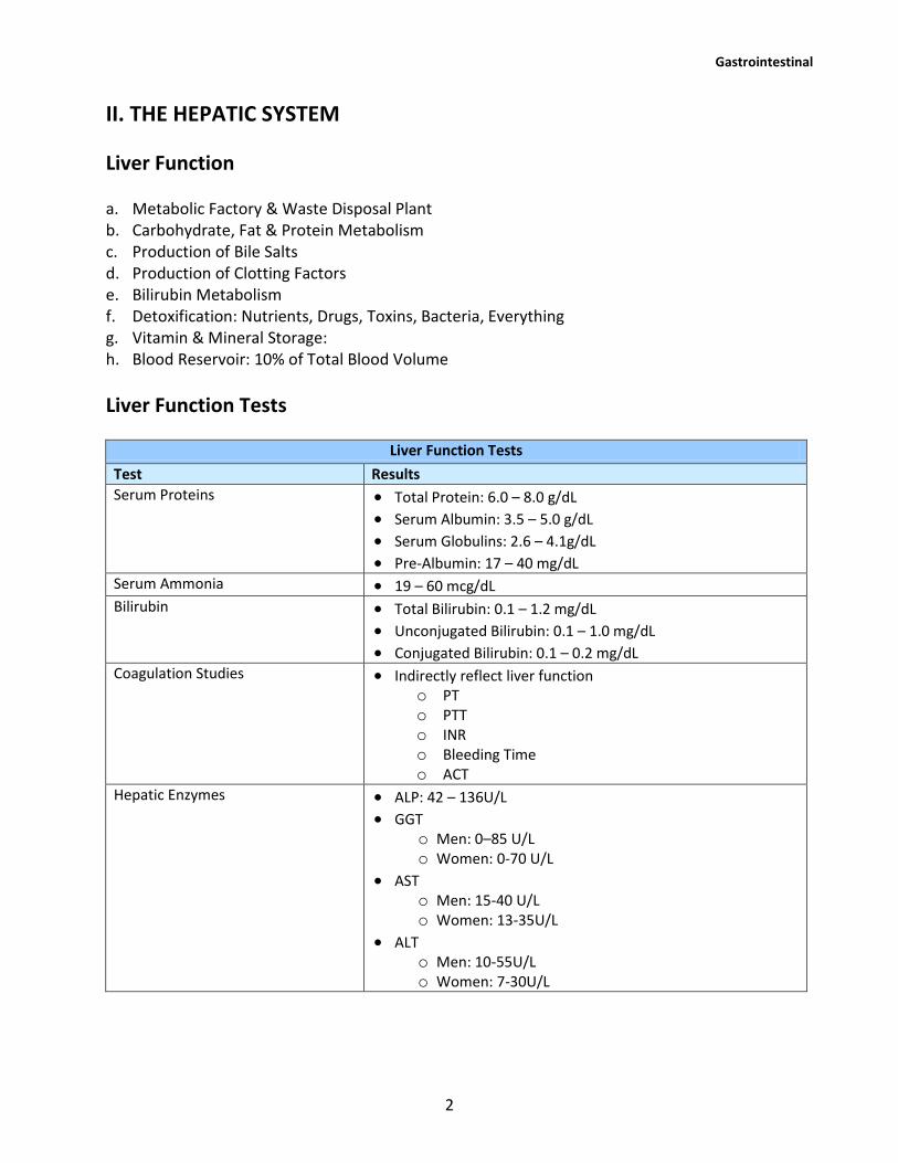
Liver Function Tests
Test Results Serum Proteins Total Protein: 6.0 – 8.0 g/dL
Serum Albumin: 3.5 – 5.0 g/dL Serum Globulins: 2.6 – 4.1g/dL Pre-Albumin: 17 – 40 mg/dL
Serum Ammonia 19 – 60 mcg/dL Bilirubin Total Bilirubin: 0.1 – 1.2 mg/dL
Unconjugated Bilirubin: 0.1 – 1.0 mg/dL Conjugated Bilirubin: 0.1 – 0.2 mg/dL
Coagulation Studies Indirectly reflect liver function o PT o PTT o INR o Bleeding Time o ACT
Hepatic Enzymes ALP: 42 – 136U/L GGT
o Men: 0–85 U/L o Women: 0-70 U/L
AST o Men: 15-40 U/L o Women: 13-35U/L
ALT o Men: 10-55U/L o Women: 7-30U/L

Gastrointestinal
3
Liver Dysfunction & Failure
Pathophysiology a. Liver Tissue (cells) are Destroyed and Replaced with Fibrotic Tissue b. Functions are Altered c. Organ Changes Shape d. Vascular Flow is Obstructed e. Portal Hypertension
Cirrhosis A chronic progressive liver disease where diffuse fibrotic bands of connective tissue, distort the liver’s normal architecture and functional ability. The liver loses its ability to regulate fluids, metabolize waste, regulate coagulation and nutrition. a. Causes
Alcoholic, Laennec's Portal, or Fatty
Post Necrotic: Toxic, Nodular, or Post Hepatic
Biliary: Cholangitic or Obstructive
Hepatitis Widespread Inflammation of Liver Cells a. Causes
Primary Viral – Most Common
Hepatotoxins - Toxic or Drugs
Secondary Viral, Low Mortality
Clinical Presentation of Liver Dysfunction Hepatic Encephalopathy The liver is unable to perform its detoxification function and toxins build up. Primarily ammonia causing altered LOC, behavior and motor abilities. a. Clinical Presentation
Confusion Coma
Agitation Unsafe Behavior
Asterixis: Flap like Tremor of Hands
Apraxia: Inability to Perform Purposeful Acts
Elevated Ammonia b. Common Treatment Modalities
Limit Protein Intake
Limit Hepatotoxic Drugs
Lactulose & Neomycin
Safe Environment

Gastrointestinal
4
Malnutrition The liver is unable to perform its function of carbohydrate, protein and fat metabolism. This leads to malnutrition a. Clinical Presentation b. Common Treatment Modalities
Need to tx the Cause of Liver Failure
Parenteral Nutrition
Limit Protein Intake
Restrict Fluids Coagulopathy The liver is unable to synthesize fibrinogen, prothrombin and factors V, VII, IX, X, XI, XIII, fibrinolytic factors and Vit. K. These are needed to maintain the ability to clot. Platelet aggregation and adhesion are also effected by liver dysfunction. a. Clinical Presentation
Bleeding Tendencies
Nonspecific Bleeding b. Common Treatment Modalities
Monitor Coagulation Studies & Platelet Ct
Decrease Bleeding and Bruising Risk
Administer Blood Products Portal Hypertension Increased pressure in the portal vein occurs secondary to flow obstruction from inflammation, bands, or fibrotic hepatic tissue. This retrograde pressure leads to formation of varices in the esophagus, stomach and rectal vault. a. Clinical Presentation
Caput Medusae: dilated cutaneous veins radiating from the umbilical
Spider angiomas commonly seen in Cirrhosis
Upper GI Bleeding b. Common Treatment Modalities
Surgical Shunting
TIPSS - Transjugular Intrahepatic Portosytemic Stent Shunt
Treat Bleeding
Treat Cause Hepatorenal Syndrome A form of pre-renal failure caused by the liver dysfunction. Mortality of liver failure is very high once renal failure develops. a. Clinical Presentation
S&S of Renal Dysfunction

Gastrointestinal
5
b. Common Treatment Modalities
Maintain Adequate Renal Perfusion
Restrict Fluids
Restrict Nephrotoxic Agents
Continuous Renal Replacement Therapies
Ascites Fluid accumulation in the peritoneal space secondary to decreased production of albumin, decreased systemic oncotic pressure, increased hepatic lymph production and increased capillary permeability. The fluid accumulation impacts the respiratory (diaphragm) and cardiac (hemodynamic) systems primarily as well as comfort and body image. a. Clinical Presentation
Inc. Abdominal Girth
Hypotension and Tachycardia
Dyspnea, Orthopnea, Tachypnea
S&S of Dehydration
N&V b. Common Treatment Modalities
Restrict PO Fluid
Diuretics (if tolerated hemodynamically)
Restrict Na
Respiratory Support
Paracentesis
Peritoneovenous Shunt Surgery
Infection One of the functions of the liver cells (Kuppfer cells) is to clean the blood of bacteria. With liver failure this function is not provided and bacteria builds up (primarily gram negative bugs) in the systemic circulation increasing the risk of infection. a. Clinical Presentation
Poor Wound Healing
Increased Risk of Infection b. Common Treatment Modalities
Heightened Prevention Measures
Abx Therapy – w Caution

Gastrointestinal
6
III. THE PANCREAS
Function a. Endocrine Functions
Synthesis & Release of Hormones: o Glycogen o Insulin o Gastrin
b. Exocrine Functions
Pancreatic Enzymes Break Down Protein, Starch & Fat. > 2L/day
Bicarbonate Raise pH c. PNS, Gastrin & Hormones Regulate Secretions
Pancreatic Enzymes a. Trypsin: Aids in Protein Digestion b. Amylase: Aids in Carbohydrate Digestion c. Lipase: Aids in Fat Digestion
Acute Pancreatitis
Pathophysiology a. Auto Digestion
Tissue Damage
Fat Necrosis
Vascular Damage & Hemorrhage
Increased Capillary Permeability
Hypotension b. Forms/Types
Edematous
Hemorrhagic c. Classifications
Acute Pancreatitis
Recurrent Acute
Recurrent Chronic
Chronic Pancreatitis
Cause (blocked enzyme release) a. Alcoholism b. Biliary Stones c. Hyperlipidemia d. Abd Trauma e. Infection (bacterial or viral)

Gastrointestinal
7
f. Shock g. Drugs (Most Common: Cyclosporine, Acetaminophen, Cimetidine, Steroids, Salicylates,
Furosemide, Thiazides, Estrogens)
Clinical Presentation a. Pain b. Low Grade Fever c. N&V d. Distended/Tender/Rigid Abd e. Guarding with Rebound Tenderness f. Jaundice g. Hypoactive Bowel Sounds h. Steatorrhea: bulky, pale, foul-smelling stools i. ? Ascites j. Hypovolemic Shock k. In Necrotizing Pancreatitis
Cullen’s Sign: o Bluish Discoloration Umbilical
Grey Turner’s Sign: o Bluish Discoloration Flanks
Labs Underlined labs are the MOST diagnostic a. Hypocalcemia (classic sign) b. Low Ca, Mg, K c. Hyperglycemia d. Hyperbilirubinemia e. Hypertriglyceridemia f. Increased BUN & Creatinine g. Elevated Amylase h. Elevated Lipase i. Elevated LFTs j. Elevated WBC k. Decreased H/H l. ? Increased H/H
Treatment Options a. Fluid Resuscitation b. Rest the Pancreas: NPO, NGT c. Pain Management d. Monitor & Replace Electrolytes e. Tx Multisystem f. Nutritional Support g. Surgery

Gastrointestinal
8
IV. ACUTE GI HEMORRHAGE
Lower GI Bleeding Not Typically Life Threatening a. Causes
Diverticulitis
Angiodysplasia
Cancer
Hemorrhoids
Inflammatory Bowel Disease (Ulcerative Colitis; Crohn's Disease)
Bowel Infarction
Upper GI Bleeding a. Causes
Peptic Ulcer Disease: Duodenal, Gastric and Stomal ulcers account for 50% bleeding episodes
Gastritis or Esophagitis
Esophageal Varices
Mallory -Weiss Syndrome b. Clinical Presentation
Hematemesis
Melena
PUD
Distended & Tender Abd
Hyperactive Bowel Sounds
Hypovolemia
Shock c. Assessment
H & H
Coags & Platelets
Hemoconcentration
Elevated BUN
LFTs
Endoscopy
Angiography
Raionuclide Scans d. Treatment
NG Decompression/Lavage – Room Temp vs Iced
Fluid Resuscitation
Blood Product Admin
Endoscopic Sclerotherapy

Gastrointestinal
9
Pharmacology
Surgery o Vagotomy and Pyloroplasty o Oversew Ulcer or Tear o Total and Subtotal Gastric Resection o Billroth I: Vagotomy, Antrectomy, AnastomosisStomach and Duodenum o Billroth II: Vagotomy, Antrectomy, Anastomosis Stomach and Jejunum o Whipple: Removal of the Distal 3rd of Stomach, Entire Duodenum, Head of
Pancreas, Gastrojejunotomy o Colon Resection
Bleeding Esophageal Varices o TIPSS: Transjugular Intrahepatic Portosytemic Stent Shunt o Beta Blocker – Decreases Pressure o Portal Caval Shunt
V. DISORDERS OF THE BOWEL
Bowel Infarction a. Etiology
Embolic or Thrombotic Occlusion
Typically from the Superior Mesenteric Artery b. Clinical Presentation
Severe Epigastric Pain
Rebound Tenderness
Guarding & Rigidity
Stimulated Sympathetic Response from Pain c. Treatment Options
Angiography to Identify/Confirm Occlusion
Surgery to Remove Occlusion & Dead Bowel
Bowel Obstruction a. Etiology
Internal Lumen Obstruction ex. Tumor
External Lumen Obstruction ex. Adhesions
Emboli: no blood flow
Paralytic Ileus

Gastrointestinal
10
b. Clinical Presentation
Complete vs Partial
Distended Edematous Bowel
Fluid and Electrolytes Leaking from Bowel
Elevated WBC
Fever
Small Intestine o Acute Pain w Sudden Onset o N & V (movement on both ends) o Wave-Like Hyperactive High Pitched Bowel Sounds o May Have Some Gas or Feces o Distention (mild)
Large Intestine o Slow Onset Pain Progression Mild Severe, Lower Abd o No N & V (nothing moving) o No Stool o Low Pitched Bowel Sounds o Distention (large amount)
Treatment Options o Diagnosis Obstruction by Hx, X-Ray, CT, Upper or Lower Barium Radiology
Tests o Pain Management o IV Fluids o Decompress w NG, Rectal or Intestinal Tube o Abx o NPO and Time (rest the bowel) o Surgery
Perforation/Peritonitis a. Etiology
Gastric/Intestinal Contents Leak into Peritoneal Cavity
Ulcer Perforation
Diverticular Rupture
Trauma
Bowel Infarction b. Clinical Presentation
Infection/Sepsis (all the S&S)
Sudden Onset of Severe Pain
Rigid Abdomen w Rebound Tenderness
Hypoactive Bowel Sounds No Bowel Sounds

Gastrointestinal
11
c. Treatment Options
Surgery to Repair Cause & Clean Up
ABX
Fluids
Tx of Sepsis
Tx of MODS
Functional GI Disorders a. Ileus b. Diabetic Gastroparesis c. Gastroesophageal Reflux d. Irritable Bowel Syndrome


