Embed Size (px)
Citation preview
A Sponsored Supplement to Science
Sponsored by Produced by the
Science/AAAS Custom Publishing Office
Translational Medicine at Capital MedicalUniversityInvestigatingMajor ChronicDiseases

Join AAAS. Get instant access to Science. Support all of the sciences.
When you subscribe to Science, you become part of the American Association for the Advancement of Science (AAAS), a nonprofit community of more than 120,000 members worldwide who believe in the power of science to make the world a better place. AAAS is hard at work promoting science in government, schools, and in the public commons around the globe.
AAAS’s award-winning journal Science offers the top peer-reviewed research across multiple disciplines.With your subscription, you'll get:
§ 51 weeks of home delivery of Science
§ Instant online retrieval of every Science article ever published, dating back to 1880
§ Full access to the Science mobile site and apps
§ Career advice, webinars, blogs and fascinating features exclusively for AAAS members
§ Members-only newsletters, and much more
With increasing public skepticism about science–and public funding for research more uncertain than ever–our work has never been more important. Join hands with us today!
Visit promo.aaas.org/joinaaas. Together we can make a difference.
Translational Medicine at Capital Medical UniversityInvestigatingMajor ChronicDiseases
Materials that appear in this supplement were not reviewed or assessed by the Science Editorial staff. Articles can be cited using the following format: [AUTHOR NAME(S)] [CHAPTER TITLE] in Translational Medicine at Capital Medical University: Investigating Major Chronic Diseases (Science/AAAS, Washington, DC, 2015), p. [xx-xx].
Bill Moran, Global Director Custom [email protected]+1-202-326-6438
Ruolei Wu, Associate Director, Asia Custom [email protected]+86-186-0082-9345
© 2015 by The American Association for the Advancement of Science. All rights reserved. 17 July 2015
Editors: Sean Sanders, Ph.D. and Tianna Hicklin, Ph.D.Proofreader/Copyeditor: Yuse Lajiminmuhip Designer: Amy Hardcastle
About the cover: Capital Medical University’s Basic Research Building
This supplement was produced by the Science/AAAS Custom Publishing Office and sponsored by Capital Medical University.
Prefaces 3 Domestic research with global implications Sean Sanders, Ph.D. Science/AAAS
4 Translational Medicine at China Capital Medical University
Prof. Xiaomin Wang, M.D., Ph.D. Senior Vice President, CCMU
Introduction 5 China Capital Medical University—A prestigious
name in China and around the world Prof. Qunyuan Xu, M.D., Ph.D. Past President, CCMU
Translational research in neuroscience 7 Investigating the epidemiology, neuropsychology,
and genetics of dementia and mild cognitive impairment in China
Jianping Jia, Aihong Zhou, Fen Wang et al.
10 Advances in diagnostics, treatment, and prevention of ischemic cerebrovascular disease
Yilong Wang and Yongjun Wang
12 Longitudinal resting-state functional MRI studies of amnestic mild cognitive impairment and Alzheimer’s disease
Yuxia Li, Can Sheng, Yu Sun et al.
16 Discovery of novel glioma biomarkers using the Chinese Glioma Genome Atlas database
Zhaoshi Bao and Tao Jiang
Translational research in internal medicine 20 The role of inflammation in hypertensive cardiac injury Yulin Li, Congcong Zhang, Jie Du
25 Pathogenesis, epidemiology, clinical characteristics, and therapy of the 2009 pH1N1 and H7N9 influenza virus outbreaks
Pengjun Zhang, Bin Cao, Chen Wang
28 Chronic obstructive pulmonary disease in China Zengyan Li, Miao Miao, Hongyan Hou et al.
32 Nonsteroidal anti-inflammatory drugs in the prevention of esophageal squamous cell carcinoma
Rui Cheng and Shutian Zhang
34 Integrating traditional Chinese medicine and Western medicine to stimulate regression of liver fibrosis
Aiting Yang, Baoen Wang, Xiaojuan Ou et al.
36 Nitrate and endothelium-protection in uremia Han Li, Sujuan Feng, Shixiang Wang
continued>>
TABLE OF CONTENTS 1
2 TRANSLATIONAL MEDICINE AT CAPITAL MEDICAL UNIVERSITY: INVESTIGATING MAJOR CHRONIC DISEASES
Translational research in opthalmology and otolaryngology 39 Glaucoma: A neurodegenerative disease affecting the entire
visual pathway Shaodan Zhang, Huaizhou Wang, Guoping Qing et al.
42 The role of regulatory immune cells in prolonging corneal graft survival
Yan He and Zhiqiang Pan
45 The impact of topical drugs on nasal mucociliary function Jian Jiao, Demin Han, Luo Zhang
48 The epigenetics of allergic rhinitis Yuan Zhang, Xu Zhang, Demin Han et al.
Translational research in surgery and rehabilitation medicine 52 miRNAs associated with metastasis in colorectal cancer Jie Yin, Jun Cai, Zhigang Bai et al.
56 Lateral lumbar interbody fusion: A novel, minimally invasive treatment for diseases of the spine
Xiang Li, Shudong Jiang, Yi Hong
58 The management of neurogenic bladder Guoqing Chen, Fan Zhang, Limin Liao
62 Genotypic prediction of the intercellular adhesion molecule 1 K469E (A/G) polymorphism in diabetes and diabetic microvascular complications
Jun Ma, Zhanjie Ren, Tianwei Gu et al.
64 Experience-induced neuroplasticity following spinal cord injury
Feng Gao, Chetwyn C. H. Chan, Mingliang Yang et al.
Translational research in integrated traditional Chinese and Western medicine 68 Individualized traditional Chinese medicine treatment
for acute stroke Lei Chen, Juexian Song, Yumin Luo et al.
Translational research in preventive medicine and clinical diagnosis 72 Diagnostic imaging of neurological disease in HIV/AIDS Yunfang Li, Hongjun Li, Jiaojiao Liu et al.
76 Urinary proteomics in biomarker discovery Man Zhang and Qian Meng
79 Use of the CRISPR/CAS system to develop bacteriophage resistance to Pseudomonas aeruginosa
Fangfang Dai, Yanhua Yu, Chen Ming et al.
Table of contentscontinued
PREFACES 3
T ranslational medicine might have many different interpretations depending on where you stand, but one thing is certain: it is a phrase that is now front and center in biomedical research around the globe. Touching many different fields of research, the intention
of translational medicine is that basic research is “translated” rapidly into clinical applications that help patients by improving diagnosis, treatment, and follow up. The endeavor is multidisciplinary, intentionally breaking down walls between previously siloed research areas to bring scientists together in a more collaborative setting. Many institutions are incorporating this new way of thinking, moving away from research as an endeavor unto itself and toward a more application-based discovery model.
China Capital Medical University (CCMU) in Beijing, has clearly taken translational medicine to heart. Founded in the 1960’s as the Beijing Sec-ond Medical College, CCMU has expanded to become a premier research institute in China, attracting international scientists and building a global reputation for its excellent science and quality clinical practice.
This supplement to Science presents an overview of just some of the translational research being undertaken at CCMU. It covers a range of re-search areas, namely neuroscience, internal medicine, ophthalmology and otolaryngology, surgery and rehabilitation medicine, traditional Chinese medicine, and preventative medicine and clinical diagnosis. In the accom-panying articles, the authors provide reviews of their subject areas, placing their own research into context. Although this work often focuses on transla-tion medicine in China, and the associated medical and scientific challenges, it is most certainly more broadly applicable to similar issues encountered worldwide as evidenced by the global nature of the diseases discussed, in-cluding Alzheimer’s disease, HIV/AIDS, and diabetes.
No doubt CCMU is looking forward to continuing to grow as a research and medical institute, and we look forward to seeing further top quality research emanating from it in the future.
Sean Sanders, Ph.D.Editor, Custom PublishingScience/AAAS
Domestic research with global implications
4 TRANSLATIONAL MEDICINE AT CAPITAL MEDICAL UNIVERSITY: INVESTIGATING MAJOR CHRONIC DISEASES
W e live in an exciting time of translational medicine, built upon two major scientific revolutions. The deciphering of the hu-man genome at the turn of the century ushered in a new era of medical research, and advancements in the biological sciences
began to transform a broad range of medical practices, from diagnosis, therapy, and prevention to public health. The dawn of translational medicine is rapidly producing health care innovations and changing the pattern of clinical medicine around the world.
As the world’s second largest economy and one still emerging from major reforms that started in the 1980s, China faces unique challenges and op-portunities in this dynamic environment. To serve the needs of its 1.4 billion citizens, China must quickly implement a modern health care infrastructure and significantly boost top-quality medical research and education. Medical institutes across the country should focus on translational medicine and em-phasize the importance of research directly related to human health. These are the guiding principles for China Capital Medical University (CCMU), a key medical school in the country’s capital, Beijing. It must continue to improve its medical research and education in order to serve the health needs of people from all over China.
CCMU is well positioned to lead the charge in translational medicine, having assembled the largest health care resource within a single institute in China (and very likely in the world), with more than 20,000 beds across 20 affiliated hospitals plus 11 teaching hospitals. It is noteworthy that the majority of these hospitals have received the top rating for hospitals in China (AAA-equivalent) and many are considered to be at the top of their most renowned specialty. In the past 5 years, CCMU has been awarded 1,723 research projects; it has also received more than CN¥1.2 billion (US$190 mil-lion) of research funding over the last three years. Putting these clinical and funding resources together, CCMU has won 53 national and provincial prizes for excellent research in the past three years and entered an explosive phase of growth in both research and education.
Many examples of the prize-winning research at CCMU are featured in this supplement. However, because of space constraints, many equally excit-ing projects and achievements from various frontiers of translational medi-cine could not be included. I sincerely invite readers to get in touch with our International Collaboration Department ([email protected]) to learn more about the research, collaboration, and career opportunities at CCMU.
Finally, I would like to thank Science/AAAS for the professional support throughout the development of this supplement. I also want to thank my colleagues Prof. Qunyuan Xu, Prof. Zhe Dong, Dr. Junmin Zhang, Dr. Jianjun Zhang, and Ms. Ying Zhuang for their great efforts in putting together the wide range of research reports. I hope you enjoy reading about our vision and research in translational medicine in these pages, and look forward to your comments as well as fruitful collaborations in the future.
Prof. Xiaomin Wang, M.D., Ph.D.Senior Vice President, CCMU
Translational Medicine at China Capital Medical University

China Capital Medical University (CCMU) was founded in 1960 as Beijing Second Medical School by Dr. Jieping Wu, a renowned urologist, scientist, and educator. Located in
the Fengtai District of China’s capital, Beijing, CCMU is a multidisciplinary university, offering the highest level of medical education in China. With a unique approach combining ancient wisdom with state-of-the-art technology, CCMU strives to meet and exceed the international standards for academic excellence. CCMU is one of the top-10 accredited medical schools in China, and receives stable financial support from the Beijing municipality and the central government.
Mission statementThe mission statement of CCMU is three-pronged:
education, research and health care. For faculty members and students, there are four guiding principles—serving patients, contributing to the community, respecting ethics, and striving for expertise—that help them achieve these missions through daily activities.
Students and facultyAs of 2014, there are 10,604 full-time students
studying at CCMU, including 3,132 postgraduates and 3,368 undergraduates. There are also 576 international students from 67 countries around the world. A strong faculty, comprising 1,875 professors, 3,275 associate professors, and more than 28,000 clinical physicians and surgeons serves the missions of CCMU.
The CCMU faculty is a highly selective group with a strong emphasis on intellectual development. It currently includes six academicians of the Chinese
China Capital Medical University— A prestigious name in China and around the world
INTRODUCTION 5
Academy of Sciences and the Chinese Academy of Engineering. In addition, many faculty members hold national and international distinctions. Together, they help evolve medical research and education at CCMU.
EducationCCMU offers the full spectrum of health care-
related training programs in 10 schools and 20 affiliated hospitals. Through rigorous courses, these comprehensive training programs prepare CCMU students to become health care professionals in 16 specialized tracks, such as basic medical sciences, clinical medicine, stomatology, pediatrics, preventive medicine, biomedical engineering, traditional Chinese medicine, pharmacy, nursing, and health service management. Students can choose to pursue any of the 59 accredited doctoral programs, 78 Master’s degree programs, 16 Bachelor’s degree programs, and 8 certificate programs offered by CCMU.
There are several distinctions at CCMU compared with other medical universities in China. It is the only university with a “talent cultivation program for clinical medicine” sponsored by the Ministry of Education. The goal of the program is to accelerate the development of translational medicine scientists. The World Health Or-ganization lists CCMU in its World Directory of Medical Schools; as a result, CCMU receives about 100 interna-tional applications for its 6-year Bachelor of Medicine/Bachelor of Surgery degree program every year. The medical degree granted by CCMU is recognized by the Educational Commission for Foreign Medical Gradu-ates, and CCMU graduates are therefore eligible for the United States Medical Licensing Examination (USMLE). Every year, more than 15 CCMU graduates take USMLE and the majority of them pass the Step 1 Exam.
ResearchCCMU has long been recognized in China for
robust medical research. Ten clinical departments have received research funding directly from the central government, highlighting their achievement at the national level. Currently, CCMU operates five distinguished National Clinical Disease Centers for cardiovascular disease, neurological disease, respiratory disease, digestive system disease, and mental illness, along with 41 Key Laboratories at the national and provincial level. continued>
6 TRANSLATIONAL MEDICINE AT CAPITAL MEDICAL UNIVERSITY: INVESTIGATING MAJOR CHRONIC DISEASES
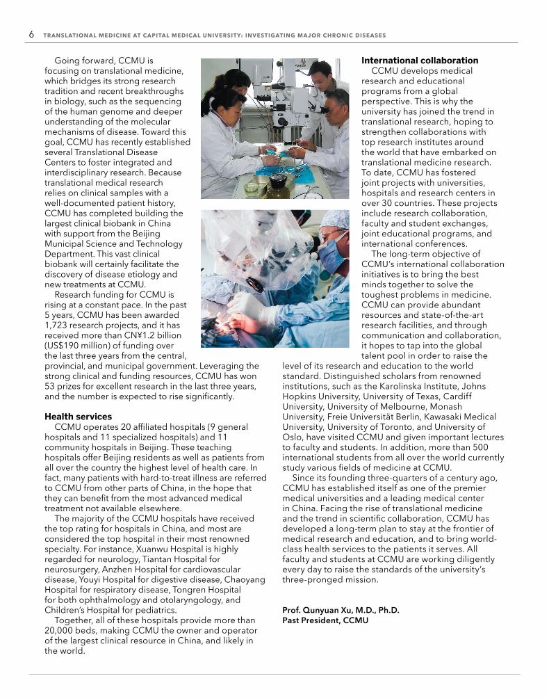
Going forward, CCMU is focusing on translational medicine, which bridges its strong research tradition and recent breakthroughs in biology, such as the sequencing of the human genome and deeper understanding of the molecular mechanisms of disease. Toward this goal, CCMU has recently established several Translational Disease Centers to foster integrated and interdisciplinary research. Because translational medical research relies on clinical samples with a well-documented patient history, CCMU has completed building the largest clinical biobank in China with support from the Beijing Municipal Science and Technology Department. This vast clinical biobank will certainly facilitate the discovery of disease etiology and new treatments at CCMU.
Research funding for CCMU is rising at a constant pace. In the past 5 years, CCMU has been awarded 1,723 research projects, and it has received more than CN¥1.2 billion (US$190 million) of funding over the last three years from the central, provincial, and municipal government. Leveraging the strong clinical and funding resources, CCMU has won 53 prizes for excellent research in the last three years, and the number is expected to rise significantly.
Health servicesCCMU operates 20 affiliated hospitals (9 general
hospitals and 11 specialized hospitals) and 11 community hospitals in Beijing. These teaching hospitals offer Beijing residents as well as patients from all over the country the highest level of health care. In fact, many patients with hard-to-treat illness are referred to CCMU from other parts of China, in the hope that they can benefit from the most advanced medical treatment not available elsewhere.
The majority of the CCMU hospitals have received the top rating for hospitals in China, and most are considered the top hospital in their most renowned specialty. For instance, Xuanwu Hospital is highly regarded for neurology, Tiantan Hospital for neurosurgery, Anzhen Hospital for cardiovascular disease, Youyi Hospital for digestive disease, Chaoyang Hospital for respiratory disease, Tongren Hospital for both ophthalmology and otolaryngology, and Children’s Hospital for pediatrics.
Together, all of these hospitals provide more than 20,000 beds, making CCMU the owner and operator of the largest clinical resource in China, and likely in the world.
International collaborationCCMU develops medical
research and educational programs from a global perspective. This is why the university has joined the trend in translational research, hoping to strengthen collaborations with top research institutes around the world that have embarked on translational medicine research. To date, CCMU has fostered joint projects with universities, hospitals and research centers in over 30 countries. These projects include research collaboration, faculty and student exchanges, joint educational programs, and international conferences.
The long-term objective of CCMU’s international collaboration initiatives is to bring the best minds together to solve the toughest problems in medicine. CCMU can provide abundant resources and state-of-the-art research facilities, and through communication and collaboration, it hopes to tap into the global talent pool in order to raise the
level of its research and education to the world standard. Distinguished scholars from renowned institutions, such as the Karolinska Institute, Johns Hopkins University, University of Texas, Cardiff University, University of Melbourne, Monash University, Freie Universität Berlin, Kawasaki Medical University, University of Toronto, and University of Oslo, have visited CCMU and given important lectures to faculty and students. In addition, more than 500 international students from all over the world currently study various fields of medicine at CCMU.
Since its founding three-quarters of a century ago, CCMU has established itself as one of the premier medical universities and a leading medical center in China. Facing the rise of translational medicine and the trend in scientific collaboration, CCMU has developed a long-term plan to stay at the frontier of medical research and education, and to bring world-class health services to the patients it serves. All faculty and students at CCMU are working diligently every day to raise the standards of the university’s three-pronged mission.
Prof. Qunyuan Xu, M.D., Ph.D.Past President, CCMU

TRANSLATIONAL RESEARCH IN NEUROSCIENCE 7
Investigating the epidemiology, neuropsychology, and genetics of dementia and mild cognitive impairment in ChinaJianping Jia*, Aihong Zhou, Fen Wang, Wei Qin, Yi Tang, Cuibai Wei, Xiumei Zuo, Dan Li, Haiqing Song, Liyong Wu, Lu Lu, Junhua Liang, and Lina Zhao
Dementia and mild cognitive impairment (MCI) are prevalent in China (1, 2). We have systematically investigated the epidemiology of MCI and dementia in
the Chinese elderly, including carrying out cognitive assessment for MCI and studying the genes involved in the pathogenesis of familial Alzheimer’s disease (FAD) and sporadic Alzheimer’s disease (sAD).
Prevalence of MCI and dementia in the Chinese elderly
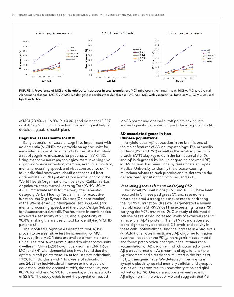
Until recently, very few epidemiologic studies on MCI and dementia had been carried out in China. We conducted a population-based survey using multistage cluster sampling to estimate the prevalence of MCI (1) and dementia (2) in the elderly population around five geographical regions cross China. A total of 10,276 community residents aged 65 years and older were included (6,096 urban and 4,180 rural). In addition to MCI, four additional subtypes were categorized: MCI caused by prodromal AD (MCI-A), MCI resulting from cerebrovascular disease (MCI-CVD), MCI with vascular risk factors (MCI-VRF), and MCI caused by other diseases (MCI-O). Dementia was categorized as either AD, vascular dementia (VaD), or other types of dementia (ODs). It was found that the prevalences of overall MCI, MCI-A, MCI-CVD, MCI-VRF, and MCI-O were 20.8% (95% CI = 20.0–21.6), 6.1% (95% CI = 5.7–6.6), 3.8% (95% CI = 3.4–4.2), 4.9% (95% CI = 4.5–5.4), and 5.9% (95% CI = 5.5–6.4), respectively (Figure 1) (1). The prevalence of dementia, AD, and VaD were 5.14% (95% CI = 4.71–5.57), 3.21% (95% CI = 2.87–3.55), and 1.50% (95% CI = 1.26–1.74), respectively (Figure 2) (2). Compared with the urban population, the rural subjects had a higher prevalence
Translational research in neuroscience
Department of Neurology, Xuan Wu Hospital, Capital Medical University, Beijing, ChinaCenter of Alzheimer’s Disease, Beijing Institute for Brain Disorders, Beijing, ChinaBeijing Key Laboratory of Geriatric Cognitive Disorders and Neurodegenerative, Laboratory of Ministry of Education of the People’s Republic of China, Beijing, China.*Corresponding Author: [email protected]
8 TRANSLATIONAL MEDICINE AT CAPITAL MEDICAL UNIVERSITY: INVESTIGATING MAJOR CHRONIC DISEASES
of MCI (23.4% vs. 16.8%, P < 0.001) and dementia (6.05% vs. 4.40%, P < 0.001). These findings are of great help in developing public health plans.
Cognitive assessments for MCIEarly detection of vascular cognitive impairment with
no dementia (V-CIND) may provide an opportunity for early intervention. A recent study looked at establishing a set of cognitive measures for patients with V-CIND. Using extensive neuropsychological tests involving five cogitive domains (attention, memory, executive function, mental processing speed, and visuoconstructive skill), four individual tests were identified that could best differentiate V-CIND patients from normal controls: the World Health Organization-University of California-Los Angeles Auditory Verbal Learning Test (WHO-UCLA AVLT) immediate recall for memory; the Semantic Category Verbal Fluency Test (animal) for executive function; the Digit Symbol Subtest (Chinese version) of the Wechsler Adult Intelligence Test (WAIS-RC) for mental processing speed; and the Block Design Subtest for visuoconstructive skill. The four tests in combination achieved a sensitivity of 92.5% and a specificity of 98.8%, making them a useful tool for identifying V-CIND patients (3).
The Montreal Cognitive Assessment (MoCA) has proven to be a sensitive test for screening for MCI. However, little MoCA data are available from mainland China. The MoCA was administered to older community dwellers in China [6,283 cognitively normal (CN), 1,687 MCI, and 441 with dementia] and it was found that the optimal cutoff points were 13/14 for illiterate individuals, 19/20 for individuals with 1 to 6 years of education, and 24/25 for individuals with seven or more years of education. With the optimal cutoffs, the sensitivity was 80.5% for MCI and 96.9% for dementia, with a specificity of 82.5%. The study established the population-based
MoCA norms and optimal cutoff points, taking into account specific variables unique to local populations (4).
AD-associated genes in Han Chinese populations
Amyloid beta (Aβ) deposition in the brain is one of the major features of AD neuropathology. The presenilin proteins (PS1 and PS2) as well as the amyloid precursor protein (APP) play key roles in the formation of Aβ (5), and Aβ is degraded by insulin degrading enzyme (IDE) (6). Much work has been done by researchers at Capital Medical University to identify the disease-causing mutations related to such proteins and to determine the genetic predisposition for both FAD and sAD.
Uncovering genetic elements underlying FADTwo novel PS1 mutations (V97L and A136G) have been
reported in Chinese pedigrees (7), and researchers have since bred a transgenic mouse model harboring the PS1 V97L mutation (8) as well as generated a human neuroblastoma SH-SY5Y cell line expressing human PS1 carrying the V97L mutation (9). Our study of this model cell line has revealed increased levels of extracellular and intracellular Aβ42 protein. The PS1 V97L mutation also led to significantly decreased IDE levels and activity in these cells, potentially causing the increase in Aβ42 levels (9). Additionally, we investigated Aβ oligomer formation over the lifespan of the PS1V97L transgenic mouse model and found pathological changes in the intraneuronal accumulation of Aβ oligomers, which occurred without Aβ plaque formation. At 6 months of age, for example, Aβ oligomers had already accumulated in the brains of PS1V97L transgenic mice. We detected impairments in synaptic plasticity, spatial memory deficits, and synaptic loss as well as abnormal tau phosphorylation and glial activation (8, 10). Our data supports an early role for Aβ oligomers in the onset of AD and suggests that Aβ
FIGURE 1. Prevalence of MCI and its etiological subtypes in total population. MCI, mild cognitive impairment; MCI-A, MCI prodromal Alzheimer’s disease; MCI-CVD, MCI resulting from cerebrovascular disease; MCI-VRF, MCI with vascular risk factors; MCI-O, MCI caused by other factors.

TRANSLATIONAL RESEARCH IN NEUROSCIENCE 9
plaques may not be the only prerequisite. This model also provides new insights into AD pathogenesis.
Genetic polymorphisms and sAD A systematic screening of more than 50 candidate
genes that have been implicated in multiple AD pathogeneses has been carried out. The target sequences included the promoters, coding and noncoding regions, and regulatory elements of genes associated with amyloid metabolism, tau hyperphosphorylation, inflammation, vascular factors, and oxidative stress. This work uncovered over 20 novel polymorphisms associated with sAD in the Chinese population (11, 12). It was also discovered that variations in the promoter region of the amyloid precursor protein (13), disintegrin and metalloproteinase 9 (14), IDE (15), microtubule-associated protein tau (16), and anterior pharynx defective 1 (17) genes may regulate protein expression by modifying transcriptional activity and thereby impact Aβ and tau metabolism.
We are continuing to seek new pathogenic genes underlying FAD and dementia in the Chinese population, with the hope of finding treatments using traditional Chinese medicine. We expect that, as a result of these studies and future research, the diagnosis and treatment of dementia and MCI in China will be significantly improved.
References 1. J. Jia et al., Alzheimers Dement. 10, 439 (2014). 2. J. Jia et al., Alzheimers Dement. 10, 1 (2014). 3. A. Zhou, J. Jia, Int. J. Geriatr. Psych. 24, 1352 (2009). 4. J. Lu et al., J. Geriatr. Psych. Neur. 24, 184 (2011). 5. D. J. Selkoe, Physiol. Rev. 81, 741 (2001). 6. W. Qiu, M. F. Folstein, Neurobiol. Aging 27, 190 (2006). 7. J. Jia, E. Xu, Y. Shao, Y. Sun, D. Li, J. Alzheimers Dis. 7, 119; discussion 173–180 (2005). 8. Y. Wang, Z. Cheng, W. Qin, J. Jia, J. Neurochem. 121, 135 (2012). 9. W. Qin, J. Jia, Eur. J. Neurosci. 27, 2425 (2008).10. Y. Zhang et al., PLOS ONE 9, e85885 (2014).
11. S. Wang, J. Jia, Am. J. Med. Genet. B Neuropsychiatr. Genet. 153B, 159 (2010).12. M. Wang, H. Song, J. Jia, Brain Res. 1327, 1 (2010).13. H. Lv et al., Neurobiol. Aging 29, 194 (2008).14. L. Cong, J. Jia, Neurobiol. Aging 32, 54 (2011).15. X. Zuo, J. Jia, Brain Res. 1249, 1 (2009).16. W. Sun, J. Jia, Neurosci. Lett. 450, 340 (2009).17. W. Qin et al., Aging Cell 10, 711 (2011).
AcknowledgmentsThis work was supported by the National Key Technology R&D Program in the Eleventh Five-Year Plan Period (2006BAI02B01), the key project of the National Natural Science Foundation of China (30830045), the National High Technology Research and Development Program 863 (2006AA02A408), the key project of the Science and Technology Plan of the Beijing Municipal Education Commission (KZ201010025023), the key project of the Science and Technology Plan of the Beijing Municipal Education Commission (KZ200910025005), the CHINA-CANADA Joint Initiative on Alzheimer’s Disease and Related Disorders (81261120571), and the National Science and Technology Major Projects for “Major New Drug Innovation and Development” of the Twelfth Five-Year Plan Period (2011ZX09307-001-03).
FIGURE 2. Prevalence for AD and VaD. AD, Alzheimer's disease; VaD, vascular dementia.

10 TRANSLATIONAL MEDICINE AT CAPITAL MEDICAL UNIVERSITY: INVESTIGATING MAJOR CHRONIC DISEASES
year increased with the severity of ICAS. Specifically, the recurrence rate increased from 3.27% in patients without ICAS to 7.27% in subjects with complete occlusion of the intracranial artery (7). Our findings suggested that about eight out of every one hundred acute stroke patients with complete intracranial occlusion will develop another cerebrovascular disease episode within one year.
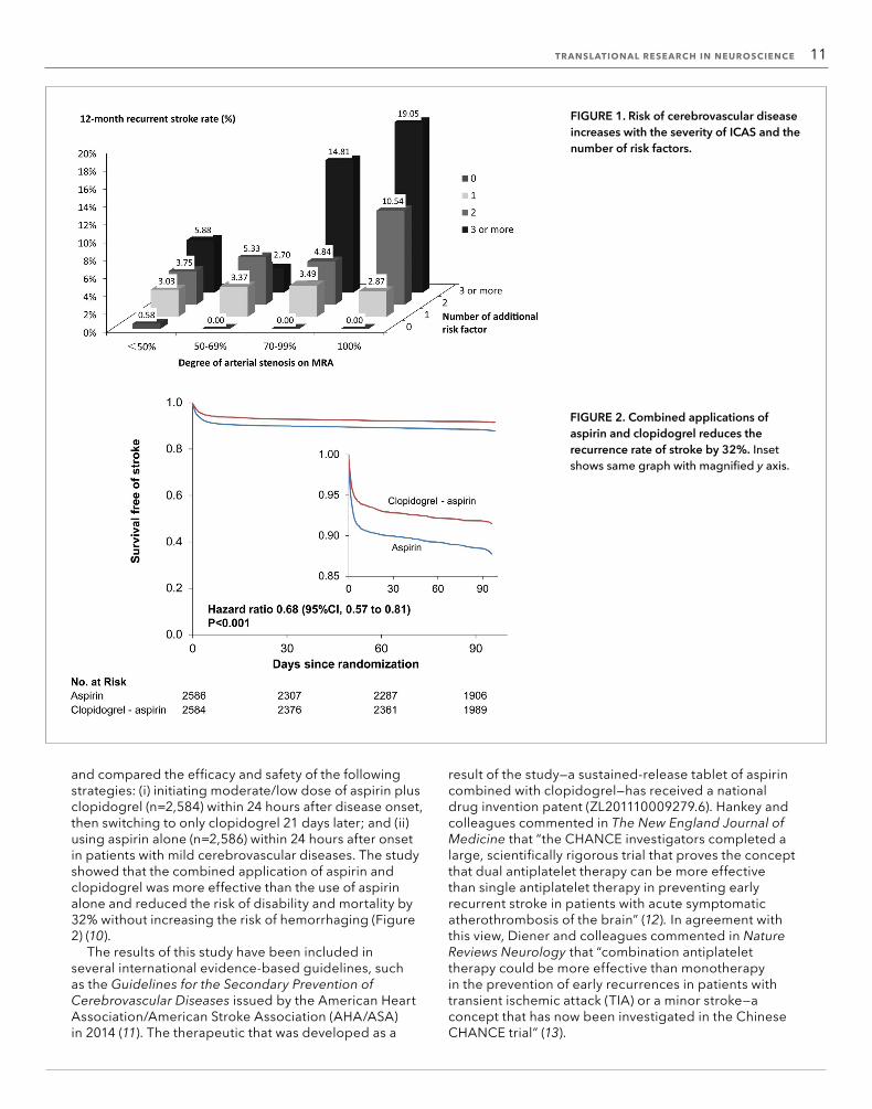
Our analysis of the disease etiology in the study’s subjects revealed that the risk of recurrence within one year increased in patients with more severe ICAS, age, family history of cerebrovascular diseases, previous history of cerebral infarction or heart disease, an incomplete Circle of Willis, and other serious neurological defects at stroke onset. The recurrence risk increased with the number of risk factors. For patients with intracranial artery occlusion and three or more risk factors, the recurrence rate reached 19.05% (Figure 1). Based on these data, we developed a stratified approach to evaluate the recurrence risk of ischemic stroke in China, which targeted inclusion of all ischemic stroke patients and recommended examination of intracranial blood vessels as a critical step for the evaluation of stroke recurrence risk. This strategy was approved by the National Health and Family Planning Commission of China and is described in more detail in the Quality Control of Diagnosis and Treatment of Ischemic Stroke guide, a health industry standard of the People’s Republic of China (WS/T398-2012).
Combination therapy for ischemic cerebrovascular diseases
A high early risk of recurrence is especially prevalent in patients with mild cerebrovascular diseases. Aspirin’s effectiveness for preventing recurrences is limited when used alone. Therefore, boosted-dual antiplatelet therapies have been proposed for prevention in several international clinical studies; however, a risk of hemorrhaging is considered unacceptable in evidence-based international guidelines for preventing the recurrence of cerebrovascular disease.
We set out to test whether treatment of mild cerebrovascular diseases with early, short-term, and moderate/low doses of aspirin, combined with clopidogrel (an antiplatelet drug, marketed as Plavix), would reduce the recurrence rate without increasing the risk of hemorrhage. We pooled and further mined the large amount of data described in previous studies and performed a preliminary study with a small sample size. Based on these data, for patients with mild ischemic cerebrovascular disease and with high early stage recurrence risk, but relatively low risk of hemorrhaging, we recommend early, short-term application of dual antiplatelet therapy, consisting of moderate/low doses of aspirin plus clopidogrel (9).
We also tested the effectiveness of clopidogrel in a multicenter, randomized, double-blind, parallel-group, placebo controlled clinical trial in China called CHANCE (Clopidogrel in High-risk patients with Acute Nondisabling Cerebrovascular Events). CHANCE included 5,170 patients at 114 medical sites
Advances in diagnostics, treatment, and prevention of ischemic cerebrovascular disease Yilong Wang and Yongjun Wang*
Cerebrovascular diseases have become the leading cause of death and disability in Chinese adults (1). More than 75% of these cases are ischemic cerebrovascular diseases, which are particularly challenging due to their high risk of recurrence. Aspirin is the only effective medicine recommended in international guidelines for the early prevention of ischemic cerebrovascular diseases (2, 3); however, the recurrence rate remains as high as 10%–20%, even with aspirin use at early stages. Dual antiplatelet and anticoagulant therapies have been tested in several studies for the prevention of ischemic cerebrovascular diseases, but the inherent risk of hemorrhage they present precludes their therapeutic application. Here we review the risk factors, risk of recurrence, and preventative strategies for ischemic cerebrovascular diseases based on our research findings.
Intracranial arterial stenosis in ChinaExtracranial carotid atherosclerotic stenosis is the most
common vascular lesion found in stroke patients who are white. In contrast, intracranial arterial stenosis (ICAS) is found commonly among stroke patients of Asian descent (4). Previous single-center studies have reported that the prevalence of intracranial atherosclerotic stenosis accounts for 33% to 67% of stroke or transient ischemic attack (TIA) cases in China and other countries in Asia, but there have been no large multicenter prospective studies to establish the prevalence of ICAS among acute stroke patients in clinical settings (5, 6). We therefore carried out the first prospective, multicenter cohort study in China to clarify the prevalence, recurrence, and risk factors of ischemic stroke in consecutively enrolled hospitalized Chinese patients (7, 8). The study, conducted from October 2007 to June 2009, included 2,864 patients with acute ischemic stroke from 22 hospitals in mainland China. The patients were assessed 3, 6, and 12 months after enrollment. The results showed that the most common cause of acute ischemic stroke in Chinese patients is ICAS, with a prevalence of 46.6%. The risk of ischemic cerebrovascular disease relapse within 1
Stroke Center, Department of Neurology, Beijing Tiantan Hospital, Capital Medical University, China National Clinical Research Center for Neurological Diseases,Center of Stroke, Beijing Institute for Brain Disorders, Beijing, China.*Corresponding Author: [email protected]
TRANSLATIONAL RESEARCH IN NEUROSCIENCE 11
and compared the efficacy and safety of the following strategies: (i) initiating moderate/low dose of aspirin plus clopidogrel (n=2,584) within 24 hours after disease onset, then switching to only clopidogrel 21 days later; and (ii) using aspirin alone (n=2,586) within 24 hours after onset in patients with mild cerebrovascular diseases. The study showed that the combined application of aspirin and clopidogrel was more effective than the use of aspirin alone and reduced the risk of disability and mortality by 32% without increasing the risk of hemorrhaging (Figure 2) (10).
The results of this study have been included in several international evidence-based guidelines, such as the Guidelines for the Secondary Prevention of Cerebrovascular Diseases issued by the American Heart Association/American Stroke Association (AHA/ASA) in 2014 (11). The therapeutic that was developed as a
FIGURE 2. Combined applications of aspirin and clopidogrel reduces the recurrence rate of stroke by 32%. Inset shows same graph with magnified y axis.
result of the study—a sustained-release tablet of aspirin combined with clopidogrel—has received a national drug invention patent (ZL201110009279.6). Hankey and colleagues commented in The New England Journal of Medicine that “the CHANCE investigators completed a large, scientifically rigorous trial that proves the concept that dual antiplatelet therapy can be more effective than single antiplatelet therapy in preventing early recurrent stroke in patients with acute symptomatic atherothrombosis of the brain” (12). In agreement with this view, Diener and colleagues commented in Nature Reviews Neurology that “combination antiplatelet therapy could be more effective than monotherapy in the prevention of early recurrences in patients with transient ischemic attack (TIA) or a minor stroke—a concept that has now been investigated in the Chinese CHANCE trial” (13).
FIGURE 1. Risk of cerebrovascular disease increases with the severity of ICAS and the number of risk factors.

12 TRANSLATIONAL MEDICINE AT CAPITAL MEDICAL UNIVERSITY: INVESTIGATING MAJOR CHRONIC DISEASES
Longitudinal resting-state functional MRI studies of amnestic mild cognitive impairment and Alzheimer’s diseaseYuxia Li1,3, Can Sheng1, Yu Sun1, Hongyan Li1,5, Zhongjie Hu1, Xuanyu Li1, Xiaoni Wang1, Jianping Jia1,2, Yong He4, and Ying Han1,2*
Amnestic mild cognitive impairment (aMCI) is a strong indicator of risk of Alzheimer’s disease (AD); more than 70% of patients with aMCI will eventually develop AD. Early detection of aMCI is crucial for identifying patients who are at high risk of developing AD within 2 to 3 years. There is, however, a lack of knowledge of the functional changes that the brain undergoes during the progression of AD, from nondementia to aMCI and from aMCI to AD. Moreover, differences in brain activity between aMCI patients who develop AD (converters) and those who do not (nonconverters) are poorly understood. Longitudinal studies of brain function in these patient populations will be important to address these issues and can provide valuable information to complement cross-sectional studies.
Magnetic resonance imaging (MRI) studies examining the brain structure of patients with AD and healthy elderly subjects have revealed differences in the distributed regions that are characteristically active during a resting state, the default-mode network (DMN). The dynamic changes in brain function observed in these patients over time suggest that functional MRIs (fMRIs)—which measure the brain’s metabolic activity, and thereby function—may provide new avenues for disease diagnosis or progression evaluation using image-based biomarker detection. Here, we review several longitudinal studies that use fMRI to monitor the brain function of patients with aMCI and AD.
Assessing brain function in AD using fMRIRecently a working group of the National Institute
on Aging-Alzheimer’s Association (NIA-AA) published diagnostic guidelines to define the preclinical stages of AD (1), which can be used in longitudinal clinical studies of aMCI and AD. According to the NIA-AA guidelines, and a commentary by Jack et al. (2), fMRIs can be used
1Department of Neurology, Xuan Wu Hospital, Capital Medical University, Beijing, China2Center of Alzheimer’s Disease, Beijing Institute for Brain Disorders, China3Department of Neurology, Tangshan Gongren Hospital, Tangshan, China4State Key Laboratory of Cognitive Neuroscience and Learning & IDG/McGovern Institute for Brain Research, Beijing Normal University, Beijing, China5Department of Neurology, Civil Aviation General Hospital, Beijing, China.*Corresponding Author: [email protected]
Intracranial drug eluting stentsFor patients with severe ICAS, intracranial stent
implantation is a potentially effective treatment and prevention method (14). However, the widely available intracranial bare metal stents introduce a high risk of restenosis. Moreover, following stent implantation, long-term dual antiplatelet therapy is required (15). This treatment is controversial because when it is continued longer than 3 months after implantation of the intracranial stents, a high risk of hemorrhaging is introduced. These factors greatly restrict the clinical application of intracranial stents (16).
Therefore, we have developed a new generation of intracranial drug eluting stents that are coated with the biodegradable drug poly(lactic-co-glycolic acid) (PLGA). Electrochemical reactions are used to chemically bond the coating to the metal. This technology improves the repair of vascular endothelium at an early stage, effectively inhibiting stent restenosis and shortening the duration of dual antiplatelet treatment (unpublished data). It has received three utility model patents in China.
The studies described in this review constitute only a small portion of clinical studies designed to assess ischemic cerebrovascular diseases. The etiologies, pathogenesis, diagnosis, treatment, and prevention of such diseases remain to be further elucidated. In the future, we plan to further investigate the epidemiology, predictive model, etiologies, pathogenesis, mechanisms of early deterioration, treatment, and pharmacogenomics of mild cerebrovascular and atherosclerotic cerebrovascular diseases. These studies will provide the basis for further improving cerebrovascular disease treatment and prevention in China.
References 1. Z. Chen. The Third National Mortality Retrospective Sampling Survey. Peking Union Medical College Press (2008). 2. CAST (Chinese Acute Stroke Trial) Collaborative Group. Lancet 349, 1641 (1997). 3. International Stroke Trial Collaborative Group. Lancet 349, 1569 (1997). 4. K. S. Wong et al., Int. J. Stroke 1, 158 (2006). 5. K. S. Wong et al., Neurology 50, 812 (1998). 6. Y. N. Huang et al., Neurology 48, 524 (1997). 7. Y. Wang et al., Stroke 45, 663 (2014). 8. Y. Pu et al., Stroke 44, 2109 (2013). 9. Y. Wang et al., Am. Heart J. 160, 380 (2010).10. Y. Wang et al., New Engl. J. Med. 369, 11 (2013).11. W. N. Kernan et al., Stroke 45, 2160 (2014).12. G. J. Hankey et al., New Engl. J. Med. 369, 82 (2013).13. H. C. Diener et. al., Nat. Rev. Neurol. 9, 549 (2013).14. Z. R. Miao et. al., Stroke 43, 3284 (2012).15. O. Eberhardt et. al., J. Vasc. Surg. 43, 1145 (2006).16. M. I. Chimowitz et. al., N. Engl. J. Med. 365, 993 (2011).
Acknowledgments This work was supported by grants from the Ministry of Science and Technology of the People’s Republic of China (2008ZX09312-008, 2011BAI08B02, and 2012ZX09303).

TRANSLATIONAL RESEARCH IN NEUROSCIENCE 13
to noninvasively detect subtle changes in brain function before the onset of the early stages of AD and before detection of Aβ protein deposition.
Over the past two decades, functional “connectomics” approaches using fMRI, whereby neural pathways
underlying brain function are mapped, have become powerful tools in the study of task-evoked or spontaneous brain activities associated with mind, behavior, and disease (3, 4). For example, fMRI studies have revealed synaptic dysfunction in the brain, which is thought to occur earlier
Study (reference number)
Population (disease, n,age range, MMSE)
Time of followup
Conversion (disease, n)
Analysis approach Main findings
Bai et al., 2011 (22) NC, n=18, (70.3±4.7)y, 28.3±1.3; aMCI, n=26, (71.4±4.3)y, 27.2±1.5
20 months Not reported PCC as the seed region; ICA
1) At baseline, aMCI patients showed hyper-FC in PCC/PCu, but a substantial decrement of FC at follow-up compared with the NCs.
2) PCC/PCu dysfunction was positively related to the impairments of episodic memory from the baseline to the follow-up in the aMCI patients (r=0.462, p=0.018).
Bai et al. 2011 (14) NC, n=18, (70.3±4.7)y, 28.3±1.3; aMCI, n=26, (71.4±4.3)y, 27.2±1.5
20 months Not reported VBM; Random effects one-samplet-test; ANOVA; ROC curve
1) Six longitudinal hippocampus subregional FC networks showed similar changes in aMCI patients over time; these changes were mainly associated with the medial frontal gyrus, lateral temporal cortex, insula, PCC, and cerebellum.
2) Disconnection of the hippocampal subregions and PCC may be a key factor in impaired episodic memory in aMCI patients; this study obtained well-classified independent samples of aMCI-P from aMCI-S (sensitivity, 83.3%; specificity, 83.3%) and NCs (sensitivity, 83.3%; specificity, 91.7%).
Bai et al., 2011 (11) NC, n=18, (70.3±4.7)y, 28.3±1.3; aMCI, n=26, (71.4±4.3)y, 27.2±1.5
20 months Not reported Anatomicallylabeled template; Temporal correlation analysis; Fisher’sz-transform
1) Compared to the NCs, the disturbances were located more in the subcortical regions and frontal cortex, which also changed with disease progression.
2) Significantly decreased negative FC may be specifically associated with the disease progression of aMCI patients.
3) Increased connectivity eventually weakened with the disease progression.
Wang et al., 2012 (16)
NC, n=14; (69.64±6.88)y, 26.64±1.01; MCI, n=14, (68.07±7.46)y, 28.57±0.65
3.0 years Not reported PCC as the seed region; Functional connectivity analysis; VBM; random effect one samplet-test; Pearson’s correlation analysis
1) FC between the PCC and a set of regions is decreased in MCI patients. Most of these regions are within DMN.
2) Three years later, the regions of SFG and MFG presented further decreased connectivity to PCC in MCI. In addition, enhanced FC between PCC and mPFC, PCC, and ACC in MCI patients.
3) PCC connectivity with some regions significantly correlated with the MMSE, and CVLT scores.
Binnewijzend et al., 2012 (12)
NC, n=43, (69±7)y, 29±1; MCI, n=23, (71±8)y, 27±3; AD, n=39, (67±8)y, 22±3
MCI, (2.8±1.9) years
MCI-S, n=14; MCI-AD converters, n= 6; Converted to FTD, n=1
ICA 1) Regional FC values of MCI (4.9±1.1) were numerically situated between the AD patients (4.3±1.0) and the NCs (5.5±1.1).
2) Regional FC values of MCI-S (5.0±1.1) and MCI-AD converters (4.8±1.0) showed no difference.
3) Correlation with cognitive dysfunction demonstrated the clinical relevance of FC changes within the DMN.
Bai et al., 2011 (15) NC, n=18, (70.3±4.7)y, 28.3±1.3; aMCI, n=26, (71.4±4.3)y, 27.2±1.5
20 months Not reported VBM; ALFF analysis; Random effects two-samplet-test
1) Compared to the NCs, the posterior cerebellar lobe showed increased ALFF at baseline and follow-up.
2) Greater decreased FCs to the posterior cerebellar lobe were identified in the longitudinal study of aMCI patients.
Abbreviations: MMSE, mini-mental state examination; NC, normal control; MCI-S, MCI patients who remain stable throughout the follow-up period; rs-fMRI, resting-state functional magnetic resonance imaging; ICA, independent component analysis; FC, functional connectivity; DMN, default-mode network; PCC, posterior cingulate cortex; PCu, precuneus; SFG, superior frontal gyrus; MFG, middle frontal gyrus; mPFC, medial prefrontal cortex; ACC, anterior cingulate cortex; CVLT, California verbal learning test; VBM, voxel-based morphometry; ANOVA, analysis of variance; ROC curve, receiver operating characteristic curve; aMCI-P, aMCI patients who converted to AD during the study period; aMCI-S, aMCI patients who remained stable throughout the follow-up period; ALFF, amplitude of low frequency fluctuation.
TABLE 1. Overview of Alzheimer’s disease (AD) and amnestic mild cognitive impairment (aMCI) longitudinal studies based on resting-state functional magnetic resonance imaging (rs-fMRI).

14 TRANSLATIONAL MEDICINE AT CAPITAL MEDICAL UNIVERSITY: INVESTIGATING MAJOR CHRONIC DISEASES
than the structural deficits detected by MRI measurements of brain structure (such as volume) and indicates neuronal loss correlated with clinical cognitive decline (5).
Recent studies suggest that fMRI scans provide a powerful tool for screening and diagnosing patients with aMCI and AD. The technique can be divided into two types, resting state (rs-fMRI) and task-based, which is categorized based on the state of the subjects when they undergo scanning. Patients with cognitive impairment may have difficulty understanding directions or might not cooperate completely with the examiner when executing cognition-related tasks. For this reason, rs-fMRI is preferred for clinical studies involving aMCI and AD. It has the additional advantage that it can also mitigate the lack of comparability created when different laboratories use different experimental designs. A number of studies using rs-fMRI have suggested that several DMN regions, including the posterior cingulate cortex/precuneus (PCC/PCu), hippocampus, medial prefrontal cortex, and inferior parietal cortex, exhibit high levels of metabolic activity during rest (6). Normal function of the DMN has been linked to episodic memory (7), while dysfunction has been observed in several neurodegenerative diseases, including AD (8).
A promising biomarker for aMCI and ADRs-fMRI is a promising imaging technique that can be
used to investigate the abnormal functional connectivity (FC) in the human brain (9). In 2007, the National Institute of Neurological and Communicative Disorders and Stroke and the Alzheimer Disease and Related Disorders Association introduced the use of rs-fMRI for measuring the imaging biomarkers used in clinical studies of AD in the associations’ diagnostic guidelines for AD (10). Given that it is nonradioactive and noninvasive, rs-fMRI provides important advantages for biomarker detection over previous techniques such as radioactive positron emission tomography tracer uptake and invasive cerebrospinal fluid assays that have met many difficulties in clinical application.
A recent rs-fMRI study showed that patients with AD exhibit decreased FC in the DMN regions, particularly in the PCC/PCu (11). Other studies have used rs-fMRI to monitor FC patterns in brains of patients with mild cognitive impairment (MCI), which is characterized by mild memory loss that occurs during the progression to AD. One study recruited 39 subjects with AD, 23 subjects with MCI, and
43 healthy controls. During a mean follow-up of 2.8±1.9 years, seven MCI patients progressed to AD (MCI-P), while 14 remained stable (MCI-S). The investigators measured FC among brain regions using independent component analysis. The AD patients showed lower FC within the DMN compared with the controls. The regional FC values of the MCI patients (4.9±1.1) fell between those of the AD patients (4.3±1.0) and the controls (5.5±1.1); however, no significant differences in regional FC values were found between the MCI-S (5.0±1.1) and MCI-P (4.8±1.0) groups (12). Jones and colleagues believed that the DMN abnormalities observed in patients with AD were related to an accelerated aging effect compared with the controls (13). A rs-fMRI study from our group suggested that aMCI patients have widespread abnormalities in intrinsic brain activity in the DMN regions, and that these abnormalities are revealed in the low frequency band of rs-fMRI data (i.e., 0.01 Hz to 0.027 Hz) (8). Together these studies show that rs-fMRI can potentially benefit clinical assessment of aMCI and AD by virtue of its ability to measure spontaneous and intrinsic brain activities.
Functional connectivity in cognition-related brain regions
Many studies using rs-fMRI have focused on FC abnormalities within the DMN, particularly the key areas impacted during the conversion of aMCI into AD. Detection of FC changes in the DMN has been proposed as a biomarker for diagnosing and monitoring the progression of the aMCI to AD transition. For instance, Bai et al. performed bilateral monitoring of hippocampal subregions (i.e., the cornuammonis, dentate gyrus, and subicular complex) and the PCC area. Twenty-six MCI patients and 18 well-matched healthy controls were enrolled and all participants underwent two rs-fMRI scans at 20-month intervals. The characteristics of the control and aMCI groups were evaluated to identify aMCI converters, nonconverters, and controls. Based on the longitudinal changes of FC observed in the brain regions under study, it was possible to separate the aMCI converters from the nonconverters (sensitivity, 83.3%; specificity, 83.3%), and from healthy individuals (sensitivity, 83.3%; specificity, 91.7%) (Table 1) (14).
Bai et al. went on to measure the altered patterns of cerebellar FC in patients with aMCI (15). They performed rs-fMRI brain scans on 26 aMCI subjects and 18 matched
FIGURE 1. Functional connectivity (FC) differences in the posterior cingulate cortex (PCC) in amnestic mild cognitive impairment progressors (aMCI-P) and aMCI stable (aMCI-S) patients. Compared with the aMCI-S group, the aMCI-P group exhibited decreased FC in the PCC with the right angular gyrus. The color calibration column in the left and middle represent single sample t test values within groups. Warm color represents positive connectivity, while cool color represents negative connectivity. The color calibration column in the right represents two-sample t test values between groups. This figure was adapted from Chin. J. Neurol. (21) with the permission of the authors.
TRANSLATIONAL RESEARCH IN NEUROSCIENCE 15
controls and found that compared with controls, the aMCI patients showed a higher amplitude of low frequency fluctuation values in the posterior cerebellar lobe at baseline and the follow-up time points. Moreover, significant reductions in FC of the posterior cerebellar lobe, together with the frontal cortex, temporal cortex and parietal cortex, were shown in the longitudinal study of aMCI individuals when compared with normal controls. This study suggests that in patients with aMCI, measuring FC changes of the cerebellum provides a more sensitive biomarker of functional disturbance than measuring regional activity. Moreover, this group has proposed that cerebellar dysfunction might contribute to the underlying mechanism of aMCI progression, a hypothesis that will require further investigation. Given that these studies indicate that patients with aMCI exhibit widespread abnormalities in intrinsic brain activity, systematic studies of widely distributed functional network abnormities may be more informative than focused studies of specific regions (8, 16–19).
Rs-fMRI–based longitudinal studiesThe progression from MCI to AD is a gradual process
during which structural brain damage and decreased brain function occur. A longitudinal clinical study with 229 normal controls, 398 subjects with MCI, and 192 subjects with mild AD was carried out in which patients were followed for 12 months using standard cognitive and functional measures. The study showed that subjects with MCI developed dementia at a rate of 16.5% per year (20). Our rs-fMRI studies of patients with aMCI revealed abnormalities in spontaneous neural activity and resting-state FC in regional components of the DMN, including the PCC, medial prefrontal cortex, and lateral temporal and parietal cortices. Moreover, these results were not significantly influenced by the gray matter atrophy seen in the patients of aMCI, according to our functional analysis of the rs-fMRI data (8).
To detect abnormalities in FC of the PCC region in patients with aMCI, we carried out a preliminary longitudinal study using rs-fMRI that focused on aMCI patients who progressed to AD (aMCI-P) and those who remained stable (aMCI-S). We performed rs-fMRI scans on 32 aMCI and 22 AD patients, and 28 age-matched controls, at baseline and annual time points from 1 to 5 years. Baseline structural MRI (sMRI) and rs-fMRI data were collected for subjects in the control and aMCI groups. During the course of the study, 5 of the 13 aMCI subjects progressed to probable AD (aMCI-P), while the others remained stable (aMCI-S). We observed that the aMCI subjects showed a greater decline in PCC FC than the control group, but less than the AD patients. As shown in Figure 1, the aMCI-P subjects exhibited decreased FC in the right angular gyrus, compared with the aMCI-S group (21). Our study confirmed that PCC connectivity is disrupted in several DMN regions in aMCI-to-AD converters, compared with nonconverters, supporting the use of imaging-based biomarkers for AD diagnosis.
A recent study by Bai et al. provides further evidence
that rs-fMRI data can be used as an effective predictor of which aMCI patients will develop AD (22). Researchers performed a 20-month longitudinal study of 26 aMCI subjects and 18 healthy controls. They found that compared with controls, aMCI patients exhibited a higher baseline FC in the PCC/PCu, which was markedly reduced in follow-up scans. In addition, patients with aMCI exhibited abnormal FC in the PCC/PCu regions, which correlated with losses of episodic memory (r=0.462, P=0.018, two-tailed) (22). Their data suggests that monitoring the activity of the PCC/PCu regions using longitudinal rs-fMRI studies can reveal regional dysfunction in patients with aMCI, as well the functional decline that occurs during the progression of aMCI to AD. Their study has also provided a means to evaluate temporal correlations between spatially distinct regions and to obtain insight into the dysfunction of whole-brain connectivity. They detected disturbances mainly in the subcortical regions and frontal cortex of patients with aMCI, which changed during development of AD. These changes included increased FC of the prefrontal cortex at the follow up stage in patients with aMCI compared with controls, but no significant alteration in baseline.
In another longitudinal study, Bai et al. observed a compensatory mechanism whereby increased FC dispersed throughout the cortical regions appeared to counterbalance the specific regional functional deficits. These compensatory abilities appeared weakened in aMCI patients compared with normal controls after a follow-up period (11). Wang et al. also concluded that such compensatory mechanisms accompany the impairments seen with a patient’s MCI disease progression. Specifically, their 3-year longitudinal study showed that FC between PCC and other regions of DMN was reduced in patients with MCI compared with the
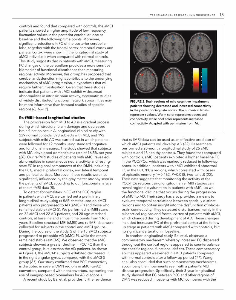
FIGURE 2. Brain regions of mild cognitive impairment patients showing decreased and increased connectivity in the posterior cingulate cortex. The numerical labels represent t values. Warm color represents decreased connectivity, while cool color represents increased connectivity. Adapted with permission from 16.

16 TRANSLATIONAL MEDICINE AT CAPITAL MEDICAL UNIVERSITY: INVESTIGATING MAJOR CHRONIC DISEASES
Discovery of novel glioma biomarkers using the Chinese Glioma Genome Atlas databaseZhaoshi Bao1,2 and Tao Jiang1,2,3,4,5*
Glioma is a particularly deadly form of cancer, and our understanding of the biological and clinical impact of the genetic alterations gliomas undergo is limited. To address this problem, in 2004, we founded a comprehensive Chinese glioma tissue bank comprising biopsies, clinical records, and follow-up data from tissue donors (1). In 2012, the Chinese Glioma Genome Atlas (CGGA) was established, extending the importance and usefulness of the tissue bank by providing a database of genomic and gene expression data derived from the banked glioma tissue (2). Here we review the aims and achievements of CGGA in its mission to improve diagnosis, treatment, and prevention of glioma through a better understanding of the molecular basis of this disease. The CGGA database will provide a means to classify gliomas based on molecular signatures and patient survival and to identify novel targets to stratify patient populations and tailor therapeutics based on the molecular profiles of the patients’ tumors.
History of CGGACGGA represents the first comprehensive dataset
generated from East Asian glioma samples. Under the guidance and direction of Zhongcheng Wang of the Chinese Academy of Engineering, and Tao Jiang, vice director of Neusurgical Department at Beijing Tiantan Hospital, CGGA focuses on both translational medicine and molecular classification of gliomas. Through their 6 years of sample collection, Jiang and colleagues have established the largest collection of glioma tissue and associated patient records in China. In addition, hundreds of glioma biopsies in the bank have been analyzed for their microRNA, mRNA, and DNA methylation profiles. CGGA represents a landmark for glioma research in China by providing a large and valuable dataset for basic and clinical research.
The aim of the CGGA project is to catalogue the genetic mutations responsible for causing glioma in Chinese patients using next generation genome analysis
1Department of Neurosurgery, Beijing Tiantan Hospital, Capital Medical University, Beijing, China2Chinese Glioma Cooperative Group (CGCG), Beijing, China3Beijing Neurosurgical Institute, Beijing, China4Center of Brain Tumor, Beijing Institute for Brain Disorders, Beijing, China5China National Clinical Research Center for Neurological Diseases, Beijing, China.*Corresponding Author: [email protected]
normal controls, and, in particular, diminished connectivity between PCC and the superior frontal gyrus and middle frontal gyrus. Interestingly, they also observed increased FC between PCC and medial prefrontal cortex and between PCC and anterior cingulate cortex in MCI patients (Figure 2). Moreover, the alterations in PCC connectivity in certain brain regions correlated with performance on the mini-mental state examination (r=0.57, P<0.05) and California verbal learning test scores (long-delayed memory scores, r=0.48, P<0.05) (16).
Taken together, these studies indicate that resting-state FC in special brain regions, especially related regional components within DMN, will change gradually in the process of conversion from aMCI to AD, accompanied by damage as well as concomitant compensation.
ConclusionsRs-fMRI is emerging as an important tool to detect
functional abnormalities in the DMN of patients with AD and aMCI and to monitor longitudinal changes during disease progression. Rs-fMRI also provides a powerful way to diagnose AD earlier, predict MCI-to-AD conversion, monitor disease progression, and measure therapeutic efficacy. Additional studies using rs-fMRI are needed to further elucidate dynamic changes in the networks underlying brain function that occur during the progression of MCI to AD.
References 1. R. A. Sperling et al., Alzheimers Dement. 7, 280 (2011). 2. C. R. Jack Jr. et al., Lancet Neurol. 9, 119 (2010). 3. R. C. Craddock et al., Nat. Methods 10, 6 (2013). 4. F. X. Castellanos et al., Neuroimage 80, 527 (2013). 5. P. Vemuri et al., Neurology 75, 143 (2010). 6. M. E. Raichle et al., Proc. Natl. Acad. Sci. U.S.A. 98, 676 (2001). 7. R. L. Buckner et al., J. Neurosci. 25, 34 (2005). 8. Y. Han et al., NeuroImage 55, 287 (2011). 9. B. Biswal et al., Magn. Reson. Med. 34, 537 (1995).10. B. Dubois et al., Lancet Neurol. 6, 734 (2007).11. F. Bai et al., Behav. Brain Res. 216, 666 (2011).12. M. A. Binnewijzend et al., Neurobiol. Aging 33, 2018 (2012).13. D. T. Jones et al., Neurology 77, 1524 (2011).14. F. Bai et al., PLOS ONE 6, e29288 (2011).15. F. Bai et al., J. Alzheimers Dis. 23, 87 (2011).16. Z. Wang et al., PLOS ONE 7, e36838 (2012).17. J. Wang et al., Biol. Psychiatry 73, 472 (2013).18. C. H. Yin et al., Transl. Neurosci. 5, 253 (2014).19. Y. Han et al., PLOS ONE 7, e28664 (2012).20. R. C. Petersen et al., Neurology 74, 201 (2010).21. Z. J. Hu et al., Chin. J. Neurol. 47, 824 (2014).22. F. Bai et al., PLOS ONE 6, e24271 (2011).
AcknowledgmentsWork was supported by the National Natural Science Foundation of China (81430037, 31371007, 81261120571, 81171403, and 81030028), the Beijing Municipal Science & Technology Commission (Z131100006813022), and the National Key Department of Neurology funded by the Chinese Health and Family Planning Committee.
TRANSLATIONAL RESEARCH IN NEUROSCIENCE 17
technologies. By applying high throughput genome analysis techniques to glioma research, we hope to improve diagnosis, treatment, and prevention of this disease. The CGGA database will provide an important platform for researchers in the field of glioma research, and can be accessed through the CGGA Data Portal (www.cgga.org.cn).
Classification of gliomas One way of classifying gliomas is to identify the key
signaling molecules that are activated during their initial origins. To this end, gene coexpression modules have been created based on the epidermal growth factor receptor (EGFR, 29 genes) and platelet derived growth factor receptor A (PDGFRA, 40 genes) signaling axes (3). Three glioma subtypes—EGFR module (EM), PGDGRA module (PM), and EMlowPMlow—have been identified, independent of cell morphology.
EM gliomas show a distinct pattern of genomic alterations and are associated with an older age of diagnosis, poorer prognosis, and stronger expression of neural stem cell and astrogenesis genes. In contrast, both PM and EMlowPMlow gliomas are correlated with better prognosis and a younger age of diagnosis. This molecular classification has provided a diagnostic framework to carry out additional studies aimed at identifying new glioma therapeutic targets (4).
In addition, a different classification has been created using a large number of samples from an East Asian population to complement The Cancer Genome Atlas (TCGA) glioma classification system. In this study, three major groups of gliomas were identified (referred to as G1, G2, and G3). The G1 subgroup was correlated with a good clinical outcome, young age, and extremely high frequency of IDH1 [isocitrate dehydrogenase 1 (NADP+),
soluble] mutations. Relative to the G1 subgroup, the G3 subgroup was correlated with a poorer clinical outcome, older age, and a very low rate of mutations in the IDH1 gene. Correlations of the G2 subgroup with respect to clinical outcome, age, and IDH1 mutation fell between the G1 and G3 subgroups. Using the TCGA classification system, proneural, neural, and mesenchymal subtypes, but not the classical subtype, were clearly defined. The G1/G2/G3 scheme may reflect the clinical and genetic alterations more clearly (5).
Biomarkers associated with glioma progression and prognosis
Insulin-like growth factor binding protein 2 (IGFBP-2) is a malignancy-associated protein measurable in tumor biopsies and blood (6). Lin et al. examined plasma IGFBP-2 levels in glioma patients and healthy controls to evaluate its value as a plasma biomarker for glioma. Preoperative plasma IGFBP-2 levels were significantly higher in high-grade glioma patients than in low-grade glioma patients and healthy controls. After recurrence, plasma IGFBP-2 levels were significantly increased in glioblastoma multiforme (GBM) patients and correlated negatively with disease-free survival (7).
Another strategy for biomarker discovery is mapping DNA methylation patterns associated with tumors. A study of DNA methylation profiling was performed on primary GBM samples from 13 long-term survivors (LTS; overall survival ≥18 months) and 20 short-term survivors (STS; overall survival <9 months). Differentially expressed CpG loci were identified between 18 STS and 9 LTS glioma CpG island methylation phenotype (G-CIMP)-negative samples. Methylation levels at 11 CpG loci (10 genes) were significantly lower, and 43 CpG loci (40 genes) were significantly higher, in tumor tissues from
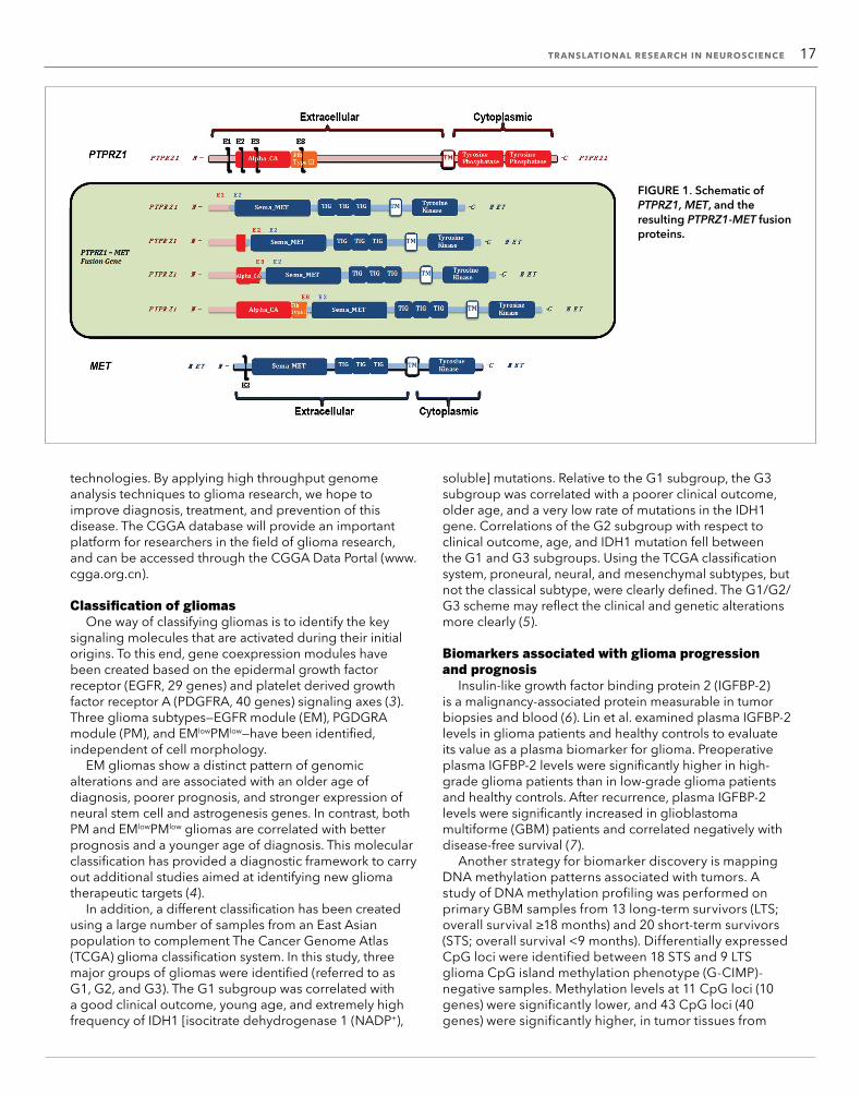
FIGURE 1. Schematic of PTPRZ1, MET, and the resulting PTPRZ1-MET fusion proteins.

18 TRANSLATIONAL MEDICINE AT CAPITAL MEDICAL UNIVERSITY: INVESTIGATING MAJOR CHRONIC DISEASES
LTS patients when compared with STS G-CIMP-negative samples. These results for the first time systematically define prognosis-related methylation signatures in G-CIMP-negative primary GBM tumors. Additionally, it was found that methylation of the aldehyde dehydrogenase 1 family, member A3 promoter region conferred a favorable prognosis in G-CIMP-negative primary GBM tumors (8).
Risk factors for low-grade gliomas with seizuresSeizure is a common presenting manifestation and
plays an important role in the clinical presentation and quality of life for glioma patients (9). We next set out to identify the factors that influence preoperative seizure characteristics and postoperative control of seizures in patients with gliomas (10). We retrospectively reviewed and analyzed cases with adult patients who have undergone initial surgery for low-grade gliomas at a single institution between 2005 and 2009 (11). For the cohort of 350 patients with seizures, we used the Engel classification to evaluate 6- and 12-month outcomes following surgery. Multivariate logistic analysis showed that a favorable seizure prognosis was more common in patients exhibiting secondary generalized seizures and calcification on MRI scans. Patients achieved much better seizure control after gross total tumor resection than after subtotal resection. Ki-67, if overexpressed, was shown to be an independent molecular marker predicting poor seizure control in the patients with a history of seizure, but was not a good predictor for those without
preoperative seizures. These factors can be applied when developing more effective treatment strategies aimed at prolonging patient survival (9).
Mechanisms underlying glioma progressionIn order to elucidate the aberrations in the DNA
methylation patterns that are associated with different prognoses of patients with G-CIMP-negative primary GBM, Zhang et al. profiled 82 primary glioblastomas using a genome-wide microRNA (miRNA) array. The results demonstrated that miR-181d expression was inversely associated with overall patient survival and was validated in independent cohorts. Transfection of miR-181d in GBM cell lines downregulated O6-methylguanine-DNA methyltransferase (MGMT) mRNA and protein expression. Furthermore, luciferase reporter assays and coprecipitation studies showed that the suppressive effect of miR-181d on MGMT expression was rescued by the introduction of an MGMT cDNA (12).
Epidermal growth factor receptor (EGFR) has been shown to be amplified in 40% of human glioblastomas (13). Zhang et al. demonstrated that miR-566 is upregulated in human glioma cell lines and inhibition of miR-566 decreases the activity of the EGFR pathway. Further, the von Hippel-Lindau (VHL) gene mRNA was identified as a novel functional target of miR-566. VHL regulates the formation of the β-catenin/hypoxia-inducible factors-1α complex under miR-566 regulation (14).
FIGURE 2. PCR (A) and Sanger sequencing (B) validation of the positive fusion samples in training and validation sets. Red line in panel B denotes fusion point.

TRANSLATIONAL RESEARCH IN NEUROSCIENCE 19
Characterization of fusion profiles and novel fusion transcripts
To identify oncogenic fusions associated with glioma progression, Bao et al. catalogued fusion transcripts in 272 gliomas by RNA-seq. Fusion transcripts were more frequently found in high-grade gliomas, in the classical subtype of gliomas, and in gliomas treated with radiation/temozolomide. Sixty-seven in-frame fusion transcripts have been identified, including three recurrent fusion transcripts: FGFR3-TACC3 (16), RNF213-SLC26A11 (17), and PTPRZ1-MET (ZM) (15). Interestingly, Bao et al. also showed that the ZM fusion is found only in grade III astrocytomas (1/13, 7.7%) or secondary GBM tumors (sGBM; 3/20, 15.0%). A genomic analysis revealed that the fusion arose from translocation events involving introns three or eight of PTPRZ1 and intron one of MET. Further, expression of the ZM fusion and overexpression of EGFR are mutually exclusive in sGBM tumors; this describes a new subtype of GBM. We also demonstrated that exogenous expression of the ZM fusion in the U87MG glioblastoma line enhances cell migration and invasion (Figure 1). Clinically, patients afflicted with ZM-fusion–harboring glioblastomas exhibited poor survival relative to those afflicted with non-ZM–harboring sGBM tumors. This study highlights the altered RNA landscape in gliomas during progression and has identified ZM as a novel, recurrent fusion transcript in sGBM tumors (15).
SummaryThe goal of the CGGA is to provide systematic,
comprehensive genomic characterization, and analysis of glioma samples from China. Further elucidating the mechanisms underlying glioma initiation and progression, and identifying possible candidates for targeted therapy, will largely depend on the improvement of the tissue bank through the addition of glioma samples. To this end, we plan to supplement the
genomics and proteomics databases with more than 500 various grades of glioma samples. Further analysis of our CGGA database will be performed to improve glioma classification, find pathways that correlate with glioma malignant outcomes, and identify novel targets for future therapies. Analysis of a broad range of samples with different clinical outcomes will aid in establishing a paradigm for making better and more educated clinical decisions, advancing us towards truly personalized medicine.
References 1. B. Vogelstein et al., Science 339, 1546 (2013). 2. www.cgga.org.cn 3. D. N. Louis et al., Acta Neuropathol. 114, 97 (2007). 4. Y. Sun et al., Proc. Natl. Acad. Sci. U.S.A. 111, 3538 (2014). 5. W. Yan et al., Neuro. Oncol. 14, 1432 (2012). 6. G. N. Fuller et al., Cancer Res. 59, 4228 (1999). 7. Y. Lin et al., Neuro. Oncol. 11, 468 (2009). 8. W. Zhang et al., Cancer Lett. 328, 120 (2013). 9. G. You et al., Neuro. Oncol. 14, 230 (2012).10. G. You et al., Eur. J. Neurol. 19, 298 (2012).11. L. Huang et al., J. Neurol. Sci. 302, 63 (2011).12. W. Zhang et al., Neuro Oncol. 14, 712 (2012).13. J. S. Smith et al., J. Natl. Cancer Inst. 93, 1246 (2001).14. K. L. Zhang et al., Mol. Cancer 13, 63 (2014).15. Z. S. Bao et al., Genome Res. 24, 1765 (2014).16. D. Singh et al., Science 337, 1231 (2012).17. J. B. Zhou, T. Zhang, B. F. Wang, H. Z. Gao, X. Xu, Mol. Med. Rep. 7, 591 (2013).
AcknowledgmentsThis work was supported by grants from National High Technology Research and Development Program (2012AA02A508), the International Science and Technology Cooperation Program (2012DFA30470), and the National Natural Science Foundation of China (91229121).

20 TRANSLATIONAL MEDICINE AT CAPITAL MEDICAL UNIVERSITY: INVESTIGATING MAJOR CHRONIC DISEASES
The role of inflammation in hypertensive cardiac injuryYulin Li, Congcong Zhang, and Jie Du*
Hypertensive cardiac injury is the leading cause of ventricular remodeling and eventually leads to heart failure. Despite marked progress in the understanding of ventricular remodeling, the underlying mechanism is not completely clear. It is known, however, that inflammatory processes underlie many cardiovascular diseases, such as atherosclerosis, myocardial ischemia and reperfusion, and viral myocarditis. Moreover, there is evidence supporting the hypothesis that hypertensive ventricular remodeling is a low-grade chronic inflammatory condition of the heart. Studies have provided significant insight into the role of inflammation in hypertensive cardiac remodeling (1). In patients with heart failure,
signs of inflammation, such as elevated levels of cytokine and C-reactive protein (CRP), are closely related to detrimental ventricular remodeling (2).
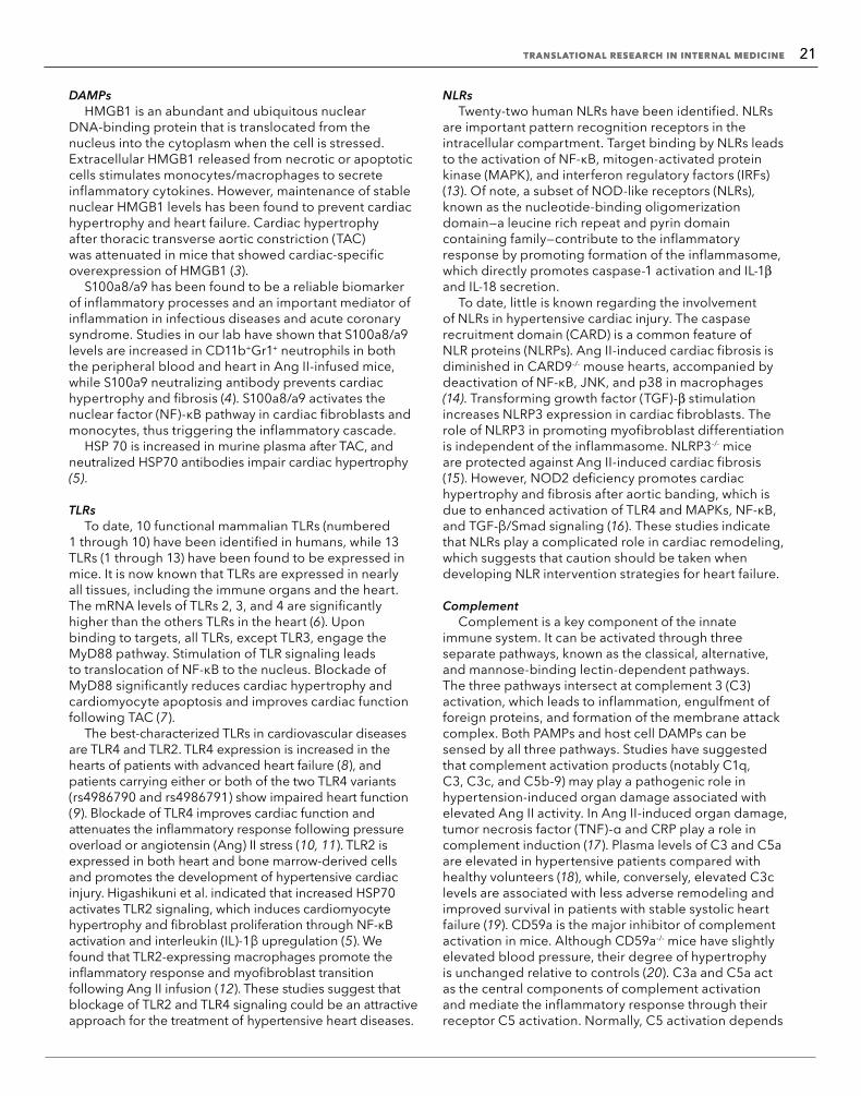
An overview of the inflammatory response in hypertensive heart remodeling is provided in Figure 1. The present review focuses on the role of the immune system in hypertensive cardiac injury and remodeling processes.
The innate immune systemThe innate immune system is regarded as the body’s
first line of defense against infection and signals that indicate potential danger, such as from microbial invasion or tissue injury. Traditionally, a set of responses is triggered when pathogen-associated molecular patterns (PAMPs) are recognized by circulating complement proteins, cellular Toll-like receptors (TLRs), or nucleotide-binding oligomerization domain-like (NOD-like) receptors (NLRs). These responses facilitate clearance of foreign organisms. However, emerging studies have indicated that the innate immune system also acts as a fast sensor of noninfectious damage by detecting danger-associated molecular patterns (DAMPs) released from damaged or stressed cells, damaged extracellular matrix proteins, or circulating oxidized proteins. DAMPs, such as high-mobility group box 1 (HMGB1), two small calcium-regulated molecules (S100a8 and S100a9), or heat shock proteins (HSPs), function as indictors of cellular stress and bind to TLR or NLRs to evoke and amplify a proinflammatory response in cells.
Translational research in internal medicine
Beijing An Zhen Hospital, Capital Medical University; The Key Laboratory of Remodeling-Related Cardiovascular Diseases, Ministry of Education; Beijing Institute of Heart, Lung, and Blood Vessel Diseases, Beijing, China.*Corresponding Author: [email protected]
TRANSLATIONAL RESEARCH IN INTERNAL MEDICINE 21TRANSLATIONAL RESEARCH IN INTERNAL MEDICINE 21
DAMPsHMGB1 is an abundant and ubiquitous nuclear
DNA-binding protein that is translocated from the nucleus into the cytoplasm when the cell is stressed. Extracellular HMGB1 released from necrotic or apoptotic cells stimulates monocytes/macrophages to secrete inflammatory cytokines. However, maintenance of stable nuclear HMGB1 levels has been found to prevent cardiac hypertrophy and heart failure. Cardiac hypertrophy after thoracic transverse aortic constriction (TAC) was attenuated in mice that showed cardiac-specific overexpression of HMGB1 (3).
S100a8/a9 has been found to be a reliable biomarker of inflammatory processes and an important mediator of inflammation in infectious diseases and acute coronary syndrome. Studies in our lab have shown that S100a8/a9 levels are increased in CD11b+Gr1+ neutrophils in both the peripheral blood and heart in Ang II-infused mice, while S100a9 neutralizing antibody prevents cardiac hypertrophy and fibrosis (4). S100a8/a9 activates the nuclear factor (NF)-κB pathway in cardiac fibroblasts and monocytes, thus triggering the inflammatory cascade.
HSP 70 is increased in murine plasma after TAC, and neutralized HSP70 antibodies impair cardiac hypertrophy (5).
TLRsTo date, 10 functional mammalian TLRs (numbered
1 through 10) have been identified in humans, while 13 TLRs (1 through 13) have been found to be expressed in mice. It is now known that TLRs are expressed in nearly all tissues, including the immune organs and the heart. The mRNA levels of TLRs 2, 3, and 4 are significantly higher than the others TLRs in the heart (6). Upon binding to targets, all TLRs, except TLR3, engage the MyD88 pathway. Stimulation of TLR signaling leads to translocation of NF-κB to the nucleus. Blockade of MyD88 significantly reduces cardiac hypertrophy and cardiomyocyte apoptosis and improves cardiac function following TAC (7).
The best-characterized TLRs in cardiovascular diseases are TLR4 and TLR2. TLR4 expression is increased in the hearts of patients with advanced heart failure (8), and patients carrying either or both of the two TLR4 variants (rs4986790 and rs4986791) show impaired heart function (9). Blockade of TLR4 improves cardiac function and attenuates the inflammatory response following pressure overload or angiotensin (Ang) II stress (10, 11). TLR2 is expressed in both heart and bone marrow-derived cells and promotes the development of hypertensive cardiac injury. Higashikuni et al. indicated that increased HSP70 activates TLR2 signaling, which induces cardiomyocyte hypertrophy and fibroblast proliferation through NF-κB activation and interleukin (IL)-1β upregulation (5). We found that TLR2-expressing macrophages promote the inflammatory response and myofibroblast transition following Ang II infusion (12). These studies suggest that blockage of TLR2 and TLR4 signaling could be an attractive approach for the treatment of hypertensive heart diseases.
NLRsTwenty-two human NLRs have been identified. NLRs
are important pattern recognition receptors in the intracellular compartment. Target binding by NLRs leads to the activation of NF-κB, mitogen-activated protein kinase (MAPK), and interferon regulatory factors (IRFs) (13). Of note, a subset of NOD-like receptors (NLRs), known as the nucleotide-binding oligomerization domain—a leucine rich repeat and pyrin domain containing family—contribute to the inflammatory response by promoting formation of the inflammasome, which directly promotes caspase-1 activation and IL-1b and IL-18 secretion.
To date, little is known regarding the involvement of NLRs in hypertensive cardiac injury. The caspase recruitment domain (CARD) is a common feature of NLR proteins (NLRPs). Ang II-induced cardiac fibrosis is diminished in CARD9-/- mouse hearts, accompanied by deactivation of NF-κB, JNK, and p38 in macrophages (14). Transforming growth factor (TGF)-b stimulation increases NLRP3 expression in cardiac fibroblasts. The role of NLRP3 in promoting myofibroblast differentiation is independent of the inflammasome. NLRP3-/- mice are protected against Ang II-induced cardiac fibrosis (15). However, NOD2 deficiency promotes cardiac hypertrophy and fibrosis after aortic banding, which is due to enhanced activation of TLR4 and MAPKs, NF-κB, and TGF-β/Smad signaling (16). These studies indicate that NLRs play a complicated role in cardiac remodeling, which suggests that caution should be taken when developing NLR intervention strategies for heart failure.
ComplementComplement is a key component of the innate
immune system. It can be activated through three separate pathways, known as the classical, alternative, and mannose-binding lectin-dependent pathways. The three pathways intersect at complement 3 (C3) activation, which leads to inflammation, engulfment of foreign proteins, and formation of the membrane attack complex. Both PAMPs and host cell DAMPs can be sensed by all three pathways. Studies have suggested that complement activation products (notably C1q, C3, C3c, and C5b-9) may play a pathogenic role in hypertension-induced organ damage associated with elevated Ang II activity. In Ang II-induced organ damage, tumor necrosis factor (TNF)-α and CRP play a role in complement induction (17). Plasma levels of C3 and C5a are elevated in hypertensive patients compared with healthy volunteers (18), while, conversely, elevated C3c levels are associated with less adverse remodeling and improved survival in patients with stable systolic heart failure (19). CD59a is the major inhibitor of complement activation in mice. Although CD59a-/- mice have slightly elevated blood pressure, their degree of hypertrophy is unchanged relative to controls (20). C3a and C5a act as the central components of complement activation and mediate the inflammatory response through their receptor C5 activation. Normally, C5 activation depends

22 TRANSLATIONAL MEDICINE AT CAPITAL MEDICAL UNIVERSITY: INVESTIGATING MAJOR CHRONIC DISEASES
on C3 activation to generate C5 convertase, but it is also known that C5a can be generated in the absence of C3. Our study indicated that C5aR-deficient, but not C3a receptor–deficient, mice exhibited markedly reduced cardiac remodeling and inflammation after Ang II infusion. This is likely a result of the fact that initiation of inflammation is dependent on activation of adhesive interactions between endothelial cells and leukocytes. The Ang II-activated C5a-C5aR pathway enhances expression of adhesion molecules, which increases monocyte adherence and migration to endothelial cells (21). Moreover, a C5aR antagonist (PMX53) prevents cardiac remodeling in Ang II–induced hypertension
(22). The C5a-C5aR pathway may therefore represent an interesting future therapeutic target for hypertensive injury/remodeling.
Immune cellsOverwhelming evidence indicates
that injurious stimuli, such as Ang II stimulation or pressure overload, induce DAMP and chemokine release, attracting various immune cells to the site of injury. Immune cells are generally categorized into two lineages: myeloid, which includes monocytes/macrophages, dendritic cells (DCs), mast cells, and granulocytes; and lymphoid, which includes T and B lymphocytes, natural killer (NK) cells, and NKT cells. Immune cells play important roles in mediating both innate and adaptive immune responses. Cells that infiltrate the heart are predominantly monocytes/macrophages, neutrophils, and T and B cells in human cases of heart failure and in murine models responding to Ang II infusion or TAC.
MacrophagesMacrophages are perhaps the most
plastic cells of the immune system, exhibiting great functional diversity, including maintenance of homeostasis, tissue repair, and immunity (23). Since the 1960s, the prevailing understanding was that bone-marrow–derived monocytes infiltrated tissues and gave rise to tissue macrophages. In this process, chemokines play an important role in attracting the circulating monocytes into the tissue. For example, monocyte-chemoattractant protein-1 (MCP-1), binds to its receptor, CCR2, on monocytes, stimulating their migration. Deletion of MCP-1 or monocyte CCR2 blunts Ang II-induced heart remodeling (23, 24), suggesting a critical detrimental
role for macrophages. However, studies using liposomal clodronate to induce macrophage depletion in vivo support a protective role for macrophages in hypertensive cardiomyopathy (24).
Macrophages can be divided into two major subpopulations: classically activated macrophages (M1) and alternatively activated macrophages (M2), depending on the local inflammatory microenvironment. These two subpopulations play opposite roles in heart remodeling. It is well known that processes such as death and hypertrophy of cardiomyocytes, the transdifferentiation and proliferation of fibroblasts, and the balance between extracellular matrix production and
FIGURE 1. Regulation of the inflammatory response in hypertensive heart remodeling. Multiple stressors (e.g., Ang II or high blood pressure (BP↑)) promote danger-associated molecular pattern (DAMP) release and complement activation. DAMPs, such as S100a8/a9 released by neutrophils (Neu) activate the nuclear factor (NF)-kB pathway in cardiac fibroblasts (CFs) via RAGE and monocytes (Mφ) via or TLR2, triggering the inflammatory cascade. Activation of the C5a-C5aR pathway enhances monocyte adherence to endothelial cells and their subsequent migration. CD8 T cells promote cardiac injury by amplifying monocyte chemoattractant protein–1 (MCP-1) production. IFN-g–producing CD4 T cells enhance the inflammatory response. Monocyte-derived IL-1b promotes IL-17 production from gd T cells, which enhance IL-6 secretion from cardiac fibroblasts. Treg-released IL-10 suppresses inflammation. M2 macrophage–produced TGF-b and CF-produced IL-6 promote heart remodeling.

TRANSLATIONAL RESEARCH IN INTERNAL MEDICINE 23
degradation, are central to heart remodeling. Our studies have shown that M2 macrophages promote fibroblast differentiation and fibrosis formation through increasing TGF-β production following Ang II stimulation (25). The transcription factor IRF5 promotes M1 macrophage polarization, and silencing IRF5 in vivo attenuates post-myocardial infarction (MI) remodeling and heart failure by suppressing inflammation (26). This suggests a new therapeutic avenue for heart inflammation and remodeling by regulating the macrophage phenotype. Further studies to clarify the phenotype, regulatory mechanisms, and functions of M1/M2 macrophages in hypertensive remodeling are needed.
Recent studies show that circulating monocytes are not the exclusive source of macrophages. A newly identified population of monocytes that resides in splenic reservoirs was shown to be mobilized, enter injured tissue during inflammation, and participate in wound healing (27). Studies show that some steady-state tissue macrophages are derived from progenitors of seeded tissue from the yolk sac, but not from hematopoietic precursors (28). Genetic fate mapping has revealed that resident cardiac macrophages are derived from yolk sac and fetal monocyte progenitors. Cardiac resident macrophages maintain their steady state and expand during Ang II stimulation through self-proliferation; in addition, the macrophage population expands through infiltration. Meanwhile, during Ang II-induced injury, hematopoietic monocytes also contribute to macrophage expansion in the heart (29). These data highlight the fact that there multiple sources of cardiac macrophages are present following injury. The differences between the resident macrophages and monocyte-derived macrophages at the functional level in this pathologic context remain to be explored.
NeutrophilsNeutrophils are recruited very early after cardiac injury,
followed by monocytes and lymphocytes. However, limited data is available regarding the mechanism of neutrophil accumulation and its function in hypertensive heart injury/remodeling. Circulating neutrophils play an important role in maintaining physiologic blood pressure and depletion of neutrophils via genetic and pharmacologic approaches leads to acute hypotension (30). Neutrophil-derived myeloperoxidase (MPO) acts as a profibrotic mediator (31). MPO-deficient mice pretreated with Ang II show reduced activity of matrix metalloproteinases-9 (MMP) and blunted atrial fibrosis. We found that neutrophil-generated S100a8/a9 can also initiate Ang II–induced cardiac inflammation and fibrosis via the receptor for advanced glycation endoproducts (RAGE), which activates NF-κB in cardiac fibroblasts (4). Moreover, we have demonstrated a crucial role for granulocyte colony–stimulating factor (G-CSF) as a key mediator of Ang II-induced neutrophil recruitment and cardiac fibrosis (32–35). Future studies are needed to evaluate the effects of neutrophils and related neutrophil-related molecules (S100a8/a9, G-CSF, and
MMP-9) on hypertension in human patients, as well as their contribution to cardiac remodeling and sudden death.
T lymphocytesA series of studies have demonstrated that T
cells, but not B cells, are crucial for the development of hypertension. For example, Th17 cells promote hypertension while regulatory T cells (Tregs) prevent hypertension in an Ang II-induced mice model. Moreover, T cells play an important role in the genesis of Ang II-induced hypertension and vascular dysfunction (33). CD4 T cells shift from naïve to effector states during the immune response and can be further categorized according to the production of specific cytokines. The signature cytokines for each population is as follows: Th1, interferon-g (IFN-g); Th2, IL-4, and IL-13; Th17, IL-17, and IL-21; and Tregs, IL-10, and TGF-β. Based on the expression of surface markers, T cells can be divided into CD4 and CD8 subtypes and, based on their T cell receptor (TCR), also into abT and gdT cells. The differentiation of pro-fibrogenic M2 macrophages in hypertension is dependent on CD4 T cells, as is the differentiation of Gr1+ monocytes into fibrocytes (34). The Th17 subpopulation promotes hypertension, while Tregs prevent Ang II-induced hypertension. Deficiency in Th1 cytokines, and thus IFN-g signaling, reduces cardiac hypertrophy and fibrosis, decreases macrophage and T-cell infiltration into the heart, and is associated with less arrhythmogenic electric remodeling following Ang II infusion (35). In experimental mouse models of lung and liver fibrosis, Th2 cytokines (IL-4 and IL-13) are strongly pro-fibrogenic.
Our studies and others have revealed that IL-17A expression increases in Ang II-infused hearts. The IL-17 receptor is expressed on numerous cell types, including cardiomyocytes, fibroblasts, and monocytes. We found that IL-17 not only participates in the development of long-term hypertension, but is also a key regulator of cardiac fibrosis associated with hypertension. IL-17 accelerates myofibroblast formation by promoting IL-6 production by cardiac fibroblasts (36). However, we have demonstrated that gdT cells, rather than classical abT cells, are the major source of IL-17. gdT cells represent a small subset of T cells, which are triggered by alarm signals such as HSPs. Our results show that IL-1β secretion by monocytes is required for IL-17A production from gdT cells. Moreover, depletion of gdT cells prevents Ang II-induced cardiac fibrosis (36).
It is known that Tregs regulate the immune response in a dominant-negative manner (37). Adoptive Treg cell transfer prevents cardiac hypertrophy, reduces cardiac fibrosis, and improves arrhythmogenic electric remodeling in Ang II-infused mice. These effects are accompanied by a marked reduction in CD4 T cells, CD8 T cells, and macrophage infiltration (38). Moreover, Treg-secreted IL-10 improves microvascular endothelial function by inhibiting NADPH oxidase activity in hypertensive mice (39). Thus, Tregs/IL-10 may be a

24 TRANSLATIONAL MEDICINE AT CAPITAL MEDICAL UNIVERSITY: INVESTIGATING MAJOR CHRONIC DISEASES
therapeutic target for the treatment of vasculopathy and cardiac damage in hypertension.
CD8 T cells act as cytotoxic T cells during the immune response. We found that myocardial infiltrating CD8 T cells are activated by Ang II via IFN-g receptor signaling (40). Adoptive transfer and deletion experiments have demonstrated that CD8 T cells initiate cardiac inflammation and fibrosis by amplifying MCP-1 production from macrophages (43). These studies highlight T lymphocytes as a promising therapeutic target for cardiac injury/remodeling in hypertension.
Clinical relevance Clinical studies suggest a link among inflammation,
hypertension, and cardiac remodeling, but the mechanism underlying the transition from adaptive hypertrophy to decompensation is incompletely understood. We believe that the inflammatory response might mediate this fundamental change. In patients with coronary artery disease, a cohort study found that proinflammatory cytokines are associated with coronary heart disease risk, independent of conventional risk factors (41). However, clinical evidence implicating inflammation in the development of myocardial hypertrophy and heart failure remains sparse. Further understanding the underlying signaling mechanisms and function of the immune system during heart failure will support the search for specific therapies.
References 1. L. Jia et al., Front. Biosci. (Landmark Ed) 17, 221 (2012). 2. D. L. Dixon et al., Cytokine 56, 593 (2011). 3. A. Funayama et al., Cardiovasc. Res. 99, 657 (2013). 4. Y. Wu et al., Hypertension 63, 1241 (2014). 5. Y. Higashikuni et al., J. Am. Heart Assoc. 2, e000267 (2013). 6. M. Nishimura et al., Biol. Pharm. Bull. 28, 886 (2005). 7. T. Ha et al., Am. J. Physiol. Heart Circ. Physiol. 290, H985 (2006). 8. S. Frantz et al., J. Clin. Invest. 104, 271 (1999). 9. A. Riad et al., J. Biol. Chem. 287, 27236 (2012).10. H. Ehrentraut et al., Eur. J. Heart Fail. 13, 602 (2011).11. R. B. Dange et al., Cardiovasc. Res. 103, 17 (2014).12. L. Wang et al., Cardiovasc. Res. 101, 383 (2014).13. Y. Zhong et al., Front. Immunol. 4, 333 (2013).14. J. Ren et al., Am. J. Hypertens. 24, 701 (2011).15. N. A. Bracey et al., J. Biol. Chem.289, 19571 (2014).16. J. Zong et al., Lab. Invest. 93, 1128 (2013).17. E. Shagdarsuren et al., Circ. Res. 97, 716 (2005).18. G. Engstrom et al., J. Hum. Hypertens. 21, 276 (2007).19. A. Frey et al., Mediators Inflamm. 2013, 716902 (2013).20. B. Coles et al., Immunology 121, 518 (2007).21. C. Zhang et al., Arterioscler. Thromb. Vasc. Biol. 34, 1240 (2014).22. C. Zhang et al., Am. J. Hypertens. 27, 857 (2014).23. T. A. Wynn et al., Nature 496, 445 (2013).24. H. R. Zandbergen et al., J. Cardiovasc. Pharmacol. Ther. 14, 68 (2009).25. Y. Li et al., Arterioscler. Thromb. Vasc. Biol. 32, 1662 (2012).26. G. Courties et al., J. Am. Coll. Cardiol. 63, 1556 (2014).
27. F. K. Swirski et al., Science 325, 612 (2009).28. C. Schulz et al., Science 336, 86 (2012).29. D. Hashimoto et al., Immunity 38, 792 (2013).30. J. Morton et al., Blood 111, 5187 (2008).31. V. Rudolph et al., Nat. Med. 16, 470 (2010).32. H. M. Jiang et al., Am. J. Hypertens. 26, 1224 (2013).33. D. G. Harrison et al., Hypertension 57, 132 (2011).34. M. Niedermeier et al., Proc. Natl. Acad. Sci. U.S.A. 106, 17892 (2009).35. L. Marko et al., Hypertension 60, 1430 (2012).36. Y. Li et al., Hypertension 64, 305 (2014).37. S. Sakaguchi et al., Cell 133, 775 (2008).38. H. Kvakan et al., Circulation 119, 2904 (2009).39. M. Kassan et al., Arterioscler. Thromb. Vasc. Biol. 31, 2534 (2011).40. F. Ma et al., J. Immunol. 192, 3365 (2014).41. S. Kaptoge et al., Eur. Heart J. 35, 578 (2014).
AcknowledgmentsThis work was supported by the National Natural Science Foundation of China (81230006 and 81470428).

TRANSLATIONAL RESEARCH IN INTERNAL MEDICINE 25
fashion; we focused on clinical management of patients, on developing guidelines for diagnosis and treatment, and on establishing a national network for clinical investigation. A National Clinical Investigation Group was also established under the leadership of Chen Wang at Capital Medical University to study emerging infectious diseases in general.
H1N1Epidemiology and Clinical Characteristics
We reported the first three cases of confirmed pH1N1 influenza virus infection in China between May 10 and May 15, 2009. The diagnoses were made using real-time reverse transcription polymerase chain reaction real-time (RT-PCR). The fever and other signs and symptoms exhibited by the pH1N1 patients were indistinguishable from those of seasonal influenza. Serial virologic monitoring of pharyngeal swabs showed that they were negative for the virus 4 to 6 days after the onset of illness (3). This analysis was the first time in China that the duration of viral shedding among patients infected with pH1N1 influenza had been observed, and the study has been cited as a fundamental basis for disease management in domestic medical guidelines. Commenting in a podcast at the time, Scott Dowell, director of the Division of Global Disease Detection and Emergency Response at the United States Centers for Disease Control and Prevention (CDC), said that “China has an impressive ability to bring together public health surveillance. They really ramped up these efforts in the wake of the severe acute respiratory syndrome [SARS] epidemic and their experience with H5N1 virus.”
Based on the close observation of 426 hospitalized patients who had confirmed 2009 pH1N1 influenza virus cases in May and June of 2011, we described the epidemiological and clinical characteristics of the infection. Our main findings were as follows: The median incubation period of the virus was two days (with a range of 1 to 7 days); 33% of the patients had a mild disease with no fever; the most common symptom, found in 70% of patients, was a cough; lymphopenia was common in adults (68%) and in children (92%); and late initiation of oseltamivir therapy (more than 48 hours after the onset of illness) was an independent risk factor for prolonged RT-PCR positivity (4). Nicole Lurie, the assistant secretary for preparedness and response at the United States Department of Health and Human Services, evaluated our report positively in an editorial, writing “It demonstrates the progress China has made in developing robust surveillance in a relatively short period.” Five findings from our study were included in the World Health Organization (WHO) Consultation on Clinical Aspects of Pandemic (H1N1) 2009 Influenza (5).
In addition to reporting the clinical manifestations of pH1N1 pneumonia during the acute phase of illness, we also reported symptoms, radiographic changes, and lung dysfunction during convalescence. Survivors were followed up after discharge for a period of 3 months. Both progressive dyspnea after resolution of fever and a
Pathogenesis, epidemiology, clinical characteristics, and therapy of the 2009 pH1N1 and H7N9 influenza virus outbreaks Pengjun Zhang1,4, Bin Cao2,4, and Chen Wang3,4*
Influenza pandemics occur when a new influenza type A virus emerges to which the population has no immunity. They pose a serious threat to public health as the influenza virus spreads efficiently between humans, potentially causing worldwide outbreaks of severe disease. There have been three influenza A global pandemics in the last century: the Spanish flu in 1918, the Asian flu in 1957, and the Hong Kong flu in 1968. Each of these resulted in a large number of fatalities. Here, we provide an overview of the two influenza pandemics that have occurred since 1968, namely the 2009 pH1N1 and the H7N9 influenza pandemics, focusing on the work of our team and our colleagues in China.
In April 2009, the first cases of human infection with the 2009 pandemic H1N1 (pH1N1) influenza A virus were identified in the United States and Mexico. It spread rapidly to other regions of the world, resulting in more than 60 million laboratory-confirmed cases in 214 countries and over 18,449 deaths by August 2010 (1).
In March 2013, a novel reassortment of avian-origin influenza A (H7N9) virus was first detected in Shanghai. It caused a severe disease in humans, with the majority of patients presenting with a rapidly progressive pneumonia and developing acute respiratory distress syndrome (ARDS). The epidemic quickly became a global concern (2).
Such pandemics occur infrequently, but we are faced with the threat of devastating new ones developing at any time. Despite this danger, the process for understanding the pathogenesis, clinical characteristics, diagnosis, and treatment of a new virus remains rather poorly defined even though much experience was gained from the 2009 pH1N1 influenza pandemic. In China, our team and others responded in a timely
1Beijing Hospital, Beijing, China2Beijing Chaoyang Hospital, Capital Medical University, Beijing, China 3Zhong Ri Hospital, Beijing, China4Department of Respiratory Diseases, Capital Medical University, Beijing, China.*Corresponding Author: [email protected]

26 TRANSLATIONAL MEDICINE AT CAPITAL MEDICAL UNIVERSITY: INVESTIGATING MAJOR CHRONIC DISEASES
higher Acute Physiology and Chronic Health Evaluation (APACHE) II score on presentation were independent risk factors associated with death in patients with pH1N1 viral pneumonia. At the 3 month follow-up visit of survivors of pH1N1 pneumonia, some degree of ground-glass opacity (GGO) persisted in most patients and decreased diffusion capacity for carbon monoxide was common (6).
Another focus of our team was to identify the characteristics of infection and risk factors in special populations, including children and pregnant women (7). Severe hypoxemia and high body mass index (BMI) on admission were associated with adverse outcomes for pregnant women and preterm delivery was a risk factor for neonatal death among pregnant women with pH1N1 influenza infection. Noninvasive ventilation may be useful in selected pregnant women without septic shock. Li et al. analyzed the course of disease and the outcome of severe or critical cases among children admitted to hospital with 2009 pH1N1 infections in China. This study concluded that clinicians should be especially vigilent with pediatric patients, particularly in children up to two years of age and those with immunosuppression. Severe hypoxemia was the main cause of death in children (8).
The above studies proved to be invaluable in establishing China’s national management guidelines and WHO’s Expert Committee review (9).
Therapy The antiviral agent oseltamivir was widely used during
the pH1N1 influenza pandemic, as recommended by WHO. Observational studies of hospitalized patients with seasonal influenza infection suggested that oseltamivir reduced the severity and mortality of the disease (10). In uncomplicated seasonal influenza cases it has been noted that oseltamivir is effective only if administered within 48 hours of the onset of symptoms, but no direct comparative evidence on the role of oseltamivir in the 2009 pH1N1 influenza has been reported (11).
During the pandemic, a cohort of 3,066 patients with pneumonia caused by 2009 pH1N1 virus was studied by the National 2009 pH1N1 Influenza Clinical Investigation Group of China. Our group assessed the effectiveness of oseltamivir treatment for pneumonia in this cohort and analyzed the optimal timing and dosing of oseltamivir in the treatment of adults and children. We concluded that antiviral therapy may improve survival in patients with severe pH1N1 viral pneumonia. The strongest protective effects against fatality were found in male patients aged 14 to 60 years and in patients with PaO2/FiO2 (ratio of arterial oxygen partial pressure to fractional inspired oxy-gen) lower than 200. Administration of a double dose of oseltamivir for patients whose body weight was less than 78 kg conferred no additional survival benefits (12). These results provided important evidence and were used to update antiviral therapy procedures internationally.
During the 2009 pH1N1 influenza epidemic, there was a shortage of effective, low-cost medicines in China. Traditional Chinese medicine (TCM) has been used to treat seasonal influenza for thousands of years although
precise clinical evidence about the efficacy of the TCM regimen was lacking. In the early days of the 2009 epidemic, the popular herbal formula Maxingshigan-Yinqiaosan (MY) was used widely by TCM practitioners to treat symptoms. Our group conducted a prospective, randomized, controlled, non-blinded, multicenter trial between July and November 2009 at 11 medical sites in China. This was the first study of the efficacy and safety of TCM in the treatment influenza using the study methods of modern Western medicine (13). We recruited 410 participants who were randomly assigned to one of four groups: untreated controls, an oseltameivir group, an MY group, and an oseltamivir plus MY group. The trial revealed that oseltamivir and MY, alone or in combination, reduced the time to fever resolution in patients with H1N1 influenza virus infection, suggesting that MY may be used as an alternative treatment of H1N1influenza virus infection. The value of the study was acknowledged by its inclusion in five international medical databases, recognizing TCM as a treatment of influenza A (H1N1).
Pathogenesis Gross examination of lung tissue revealed that severe
H1N1 influenza is accompanied by serious lung injury although there was no obvious correlation between viral load and the severity of lung disease (6). Microscopically, the lungs showed diffuse alveolar damage, pulmonary hemorrhage, and inflammatory cell infiltration.
In collaboration with the research team of Cheng-yu Jiang at the Chinese Academy of Medical Sciences, we found that excessive early cytokine responses with early recruitment of inflammatory immune cells to the lung were key contributors to the morbidity of the 2009 pH1N1 influenza (14). We also confirmed that the mol-ecule IP-10 plays an important role in mediating acute lung injury induced by 2009 pH1N1 infection, suggesting a new approach for developing immunotherapy to treat future influenza A (H1N1) virus pandemics (15).
Collaborating with a group led by Chuan Qin at the Chinese Academy of Medical Sciences, we developed mouse models of mild and severe H1N1 influenza (16). Zhang et al. also clarified changes in the distribution pattern of T cells caused by aggravated lung injury and identified a role for natural killer cells in severe pH1N1 influenza infection (17).
In BALB/c mice, severe pH1N1 infection leads to progressive thymic atrophy and depletion of CD4+CD8+ double-positive T cells due to apoptosis (18). Neutral-ization of interferon-g (IFN-g) significantly reduces the atrophy and CD8+CD44hi T cells were found to play a criti-cal role in influenza-induced thymic atrophy by secreting IFN-g. This finding suggests that IFN-g might serve as a target for the prevention and treatment of thymic atrophy induced by influenza A (H1N1).
H7N9 A novel influenza A (H7N9) virus of avian origin
emerged in eastern China in the spring of 2013. This virus
TRANSLATIONAL RESEARCH IN INTERNAL MEDICINE 27
causes severe disease in humans, including acute and lethal respiratory failure. As of January 2014, 275 cases of H7N9-infected patients had been reported. Based on the experience accumulated during the 2003 SARS and 2009 pH1N1 influenza epidemics, our team directed the clinical management of patients and carried out clinical research and applied basic research during this episode.
Gao et al. generated a portrait of the new disease from the first three fatal cases in China, identifying a novel reassortment influenza A (H7N9) virus. The Q226L and T160A mutations in the hemagglutinin protein and the deletion of five amino acids in the viral neuraminidase stalk of this avian-origin influenza virus were responsible for the alteration in viral tropism to the human respiratory tract and for the enhancement of viral replication (19).
The “clinical iceberg” phenomenon, whereby milder cases escape detection, is a common feature of influenza. Most reports of infection from the novel influenza A (H7N9) virus presented a severe clinical picture and Ip et al. confirmed the existence of a clinical iceberg, reinforcing the need for vigilance to the diverse presentation associated with A(H7N9) infection. At the public health level, indirect evidence suggested a substantial proportion of those infected suffered mild disease (20).
Together with the group of Lanjuan Li at Zhejiang University, our team described the clinical characteristics and laboratory abnormalities in 111 confirmed cases of H7N9 virus infection (2). The main findings were that the median incubation period was 5 days (ranging from 2 to 8 days); bilateral GGO and consolidation were the most common radiologic findings; infection was fatal in 30% of cases, and patients with fatal infections had pneumonia and ARDS; and using multivariate analysis, the presence of coexisting medical conditions was the only independent risk factor for moderate-to-severe ARDS.
Yu et al. estimated that the severity of disease caused by A(H7N9) was somewhat lower than A(H5N1), but higher than seasonal influenza viruses and influenza A(H1N1)pdm09 virus (21). In a comparison of risk factors, clinical presentation, and progression of patients hospitalized with H7N9, H5N1, and 2009 pH1N1, Wang et al. showed that patients hospitalized with H7N9 virus infection shared some risk factors with those hospitalized with pH1N1 infection, but had a clinical profile more closely resembling that of H5N1 patients. The identification of known risk factors for severe seasonal influenza and the more protracted clinical course compared with that of H5N1 suggested that host factors are important contributors to H7N9 severity (22).
Therapy The broad similarity of clinical, virologic, and
histopathologic features of viral pneumonias caused by A(H7N9), A(H5N1), and A(H1N1)pdm09 viruses suggest that the therapeutic recommendations for the other two viruses are appropriate for A(H7N9) (23).
Progressive viral pneumonia and ARDS are prominent features of patients with A(H7N9) infection. In addition
to correcting hypoxemia and providing critical care support, an increased dose of oral oseltamivir or, preferably, intravenous peramivir or zanamivir, is the foundation for specific treatment of A(H7N9) pneumonia. Optimal therapy for neuraminidase inhibitor-resistant A(H7N9) possessing the R294K mutation remains uncertain: several antiviral combinations are of interest, but they require systematic clinical testing. Systemic corticosteroids are not recommended for treating pneumonia or ARDS and appear to cause harm in some patients (23).
Pathogenesis The plasma level of angiotensin II, a major regulatory
peptide of the renin-angiotensin system, is markedly elevated in H7N9 patients and is associated with disease progression (24). Moreover, sustained high levels of angiotensin II in these patients is strongly correlated with mortality and the predictive value of this is greater than that of C-reactive protein and some clinical parameters such as the PaO2/FiO2 ratio. These findings indicate that angiotensin II is a biomarker of lethality in influenza infections.
ConclusionsWhen the first pneumonia case with unknown etiology
was reported to the National Health and Family Planning Commission and the China Center of Disease Control and Prevention, the authorities responded in a timely fashion: A national network was established, nationwide clinic-based surveillance work was carried out, guidelines for diagnosis and treatment were developed, and directed scientific research was initiated. Infectious disease surveillance efforts were markedly increased following the outbreak of SARS in 2003. Consequently, Chinese doctors and scientists today have more confidence in facing emerging infectious diseases than they had 10 years ago; however, we must tread carefully when responding to unknown, sudden diseases despite the significant advances that have been made in the prevention and treatment of emerging infectious diseases.
References 1. Centers for Disease Control and Prevention, MMWR Morb. Mortal. Wkly Rep. 58, 400 (2009). 2. H. N. Gao et al., N. Engl. J. Med. 368, 2277 (2013). 3. B. Cao et al., Emerg. Infect. Dis. 15, 1418 (2009). 4. B. Cao et al., N. Engl. J. Med. 361, 2507 (2009). 5. N. Lurie, N. Engl. J. Med. 361, 2571 (2009). 6. L. Bai et al., Chest 139, 1156 (2011). 7. P. J. Zhang et al., BMC Infect. Dis. 12, 29 (2012). 8. X. L. Li et al., Chin. J. Clinicians 6, 2663 (2012). 9. Writing Committee of the WHO Consultation on Clinical Aspects of Pandemic (H1N1) 2009 Influenza, N. Engl. J. Med. 362, 17089 (2010).10. A. McGeer et al., Clin. Infect. Dis. 45, 1568 (2007).11. K. G. Nicholson et al., Lancet 355, 1845 (2009).12. S. G. Yang et al., PLOS ONE 7, e29652 (2012).

28 TRANSLATIONAL MEDICINE AT CAPITAL MEDICAL UNIVERSITY: INVESTIGATING MAJOR CHRONIC DISEASES
Chronic obstructive pulmonary disease in ChinaZengyan Li, Miao Miao, Hongyan Hou, Yaning Gao, and Xiaoning Bu*
C hronic obstructive pulmonary disease (COPD) has a high prevalence of morbidity and mortality that is threatening human health around the world. In China, respiratory diseases—of which COPD is a major component—are the third leading cause of death in rural areas and the fourth leading cause of death in urban areas. They account for one million deaths and over five million disabilities each year, according to the 2013 China Health Statistics Yearbook. Yet, the country still lacks a widespread understanding of the prevalence, causes, clinical characteristics, and treatment of COPD. Here, we review a number of studies on COPD undertaken at Capital Medical University as well as on the assessment of the drug carbocisteine, which we propose is a useful intervention to prevent exacerbation of COPD in Chinese patients.
PrevalenceThe exact prevalence of COPD in China is unknown.
In 2005, a study of random clusters of rural and urban populations was undertaken across seven provinces and cities. Within these clusters, every resident 40 years of age and older was interviewed using a standardized questionnaire, and all eligible participants received a spirometry test for lung function. Among the 25,627 subjects identified, 20,245 completed the questionnaire and received the spirometry test—a response rate of 79%. Using the diagnostic criteria of a post-bronchodilator FEV1/FVC (the forced expiratory volume in 1 second divided by forced vital capacity) result of less than 70% (normal values are approximately 80%), the overall prevalence of COPD was estimated to be 8.2% (12.4% in men and 5.1% in women) (1). Stratification analysis of COPD prevalence examined smoking status, chronic bronchitis, and respiratory symptoms. A higher prevalence of COPD, 32.1%, was seen in smokers compared with nonsmokers, at 5.2%.
Multiple factors have been independently associated with a higher risk of COPD among nonsmokers, which include advanced age; male gender; lower body mass index (BMI); lower educational level; exposure to environmental tobacco smoke, coal, and/or biomass
Department of Respiratory and Critical Care Medicine, Beijing Chaoyang Hospital, Capital Medical University, Beijing, China.*Corresponding Author: [email protected]
13. C. Wang et al., Ann. Intern. Med. 155, 217 (2011).14. H. Wang, C. Jiang, Sci. China C. Life Sci. 52, 464 (2009).15. W. Wang et al., Cell Res. 23, 577 (2013).16. L. Xu et al., PLOS ONE 6, e20698 (2011).17. X. L. Zhang et al., 9th International Conference on Innate Immunity, 2012/6/23-2012/6/28, pp. 113–114, Rhodes, June 23, 2012.18. B. Liu et al., Cell Death Dis. 5, e1440 (2014).19. R. Gao et al., N. Engl. J. Med. 368, 1888 (2013).20. D. K. Ip et al., Br. Med. J. 346, f3693 (2013).21. H. J. Yu et al., Lancet 382, 138 (2013).22. C. Wang et al., Clin. Infect. Dis. 58, 1095 (2014).23. B. Cao, F. G. Hayden. Expert Rev. Anti Infect. Ther. 11, 1123 (2013).24. F. Huang et al., Nature Commun. 5, 3595 (2014).
AcknowledgmentsThis work was supported by the National Natural Science Foundation of China (81030032 and 81271840), the National Science for Distinguished Young Scholars (81425001), and the National Key Technology R&D Program of the Ministry of Science and Technology (2012BAI05B02).

TRANSLATIONAL RESEARCH IN INTERNAL MEDICINE 29
smoke; a poorly ventilated kitchen; a family history of respiratory disease; and recurrent childhood coughs. Nonsmokers with COPD were less likely to present with chronic productive coughs and have a lower BMI, while they were more likely to have had a physician diagnose them with asthma and respiratory diseases in childhood compared with smokers with COPD (2). Of the 20,245 survey respondents, 70% of the 1,668 patients who were diagnosed with COPD reported no history of chronic bronchitis (CB). A higher risk of COPD with CB was associated with male gender, residence in a rural area, a lower level of education, exposure to tobacco smoke or biomass fuels, a poorly ventilated kitchen, and a family history of respiratory disease. Patients without CB had less difficulty in walking and higher FEV1/FVC values than patients with CB, but were more likely to be underdiagnosed. The strongest predictors of CB were male gender, currently a smoker, and the severity of dyspnea (shortness of breath).
CB is not essential to the diagnosis of COPD (3). Among the 1,668 patients who were diagnosed with COPD, 35.3% were asymptomatic. Those in this asymptomatic COPD group were less likely to have a poorly ventilated kitchen, a family history of respiratory disease, and recurrent childhood coughs. Cardiovascular disease and lung cancer were more common among symptomatic COPD patients than the asymptomatic group. Asymptomatic COPD patients scored significantly higher than the symptomatic group in terms of FEV1 (73.1% vs. 61.0%), FVC (91.9% vs. 82.0%), and FEV1/FVC (62.9% vs. 58.7%) (4).
Clinical characteristicsOh et al. evaluated the characteristics of 70 stable
COPD patients (69 male), with an average age of 68.9 years, at Beijing Chaoyang Hospital between September 2009 and September 2010. Forty-one patients (58.6%) had a history of exposure to biomass fuel smoke and 31 (44.3%) had an occupational history of exposure to dust. Two (2.9 %) had no history of cigarette smoking while 68 (97.1%) were cigarette smokers, including 14 (20.0%) current smokers. Just over two-thirds (68.6%) of these patients had fathers who smoked and 28.6% had mothers who smoked. Sixteen of the patients (22.9%) had symptoms of chronic bronchitis and COPD patients with a cough, phlegm, and wheeze was 32 (45.7%), 51 (72.9%), and 63 (90.0%) respectively. The levels of dyspnea, evaluated using the modified Medical Research Council (mMRC) dyspnea grade, were 22.9% at grade 0; 20.0% at grade 1; 38.6% at grade 2; 8.6% at grade 3; and 10.0% at grade 4. The mean BMI was 24.0 kg/m2, the mean predicted post-bronchodilator FEV1 was 47.2%, and the mean total St. George’s Respiratory Questionnaire (SGRQ) score was 29.8. The proportion of under weight and overweight COPD patients was 10.1% and 42.9%, respectively. According to the severity criteria of the Global Initiative for Chronic Obstructive Lung Disease (GOLD), the prevalence of stage I (mild), stage II (moderate), stage III (severe), and stage IV
(very severe) COPD was 1.4%, 41.4%, 45.7%, and 11.4%, respectively (5).
PathogenesisBoth genetic and environmental factors play a role
in the development of COPD. One novel candidate risk factor is the serine protease inhibitor E2 (SERPINE2) gene, a member of the SERPIN family, located on chromosome 2q33-35. An et al. genotyped 20 single nucleotide polymorphisms (SNPs) in SERPINE2 from 310 patients with COPD and 203 controls, all of whom were of Han descent from North China. Two single nucleotide polymorphisms (rs729631and rs975278) in SERPINE2, which are in strong linkage disequilibrium (LD) and located in block 1 on the LD map, showed significant association with both a risk for COPD and a decline in baseline lung function (6). This provides further evidence that SERPINE2 may be a COPD susceptibility gene and that block 1 of SERPINE2 may be an important genetic variant region playing a role in the genetic susceptibility of the Han Chinese.
The angiotensin-converting enzyme (ACE) gene contains a polymorphism based on the presence [insertion (I)] or absence [deletion (D)] within an intron of a 287 basepair nonsense DNA domain, resulting in three genotypes (II homozygous, ID heterozygous, and DD homozygous). The ACE gene is involved in the regulation of inflammatory reactions to lung injury, respiratory drive, erythropoiesis, and tissue oxygenation. In one study, ACE genotypes were determined for 61 Chinese Han COPD patients and 57 healthy control subjects who also performed incremental cardiopulmonary exercise testing on a cycle ergometer. In patients with COPD, maximal work load (Wmax) was significantly higher and VO2max/WRmax was significantly lower in patients with the II genotype relative to those with the ID and DD genotypes who were performing the same exercise. This suggests that the ACE gene may be involved in the regulation of skeletal muscle aerobic work efficiency, but is not associated with the ventilatory responses to exercise in COPD patients (7).
T lymphocytes are believed to be key cells in regulating airway inflammation in COPD. Human regulatory T cells (Tregs) are composed of three distinct subpopulations: CD25++ CD45RA+ resting Tregs (rTregs), CD25+++ CD45RA− activated Tregs (aTregs, which are suppressive), and CD25++ CD45RA− cytokine-secreting (Fr III) cells, which have proinflammatory capacity. Hou et al. evaluated the dynamic changes in circulating and pulmonary Treg subpopulations, using blood samples from 57 never-smokers, 32 smokers with normal lung function, and 66 patients with COPD as well as bronchoalveolar lavage (BAL) samples from 12 never-smokers, 12 smokers, and 18 patients with COPD. In the peripheral blood and BAL samples, patients with COPD showed decreased rTregs and aTregs and significantly increased Fr III cells compared with smokers, and the (aTreg+rTreg):(Fr III) ratio was correlated with the FEV1 percentage-predicted value. This imbalance between

30 TRANSLATIONAL MEDICINE AT CAPITAL MEDICAL UNIVERSITY: INVESTIGATING MAJOR CHRONIC DISEASES
the anti-inflammatory subsets (aTreg+rTreg) and the proinflammatory subset (Fr III) of Tregs may play an important role in COPD progression (8).
ComorbiditiesDeep venous thrombosis
Patients who are admitted for acute exacerbations of COPD are generally considered to be at moderate risk for the development of venous thromboembolism due to some concomitant risk factors such as immobilization and bronchial superinfection. Between March 2007 and March 2009, 520 patients were included in a study. Upon admission, color Doppler ultrasound of the patients’ lower extremities was performed to diagnose deep venous thrombosis (DVT). Patients with and without DVT were compared in terms of demographics, symptoms, physical signs, and risk factors. Of the 520 patients, DVT was found in 46 (9.7%). The frequency was higher in those who had risk factors such as immobilization exceeding three days, current smoker, respiratory failure type II, and pneumonia. In addition, DVT may exist in COPD patients who have lower extremity pain (9).
Depression and anxietyDepression and anxiety are significant comorbid
disorders and potentially worsening factors for patients with COPD. Xu et al. conducted a multicenter prospective cohort study in Beijing of 491 patients with stable COPD from August 2004 to June 2006. Depression and anxiety among COPD patients were evaluated using the Hospital Anxiety and Depression Scale (HADS) and patients were monitored monthly for 12 months to document the occurrence and characteristics of COPD exacerbations and hospitalizations. To estimate the adjusted incidence rate ratios (IRRs) and adjusted effects on the duration of events, multivariate Poisson and linear regression analyses were used, respectively.
Probable depression (a HADS depression score greater than 11) was associated with an increased risk of symptom-based exacerbations, event-based exacerbations, and hospitalization compared with nondepression (score less than 7). The duration of event-based exacerbations was 1.92 times longer for patients with probable anxiety than those with no anxiety. This suggests a possible causal relationship between depression and the risk of COPD exacerbation and hospitalization (10).
Poor periodontal healthOral pathogens and inflammatory cytokines from
periodontal lesions induce systemic inflammation. To investigate whether this contributes to the pathogenesis of COPD, Wang et al. conducted a case-controlled study of 306 COPD patients and 328 controls with normal pulmonary function in Beijing ChaoYang Hospital and seven other hospitals in Beijing between March 2007 and November 2008. Periodontal status and respiratory function were clinically evaluated and information on oral health behaviors was obtained using a validated questionnaire. Patients with COPD had fewer teeth
and a higher plaque index than controls. Univariate analysis showed that the length of time and method of tooth brushing, experience of dental floss use, regular dentist visits and supra-gingival scaling, and oral health knowledge were significantly correlated to the risk of developing COPD (11).
Poor periodontal health has also been associated with the quality of life of patients with COPD. Zhou et al. clinically evaluated periodontal status and respiratory function in 306 COPD patients and assessed their quality of life using the standardized St. George’s Respiratory Questionnaire (SGRQ). Of the periodontal health parameters, the number of missing teeth and plaque index score appeared to be related to quality of life scores. The age- and gender-adjusted Pearson’s correlation coefficients for missing teeth and the total score, symptoms score, and activity score were 0.09, 0.12, and 0.12, respectively (all P < 0.05). The Pearson’s correlation coefficients between the plaque index score, symptoms score, and activity score were 0.09 and 0.09 (P < 0.05). After adjusting for age, gender, body mass index, and smoking status, the number of missing teeth was significantly associated with the symptoms score and activity score, while the plaque index was significantly associated with the symptoms score. These findings indicate the importance of promoting dental care in current public health strategies to improve the quality of life in COPD patients (12).
ManagementCarbocisteine
Carbocisteine is a mucolytic agent with anti-inflammatory and antioxidation activities. Zheng et al. performed a randomized, double-blind, placebo-controlled study for the use of carbocisteine to treat COPD with 709 patients across 22 centers in China. The participants were all diagnosed COPD patients with a post-bronchodilator FEV1/FVC of less than 70% and an FEV1 of between 25% and 79% of the predicted value. They were all between the ages of 40 and 80 years, had a history of at least two COPD exacerbations within the previous two years, and had remained clinically stable for over 4 weeks prior to the study. Three hundred and fifty four patients were assigned to the carbocisteine group and 355 to the placebo group. For the group given a dose of 1,500 mg carbocisteine each day, the number of COPD exacerbations over the following year declined significantly compared with the placebo group, and carbocisteine was well tolerated (13). These results suggest that carbocisteine should be recognized as a potential treatment for prevention of exacerbations in Chinese patients with COPD.
Zafirlukast: A leukotriene receptor antagonist Leukotriene receptors are found in both the airways and
the pulmonary vasculature. Bu and colleagues examined the effects of Zafirlukast in 11 cases of COPD-induced chronic cor pulmonale at an acute exacerbation stage. The patients’ hemodynamic, oxygen-dynamic parameters and

TRANSLATIONAL RESEARCH IN INTERNAL MEDICINE 31
respiratory rate, plasma endothelin-1 (ET-1) level, and urea leukotriene E4 (LTE4) levels were measured before and at 1, 2, 3, 5, 7, 9, and 12 hours after taking 40 mg Zafirlukast orally. The mean pulmonary arterial pressure (mPAP) and pulmonary vascular resistance index decreased 3 hours after taking Zafirlukast. In addition, these patients’ respiratory rate decreased significantly within the third and seventh hour, and their LTE4 and ET-1 levels were lower after three hours and showed a positive correlation with a change in mPAP (14). Zafirlukast can reduce mPAP and pulmonary vascular resistance and does not affect the ambulatory blood pressure monitoring and oxygenation in cases of chronic cor pulmonale at the acute exacerbation stage. Zafirlukast may therefore provide an option to decrease PAP in patients with COPD.
Sequential noninvasive MV following invasive MVMany cases of COPD are exacerbated by bronchial-
pulmonary infection. In some cases severe respiratory failure occurs, often requiring mechanical ventilation (MV).
Wang et al. studied the feasibility and efficacy of early extubation and sequential noninvasive MV in 22 intubated COPD patients with severe hypercapnic respiratory failure due to pulmonary infection. Early extubation was conducted at the onset of the pulmonary infection control (PIC) window when pulmonary infection is significantly controlled, as assessed by resolution of fever and a decrease in purulent sputum, radiographic infiltrations, and leukocytosis following antibiotic and comprehensive therapy. This was followed by immediate noninvasive MV via a face mask in 11 cases, while the 11 other COPD cases with similar clinical characteristics continued to receive invasive MV after the PIC window. The duration of invasive MV, the total time of ventilation support, the incidence of ventilator-associated pneumonia (VAP), and the duration of intensive care unit stay were all decreased in the former group (15).
Future developmentsWe recently conducted a cross-sectional, nationally
representative survey of 60,800 noninstitutionalized adults aged 20 years and older from two municipalities (Beijing and Shanghai) and eight provinces (Liaoning, Zhejiang, Sichuan, Guiyang, Guangdong, Wuhan, Shaanxi, and Shanxi). A multistage stratified sampling procedure was used to select one person per household as a study participant, and the variables assessed included individual post-bronchodilator spirometer tests, demographic data (age, gender, years of formal education, residential area, and economic level), smoking habits, passive smoke exposure, overall health status, respiratory symptoms, cooking fuel, occupational exposure to dust or harmful gas, and history of childhood lung diseases. The study used a centralized sampling process, standardized spirometry testing, standardized survey procedures, and stringent training and quality control processes to ensure high-quality data. With the data we have collected, we aim to establish prediction equations for pulmonary function for Chinese adults
aged 20 years and older based on individual spirometry tests. This information will be used to study the changes in pulmonary function over time and the potential risk factors that lead to a decline in pulmonary function. This will allow us a more in-depth understanding of COPD that has burdened China for years.
References 1. N. Zhong et al., Am. J. Respir. Crit. Care Med. 176, 753 (2007). 2. Y. Zhou et al., Eur. Respir. J. 33, 509 (2009). 3. M. Lu et al., Respirology 15, 1072 (2010). 4. M. Lu et al., Chin. Med. J. 123, 1494 (2010). 5. Y. M. Oh et al., Int. J. Chron. Obstruct. Pulmon. Dis. 8, 31 (2013). 6. L. An et al., Mol. Biol. Rep. 39, 1427 (2012). 7. D. Xiao et al., Chin. Med. J. 117, 661 (2004). 8. J. Hou et al., Thorax 68, 1131 (2013). 9. S. C. Duan et al., Chin. Med. J. 123, 1510 (2010). 10. W. Xu et al., Am. J. Respir. Crit. Care Med. 178, 913 (2008). 11. Z. Wang et al., J. Clin. Periodontol. 36, 750 (2009). 12. X. Zhou et al., Respir. Med. 105, 67 (2011). 13. J. P. Zheng et al., Lancet 371, 2013 (2008). 14. X. Bu, C. Wang, B. Pang, Chin. Med. J. 116, 459 (2003). 15. C. Wang et al., Chin. Med. J. 116, 39 (2003).
AcknowledgmentsThis work was supported by the New-Star of Science and Technology program.

32 TRANSLATIONAL MEDICINE AT CAPITAL MEDICAL UNIVERSITY: INVESTIGATING MAJOR CHRONIC DISEASES
Nonsteroidal anti-inflammatory drugs in the prevention of esophageal squamous cell carcinomaRui Cheng and Shutian Zhang*
C ancers of the esophagus are the fourth leading cause of death from cancer worldwide. In 2008 more than 400,000 deaths and in excess of 480,000 new cases of esophageal cancer were recorded (1, 2). There are two major histological types, namely esophageal squamous cell carcinoma (ESCC) and esophageal adenocarcinoma (EAC) (3). The incidence of ESCC has increased rapidly, especially in some developing countries and, despite efforts to improve detection and treatment, the 5-year survival rate for esophageal carcinoma remains below 20% (4). The identification of chemopreventive agents would be of enormous benefit.
Over the past few years, both case-control and cohort studies have suggested that daily ingestion of nonsteroidal anti-inflammatory drugs (NSAIDs) and aspirin reduces the risk of several common cancers, notably colon adenocarcinoma and stomach, lung, breast, and esophageal carcinomas (5). Several epidemiological studies have defined an association between the regular long-term ingestion of NSAIDs and aspirin, and reduced the incidence of ESCC (5–7). These medications inhibit cyclooxygenase (COX), and thereby prevent the formation of prostanoids including prostaglandins, prostacyclin, and thromboxane. They are understood to alter prostaglandin-mediated inflammation and immune responses, to suppress tumor neovascularization, and to induce tumor cell apoptosis (8, 9). Recent experiments in vitro and in vivo have raised the possibility that COX-2 plays an important role in carcinogenesis of ESCC (10–12). The antitumor properties of aspirin may be selective for reasons that are not yet clear; accordingly, it may be appropriate to take a personalized approach to using aspirin for the therapy of ESCC.
Here, we describe what is known about the mechanism of action of NSAIDs and aspirin in ESCC prevention, drawing on clinical and experimental evidence.
Clinical studiesNumerous studies have indicated that regular and
long-term NSAID and aspirin use is associated with reduced risk of death inESCC patients. Besides aspirin and acetaminophen, the most frequently used classes of NSAIDs in these studies were ibuprofen (23%), diclofenac (19%), celecoxib (14%), naproxen (9%), indomethacin (8%), and piroxicam (6%). A meta-analysis conducted in 2008 reported a significant reduction in risk for both EAC and ESCC with the ingestion of aspirin/NSAIDs, particularly among patients with frequent symptoms of gastroesophageal reflux (7). The analysis also revealed that NSAID consumption over a 5-year period was less common among ESCC patients than in controls; this held at all levels of intake and was statistically significant for the highest level of consumption [odds ratio (OR), 0.46; 95% confidence interval (CI), 0.30–0.73]. More than weekly use of aspirin, NSAIDs, or both was associated with a significantly lower risk of ESCC (OR, 0.54; 95% CI, 0.36–0.83). Moreover, the analysis provided evidence of a dose-effect trend.
A more recent meta-analysis also found a protective association between intake of aspirin/NSAIDs and cancer incidence and mortality. This analysis assessed data from seven case-control studies, which included a total of 1,075 cases and four cohort studies, which included a total of 1,118 cases, and reported information on aspirin use and the risk of ESCC. It described a 39% reduction in the risk of ESCC for regular aspirin users (13).
The role of COX-2The pharmacological effects of NSAIDs are thought
to result from their inhibitory action on COX, of which there are two isoforms. COX-1 is a constitutive enzyme expressed in most normal tissues; its inhibition is associated with epithelial injuries, bleeding, and other adverse reactions. COX-2 in not constitutively expressed and is induced by a variety of exogenous and endogenous stimuli, including cytokines, growth factors, tobacco, and bile acids (8, 9, 14). Inflammation plays a critical role in esophageal carcinogenesis, which raises the question of the involvement of COX-2, the overexpression of which has been reported in many human malignancies, including esophageal cancer. Immunohistochemistry revealed that there are high levels of COX-2 protein in human ESCC surgical specimens, whereas there is little or no expression in normal esophageal epithelium (15, 16). COX-2 has also been reported to be a sensitive marker for high grade dysphagia which is the early stage of squamous carcinogenesis of the esophagus (17). It has been suggested that COX-2 is associated with the early stages of ESCC.
In addition, previous evidence has shown that elevated expression of COX-2 correlates with a reduced survival rate in patients undergoing surgery (18), and COX-2 is overexpressed in seven out of the ten ESCC cell lines tested (19). Research in our laboratory found a significant difference in COX-2 mRNA expression between human
Department of Gastroenterology, Beijing Friendship Hospital, Capital Medical University, Beijing, China.*Corresponding Author: [email protected]
TRANSLATIONAL RESEARCH IN INTERNAL MEDICINE 33
ESCC tissue samples and adjacent normal esophageal squamous epithelium using reverse transcription polymerase chain reaction (RT-PCR). Of the 22 ESCC tissue samples assessed, COX-2 mRNA expression was detected in 12 (54%) cases, while it was undetectable in adjacent normal esophageal squamous epithelium in all specimens. Work from our group has shown that aspirin suppresses COX-2 mRNA expression in ESCC cell lines in a dose-dependent fashion (20).
The molecular mechanism that regulates COX-2 expression in ESCC is poorly understood. One possible contributor is DNA methylation, an epigenetic mechanism for gene silencing. Aberrant hypermethylation of CpG islands in the promoter region is associated with transcriptional silencing of various genes involved carcinogenesis (21). The DNA methyltransferase inhibitor, 5-aza-DC, demethylates the COX-2 promoter, inducing COX-2 expression in experimental studies (22–24). Our laboratory has shown in ESCC cell lines KYSE 150 and TE-1, which have hypermethylated COX-2 promoters, that the addition of 5-aza-DC causes demethylation and brings about the reactivation of the gene resulting in transcription and translation of COX-2 mRNA (25). These findings suggest that methylation of the COX-2 gene promoter may indeed mediate transcriptional downregulation of the gene. The methylation site in the promoter region is located close to the NF-kB binding site, which is known to be heavily involved in the transcriptional expression of COX-2 (26).
Long-term exposure to cigarette smoke extracts (CSE), which induces methylation in certain target genes, may regulate the initiation, promotion, and progression of the cancer (27). The expression of COX-2 in ESCC cell lines was enhanced when the cells were pretreated with 5-aza-DC and then stimulated with an ethanol extract of smoke. The high concentration of prostaglandin E2 (PGE2) following treatment with 5-aza-DC and ethanol extract indicated that methylation of the COX-2 gene promoter may be associated with transcriptional downregulation of the PGE2 gene (28).
Cellular effects of NSAIDsExperimental studies suggest that aspirin might
prevent the progression of ESCC (10, 15, 19, 20, 29–31). To explore this, we examined the effects of aspirin on esophageal cancer cell lines. DNA fragmentation and terminal deoxynucleotidyl transferase UTP nick end labeling (TUNEL) assays have shown that a range of doses of aspirin can inhibit growth and induce apoptosis in the cell lines (15).
Further, studies have demonstrated that the promotion of apoptosis by NSAIDs is independent of COX inhibition, cell cycle arrest, and p53 induction (30, 31). In our work, aspirin reduced Bcl-2 expression in two esophageal cancer cell lines after as little as 6 hours of treatment, also independent of COX expression (19). Interestingly, we previously showed that aspirin, a nonselective COX inhibitor, significantly inhibits the proliferation of both the EC-109 and EC-9706 ESCC cell lines. In contrast, the
selective inhibitor of COX-2 nimesulide inhibited only the EC-9706 cells (20).
Taken together, these results support the view that multiple factors are involved in the pathogenesis of ESCC and that the anticancer effects of the COX-2–specific inhibitors are limited to a subset of these. Various lines of evidence support this. For example, meloxicam decreases the levels of COX-2 mRNA, COX-2 protein, and nuclear NF-kB protein, and increases cytoplasmic IkB protein in ESCC cell lines (29). The research concluded that meloxicam induces apoptosis in ESCC in vivo by inhibiting the NF-kB pathway. In our laboratory, we found that in EC109 cells, the induction of apoptosis was linked to downregulation of Bcl-2, but not of Bax. Dose-dependent inhibition of proliferation of EC9706 cells by aspirin and nimesulide has also been linked to an increased proportion of cells in G0/G1 phase and a concomitant decrease in M- and G2/S-phase cells, indicating G0/G1 cell-cycle arrest (31). Finally, Zhang et al. treated two ESCC cell lines (EC109 and EC9706) with COX-2–specific siRNA, which reduced the percentage of proliferating tumor cells and increased the rate of apoptosis, suggesting that COX-2 expression is important for the proliferation of ESCC cells (10).
Cigarettes, NSAIDs, and ESCCCigarette smoking is one of the major risk factors for
ESCC. Tobacco plays an important role in the formation of DNA adducts and modulates cell proliferation, apoptosis, and angiogenesis in carcinogenesis. Ethanol extracts of cigarette smoke can directly stimulate the proliferation of ESCC cell lines. Aspirin decreased the ethanol extract-induced COX-2 expression in these cells, and pretreatment of the cells with aspirin prevents ethanol extract-induced COX-2 upregulation (15).
CyclinD1 is an important regulator of cell cycle progression and a transcription co-regulator. Its overexpression is associated with the development and progression of cancers, including those of the breast, esophagus, and lung. Cigarette smoke extract, which significantly promotes the proliferation of EC109 cells, also upregulates the level of cyclin D1 protein in a dose-dependent manner, while causing a decrease in cells in G0/G1 and an increase in cells in S and G2/M phases. Aspirin both inhibited cell line growth and suppressed cyclin D1 induction by the cigarette smoke extract. Indeed, aspirin increased the proportion of cells in G0/G1 phase and reduced the number in S and G2/M phases, possibly by decreasing the expression of cyclin D1 (32).
Reduction of ESCC growth in vivoAspirin at high doses can effectively inhibit the growth
of xenografted tumors in nude mice. In these experiments, aspirin was administered to the nude mice start ing one day before the implantation of EC109 cells with or without stably transfected COX-2. In a group of animals exposed to a low dose of aspirin (0.016 mg/dL, once per day), the rate of tumor inhibition was 6%, while a high dose of aspirin (0.315 mg/dL, once per day) significantly decreased the volume and weight of tumors in 52% of the xenografted

34 TRANSLATIONAL MEDICINE AT CAPITAL MEDICAL UNIVERSITY: INVESTIGATING MAJOR CHRONIC DISEASES
Integrating traditional Chinese medicine and Western medicine to stimulate regression of liver fibrosisAiting Yang1, Baoen Wang1,2, Xiaojuan Ou1, Hong Ma1, Jidong Jia1*, and Hong You1,2*
Hepatic fibrosis is a reversible scarring process that is characterized by the overexpression and excessive accumulation of extracellular matrix (ECM) proteins (1, 2). Fibrogenesis is instigated by activated hepatic stellate cells (HSCs) and most therapeutic approaches, including both Western medicine and traditional Chinese medicine (TCM), have focused on regulating different aspects of HSC activation, including proliferation, contractility, and ECM production (3, 4). A recently published meta-analysis has shown that Chinese herbal medicines have a greater impact on fibrosis both when used alone or when combined with Western medicines compared with Western medicines alone, as indicated by reductions in serum levels of hyaluronic acid, type III procollagen, laminin, and type IV collagen (5).
Herbal compound 861 (Cpd 861), formulated by Professor Baoen Wang, is a TCM preparation comprising 10 herbs, among which Salvia miltiorrhiza, Astragalus membranaceus, and Spatholobus suberectus are the most prominent (6). Cpd 861 has been extensively studied for its antifibrotic effect preclinically and clinically (7–9). This review provides an overview of the clinical studies and basic research on Cpd 861-induced regression of fibrosis and early cirrhosis.
Clinical studiesProfessor Wang first proposed the concept of
reversibility of liver fibrosis in the 1980s (10) and subsequently focused on the anti-fibrotic potential of TCM. He formulated Cpd 861 at Beijing Friendship Hospital, Capital Medical University, in 1986 and undertook observational studies of its efficacy in the treatment of chronic liver disease. After 15 years of clinical use on more than 2,000 chronic liver disease patients, it was clear that the compound generates significant improvements in liver histology and portal hypertension.
1Liver Research Center, Beijing Friendship Hospital, Capital Medical University, Beijing Clinical Medicine Institute; Beijing Key Laboratory of Translational Medicine in Liver Cirrhosis and National Clinical Research Center of Digestive Diseases, Beijing, China2Faculty of Integrative Medicine, Capital Medical University, Beijing, China.*Corresponding Authors: [email protected] (J.J.) and [email protected] (H.Y.)
animals. The expression of Ki-67 protein, which is strictly associ ated with cell proliferation, was assessed in the nuclei of the xeno grafted tumor cells. This revealed that proliferation of tumor xenograftcells transfect ed with COX-2 was greater than parent cells or vector control-transfected cells (10). In light of these in vivo results, we demonstrated that COX-2 may be a new gene target for ESCC treatment and that inhibition of COX-2 expression using RNAi and aspirin may serve as an effective prevention measure.
References 1. J. Ferlay et al., Int. J. Cancer 127, 2893 (2010). 2. A. Jemal et al., CA Cancer J. Clin. 59, 225 (2009). 3. P. C. Enzinger, R. J. Mayer, New Engl. J. Med. 349, 2241 (2003). 4. R. S. Holmes, T. L. Vaughan, Semin. Radiat. Oncol. 17, 2 (2007). 5. S. Sadeghi et al., Cancer Epidemiol. Biomarkers Prev. 17, 1169 (2008). 6. G. Yusup et al., Int. J. Oncol. 44, 1146 (2014). 7. M. Lindblad et al., Cancer Epidemiol. Biomarkers Prev. 14, 444 (2005). 8. J. R. Vane, Y. S. Bakhle, R. M. Botting, Annu. Rev. Pharmacol. Toxicol. 38, 97 (1998). 9. D. A. Kujubu et al., Herschman, J. Biol. Chem. 266, 12866 (1991).10. L. Zhang et al., World J. Gastroenterol. 17, 4572 (2011).11. L. Zhang et al., Pathol. Oncol. Res. 16, 39 (2010).12. H. Zhi et al., Carcinogenesis 27, 6 (2006).13. C. Bosetti, V. Rosato, S. Gallus, J. Cuzick, C. La Vecchia, Ann. Oncol. 23, 1403 (2012).14. D. J. Kelley et al., Carcinogenesis 18, 795 (1997).15. K. C. Zimmermann et al., Cancer Res. 59, 198 (1999).16. H. Zhi et al., Carcinogenesis 27, 1214 (2006).17. A. Shamma et al., Clin. Cancer Res. 6, 1229 (2000).18. H. H. Xia et al., Carcinogenesis 26, 11 (2005).19. M. Li et al., Cancer Epidemiol. Biomarkers Prev. 9, 545 (2000).20. X. Liu, P. Li, S. T. Zhang, J. D. Jia, Z. L. Yu, World J. Gastroenterol. 12, 2527 (2004).21. A. Rashid, J. P. Issa, Gastroenterology 127, 1578 (2004).22. L. Huang et al., Hum. Pathol. 37, 557 (2006).23. M. Toyota et al., Cancer Res. 60, 4044 (2000). 24. H. Murata et al., Lab. Invest. 84, 1050 (2004).25. X. Y. Meng et al., Dis. Esophagus 24, 444 (2011).26. K. S. Chun et al., Biochem. Pharmacol. 68, 1089 (2004)27. R. E. Watson, G. M. Curtin, D. J. Doolittle, J. I. Goodman, Toxicol. Sci. 75, 289 (2003).28. X. Y. Meng et al., J. Dig. Dis. 13, 208 (2012).29. J. F. Liu et al., Int. J. Cancer 122, 1639 (2008).30. G. A. Piazza et al., Cancer Res. 57, 2452 (1997).31. D. J. E. Elder, D. E. Halton, A. Hague, C. Paraskeva, Clin. Cancer Res. 3, 1679 (1997).32. P. Li et al., Dis. Esophagus 22, 21 (2009).

TRANSLATIONAL RESEARCH IN INTERNAL MEDICINE 35
A randomized, double-blinded, placebo-controlled clinical trial to evaluate the efficay of Cpd 861 on fibrosis in patients with chronic hepatitis B was started in 2000, with liver biopsy performed before and after treatment (9). This 6 month trial included 52 patients in the Cpd 861 treatment group and 50 patients in a placebo-controlled group. Liver necroinflammation and serum fibrosis markers were significantly reduced in the Cpd 861 group. Most importantly, in Cpd 861-treated group, liver fibrosis was improved in 39% of patients with moderate fibrosis, 53% of those with advanced fibrosis, and 79% of those with early cirrhosis; whereas the in the numbers in the placebo group were 14%, 25%, and 42%, respectively (9). This important study has been widely cited (11).
Experience with antiviral therapies for chronic hepatitis B has shown that liver fibrosis, and to certain extent cirrhosis, regresses if the underlying etiology is under control (12). Working with Xiaojuan Ou and colleagues at the Liver Research Center, Beijing Friendship Hospital, Capital Medical University, we designed a randomized, double-blind, double-dummy, controlled clinical trial to evaluate the effects of combining TCM and nucleos(t)ide analogue therapy on the regression of fibrosis. The intial 5-year follow-up data has shown that combination therapy is more effective than monotherapy in inducing regression of liver fibrosis (unpublished).
Cpd 861 has been used to treat more than 200,000 patients. Through almost 30 years of clinical experience, Cpd 861 has consistently generated a significant anti-fibrotic effect and shown a good safety profile. To better understand its mechanism of action, we have conducted a basic research program studying regression of fibrosis following Cpd 861 treatment.
Group Progression (n) No change (n) Regression (n) Regression (%)
Cpd 861 (n = 50) 6 18 26* 52
S1 (n = 3) 0 3 0
S2 (n = 18 5 6 7 38.9
S3 (n = 15) 1 6 8 53.3
S4 (n = 14) 0 3 11 78.6
S3+S4 (n = 29) 1 9 19*
Placebo (n = 50) 13 27 10 20
S1 (n = 12) 2 10 0
S2 (n = 14) 8 4 2 14.3
S3 (n = 12) 1 8 3 25.0
S4 (n = 22) 2 5 5 41.7
S3+S4 (n = 24) 3 13 8
*Significantly different when compared with placebo group (P < 0.05). S, stage of fibrosis; S1, mild fibrosis; S2 /S3, moderate fibrosis; S4,cirrhosis; S3 + S4, advanced fibrosis.
Basic ResearchAn effective anti-fibrosis treatment should suppress
the synthesis and/or promote the degradation of the ECM, and experimental studies have shown that Cpd 861 achieves both of these goals. Transforming growth factor (TGF)-β1 is the most potent stimulator of ECM synthesis and inhibitor of ECM degration (13). In vitro studies have shown that Cpd 861 can downregulate TGF-β1 expression and consequently decrease the synthesis and increase degradation of collagen in HSCs (14). Further, Li et al. has reported that the TGFβ1/ALK1/Smad1 pathway represents one of the targets for Cpd 861’s antifibrotic activity (15).
In vivo studies have demonstrated that Cpd 861 is effective against rat liver cirrhosis induced by carbon tetrachloride or albumin immune complex and that this effect is associated with decreased expression of TGF-b1 (7). The underlying mechanism has not yet been fully established, but it seems probable that inhibition of TGF-b1 plays a key role, as its suppression increases collagenase activity and decreases the activity of the tissue inhibitor of TIMPs.
Therefore, the antifibrotic function of Cpd 861 is mediated not only by inhibiting collagen synthesis (by downregulating collagen type III gene expression), but also by enhancing the degradation of collagens (by increasing the expression of matrix metalloproteinase-1 (MMP-1), an enzyme that degrades collagens).
There are several ways that Cpd 861 promotes resolution of fibrosis, including reducing hepatic stellate cells activation, enhancing the apoptosis of activated hepatic stellate cells, and decreasing HSCs contraction. Fistly, delaying or preventing the transformation of quiescent HSCs into activated HSCs is a particularly attractive goal, given the central role of this process in the fibrotic response (3, 16). We have demonstrated
TABLE 1. Comparison of liver fibrosis regression between Cpd 861 and placebo in patients with chronic hepatitis B (9).
36 TRANSLATIONAL MEDICINE AT CAPITAL MEDICAL UNIVERSITY: INVESTIGATING MAJOR CHRONIC DISEASES
that Cpd 861 both reduces the expression of α-smooth muscle actin and inhibits HSC proliferation, ultimately slowing ECM deposition (14, 17). Secondly, apoptosis is another key mechanism for reducing the number of HSCs during resolution of fibrosis (18). We found that Cpd 861 increases the rate of apoptosis in HSCs in a dose- and time-dependent manner through the NF-kB pathway (19, 20). Finally, contractile force generation by HSCs plays a key role in the liver’s response to injury. Evidence suggests that calcium (Ca2+) signaling pathways regulate stellate cell contraction by activating myosin light chain kinase. Cpd 861 influences intracellular Ca2+ levels and impacts the expression of L-type voltage-operated calcium channels in activated HSCs (21, 22).
ConclusionsA large body of evidence indicates that Cpd 861
can slow progressive liver fibrosis. The mechanisms are multifold but downregulation of TGF-β1 may play a key role in inhibiton of collagen production and promotion of collagen degradation in HSCs. Cpd 861 is potentially a promising therapeutic approach to preventing the progression of liver fibrosis; however, more long-term studies and high-quality clinical trials are required to further validate its therapeutic efficacy.
References 1. D. Schuppan, Y. O. Kim, J. Clin. Invest. 123, 1887 (2013). 2. A. Ahmad, R. Ahmad, Saudi J. Gastroenterol. 18, 155 (2012). 3. Z. Ghiassi-Nejad, S. L. Friedman, Expert Rev. Gastroenterol. Hepatol. 2, 803 (2008). 4. J. M. Luk et al., Liver Int. 27, 879 (2007). 5. F. Cheung et al., Chin. Med. 7, 5 (2012). 6. S. Y. Yin, W. C. Wei, F. Y. Jian, N. S. Yang, Evid. Based Complement. Alternat. Med. 2013, 302426 (2013). 7 . B. E. Wang et al., Chin. J. Integ. Trad. Western Med. 5, 6 (1999). 8. T. L. Wang et al., Chin. J. Gastroenterol. Hepatol. 7, 148 (1998). 9. S. S. Yin et al., Chin. J. Hepatol. 12, 467 (2004).10. B. E. Wang et al., Trad. Med. Herbs 21, 23 (1990).11. L. Zhang, D. Schuppan, J. Hepatol. 61, 166 (2014).12. P. Marcellin et al., Lancet 381, 468 (2013).13. Y. Inagaki, R. Higashiyama, K. Higashi, J. Gastroenterol. Hepatol. Suppl. 2, 85 (2012).14. L. Wang, B. E. Wang, J. Wang, P. G. Xiao, X. H. Tan, World J. Gastroenterol. 14, 1790 (2008).15. L. Li, X. Y. Zhao, B. E. Wang, World J. Gastroenterol. 14, 2894 (2008). 16. S. L. Friedman, D. Sheppard, J. S. Duffield, S. Violette, Sci. Transl. Med. 5, 167sr1 (2013).17. L. Wang et al., World J. Gastroenterol. 10, 2831 (2004).18. A. M. Elsharkawy, F. Oakley, D. A. Mann, Apoptosis 10, 927 (2005).19. H. You, B. Wang, T. Wang, Chin. J. Hepatol. 8, 78 (2000). 20. H. You, B. Wang, X. Ma, Chin. J. Hepatol. 9, 73 (2001).21. H. G. Ding, S. Z. Tang, B. E. Wang, J. D. Jia, C. H. Zhao, Chin. J. Hepatol. 11, 308 (2003).22. H. G. Ding, B. E. Wang, H. W. Shang, Chin. J. Integ. Trad. Western Med. 22, 362 (2002).
Nitrate and endothelium-protection in uremiaHan Li, Sujuan Feng, and Shixiang Wang*
Cardiovascular disease is the leading cause of morbidity and mortality in patients with end-stage renal disease, accounting for 45% to 50% of the total mortality in maintenance hemodialysis patients (1). This is, in part, because the blood pressure of dialysis patients can be difficult to control, even when three or more antihypertensive drugs are used in combination. Hypertension is a major risk factor for the development of cardiovascular disease and often leads to left ventricular hypertrophy, an important indicator of cardiovascular prognosis in dialysis patients (2). In addition to volume overload, increased peripheral vascular resistance contributes to hypertension. Dialysis patients are prone to high blood levels of asymmetric dimethylarginine (ADMA), a uremic toxin that is an independent predictor of cardiovascular disease. ADMA competitively inhibits L-arginine in the generation of nitric oxide (NO), causing an increase in systemic vascular resistance that eventually leads to endothelial dysfunction, blood pressure elevation, left ventricular hypertrophy, and a decreased ejection fraction. To combat this, nitrates—which are NO donors— have been developed as therapeutics; they induce the release of NO, which in turn plays a protective role against hypertension and cardiovascular disease. Here, we review the mechanisms underlying the protective effects of nitrate in uremic cardiovascular diseases.
Cardiovascular disease in dialysis patients Mortality due to cardiovascular disease is 30 times
higher in dialysis patients with end-stage renal disease than it is in the general population (3). Cardiovascular diseases and hypertension are independent predictors of morbidity and mortality (4), and the high blood pressure of dialysis patients is often refractory. Illustrating this, a study of 69 dialysis units in the United States found that almost 86% of patients suffered from hypertension and the control rate for blood pressure was just 30% (5). Glomerular filtration rate provides another demonstration of the problems faced by patients: once it falls below 60 mL/min, the incidence of hypertension is 50% to 75%, and this incidence increases with further decline in the filtration rate (6). Arterial pressure increases, too, are a cause for concern. A rise of 10 mmHg in chronic hemodialysis patients increases the occurrence of left ventricular
Department of Blood Purification, Beijing Chaoyang Hospital, Capital Medical University, Nephrology Faculty, Capital Medical University, Beijing, China.*Corresponding Author: [email protected]

TRANSLATIONAL RESEARCH IN INTERNAL MEDICINE 37
hypertrophy by 48%, ischemic heart disease by 39%, and congestive cardiac failure by 44% (7).
A number of risk factors for high blood pressure have been identified in dialysis patients, including excessive volume load (8), activation of the renin-angiotensin system (9), overexcitation of the sympathetic nervous system (10), and an increase in the concentration of nitric oxide (NO)-inhibitory substances (11). This makes the control of blood pressure difficult. The patients may also display increases in returned blood volume, cardiac preload, left ventricular end-diastolic volume, and pressure of the left ventricular wall, eventually leading to volume-dependent hypertension, enlarged ventricle and myocardial hypertrophy, and to a greatly increased risk of acute cardiac failure (12).
Given the dangers of hypertension and cardiovascular disease in dialysis patients, the mechanisms by which they occur have been studied intensively. Increased peripheral vascular resistance due to interference in the function of NO, which plays a crucial role in the modulation of vascular tone, is one well-established factor. NO is synthesized by nitric oxide synthase (NOS) via a series of redox reactions involving L-arginine, oxygen, and nicotinamide adenine dinucleotide phosphate. The generation of NO in the vascular endothelium ensures the maintenance of vascular tone and, thereby, of blood pressure (13). Numerous experimental studies have documented the protective effect of NO in the cardiovascular system. It regulates arterial compliance and vasodilation, reduces peripheral vascular resistance, and inhibits vascular smooth muscle cell proliferation, platelet aggregation, and monocyte adhesion. Reduced NO production in the circulatory system promotes and increases the incidence of atherosclerosis and cardiovascular disease (14).
ADMA and NO release ADMA is an endogenous competitive antagonist of
NOS. It suppresses NO production by competing with L-arginine to directly inhibit NOS activity (15). ADMA is produced by the enzyme arginine methyltransferase,
which methylates proteins containing arginine to generate free ADMA. By competitively inhibiting NO production, ADMA causes endothelial dysfunction, hypertension, left ventricular hypertrophy, and ejection fraction reduction in patients (16, 17). The molecular weight of ADMA is only 202 daltons, but it has a high protein-binding constant, which means that it is not effectively removed by hemodialysis. ADMA levels negatively correlate with the thickness of vascular intima in the early stage of atherosclerosis (18) making it a marker and mediator of endothelial dysfunction (19). Plasma levels of ADMA have also been shown to provide a strong and independent determinant of intima-media thickness (IMT) of the carotid artery in the large number of patients who do not have overt cerebro-cardiovascular diseases (20).
Patients with chronic renal failure fail to excrete ADMA. Consequently, plasma concentrations in uremic patients can be two to six times higher than in healthy control individuals. These ADMA concentrations are higher still in dialysis patients who clinically manifest atherosclerosis than in patients without atherosclerotic disease. ADMA increases renal vascular resistance and reduces renal plasma flow by suppressing endothelial-dependent vasodilation through the inhibition of NO production (21). In patients with end-stage renal disease, the incidence of cardiovascular diseases such as atherosclerosis and hypertension is increased significantly,
FIGURE 1. Proposed protective mechanisms of nitrate against cardiovascular disease in dialysis patients. Nitrate can reverse left ventricular hypertrophy (LVH) and decrease blood pressure through upregulation of plasma NO levels. In addition, nitrate may reduce plasma ADMA levels, reversing the inhibition of NO generation.

38 TRANSLATIONAL MEDICINE AT CAPITAL MEDICAL UNIVERSITY: INVESTIGATING MAJOR CHRONIC DISEASES
partly due to the extent of ADMA-induced L-arginine/NO pathway dysfunction. Therefore, ADMA is considered to be a key cause of high blood pressure, and one of the uncontrollable factors associated with elevated blood pressure in dialysis patients. ADMA serum levels of maintenance hemodialysis patients provide a strong independent predictor for cardiovascular events and overall mortality, correlating positively with left ventricular mass and negatively with ejection fraction (22). From this perspective, ADMA is a uremic toxin (23).
Protective mechanism of nitrateNitrate is catalyzed by thiol-containing enzymes, such as
glutathione transferase in smooth muscle cells, to release NO. As the plasma NO concentration increases, the function of the endothelium improves, along with reversal of inhibition of smooth muscle cell proliferation and reduction in myocardial hypertrophy. As a donor of NO, nitrates decrease cardiac preload and left ventricular end-diastolic pressure to reduce cardiac afterload by dilating the smooth muscle of both veins and arteries. Bryan et al. tracked blood and saliva nitrate levels in dialysis patients and found that its depletion reduced NO bioavailability, partially explaining the increased cardiovascular mortality (24). Another group has confirmed that prolonged nitrate treatment effectively suppresses left ventricular loading and remodeling (25), and we have observed that nitrate can reverse the left ventricular hypertrophy in dialysis patients, an effect that is independent of the reduction in systemic blood pressure (26). However, data on the association between nitrate and decreased plasma ADMA concentration is still rare.
In a further study, we found that nitrates were effective in peritoneal dialysis patients with refractory hypertension who need combinational therapy of three or more antihypertensive drugs (27). The data suggested that nitrates could reduce serum ADMA levels, suggesting that their protective role results either from inhibiting ADMA production or facilitating its degradation. An integrated view of the cardiovascular protective effects of nitrate in dialysis patients is given in Figure 1.
Renal excretion plays a role in the elimination of endogenous ADMA. However, even under normal conditions, the rate of clearance of ADMA is very low, with around 50mmol/L being eliminated via the kidneys every 24 hours. In vivo, ADMA is cleared primarily by dimethylarginine dimethylaminohydrolase (DDAH), which degrades ADMA into citrulline and dimethylamine (28). DDAH is widely distributed, most abundantly in the liver, vascular endothelium, and kidney (29). It exists as two isoforms, DDAH-1, which is encoded by the chromosome 1, and DDAH-2, encoded on chromosome 6 (30). Considering that the amount and activity of DDAH is a key regulator of ADMA levels, we hypothesize that nitrate may play a protective role in hypertension and
cardiovascular disease in dialysis patients by modulating the ADMA/DDAH pathway. Our future work will focus on the regulatory impact of nitrate on the ADMA/DDAH pathway. We believe that this may provide insights into new molecular targets for future clinical therapy of hypertension and cardiovascular disease in dialysis patients.
References 1. J. J. Carrero et al., Clin. J. Am. Soc. Nephrol. 6, 1722 (2011). 2. Z. Bircan et al., Pediatr. Nephrol. 25, 1311 (2010). 3. F. Verbeke et al., Clin. J. Am. Soc. Nephrol. 6, 153 (2010). 4. M. J. Sarnak, R. N. Foley, Am. J. Kidney Dis. 58, 4 (2011). 5. R. Agarwal et al., Am. J. Med. 115, 291 (2003). 6. C. Suzuki, S. Nakamura, H. Ishibashi-Ueda, F. Yoshihara, Y. Kawano, Ther. Apher. Dial. 15, 51 (2011). 7. A. J. Collins et al., Am. J. Kidney Dis. 53, S71 (2009). 8. A. Merouani, W. Kechaou, C. Litalien, T. Ducruet, P. Jouvet, Nephrol. Dial. Transplant. 26, 3315 (2011). 9. N. Nakagawa et al., Hypertens. Res. 34, 592 (2011).10. E. Ino-Oka et al., Intern. Med. 50, 2741 (2011).11. C. Meyer et al., J. Am. Coll. Cardiol. 55, 454 (2010).12. G. London, Nephrol. Dial. Transplant. 16 Suppl 2, 3 (2001).13. H. Shimokawa, M. Tsutsui, Pflugers Arch. 459, 959 (2010).14. D. Ozbasar et al., J. Obstet. Gynaecol. Res. 36, 133 (2010).15. A. L. Sverdlov et al., Nitric Oxide 25, 41 (2011).16. J. Eiselt et al., Artif. Organs 34, 420 (2010).17. G. Tripepi et al., Clin. J. Am. Soc. Nephrol. 6, 1714 (2011).18. G. Riccioni et al., J. Biol. Regul. Homeost. Agents 24, 351 (2010).19. G. Yetik-Anacak, J. D. Catravas, Vascul. Pharmacol. 45, 268 (2006).20. K. Furuki et al., Atherosclerosis 191, 206 (2007).21. S. Ueda, S. Yamagishi, S. Okuda, J. Nephrol. 23, 377 (2010).22. H. I. Lehmann et al., Europace 13, 1428 (2011).23. R. H. Boger, R. Maas, F. Schulze, E. Schwedhelm, Pharmacol. Res. 60, 481 (2009).24. N. S. Bryan et al., Free Radic. Biol. Med. 58, 46 (2013).25. B. I. Jugdutt, M. I. Khan, Circulation 89, 2297 (1994).26. H. Li, S. X. Wang, J. Nephrol. 24, 236 (2011).27. H. Li, S. Wang, Int. J. Mol. Sci. 14, 1069 (2013).28. F. Arrigoni, B. Ahmetaj, J. Leiper, Curr. Pharm. Des. 16, 4089 (2011).29. F. Palm, M. L. Onozato, Z. Luo, C. S. Wilcox, Am. J. Physiol. Heart Circ. Physiol. 293, H3227 (2007).30. X. Hu et al., Arterioscler. Thromb. Vasc. Biol. 31, 1540 (2011).
AcknowledgmentsThis work was supported by the Basic-Clinical Cooperation Fund of Capital Medical University (12JL44), the Excellent Person Project of Beijing (2012D003034000010), the National Natural Science Foundation of China (81200543), the Beijing Natural Science Foundation (7142057), and the Beijing Municipal Health Bureau High-level Medical Professionals Promotion Project (2013-3-016).
TRANSLATIONAL RESEARCH IN OPTHALMOLOGY AND OTOLARYNGOLOGY 39
Glaucoma: A neuro-degenerative disease affecting the entire visual pathway Shaodan Zhang1,2, Huaizhou Wang1, Guoping Qing1, Weiwei Chen1, Li Zhao1,3, Yuan Xie1, Jie Hong1, and Ningli Wang1*
Primary open-angle glaucoma (POAG) is a multifactorial optic neuropathy characterized by progressive retinal ganglion cell loss, morphologic changes at the optic nerve head, and visual field defects (1). POAG has been traditionally viewed as a neurodegenerative eye disease that only affects the
retina; however, new ophthalmologic, neurologic, and neuroimaging techniques suggest that signs of glaucomatous neuropathy can also be seen in the central nervous system (CNS). For example, studies of primate models of glaucoma and patients with glaucoma have shown that long-standing elevated intraocular pressure (IOP) results in shrinkage and loss of neurons, reduced metabolic activity, and changes in the expression patterns of several synaptic plasticity markers in the lateral geniculate nucleus (LGN) and visual cortex (2–9). How these changes occur in the brain and whether they actively promote the progression of glaucoma is unclear. Understanding the brain’s involvement in glaucoma can help advance our understanding of the disease and drive development of new clinical strategies. Here we summarize our recent work investigating glaucoma-related visual pathway alterations from both animal models and clinical studies.
Glaucoma-induced neuronal and glial responses in the visual pathway
To determine whether an episode of IOP elevation affects synaptic activity in the brain, we investigated neuronal and glial responses in the retina and the CNS using a rat model of acute and transient intraocular hypertension (110 mmHg, 75 minutes). Three days after injury, we observed loss of retinal ganglion cells (RGCs) as well as a decrease in the cross-sectional area of neurons in the contralateral dorsal LGN (dLGN) and superior
Translational research in opthalmology and
otolaryngology
1Department of Ophthalmology, Beijing Tongren Hospital, Capital Medical University; Beijing Ophthalmology & Visual Science Key Lab, Beijing, China2Department of Ophthalmology, The 4th People’s Hospital of Shenyang, Shenyang Eye Research Institute, Key Laboratory of Ophthalmology of Shenyang, Shenyang, Liaoning Province, China3Department of Ophthalmology, Beijing Chaoyang Hospital, Capital Medical University, Beijing, China.*Corresponding Author: [email protected]

40 TRANSLATIONAL MEDICINE AT CAPITAL MEDICAL UNIVERSITY: INVESTIGATING MAJOR CHRONIC DISEASES
colliculus (SC). This neurodegeneration progressed over time in both the retina and the central axon terminals. We also observed a noticeable increase in immunoreactivity of glial fibrillary acid protein (GFAP)—a marker for astrocytes—in the retina as early as 1 day after injury. After 3 days, densely GFAP-stained astrocytes were homogeneously distributed throughout the contralateral dLGN and superficial layers of the SC. We also observed co-expression of GFAP and glutamine synthetase (GS) in the astrocytes extending from the retina to the LGN and SC, suggesting that these cells were undergoing anti-glutamate cytotoxicity (10).
Further, we have shown that unilateral optic nerve transection in rodents results in a massive loss of RGCs three days after axotomy. In this same study, we detected increased expression of heat shock protein 70 (HSP70), an endogenous factor involved in protecting cells from injury, bilaterally in the LGN. HSP70 was detectable one day after injury and reached its peak expression at three days. This early upregulation of Hsp70 expression suggests that LGN neurons play a neuroprotective role following optic nerve injury (11).
Together, these findings indicate that an episode of IOP elevation can trigger early and sustained neuronal and glial responses in the visual pathway; thus, changes in the brain may occur in parallel with changes in the retina. Neuronal and glial responses in the central visual targets may actively participate in the cascade of glaucomatous neurodegeneration through mechanisms such as neurotrophic factor deprivation, clearance of aberrantly released glutamate, or upregulation of HSP expression. Elucidating the neuroprotective mechanisms that can be activated throughout the visual pathway may bring new insights for creating targeted therapies for glaucoma.
Glaucoma-induced brain function and structure changes
Our studies using rat models of glaucoma have demonstrated some of the brain’s early molecular responses following induction of IOP; however, there is a lack of clinical data on the effects of IOP in patients’ brains. The visual field defects observed in patients with glaucoma usually begin in the midperipheral regions, leaving the central visual field preserved even in late stage of the disease, resulting in “tunnel vision.” It is generally believed that the retinal ganglion cells of the central visual field—the papillomacular bundle—are relatively resistant to the pathological increase in IOP. To investigate whether this is the case, we used functional magnetic resonance imaging (fMRI) to measure cortical responses to visual stimuli presented to the apparently normal central visual field in patients with asymmetric POAG. There were no differences observed in macular thickness between the glaucomatous eye (the eye with more severe visual field loss) and the fellow eye (the eye with less or no visual field loss). We compared the blood oxygen-level dependent (BOLD) responses in the occipital visual cortex elicited by the glaucomatous
eye and the fellow eye. As a stimulus, a central black-and-white checkerboard reversing at 8 Hz was used. In all of the subjects tested, viewing the stimuli with the glaucomatous eye consistently elicited a less pronounced BOLD signal in the primary visual cortex compared with the fellow eye. Based on these findings, we conclude that the function of the apparently normal central visual field is actually diminished in POAG patients. Moreover, this decreased cortical neuronal activity precedes detectable losses of the retinal nerve fiber layer (RNFL) and the visual field (12). These findings shift our focus from visual fields that are lost to those that are spared and suggest that fMRIs may be a promising tool for early detection of glaucoma effects before the onset of visual field defects.
To probe the structural changes that occur in the brains of glaucoma patients, we measured cross-sectional areas of the optic nerve, optic chiasm, and the gray matter volume of the whole brain, using three-dimensional MRI and unbiased, semi-automated morphological analysis. We observed that cross-sectional areas of the optic nerve and the optic chiasm were significantly smaller in glaucoma patients than in controls. Compared with subjects in the control group, patients with glaucoma showed significant decreases in the volume of gray matter bilaterally for the following: lingual gyrus, calcarine sulcus, postcentral gyrus, superior frontal gyrus, inferior frontal gyrus, and rolandic operculum. Further, the subjects with glaucoma showed decreases in gray matter volume for the right inferior occipital gyrus, left paracentral lobule, right supramarginal gyrus, and right cuneus. Each of the brain regions where atrophy was seen is associated with visual functions. In addition, patients with glaucoma had significantly increased gray matter volume bilaterally in the middle temporal gyrus, inferior parietal gyrus, angular gyrus as well as in the left superior parietal gyrus, left precuneus, and left middle occipital gyrus. These changes suggest that after sustained vision loss, the brain may exhibit plasticity and reconstruct function (13).
Glaucoma-induced hemodynamic and cerebral blood flow changes
Different mechanisms may underlie the decreased BOLD responses we observed in the areas corresponding with central, apparently normal, visual fields in patients with POAG, including abnormal neural activity or a disturbed cerebral blood flow. In the CNS, neurons, vascular cells, and surrounding glial cells form a functionally integrated network that is collectively termed the neurovascular unit (14). Changes in the function, structure, or metabolism of neurons or abnormal activation of the glial cells may lead to disturbance of the local cerebral blood flow (15). Therefore, we investigated whether the glaucoma-related neurodegeneration and glial activation seen in the brain is accompanied by changes in the cerebral vascular system.
Using transcranial Doppler (TCD), we studied the hemodynamic characteristics of the posterior cerebral artery (PCA)—the main blood supply to the visual pathway
TRANSLATIONAL RESEARCH IN OPTHALMOLOGY AND OTOLARYNGOLOGY 41
posterior to the lateral geniculate ganglion. We chose to examine the eye with the more severe visual field loss in patients with glaucoma, while the study eye was chosen at random for the control group. Ipsilateral and contralateral PCA were defined according to the tested eye, and we measured the velocity parameters, including peak systolic velocity (PSV); end diastolic velocity (EDV); mean velocity (MV); mean velocity change percent (MV% = [MVtask-MVbaseline]/MVbaseline×100%); and vasoresistance parameters, including resistance index (RI) and pulsatility index (PI). Measurements were recorded at baseline, during monocular central visual stimulation, and during hyperventilation. We observed that the velocity parameters of PCAs did not differ at baseline between groups; however, the resistance parameters in the ipsilateral PCA were significantly elevated in POAG patients compared with controls. Further, monocular visual stimulation of central, normal, or reserved visual field of controls and POAG patients consistently led to increased vasovelocity and decreased vasoresistance in bilateral PCAs compared with baseline. The MV% was significantly lower in bilateral PCA of POAG patients than in controls. In POAG patients and controls, hyperventilation at 2 Hz significantly decreased vascular velocity and increased vascular resistance in PCAs. Though the MV% of bilateral PCA was much lower in POAG patients than in controls, the difference was only significant in the contralateral side. The reduced responsiveness to hyperventilation, as a systemic vasoconstrictive stimulation, in PCAs of POAG patients suggests a general impairment of the vessels (16).
Our data has established that patients with POAG incur cerebral vascular insufficiencies in PCA prior to the loss of visual function. This change may be a manifestation of widespread systemic vascular insufficiency or could be secondary to the visual pathway neurodegeneration through neurovascular coupling. Deregulated vasoreactivity of PCA may lead to an unstable oxygen supply to the central visual pathway, and therefore drive IOP-independent neurodegeneration in the posterior visual pathway. Based on these data, patients with glaucoma may benefit from therapies aimed at improving the regulation of cerebral vasoreactivity as well as strategies to lower IOP.
Glaucoma-induced changes in the non-image-forming visual pathway
In addition to the classic “retina-LGN-visual cortex” image-forming visual pathway, a role for a non-image-forming functions in the eye has been established (17, 18). Specifically, these non-image forming functions comprise intrinsically photosensitive retinal ganglion cells (ipRGCs), a specialized subset of retinal ganglion cells expressing melanopsin, which form the retinohypothalamic tract that directly innervates the suprachiasmatic nucleus (SCN) of the hypothalamus. Recently, we have reported ipRGC loss in a rat model of glaucoma. We observed that acute IOP elevation (13.3 kPa, 75 minutes) led to loss of nearly 30% of ipRGCs at one week, whereas a sustained, moderate
IOP elevation (about 30 mmHg) led to a loss of 21% of ipRGC at 12 weeks (19, 20).
Degenerative loss of ipRGCs may cause impairment of circadian rhythm regulation, pupillary light responses, and other non-image–forming functions. In humans, the most characteristic activity of circadian rhythm is the sleep/wake cycle. Thus, we compared sleep disturbances among patients with POAG, patients with primary angle-closure glaucoma (PACG), and healthy controls. To assess sleep quality and disturbances, we used the Pittsburgh Sleep Quality Index (PSQI), a validated self-rated questionnaire. The POAG and PACG patient groups both reported a higher prevalence of sleep disturbances than control subjects; however, patients with PACG in the 61 to 80 age group reported significantly more sleep problems than the POAG patients in the same age range. The percentage of POAG patients with sleep disorders appeared to increase with augmented impairment of the visual field, although the finding was not statistically significant (21).
In summary, our data has shown that an episode of IOP elevation can trigger early neuronal and glial responses in the retina as well as distal, vision-related brain regions. Patients with glaucoma show decreased cortical neuronal activity in the occipital poles, the projecting zone in the brain that receives input from the central preserved visual field. Cortical plasticity has been detected in the brains of glaucoma patients. In addition to neurodegeneration in the central visual targets, patients with glaucoma also display altered hemodynamic activities in PCA. This may lead to unstable oxygen supply to local neurons and, therefore, enhance the acceleration of neurodegeneration in the brains of patients. The pathological and behavioral findings related to the non-image–forming visual pathway suggest that the pathophysiology of glaucoma is more complicated than previously believed. Optimizing techniques such as fMRI and TCD to sensitively identify and measure the structural and functional parameters in vivo will be crucial to understanding how the brain changes in response to glaucoma. We propose that to effectively prevent visual impairments in glaucoma, neuroprotective strategies targeting the entire visual pathway should be added to the arsenal of therapies aimed at lowering IOP.
References 1. R. N. Weinreb, P. T. Khaw, Lancet 363, 1711 (2004). 2. N. Gupta, L. C. Ang, L. Noel de Tilly, L. Bidaisee, Y. H. Yucel, Br. J. Ophthalmol. 90, 674 (2006). 3. N. Gupta et al., Br. J. Ophthalmol. 93, 56 (2009). 4. A. J. Weber, H. Chen, W. C. Hubbard, P. L. Kaufman, Invest. Ophthalmol. Vis. Sci. 41, 1370 (2000). 5. Y. H. Yucel, Q. Zhang, R. N. Weinreb, P. L. Kaufman, N. Gupta, Prog. Retin. Eye Res. 22, 465 (2003). 6. D. Y. Lam, P. L. Kaufman, B. T. Gabelt, E. C. To, J. A. Matsubara, Invest. Ophthalmol. Vis. Sci. 44, 2573 (2003). 7. M. L. Crawford, R. S. Harwerth, E. L. Smith, 3rd, S. Mills, B. Ewing, Invest. Ophthalmol. Vis. Sci. 42, 358 (2001). 8. M. L. Crawford, R. S. Harwerth, E. L. Smith, 3rd, F. Shen, L.

42 TRANSLATIONAL MEDICINE AT CAPITAL MEDICAL UNIVERSITY: INVESTIGATING MAJOR CHRONIC DISEASES
The role of regulatory immune cells in prolonging corneal graft survivalYan He and Zhiqiang Pan*
Corneal transplants have been successfully performed in human patients for over 100 years and are the most common tissue transplantation procedure worldwide today. Orthotopic corneal allografts have the benefits of immune privilege, which confers a reduced incidence of immune rejection compared to allografts of other organs. Long-term survival rates for corneal grafts range from 50% to over 90% without the need for human leukocyte antigen (HLA) matching or the use of systemic immunosuppressive drugs (1, 2).
The anterior chamber of the eye is an immunologically privileged site not only because it is sequestered from the immune apparatus (passive immune privilege), but also due to an immunomodulatory process that prevents rejection (active immune privilege). However, immune privilege can be abolished by inflammation, neovascularization, or corneal trauma. For example, corneal vascularization significantly increases the risk of corneal graft rejection because new blood vessel growth elicits the proliferation of lymph vessels, which contribute significantly to graft rejection (3). Under high-risk neovascularization conditions, allografts incur earlier and more serious rejection episodes that are resistant to therapy. Topical application corticosteroids, which keep the immune rejection at bay, are contraindicated in these cases (1–3). Interventions that push the immune system toward tolerance offer promising solutions for rejection-related issues. This article describes recent insights into the regulatory immune cell populations that may restore immune privilege to corneal allografts and enhance their survival.
Induced immune privilege Various immunomodulatory strategies have been used
to induce immune privilege for experimental corneal transplantation, including the depletion of T cells and macrophages, manipulation of costimulatory molecule function, and cytokine modulation. High-risk transplants can be simulated in mice or rats by inserting sutures into transplanted corneas, which induce the formation of new blood and lymph vessels. This neovascularization process exposes alloantigens to the peripheral immune apparatus and facilitates the migration of circulating effector immune elements into the corneal allograft.
Beijing TongRen Eye Center, Beijing TongRen Hospital, Capital Medical University, Beijing Ophthalmic and Visual Science Key Laboratory, Beijing, China *Corresponding Author: [email protected]
Carter-Dawson, Invest. Ophthalmol. Vis. Sci. 41, 1791 (2000). 9. K. Imamura et al., Neuroreport 20, 139 (2009).10. S. Zhang et al., Brain Res. 1303, 131 (2009).11. L. Zhao, N. Wang, L. Jiang, C. Long, J. Li, Neurosci. Lett. 404, 44 (2006).12. G. Qing, S. Zhang, B. Wang, N. Wang, Invest. Ophthalmol. Vis. Sci. 51, 4627 (2010).13. W. W. Chen et al., Invest. Ophthalmol. Vis. Sci. 54, 545 (2013).14. M. R. Metea, E. A. Newman, J. Neurosci. 26, 2862 (2006).15. M. Zonta et al., Nat. Neurosci. 6, 43 (2003).16. S. Zhang et al., Ophthalmology 120, 2501 (2013).17. S. Hattar, H. W. Liao, M. Takao, D. M. Berson, K. W. Yau, Science 295, 1065 (2002).18. S. Panda et al., Science 301, 525 (2003).19. H. Wang, J. Hong, N. Wang, Chin. Ophthalmic. Res. 27, 558 (2009).20. H. Z. Wang et al., Chinese Medical Journal 121, 1015 (2008).21. H. Wang, Y. Zhang, J. Ding, N. Wang, PLOS ONE 8, e62841 (2013).
AcknowledgmentsThis work was supported by the National Nature Science Foundation of China (81100661, 30973261, and 30571991), the Postdoctoral Science Foundation of China (20100480170), the New Star Project on Science and Technology of Beijing Municipal Science and Technology Commission (2006B51), the Beijing Scholar Project (2013-247), and the Key Lab Open Foundation of Capital Medical University (2010YKSJ04).

TRANSLATIONAL RESEARCH IN OPTHALMOLOGY AND OTOLARYNGOLOGY 43
Studies have shown that CD4+ T cells, but not CD8+ T cells, play an important role in acute rejection of orthotopic corneal allografts. Moreover, if corneal allografts survive for 8 weeks without acute rejection, the CD4+ T cell population switches roles: it promotes donor-specific anterior chamber-associated immune deviation (ACAID), which facilitates long-term graft acceptance (4). The adoptive transfer of such regulatory immune cells offers a novel tool for the management of corneal graft rejection in patients. Various cell populations have been tested in corneal transplant animal models and have been shown to modulate immune responses via up- or downregulation via direct cell contact or by promoting the secretion of Th1, Th2, or immune regulatory cytokines. These include natural killer T (NKT) cells, CD4+CD25+FoxP3+ regulatory T cells (Tregs), dendritic cells (DCs), and myeloid-derived suppressor cells (MDSCs). The experimental approach used to assess the ability of these different populations to promote ACAID is shown in Figure 1.
NKT cells NKT cells regulate a wide range of immune activities,
including autoimmunity, infectious diseases, and cancer, and can be used for transplantation. They belong to a specialized T lymphocyte sublineage that shares characteristics with NK cells, such as the co-expression of the T cell receptor (TCR) α/β chain and CD161 (CD161c is known as NK1.1 in mice) or NKR-P1 (5). In contrast to conventional T lymphocytes, the TCR expressed by NKT cells does not interact with the peptide antigens presented by classical major histocompatibility complex (MHC)-encoded class I or II molecules. Instead, it recognizes the non-classical class I antigen–presenting molecule CD1d. When activated in vivo, NKT cells produce cytokines that stimulate both Th1 and Th2 cells, including interferon-g (IFN-g), tumor necrosis factor (TNF), interleukin 4 (IL-4), and IL-13 (6).
Some reports have indicated that CD1d-reactive NKT cells play an important role in the induction of immune tolerance and are essential for the survival of corneal
allografts in mice (7, 8). For example, Sonoda et al. found no differences in the early phase of rejection between normal mice and Jα281 knockout mice, which lack NKT cells (8). However, the survival rates at 12 week after grafting in the normal and Jα281 knockout mice were 50% and 0%, respectively. Our group has provided supporting evidence for this work. We observed longer survival times for corneal grafts and increased levels of NKT cells in rats injected with the superantigen staphylococcal enterotoxin B (SEB) (9). We also found that conjunctival injection of purified NKT cells significantly prolonged corneal graft survival in the rat model (10).
Other researchers have shown that CD1d-reactive NKT cells induce regulatory T cells via IL-10 production. It is also notable that, in our hands, CD25 is expressed at low levels by CD4+ NKT cells (11). This raises the possibility that some of the immunosuppressive activity ascribed to NKT cells might really be due to CD4+ CD25+ Tregs.
Tregs The removal of Tregs from a normal immune system
results in autoimmune disease, indicating that this population plays an indispensable role in natural self-tolerance (12). Corneal allograft survival is enhanced in mice enriched for Tregs (13), a finding supported by our observation that subconjunctival injection of Tregs significantly prolongs graft survival in a high-risk rat corneal transplant model (unpublished data). The exact mechanism by which Tregs exert their immunosuppressive effect remains elusive. What is known is that Tregs can suppress effector T cell proliferation in vitro through a cell contact–dependent mechanism that is largely cytokine-independent (14).
There are multiple populations of Tregs with different cell surface phenotypes and different mechanisms of action. The naturally occurring CD4+CD25+ Tregs (nTregs) develop in the thymus. nTregs constitute 5% to 10% of mature CD4+CD8– thymocytes and about 10% of the peripheral CD4+ T cell population (12). Other well-studied populations are the induced Treg (iTreg) subsets that
FIGURE 1. Schematic plot for adoptive transfer of regulatory immune cells. Activated regulatory immune cells can be obtained from preprocessing mice with activated regulatory cells in vivo or cells can be harvested from naive mice and induced in vitro under certain conditions. Adoptive transfer the regulatory immune cells into transplant models is used as an experimental corneal transplant treatment.
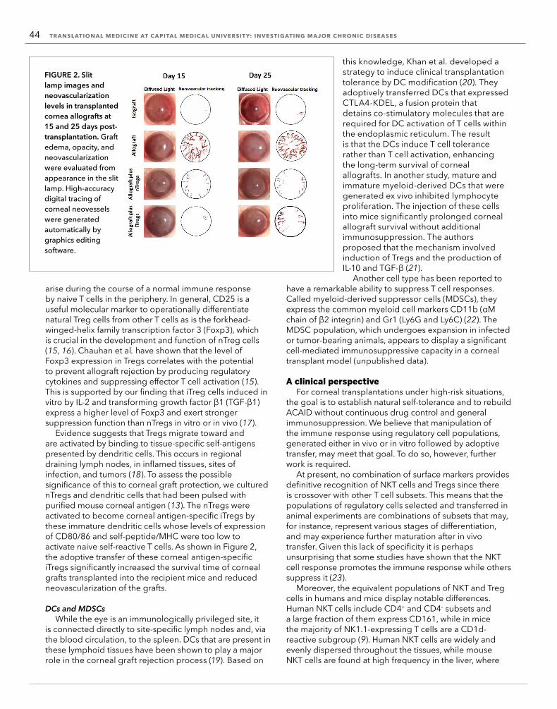
44 TRANSLATIONAL MEDICINE AT CAPITAL MEDICAL UNIVERSITY: INVESTIGATING MAJOR CHRONIC DISEASES
arise during the course of a normal immune response by naive T cells in the periphery. In general, CD25 is a useful molecular marker to operationally differentiate natural Treg cells from other T cells as is the forkhead-winged-helix family transcription factor 3 (Foxp3), which is crucial in the development and function of nTreg cells (15, 16). Chauhan et al. have shown that the level of Foxp3 expression in Tregs correlates with the potential to prevent allograft rejection by producing regulatory cytokines and suppressing effector T cell activation (15). This is supported by our finding that iTreg cells induced in vitro by IL-2 and transforming growth factor β1 (TGF-β1) express a higher level of Foxp3 and exert stronger suppression function than nTregs in vitro or in vivo (17).
Evidence suggests that Tregs migrate toward and are activated by binding to tissue-specific self-antigens presented by dendritic cells. This occurs in regional draining lymph nodes, in inflamed tissues, sites of infection, and tumors (18). To assess the possible significance of this to corneal graft protection, we cultured nTregs and dendritic cells that had been pulsed with purified mouse corneal antigen (13). The nTregs were activated to become corneal antigen-specific iTregs by these immature dendritic cells whose levels of expression of CD80/86 and self-peptide/MHC were too low to activate naive self-reactive T cells. As shown in Figure 2, the adoptive transfer of these corneal antigen-specific iTregs significantly increased the survival time of corneal grafts transplanted into the recipient mice and reduced neovascularization of the grafts.
DCs and MDSCs While the eye is an immunologically privileged site, it
is connected directly to site-specific lymph nodes and, via the blood circulation, to the spleen. DCs that are present in these lymphoid tissues have been shown to play a major role in the corneal graft rejection process (19). Based on
this knowledge, Khan et al. developed a strategy to induce clinical transplantation tolerance by DC modification (20). They adoptively transferred DCs that expressed CTLA4-KDEL, a fusion protein that detains co-stimulatory molecules that are required for DC activation of T cells within the endoplasmic reticulum. The result is that the DCs induce T cell tolerance rather than T cell activation, enhancing the long-term survival of corneal allografts. In another study, mature and immature myeloid-derived DCs that were generated ex vivo inhibited lymphocyte proliferation. The injection of these cells into mice significantly prolonged corneal allograft survival without additional immunosuppression. The authors proposed that the mechanism involved induction of Tregs and the production of IL-10 and TGF-β (21).
Another cell type has been reported to have a remarkable ability to suppress T cell responses. Called myeloid-derived suppressor cells (MDSCs), they express the common myeloid cell markers CD11b (αM chain of β2 integrin) and Gr1 (Ly6G and Ly6C) (22). The MDSC population, which undergoes expansion in infected or tumor-bearing animals, appears to display a significant cell-mediated immunosuppressive capacity in a corneal transplant model (unpublished data).
A clinical perspective For corneal transplantations under high-risk situations,
the goal is to establish natural self-tolerance and to rebuild ACAID without continuous drug control and general immunosuppression. We believe that manipulation of the immune response using regulatory cell populations, generated either in vivo or in vitro followed by adoptive transfer, may meet that goal. To do so, however, further work is required.
At present, no combination of surface markers provides definitive recognition of NKT cells and Tregs since there is crossover with other T cell subsets. This means that the populations of regulatory cells selected and transferred in animal experiments are combinations of subsets that may, for instance, represent various stages of differentiation, and may experience further maturation after in vivo transfer. Given this lack of specificity it is perhaps unsurprising that some studies have shown that the NKT cell response promotes the immune response while others suppress it (23).
Moreover, the equivalent populations of NKT and Treg cells in humans and mice display notable differences. Human NKT cells include CD4+ and CD4– subsets and a large fraction of them express CD161, while in mice the majority of NK1.1-expressing T cells are a CD1d-reactive subgroup (9). Human NKT cells are widely and evenly dispersed throughout the tissues, while mouse NKT cells are found at high frequency in the liver, where
FIGURE 2. Slit lamp images and neovascularization levels in transplanted cornea allografts at 15 and 25 days post-transplantation. Graft edema, opacity, and neovascularization were evaluated from appearance in the slit lamp. High-accuracy digital tracing of corneal neovessels were generated automatically by graphics editing software.
TRANSLATIONAL RESEARCH IN OPTHALMOLOGY AND OTOLARYNGOLOGY 45
The impact of topical drugs on nasal mucociliary functionJian Jiao1,2, Demin Han1,2*, and Luo Zhang1,2,3*
Intranasal administration of drugs has generated much interest within the pharmaceutical industry in recent years as a noninvasive route for delivery. Nasal mucosa provides several advantages for both topical and systemic drug delivery including a large surface area for delivery, rapid onset of therapeutic effect, the potential for central nervous system delivery, lack of first-pass metabolism, and the likelihood for maximal patient comfort and compliance due to the noninvasive nature of the process (1).
Mucociliary clearance is an intrinsic first-line defense mechanism in the respiratory tract that, under normal conditions, removes inhaled particles and irritants such as dust, bacteria, and air pollutants from the airways (2). The driving force of mucociliary clearance is the coordinated and unidirectional beating of epithelial cilia. Mathematical modeling has demonstrated that the energy transferred by cilia to the mucus blanket is proportional to the square of ciliary beat frequency (CBF), while experimental data show that a relatively modest increase of 16% in CBF results in a 56% increase in surface liquid velocity, that is, mucociliary transport (3, 4). These data indicate the importance of CBF as a parameter for assessing ciliary function and mucociliary clearance.
Evidence-based international treatment guidelines recommend the use of intranasal topical corticosteroids, antihistamines, and decongestants for the treatment of rhinitis, sinusitis, and related allergic or chronic nasal conditions (5–7). Intranasal drug preparations, however, are composed of the active drug and various formulation excipients such as preservatives and absorption-enhancing compounds, which individually or in combination may affect mucociliary clearance or CBF of the nasal epithelial cells and thus impact the integrity of the nasal defense mechanisms. This article discusses the current literature and our own findings on the effects of currently available intranasal formulations on nasal mucociliary clearance and CBF.
Effects of topical steroids The use of corticosteroid nasal sprays for the treatment
of allergic rhinitis (and in some cases other forms of
1Department of Otolaryngology, Head and Neck Surgery, Beijing TongRen Hospital, Capital Medical University, Beijing, China2Beijing Key Laboratory of Nasal Diseases, Beijing Institute of Otolaryngology, Beijing, China3Department of Allergy, Beijing Tongren Hospital, Capital Medical University, Beijing, China.*Corresponding Authors: [email protected] (D.H.) and [email protected] (L.Z.)
they represent 10% to 40% of lymphocytes and are present at much lower frequencies (typically less that 1% of lymphocytes) in thymus, bone marrow, spleen, lymph node, and blood (5). A fatal disorder known as immune dysregulation, polyendocrinopathy, enteropathy, X-linked syndrome (IPEX) in humans was found to be caused by mutations of FOXP3 (called Scurfy in mice lacking Foxp3) (16). Although the identification and characterization of NKT and Treg cells in mice has been extensive, the significance of species-specific differences in the frequency of subpopulations remains unclear.
The transfer of immune cell populations appears not to have severe side effects in mouse models; however, a greater understanding of the cellular and molecular microenvironment changes that result from the injection of regulatory cells would be helpful for establishing new strategies for the treatment and prevention of immunological diseases in human patients. Moreover, this knowledge could improve our ability to affect a wide spectrum of physiological immune responses associated with corneal transplantation.
References 1. A. J. George, D. F. Larkin, Am. J. Transplant. 4, 678 (2004). 2. J. Y. Niederkorn, J. Leukoc. Biol. 74, 167 (2003). 3. J. Y. Niederkorn, Curr. Eye Res. 32, 1005 (2007). 4. J. Y. Niederkorn, Curr. Opin. Allergy Clin. Immunol. 10, 493 (2010). 5. D. I. Godfrey, H. R. MacDonald, M. Kronenberg, M. J. Smyth, L. Van Kaer, Nat. Rev. Immunol. 4, 231 (2004). 6. J. L. Matsuda et al., Proc. Natl. Acad. Sci. U.S.A. 100, 8395 (2003). 7. K. H. Sonoda, M. Taniguchi, J. Stein-Streilein, J. Immunol. 168, 2028 (2002). 8. K. H. Sonoda, M. Exley, S. Snapper, S. P. Balk, J. S. Stein- Streilein, J. Exp. Med. 190, 1215 (1999). 9. Z. Pan et al., Invest. Ophthalmol. Vis. Sci. 44, 3346 (2003). 10. Y. Liu, Y. Jie, Z. Pan, Y. Wu, Ophthalmol. CHN 16, 175 (2007).11. G. Benichou, Y. Yamada, A. Aoyama, J. C. Madsen, Curr. Opinion. Organ. Transplant. 16, 47 (2011).12. S. Sakaguchi, T. Yamaguchi, T. Nomura, M. Ono, Cell 133, 775 (2008).13. Y. He et al., Cornea 29, S25-S31 (2010).14. C. A. Piccirillo et al., J. Exp. Med. 195, 237 (2002).15. S. K. Chauhan, D. R. Saban, H. K. Lee, R. Dana, J. Immunol. 182, 148 (2009).16. J. D. Fontenot, M. A. Gavin, A. Y. Rudensky, Nat. Immunol. 4, 330 (2003).17. X. Guo et al., Human Immunol. 73, 1061 (2012).18. Y. Belkaid, C. A. Piccirillo, S. Mendez, E. M. Shevach, D. L. Sacks, Nature 420, 502 (2002).19. J. V. Forrester, H. Xu, L. Kuffová L, A. D. Dick, P. G. McMenamin, Immunol. Rev. 234, 282 (2010).20. A. Khan et al., Eur. J. Immunol. 43, 734 (2013). 21. L. O’Flynn et al., Mol. Ther. 21, 2102 (2013). 22. D. Wang et al., Biol. Blood. Marrow. Transplant. 19, 692 (2013).23. T. Kenna et al., J. Immunol. 171, 1775 (2003).

46 TRANSLATIONAL MEDICINE AT CAPITAL MEDICAL UNIVERSITY: INVESTIGATING MAJOR CHRONIC DISEASES
rhinosinusitis) is widely accepted due to the combination of their efficacy, tolerability, and ease of use. However, the debate over potential side effects on nasal structure and ciliary function has going on for many years. Some of the current, commercially available intranasal corticosteroids with widespread use include budesonide, triamcinolone acetonide, fluticasone propionate/furoate, and mometasone furoate (8), along with the newer nasal aerosol preparations beclomethasone (9, 10) and ciclesonide (11, 12).
Several studies investigating the long-term use of intranasal corticosteroids on the nasal mucosa have demonstrated histologically that the structural integrity of the nasal mucosa is preserved after prolonged exposure to these agents; indeed, some studies have demonstrated an improvement in the overall health of the nasal mucosa (13–17).
Comparatively few studies have investigated the influence of intranasal corticosteroids on mucociliary clearance and ciliary function. Klossek et al. performed a prospective, randomized, multi-institutional study addressing the effect of long-term treatment with triamcinolone acetonide (220 µg/day) in patients with perennial allergic rhinitis (14). They demonstrated that there was no atrophy of the nasal mucosa or impairment of mucociliary function after six months of treatment, as indicated by use of the indigocarmine saccharine test. Similarly, Naclerio et al. compared the effect of treatment for two weeks with either budesonide or mometasone on nasal mucociliary function using a radiotracer technique and found that these drugs also did not alter mucociliary clearance (18). Using an excised ciliated chicken trachea tissue model, Merkus et al. showed that although most of the widely prescribed intranasal steroid formulations—such as fluticasone, mometasone furoate, budesonide, and triamcinolone acetonide—reduced CBF of the tracheal preparation, this effect was partially or entirely reversible following a wash-out of the intranasal formulation (19). Hofmann et al. employed cultured human nasal ciliated epithelial cells to investigate the effect of budesonide, fluticasone propionate, and mometasone furoate on CBF of these cultures using a photometer (20). The authors reported that the budesonide spray, containing the preservative potassium sorbate, did not affect the CBF at 10% dilution and showed moderate reversible decrease of CBF at 50% dilution. In contrast, the fluticasone propionate and mometasone furoate sprays, containing the preservative benzalkonium chloride, caused a reversible decrease of CBF at 10% dilution and a complete, irreversible standstill at 50% dilution.
Using high-speed digital imaging methods combined with a beat-by-beat CBF analysis, we have recently shown that budesonide caused a rapid but reversible ciliostasis at undiluted therapeutic concentrations, a gradual but fully reversible decrease of CBF at 50% dilution, and no ciliotoxic effect at 10% dilution in primary cultures of human nasal epithelial cells. Similarly, fluticasone propionate induced irreversible ciliostatic activity when used undiluted and at up to 50% dilution of therapeutic
concentration, and a reversible decrease of CBF at 10% dilution in this culture model (21).
Effects of topical antihistamines Intranasal antihistamines, along with topical
corticosteroids, have been established as first-line therapies in patients with allergic rhinitis (5, 6). In contrast to intranasal corticosteroids, which are considered the most effective therapy for allergic rhinitis, intranasal antihistamines have a more rapid onset of action. Furthermore, second generation antihistamines are also highly potent and selective H1-receptor antagonists, with many of these compounds possessing additional anti-allergic and anti-inflammatory properties (22). Studies investigating the effects of intranasal antihistamines on nasal ciliary function, however, have provided contradictory results. In particular, while some studies have demonstrated no adverse effects of intranasal formulations containing azelastine or levocabastine on ciliary activity either in vitro or in vivo (23–25), other studies have reported that formulations containing azelastine and levocabastine do produce ciliotoxic effects in the human nasal epithelium (26, 27).
We have investigated the effect of azelastine hydrochloride and levocabastine hydrochloride nasal sprays on CBF of human nasal epithelial cell cultures. Our data showed that undiluted aqueous azelastine causes a rapid, irreversible ciliostasis, but a 10% dilution resulted in a gradual but reversible decrease in CBF. In contrast, no ciliotoxic effect was observed for concentrations up to 5% dilution of azelastine spray. Levocabastine spray showed comparable effects on CBF: there was induction of an irreversible ciliostasis at an undiluted concentration, a minor reversible decrease of CBF at 10% dilution, and no effect on CBF at up to 5% dilution of levocabastine spray (21).
Effects of topical decongestants Nasal decongestants are widely used to relieve
allergic rhinitis and acute or chronic rhinitis-induced nasal obstruction by reducing nasal blood flow via vasoconstriction. Clinically, nasal decongestants are divided into two groups: sympathomimetic amines (such as ephedrine and phenylephrine) and imidazoline derivatives (oxymetazoline hydrochloride and xylometazoline hydrochloride) (29). Ephedrine acts as a nonselective α, β-adrenoceptor agonist, while phenylephrine, oxymetazoline, an d xylometazoline act as selective α-adrenoceptor agonists.
Phenylephrine, oxymetazoline and xylometazoline have been reported to inhibit both ciliary movement in the human airway (30–32) and airway ciliary activity in several animal models, including chicken, rabbit, and rat (26, 33, 34). Similarly, ephedrine has been shown to have an inhibitory effect on rat tracheal CBF (26). Phillips et al. investigated the effect of phenylephrine on nasal ciliary function in the noses of healthy volunteers. They demonstrated that, while this drug significantly increased CBF in vivo, it had a ciliostimulatory effect at
TRANSLATIONAL RESEARCH IN OPTHALMOLOGY AND OTOLARYNGOLOGY 47
low concentration (0.01%) and a cilioinhibitory effect at higher concentrations (0.25% and 0.5%) in vitro (32). Another investigation of the effects of phenylephrine (0.5%) and tetrahydrozoline decongestant sprays on nasal ciliary function in healthy nonsmokers reported that both of these agents significantly increased nasal mucus velocity, compared to topical application of phenylephrine and tetrahydrozoline vehicles, normal saline, and a sham treatment (empty aerosol container) (35).
Data from our laboratory have also indicated that ephedrine induces an instant and moderate increase in human nasal CBF followed by an inhibitory response. Moreover, while the increment by which CBF increased was independent of ephedrine concentrations ranging from 0.25% to 2.0%, the inhibitory effect was shown to be concentration-dependent. Our findings suggest that the concentration at which ephedrine is used in the clinic (0.5%) has a maximum stimulatory effect without an obvious inhibitory effect on human nasal CBF (36).
We also investigated the effects of oxymetazoline on human nasal CBF and its influence on mucociliary transport (37). At lower concentrations, 0.025% or 0.05%, oxymetazoline did not significantly alter CBF, while higher concentrations, 0.10% and 0.20%, significantly decreased CBF. Furthermore, 0.05% oxymetazoline increased the mean human nasal mucociliary transport time.
Effects of intranasal formulation preservatives Preservatives are indispensable components of
traditional multidose nasal formulations due to the necessity for repeated administration and because their aqueous nature makes formulations susceptible to microorganism infestation. Benzalkonium chloride (BKC), a quaternary ammonium compound, was approved for use as a preservative by the U.S. Food and Drug Administration in 1982 and is by far the most commonly used preservative in a variety of prescription and over-the-counter products. A MEDLINE literature search for publications addressing the issue of BKC safety shows that results are ambiguous. There are both short-term and long-term studies demonstrating that BKC has no toxic effects on the nasal mucosa, but are also several studies that report BKC causes degenerative changes in human nasal epithelia, as measured by CBF, ciliary morphology, and mucociliary clearance (38). Our studies have demonstrated that BKC inhibits CBF in a concentration-dependent manner in cultured rabbit tracheal ciliated cells, with ciliary activity stopping after exposure for 5 minutes to 0.01% BKC (39). We have confirmed the toxic effect of BKC on CBF in human nasal ciliated cells, and additionally demonstrated that BKC causes an irreversible ciliostasis at concentrations of 0.005% or 0.01% (21).
It is important to note that there is a striking difference between in vitro and in vivo data for the safety of BKC. While data generated in vitro raises concerns regarding the safety of BKC, in vivo data generally indicates that BKC is safe. This discrepancy may result from the differences in the local environmental conditions of the nasal mucosal cells in vitro and in vivo. In particular, numerous intrinsic
mechanisms which protect the ciliated epithelium in the nasal cavity are missing in vitro. Furthermore, evidence that BKC can be inactivated by nonionic surfactants raises the possibility that in vivo BKC may be neutralized by the surfactant properties of nasal mucus (40). Additionally, the mucus layer leads to variable dilutions of topically administered drugs. As the healthy human nose contains about 0.4 mL mucus 41and in clinical practice spray dose volumes are generally in the order of 0.1 mL, the nasal products are diluted fourfold after delivery into the nose (41). The acidic environment of the nasal cavity is another local factor that is likely to contribute to amelioration of exogenous drug toxicity. An in vitro study reported that the toxic effect of BKC on CBF was significantly attenuated when the pH was decreased (42).
Potassium sorbate, a white crystalline powder used to inhibit mold, yeast, and bacterial growth in foods, cosmetics, and drug preparations is another commonly used preservative in nasal formulations. In contrast to the ciliotoxic effects of BKC, studies investigating the effects of potassium sorbate (20, 39, 43) have not shown any toxic effects on CBF at concentrations used clinically. Potassium sorbate can therefore be considered to be a safe preservative for use in topical formulation.
In summary, nasal mucociliary clearance is an important defense mechanism of the upper respiratory tract. A review of the published literature and our own studies suggest that intranasal topical formulations, including topical steroids, decongestants, antihistamines, and various formulation excipients, such as preservatives, may influence nasal mucociliary clearance or nasal mucosal ciliary function. Thus, an investigation of the influence and safety of topical drugs on mucocililary clearance and ciliary function is of great importance in the development of new nasal drugs and selection of appropriate safe excipients.
References 1. H. R. Costantino, L. Illum, G. Brandt, P. H. Johnson, S. C. Quay, Int. J. Pharm. 337, 1 (2007). 2. D. A. Gudis, N. A. Cohen, Otolaryngol. Clin. North. Am. 43, 461 (2010). 3. A. Silberberg, Biorheology 27, 295 (1990). 4. Z. V. Seybold et al., J. Appl. Physiol. 68, 1421 (1990). 5. J. Bousquet et al., Allergy 63 (Suppl 86), 8 (2008). 6. J. L. Brozek et al., J. Allergy Clin. Immunol. 126, 466 (2010). 7. M. Desrosiers et al., Allergy Asthma. Clin. Immunol. 7, 2 (2011). 8. M. S. Benninger, N. Ahmad, B. F. Marple, Otolaryngol. Head Neck Surg. 129, 739 (2003). 9. E. O. Meltzer et al., Allergy. Asthma. Proc. 33, 249 (2012).10. G. D. Raphael et al., Curr. Med. Res. Opin. 29, 1329 (2013).11. D. Mohar et al., Allergy. Asthma. Proc. 33, 19 (2012).12. R. L. Jacobs, Expert. Rev. Clin. Immunol. 7, 735 (2011).13. D. J. Verret, B. F. Marple, Curr. Opin. Otolaryngol. Head Neck Surg. 13, 14 (2005).14. J. M. Klossek, F. Laliberte, M. F. Laliberte, N. Mounedji, J. Bousquet, Rhinology 39, 17 (2001).15. F. M. Baroody et al., Arch. Otolaryngol. Head Neck Surg. 127, 193 (2001).

48 TRANSLATIONAL MEDICINE AT CAPITAL MEDICAL UNIVERSITY: INVESTIGATING MAJOR CHRONIC DISEASES
The epigenetics of allergic rhinitisYuan Zhang1,2, Xu Zhang2, Demin Han1,2*, and Luo Zhang1,2,3*
A llergic rhinitis (AR) is an inflammatory disease of the nasal mucosa induced by an immunoglobulin E (IgE) - mediated reaction in allergen - sensitized subjects. Over the last decade, AR has increased in prevalence worldwide (1–4). The plethora of evidence linking it with asthma has led to the concept of “the united air way,” also known as “one airway, one disease.” AR is widely agreed to be an independent risk factor for the development of asthma (5, 6). Barnes has proposed that since allergic diseases, such as asthma, AR, and atopic dermatitis share common systemic characteristics (for example, high total and/or specific IgE), the same susceptibility genes likely contribute to the allergic process, regardless of the specific clinical phenotype. Thus, the many genetic linkages that have been reported to be associated with asthma may not be specific for asthma per se, but rather may be reflective of an overall predisposition for allergic disease (7).
Genetic architecture of allergic diseaseIt has long been recognized that asthma and asthma-
related traits run strongly in families and have a clear hereditary component (8). Furthermore, it is believed that allergic diseases which include AR as a comorbid condition result from the effect of multiple interacting genetic and environmental factors (9). Genetic studies of allergic diseases over the years have involved mainly three tools: genome—wide linkage studies, candidate—gene association studies, and genome-wide association studies (GWASs).
The first GWAS for an asthma trait, which represented a quantum leap for the field, was published in 2007 (10). Since then, more than 60 susceptibility genes involving almost 150 loci have been reported to be associated with asthma through GWASs. Four years later, the first GWAS to specifically investigate AR was conducted in a cohort of Singaporean Chinese subjects (11). This study reported two novel candidate genes for atopy and AR: mitochondrial ribosomal protein L4 (MRPL4) and B-cell adaptor for phosphatidylinositol 3-kinase (BCAP). More
1Department of Otolaryngology, Head and Neck Surgery, Beijing Tongren Hospital, Capital Medical University, Beijing, China2Beijing Key Laboratory of Nasal diseases, Beijing Institute of Otolaryngology, Beijing, China3Department of Allergy, Beijing Tongren Hospital, Capital Medical University, Beijing, China*Corresponding Authors: [email protected] (D.H.) and [email protected] (L.Z.)
16. U. Pipkorn, J. Pukander, J. Suonpaa, J. Makinen, N. Lindqvist, Clin. Allergy 18, 253 (1988).17. F. Laliberte et al., Allergy 55, 718 (2000).18. R. M. Naclerio, F. M. Baroody, N. Bidani, M. De Tineo, B. C. Penney, Otolaryngol. Head Neck Surg. 128, 220 (2003).19. P. Merkus, S. G. Romeijn, J. C. Verhoef, F. W. Merkus, P. F. Schouwenburg, Laryngoscope 111, 595 (2001).20. T. Hofmann, M. Gugatschga, B. Koidl, G. Wolf, Arch. Otolaryngol. Head Neck Surg. 130, 440 (2004).21. J. Jiao, N. Meng, L. Zhang, ORL. J. Otorhinolaryngol. Relat. Spec. 76, 127 (2014).22. L. Zhang, L. Cheng, J. Hong, Pharmacology 92, 14 (2013).23. U. Achterrath-Tuckermann et al., Lung 170, 201 (1992).24. U. Achterrath-Tuckermann et al., Arzneimittelforschung 47, 1230 (1997).25. F. W. Merkus, M. T. Schusler-van Hees, Allergy 47, 230 (1992).26. X. Y. Su, A. Li Wan Po, J. Clin. Pharm. Ther. 18, 219 (1993).27. T. Hofmann, G. Wolf, B. Koidl, HNO 46, 146 (1998).28. J. Alberty, W. Stoll, Allergy 53, 986 (1998).29. D. Passali, L. Salerni, G. C. Passali, F. M. Passali, L. Bellussi, Expert. Opin. Drug Saf. 5, 783 (2006).30. W. M. Boek et al., Acta. Otolaryngol. 119, 93 (1999).31. Y. G. Min et al., Laryngoscope 108, 418 (1998).32. P. P. Phillips, T. V. McCaffrey, E. B. Kern, Otolaryngol. Head Neck Surg. 103, 558 (1990).33. J. P. Dudley, J. D. Cherry, Laryngoscope 88, 110 (1978).34. J. C. Hybbinette, U. Mercke, Acta. Otolaryngol. 94, 121 (1982).35. K. Saketkhoo, B. M. Yergin, A. Januszkiewicz, K. Kovitz, M. A. Sackner, Am. Rev. Respir. Dis. 118, 251 (1978).36. L. Zhang et al., ORL. J. Otorhinolaryngol. Relat. Spec. 70, 91 (2008).37. L. Zhang, D. Han, X. Song, K. Wang, H. Wang, Ann. Otol. Rhinol. Laryngol. 117, 127 (2008).38. B. Marple, P. Roland, M. Benninger, Otolaryngol. Head Neck Surg. 130, 131 (2004).39. C. Wang, Q. Deng, D. Han, L. Zhang, ORL. J. Otorhinolaryngol. Relat. Spec. 74, 149 (2012).40. K. Khanvilkar, M. D. Donovan, D. R. Flanagan, Adv. Drug Deliv. Rev. 48, 173 (2001).41. P. J. Stanley, R. Wilson, M. A. Greenstone, I. S. Mackay, P. J. Cole, Br. J. Dis. Chest 79, 77 (1985).42. H. J. M. van de Donk, Pharma. Weekblad. Sci. Ed. 5, 2 (1983).43. C. Y. Ho, M. C. Wu, M. Y. Lan, C. T. Tan, A. H. Yang, Am. J. Rhinol. 22, 125 (2008).
AcknowledgmentsThis work was supported by the Program for Changjiang Scholars and Innovative Research Team (IRT13082), the National Science Fund for Distinguished Young Scholars and for the Major International Joint Research Program (81025007 and 81420108009), the National Natural Science Foundation of China (81100704), the Beijing Natural Science Foundation (7131006), the Ministry of Health Foundation (201202005), the Beijing Nova Program (Z111107054511061), the Specialized Research Fund for the Doctoral Program of Higher Education of China (20111107120004), the Capital Health Research and Development of Special (2011-1017-03), and the Science Foundation for High-Level Medical Talents of Beijing Health System (2009-02-007).

TRANSLATIONAL RESEARCH IN OPTHALMOLOGY AND OTOLARYNGOLOGY 49
recently, Ramasamy and colleagues conducted a meta-analysis of existing GWAS data of single nucleotide polymorphisms among four large European adult cohorts to identify common genetic variants associated with prevalent AR and grass sensitization. They identified three loci that were strongly associated with both AR and grass sensitization phenotypes: the human leukocyte antigen (HLA) variant rs7775228; variants in chromosome 11 open reading frame 30 (C11orf30) and leucine rich repeat containing 32 (LRRC32); and variant rs17513503 located near transmembrane protein 232 (TMEM232) and solute carrier family 25, member 46 (SLC25A46) (12). Another recently published meta-analysis of GWASs compared both physician-diagnosed asthma and rhinitis with healthy control subjects (13). This study indicated that at least 11 independent variants may be associated with the risk of having asthma with hay fever, 2 of which were shown to have reached the level of genome-wide significance with allergic disease.
GWASs have provided valuable insights into the genetic architecture of allergic disease. However, most variants identified so far confer relatively small increments in risk and explain only a small proportion of familial clustering. A substantial component of the genetic profile of the disease is still “missing” (14). Several explanations for this missing heritability have been put forward, which are not necessarily mutually exclusive. One factor is that GWAS is reliant on the assessment of common haplotype blocks and the genotyping of common variants. This restricts the ability to detect rare risk alleles that might otherwise be identified as contributing to the disease (15). Copy number variations provide another explanation: segmentally duplicated sequences in the genome, which may contribute a sizeable effect on the variability of gene expression, are not identified using a GWAS approach (16). There is also increasing evidence for the existence of epigenetic mechanisms, which likewise cannot be identified by GWAS or next generation sequencing of GWAS. To us, this may be the biggest contributory factor, particularly as the ability of environmental factors to shape health and disease mainly involves epigenetic mechanisms (17, 18).
EpigeneticsEpigenetics refers to the heritable changes in gene
expression that occur in the absence of changes to the DNA sequence itself. These changes may be induced by environmental factors and transmitted through generations via a number of mechanisms, including DNA methylation, modification of histone tails, chromatin remodeling, and noncoding RNAs (19). The influence of regulatory microRNAs (miRNAs) on gene transcription is also increasingly recognized as a key mechanism of epigenetic gene regulation (17, 20). Conventional gene-environment interaction studies strive to understand how individuals with different genotypes respond to various environmental factors and how these responses change over time. Such research efforts have highlighted the important contribution of both genetic and environmental
variability in human diseases; however, a full understanding of gene-environment interactions requires that epigenetic mechanisms also be taken into account (21). For example, epigenetics may explain immune modulation induced by the environment, even when it is not possible to measure the inciting environmental exposure itself. Indeed, it has been suggested that a disease and its phenotypic variability may be due to the environmental origins and, possibly, related to epigenetic mechanisms (22).
Several types of environmental exposure have been associated with an increased risk of allergic disease or altered immune effects on the fetus, including maternal medications in pregnancy, smoking, and pollutants. Exposure to cigarette smoke is particularly well recognized as inducing epigenetic changes that alter gene expression, and the finding that grandmaternal smoking increases the risk of childhood asthma in grandchildren supports the concept that transgenerational epigenetic effects (mediated by DNA methylation) might also be operating in allergic diseases (23). Moreover, a recent study in children demonstrated that secondhand smoke in combination with ambient air pollution exposure is associated with increased CpG methylation and decreased expression of both interferon (IFN)-g in T effector cells and forkhead box P3 (Foxp3) in T regulatory cells (24). Similarly, exposure to inhaled traffic-related pollutants has been demonstrated in humans and animal models to alter methylation patterns in several genes. The findings that exposure to inhaled traffic pollutants during pregnancy is associated with changes in DNA methylation and to higher parental reporting of early childhood asthma symptoms is of particular interest (25). Indeed, increased environmental particulate exposure from traffic pollution results in a dose-dependent increase in peripheral blood DNA methylation (26). A community-based study by Sofer and colleagues revealed a correlation between the methylation patterns of specific genes related to asthma and the level of exposure subjects experienced to the airborne particulates black carbon and sulfate (27). These results led the authors to conclude that the effect of air pollution on asthmatic and respiratory responses may be mediated through gene methylation. In a follow-up study, increased concentrations of air pollutants was significantly associated with hypomethylation of tissue factor (F3), intercellular adhesion molecule (ICAM)-1, and toll-like receptor (TLR)-2 gene promoter regions, and hypermethylation of IFN-g and interleukin (IL)-6 gene promoter regions (28). Other studies have shown that several additional atopy- and asthma-related genes are susceptible to epigenetic regulation, including genes important to T-effector pathways (IFN-g, IL-4, IL-13, IL-17) (29–38), T-regulatory pathways (FoxP3) (24, 39), airway inflammation [inducible nitric oxide synthase (iNOS)], and CD14 (40, 41).
Recently it has been postulated that epigenetic mechanisms might mediate the effects of behavioral and environmental exposures early in life, as well as lifelong environmental exposures and the susceptibility to allergic disease later in life (42). Furthermore, Murphy and

50 TRANSLATIONAL MEDICINE AT CAPITAL MEDICAL UNIVERSITY: INVESTIGATING MAJOR CHRONIC DISEASES
colleagues have proposed that stress might be a possible link between genetics, epigenetics, and childhood asthma (43, 44).
miRNAs Among the mediators of epigenetic effects, a class of
small noncoding RNAs (19 to 25 nucleotides in length) that can regulate genes through effects on mRNA stability and translation is currently attracting a lot of attention (45). These miRNAs appear to regulate homeostatic immune architecture and acquired immunity, as well as many other functions. Recent studies of miRNA profiles for patients with various allergic inflammatory diseases have identified critical roles for these molecules in regulating the pathogenic mechanisms in allergic inflammation (46), including the polarization of adaptive immune responses and activation of T cells [miR-21 (47–49) and miR-146 (50)]; regulation of eosinophil development [miR-21 (51) and miR-223 (52)], and modulation of IL-13–driven epithelial responses [miR-375 (53–55)].
The role of specific miRNAs in AR was first investigated by Chen and colleagues. They screened the expression of 157 miRNAs in mononuclear leukocytes from human umbilical cord blood samples and identified three miRNAs that were downregulated in the cord blood samples that showed increased levels of IgE. Two of these, namely miR-21 and miR-26, were found to be reduced in monocytes from children with AR. Downregulation of miR-21 expression was also associated with significantly increased expression of transforming growth factor, beta receptor II (TGFβR2) on monocytes from cord blood with raised IgE and from AR patients, and transfection of miR-21 precursor into monocytes from AR patients increased miR-21 expression and decreased TGFβR2 expression. Collectively, these experiments suggest that lower miR-21 expression in cord blood and increased TGFβR2 expression is likely to be associated with antenatal IgE production and the development of AR (49).
An analysis of miRNA expression in the nasal mucosa using biopsy specimens from AR and nonallergic patients identified 421 differentially expressed miRNAs, nine of which showed a greater than twofold difference. Using reverse transcription polymerase chain reaction (RT-PCR) the authors confirmed that hsa-miR-224, hsa-miR-187, and hsa-miR-143 were downregulated in AR, suggesting that they were involved in the pathogenesis of the disorder (56). Zhang and colleagues investigated the differential expression of miRNA in sinonasal mucosa from controls, patients with chronic rhinosinusitis (CRS) without nasal polyps (CRSsNP), and patients with eosinophilic CRS with nasal polyps (CRSwNP). They reported an overexpression of miR-125b specifically in patients with eosinophilic CRSwNP. Moreover, miR-125b exerted a crucial role as a regulator of innate immunity via the miR-125b-EIF4E-binding protein 1 IFN pathway for mucosal eosinophilia in such patients (57). Another study from this same group assessed the expression of miRNA machinery proteins in different types of CRS. This work revealed that PACT, a protein activator of the interferon-induced protein
kinase that is associated the miRNA machinery, was significantly upregulated in CRSwNP patients compared with control and CRSsNP patients. The expression of PACT was correlated with the severity of disease and of eosinophil infiltration and the authors proposed that PACT may be involved in plasma cell function and eosinophilic inflammation in CRSwNP (58).
ConclusionsThe field of genetics continues to evolve, expanding
our knowledge of the aetiology of AR and other allergic diseases. Epigenetics is still a relatively new concept, even though developmental biologist Conrad Waddington first envisioned the epigenetic landscape as a metaphor more than 50 years ago. Over this period, the concept has evolved from a collection of diverse phenomena to a defined and far-reaching field of study, one that Goldberg and colleagues described as the “bridge between genotype and phenotype” (59). Specifically, epigenetics serves as a link between nature and nurture by bridging environmental exposures in early life with gene regulation, phenotypic changes, and diseases in adults (21). The challenge ahead is to understand how genetic variation, epigenetic regulation, and environmental factors interact to cause allergic disease.
References 1. J. Bousquet et al., Allergy 63 (Suppl 86), 8 (2008). 2. V. Bauchau, S. R. Durham, Eur. Respir. J. 24, 758 (2004). 3. R. A. Nathan et al., Allergy Asthma Proc. 29, 600 (2008). 4. L. Zhang et al., Int. Arch. Allergy Immunol. 149, 47 (2009). 5. C. Bachert, M. Jorissen, B. Bertrand, N. Khaltaev, J. Bousquet, B-ENT 4, 253 (2008). 6. J. Grossman, Chest 111, 11S (1997). 7. K. C. Barnes, J. Allergy Clin. Immunol. 106, S192 (2000). 8. H. Los, P. E. Postmus, D. I. Boomsma, Twin Res. 4, 81 (2001). 9. J. W. Holloway, I. A. Yang, S. T. Holgate, J. Allergy Clin. Immunol. 125, S81 (2010).10. M. F. Moffatt et al., Nature 448, 470 (2007).11. A. K. Andiappan et al., PLOS ONE 6, e19719 (2011).12. A. Ramasamy et al., J. Allergy Clin. Immunol. 128, 996 (2011).13. M. A. Ferreira et al., J. Allergy Clin. Immunol. 133, 1564 (2014).14. T. A. Manolio et al., Nature 461, 747 (2009).15. K. A. Frazer, S. S. Murray, N. J. Schork, E. J. Topol, Nat. Rev. Genet. 10, 241 (2009).16. C. N. Henrichsen, E. Chaignat, A. Reymond, Hum. Mol. Genet. 18, R1 (2009).17. B. Maher, Nature 456, 18 (2008).18. D. Vercelli, J. Allergy Clin. Immunol. 113, 381 (2004).19. A. Bird, Nature 447, 396 (2007).20. M. I. McCarthy, J. N. Hirschhorn, Hum. Mol. Genet. 17, R156 (2008).21. D. C. Dolinoy, Nutr. Rev. 66 (Suppl 1), S7 (2008).22. S. Lovinsky-Desir, R. L. Miller, Curr. Allergy Asthma Rep. 12, 211 (2012).23. Y. F. Li, B. Langholz, M. T. Salam, F. D. Gilliland, Chest 127, 1232 (2005).24. A. Kohli et al., Clin. Epigenetics 4, 17 (2012).
TRANSLATIONAL RESEARCH IN OPTHALMOLOGY AND OTOLARYNGOLOGY 51
25. F. Perera et al., PLOS ONE 4, e4488 (2009).26. A. Baccarelli et al., Am. J. Respir. Crit. Care Med. 179, 572 (2009).27. T. Sofer et al., Epigenomics 5, 147 (2013).28. M. A. Bind et al., Epigenetics 9, 448 (2014).29. J. Liu et al., Toxicol. Sci. 102, 76 (2008).30. R. B. Webster, Y. Rodriguez, W. T. Klimecki, D. Vercelli, J. Biol. Chem. 282, 700 (2007).31. G. P. White et al., Pediatr. Allergy Immunol. 17, 557 (2006).32. B. Jones, J. Chen, EMBO J. 25, 2443 (2006).33. J. R. Schoenborn et al., Nat. Immunol. 8, 732 (2007).34. A. M. Akimzhanov, X. O. Yang, C. Dong, J. Biol. Chem. 282, 5969 (2007).35. V. K. Patil et al., Clin. Epigenetics 5, 22 (2013).36. N. Soto-Ramirez et al., Clin. Epigenetics 5, 1 (2013).37. A. T. Ooi et al., Am. J. Transl. Res. 4, 219 (2012).38. W. Y. Tang et al., Environ. Health Perspect. 120, 1195 (2012).39. J. Huehn, J. K. Polansky, A. Hamann, Nat. Rev. Immunol. 9, 83 (2009).40. M. C. Munthe-Kaas et al., Pediatr. Allergy Immunol. 23, 747 (2012).41. G. G. Slaats et al., Allergy 67, 895 (2012).42. M. Szyf, M. J. Meaney, Allergy Asthma Clin. Immunol. 4, 37 (2008).43. S. K. Murphy, J. W. Hollingsworth, Am. J. Respir. Crit. Care Med. 187, 563 (2013).44. R. J. Wright, Immunol. Allergy Clin. North Am. 31, 19 (2011).45. J. Winter, S. Jung, S. Keller, R. I. Gregory, S. Diederichs, Nat. Cell Biol. 11, 228 (2009).
46. T. X. Lu, M. E. Rothenberg, J. Allergy Clin. Immunol. 132, 3 (2013).47. T. X. Lu, A. Munitz, M. E. Rothenberg, J. Immunol. 182, 4994 (2009).48. T. X. Lu et al., J. Allergy Clin. Immunol. 129, 1064 (2012).49. R. F. Chen et al., Clin. Exp. Allergy 40, 1482 (2010).50. K. D. Taganov, M. P. Boldin, K. J. Chang, D. Baltimore, Proc. Natl. Acad. Sci. U.S.A. 103, 12481 (2006).51. C. K. Wong et al., Immunobiology 218, 255 (2013).52. T. X. Lu et al., J. Immunol. 190, 1576 (2013).53. B. Bleck et al., J. Immunol. 190, 3757 (2013).54. T. X. Lu et al., Mucosal Immunol. 5, 388 (2012).55. M. Biton et al., Nat. Immunol. 12, 239 (2011).56. Y. Shaoqing et al., Am. J. Rhinol. Allergy 25, e242 (2011).57. X. H. Zhang et al., Am. J. Respir. Crit. Care Med. 185, 140 (2012).58. Y. N. Zhang, P. P. Cao, X. H. Zhang, X. Lu, Z. Liu, Laryngoscope 122, 2621 (2012).59. A. D. Goldberg, C. D. Allis, E. Bernstein, Cell 128, 635 (2007).
AcknowledgmentsThis work was supported by grants from National Science Fund for Distinguished Young Scholars and for the Major International Joint Research Program (81025007 and 81420108009), Program for Changjiang Scholars and Innovative Research Team (IRT13082), National Natural Science Foundation of China (30973282, 81100706, and 81441031), Ministry of Health Foundation (201202005), Beijing Natural Science Foundation (7131006), and Beijing Nova Program (2010B022).

52 TRANSLATIONAL MEDICINE AT CAPITAL MEDICAL UNIVERSITY: INVESTIGATING MAJOR CHRONIC DISEASES
miRNAs associated with metastasis in colorectal cancerJie Yin, Jun Cai, Zhigang Bai, Jun Zhang, Jin Wang, Fei Zheng, and Zhongtao Zhang*
Colorectal cancer (CRC) is the third most common cancer and the third leading cause of cancer-related deaths worldwide. Complications arising from metastasis are the major cause of CRC-related deaths. Effective new treatments that target metastatic disease would have a substantial impact on morbidity and mortality; however, the molecular mechanisms underlying metstasis are not yet completely understood. Knowledge of CRC metastasis would be enhanced by the development of novel screening tools and diagnostic biomarkers. Recently, a family of small regulatory RNAs, called microRNAs (miRNAs), has emerged as possible serum markers for
human diseases, including cancers, due to their relative stability in the circulation (1). This article describes the progress made thus far in the application of miRNAs to the study of metastatic CRC.
Introducing miRNAsmiRNAs are small, noncoding RNAs of 18–22
nucleotides in length. More than 900 human miRNAs have been identified to date (2), and each has been assigned a numerical identifier. They regulate gene expression by interfering with transcription or inhibiting translation (3) and are known to be involved in the regulation of cell differentiation, cell cycle progression, and apoptosis pathways (4). Studies have shown that the expression levels of certain miRNAs differ between normal and tumor tissues as well as among tumor types (5).
The progression from primary tumor to metastatic lesion is a multistep process that involves local invasion, intravasation, survival in the circulatory system, extravasation, and colonization of a secondary anatomical site (6). Researchers have identified miRNAs that participate in specific steps of this process by either promoting or suppressing metastasis. These miRNAs are specific to metastatic development and are not involved in tumorigenesis, prompting Hurst and colleagues to coin the term “metastamir” to describe them (7). Our group focuses on the correlations between miRNAs and metastasis of CRC with the goal of revealing metastasis-
Department of General Surgery, Beijing Friendship Hospital, Capital Medical University, Beijing, China*Corresponding Author: [email protected]
Translational research in surgery and rehabilitation
medicine

TRANSLATIONAL RESEARCH IN SURGERY AND REHABILITATION MEDICINE 53
associated miRNAs and identify novel screening tools and diagnostic biomarkers involved in CRC metastasis.
Metastasis-associated miRNAs Eight miRNAs have been associated with metastasis
in CRC. Here, and in Table 1 below, we summarize the findings to date.
Let-7a Expressed at higher than normal levels in many
cancers including CRC, let-7a plays a role in metastasis promotion by targeting oncogenes such as KRAS and several cell cycle-related genes (8). Experimental and early clinical data have revealed that let-7 is upregulated in samples from patients with KRAS mutation-positive metastatic disease when compared with samples from patients with normal mucosa, nonmetastatic disease (8). Further, our group has found that serum levels of let-7a are significantly upregulated in metastatic CRC (mCRC) compared with localized CRC (L-CRC; P = 0.0120) (9).
miR-21 The tumor-suppression genes phosphatase and tensin
homolog (PTEN), programmed cell death 4 (PDCD4), and tropomyosin1 (TPM1) are hypothesized to be targets of miR-21 (8). Invasion and migration are increased and apoptosis decreased by miR-21 expression in multiple model systems including colon cancer and breast cancer (10). Clinically, miR-21 expression increases with a tumor’s metastatic potential, according to a study which assessed microdissected paraffin-embedded CRC tumors, the adjacent normal tissue, and the corresponding liver metastatic tissues (8). Toiyama et al. found serum miR-21 to be a promising biomarker for the early detection and prognosis of CRC in a comparison of the its expression levels in CRC patients, advanced adenoma patients, and control subjects (11).
miR-135 miR-135 has been shown to suppress the expression
of adenomatous polyposis coli (APC), a tumor suppressor gene that induces the Wnt signaling pathway involved in gene transcription regulation, and possibly contributes to the development of colorectal adenomas and carcinomas (12). miR-135 has been reported to be expressed at significantly higher levels in exfoliated colonocytes isolated from the feces of patients with CRC compared with healthy volunteers (13), and a more recent study has shown that miR-135 is more highly expressed in primary colorectal tumors compared with normal adjacent tissues and in tumors that have the ability to metastasize (8). Taken together, these data suggest miR-135 is associated with metastasis in CRC.
miR-206miR-206 was the first miRNA found to suppress
metastasis. This was discovered by measuring miRNA expression levels in metastatic and nonmetastatic variants of the human breast carcinoma cell line MDA-
MB-231 (14). Early studies identified miR-206 as skeletal muscle-specific. More recently, it has been shown to target the estrogen receptor and to be downregulated in estrogen receptor-positive breast cancer, and has been postulated to be associated with breast cancer metastasis. Overexpression of miR-206 has pro-apoptotic properties through inhibition of Notch3 signaling and inhibits cell migration, focus formation, and proliferation. Moreover, miR-206 expression is inversely correlated with c-met expression in rhabdomyosarcoma cells, and inhibition of miR-206 promotes cell proliferation and migration (15). Vickers et al. has shown that downregulation of miR-206 is associated with a more malignant CRC phenotype, suggesting a tumor suppressor role for miR-206 in CRC (8).
miR-335miR-335 targets sex-determining region Y (SRY)-box
containing transcription factor (SOX4), receptor type tyrosine protein phosphatase (PTPRN2), c-Mer tyrosine kinase (MERTK), and possibly tenascin C (TNC) (14, 16). Additionally, inhibition of SOX4 or TNC by short hairpin RNAs (shRNAs) has been demonstarted to block invasion in vitro and metastasis in vivo—a prime example of how a single miRNA can impact multiple downstream pathways through arborizing signaling pathway components (14). Clinically, miR-335 expression has been associated with metastasis-free survival in a set of 20 primary breast tumor samples (17); however, Vickers et al. have demonstrated elevated miR-335 expression with increased metastatic potential in CRC (8). This apparent contradiction may indicate that the biologic impact of specific miRs is likely dependant on the cell type and the transcription level of target mRNA species.
miR-126The metastasis-suppressing miRNA, miR-126, has been
shown to interfere with angiogenesis. By inhibiting the regulatory units of the RAS-ERK and PI3K-AKT pathways, miR-126 can modulte vascular endothelial growth factor receptor (VEGFR-2)-related signal transduction. For example, high levels of miRNA-126 in tumour tissue samples derived from patients with CRC are associated with high VEGFR-2 mRNA and protein levels and a higher density of newly formed microvessels (18).
Zhang et al. was the first to describe a role for miR-126 in breast cancer. Their study suggests that miR-126 and miR-126* inhibit breast cancer metastasis by impeding cytokine-dependent infiltration of two important metastasis-promoting stromal cell types in the primary tumor (19). Recently, miR-126 has also been shown to suppress metastatic endothelial cell recruitment and angiogenesis at the site of metastatic colonization by coordinating the targeting of the pro-angiogenic genes IGFBP2, PITPNC1, and MERTK (20). Kang et al. have demonstrated that both strands of the miR-126 duplex can inhibit lung metastasis and reduce the recruitment of both mesenchymal stem cells and inflammatory monocytes to the primary tumor (21). Similarly, our
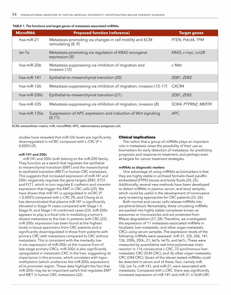
54 TRANSLATIONAL MEDICINE AT CAPITAL MEDICAL UNIVERSITY: INVESTIGATING MAJOR CHRONIC DISEASES
studies have revealed that miR-126 levels are significantly downregulated in mCRC compared with L-CRC (P < 0.0001) (9).
miR-141 and 200cmiR-141 and 200c both belong to the miR-200 family.
They function as a switch that regulates the epithelial to mesenchymal transition (EMT) and the mesenchymal to epithelial transition (MET) in human CRC metastasis. This suggests that increased expression of miR-141 and 200c negatively regulate the gene targets ZEB1, ETS1, and FLT1, which in turn regulate E-cadherin and vimentin expression that trigger the EMT in CRC cells (22). We have shown that miR-141 is upregulated in mCRC (P < 0.0001) compared with L-CRC (9), and Cheng et al. has demonstrated that plasma miR-141 is significantly elevated in Stage IV cases compared with Stage I–II, Stage III, and Stage I–III combined cases (23). miR-200c appears to play a critical role in mediating a tumor’s distant metastasis to the liver in patients with CRC (22). miR-200c expression has been found at the highest levels in tissue specimens from CRC patients and is significantly downregulated in those from patients with primary CRC with metastasis compared to CRC without metastasis. This is consistent with the markedly low in situ expression of miR-200c at the invasive front of late-stage primary CRCs. miR-200c is also significantly upregulated in metastatic CRC in the liver, suggesting its importance in this process, which correlates with hypo-methylation (which unsilences the miR-200c expression) of its promoter region. These data highlight the fact that miR-200c may be an important switch that regulates EMT and MET in human CRC metastasis (22).
Clinical implicationsThe notion that a group of miRNAs plays an important
role in metastasis raises the possibility of their use as biomarkers for early detection of metastasis, for predicting prognosis and response to treatment, and perhaps even as targets for cancer treatment strategies.
miRNAs as diagnostic markersOne advantage of using miRNAs as biomarkers is that
they are highly stable in archived formalin-fixed paraffin embedded (FFPE) tissues and body fluids (24, 25). Additionally, several new methods have been developed to detect miRNAs in plasma, serum, and stool samples, which could be useful in the development of noninvasive early screening approaches for CRC patients (23, 26).
Both normal and cancer cells release miRNAs into peripheral blood. Remarkably, these circulating miRNAs are packed into highly stable complexes known as exosomes or microvesicles and are protected from RNase degradation (27, 28). Therefore, we investigated the expression of 11 metastasis-associated miRNAs in localized, liver-metastatic, and other organ-metastatic CRCs using serum samples. The expression levels of the following miRNAs were assessed: miR-31, 335, 206, 141, 126, 200b, 200c, 21, let7a, let7b, and let7c. These were measured by quantitative real-time polymerase chain reaction in 116 consecutive L-CRC, 72 synchronous liver-metastatic CRC (SLM-CRC), and 36 other organ-metastatic CRC (OM-CRC). Seven of the eleven tested miRNAs could be detected in serum and of these, four, namely miR-126, Let-7a, miR-141, and miR-21, were associated with metastasis. Compared with L-CRC, there was significantly increased expression of miR-141 and miR-21 in SLM-CRC
MicroRNA Proposed function (reference) Target geneshsa-miR-21 Metastasis-promoting via changes in cell motility and ECM
remodeling (8, 9)PTEN, Pdcd4, TPM
let-7a Metastasis-promoting via regulation of KRAS oncogene expression (8)
KRAS, c-myc, Lin28
hsa-miR-206 Metastasis-suppressing via inhibition of migration and invasion (12)
c-Met
hsa-miR-141 Epithelial-to-mesenchymal transition (20) ZEB1, ZEB2
hsa-miR-126 Metastasis-suppressing via inhibition of migration, invasion (15–17) CXCR4
hsa-miR-200c Epithelial-to-mesenchymal transition (21) ZEB1, ZEB2
hsa-miR-335 Metastasis-suppressing via inhibition of migration, invasion (8) SOX4, PTPRN2, MERTK
hsa-miR-135a Suppression of APC expression and induction of Wnt signaling (8,11)
APC
ECM, extracellular matrix; miR, microRNA; APC, adenomatous polyposis coli.
TABLE 1. The functions and target genes of metastasis-associated miRNAs.

TRANSLATIONAL RESEARCH IN SURGERY AND REHABILITATION MEDICINE 55
and OM-CRC; significantly reduced expression of miR-126 in SLM-CRC and OM-CRC; and significantly increased expression of let-7a in OM-CRC. The receiver operating characteristic (ROC) curve showed that serum miR-126 had a cut-off with 78% sensitivity and 69% specificity, miR-141 had a cut-off with 86% sensitivity and 76% specificity, and miR-21 had a cut-off with 74% sensitivity and 66% specificity. Thus, we have identified liver metastasis-associated miRNAs, including serum miR-126, miR-141, and miR-21 levels, as novel biomarkers for the clinical diagnosis of early stage liver-metastatic CRC (9). Additionally, Vickers et al. previously demonstrated that the prognostic signature of miR-21,135a, 335, 206, and let-7a detects the presence of metastases with a specificity of 87% and sensitivity of 76% (8). Therefore, we have good reason to believe miRNAs can play a significant role in the diagnosis of CRC metastasis.
miRNAs as prognostic markersThe association between miRNA dysregulation and
disease outcome in patients with CRC has been explored in a number of studies, with miR-21 perhaps the best-studied. High levels of miR-21 are associated with lymph node positivity; the development of distant metastases; advanced tumor, node and metastasis staging; poor survival; and poor therapeutic outcome (29, 30). Other miRNAs specifically associated with increased tumor size, metastasis, and/or patient survival have also been described. For instance, miR-31 is positively correlated with deeper tumor invasion and more advanced tumor, node, and metastasis staging (31). Xi et al. evaluated 10 miRNAs for their potential prognostic value in patients with colorectal cancer using snap-frozen clinical colorectal samples; patients with higher levels of miR-200c expression had shorter survival times compared with patients with lower levels of expression, suggesting that miR-200c may be a novel prognostic factor in CRC (32).
miRNAs and therapeutic responses A tumor’s response to treatment may be affected
by its miRNA expression profile. Moreover, altered miRNA expression, or polymorphisms in miRNA genes or miRNA-binding sites, are correlated with treatment outcome in colon cancer patients. Nakajima et al. demonstrated that let-7g and miR-181b expression levels are strongly associated with the chemoresponse to S-1, a 5-fluorouracil-based anti-metabolite (33). A let-7 miRNA-binding site polymorphism in the 3΄ untranslated region of the KRAS gene is positively related to cetuximab responsiveness in mCRC patients with a nonmutated (or wild-type) KRAS gene (34). Finally, miR-31 suppression has recently been shown to increase sensitivity to 5-fluorouracil and thus the inhibition of proliferation in colon cancer cell lines (35).
Looking aheadmiRNAs play a significant role in CRC metastasis,
including proliferation, cell cycle control, invasion, EMT, and the maintainance of a tumor stem-cell phenotype.
From a translational medicine perspective, we are optimistic that miRNA-based diagnostics, prognostics, and therapeutics will be developed in the near future to benefit patients. Large, multicenter patient cohort studies of potential miRNA markers are required to establish the clinical usefulness of miRNA-based diagnosis and prognosis. The identification of disease-associated changes in miRNA expression suggests that manipulation of miRNA expression levels offers a potential therapeutic approach for the inhibition of metastatic development. With the advancement of RNA delivery technology, we anticipate that miRNA-based therapeutics will be added to the armamentarium in the fight against human cancer in the near future.
References 1. M. A. Cortez, G. A. Calin, Expert Opin. Biol. Ther. 9, 703 (2009). 2. S. Griffiths-Jones, H. K. Saini, S. van Dongen, A. J. Enright, Nucleic Acids Res. 36, D154 (2008). 3. M. Fabbri, C. M. Croce, G. A. Calin, Cancer J. 14, 1 (2008). 4. L. He, G. J. Hannon, Nat. Rev. Genet. 5, 522 (2004). 5. J. Lu et al., Nature 435, 834 (2005). 6. D. X. Nguyen, P. D. Bos, J. Massague, Nat. Rev. Cancer 9, 274 (2009). 7. D. R. Hurst, M. D. Edmonds, D. R. Welch, Cancer Res. 69, 7495 (2009). 8. M. M. Vickers et al., Clin. Exp. Metastasis 29, 123 (2012). 9. J. Yin, et al., Chin. J. Cancer Res. 26, 95 (2014).10. I. A. Asangani et al., Oncogene 27, 2128 (2008).11. Y. Toiyama et al., J. Natl. Cancer Inst. 105, 849 (2013).12. R. Nagel et al., Cancer. Res. 68, 5795 (2008).13. Y. Koga et al., Cancer Prev. Res. 3, 1435 (2010).14. S. F. Tavazoie et al., Nature 451, 147 (2008).15. D. Yan et al., J. Biol. Chem. 284, 29596 (2009).16. A. Varadi et al., Mol. Biol. Cell 16, 2670 (2005).17. D. R. Hurst et al., Cancer Res. 69, 7495 (2009).18. T. F. Hansen et al., Oncol. Lett. 2, 1101 (2011).19. Y. Zhang et al., Nat. Cell Biol. 15, 284 (2013).20. K. J. Png, N. Halberg, M. Yoshida, S. F. Tavazoie, Nature 481, 190 (2011).21. G. Ren, Y. Kang, Nat. Cell Biol. 15, 231 (2013).22. K. Hur et al., Gut 62, 1315 (2012).23. H. Cheng et al., PLOS ONE 6, e17745 (2011).24. A. B. Hui et al., Lab. Invest. 89, 597 (2009).25. X. Chen et al., Cell Res. 18, 997 (2008).26. Z. Huang et al., Int. J. Cancer 127, 118 (2010).27. M. P. Hunter et al., PLOS ONE 3, e3694 (2008).28. N. Kosaka et al., Cancer Sci. 101, 2087 (2010).29. O. Slaby et al., Oncology 72, 397 (2007).30. A. J. Schetter et al., J. Amer. Med. Assoc. 299, 425 (2008).31. C. J. Wang et al., Dis. Markers 26, 27 (2009).32. Y. Xi et al., Biomarker Insights 1, 113 (2006).33. G. Nakajima et al., Cancer Genomics Proteomics 3, 317 (2006).34. W. Zhang et al., Ann. Oncol. 22, 104 (2011).35. C. J. Wang, J. Stratman, Z. G. Zhou, X. F. Sun, BMC Cancer 10, 616 (2010).

56 TRANSLATIONAL MEDICINE AT CAPITAL MEDICAL UNIVERSITY: INVESTIGATING MAJOR CHRONIC DISEASES
Lateral lumbar interbody fusion: A novel, minimally invasive treatment for diseases of the spineXiang Li, Shudong Jiang, and Yi Hong*
With the evolution of advanced surgical tools, minimally invasive spine surgery has undergone tremendous growth in recent years. Lateral lumbar interbody fusion (LLIF), also called XLIF and DLIF (direct lateral interbody fusion), is a novel, minimally invasive technique that provides safe and effective anterior spinal fusion with fewer complications compared with a traditional anterior approach. With the assistance of special dilators, this technique can access the anterolateral spine through the retroperitoneal space and psoas muscle through a 3 to 4 cm incision (1). The advantages of this technique include being able to insert a larger cage in the intervertebral space, preservation of the integrity of the anterior-posterior annulus, a higher fusion rate, lower risk of visceral and great vessel injuries, and fewer perioperative complications. Satisfactory results have been achieved using this technique for the treatment of thoracic and lumbar spine disease, including adult degenerative scoliosis, discogenic pain, spondylolisthesis, lumbar infectious spondylitis, spinal fractures, and spinal tumors (2–6). However, postoperative complications such as lumbar plexus injury using the transpsoas approach (7), cage overhang (8), cage migration (9), contralateral femoral nerve compression (10), and retroperitoneal hematoma (11), have all been reported.
Here we review the literature on this surgical technique, examining the clinical results and complications associated with lateral lumbar interbody fusion to raise awareness of this technique.
Surgical techniqueApproach to lumbar spine
Lateral lumbar interbody fusion was first described by Dr. Luiz Pimenta on the VIII Brazilian Spine Society Meeting in 2001. Ozgur et al. (1) reported the first clinical results in patients with axial low back pain. During the operation, the patient is placed in a true right lateral decubitus position and general endotracheal anesthesia is administered. Fluoroscopic imaging is used to identify the affected disc space. Two small incisions are made, the first on the
patient’s lateral side just over the center of the index disc space. This will be used to insert the tissue dilator and retractor. The second incision is located behind the first. During the operation, the surgeon inserts an index finger through the second incision to dissect the muscle fiber and access the retroperitoneal space, after which the same finger palpates down to touch the psoas muscle. Once the psoas muscle has been identified, the index finger sweeps up towards the first incision. A guide-wire is inserted through the first incision into the retroperitoneal space to access the psoas muscle, using the index finger as a guide. A tissue dilator and retractor are then are introduced and placed over the surface of the psoas muscle that lies just over the affected disc. The psoas is dissected between the middle and anterior third of the muscle to minimize injury to the lumbar plexus. A real-time electromyography (EMG) monitoring system can be employed to prevent this complication. Once the disc is exposed and the retractor is fixed, the standard disectomy and interbody fusion can be performed by viewing the surgery sight directly. The posterior annulus should be left intact and the lateral annulus released. Inserting a large implant resting on both lateral margins of the epiphyseal can provide strong end plate support as well as sagittal and coronal plane correction. In the initial 13 patients treated with this technique, satisfactory results were received and no complications were noted.
Approach to thoracolumbar and thoracic spineKarikari et al. expanded the indications of this technique
to the thoracic and thoracolumbar spine, and we further extended its indication to thoracolumbar fractures (12). Newly designed dilators, which consisted of five sequential cannulas with an inner diameter of 24 mm, were used to provide the surgical field. Each cannula can accommodate an endoscope with an illumination system and the lumbar interbody fusion instrumentation simultaneously. Two projections at the end of each cannula can be helpful in fixing this system on the lateral aspect of adjacent vertebrae. The surgeon can perform the discectomy either under a direct or endoscopic view, which can improve the safety of the procedure and avoid the steep learning curve generally associated with simple endoscopic techniques. For patients undergoing LLIF in the lumbar spine region at L1/L2, the retroperitoneal approach was used according to Ozgur et al. (1), while those undergoing anterior fusion of the thoracic region at T11/T12 or T12/L1, a thoracotomy approach was used, without the involvement of the diaphragm. Real-time, evoked electromyography (EMG) was not routinely used in this study because of the low risk of lumbar injury at the thoracolumbar segments.
Clinical outcomesLumbar spine degenerative disease
Satisfactory results have been reported using LLIF to treat thoracic and lumbar spine disease, including adult degenerative scoliosis, axial low back pain, spondylosisthesis, adjacent segment disease after fusion,
Orthopedic Surgery Department of Capital Medical University, Beijing, China, and Department of Spine Surgery, Beijing Bo’ai Hospital, China Rehabilitation Research Center, Beijing, China.*Corresponding Author: [email protected]
TRANSLATIONAL RESEARCH IN SURGERY AND REHABILITATION MEDICINE 57
spinal infectious spondylitis, reconstruction following spinal tumor resection, and spinal fractures.
In a series of 43 patients who underwent LLIF for the treatment of lumbar degenerative disease, spondylosisthesis, and de novo scoliosis, Sharma et al. reported satisfactory correction of the coronal and sagittal plane (13). In 25 patients with scoliosis, the mean scoliosis correction was 10.4º and the correction rate was 43% at the one-year follow-up exam. The clinical outcomes, including visual analog scale (VAS), Oswestry disability index (ODI), and Short Form-12 (SF-12), improved significantly. The most common complication was anterior thigh pain that occurred in 25% patients. The majority of the symptoms were transient and resolved within 6 weeks.
Karikari et al. reported 22 patients treated with this technique for thoracic and thoracolumbar spine disease, including degenerative scoliosis, pathological fractures from tumors, adjacent level disease, thoracic disc herniations, and discitis/osteomyelitis (12). At the 6-month follow-up, all of the patients showed bony fusion in their computed tomography scans. Complications included wound infection, subsidence, and adjacent level disease, none of which affected the clinical outcome.
Acosta et al. analyzed the effect of LLIF on changes in the coronal and sagittal planes (13). In a series of eight patients treated by LLIF for lumbar degenerative scoliosis, the segmental coronal angle was corrected from 21.4º to 9.7º, and the global coronal alignment improved from 19.5 mm preoperatively to 12.5 mm postoperatively. The segmental sagittal angle was corrected from -5.3º to -8.2º, but the global sagittal alignment was not improved significantly. VAS and ODI improved from 7.7 and 43 preoperatively to 2.9 and 21 postoperatively, respectively. It was concluded that LLIF can significantly improve the coronal plane in patients with lumbar degenerative scoliosis, but had no effect on sagittal alignment.
Berjano et al. reviewed recent literature and found that LLIF combined with posterior pedicle instrumentation provided 40% to 75% correction of coronal alignment with modest improvement of sagittal alignment (2). Therefore, combining LLIF with minimally invasive surgery for the posterior approach could minimize the risk of complications association with a traditional open surgery approach.
Thoracolumbar spine fracturesLi et al. reported preliminary results of invasive LLIF
combined with posterior short-segment instrumentation for the treatment of thoracolumbar fractures (14). In a group of 12 patients with thoracolumbar fractures, this combined procedure achieved satisfactory results in terms of kyphosis correction (Cobb angle improved from 31º±8.7º preoperative to 5.1º±3.5º postoperative), correction maintenance (mean 4.3°±2.1º of correction loss at final follow-up) with less blood loss (185.8±62.3 mL), shorter operative time (mean 127.1±21.7 minutes), and lower risk of complication. These results are similar with that of Tofuku et al. (15).
In our own unpublished study, we compared clinical outcomes between LLIF combined with posterior short-segment instrumentation and traditional wide-open anterior-posterior (A-P) approach for the treatment of selective thoracolumbar fractures. The results showed that posterior short-segment pedicle instrumentation combined with LLIF can achieve similar clinical results with significantly less operative time, blood loss, and surgical complications compared with traditional wide-open A-P approach. This minimally invasive procedure seems to be a reasonable treatment option for selective patients with thoracolumbar fractures.
ComplicationsComplications associated with surgical approach
The LLIF procedure involves a retroperitoneal approach, directly through the psoas muscle to gain access to the lumbar spine. The lumbosacral plexus passes through this region, making it richly innervated. Cummock et al. reviewed a series of 69 patients who underwent LLIF (16). The study showed that 62.7% patients presented with postoperative issues in the upper leg, including thigh pain (39%), numbness (42.4%), paresthesias (11.9%), and weakness (23.7%). The risk of thigh complications is higher in L4/5 transpsoas interbody fusion when compared with proximal segments, although the difference does not reach statistical significance. Direct trauma to the psoas muscle belly, retractor compression, hematoma, and patient hip position may cause the motor deficits of the iliopsoas and quadriceps muscles and sensory deficits in the thigh region. Defining the surgical safe zone, using real-time EMG monitoring, and intermittent retractor release can each minimize the risk of complications associated with this approach.
Other complications Complications associated with an implant, such
as displacement of the cage and contralateral nerve compression, have been reported. Daffner et al. (17) described a patient presenting with leg pain due to the migrated cage after an XLIF procedure. Revision surgery that included reinsertion of the cage with an additional lateral plate was performed, and the patient’s pain resolved shortly after the procedure. The author recommended considering an additional lateral plate for patients with significant coronal deformity.
Papanastassiou et al. (10) described two patients with contralateral nerve root compression that resulted from a displaced endplate fragment and a far-lateral herniation, respectively, after an XLIF procedure. Therefore, the authors suggest that both the overzealous removal of the endplate and placing the cage diagonally toward the neural foramen should be avoided to prevent these types of complications.
Another complication, delayed abscess due to retroperitoneal hematoma, has also been reported, but this can be resolved by aspiration and oral antibiotics (11).
58 TRANSLATIONAL MEDICINE AT CAPITAL MEDICAL UNIVERSITY: INVESTIGATING MAJOR CHRONIC DISEASES
ConclusionsLLIF is a relatively novel technique that has received
considerable interest. Results using this procedure have been satisfactory, although complications have also been reported. Further studies that include a better understanding of how the anatomy is affected by the procedure and how to prevent complications as well as further evaluation of the safety and effectiveness of the technique will help advance our understanding of the potential advantages and disadvantages of the procedure.
References 1. B. M. Ozgur, H. E. Aryan, L. Pimenta, W. R. Taylor, Spine J. 6, 435 (2006). 2. P. Berjano, C. Lamartina, Eur. Spine J. 22 (Suppl 2), S242 (2013). 3. A. K. Sharma et al., J. Spinal Disord. Tech. 24, 242 (2011). 4. R. E. Isaacs, J. Hyde, J. A. Goodrich, W. B. Rodgers, F. M. Phillips, Spine 35, S322 (2010). 5. W. B. Rodgers, E. J. Gerber, J. Patterson, Spine 36, 26 (2011). 6. A. M. Caputo et al., J. Clin. Neurosci. 20, 1558 (2013). 7. W. K. Hu et al., Eur. Spine J. 20, 557 (2011). 8. G. J. Regev et al., Spine 35, 1406 (2010). 9. S. D. Daffner, J. C. Wang, Orthopedics 33, 518 (2010).10. I. D. Papanastassiou, M. Eleraky, F. D. Vrionis, J. Clin. Neurosci. 18, 149 (2011).11. M. R. Murray, J. K. Weistroffer, M. F. Schafer, Spine J. 12, e1 (2012).12. I. O. Karikari et al., J. Spinal Disord. Tech. 24, 368 (2011).13. F. L. Acosta et al., J. Neurosurg. Spine 15, 92 (2011).14. X. Li et al., Chinese J. Spine Spinal Cord 24, 400 (2014).15. K. Tofuku et al., J. Spinal Disord. Tech. 25, 38 (2012).16. M. D. Cummock, S. Vanni, A. D. Levi, Y. Yu, M. Y. Wang, J. Neurosurg. Spine 15, 11 (2011).17. K. Alpantaki et al., Orthopedics 33, 422 (2010).
The management of neurogenic bladderGuoqing Chen, Fan Zhang, and Limin Liao*
Neurogenic bladder, a dysfunction of the urinary bladder due to spinal cord injury (SCI) or other diseases of the central nervous system or peripheral nerves, is a major medical and social problem. The loss of supraspinal control leads to neurogenic detrusor overactivity (NDO), causing urinary incontinence, and to detrusor sphincter dyssynergia (DSD), which results in elevated bladder pressure during the storage and voiding phases.
NDO, DSD, and high pressure often lead to structural bladder damage, vesicoureteral reflux, upper urinary tract dilation, and renal insufficiency. Therefore, the management and treatment for neurogenic bladder should aim to protect upper urinary tract function, reduce urinary infection, improve quality of life, and maintain normal lower urinary tract function. The current methods used to treat neurogenic bladder include medication, botulinum toxin A (BTX-A) injection, neuromodulation, and surgical procedures, each of which has advantages and disadvantages. In recent years, we have worked to translate progress in basic science into improvements in these established therapeutic areas, as well as investigating new treatment possibilities for neurogenic bladder, including tissue engineering, stem cell transplantation, and gene therapy. Here we review our findings.
MedicationCurrently, the “gold standard” treatment for NDO
is clean intermittent catheterization combined with anticholinergic agent therapy. There is a new oral anticholinergic drug, solifenacin, with a high affinity for the M3 muscarinic receptor in the bladder, but data on its impact in NDO are limited. We recently assessed the efficacy and safety of solifenacin in 50 patients with NDO secondary to SCI (1), and showed that it increases bladder capacity, decreases bladder leakage volume, and significantly improves the quality of life (1). Of note, NDO recurs immediately after withdrawal of the anticholinergic agent, even after long-term treatment, suggesting that there are no long-lasting therapeutic effects of pharmacologic treatment. Thus, patients with NDO must be permanently treated with anticholinergic agents and face lifelong associated side effects unless completely safe and effective drugs are developed.
Department of Urology, Capital Medical University, Beijing, China; Department of Urology, China Rehabilitation Research Center, No.10 Jiaomen Beilu, Fentai District, Beijing, China;. and Center of Neural Injury and Repair, Beijing Institute for Brain Disorders, Capital Medical University, Beijing, China. *Corresponding Author: [email protected]

TRANSLATIONAL RESEARCH IN SURGERY AND REHABILITATION MEDICINE 59
BTX-A was introduced to treat NDO by injection into the detrusor muscle on the theoretical basis that it would temporarily block the presynaptic release of acetylcholine from parasympathetic innervation and produce a paralysis of the detrusor smooth muscle. We began using this procedure over 10 years ago. At present, Botox (Allergen) is widely used globally; however, we use Lantox (Lanzhou Institute of Biological Products, Lanzhou, China) to manage NDO in patients with SCI and have achieved similar efficacy, safety, and tolerability profiles to Botox at the same doses (2). Lantox injection into the detrusor muscle in patients with NDO secondary to SCI significantly improves bladder function, as evidenced by continence and subjective patient satisfaction. The urodynamic parameters measured at the follow-up assessment have shown that the reflex volume, maximum detrusor pressure, bladder compliance, and maximum cystometric capacity are all significantly improved. We have also shown that injection of Lantox into the detrusor muscle significantly reduced urinary tract infections in patients with SCI; we suggest that this reduction in is related to the decrease in detrusor pressure (3). The cost of Lantox is one-third that of Botox. This new procedure has been approved for use in clinical practice in China based on our studies.
Recently, nitric oxide (NO) has been shown to participate in the neural pathways that control the lower urinary tract. Expression of neural NO synthase (nNOS) can be upregulated after SCI, and altered nNOS activity may participate in the resulting lower urinary tract dysfunction. We examined the distribution of nNOS immunoreactivity in rat neurons following SCI, and the impact of nNOS inhibitors (4), and concluded that the strategic manipulation of NO production could help restore function or reduce undesired functional effects in the lower urinary tract (4). Indeed, we believe that this may become a focus for the development of new pharmacological interventions, a first step toward which would be the study of nNOS immunoreactivity and the effect of nNOS inhibitor administration in patients at different stages of SCI.
Urinary tract reconstruction surgeryUrinary infection and vesicoureteral reflux,
detrimental effects on the upper urinary tract secondary to high bladder pressure, are common and devastating problems for patients with neurogenic bladder. Preservation of the upper urinary tract is the most important goal in any type of lower urinary tract reconstruction. The gold standard treatment for urinary infection and vesicoureteral reflux is augmentation enterocystoplasty, which aims to create a reservoir with a large capacity and good compliance while preserving the upper urinary tract, thus allowing for socially-acceptable continence (5). All segments of the bowel have been utilized for augmentation enterocystoplasty. The sigmoid has advantages, including its anatomic proximity to the bladder as
FIGURE 1. Macroscopic and histological evaluation of bladder regenerated with small intestine submucosa in a rabbit model system. (A) Submucosa patch grafted onto the host bladder. (B) Regenerated tissue (arrows) at 24 weeks postoperation. (C) Thin arrow marks the regenerated transitional epithelium. Yellow arrow marks infiltrating inflammatory cells. (D) Thin arrow marks the regenerated transitional epithelium. Broad arrow indicates new blood vessels.
FIGURE 2. Cystoplasty with small intestine submucosa in neurogenic bladder patient. (A) Small intestine submucosa-grafted bladder, with drainage tube, from a human patient. (B) Cystogram in patient at 6-month follow-up. (C), (D) Cystoscopic biopsies of implanted engineered bladders taken between 1 and 3 months postoperatively, showing the extent of regeneration (Hematoxylin-eosin ×100).

60 TRANSLATIONAL MEDICINE AT CAPITAL MEDICAL UNIVERSITY: INVESTIGATING MAJOR CHRONIC DISEASES
well as a thick muscular wall, large lumen, and abundant mesentery, which together guarantee adequate capacity and maneuverability for the bladder. We retrospectively reviewed a total of 78 augmentation enterocystoplasties performed between 2005 and 2011 and identified 47 patients who underwent sigmoidocolocystoplasty alone or in combination with ureteral re-implantation. Sigmoidocolocystoplasty was found to be a safe and effective treatment for neurogenic bladder, and the concomitant ureteral re-implantation was beneficial for patients with a long history of illness. Moreover, in the majority of patients the procedure resolved urinary infection under clean intermittent catheterization (6). Consequently, sigmoidocolocystoplasty is included in our routine neuro-urologic practice, and we have performed it in more than 180 patients to date.
To improve our ability to make clinical decisions and identifying changes in upper urinary tract dilation pre- and posttreatment, we recently introduced two innovations: an upper urinary tract dilation grading system based on magnetic resonance urography (7, 8) and a comprehensive classification system (Liao’s classification) for lower and upper urinary tract dysfunction in patients with neurogenic bladder (8, 9). Evaluating the midterm outcomes using these tools demonstrated conclusively that augmentation enterocystoplasty provides effective and safe upper urinary tract protection (8). The presence of gastrointestinal segments in the urinary tract has, however, been associated with many complications, such as adhesive intestinal obstruction, metabolic disturbances, urolithiasis, excessive mucous production, and malignant diseases. Composite cystoplasty and the use of substitution materials have been proposed as means of overcoming these complications, and we have been testing tissue-engineering technology during cystoplasty in animal experiments (10). Specifically, we have used small intestine submucosa as a bladder wall replacement in a rabbit augmentation model. Histologically, the small intestine submucosa-regenerated bladders resemble normal bladder, and all three tissue layers (mucosa with submucosa, smooth muscle, and serosa) are present (Figure 1). In an in vitro study of the detrusor strip there were no significant differences in autorhythmicity and contractility between regenerated and normal detrusor muscles. An immunohistochemical analysis has indicated that the quantity of α-actin developed to a normal level and urodynamic testing showed that compliance remained stable postoperatively, and the volume increased significantly.
On the basis of these animal experiments, we explored the use of a small intestine submucosa scaffold for bladder augmentation in neurogenic bladder patients (11). Preliminary data from 14 cases have shown that cystoplasty with small intestine submucosa can improve the functionality of bladders in neurogenic bladder patients (Figure 2). No metabolic consequences were noted, no urinary calculi were observed, and renal
function was preserved (11). We do not currently recommend bladder augmentation using small intestine submucosa as a substitute for enterocystoplasty; nevertheless, this tissue-engineering technology does provide a potentially viable option for urinary tract reconstruction in neurogenic bladder patients.
NeuromodulationThe use of electrical currents for the treatment of
lower urinary tract dysfunction was first reported in 1878, when Saxtorph, a Danish surgeon, described intravesical electrostimulation in patients with an acontractile bladder and complete urinary retention. Today, an increasing number of studies are focused on somatic nerve stimulation to treat bladder dysfunction.
Sacral neuromodulation is an established, minimally invasive, reversible surgical treatment for both lower urinary tract and bowel dysfunction. The mechanism of action is not entirely clear but it is thought that the inhibition of afferent signals interrupts inappropriate detrusor contractions. While it was not thought to be a promising treatment option for patients with neurogenic bladder, our study has suggested that patients can benefit from neuromodulation of sacral spinal nerve 3 (S3). Patients with neurologic disease or injury often exhibit multiple symptoms and, while sacral neuromodulation may not resolve all of them, it may be a good option in combination with other treatments (12).
Pudendal nerve stimulation is another potential treatment for NDO and has achieved good results in patients with dysfunctional voiding. Our animal studies have shown that stimulation of the pudendal nerve increases bladder capacity only during the early period after SCI (13). In chronic SCI, the bladder becomes hypertrophic and/or fibrotic, and bladder compliance is significantly altered. We believe that pudendal nerve stimulation should be used to treat NDO secondary to SCI immediately after other conservative treatments fail. Further work is needed to determine definitively if early application of pudendal nerve stimulation also prevents bladder fibrosis and, if so, what the optimal timing is for the treatment. In an animal model, pudendal nerve stimulation from the time of SCI inhibits NDO, increases bladder capacity, and delays the progression of bladder fibrosis, leading us to conclude that pudendal nerve stimulation should be used as early as possible after injury (14). This finding needs to be translated into clinical practice as soon as possible.
Sacral and pudendal neuromodulation, as practiced today, is invasive and requires surgery to implant an InterStim electrical stimulator and an electrode. However, we have recently found that transcutaneous tibial nerve stimulation using adhesive skin-surface electrodes placed on the ankle is effective in treating NDO. A branch of the tibial nerve courses through the foot, which likely explains this effect. Given the noninvasiveness of this stimulation method a clinical trial could easily be conducted, for which we would recommend an intermittent stimulation pattern (15). A study in cats
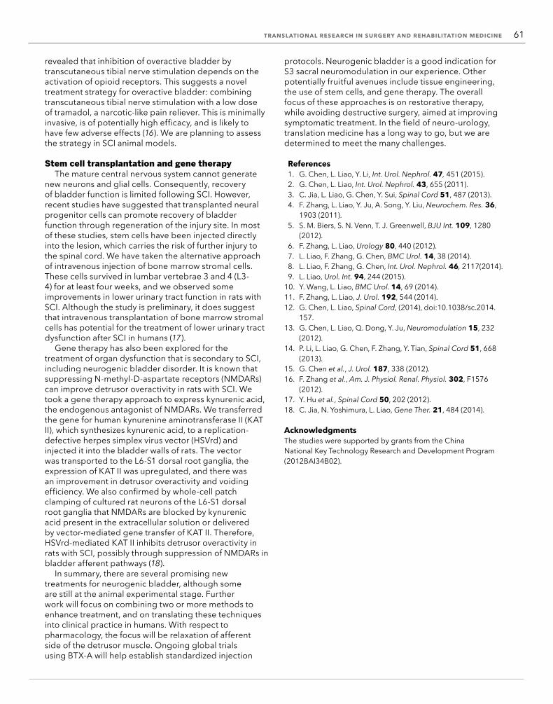
TRANSLATIONAL RESEARCH IN SURGERY AND REHABILITATION MEDICINE 61
revealed that inhibition of overactive bladder by transcutaneous tibial nerve stimulation depends on the activation of opioid receptors. This suggests a novel treatment strategy for overactive bladder: combining transcutaneous tibial nerve stimulation with a low dose of tramadol, a narcotic-like pain reliever. This is minimally invasive, is of potentially high efficacy, and is likely to have few adverse effects (16). We are planning to assess the strategy in SCI animal models.
Stem cell transplantation and gene therapyThe mature central nervous system cannot generate
new neurons and glial cells. Consequently, recovery of bladder function is limited following SCI. However, recent studies have suggested that transplanted neural progenitor cells can promote recovery of bladder function through regeneration of the injury site. In most of these studies, stem cells have been injected directly into the lesion, which carries the risk of further injury to the spinal cord. We have taken the alternative approach of intravenous injection of bone marrow stromal cells. These cells survived in lumbar vertebrae 3 and 4 (L3-4) for at least four weeks, and we observed some improvements in lower urinary tract function in rats with SCI. Although the study is preliminary, it does suggest that intravenous transplantation of bone marrow stromal cells has potential for the treatment of lower urinary tract dysfunction after SCI in humans (17).
Gene therapy has also been explored for the treatment of organ dysfunction that is secondary to SCI, including neurogenic bladder disorder. It is known that suppressing N-methyl-D-aspartate receptors (NMDARs) can improve detrusor overactivity in rats with SCI. We took a gene therapy approach to express kynurenic acid, the endogenous antagonist of NMDARs. We transferred the gene for human kynurenine aminotransferase II (KAT II), which synthesizes kynurenic acid, to a replication-defective herpes simplex virus vector (HSVrd) and injected it into the bladder walls of rats. The vector was transported to the L6-S1 dorsal root ganglia, the expression of KAT II was upregulated, and there was an improvement in detrusor overactivity and voiding efficiency. We also confirmed by whole-cell patch clamping of cultured rat neurons of the L6-S1 dorsal root ganglia that NMDARs are blocked by kynurenic acid present in the extracellular solution or delivered by vector-mediated gene transfer of KAT II. Therefore, HSVrd-mediated KAT II inhibits detrusor overactivity in rats with SCI, possibly through suppression of NMDARs in bladder afferent pathways (18).
In summary, there are several promising new treatments for neurogenic bladder, although some are still at the animal experimental stage. Further work will focus on combining two or more methods to enhance treatment, and on translating these techniques into clinical practice in humans. With respect to pharmacology, the focus will be relaxation of afferent side of the detrusor muscle. Ongoing global trials using BTX-A will help establish standardized injection
protocols. Neurogenic bladder is a good indication for S3 sacral neuromodulation in our experience. Other potentially fruitful avenues include tissue engineering, the use of stem cells, and gene therapy. The overall focus of these approaches is on restorative therapy, while avoiding destructive surgery, aimed at improving symptomatic treatment. In the field of neuro-urology, translation medicine has a long way to go, but we are determined to meet the many challenges.
References 1. G. Chen, L. Liao, Y. Li, Int. Urol. Nephrol. 47, 451 (2015). 2. G. Chen, L. Liao, Int. Urol. Nephrol. 43, 655 (2011). 3. C. Jia, L. Liao, G. Chen, Y. Sui, Spinal Cord 51, 487 (2013). 4. F. Zhang, L. Liao, Y. Ju, A. Song, Y. Liu, Neurochem. Res. 36, 1903 (2011). 5. S. M. Biers, S. N. Venn, T. J. Greenwell, BJU Int. 109, 1280 (2012). 6. F. Zhang, L. Liao, Urology 80, 440 (2012). 7. L. Liao, F. Zhang, G. Chen, BMC Urol. 14, 38 (2014). 8. L. Liao, F. Zhang, G. Chen, Int. Urol. Nephrol. 46, 2117(2014). 9. L. Liao, Urol. Int. 94, 244 (2015).10. Y. Wang, L. Liao, BMC Urol. 14, 69 (2014).11. F. Zhang, L. Liao, J. Urol. 192, 544 (2014).12. G. Chen, L. Liao, Spinal Cord, (2014), doi:10.1038/sc.2014. 157.13. G. Chen, L. Liao, Q. Dong, Y. Ju, Neuromodulation 15, 232 (2012).14. P. Li, L. Liao, G. Chen, F. Zhang, Y. Tian, Spinal Cord 51, 668 (2013).15. G. Chen et al., J. Urol. 187, 338 (2012). 16. F. Zhang et al., Am. J. Physiol. Renal. Physiol. 302, F1576 (2012).17. Y. Hu et al., Spinal Cord 50, 202 (2012). 18. C. Jia, N. Yoshimura, L. Liao, Gene Ther. 21, 484 (2014).
AcknowledgmentsThe studies were supported by grants from the China National Key Technology Research and Development Program (2012BAI34B02).

62 TRANSLATIONAL MEDICINE AT CAPITAL MEDICAL UNIVERSITY: INVESTIGATING MAJOR CHRONIC DISEASES
Genotypic prediction of the intercellular adhesion molecule 1 K469E (A/G) polymorphism in diabetes and diabetic microvascular complicationsJun Ma1*, Zhanjie Ren1, Tianwei Gu2, Liyun Zhao1, Duomao Lin1, Kerstin Brismar2, and Harvest F. Gu2*
In recent years, a strong translational research effort has been underway to identify genetic polymorphisms associated with susceptibility to diabetes and diabetic microvascular complications. Although the pathogenesis of diabetes and diabetic microvascular complications is multifactorial, local inflammatory stress may result from both metabolic and hemodynamic derangements in the diseases (1, 2). Intercellular adhesion molecule 1 (ICAM-1) is an acute phase marker of inflammation. Previously, we reported a genetic study of the ICAM1 gene in Swedish patients with type 1 diabetes (T1D) and diabetic nephropathy (DN) (3). Recently, we have performed genotyping analyses of the ICAM1 genetic polymorphisms in Chinese subjects with type 2 diabetes (T2D) and diabetic peripheral neuropathy (DPN) (4). In this review, we summarize previous analyses of the genotype distribution of the ICAM1 K469E(A/G) polymorphism among Chinese and Swedish populations and discuss the potential use of this polymorphism for assessing risk susceptibility in diabetes and diabetic microvascular complications.
Elevation of ICAM-1 levels in diabetes patientsICAM-1 is a 90 kiloDalton (kD) inducible surface
glycoprotein expressed in leukocytes and endothelial cells. The protein promotes adhesion in immunological and inflammatory reactions and plays an important role in T lymphocyte activation and leukocyte-endothelial cell interaction (5–7). Clinical studies have demonstrated that increased serum ICAM-1 levels are associated with increased mortality and cardiovascular morbidity in T1D and T2D patients (8). Similarly, serum ICAM-1 levels were found to be positively correlated with albuminuria in T2D (9, 10).
Genetic association studies of the ICAM1 K469E polymorphism in diabetes and diabetic microvascular complications
The ICAM1 gene is located on chromosome 19p13.2 and resides in a region linked to diabetes (11). We previously conducted a genetic association study of the ICAM1 gene in Swedish subjects with normal glucose tolerance (NGT), and T1D patients with or without DN. Six single nucleotide polymorphisms (SNPs) including three Tag SNPs rs5498 (K469E A/G), rs1799969 (R241G A/G), and rs281432 (C/G) were selected from the ICAM1 gene for this study (3). Genotyping experiments were conducted using dynamic allele-specific hybridization, a high-throughput SNP scoring technique (12). Results showed that there was a significant association of SNP rs5498 (K469E A/G) in the ICAM1 gene with T1D (P=0.002, OR=1.783 95%CI 1.226-2.594) in the Swedish population. Further analysis of T1D patients with and without DN indicated that this polymorphism was associated with DN (P=0.001, OR=1.957, 95%CI 1.303-2.941) (Table 1). Interestingly, we found a high heterozygous index —whereby the number of heterozygous genotype carriers was higher than both homozygous genotypes—of this polymorphism in the Swedish population (Figure 1) (3). A high heterozygous index can be caused by genotyping errors, a SNP being located within segmental duplications (duplicons), or other genetic factors related to the disease.
In order to exclude the possibility that the high heterozygous index was caused by a genotyping error, we replicated the genotyping experiments using pyrosequencing and obtained the same results (13). In the human genome, duplicons with >90% similarity between two copies comprise at least 5% of the genomic DNA (14–16). If a SNP is present in a duplicon region, a high heterozygous index may be expected because there will be two copies of the SNP in the genome. To eliminate this as a potential explanation for our results, we performed extensive sequencing of the ICAM1 gene in Swedish subjects, but no duplicated sequence was found in this gene region (3).
We went on to investigate whether the high heterozygous index was present in other populations. We genotyped 1,449 Chinese subjects with NGT, and T2D with or without DPN (4). The data showed that the ICAM1 K469E (A/G) polymorphism was moderately associated with DPN in Chinese subjects with T2D (P=0.037, OR=1.715, 95% CI 1.027-2.865) (Table 1). Unlike the Swedish population, however, the Chinese population did not have a high heterozygous index of the ICAM1 K469E(A/G) polymorphism. The frequencies of homozygous K469(A/A), heterozygous K469E(A/G), and homozygous 469E(G/G) genotypes among the subjects with NGT were 48.8%, 43.7%, and 7.5%, respectively (Figure 1). Upon searching the HapMap database for additional genotype information, we confirmed that the genotype distribution of the ICAM1 K469E(A/G) polymorphism among 86 Han Chinese from Beijing was consistent with our experiments (17). Over a third (39.8%) of subjects in the Chinese population carried
1Center for Anesthesiology, Anzhen Hospital, Capital Medical University, Beijing, China2Rolf Luft Research Center for Diabetes and Endocrinology, Department of Molecular Medicine and Surgery, Karolinska University Hospital, Karolinska Institutet, Stockholm, Sweden.*Corresponding Authors: [email protected] (J.M.) and [email protected] (H.F.G.)
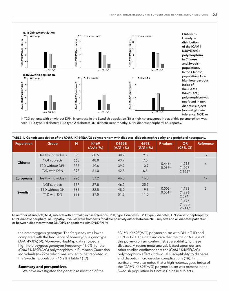
TRANSLATIONAL RESEARCH IN SURGERY AND REHABILITATION MEDICINE 63
the heterozygous genotype. The frequency was lower compared with the frequency of homozygous genotype (A/A, 49.8%) (4). Moreover, HapMap data showed a high heterozygous genotype frequency (46.0%) for the ICAM1 K469E(A/G) polymorphism in European Caucasian individuals (n=226), which was similar to that reported in the Swedish population (46.2%) (Table 1) (3).
Summary and perspectives We have investigated the genetic association of the
ICAM1 K469E(A/G) polymorphism with DN in T1D and DPN in T2D. The data indicate that the major A allele of this polymorphism confers risk susceptibility to these diseases. A recent meta-analysis based upon our and other studies confirmed that the ICAM1 K469E(A/G) polymorphism affects individual susceptibility to diabetes and diabetic microvascular complications (18). In particular, we also noted that a high heterozygous index of the ICAM1 K469E(A/G) polymorphism was present in the Swedish population but not in Chinese subjects.
FIGURE 1. Genotype distribution of the ICAM1 K469E(A/G) polymorphism in Chinese and Swedish populations.In the Chinese population (A), a high heterozygous index of the ICAM1 K469E(A/G) polymorphism was not found in non-diabetic subjects (normal glucose tolerance, NGT) or
in T2D patients with or without DPN. In contrast, in the Swedish population (B), a high heterozygous index of this polymorphism was seen. T1D, type 1 diabetes; T2D, type 2 diabetes; DN, diabetic nephropathy; DPN, diabetic peripheral neuropathy.
TABLE 1. Genetic association of the ICAM1 K469E(A/G) polymorphism with diabetes, diabetic nephropathy, and peripheral neuropathy.
Population Group N K469 (A/A) (%)
K469E (A/G) (%)
469E (G/G) (%)
P-values OR (95% CI)
Reference
Chinese
Healthy individuals 86 60.5 30.2 9.3 17NGT subjects 668 48.8 43.7 7.5
0.446a
0.037b1.715
(1.027-2.865)b
4T2D without DPN 383 49.6 39.7 10.7T2D with DPN 398 51.0 42.5 6.5
Europeans Healthy individuals 226 37.2 46.0 16.8 17
Swedish
NGT subjects 187 27.8 46.2 25.70.002a
0.001b1.783
(1.226-2.594)a
1.957 (1.303-2.941)b
3T1D without DN 535 32.5 48.0 19.5T1D with DN 328 37.5 51.5 11.0
N, number of subjects; NGT, subjects with normal glucose tolerance; T1D, type 1 diabetes; T2D, type 2 diabetes; DN, diabetic nephropathy;DPN, diabetic peripheral neuropathy. P-values were from tests for allele positivity either between NGT subjects and all diabetes patients (a) or between diabetes without DN/DPN andpatients with DN/DPN (b).

64 TRANSLATIONAL MEDICINE AT CAPITAL MEDICAL UNIVERSITY: INVESTIGATING MAJOR CHRONIC DISEASES
Experience-induced neuroplasticity following spinal cord injury
Feng Gao1,2,3,4, Chetwyn C. H. Chan2*, Mingliang Yang1,3,4, Liangjie Du3,4, Hongyu Chu3,4, and Jianjun Li1,3,4*
Spinal cord injuries (SCIs) disrupt the nerve fiber bundles that convey sensorimotor information between the brain and peripheral parts of the body, such as the limbs. This interference in the flow of sensorimotor information leads to profound and persistent impair-ment to the function of body parts below the lesion site. Therefore, researchers have begun developing solutions to compensate for the dysfunction resulting from SCI. For example, a recent advance has been the capture of voluntary brain signals in order to stimulate muscles to produce purposeful movements (1). The assumption is that, as the anatomical structure is not involved in the SCI, the brain should have retained the capacity to remediate the lost sensorimotor functions. Other studies, however, have demonstrated a noticeable reorganization of the brain after SCI (2, 3), which has an impact on brain func-tion. These somewhat counter-intuitive findings have prompted researchers to explore how SCIs that originally affected sensorimotor signal exchanges might modulate brain function.
In this article, we review the existing evidence for modulation of brain function by SCI and present our views on the possible mechanisms that underlie reorganization of the brain after SCI. Understanding these mechanisms will provide a crucial signpost for the development and refinement of therapeutic interventions for the rehabilitation of SCI patients.
Post-SCI neuroplasticityRecent studies have revealed anatomical and functional
changes in the brains of patients who have suffered SCI. These changes can involve sensory afferents, motor control outputs, and other motor-related cortical and subcortical areas. They are closely related to the specific characteristics of the injury to the spinal cord, including the lesion site and the severity and duration of the injury (4, 5).
1Center of Neural Injury and Repair, Beijing Institute for Brain Disorders, Beijing, China2Applied Cognitive Neuroscience Laboratory, Department of Rehabilitation Sciences, The Hong Kong Polytechnic University, Kowloon, Hong Kong, China3Department of Spinal and Neural Function Reconstruction, China Rehabilitation Research Center, Beijing, China4School of Rehabilitation Medicine, Capital Medical University, Feng Tai District, Beijing, China.*Corresponding Authors: [email protected] (C.C.H.C.) and [email protected] (J.L.)
The ICAM-1 protein acts as a ligand for lymphocyte function-associated antigen 1 (LFA-1), an integrin (transmembrane receptor) found in leukocytes. When activated, leukocytes bind to endothelial cells via ICAM-1/LFA-1 and then transmigrate into tissues (5-7). Unlike most integrin-binding proteins, ICAM-1 does not contain an Arg-Gly-Asp (RGD) motif to promote integrin binding (19, 20). The ICAM1 K469E(A/G) polymorphism resides in the 5th Ig-like domain of the ICAM-1 protein. This domain is essential for dimerization, surface presentation, and solubilization of the protein, and plays a crucial role in the interaction of ICAM-1 with LFA-1 and the adhesion of B cells (21, 22). The ICAM1 K469E(A/G) polymorphism is nonsynonymous and results in a glutamic acid to lysine substitution. Analyzing the ratio of the two forms of ICAM-1 protein will provide a better understanding of the biological effects of the ICAM1 K469E(A/G) polymorphism, and may be useful for predicting susceptibility of patients to diabetes and diabetic microvascular complications.
References 1. G. L King. J. Periodontol. 79, 1527 (2008). 2. J. F. Navarro-Gonzández et al. Nat. Rev. 7, 327 (2011). 3. J. Ma et al., Diabet. Med. 23, 1093 (2006). 4. Z. Ren et al., Gene & Genomics, (2014), doi:10.1007/s13258- 014-0230-9. 5. A. van de Stolpe, P. T. van der Saag, J. Mol. Med. 74, 13 (1996). 6. K. Sahakyan et al., Eur. J. Endocrinol. 162, 1101 (2010). 7. K. Sahakyan et al., Am. Heart J. 159, 1074 (2010). 8. A. Becker et al., Eur. J. Clin. Invest. 32, 1 (2002). 9. A. F. Rubio-Guerra et al., Med. Sci. Monit. 13, CR349 (2007).10. K. Mastej, R. Adamiec, Acta Diabetol. 45, 183 (2008).11. H. F. Gu et al., Front. Endocrinol. 3, 179 (2013).12. W. M. Howell, M. Jobs, U. Gyllensten, A. J. Brookes, Nat. Biotechnol. 17, 87 (1999).13. M. Ronaghi, M. Uhlén, P. Nyrén, Science 281, 363 (1998).14. J. C. Venter et al., Science 291, 1304 (2001). 15. D. Fredman et al., Nat. Genet. 36, 861 (2004). 16. Z. Cheng et al., Nature 437, 88 (2005). 17. http://hapmap.ncbi.nlm.nih.gov/cgi-perl/gbrowse/ hapmap24_B36/ICAM118. X. Su et al., PLOS ONE 8, e69940 (2013).19. A. van de Stolpe, P. T. van der Saag, J. Mol. Med. 74, 13 (1996).20. C. A. Janeway, Proc. Natl. Acad. Sci. U.S.A. 98, 7461 (2001).21. J. Miller et al., J. Exp. Med. 182, 1231 (1995).22. A. Brown et al., J. Biol. Chem. 288, 5992 (2013).
AcknowledgmentsThe authors wish to thank all Swedish and Chinese subjects for participating in our studies. The studies were supported by the Beijing Health System High-level Health Technical Talents Cultivation Fund (2013-2-004), the Capital Medical Development Research Fund (2009-3145), the Beijing Science and Technology Project (Z121107001012159), the National Natural Science Foundation in China (30972851), the Stig and Gunborg Westmans Foundation, the Karolinska Institutet Fund, and the Family Erling-Persson Foundation in Sweden.

TRANSLATIONAL RESEARCH IN SURGERY AND REHABILITATION MEDICINE 65
Animal studies have indicated that there is a reduction in the number of neurons in the primary motor cortex after SCI, related to disrupted neural pathways in the damaged spinal cord (6). In human patients, voxel-based morphometry (VBM) studies have shown a reduction in the volume of the gray matter in the primary motor cortex, the medial prefrontal cortex, and the adjacent anterior cingulate cortices (7). These findings suggest that there are compensatory plastic changes in the brain subsequent to SCI, but it is unclear on whether these changes augment or hamper the functional capability of patients with SCI and, hence, on what their impact on the rehabilitation processes might be.
One possible explanation for the post-SCI structural changes in the brain, particularly those related to the primary motor cortex, is that the neural substrates that previously received afferent input via the spinal cord are deprived of stimulation after injury (1, 7). The intact sensorimotor cortices that are deprived of stimulation do eventually receive afferent inputs via the adjacent cortices, and the same applies to efferent outputs from the sensorimotor cortices to other associative areas (7, 8). However, the temporary deprivation of afferent and efferent signals results in topographic reorganization in the sensory and motor cortices. For example, in the rat model, the cortical representation of the forelimb is magnified and invades the adjacent de-afferent hind limb area (6). Among paraplegic individuals, the post-SCI experience eventually results in an increase in the volume of the primary motor cortex representing the hand and other nonprimary cortical and subcortical regions representing the forearm (9), while in individuals with tetraplegia the cortical areas representing the forearm extend into those representing the hand and fingers (6). Nardone et al. postulated that these plastic changes are due to the overriding dominance of the activities of the forearms over hands and fingers, while the extent of the changes is proposed to be related to the level of the injury and the functions preserved (8). Other controlled studies have offered further evidence of the relationship between the post-SCI motor experience and plastic cortical changes. Research on monkeys with cervical subtotal hemisections showed that plastic cortical changes are observed in the contralateral, but not in the ipsilateral, hemisphere (10). The use of transcranial magnetic stimulation in individuals with chronic incomplete SCI also revealed that the ipsilateral M1 was not involved in motor improvements (5).
Thus, there are consistent findings of post-SCI experience-induced reorganization in the brain. This raises questions about the mechanisms underlying neural reorganization and whether the reorganization is beneficial or detrimental to patient rehabilitation.
Mechanisms underlying cortical reorganizationObservations from the recovery of the dorsal
column of the spinal cord after SCI have shed light on the mechanism of post-SCI, experience-induced reorganization. In one study, researchers tested the
intact dorsal column of patients with SCI, which showed normal functioning. The cortical areas supplied by the intact dorsal column were found to expand in size over the 5 or more weeks following the injury (11). Interestingly, the expanded cortical areas, which originally received afferent inputs from the limbs, now responded only to stimulations from the face. This suggests that reorganization of the sensorimotor cortex is likely to originate from the intact dorsal column, rather than from an intrusion of the nearby spinal pathways.
Another study postulated that reorganization of the sensorimotor cortex after spinal cord injury is due to the sprouting of intact afferent fibers (12). This was supported by the finding that the degree of deafferentation (or the amount of spared neurons in the dorsal column) is directly proportional to the extent of the reorganization in the contralateral primary sensorimotor cortex and, to some extent, to the level of functional recovery of patients with SCI. Post-SCI reorganization was found to take place in the corticospinal and extrapyramidal tracts (13, 14).
Changes in excitability, the formation of new neuronal connections, or a combination of these have been proposed as an explanation for the post-SCI plastic changes in the brain. Abnormal excitability in the brain near deafferented areas was found to be related to the distance from the deafferented area and the period of deafferentation persistence (15). Yague et al. noted the presence of cortical hyperexcitability in both primary sensorimotor cortices in response to the preserved spinothalamic inputs after spinal cord hemisection (16). In unilateral dorsal horn injury, the neural activity induced by below-threshold electrical stimuli was also considered to be cortical hyperexcitability, acting as amplified inputs from the periphery (17). These inputs play an important role in the generation of neuropathic pain and autonomic dysreflexia.
Finally, in a study by Massey et al., sprouting and synaptic regeneration and formation were assumed to be mechanisms of reorganization (18). Other studies have shown that reduced and altered spinothalamic and spinocerebellar input leads to adaptation to a new body reference scheme and that abnormal brain activation is quantitatively related to the severity of the injury (19).
Modulation of neural processesExperience-induced reorganization of the brain after
SCI might be expected to result in modulation of neural processes, and evidence suggests that this is the case (20–22). Patients who received task-specific training involving brain-signal driven prosthesis and a robot showed significant changes in remodeling of cortical projections (20). Castro et al. found that patients with chronic SCI (6 to 24 years postinjury) have modulated motor preparatory and motor execution processes, with lower amplitudes of both readiness potential and motor potential, as well as altered topographical distribution of the motor potential when compared to healthy controls (21). These modulations in neural
66 TRANSLATIONAL MEDICINE AT CAPITAL MEDICAL UNIVERSITY: INVESTIGATING MAJOR CHRONIC DISEASES
processes are associated with reorganization of the primary sensorimotor cortex, which is responsible for motor control. Another study, which used event-related potentials, also reported that patients with SCI presented with modulated neural processes. The authors demonstrated that, in the early sensory stage, when compared with normal controls, the patients with SCI had altered encoding and executive processes which were correlated with significant differences in the right posteriorly distributed P200 (22). In the later cognitive stage, modulation of neural processes resulted from the diminished P300 amplitude elicited in the left and right posterior regions among the patients with SCI.
Our group conducted a recent study that provides further evidence that the sensorimotor cortex is under-involved when motor-related tasks are performed by patients with chronic paraplegia. In this study, the participants visualized movements of the upper and lower limbs while their blood oxygen level-dependent (BOLD) signals were captured using a 3T magnetic resonance imaging (MRI) scanner. During the visualization of movements of the upper limbs, the patients with chronic paraplegia showed substantial increases in activation over the precentral and postcentral gyri, inferior parietal gyrus, and at the subcortical level thalamus and globus pallidus, while the healthy control participants showed significant increases in activation in the frontal cortex, including the middle and medial frontal gyri and the anterior cingulate cortex. These findings suggest that the loss of afferent sensorimotor inputs modulates the functions of the sensorimotor cortices. In this example, the loss of the inputs appears to have an impact on the neural substrates involved in the visualization of upper limb movements, which might have been expected to be intact in paraplegic patients. When visualizing movement of the lower limbs, the paraplegic patients showed increased activation in the external globus pallidus (or GPe) and the lingual gyrus. In contrast, in the healthy control participants, activation was significantly increased in the superior and medial frontal gyri and the anterior cingulate cortex. Our findings of increased activation in the GPe suggest that there may be modulation of excitability of the motor system among the paraplegic patients. We speculate that the cortical hyper-excitability among the paraplegic patients may be due to the lack of activity in the motor inhibitory system, which was found to be mediated by the GPe (unpublished results).
In general, the neural changes after SCI are considered to be a positive sign of functional recovery of the patients. The issue of cause and effect between active engagement in physical activity and neural changes requires further research. However, other changes associated with dysfunctions of the motor system, such as spasticity and neuropathic pain, are less beneficial for functional regain (17, 23).
Structural and functional changes have been found to occur within a very short period of time after SCI. These changes continue over months or years, and possibly last
a lifetime. Electrophysiological studies demonstrated that deafferentation due to SCI exerts changes in the cortical networks within the first hour while changes in the topography, such as within the primary sensorimotor cortex, occur as early as three days after the injury (6, 24). Time-dependent changes vary across different regions of the brain. For example, during the first month postinjury, activities of the bilateral M1 (synaptic sprouting) were found to heavily contribute to the plastic changes, whereas by 3 to 4 months postinjury the contralateral M1 and bilateral ventral premotor cortex contributed most to the plastic changes (25).
Factors affecting post-SCI neuroplasticityFollowing SCI, the architecture of the spinal cord is
destroyed and the information flow between the brain and peripheral nerves is disrupted. As a result, the cortex, subcortical brain structure, and spinal cord are all plastically reorganized. Many factors, such as the lesion site, the level of the lesion on the spinal cord, the completeness of the injury, the time postinjury, rehabilitative training, and the extent of functional recovery, all contribute to the character and extent of neural plasticity and reorganization postinjury.
SCI causes spinal cord atrophy. Typically, there is a 30% reduction in the cord area compared with healthy subjects, while more severe spinal cord atrophy or damage leads to more extensive reorganization of the cortex. Spinal cord atrophy is associated quantitatively with disability and corticospinal tract (CST) atrophy, reflecting functional impairment of the cranial CST (26). The differential impacts of the impaired afferent feedback and efferent outputs originating from the various lesion levels have been documented by positron emission tomography (27). Our own study showed that increased activation in the left primary motor cortex (BA 4), right premotor area, and presupplementary motor area (BA 6) during visualization of upper limb movements by patients with paraplegia were negatively correlated with the time postinjury in the SCI (unpublished results).
Additionally, the lack of sustainable excitability in paralyzed SCI subjects suppresses activity-dependent plasticity. However, plastic changes that occur below the injury level can be enhanced and augmented in incomplete SCI by specialized functional training programs that provide appropriate afferent inputs (23). A number of interventions have been designed to reorganize locomotor networks, including neuroprosthetic spinal cord electrode arrays, customized pharmacological treatments, and robotically assisted locomotor training procedures. The recovery of function and improvement in daily activities should be confirmed by studies with a rigid experimental design and a big sample size. To facilitate excitation in the cortical areas, transcranial magnetic stimulation or transcranial direct current stimulation are effective approaches. Franc et al. have shown that at 10 minutes postinjury, the resting motor evoked potential increases (28). This hyperexcitability can aid functional restoration by

TRANSLATIONAL RESEARCH IN SURGERY AND REHABILITATION MEDICINE 67
extensive motor training, but its duration is too short to provide a window for intervention. Hajela et al. have found that in incomplete paraplegia, newly acquired cortical control of the spinal interneuronal circuits reorganized by locomotor training helps generate patterned motor activity, thereby modifying spinal reflex function (29). A study by Martinez et al. revealed that in rats with a C4-5 hemisection, the S1 cortex representation is reactivated by sensorimotor training, which enhances endogenous activity within spared spinal and supraspinal networks (30). In complete SCI, the combined utilization of epidural electrical stimulation and monoamine agonists promotes locomotor rehabilitation (31).
Each of these studies demonstrates that the plastic reorganization of spinal networks induced by multisensory stimuli contributes to functional restoration. Clinically, the progress of reorganization can be evaluated using imaging parameters, such as the cord area, BOLD signals, and gray matter volume in the primary motor cortex, primary sensory cortex, and by assaying nerve fibers using diffusion tensor imaging.
The major objective of studying plastic reorganization after SCI is to learn how to promote functional recovery. As described above, different cortical areas contribute to plastic reorganization at different stages postinjury. In the early stage following injury, the goal is to reduce inhibition and enhance excitability in the available preexisting neural system, while in later stages the aim is to utilize the plastic changes in neural networks in the original system or in a newly recruited system (32). One notable study indicates that sensorimotor system plasticity can be modulated by manipulation of the Nogo signaling pathway, such as by decreasing Nogo receptors and LINGO-1 expression or by increasing brain-derived neurotrophic factor levels (17).
Conclusions With the integration of neuroimaging and
neurophysiological methodologies, more thorough investigation of plasticity in the nervous system after SCIs is becoming possible. This is helping us to better understand the course of nervous system adaptation and repair. Large-scale reorganization clearly plays an important role in functional recovery, as well as in the occurrence of some complications, and it can be used to evaluate functional progression and develop more effective therapeutic strategies.
Further studies are needed to confirm some of the current controversial findings, such as the characteristics of the spatial shift in sensorimotor system activation after SCI and the rules of reorganization. Studies exploring
potential interventions that enhance brain reorganization after complete SCI and more clearly elucidate what changes occur in more complex sensorimotor neural networks are also important. Lastly, we still require a better understanding of the post-SCI plastic changes of the motor control system and its relationship with other higher cognitive function systems of SCI patients.
References 1. M. E. Schwab, Science 295, 1029 (2002). 2. S. Hotz-Boendermaker et al., Neuroimage 39, 383 (2008). 3. P. Sabbah et al., J. Neurotrauma 19, 53 (2002). 4. K. J. Kokotilo et al., J. Neurotrauma 26, 2113 (2009). 5. R. Nardone et al., Neurosci. Lett. 552, 21 (2013). 6. P. J. Wrigley et al., Cereb. Cortex 19, 224 (2009). 7. S. Tandon et al., Eur. J. Neurosci. 38, 2271 (2013). 8. R. Nardone et al., Brain Res. 1504, 58 (2013). 9. A. Curt et al., Brain 125, 2567 (2002).10. E. Schmidlin et al., Brain Res. 1017, 172 (2004). 11. N. Jain et al., Nature 386, 6624 (1997).12. J. H. Kaas et al., Exp. Neurol. 209, 407 (2008).13. P. Freud et al., J. Neurol. Neurosurg. Psychiatry 83, 629 (2012).14. B. Zaaimi et al., Brain 135, 2277 (2012).15. M. Lotzea et al., Restor. Neurol. Neurosci. 23, 1 (2005).16. J. G. Yague et al., Exp. Neurol. 227, 252 (2011).17. T. Endo et al., Exp. Neurol. 209, 155 (2008).18. J. M. Massey et al., J. Neurosci. 26, 4406 (2006).19. M. Bruehlmeier et al., Eur. J. Neurosci. 10, 3918 (1998).20. R. van den Brand et al., Science 336, 1182 (2012).21. A. Castro et al., Int. J. Psychophysiol. 87, 205 (2013).22. I. Lazzaro et al., J. Clin. Neurophysiol. 30, 59 (2013).23. V. Dietz, Exp. Neurol. 235, 110 (2012).24. T. Endo et al., Brain 130, 2951 (2007).25. Y. Nishimura et al., Exp. Neurol. 235, 152 (2012).26. P. Freund et al., PLOS ONE 7, e51729 (2012). 27. A. Curt et al., J. Neurotrauma 19, 43 (2002).28. F. D. Roy et al., J. Neurophysiol. 103, 2222 (2010).29. N. Hajela et al., BioMed. Res. Int. 2013, 516427 (2013).30. M. Martinez et al., Eur. J. Neurosci. 30, 2356 (2009).31. P. Musienko et al., Exp. Neurol. 235, 100 (2012).32. Y. Nishimura et al., Neuroscientist 15, 436 (2009).
AcknowledgmentsThis work was supported in part by funds from the Supporting Program of the Twelfth Five-Year Plan for Science and Technology Research of China (2012BAI34B02) and the State Natural Science Foundation (81272164). The work was also partially supported by the General Research Fund (560110) awarded by the Research Grant Council of Hong Kong, for which C. C. H. Chan is the principal investigator.

68 TRANSLATIONAL MEDICINE AT CAPITAL MEDICAL UNIVERSITY: INVESTIGATING MAJOR CHRONIC DISEASES
Individualized traditional Chinese medicine treatment for acute strokeLei Chen1,2‡ , Juexian Song1‡ , Yumin Luo3, Min Li4*, and Li Gao1*
Stroke is one of the leading causes of mortality and disability in adults worldwide, causing enormous patient suffering and social burden (1). Although tremendous resources have been invested in research and clinical trials, only limited progress has been made for poststroke treatments. In a search for novel treatments or therapeutic strategies, pharmaceutical
companies, scientists, and physicians as well as the public have shown increasing interest in traditional Chinese medicine (TCM). In this review, we briefly introduce the use of customized TCM treatment in acute stroke patients as a supplement to standard Western therapies.
TCM theory TCM has a more than three thousand year history,
providing a knowledge-rich, practice-based foundation that may provide insight into the pathology and treatment of stroke. The foundation of TCM theory, as documented in the “Inner Canon of the Yellow Emperor,” includes the following four basic elements (2): (i) Yin-yang (阴阳) theory, which explains that the human body is under the influence of two opposing, but complementary forces; (ii) Theory of correspondence between the universe and humans, which posits that humans are in adaptive conformity with their natural environment; (iii) Holism theory, which emphasizes the human body as an organic whole, integrated with external social circumstances; and (iv) the theory of individualized treatment based on pattern identification or syndrome differentiation.
There are four pairs of principles, also called the “Eight Principles” (八纲)—cold/heat, interior/exterior, hyperactivity/deficiency, and yin/yang—that are used to analyze a patient’s symptoms. These are applied to determine the cause, nature, and location of an illness, evaluate the patient’s overall physical condition as well
1Department of Neurology, Xuan Wu Hospital of the Capital Medical University, Beijing, China2Fishberg Departments of Psychiatry and Neuroscience, Friedman Brain Institute, Icahn School of Medicine at Mount Sinai, New York, USA3Cerebrovascular Diseases Research Institute, Xuan Wu Hospital of the Capital Medical University, Beijing, China4School of Chinese Medicine, Hong Kong Baptist University, Kowloon Tong, Hong Kong*Corresponding Authors: [email protected] (M.L.) and [email protected] (L.G.) ‡Contributed equally to the work.
Translational research in integrated traditional
Chinese and Western medicine

TRANSLATIONAL RESEARCH IN INTEGRATED TRADITIONAL CHINESE AND WESTERN MEDICINE 69
as establish an optimal, customized, and individualized treatment.
According to TCM theory, Qi (气), blood (血), and fluid and humor (津液) circulate normally through the body to maintain good health. However, when their circulation is dysfunctional, unhealthful conditions result, such as so-called Qi stagnation, Qi deficiency, blood stasis, or phlegm-dampness. These are indicated by particular biological markers such as pulse quality, changes in appearance of the skin and tongue, disturbed bowel habits, and emotional and mental disruptions.
The patient’s symptoms and signs are collectively known as Zheng (证) in TCM, which is established using four diagnostic approaches: (i) inspection, (ii) listening and smelling, (iii) verbal inquiry, and (iv) palpation. Zheng represents the patient’s disease pattern and classification and is used to evaluate the patient’s body condition and to customize TCM prescriptions.
Categorization of herbal ingredientsIn TCM, medicinal herbs are categorized in accordance
with their properties (hot, warm, cool, and cold) as well as one of the five flavors: pungent, sweet, sour, bitter, and salty. Based on TCM theory, drugs of a “hot” or “warm” nature have the opposite effect to those with a “cold” or “cool” nature. “Hot” or “warm” drugs are used to treat “coldness” in the body, while “cold” or “cool” drugs can remove toxins and nourish yin and to treat “heat” syndromes, including fever, sore throat, fetid mouth odor, reddish-yellow urine, hard-bound stool, red tongue with yellow and thick tongue fur, and rapid or surging pulse. Coptidis Rhizoma (黄连) and Rhei Palmati Radix et Rhizoma (大黄), both of “cold” nature in TCM, are among the most commonly used herbs in combined prescriptions for stroke treatment. The rationale behind their medical usage, at least in part, is due to their antioxidant and anti-inflammatory properties reported in experimental studies (3, 4).
TCM herbs used to treat stroke
Recent years have seen a surge of interest in TCM, especially in the medical application of biologically active ingredients extracted from herbs. Below are four herbal components frequently used in TCM prescriptions for ischemic stroke therapy, and their potential regulatory and neuroprotective mechanisms.
1. Salviae Miltiorrhizae Radix (丹参, Danshen, cool)Danshen is one of the most versatile herbal ingredients
used in TCM for treatment of cardiovascular diseases and stroke (5, 6). Various forms of Danshen extracts, including decoction, droplet, and injection, have been fabricated and are widely used in medical practice in China. Tanshinones, such as salvianolic acid B (Sal B) and tanshinone IIA, are part of a large family of active compounds that have been isolated and characterized from Danshen. Our previous studies have shown that Sal B inhibits hydrogen peroxide-induced endothelial cell apoptosis through regulation of phosphatidylinositide
3-kinase/Akt signaling, and attenuates the expression of adhesion molecules after tumor necrosis factor α stimulation in human aortic endothelial cells (7, 8). Moreover, tanshinone IIA has been reported to be protective following ischemic stroke, purportedly through vascular dilation, focal immune response inhibition, alleviation of oxidative stress and nitric oxide stress, suppression of the production of cytokines and chemokines, and attenuation of excitotoxicity (5, 6, 9).
2. Puerariae Lobatae Radix (葛根, Gegen, cool)Puerarin is an isoflavonoid compound isolated from
the Puerariae Lobatae Radix, which has been shown to reduce infarction volume in a transient ischemia model of rat (10). Puerarin has also been reported to demonstrate various protective effects, such as inhibition of prostaglandin E2 production, scavenging of oxidants, upregulation of superoxide dismutase activity, and inhibition of excitotoxicity (11).
3. Rhei Palmati Radix et Rhizoma (大黄, Daihuang, cold)Daihuang, also called rhubarb, is traditionally
prescribed for constipation. However, recent clinical observation and experimental studies have revealed that an extract of Daihuang can improve arterial endothelial function and stabilize vulnerable atherosclerotic plaques through inhibition of the inflammatory response and regulation of lipid metabolism (12, 13). Moreover, preclinical research has provided evidence that Daihuang extracts, such as rhubarb aglycone and emodin, can prevent the occurrence of ischemic-hemorrhagic transformation and alleviate the inflammatory cascade reaction after cerebral ischemia (14–16), suggesting that such treatment involves multiple targets and works through various mechanisms.
4. Notoginseng Radix (三七, Sanqi, warm)Unlike the herbs above, Sanqi (also known as pseudo-
ginseng) has a “warm” nature. Research on ginsenosides, the most important active compounds extracted from Panax notoginseng (pseudo-ginseng, Sanqi) and Panax ginseng (ginseng), have been shown to regulate the immune system and inhibit inflammatory responses as well as demonstrate antioxidant properties and the ability to alleviate excitotoxicity (17).
It should be emphasized that most herbal compounds are of a mixed nature and are pleiotropic in their bioactivities. Moreover, following TCM’s theory of aiming at multiple targets through multiple approaches, most TCM formulations consist of a combination of ingredients to achieve mutual effects and pharmacological synergy as well as to offset or avoid potential adverse side-effects. This strategy maximizes therapeutic effects and provides more flexibility in personalizing the therapeutic regimen to meet individual patient’s specific needs (18).
Stroke treatment and TCMIn our integrative stroke unit, inpatients who have
suffered an acute stroke receive standard stroke therapy
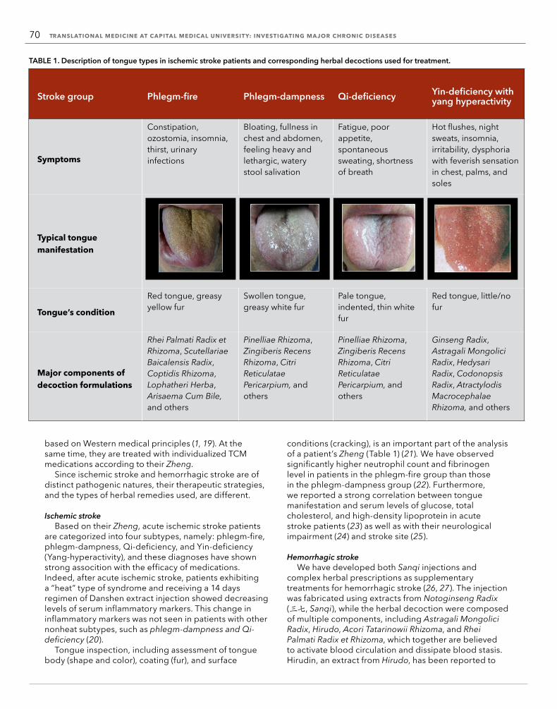
70 TRANSLATIONAL MEDICINE AT CAPITAL MEDICAL UNIVERSITY: INVESTIGATING MAJOR CHRONIC DISEASES
based on Western medical principles (1, 19). At the same time, they are treated with individualized TCM medications according to their Zheng.
Since ischemic stroke and hemorrhagic stroke are of distinct pathogenic natures, their therapeutic strategies, and the types of herbal remedies used, are different.
Ischemic strokeBased on their Zheng, acute ischemic stroke patients
are categorized into four subtypes, namely: phlegm-fire, phlegm-dampness, Qi-deficiency, and Yin-deficiency (Yang-hyperactivity), and these diagnoses have shown strong assocition with the efficacy of medications. Indeed, after acute ischemic stroke, patients exhibiting a “heat” type of syndrome and receiving a 14 days regimen of Danshen extract injection showed decreasing levels of serum inflammatory markers. This change in inflammatory markers was not seen in patients with other nonheat subtypes, such as phlegm-dampness and Qi-deficiency (20).
Tongue inspection, including assessment of tongue body (shape and color), coating (fur), and surface
conditions (cracking), is an important part of the analysis of a patient’s Zheng (Table 1) (21). We have observed significantly higher neutrophil count and fibrinogen level in patients in the phlegm-fire group than those in the phlegm-dampness group (22). Furthermore, we reported a strong correlation between tongue manifestation and serum levels of glucose, total cholesterol, and high-density lipoprotein in acute stroke patients (23) as well as with their neurological impairment (24) and stroke site (25).
Hemorrhagic strokeWe have developed both Sanqi injections and
complex herbal prescriptions as supplementary treatments for hemorrhagic stroke (26, 27). The injection was fabricated using extracts from Notoginseng Radix (三七, Sanqi), while the herbal decoction were composed of multiple components, including Astragali Mongolici Radix, Hirudo, Acori Tatarinowii Rhizoma, and Rhei Palmati Radix et Rhizoma, which together are believed to activate blood circulation and dissipate blood stasis. Hirudin, an extract from Hirudo, has been reported to
TABLE 1. Description of tongue types in ischemic stroke patients and corresponding herbal decoctions used for treatment.
Stroke group Phlegm-fire Phlegm-dampness Qi-deficiency Yin-deficiency with yang hyperactivity
Symptoms
Constipation, ozostomia, insomnia, thirst, urinary infections
Bloating, fullness in chest and abdomen, feeling heavy and lethargic, watery stool salivation
Fatigue, poor appetite, spontaneous sweating, shortness of breath
Hot flushes, night sweats, insomnia, irritability, dysphoria with feverish sensation in chest, palms, and soles
Typical tongue manifestation
Tongue’s condition
Red tongue, greasy yellow fur
Swollen tongue, greasy white fur
Pale tongue, indented, thin white fur
Red tongue, little/no fur
Major components of decoction formulations
Rhei Palmati Radix et Rhizoma, Scutellariae Baicalensis Radix, Coptidis Rhizoma, Lophatheri Herba, Arisaema Cum Bile, and others
Pinelliae Rhizoma, Zingiberis Recens Rhizoma, Citri Reticulatae Pericarpium, and others
Pinelliae Rhizoma, Zingiberis Recens Rhizoma, Citri Reticulatae Pericarpium, and others
Ginseng Radix, Astragali Mongolici Radix, Hedysari Radix, Codonopsis Radix, Atractylodis Macrocephalae Rhizoma, and others

TRANSLATIONAL RESEARCH IN INTEGRATED TRADITIONAL CHINESE AND WESTERN MEDICINE 71
regulate Aquaporin-4 and 9 expression and improve the outcome in an experimental intracerebral hemorrhagic stroke rodent model (28, 29). Our laboratory tests and animal experiments further confirmed that these TCM treatments preserved microvascular integrity and function at the disease site, improved microcirculation and blood velocity, and significantly attenuated inflammatory factors. Indeed, observations in our pilot clinical trial have revealed that TCM treatment facilitated hematoma absorption and reduced brain edema, and improved overall outcomes (26).
SummaryWe have briefly introduced TCM theory and its
application in acute stroke therapy in our department. Our clinical experience with the integration of Western medicine and TCM in stroke therapy suggests that TCM treatment can enhance recovery in stroke patients, and that differential diagnosis based on a patient’s Zheng is critical for the customization of Chinese herbal medication. We believe that TCM, as a fundamental part of Chinese ancient wisdom, carries a reservoir of knowledge that is an understudied treasure and worth further exploration to provide additional benefits for stroke patients.
References 1. G. A. Donnan, M. Fisher, M. Macleod, S. M. Davis, Lancet 371, 1612 (2008). 2. Z. W. Liu, Essentials of Chinese Medicine (Springer, New York/ London, 2010). 3. G. R. Schinella, Life Sci. 70, 1023 (2002). 4. A. Matkowski, W. Jamiołkowska-Kozlowska, I. Nawrot, Curr. Med. Chem. 20, 984 (2013). 5. X. Yan, Dan Shen (Salvia miltiorrhiza) in Medicine Volume 1. Biology and Chemistry (Springer Dordrecht Heidelberg, New York/London, 2015). 6. X. Yan, Dan Shen (Salvia miltiorrhiza) in Medicine Volume 2.
Pharmacology and Quality Control (Springer Dordrecht Heidelberg, New York/London, 2015). 7. C. L. Liu et al., PLOS ONE 2, e1321 (2007). 8. L. X. Xie et al., Clin. Hemorheol. Micro. 44, 245 (2010). 9. L. Zhou, Z. Zuo, M. S. Chow, J. Clin. Pharmacol. 45, 1345 (2005).10. L. Gao et al., Neurol. Res. 31, 402 (2009).11. H. Kim, Neurol Res. 27, 287 (2005).12. Y. F. Liu et al., Am. J. Chin. Med. 35, 583 (2007).13. Y. F. Liu et al., Phytother. Res. 22, 935 (2008).14. J. S. Li et al., Chin. J. Trad. Chin. Med. Pharm. 2011-9, 1967 (2011).15. J. S. Li et al., Chin. Pharm. Bull. 2007-01, 114 (2007).16. J. S. Lu et al., Chin. J. Chin. Mater. Med. 30, 1939 (2005).17. R. Ye, G. Zhao, X. Liu, Expert Rev. Neurother. 13, 603 (2013).18. N. J. Sucher, Epilepsy Behav. 8, 350 (2006). 19. H. P. Adams et al., Stroke 38, 1655, (2007). 20. Y. Luo et al., Chin. J. TCM WM Crit. Care, 16, 131 (2009).21. P. Tian, Nature 480, S84 (2011).22. L. Gao et al., Chin. J. Integr. Med. 18, 942 (2012).23. P. Liu et al., J. Trad. Chin. Med. 34, 162 (2014).24. L. Gao et al., Chin. J. Integr. Med. 19, 149 (2013).25. P. Liu et al., Chin. J. Integr. Med. 20, 823 (2014).26. L. Gao et al., J. Neurol. Sci. 323, 236 (2012).27. Y. Sun et al., Chin. J. Integr. Med. 9, 4 (2003).28. Y. Zhang et al., Chin. J. Chin. Mater. Med. 31, 69 (2006).29. Z. Sun et al., Mol. Biol. Rep. 36, 1119 (2009).
Acknowledgments This work was supported by the Chinese Natural Science Foundation (81401090 to J. S., and 81071058 and 81471340 to Y. L.), and by AHA Award 14SDG20480186 (to L. C.). It was also partly supported by research grants ITS/274/12, RGC/HKBU-121009/14, and HMRF 12132091 (to M. L.). The authors would like to thank Dr. Martha Dahlen for her English editing of this manuscript. Conflict of Interest: the authors declare no conflict of interest.
72 TRANSLATIONAL MEDICINE AT CAPITAL MEDICAL UNIVERSITY: INVESTIGATING MAJOR CHRONIC DISEASES
Diagnostic imaging of neurological disease in HIV/AIDSYunfang Li, Hongjun Li*, Jiaojiao Liu, Jinli Ding, and Wei Wang
Infection with human immunodeficiency virus (HIV) and the acquired immunodeficiency syndrome (AIDS) that it causes are widespread across the world and present a serious threat to human health. For example, according to the Chinese Centers for Disease Control and Prevention (CDC), there were 483,801 HIV-positive patients in China on June 30, 2014 (1). HIV-related diseases of the nervous system are one of the major features of AIDS and a common cause of death. In 10% to 20% of AIDS patients, nervous system impairment is the first symptom identified and abnormal neuropathology is present in up to 80% of cases, as assessed by autopsy.
Imaging plays a crucial role in the diagnosis and differential diagnosis of AIDS-related nervous system diseases. Recent developments in imaging technologies have enabled the diagnosis of such disorders using techniques such as computed tomography (CT) and magnetic resonance imaging (MRI), together with post-processing imaging software. Using criteria including origin, location, and spectrum, the diseases can be classified into five categories: primary HIV-related impairments, intracranial opportunistic infections, neurological tumors, cerebrovascular diseases, and peripheral neuropathies (2). Here, we describe each of these AIDS-related neurological complications and examine the current state of the art for imaging each of them.
Primary HIV-related impairmentsHIV encephalitis results from direct damage to the
nervous system by HIV. The main neuropsychiatric symptom is dementia, also known as HIV-associated dementia (HAD). This represents a severe form of HIV-related central nervous system disease and is characterized clinically as movement disorders, behavioral changes, and cognitive dysfunction (3). Using data collected over the past decade (2004 to 2013) in Beijing, Shanghai, Shenzhen, Zhengzhou, and five cities in the Xinjiang autonomous region, we found that the incidence of HIV encephalitis was slightly above 4% (4).
No obvious morphological changes are present in early stages of this disease. In the intermediate and advanced
Department of Radiology, Beijing You’an Hospital, Capital Medical University, Beijing, China.*Corresponding Author: [email protected]
Translational research in preventive medicine and
clinical diagnosis

TRANSLATIONAL RESEARCH IN PREVENTIVE MEDICINE AND CLINICAL DIAGNOSIS 73
FIGURE 1. Contrast diagrams of regions activated with right hand motion, normal volunteers compared with patients with HIV. The activated region on the right side of the HIV-infected brain is enlarged and enhanced compared with normal volunteers.
stages, diffusive or focal abnormalities in the white matter can be detected by MRI, but no significant edema, altered brain tissue around an edema-induced mass, widened sulci, enlarged ventricles, or decreased brain volume was seen. Functional and molecular imaging techniques such as blood-oxygen-level dependent functional MRI (BOLD-fMRI), magnetic resonance stimulation (MRS), and diffusion tensor imaging (DTI) have been used to assess abnormal metabolism and microstructures. These techniques have been shown to diagnose HAD before significant symptoms and morphological imaging abnormalities become apparent. Using MRS it has been shown that brain injury and/or reduced brain activity is concentrated in the basal ganglia and the frontal white matter, and that the changes appear in the basal ganglia earlier than in the frontal white matter (5). Signal changes seen using BOLD-fMRI have revealed laterality in the damage to brain functional regions, while highly active antiretroviral therapy (HAART) was shown by the same technique to suppress abnormal activation in the brain (see Figure 1) (6). DTI has been used to observe and track cerebral white matter fiber tracts (see Figure 2). It has revealed that the fractional anisotropy (FA) value, which is a measure of connectivity in the brain, is decreased and showed a preferential occult injury of frontal lobe in the early phase of HAD (7). This study also showed that a reduced CD4+ T lymphocyte count may be a risk factor for HAD. Further, it has been reported that HIV seropositive patients without noticeable neurocognitive impairment have changes in gray matter volume as detected by high-resolution T1-weighted MRI and Voxel-based morphometry (8). In particular, compared to the control group, HIV seropositive males had smaller gray matter volumes in
both the triangular and orbital part of the left inferior frontal gyrus, the left superior temporal gyrus, the right middle frontal gyrus and anterior cingulum. Additionally, significant increases in gray matter volumes were seen in the periaqueductal gray and gray around lateral ventricle for the patients with HIV.
Aseptic meningitis is another primary impairment associated with HIV infection. The acute phase generally occurs in the tentorium cerebelli and cerebral falx, with zonal thickening and linear or strip enhancement. In the chronic phase of infection, supratentorial hydrocephalus and ventriculomegaly was found (9).
HIV-related intracranial opportunistic infectionsIntracerebral parasitic infections
HIV-related toxoplasma encephalitis is a leading cause of death in HIV-positive patients. In our cohort of Chinese patients, the incidence was found to be 1.5% (4). It affects mainly the basal ganglia, cortex, and medulla junction, but can also be found in the cerebellum, brainstem, and even in the ventricles. Based on the location of the lesions, toxoplasma encephalitis can be divided into three types: parenchymal, ventricular, and mixed. MRIs have revealed single and multiple ring-shaped, spiral, or nodular enhancements that are smaller than 2 cm in diameter and surrounded by significant edema (10).
Intracerebral cysticercosis infection associated with HIV is commonly detected in the vesicular and colloidal phases. Due to the lack of immune-mediated effects, there is no usual related pathological processes and imaging of the granuloma calcification stages Another HIV-related infection, trichinosis, occurs mainly in geographically rural areas. Lesions are distributed in the cortex, medulla, and
FIGURE 2. A three-dimensional view of a patient with HIV’s brain captured using diffusion tensor imaging (DTI), showing the distribution and arrangement of the white matter fiber tracts, which are bilaterally symmetrical.

74 TRANSLATIONAL MEDICINE AT CAPITAL MEDICAL UNIVERSITY: INVESTIGATING MAJOR CHRONIC DISEASES
basal ganglia, and imaging shows mainly granuloma formation with brain edema.
Intracerebral viral infectionsCytomegalovirus
encephalomyelitis involves mainly the basal ganglia, where there is a characteristic pattern of flaky demyelination, but the ventricles, pons, and medulla oblongata may also be involved, albeit with ill-defined boundaries. No enhancement in CT or MRI enhancement scans has been reported in the brain parenchyma, but this does occur in the ependymal (11).
Progressive multifocal leukoencephalopathy (PML), caused by the John Cunningham virus (JCV), is found in the advanced stages of AIDS at an incidence rate of between 1% and 10% (12). PML is characterized by multifocal neuropathy with rapid progression. It usually occurs in the top occipital white matter, often with a scattered and asymmetric distribution, and sometimes also involves the gray matter, causing lesions that create space-occupying changes in the brain tissue. As a result, the damage gradually becomes integrated, expanding and confusing the boundaries of lesions (13). Imaging reveals ventriculomegaly, deepening and widening of the sulci, and, in advanced stages, brain atrophy (14).
Herpes simplex viral encephalitis is characterized by asymmetry in the distribution of lesions, which occurs mostly in the middle temporal lobe, hippocampus, frontal orbital plane, parietal lobe, and cingulate, and causes obvious brain edema. Punctate bleeding is also a characteristic seen in the brain. This type of encephalitis has abnormal ependymal linear or gyrus-like enhancement. Generally, it does not involve the putamen (15).
Intracranial fungal infections According to our data, the incidence of HIV-related
Cryptococcus neoformans meningitis was just below one percent (4). The disease is listed as an AIDS-defined disease in Africa by the United States Center for Disease Control and Prevention (CDC) (Figure 3) and has been
categorized into four types based on MRI evidence and pathomechanism: meningoencephalitis type, infarct type (colloid capsules), infarct type, and granulomatous type.
Cerebral aspergillosis infection is rare. It occurs mostly as multifocal and polymorphic damage in the brain parenchyma, where it can cause abscesses, cerebral infarctions, cerebral hemorrhaging, intracranial granulomas, meningitis, and encephalitis. Imaging has revealed that enhanced dura mater with local epidural abscesses might occur at the same time, together with enhancement of the soft tissue in the paranasal sinuses and orbit.
Intracranial tuberculosis We found the incidence
of AIDS-related cerebral tuberculosis to be close to 1.5% (4). As a result of the immunodeficiency caused by the virus, there was a lack of immune-mediated damage and consequently no fibrous tissue, making it difficult to identify the classic brain tuberculoma. AIDS-related cerebral tuberculosis is therefore characterized by encephalitis or meningitis. There is meningeal thickening at the base of the brain and obvious abnormal enhancement; sometimes, multiple round nodules can be identified, with
unobvious peripheral edema, irregular rings, or nodular enhancements (16).
Other infectionsRhodococcus equi infection rarely involves the
nervous system, but commonly involves deep brain tissues. It undergoes rapid progression due to general dissemination, but lacks a characteristic appearance when imaged (17).
Intracerebral Penicillium marneffei infection occurs mostly in humid geographical zones. Due to this pathogen’s mechanism of dissemination, which involves the lung and retroperitoneal lymph nodes, infection of the brain is extremely rare. When it does occur, most foci are found in the cortex-medulla junction. There can be multiple or singular low-density shadows in brain
FIGURE 3. Enhanced MRI of an HIV-positive person with Cryptococcal meningoencephalitis. (A) Honeycomb-like enhancements can be seen in the left basal ganglia. Magnetic Resonance (MR) spectroscopy (B) shows the choline (Cho) peak, representing various choline-containing compounds, is increased and the N-Acetyl Aspartate (NAA) peak, an amino acid found exclusively in neurons, is reduced.

TRANSLATIONAL RESEARCH IN PREVENTIVE MEDICINE AND CLINICAL DIAGNOSIS 75
parenchyma, and obvious edema and space-occupying lesions, without any particular pattern being obvious.
Finally, cerebral syphilis is rare in HIV patients due to the lack of immune-mediated effects of the virus and is mainly confined to meningoencephalitis.
Neurological tumorsIn our cohort of patients with AIDS, the incidence
of AIDS-related lymphoma fell between 5% and 10% while the incidence of primary central nervous system lymphoma (PCNSL) is about 0.6% (4). Imaging of PCNSL showed markedly enhanced meninges, ventricles, and ependyma, and obvious abnormal enhancement can be seen at the map-liked boundaries on the lesions’ edges. In our study, lymphoma lesions always occurred singly and with a higher incidence in the central nervous systems of AIDS patients than that of immunocompetent patients. Central necrosis of lesions is common, but infratentorial involvement is less so (18).
Other rare HIV-related neurological tumors include intracerebral Hodgkin’s lymphoma, Kaposi’s Sarcoma, and various undefined tumors that feature metastases, meningiomas, and leukemias.
Cerebrovascular diseasesThe incidence of AIDS-associated cerebral atrophy in
our cohort was 1.5%; 1% for cerebral hemorrhaging and 0.5% for cerebral infarction (4). Vascular lesions can be divided into five categories: nonspecific congestion of brain tissue, edema, and hemorrhage; cerebrovascular thrombosis and embolism, with resulting cerebral infarct; acute granulomatous cerebral vasculitis; opportunistic infection-induced vascular lesions; and tumorous vascular lesions.
Peripheral neuropathiesMorphological changes and dysfunction of peripheral
nerves occurred in 9% to 16% of AIDS patients in our cohort (4). These alterations generally included acute/chronic demyelinating polyneuropathy, multiple nerve root disease, autonomic neuropathy, and ganglionitis.
Imaging research in HIV-related neurological disease in China
Much of the research discussed here is based upon a patient cohort that has been followed for over 16 years by a research group at Capital Medical University, established by Hongjun Li (4). The research team has been recording epidemiology, clinical, imaging, anatomical, pathological, and pathogenic data. Imaging features are studied to establish criteria for the involvement of HIV in neurological disease. This has helped to illuminate some of the pathological mechanisms underlying HIV-related conditions and establish the theories underlying the progression of HIV-related neurological diseases (17, 19–22). Taken together, this information serves as a powerful resource for the prevention and treatment of HIV and will be included in future textbooks produced by the Chinese Ministry of Health.
Imaging research on HIV-related nervous system diseases is essential for five reasons. First, neurological diseases remain a leading cause of death among HIV patients, and noninvasive, digital imaging technology is an important way to monitor disease progression. Second, imaging research can capture multiple aspects of diseases that can have unusual and complicated presentations due to immunosuppression. Third, the current understanding of evidence-based and translational medicine is insufficient; digital medical imaging provides an important objective assessment for diagnosis and treatment. Fourth, HIV/AIDS presents as a spectrum of diseases with frank disease, latent disease, and many co-occurring pathologies. HIV/AIDS patients or latent HIV/AIDS patients are commonly seen in specialized and general hospitals, and professional knowledge about HIV/AIDS diagnosis or treatment is essential. Radiologists must be aware of the disease spectrum and become more familiar with the complexities of AIDS-related imaging. Fifth, an understanding of the spectrum of HIV/AIDS-related neurological diseases also needs to be more widespread among clinicians in general. Information about noninvasive imaging of HIV effects and guidelines for using HIV/AIDS imaging in clinical and research studies continues to be of great value in the ongoing efforts to prevention and treat HIV/AIDS.
References 1. NCAIDS, NCSTD, China CDC. Chin. J. AIDS STD. 20, 629 (2014). 2. H. J. Li et al., Chin. Med. J. 126, 4371 (2013). 3. A. B. Smith, et al., Radiographics 28, 2033 (2008). 4. H. J. Li, Ed. Radiology of HIV/AIDS, (Springer, New York, 2014). 5. S. Qi et al., J. Clin. Radiol. 33, 1803 (2014). 6. J. Zhao et al., J. Clin. Radiol. 33, 1787 (2014). 7. R, Li et al., J. Clin. Radiol. 33, 1796 (2014). 8. Y. Li et al., Int. J. Clin. Exp. Med. 7, 3362 (2014). 9. F. Antunes, Acta Neurochir. 146, 1071 (2004).10. V. Vladimira, B. Nevajda, M. Spero, D. Chudy, Pathog. Glob. Health 107, 325 (2013).11. H. Pamar, M. Ibrahim, Neuroimaging Clin. N. Am. 22, 707 (2012).12. C. Wüthrich et al., J. Neuropathol. Exp. Neurol, 68, 15 (2009).13. S. Dahlhaus et al., J. Neurol. Neurosurg. Psychiatry 84, 1068 (2013).14. T. Ernst et al., Radiology 210, 539 (1999).15. R. J. Bert et al., Am. J. Neuroradiol. 25, 1722 (2004).16. T. Richa et al., Indian J. Radiol. Imaging 19, 256 (2009) 17. H. J. Li et al., Chin. Med. J. 124, 968 (2011).18. M. Bilgrami, P. O’Keefe, Handb. Clin. Neurol. 121, 1321 (2014).19. H. J. Li et al., Chin. Med. J. 124, 1427 (2011). 20. H. J. Li et al., Chin. Med. J. 122, 2142 (2009).21. Y. Z. Zhang et al., Chin. Med. J. 124, 693 (2011). 22. H. J. Li, Ed. Atlas of differential Diagnosis in HIV/AIDS, (PMPH, Beijing, China, 2008).
76 TRANSLATIONAL MEDICINE AT CAPITAL MEDICAL UNIVERSITY: INVESTIGATING MAJOR CHRONIC DISEASES
Urinary proteomics in biomarker discovery
Man Zhang* and Qian Meng
A lthough urine is the final product of metabolism, a significant amount of protein remains present even after filtration by the glomeruli of the kidneys, reabsorption, and secretion via the renal tubules. Changes in abundance, composition, and character of these proteins are associated with the occurrence, development, and prognosis of disease. These changes are not indicative only of the state of the kidney and urogenital tract. The health of more distant organs are also mirrored in the protein composition of urine.
Proteomics is the most effective way to gather and interpret the information carried by proteins and to determine a profile of the constituents. Comparison of the urine protein profiles between normal subjects and patients with a defined disease can reveal differentially expressed proteins that can be used as biomarkers for the disease. The urinary proteome has huge potential for new and much-needed diagnostics; its analysis enables early detection of changes in urinary and other system functions.
Urine has several advantages over other body fluids that make it a preferred sampling source for biomarker discovery. It is convenient to collect in large quantities and can be obtained noninvasively. Urine has a simple composition that makes it easy to purify and analyze, and its protein content is relatively stable. By the time urine is voided, proteolytic degradation is often already complete because urine stays in the bladder for hours. One disadvantage of using urine for sampling is that the proteomic composition changes significantly over the course of the day as a result of meals, metabolic processes, exercise, and circadian rhythms; however, these variations affect only a small fraction of the urinary proteome (1).
Estimates of the human urine proteome’s complexity have increased dramatically as the technologies have improved. The first attempt to describe the urinary proteome, in 2001, identified 124 gene products (proteins and translations of expressed sequence tags) in pooled urine samples using liquid chromatography–mass spectrometry (LC-MS) (2). Just three years later, nearly 1,400 distinct protein spots have been screened using two-dimensional electrophoresis (2-DE). Of these, 420 spots were identified, resulting from 150 unique urine proteins (3). By 2006, Adachi et al. had identified 1,543
proteins in urine samples from healthy individuals by combining 1-D sodium dodecyl sulfate polyacrylamide gel electrophoresis (SDS-PAGE), reversed phase high-performance liquid chromatography (HPLC), linear trap quadrupole-Fourier transformation (LTQ-FT), and LTQ-Orbitrap mass spectrometry analysis (4). In this study, membrane proteins accounted for nearly half of all protein species. The number of identified proteins in urine increased to 1,823 in 2011 (5), including154 proteins that were previously known to be plasma proteins. In addition to proteins, large quantities of different peptides are present in urine; Coon et al. reported that that there are more than 100,000 (6). Urine is therefore a rich source of potential biomarkers of disease that awaits exploration.
Biomarker discovery by diseaseUrinary proteome studies have identified and validated
biomarkers in various diseases using multiple proteomics technologies. Here, we summarize some of the current literature on different types of cancer and other major diseases.
Bladder cancerBladder cancer is the second most common malignancy
in the United States affecting the urinary system (7). Cystoscopy is the reliable diagnostic method at present (8). However, it is invasive for the patient, and is expensive. The development of new methods for the diagnosis of bladder cancer is an urgent challenge.
An exploratory study to identify urinary biomarkers for bladder cancer compared proteins in urine from patients with transitional cell carcinoma of the bladder, from patients with other urogenital diseases, or from healthy donors (9). Using protein chip technology, the project identified five potential biomarkers in seven protein clusters. The method of using protein biomarkers is better than urine or bladder washing cytology, the sensitivity of detecting low-grade transitional cell carcinoma improved from 33% to 78%.
Another group identified differential expression of the proteins Reg-1 and keratin 10 between patients with bladder tumors and controls, using 2-D difference gel electrophoresis (DIGE) coupled with mass spectrometry (10). Using a similar approach, Gc-globulin was found to be a marker of high sensitivity and specificity for the early detection and surveillance of bladder cancer (11).
The first attempt to profile urine proteins for bladder cancer using the isobaric tag for relative and absolute quantitation (iTRAQ) method revealed numerous protein biomarkers, including some classical plasma proteins that differed more than two-fold in expression levels between patients and controls (12). Our group has also studied urine proteins in patients with bladder cancer (13). Using 2-DE and matrix-assisted laser desorption ionization time-of-flight mass spectrometry (MALDI-TOF-MS), we identified 14 discriminatory protein spots that differed between pooled urine samples from three patients with bladder cancer and three normal controls. One of the proteins, apolipoprotein A-I, was present at increased levels in the urine of bladder cancer patients, making it a potential biomarker for the
Department of Clinical Laboratory, Beijing Shijitan Hospital, Capital Medical University, Beijing, China*Corresponding Author: [email protected]

TRANSLATIONAL RESEARCH IN PREVENTIVE MEDICINE AND CLINICAL DIAGNOSIS 77
disease. Additionally, we identified biomarkers that discriminate between adriamycin-resistant (pumc-91/ADM) and parental (pumc-91) human bladder cancer cell lines (14). Other proteins have been found in bladder cancer tissue that may be promising biomarkers to predict recurrence and cancer drug resistance, as well as provide guidance for tumor surveillance (15, 16). We are currently working to determine if they can be validated in urine protein analysis.
Prostate cancerProstate cancer is one of the most common cancers in
men (7). The level of serum prostate-specific antigen (PSA) has been widely used for prostate cancer diagnosis and prognosis, but its measurement to lacks the specificity to accurately distinguish between men with or without prostate cancer (17). Additional biomarkers are needed to improve detection and diagnosis.
Urine has been a promising source for prostate cancer biomarker discovery since Rehman et al. first described the urine protein profiles of patients with prostate cancer in 2004 (18). They used 2-DE with MALDI-TOF-MS analysis to compare cancer and benign prostatic hyperplasia, identifying B/MRP-14 as a potential urine marker for prostate cancer. Using capillary electrophoresis coupled with mass spectrometry (CE-MS), additional urine proteins were suggested by Theodorescu and colleagues to discriminate between prostate cancer and benign prostatic hyperplasia (19). Another large-scale study for peptide biomarkers identified in urine was carried out using MALDI-TOF (20). This achieved 71% specificity and 67% sensitivity indiscriminating patients with prostate cancer and benign prostatic hyperplasia, and identified uromodulin and semenogelin I isoform b pre-pro-protein as possible peptide biomarkers.
Renal cell carcinomaRenal cell carcinoma, which is the one of the most
common primary renal malignant neoplasms, is difficult to diagnose in the early stages, making it a great candidate for validated biomarkers (21). Surface-enhanced laser desorption/ionization (SELDI) TOF-MS technology was employed by Wu et al. in a study that identified differentially excreted peptides as potential biomarkers (22). Specificity and sensitivity for renal cell carcinoma detection was 81% and 68% respectively. Urine samples were also analyzed by Bosso et al. who found three markers that can be used to discriminate patients from controls and which, in combination, provided higher diagnostic efficacy (23).
Other cancersUrinary proteomics has been used to study various
other types of cancers. For example, MALDI and SELDI were used to analyze urine samples from 67 patients with colorectal cancer and 72 noncancer control subjects (24). This study yielded 19 discriminatory peaks, three of which were quantified. Proteomic methods have also been used to identify biomarkers from urine samples of patients
with ovarian cancer, including elevated urinary eosinophil-derived neurotoxin and osteopontin COOH-terminal fragments, as shown by Ye et al. (25). Finally, in a comparison of urinary protein analyses, Tantipaiboonwong et al. identified proteins such as CD59 glycoprotein, transthyretin, GM2AP, and free immunoglobulin light chain, which may be useful as lung cancer markers (26).
Chronic kidney diseaseChronic kidney disease is a major public health concern,
with patients at high risk for cardiovascular disease and other adverse outcomes. Early diagnosis of specific renal diseases can improve therapeutic treatments, prevent deterioration of the kidney, and reduce the financial burden for patients. Biomarkers that identify individuals in the early stages of the disease are therefore highly desirable (27). As discussed above, proteins and peptides in urine are potentially a good source for such biomarkers.
The urinary peptide profile of chronic kidney disease was first reported in 2008 (28). Three urinary peptides (the precursor fragments of collagen 1a and 5a chains, and uromodulin) were shown using MALDI-MS to differ in expression between patients with or without chronic kidney disease. Good et al. also characterized urinary peptide biomarkers of chronic kidney disease (29). Using CE-MS, 273 biomarkers were found from a study of 230 patients and 379 healthy volunteers. When chronic kidney disease-specific biomarkers were examined in an independent cohort, chronic kidney disease could be detected in patients with 85% sensitivity and 100% specificity.
Type 2 diabetes mellitusAnother serious and growing public health problem
is type 2 diabetes (T2D), which is characterized by hyperglycemia and caused by an absolute or relative lack of insulin. To monitor and treat complications promptly, regular examinations are essential for patients with T2D. Blood glucose was tested in routine clinical practice to evaluate the efficacy of drugs and facilitate adjustments to drug doses. However, because blood sample testing commonly involves injuring blood vessels, it was decided to collect urine samples, as they were easier to get. We have used magnetic bead-based weak cationic exchange beads (MB-WCX) and MALDI-TOF-MS to define the urine peptidome profiles of patients with T2D and healthy controls, with the aim of identifying urinary peptides that can distinguish between the two groups (30). We identified three peptides, namely histidine triad nucleotide-binding protein 1 (HINT1), bifunctional aminoacyl-tRNA synthetase (EPRS), and cluster in precursor protein (CLU) that are reduced in T2D patients and may serve as biomarkers. Our research also identified two urinary peptides (isoform 1 of fibrinogen α chain precursor and prothrombin precursor) that enable the monitoring of T2D at different stages (31). Since diabetes is a risk factor for coronary heart disease, we compared the urinary peptide signatures in two groups of T2D patients, those with coronary artery disease and those without. Six differentially expressed peptides were found including peptide fragments of isoform 1 of fibrinogen α chain
78 TRANSLATIONAL MEDICINE AT CAPITAL MEDICAL UNIVERSITY: INVESTIGATING MAJOR CHRONIC DISEASES
precursor, prothrombin precursor, and inter-alpha-trypsin inhibitor heavy chain H4 (32).
Diabetic nephropathy, one of the serious and common complications of diabetes, leads to end-stage renal disease. Currently, microalbuminuria is used for the diagnosis of early stage diabetic neuropathy; however, one-third of neuropathy cases occur in the absence of albuminuria, and microalbuminuria can also occur in healthy individuals (33). Therefore, scientists have focused much effort on finding biomarkers for individuals who are at high risk for diabetic neuropathy, and a number of urinary candidates have been identified. Rossing et al. cataloged 65 biomarkers that differed between patients with and without nephropathy using CE-MS analysis (34). E-cadherin was discovered as a novel biomarker for diabetic nephropathy by a group using difference gel electrophoresis (DIGE) and MS. The expression of E-cadherin was significantly decreased in the diabetic nephropathy group and the sensitivity and specificity of the test was 79% and 80%, respectively (35). Finally, iTRAQ was used to uncover urinary proteins differentially excreted in T2Dpatients with and without diabetic nephropathy (36). In total, 710 protein biomarkers were identified, of which 196 were quantified by multiple reaction monitoring in urine.
Immune system disordersAutoimmune diseases are chronic diseases caused by
defects in the immune system that can lead to disability or death, making this an active area of research. Zhang et al. employed serial urine proteomics to uncover biomarkers that predict the nephritis component of systemic lupus erythematosus (SLE) (37). In an effort to identify early indicators of graft-versus-host disease (GVHD), a recent phase I diagnostic study was performed to validate urine polypeptide patterns from 40 patients following hematopoietic stem cell transplantation as well as five patients with sepsis. Sixteen differentially excreted polypeptides provided a pattern that identified early GVHD with 82% specificity and 100% sensitivity (38).
Additional urine sampling may provide an effective alternative for certain routine screenings. Our research indicates that urine may therefore provide a more convenient way to monitor maternal thyroid functional status during pregnancy. The ratio of urine free triiodothyronine (uFT3) to urine retinol binding protein (uRBP) (uFT3/uRBP) and the urine free thyroxine (uFT4) to uRBP ratio (uFT4/uRBP) correlated well with levels of serum FT3 and FT4 during pregnancy, respectively (39).
In conclusion, urinary proteomics offers multiple methods to describe protein profiles. Scientists have already successfully identified a number of biological markers from urine samples using these approaches and have promising prospects for developing new tools for clinical diagnosis; however, very few biomarkers have been applied to clinical practice. This is, perhaps, one of the biggest challenges facing the field today, making translation of such discoveries into clinical tests an important focus for the future.
References 1. S. Decramer et al., Mol. Cell. Proteomics 7, 1850 (2008). 2. C. S. Spahr et al., Proteomics 1, 93 (2001). 3. R. Pieper et al., Proteomics 4, 1159 (2004). 4. J. Adachi, C. Kumar, Y. L. Zhang, V. O. Jesper, M. Matthias, Genome Biol. 7, 80 (2006). 5. A. Marimuthu et al., J. Proteome Res. 10, 2734 (2011). 6. J. J. Coon et al., Proteomics Clin. Appl. 2, 964 (2008). 7. R. Siegel et al., CA Cancer J. Clin. 63, 11 (2013). 8. D. Mitropoulos et al., J. Endourol. 19, 861 (2005). 9. A. Vlahou et al., Am. J. Pathol. 158, 1491 (2001).10. E. Orenes-Piñero et al., J. Proteome Res. 6, 4440 (2007).11. F. Li et al., J. Proteomics 77, 225 (2012).12. Y. T. Chen et al., J. Proteome Res. 9, 5803 (2010).13. T. Lei et al., Clin. Genitourin. Cancer 1, 56 (2013).14. Q. Meng et al., J. Canc. Res. Clin. Oncol.139, 509 (2013).15. S. L. Yu, Q. Meng, H. H. Hu, M. Zhang, Int. J. Clin. Exp. Pathol. 9, 7 (2014).16. H. H. Hu, Q. Meng, T. Lei, M. Zhang, Clin. Exp. Med., (2014), doi:10.1007/s10238-014-0288-3.17. I. M. Thompson et al., N. Engl. J. Med. 350, 2239 (2004)18. I. Rehman et al., Urology 64, 1238 (2004).19. D. Theodorescu et al., Electrophoresis 26, 2797 (2005).20. A. E. M’Koma et al., Biochem. Biophys. Res. Comm. 353, 829 (2005).21. M. Li, W. Rathmell, in Kidney Cancer: Principles and Practice, P. N. Lara, Jr., E. Jonasch, Eds. (Springer Berlin, Heidelberg, 2012), pp. 47–65.22. D. L. Wu, W. H. Zhang, W. J. Wang, S. B. Jing, Y. M. Xu, Technol. Canc. Res. Treat. 7, 155 (2008).23. N. Bosso et al., Proteomics Clin. Appl. 2, 1036 (2008).24. D. G. Ward et al., Proteome Sci. 6, 19 (2008).25. B. Ye et al., Clin. Cancer Res. 12, 432 (2006).26. P. Tantipaiboonwong, S. Sinchaikul, S. Sriyam, S. Phutrakul, S. T. Chen, Proteomics 5, 1140 (2005).27. Q. L. Zhang, D. Rothenbacher, BMC Public Health 8, 117 (2008).28. A. Lapolla et al., J. Mass Spectrom. 44, 419 (2009).29. D. M. Good et al., Mol. Cell. Proteomics 9, 2424 (2010).30. L. N. Chu, G. Z. Fu, Q. Meng, H. Zhou, M. Zhang, Diabetes Res. Clin. Pract. 101, 187 (2013).31. M. Zhang, G. Z. Fu, T. Lei, PLOS ONE, e122950 (2015), doi:10.1371/journal.pone.0122950.32. G. Z. Fu, M. Hu, L. N. Chu, M. Zhang, Int. J. Endocrinol. 758402 (2015).33. V. Thongboonkerd, Contrib. Nephrol. 170, 172 (2011).34. K. Rossing et al., J. Am. Soc. Nephrol. 19, 1283 (2008).35. H. Jiang et al., Diabetes Metab. Res. Rev. 25, 232 (2009).36. J. Jin et al., J. Diabetes Res., 168602 (2012), doi:10.1155/ 2012/168602.37. X. Zhang et al., Kidney Int.74, 799 (2008).38. T. Kaiser et al., Blood 104, 340 (2004). 39. J. Cai et al., Syst. Biol. Reprod. Med. 60, 171 (2014).
AcknowledgmentsThis work was supported by the Beijing Natural Science Foundation (7122086).
TRANSLATIONAL RESEARCH IN PREVENTIVE MEDICINE AND CLINICAL DIAGNOSIS 79
Fangfang Dai, Yanhua Yu, Chen Ming, Lili Zhang, Li Ning, Xiuying Zhao, and Jinli Lou*
Recently, antibiotic resistance—the ability of bacteria to survive treatment with previously effective drugs—has captured the world’s attention. There is a clear need to develop new antibiotics to keep infectious diseases at bay. One appealing prospect is the use of bacteriophages, small viruses that can infect and lyse bacteria. Of course, there is the possibility that bacteria could become phage-resistant, again subverting our attempts to control them. Proponents point to the large number of phages that can infect bacteria, but the development of multiple phage antibiotics would be both time-consuming and expensive. A preferable approach would be to identify the mechanism that bacteria use to gain phage resistance, and eliminate it; this would provide a reliable foundation for phage treatment and for overcoming phage-resistant bacteria.
Among the resistance mechanisms described in bacteria, the one that stands out as being especially complex and important is the CRISPR/Cas system. CRISPR stands for “clustered, regularly interspaced, short palindromic repeats” and Cas for “CRISPR-associated genes.” Our research interest is focused on the CRISPR/Cas system in the opportunistic human pathogen, Pseudomonas aeruginosa, which is frequently involved in nosocomial infections that affect immunosuppressed cancer and severe burn patients as well as individuals suffering from cystic fibrosis. Due to irregular use of antibiotics and the ability of P. aeruginosa to acquire drug resistance, it is very difficult to treat an infection effectively with the current antibiotics. Phage therapies offer one possible solution; however, there are barriers to overcome: the CRISPR/Cas system in P. aeruginosa is more complicated than in other bacteria, and it is still a very new area of study (1, 2). Here, we summarize some of the recent literature about the CRISPR/Cas system and the development of bacteriophage resistance in P. aeruginosa.
The CRISPR/Cas systemCRISPR/Cas is the latest addition to the list of known
prokaryotic anti-invader systems, and it is the most complex. It is unique in being the only adaptive and inheritable prokaryotic immune system described to date (1).
In 1987, Ishino and his colleagues discovered an unusual structure of repetitive DNA downstream from the E. coli inhibitor of apoptosis (iap) gene, consisting of invariant direct repeats (29nt) and variable spacing sequences (32nt) (3). Although similar repeat clusters were subsequently identified in Haloferax mediterranei, Streptococcus pyogenes, Anabaena, and Mycobacterium tuberculosis, their function remained unknown. Jansen and colleagues coined the term CRISPR and reported that CRISPRs colocalized with specific cas genes (4). CRISPR/Cas systems are exclusively found in prokaryotes (5) and are present in approximately half of all bacteria and almost all archaea (6). Different systems have been proposed to classify the typical combinations of cas genes (7, 8). The most recent classification defines three main types, on the basis of the cas genes (Type I, Type II, and Type III), which are divided into at least ten subtypes (Types I-A to F, Types II-A and B, and Types IIIA and B). Cas1 and cas2 genes are present in each CRISPR/Cas variant (9).
The discovery of bacterial and archaeal genome sequences and the associated viral and plasmid sequences led to the realization that CRISPR spacers, which resemble fragments of foreign genetic elements, were derived from invading genomes (10–12). This breakthrough, together with the detection of CRISPR locus transcripts with defined lengths of one or more spacer repeat units (13–15), and the predicted nucleic acid-related activities for many of the cas genes, led Makarova and colleagues to propose that CRISPR/Cas neutralizes invaders via a mechanism reminiscent of RNA interference (8). Soon thereafter, Barrangou and colleagues provided the first experimental evidence that the CRISPR/Cas system of the lactic acid bacterium Streptococcus thermophilus functions as an inheritable, adaptive prokaryotic immune system (16).
Why wasn’t the CRISPR/Cas system discovered earlier? To some degree, this was due to the tight regulation of CRISPR/Cas systems in model organisms such as E. coli and Salmonella enterica that resulted in expression being silenced under normal laboratory growth conditions. Thus, from 1995 onwards, genomics researchers gathered information on the ubiquitous repetitive CRISPR loci and the associated gene clusters, but the fact that it was an adaptive and inheritable prokaryotic immune system remained hidden. Over the last 6 years, however, CRISPR research has flourished, providing numerous insights into the molecular mechanisms of this highly diverse defense system.
The CRISPR array consists of host-derived repeating sequences, typically of 30 base pairs, interspaced by similarly-sized acquired spacer sequences. A CRISPR locus may vary in length from just a few to several hundred repeat-spacer units. The repeat sequences have been
Department of Clinical laboratory,Beijing You’an Hospital, Capital Medical University, Beijing, China*Corresponding Author: [email protected]
Use of the CRISPR/CAS system to develop bacteriophage resistance to Pseudomonas aeruginosa
80 TRANSLATIONAL MEDICINE AT CAPITAL MEDICAL UNIVERSITY: INVESTIGATING MAJOR CHRONIC DISEASES
classified into 12 different clusters, some of which are predicted to form a hairpin secondary structure, while others are predicted to be unstructured (16). CRISPR loci are generally flanked by an adenine/thymine (AT)-rich leader sequence (3) that contains promoter elements (18–21) and binding sites for regulatory proteins (20, 21). Moreover, the polarized acquisition of new spacer sequences at the leader end of the CRISPR array suggests that leader sequences may contain binding sites for Cas proteins involved in CRISPR adaptation (22).
The mechanism of action of CRISPR/Cas systems is divided into three stages: adaptation, expression, and interference. During adaptation, resistance is acquired by integration of a new spacer sequence into a CRISPR array. In expression, cas genes are transcribed and translated. CRISPRs are transcribed into precursor CRISPR RNAs (pre-crRNAs) that are subsequently cleaved by a Cas6 homolog in Type I and Type III systems and by an RNase III in Type II systems (23–26). Mature crRNAs containing a single spacer sequence recognize a single target. In the third stage, interference, the crRNA guides one or more Cas-related proteins to cleave complementary invading nucleic acids.
CRISPR/Cas adaptation immunization in P. aeruginosa
In 2008, Zegans and colleagues found that infection of P. aeruginosa PA14 by the phage DMS3 resulted in lysogenized strains that were unable to form a biofilm or undergo swarming motility, two key group behaviors of this bacterium necessary for its infection of the host. This loss of function required CRISPRs and five of six cas genes. Based on these findings, the authors speculated that the CRISPR spacer content in P. aeruginosa PA14 was ineffective at resisting infection, but that incorporation of different spacers would create a resistant isolate. This raised the possibility that CRISPR regions have adapted to different functions in different species and/or play distinct roles in response to infection by lytic versus lysogenic phages. Further, the group suggested that the P. aeruginosa PA14 CRISPR region evolved to alter the effects of lysogeny or to resist phage infection or lysis. Alteration in biofilm formation and swarming behavior may have evolved as a strategy for limiting the effects of bacteriophage dissemination in bacterial communities. The authors implied that this system defined a novel mechanism of lysogenic conversion (27).
Then, in 2011, another study by Zegans and colleagues reported that DMS3 inhibition of biofilm formation was reversed in a lysogen with a transposon insertion in the CRISPR-associated gene csy3. A later study by Cady and O’Toole confirmed the requirement for CRISPR-associated genes in biofilm inhibition by DMS3 lysogens using clean deletion strains and genetic complementation (28). They queried catalytic sites within the Csy4 and Cas3 proteins. Based on this work, certain requirements for CRISPR-associated genes in the biofilm inhibition became clear. First, CRISPR2 spacer 1, but not spacer 20, is required for CRISPR-dependent biofilm inhibition. Second, specific residues within spacer 1 are required. Third, spacer 1
shares an imperfect identity with DMS3 gene 42 (DMS3-42). Fourth, deletion of DMS3-42 in a DMS3 lysogen restores biofilm formation to CRISPR-positive cells. Finally, specific nucleotides within DMS3-42 with complementarity to spacer 1 are required for biofilm inhibition. This was further evidence that P. aeruginosa CRISPR interacts with a chromosomally integrated mobile element and the results elucidated a specific target within DMS3. This interaction is mediated by a spacer which does not share full identity with DMS3 (spacer 1), rather than by a spacer that does (spacer 20) (3).
In 2012, Chung and colleagues isolated seven new phages from the culture supernatants of clinical P. aeruginosa isolates. Among them, MP29 and MP42 phages were plaque-purified using P. aeruginosa strain PA14 as a host; they, like DMS3, belong to the Siphoviridae family. However, the swarming motility of P. aeruginosa strain PA14 was not inhibited by lysogenization of either MP29 or MP42. Both of these phages contain two 32-base pair target sequences in ORF42, the nucleotide sequences of which are identical to those of MP22 and DMS3. The authors concluded that the nonidentity in this region might be necessary but is not sufficient for the phage-host CRISPR interaction that leads to swarming inhibition (29). At the same time, Cady and colleagues reported six temperate bacteriophages that can be prevented from replicating within the laboratory strain of P. aeruginosa PA14 by the endogenous CRISPR/Cas system. This work provided the first evidence that the P. aeruginosa type I-F CRISPR/Cas system can function in phage resistance. It also demonstrated that P. aeruginosa can acquire spacer content in response to lytic phage challenge, illustrating the adaptive nature of CRISPR/Cas. The authors suggested that the CRISPR/Cas system mediates a gradient of resistance to a phage based on the level of complementarity between CRISPR spacer RNA and the phage protospacer target (2).
In 2013, Bondy-Denomy and colleagues described the first examples of genes that mediate the inhibition of a CRISPR/Cas system (30). They described five distinct anti-CRISPR genes present in the genomes of bacteriophages infecting P. aeruginosa. Mutation of these anti-CRISPR genes rendered a phage unable to infect bacteria with a functional CRISPR/Cas system, and the addition of the same gene to the genome of a CRISPR/Cas-targeted phage allowed it to evade the CRISPR/Cas system. Phage-encoded anti-CRISPR genes may represent a widespread mechanism for phages to overcome the highly prevalent CRISPR/Cas systems (30). The existence of anti-CRISPR genes presents new avenues for the exploitation of CRISPR/Cas functional mechanisms as well as providing insight into the co-evolution of phages and bacteria.
In summary, the adaptive nature of the CRISPR/Cas system and the widespread occurrence of CRISPR regions in bacterial genomes suggest that this system is the most powerful weapon possessed by bacteria to resist invasion by foreign DNA. Phages also can evolve to resist the system. More work is needed to provide a full understanding of the mechanism of the CRISPR/Cas

TRANSLATIONAL RESEARCH IN PREVENTIVE MEDICINE AND CLINICAL DIAGNOSIS 81
system in P. aeruginosa and its phages. This understanding can provide a foundation for phage-based treatments for bacterial disease and for preventing the evolution of phage-resistant bacteria. References 1. E. R. Westra et al., Annu. Rev. Genet. 46, 311 (2012). 2. K. C. Cady et al., J. Bacteriology 194, 5728 (2012). 3. Y. Ishino, H. Shinagawa, K. Makino, M. Amemura, A. Nakata, J. Bacteriol. 169, 5429 (1987). 4. R. Jansen, J. D. Embden, W. Gaastra, L. M. Schouls, Mol. Microbiol. 43, 1565 (2002). 5. R. Jansen, J. D. van Embden, W. Gaastra, L. M. Schouls, OMICS 6, 23 (2002). 6. I. Grissa, G. Vergnaud, C. Pourcel, BMC Bioinform. 23, 172 (2007). 7. D. H. Haft, J. Selengut, E. F., Mongodin, K. E. Nelson, PLOS Comput. Biol. 1, e60 (2005). 8. K. S. Makarova, N. V. Grishin, S. A. Shabalina, Y. I. Wolf, E. V. Koonin, Biol. Direct. 16, 7 (2006). 9. K. S. Makarova et al., Nat. Rev. Microbiol. 9, 466 (2011).10. A. Bolotin, B. Quinquis, A. Sorokin, S. D. Ehrlich, Microbiology 151, 2551 (2005).11. F. J. Mojica, C. Diez-Villaseno, J. Garcia-Martinez, E. Soria, J. Mol. Evol. 60, 174 (2005).12. C. Pourcel, G. Salvignol, G. Vergnaud, Microbiology 151, 653 (2005).
13. R. K. Lillestol, P. Redder, R. A., Garrett, K. Brugger, Archaea 2, 59 (2006).14. T. H. Tang et al., Proc. Natl. Acad. Sci. U.S.A. 99, 7536 (2002).15. T. H. Tang et al., Mol. Microbiol. 55, 469 (2005).16. R. Barrangou et al., Science 315, 1709 (2007).17. V. Kunin, R. Sorek, P. Hugenholtz., Genome Biol. 8, R61 (2007).18. C. R. Hale et al., Mol. Cell 10, 292 (2012).19. R. K. Lillestol et al., Mol. Microbiol. 72, 259 (2009).20. K. Pougach et al., Mol. Microbiol. 77, 1367 (2010).21. U. Pul et al., Mol. Microbiol. 75, 1495 (2010).22. S. Al-Attar, E. R. Westra, J. Van der Oost, S. J. Brouns, Biol. Chem. 392, 277 (2011).23. S. J. Brouns et al., Science 321, 960 (2008).24. J. Carte, R. Wang, H. Li, R. M. Terns, M. P. Terns, Genes Dev. 22, 3489 (2008).25. R. E. Haurwitz, M. Jinek, B. Wiedenheft, K. Zhou, J. A. Doudna, Science 329, 1355 (2010).26. R. Przybilski et al., RNA Biol. 8, 517 (2011).27. M. E. Zegans et al., J. Bacteriol. 191, 1 (2009).28. K. C. Cady et al., J. Bacteriol. 193, 3433 (2011).29. I. Y. Chung, Y. H Cho, J. Virology 86, 8336 (2012).30. J. Bondy-Denomy, A. Pawluk, K. L. Maxwell, A. R. Davidson, Nature 493, 429 (2013).
AcknowledgmentsThis work was supported by the Youan Liver Disease and AIDS Foundation of Chinese Primary Healthcare (BJYAH-2011-001).
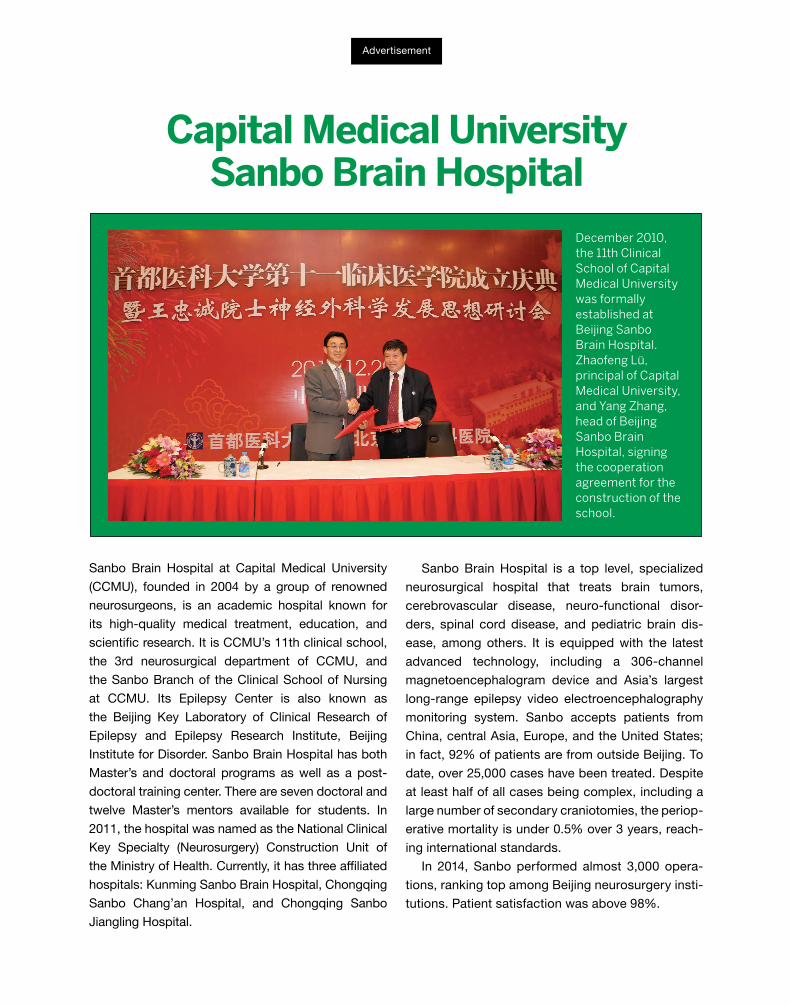
Capital Medical UniversitySanbo Brain Hospital
December 2010, the 11th Clinical School of Capital Medical University was formally established at Beijing Sanbo Brain Hospital. Zhaofeng Lü, principal of Capital Medical University, and Yang Zhang, head of Beijing Sanbo Brain Hospital, signing the cooperation agreement for the construction of the school.
Sanbo Brain Hospital at Capital Medical University (CCMU), founded in 2004 by a group of renowned neurosurgeons, is an academic hospital known for its high-quality medical treatment, education, and scientific research. It is CCMU’s 11th clinical school, the 3rd neurosurgical department of CCMU, and the Sanbo Branch of the Clinical School of Nursing at CCMU. Its Epilepsy Center is also known as the Beijing Key Laboratory of Clinical Research of Epilepsy and Epilepsy Research Institute, Beijing Institute for Disorder. Sanbo Brain Hospital has both Master’s and doctoral programs as well as a post-doctoral training center. There are seven doctoral and twelve Master’s mentors available for students. In 2011, the hospital was named as the National Clinical Key Specialty (Neurosurgery) Construction Unit of the Ministry of Health. Currently, it has three affiliated hospitals: Kunming Sanbo Brain Hospital, Chongqing Sanbo Chang’an Hospital, and Chongqing Sanbo Jiangling Hospital.
Sanbo Brain Hospital is a top level, specialized neurosurgical hospital that treats brain tumors, cerebrovascular disease, neuro-functional disor-ders, spinal cord disease, and pediatric brain dis-ease, among others. It is equipped with the latest advanced technology, including a 306-channel magnetoencephalogram device and Asia’s largest long-range epilepsy video electroencephalography monitoring system. Sanbo accepts patients from China, central Asia, Europe, and the United States; in fact, 92% of patients are from outside Beijing. To date, over 25,000 cases have been treated. Despite at least half of all cases being complex, including a large number of secondary craniotomies, the periop-erative mortality is under 0.5% over 3 years, reach-ing international standards.
In 2014, Sanbo performed almost 3,000 opera-tions, ranking top among Beijing neurosurgery insti-tutions. Patient satisfaction was above 98%.
Advertisement
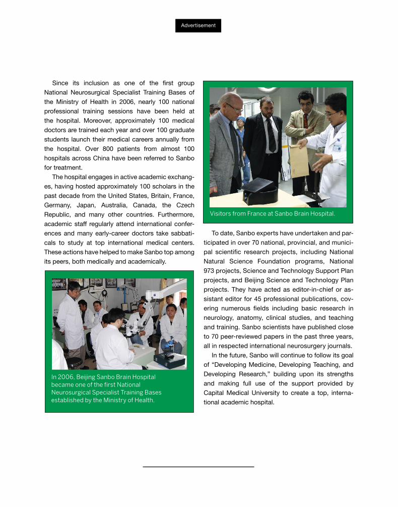
To date, Sanbo experts have undertaken and par-ticipated in over 70 national, provincial, and munici-pal scientific research projects, including National Natural Science Foundation programs, National 973 projects, Science and Technology Support Plan projects, and Beijing Science and Technology Plan projects. They have acted as editor-in-chief or as-sistant editor for 45 professional publications, cov-ering numerous fields including basic research in neurology, anatomy, clinical studies, and teaching and training. Sanbo scientists have published close to 70 peer-reviewed papers in the past three years, all in respected international neurosurgery journals.
In the future, Sanbo will continue to follow its goal of “Developing Medicine, Developing Teaching, and Developing Research,” building upon its strengths and making full use of the support provided by Capital Medical University to create a top, interna-tional academic hospital.
In 2006, Beijing Sanbo Brain Hospital became one of the first National Neurosurgical Specialist Training Bases established by the Ministry of Health.
Visitors from France at Sanbo Brain Hospital.
Advertisement
Since its inclusion as one of the first group National Neurosurgical Specialist Training Bases of the Ministry of Health in 2006, nearly 100 national professional training sessions have been held at the hospital. Moreover, approximately 100 medical doctors are trained each year and over 100 graduate students launch their medical careers annually from the hospital. Over 800 patients from almost 100 hospitals across China have been referred to Sanbo for treatment.
The hospital engages in active academic exchang-es, having hosted approximately 100 scholars in the past decade from the United States, Britain, France, Germany, Japan, Australia, Canada, the Czech Republic, and many other countries. Furthermore, academic staff regularly attend international confer-ences and many early-career doctors take sabbati-cals to study at top international medical centers. These actions have helped to make Sanbo top among its peers, both medically and academically.

T H E R E ’ S A SC IENCE T O A DVA N C I N G YO U R C A R E E R
Access the latest jobs and the most relevant career information—all for free
§ Search thousands of job listings
§ Complete an interactive, personalized career plan at “my IDP”
§ Create job alerts based on your criteria
§ View webinars and booklets
§ Post your resume/CV in our searchable database
§ Read relevant career advice articles
§ Research graduate programs
§ Visit our Career Forum to get advice.
Advance your career. Visit ScienceCareers.org
For recruitment in science, there’s only one

Find out more about the scope of the journal and submit your research today! ScienceTranslationalMedicine.org
WILL YOUR RESEARCHLEAD TO BETTER LIVESFOR PATIENTS?
Gopinath Sutendra and Evangelos D. Michelakis, “Pulmonary Arterial Hypertension: Challenges in Translational Research and a Vision for Change”, Sci. Transl. Med. 5, 208sr5 (2013) Credit: Science Source

Science Headquarters1200 New York Avenue, NWWashington, DC 20005 USA


![MSc in Translational (Neuroscience) · PDF fileMSc in Translational Pathology [Neuroscience] Why Translational Pathology? The MSc Translational Pathology (Neuroscience) course combines](https://img.dokumen.tips/doc/110x75/5a7454947f8b9a0d558bb440/msc-in-translational-neuroscience-a-msc-in-translational-pathology-neuroscience.jpg)