Embed Size (px)
Citation preview
International Journal of Osteoarcbaeology, Vol. 2: 337-340 (1 992)
SHORT REPORT
A Skull Wound and Possible Trepanation from a Roman Cemetery at Baldock, Hertfordshire JACQUELINE 1. MCKINLEY 12 Victoria Road, Warminster 8 A 12 8HE, UK
Inhumation burial 5779 was one of about 210 examined by the writer from the Romano-British Cemetery at Baldock (Area 15). T h e inhumation was supine, the upper body extended with the left arm flexed across the body. T h e short length of the grave had necessitated the legs being bent up at the knee; after burial they had slumped to the right.
About 97 per cent of the skeleton was present for examination and the bone was in a good condition. T h e individual was identified as an older adult male with mesocrany skull form, and an estimated stature of 163.9 cm. Pathology noted included dental caries; dental abscesses, osteo- arthritis in costo-vertebral, cervical, lumbar, thoracic and shoulder joints; osteophytes on
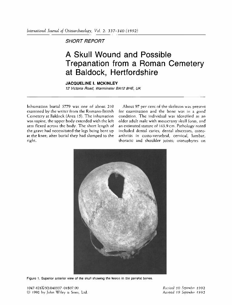
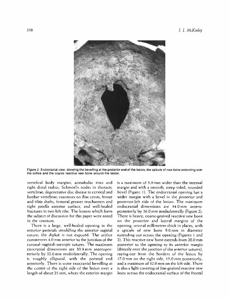
Figure 1. Superior anterior view of the skull showing the lesion in the parietal bones.
1047-42 8x/92/0403 37-04$07.00 0 1992 by John Wiley & Sons, Ltd.
Received 10 Sepleniber 1992 Accepted 1 9 September 1992
338 1. I. McKinley
Figure 2. Endocranial view, showing the bevelling at the posterior end of the lesion, the spicule of new bone extending over the orifice and the coarse reactive new bone around the lesion.
vertebral body margins, acetabulae rims and right distal radius; Schmorl’s nodes in thoracic vertebrae; degenerative disc disease in cervical and lumbar vertebrae; exostoses on iliac crests, femur and tibia shafts, femoral greater trochanters and right patella anterior surface, and well-healed fractures in two left ribs. T h e lesions which form the subject of discussion for this paper were noted in the cranium.
There is a large, well-healed opening in the anterior parietals straddling the anterior sagittal suture; the diploe is not exposed. T h e orifice commences 4.0 mm anterior to the junction of the coronal-sagittal-metopic sutures. T h e maximum exocranial dimensions are 50.8 mm anteropos- teriorly by 32.0 mm mediolaterally. T h e opening is roughly ellipsoid, with the pointed end anteriorly. There is some exocranial bevelling at the centre of the right side of the lesion over a length of about 25 mm, where the exterior margin
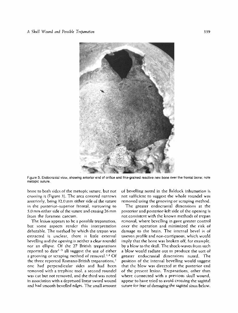
is a maximum of 5.0 mm wider than the internal margin and with a smooth, steep sided, rounded bevel (Figure I ) . T h e endocranial opening has a wider margin with a bevel in the posterior and posterior-left side of the lesion. T h e maximum endocranial dimensions are 54.0 mm antero- posteriorly by 36.0 mm mediolaterally (Figure 2). There is heavy, coarse-grained reactive new bone on the posterior and lateral margins of the opening, several millimetres thick in places, with a spicule of new bone 9.Omm in diameter extending out across the opening (Figures 1 and 2). This reactive new bone extends from 20.0 mm posterior to the opening to its anterior margin (directly over the junction of the anterior sutures), raying-out from the borders of the lesion by 17.0 mm on the right side, 15.0 mm posteriorly, and a maximum of 32.0 mm on the left side. There is also a light covering of fine-grained reactive new bone across the endocranial surface of the frontal
A Skull Wound and Possible Trepanation 339
Figure 3. Endocranial view, showing anterior e n d of orifice a n d fine-grained reactive new bone over t h e frontal bone: note metopic suture.
bone to both sides of the metopic suture, but not crossing it (Figure 3). T h e area covered narrows anteriorly, being 32.0 mm either side of the suture in the posterior-superior frontal, narrowing to 5.0 mm either side of the suture and ceasing 26 mm from the foramen caecum.
T h e lesion appears to be a possible trepanation, but some aspects render this interpretation debatable. T h e method by which the trepan was extracted is unclear, there is little external bevelling and the opening is neither a clear roundel nor an ellipse. Of the 27 British trepanations reported to date'-3 all suggest the use of either a grooving or scraping method of r e m ~ v a l . ' , ~ Of the three reported Romano-British trepanations,' one had perpendicular sides and had been removed with a trephine tool, a second roundel was cut but not removed, and the third was noted in association with a depressed linear sword wound and had smooth bevelled edges. The small amount
of bevelling noted in the Baldock inhumation is not sufficient to suggest the whole roundel was removed using the grooving o r scraping method.
T h e greater endocranial dimensions at the posterior and posterior-left side of the opening is not consistent with the known methods of trepan removal, where bevelling in gave greater control over the operation and minimized the risk of damage to the brain. T h e internal bevel is of uneven profile and non-contiguous, which would imply that the bone was broken off, for example, by a blow to the skull. The shock-waves from such a blow would radiate out to produce the sort of greater endocranial dimensions noted. T h e position of the internal bevelling would suggest that the blow was directed a t the posterior end of the present lesion. Trepanations, other than where connected with a previous skull wound, appear to have tried t o avoid crossing the sagittal suture for fear of damaging the sagittal sinus below.
3 40 J. I. McKinley
O n e of the reasons for carrying ou t a trepana- tion seems to have been to relieve pressure on the brain resulting from a skull wound. ' T h e smooth margins to this lesion would suggest that not only did the individual survive for some time following the suggested blow to the skull, bu t that the bone from the fracture was removed from position. I f this was originally a skull wound rather than a trepanation, it is possible that an operation was subsequently conducted to remove the dislodged fragment(s) of bone.
Whatever the course of events, one outcome was meningeal infection, presumably in the dura mater, illustrated by the extensive reactive new bone on the endocranial surfaces. T h e gross new bone around the opening presumably formed the original seat of the infection, which later spread to the frontal region. The eventual death of the individual may have been as a consequence of this infection.
Acknowledgements
Thanks to Gil Burleigh and Mark Stevenson of the Baldock Project Letchworth Museum for inhumation context details. Photographs by Elaine A. Wakefield.
References
1. Parker, S., Roberts, C. and Manchester, K. A review of British Trepanations with reports of two new cases. OSSA, 1986; 12: 141-158.
2. Wells, C. Probable trepanation of five early Anglo- Saxon skulls. Antiquity, 1974; 48: 298-300.
3. Mckinley, J . I . A Probable Trepanation from an Early Anglo-Saxon Cemetery a t Oxborough, Norfolk. International Journal of Osteoarcbaeology, 1992; 2: 333-335.
4. Wakely, J. and Duhig, C. A comparative microscopical study of three European trephined skulls. Journal of Paleopatbology, 1989; 3 (2) : 75-87.