Embed Size (px)
Citation preview
BRAIN COMPUTER INTERFACE
A SEMINAR REPORT
Submitted by
NITISH KUMAR
in partial fulfillment for the award of the degree
of
BACHELOR OF TECHNOLOGY in
COMPUTER SCIENCE AND ENGINEERING
SCHOOL OF ENGINEERING
COCHIN UNIVERSITY OF SCIENCE &
TECHNOLOGY, KOCHI-682022
AUGUST 2008
DIVISION OF COMPUTER ENGINEERING
SCHOOL OF ENGINEERING
COCHIN UNIVERSITY OF SCIENCE &
TECHNOLOGY, KOCHI-682022
Certified that this is a bonafide record of the seminar report entitled
“ BRAIN COMPUTER INTERFACE”
is done by “NITISH KUMAR” of the VIIth semester, Computer Science
and Engineering in the year 2008 in partial fulfillment of the requirements to
the award of Degree of Bachelor of Technology in computer Science and
Engineering of Cochin University of Science and Technology.
SHEENA MATHEW Dr. DAVID PETER
SEMINAR GUIDE HEAD OF THE DEPARTMENT
Lecturer
Computer Science and Engineering Computer Science and Engineering
School of Engineering, School of Engineering,
CUSAT CUSAT
Certificate

ACKNOWLEDGEMENT
At the outset, we thank God almighty for making our endeavor a success.
We also express our gratitude to Mr. David Peter S, Head of the
Department for providing us with adequate facilities, ways and means by
which we were able to complete this seminar.
I express my sincere gratitude to my seminar Guide Mrs. Sheena Mathew,
Lecturer, Computer Engineering Division for her constant support and
valuable suggestions without which the successful completion of this
seminar would not have been possible.
I express my immense pleasure and thanks to all the teachers and staff of the
Department of Computer Science and Engineering, CUSAT for their
cooperation and support.
Last but not the least, I thank all others, and especially
our classmates and our family members who in one way or another helped
me in the successful completion of this work.

ABSTRACT
A brain-computer interface (BCI), sometimes called a direct neural interface
or a brain-machine interface, is a direct communication pathway between a
human or animal brain and an external device. In one-way BCIs, computers
either accept commands from the brain or send signals to it (for example, to
restore vision) but not both. Two-way BCIs would allow brains and external
devices to exchange information in both directions but have yet to be
successfully implanted in animals or humans.
In this definition, the word brain
means the brain or nervous system of an organic life form rather than the
mind. Computer means any processing or computational device, from simple
circuits to silicon chips. Research on BCIs began in the 1970s, but it wasn't
until the mid-1990s that the first working experimental implants in humans
appeared. Following years of animal experimentation, early working
implants in humans now exist, designed to restore damaged hearing, sight
and movement. With recent advances in technology and knowledge,
pioneering researchers could now conceivably attempt to produce BCIs that
augment human functions rather than simply restoring them, previously only
a possibility in science fiction.

TABLE OF CONTENTS
CHAPTER NO. TITLE PAGE NO.
ABSTRACT
LIST OF TABLES
LIST OF FIGURES
1. INTRODUCTION 1
2. WORKING ARCHITECTURE 3
2.1 INTRODUCTION 3
2.2 INVASIVE BCI 3
2.3 PARTIALLY INVASIVE BCI 5
2.4 NON-INVASIVE BCI 5
2.5 ANIMAL BCI RESEARCH 7
2.5.1 Early work 8
2.5.2 Prominent research successes 8
2.6 CELL CULTURE BCI’S 11
3. THE CURRENT BCI TECHNIQUES 13
3.1 INTRODUCTION 13
3.2 P300 DETECTION 13
3.3 EEG mu-RYTHM CONDITIONING 14
3.4 VEP DETECTION 16
3.5 EEG PATTERN MAPPING 17
3.6 DETECTING LATERAL 18
HEMISPHERE DIFFERENCES
4. BRAIN GATE 19
4.1 DARPA 21
i
5. BCI APPLICATIONS 22
5.1 INTRODUCTION 22
5.2 THE MENTAL TYPEWRITER 22
5.3 BCI OFFERS PARALYZED PATIENTS 23
GOOD QUALITY OF LIFE
5.4 ADAPTIVE BCI FOR AUGMENTED 25
COGNITION AND ACTION
5.4.1 Error and conflict perception 25
5.5.1 Working memory encoding 26
5.5.2 Rapid visual recognition 26
6. THE ABI SOFTWARE 30
6.1 INTRODUCTION 30
6.2 WORK OF THE SOFTWARE 30
6.3 THE CLASSIFICATION ACHIEVED 31
BY THIS SOFTWARE
6.4 INSTRUCTIONS 31
6.4.1 Simulation and recording 33
6.4.2 Training 34
6.4.3 The trial archive 36
6.4.4 The configuration file 36
6.4.5 Electrode positions 37
6.4.6 Fast testing 38
6.4.7 Considerations 39
6.5 CLASSIFIER DESIGN 40
ii

iii

LIST OF TABLES
NO TITLE PAGE NO
6.4.1.1 SIMULATION MODE TO 34
TEST THE BCI
6.4.2.1 TRAINING 35
6.4.4.1 LIST OF VARIABLES IN 37
CONFIGURATION FILE

LIST OF FIGURES
NO TITLE PAGE NO
1.1 SCHEMATIC DIAGRAM OF BCI 2
SYSTEM
2.2.1 THE EARLY SHOW 3
2.4.1 RECORDING OF BRAINWAVES BY 6
ELECTROENCEPHALOGRAM
2.5.1 RATS IMPLANTED WITH BCI’S 7
2.5.2 RECORDINGS OF CAT VISION 9
2.5.3 BCI DEVELOPED BY MIGUEL NICOLELIS 10
2.6.1 NEUROCHIP 11
4.1 DESIGN OF BRAIN GATE INTERFACE 19
5.4.1 REAL TIME BCI 27
5.4.2 REDUCTION OF ERROR 28
5.4.3 IMAGE THROUGHPUT FOR DETECTED 29
EEG
6.4.1 THE EEG CHANNELS 32
6.4.6 FAST TESTING 38

Brain Computer Interface
Chapter 1. Introduction Man machine interface has been one of the growing fields of research and
development in recent years. Most of the effort has been dedicated to the design of
user-
friendly or ergonomic systems by means of innovative interfaces such as voice
recognition, virtual reality. A direct brain-computer interface would add a new
dimension to man-machine interaction.
A brain-computer interface, sometimes called a direct neural interface or a
brain machine interface, is a direct communication pathway between a human or
animal brain(or brain cell culture) and an external device. In one BCIs, computers
either accept commands from the brain or send signals to it but not both. Two way
BCIs will allow brains and external devices to exchange information in both
directions but have yet to be successfully implanted in animals or humans.
Brain-Computer interface is a staple of science fiction writing. In its earliest
incarnations no mechanism was thought necessary, as the technology seemed so far
fetched that no explanation was likely. As more became known about the brain
however, the possibility has become more real and the science fiction more
technically sophisticated. Recently, the cyberpunk movement has adopted the idea of
'jacking in', sliding 'biosoft' chips into slots implanted in the skull(Gibson,
W.1984).Although such biosofts are still science fiction, there have been several
recent steps toward interfacing the brain and computers.
In this definition, the word brain means the brain or nervous system of an
organic life form rather than the mind. Computer means any processing or
computational device, from simple circuits to silicon chips (including hypothetical
future technologies like quantum computing).
Research on BCIs has been going on for more than 30 years but from the
mid 1990’s there has been dramatic increase working experimental implants. The
common thread throughout the research is the remarkable cortical-plasticity of the
Division of Computer Science and Engineering, SOE, CUSAT 1

Brain Computer Interface
brain, which often adapts to BCIs treating prostheses controlled by implants and
natural limbs. With
recent advances in technology and knowledge, pioneering researches could now
conceivably attempt to produce BCIs that augment human functions rather than
simply restoring them, previously only the realm of science fiction.
Fig. 1.1: Schematic diagram of a BCI system
Division of Computer Science and Engineering, SOE, CUSAT 2

Brain Computer Interface
Chapter 2. Working architecture
2.1. Introduction:
Before moving to real implications of BCI and its application let us first
discuss the three types of BCI. These types are decided on the basis of the technique
used for the interface. Each of these techniques has some advantages as well as some
disadvantages. The three types of BCI are as follows with there features:
2.2. Invasive BCI:
Invasive BCI are directly implanted into the grey matter of the
brain during neurosurgery. They produce the highest quality signals of BCI devices .
Invasive BCIs has targeted repairing damaged sight and providing new functionality
to paralyzed people. But these BCIs are prone to building up of scar-tissue which
causes the signal to become weaker and even lost as body reacts to a foreign object in
the brain.
fig.2.2.1: Jens Naumann, a man with acquired blindness, being interviewed about his
vision BCI on CBS's The Early Show
In vision science, direct brain implants have been used to treat non-
congenital i.e. acquired blindness. One of the first scientists to come up with a
working brain interface to restore sight as private researcher, William Dobelle.
Dobelle’s first prototype was implanted into Jerry, a man blinded in
adulthood, in1978. A single-array BCI containing 68 electrodes was implanted onto
Division of Computer Science and Engineering, SOE, CUSAT 3

Brain Computer Interface
Jerry’s visual cortex and succeeded in producing phosphenes, the sensation of seeing
light. The system included TV cameras mounted on glasses to send signals to the
implant. Initially the implant allowed Jerry to see shades of grey in a limited field of
vision and at a low frame-rate also requiring him to be hooked up to a two-ton
mainframe. Shrinking electronics and faster computers made his artificial eye more
portable and allowed him to perform simple tasks unassisted.
In 2002, Jens Naumann, also blinded in adulthood, became the first in a
series of 16 paying patients to receive Dobelle’s second generation implant, marking
one of the earliest commercial uses of BCIs. The second generation device used a
more sophisticated implant enabling better mapping of phosphenes into coherent
vision. Phosphenes are spread out across the visual field in what researchers call the
starry-night effect. Immediately after his implant, Jens was able to use imperfectly
restored vision to drive slowly around the parking area of the research institute.
BCIs focusing on motor Neuroprosthetics aim to either restore
movement in paralyzed individuals or provide devices to assist them, such as
interfaces with computers or robot arms.
Researchers at Emory University in Atlanta led by Philip Kennedy and
Roy Bakay were first to install a brain implant in a human that produced signals of
high enough quality to stimulate movement. Their patient, Johnny Ray, suffered from
‘locked-in syndrome’ after suffering a brain-stem stroke. Ray’s implant was installed
in 1998 and he lived long enough to start working with the implant, eventually
learning to control a computer cursor.
Tetraplegic Matt Nagle became the first person to control an artificial
hand using a BCI in 2005 as part of the nine-month human trail of cyber kinetics
Neurotechnology’s Braingate chip-implant. Implanted in Nagle’s right precentral
gyrus(area of the motor cortex for arm movement), the 96 electrode Braingate implant
allowed Nagle to control a robotic arm by thinking about moving his hand as well as a
computer cursor, lights and TV.
Division of Computer Science and Engineering, SOE, CUSAT 4
Brain Computer Interface
2.3. Partially Invasive BCI:
Partially invasive BCI devices are implanted inside the skull but rest
outside the brain rather than amidst the grey matter. They produce better resolution
signals than non-invasive BCIs where the bone tissue of the cranium deflects and
deforms signals and have a lower risk of forming scar-tissue in the brain than fully-
invasive BCIs.
Electrocorticography(ECoG) uses the same technology as non-invasive
electroencephalography, but the electrodes are embedded in a thin plastic pad that is
placed above the cortex, beneath the dura mater. ECoG technologies were first traled
in humans in 2004 by Eric Leuthardt and Daniel Moran from Washington University
in St Louis. In a later trial, the researchers enabled a teenage boy to play Space
Invaders using his ECoG implant. This research indicates that it is difficult to produce
kinematic BCI devices with more than one dimension of control using ECoG.
Light Reactive Imaging BCI devices are still in the realm of theory.
These would involve implanting laser inside the skull. The laser would be trained on a
single neuron and the neuron’s reflectance measured by a separate sensor. When
neuron fires, The laser light pattern and wavelengths it reflects would change slightly.
This would allow researchers to monitor single neurons but require less contact with
tissue and reduce the risk of scar-tissue build up.
2.4. Non-Invasive BCI :
As well as invasive experiments, there have also been
experiments in humans using non-invasive neuroimaging technologies as interfaces.
Signals recorded in this way have been used to power muscle implants and restore
partial movement in an experimental volunteer. Although they are easy to wear, non-
invasive implants produce poor signal resolution because the skull dampens signals,
dispersing and blurring the electromagnetic waves created by the neurons. Although
the waves can still be detected it is more difficult to determine the area of the brain
that created them or the actions of individual neurons.
Division of Computer Science and Engineering, SOE, CUSAT 5

Brain Computer Interface
fig.2.4.1: Recordings of brainwaves produced by an electroencephalogram
Electroencephalography(EEG) is the most studied potential non-invasive
interface, mainly due to its fine temporal resolutions, ease of use, portability and low
set-up cost. But as well as the technology's susceptibility to noise, another substantial
barrier to using EEG as a brain-computer interface is the extensive training required
before users can work the technology. For example, in experiments beginning in the
mid-1990s, Niels Birbaumer of the University of Tübingen in Germany used EEG
recordings of slow cortical potential to give paralysed patients limited control over a
computer cursor.(Birbaumer had earlier trained epileptics to prevent impending fits by
controlling this low voltage wave.) The experiment saw ten patients trained to move a
computer cursor by controlling their brainwaves. The process was slow, requiring
more than an hour for patients to write 100 characters with the cursor, while training
often took many months.
Another research parameter is the type of waves measured. Birbaumer's later
research with Jonathan Wolpaw at New York State University has focused on
developing technology that would allow users to choose the brain signals they found
easiest to operate a BCI, including mu and beta waves.
A further parameter is the method of feedback used and this is shown in
studies of P300 signals. Patterns of P300 waves are generated involuntarily (stimulus-
feedback) when people see something they recognise and may allow BCIs to decode
categories of thoughts without training patients first. By contrast, the biofeedback
methods described above require learning to control brainwaves so the resulting brain
activity can be detected. In 2000, for example, research by Jessica Bayliss at the
University of Rochester showed that volunteers wearing virtual reality helmets could
Division of Computer Science and Engineering, SOE, CUSAT 6

Brain Computer Interface
control elements in a virtual world using their P300 EEG readings, including turning
lights on and off and bringing a mock-up car to a stop.
In 1999, researchers at Case Western Reserve University led by Hunter
Peckham, used 64-electrode EEG skullcap to return limited hand movements to
quadriplegic Jim Jatich. As Jatich concentrated on simple but opposite concepts like
up and down, his beta-rhythm EEG output was analysed using software to identify
patterns in the noise. A basic pattern was identified and used to control a switch:
Above average activity was set to on, below average off. As well as enabling Jatich to
control a computer cursor the signals were also used to drive the nerve controllers
embedded in his hands, restoring some movement.
Electronic neural-networks have been deployed which shift the learning
phase from the user to the computer. Experiments by scientists at the Fraunhofer
Society in 2004 using neural networks led to noticeable improvements within 30
minutes of training.
Experiments by Edurado Miranda aim to use EEG recordings of mental activity
associated with music to allow the disabled to express themselves musically through
an encephalophone.
Magnetoencephalography (MEG) and functional magnetic resonance
imaging (fMRI) have both been used successfully as non-invasive BCIs. In a widely
reported experiment, fMRI allowed two users being scanned to play Pong in real-time
by altering their haemodynamic response or brain blood flow through biofeedback
techniques. fMRI measurements of haemodynamic responses in real time have also
been used to control robot arms with a seven second delay between thought and
movement.
2.5. Animal BCI research:
fig.2.5.1: Rats implanted with BCIs in Theodore Berger's experiments
Division of Computer Science and Engineering, SOE, CUSAT 7
Brain Computer Interface
Several laboratories have managed to record signals from monkey and
rat cerebral cortexes in order to operate BCIs to carry out movement. Monkeys have
navigated computer cursors on screen and commanded robotic arms to perform
simple tasks simply by thinking about the task and without any motor output. Other
research on cats has decoded visual signals.
2.5.1. Early work
Studies that developed algorithms to reconstruct movements from motor
cortex neurons, which control movement, date back to the 1970s. Work by groups led
by Schmidt, Fetz and Baker in the 1970s established that monkeys could quickly learn
to voluntarily control the firing rate of individual neurons in the primary motor cortex
after closed-loop operant conditioning, a training method using punishment and
rewards.
In the 1980s, Apostolos Georgopoulos at Johns Hopkins University found
a mathematical relationship between the electrical responses of single motor-cortex
neurons in rhesus macaque monkeys and the direction that monkeys moved their arms
(based on a cosine function). He also found that dispersed groups of neurons in
different areas of the brain collectively controlled motor commands but was only able
to record the firings of neurons in one area at a time because of technical limitations
imposed by his equipment.
There has been rapid development in BCIs since the mid-1990s. Several
groups have been able to capture complex brain motor centre signals using recordings
from neural ensembles (groups of neurons) and use these to control external devices,
including research groups led by Richard Andersen, John Donoghue, Phillip
Kennedy, Miguel Nicolelis, and Andrew Schwartz.
2.5.2. Prominent research successes
Phillip Kennedy and colleagues built the first intracortical brain-computer interface by
implanting neurotrophic-cone electrodes into monkeys.
Division of Computer Science and Engineering, SOE, CUSAT 8

Brain Computer Interface
fig.2.5.2: Garrett Stanley's recordings of cat vision using a BCI implanted in the
lateral geniculate nucleus (top row: original image; bottom row: recording)
In 1999, researchers led by Garrett Stanley at Harvard University
decoded neuronal firings to reproduce images seen by cats. The team used an array of
electrodes embedded in the thalamus (which integrates all of the brain’s sensory
input) of sharp-eyed cats. Researchers targeted 177 brain cells in the thalamus lateral
geniculate nucleus area, which decodes signals from the retina. The cats were shown
eight short movies, and their neuron firings were recorded. Using mathematical filters,
the researchers decoded the signals to generate movies of what the cats saw and were
able to reconstruct recognisable scenes and moving objects.
Miguel Nicolelis has been a prominent proponent of using multiple
electrodes spread over a greater area of the brain to obtain neuronal signals to drive a
BCI. Such neural ensembles are said to reduce the variability in output produced by
single electrodes, which could make it difficult to operate a BCI.
After conducting initial studies in rats during the 1990s, Nicolelis and his
colleagues developed BCIs that decoded brain activity in owl monkeys and used the
devices to reproduce monkey movements in robotic arms. Monkeys have advanced
reaching and grasping abilities and good hand manipulation skills, making them ideal
test subjects for this kind of work.
By 2000, the group succeeded in building a BCI that reproduced owl
monkey movements while the monkey operated a joystick or reached for food.The
BCI operated in real time and could also control a separate robot remotely over
Internet protocol. But the monkeys could not see the arm moving and did not receive
any feedback, a so-called open-loop BCI.
Division of Computer Science and Engineering, SOE, CUSAT 9

Brain Computer Interface
fig.2.5.3: Diagram of the BCI developed by Miguel Nicolelis and collegues for use on
Rhesus monkeys.
Later experiments by Nicolelis using rhesus monkeys, succeeded in
closing the feedback loop and reproduced monkey reaching and grasping movements
in a robot arm. With their deeply cleft and furrowed brains, rhesus monkeys are
considered to be better models for human neurophysiology than owl monkeys. The
monkeys were trained to reach and grasp objects on a computer screen by
manipulating a joystick while corresponding movements by a robot arm were hidden.
The monkeys were later shown the robot directly and learned to control it by viewing
its movements. The BCI used velocity predictions to control reaching movements and
simultaneously predicted hand gripping force.
Other labs that develop BCIs and algorithms that decode neuron signals
include John Donoghue from Brown University, Andrew Schwartz from the
University of Pittsburgh and Richard Andersen from Caltech. These researchers were
able to produce working BCIs even though they recorded signals from far fewer
neurons than Nicolelis (15–30 neurons versus 50–200 neurons).
Donoghue's group reported training rhesus monkeys to use a BCI to
track visual targets on a computer screen with or without assistance of a joystick
(closed-loop BCI).Schwartz's group created a BCI for three-dimensional tracking in
virtual reality and also reproduced BCI control in a robotic arm. The group created
headlines when they demonstrated that a monkey could feed itself pieces of zucchini
using a robotic arm powered by the animal's own brain signals.
Division of Computer Science and Engineering, SOE, CUSAT 10

Brain Computer Interface
Andersen's group used recordings of premovement activity from the
posterior parietal cortex in their BCI, including signals created when experimental
animals anticipated receiving a reward.
In addition to predicting kinematic and kinetic parameters of limb
movements, BCIs that predict electromyographic or electrical activity of muscles are
being developed.Such BCIs could be used to restore mobility in paralysed limbs by
electrically stimulating muscles.
2.6.Cell-culture BCIs
Researchers have also built devices to interface with neural cells and
entire neural networks in cultures outside animals. As well as furthering research on
animal implantable devices, experiments on cultured neural tissue have focused on
building problem-solving networks, constructing basic computers and manipulating
robotic devices. Research into techniques for stimulating and recording from
individual neurons grown on semiconductor chips is sometimes referred to as
neuroelectronics or neurochips.
fig.2.6.1: World first: Neurochip developed by Caltech researchers Jerome Pine and
Michael Maher
Development of the first working neurochip was claimed by a Caltech
team led by Jerome Pine and Michael Maher in 1997. The Caltech chip had room for
16 neurons.
Division of Computer Science and Engineering, SOE, CUSAT 11

Brain Computer Interface
In 2003, a team led by Theodore Berger at the University of Southern
California started work on a neurochip designed to function as an artificial or
prosthetic hippocampus. The neurochip was designed to function in rat brains and is
intended as a prototype for the eventual development of higher-brain prosthesis. The
hippocampus was chosen because it is thought to be the most ordered and structured
part of the brain and is the most studied area. Its function is to encode experiences for
storage as long-term memories elsewhere in the brain.
Thomas DeMarse at the University of Florida used a culture of 25,000
neurons taken from a rat's brain to fly a F-22 fighter jet aircraft simulator.After
collection, the cortical neurons were cultured in a petri dish and rapidly begin to
reconnect themselves to form a living neural network. The cells were arranged over a
grid of 60 electrodes and trained to control the pitch and yaw functions of the
simulator. The study's focus was on understanding how the human brain performs and
learns computational tasks at a cellular level.
Division of Computer Science and Engineering, SOE, CUSAT 12
Brain Computer Interface
Chapter 3. The Current BCI Techniques
3.1. Introduction:
In todays time various techniques are used for BCI interface, there
implementations and result manipulation. These techniques are headed towards the
development of BCI in coming era.
3.2. P300 Detection:
Farwell [Farwell&Donchin 1988] of the Department of Psychology
and Cognitive Psychophysiology Laboratory at the University of Illinois at Urbana-
Champaign IL, describes a technique for detecting the P300 component of a subject's
event-related brain potential (ERP) and using it to select from an array of 36 screen
positions. The P300 component is a positive-going ERP in the EEG with a latency of
about 300ms following the onset of a rarely- occurring stimulus the subject has been
instructed to detect. The EEG was recorded using electrodes placed at the Pz
(parietal) site (10/20 International System), limited with band-pass filters to .02-35Hz
and digitized at 50Hz. Electro-oculogram (EOG) data was also recorded from each
subject via electrodes placed above and below the right eye. The "odd-ball" paradigm
was used to elicit the P300, where a number of stimuli are presented to the
experimental subject who is required to pay attention to a particular, rarely-occurring
stimulus and respond to it in some non- motor way, such as by counting occurrences.
Detecting the P300 response reliably requires averaging the EEG response over many
presentations of the stimuli. The purpose of the current experiment was to discover
the minimum number of presentations at two different inter-stimulus intervals (ISI)
required to detect the P300 response. The experiment presented a 36-position array of
letters, plus common typing characters and controls (e.g. space, backspace), made to
flash in a random sequence first by rows and then columns. Each trial consisted of a
complete set of six column or row flashes. Trials contaminated with muscular or
Division of Computer Science and Engineering, SOE, CUSAT 13
Brain Computer Interface
EOG response were rejected and additional trials presented until data were collected
from a block of 30 good trials, during which subjects were to fixate on a particular
position, and count the number of times it flashed while a control message was
elsewhere on the screen. After each block the
fixated letter (one of B-R-A-I-N) was added to the screen so that subjects were
conscious of slowly spelling out the word "BRAIN" through a succession of five
blocks. A set of five blocks was run at each ISI -- 125ms and 500ms. The two
presentation rates were chosen to bracket a range of communication rates from a low
of 30 averaged trials at 500ms ISI (93.6 seconds of presentation per character) to a
high of one trial at 125ms (1.245 seconds of presentation per character), an effective
communication rate range of .01 to .8 characters-per- second, respectively. The
authors used four techniques to analyze the data for reliable P300 response detection –
stepwise descriminant analysis (SWDA), peak picking, area, and covariance, and
identified SWDA as leading to the greatest accuracy at the fastest presentation rate.
Results indicated that a character chosen from among 36 items can be detected with
95% accuracy within 26 seconds.
3.3. EEG mu-rhythm Conditioning:
Three papers using this technique were reviewed including Wolpaw
[Wolpaw et al 1991], McFarland [McFarland et al 1993], and colleagues at the
Wadsworth Center for Laboratories and Research, Albany, NY, and Pfurtscheller
[Pfurtscheller et al 1993] and colleagues at the Ludwig Boltzmann Institute of
Medical Informatics and Neuroinformatics, Department of Medical Informatics,
Institute
of Biomedical Engineering, University of Technology Graz, Austria. All three papers
describe subjects' abilities to move a cursor toward a target on a computer screen by
manipulating their mu- rhythm, a detectable pattern in a great majority of individuals
in the EEG 8-12Hz frequency range, centered about 9.1Hz. Work is based on earlier
research efforts by Kuhlman [Kuhlman 1978b] who described the mu-rhythm in
normal and epileptic subjects. Wolpaw describes detecting subjects' mu-rhythm
amplitude, defined as the square-root of the spectral EEG power at 9Hz, using two
scalp-mounted electrodes located near location C3 in the International 10/20 System
Division of Computer Science and Engineering, SOE, CUSAT 14

Brain Computer Interface
and a digital signal processing board analyzing continuous EEG in 333ms segments,
and using it to drive a cursor up or down on a screen toward a target placed randomly
at the top or bottom. An experiment operator preset the size of the ranges and number
of cursor movement steps assigned to each range for each subject during testing prior
to each experimental run.
Ranges were set so that the commonest mu-rhythm amplitudes (<4 microvolts) left
the cursor in place or moved it downwards moderately while higher amplitudes (>4
microvolts) moved it upwards in increasing jumps. Weights were adjusted as subjects
exhibited better control of their mu-rhythm amplitudes for up and down targets in
repeated trials. Wolpaw substantiates subjects' learned intentional control over mu-
rhythm amplitude in three ways: by performing frequency analysis up to 192Hz on
subjects during cursor movement trials and failing to find any relationship between
mu-
rhythm changes and the higher frequencies associated with muscular (EMG) activity;
by subjects statements about not making contralateral movements and observing
none; and by failing to find any relationship between mu-rhythm changes and
posterior scalp recordings of the visual alpha-rhythm. Four out of five subjects
acquired impressive control over their mu-rhythm amplitude during 12 45-minute
sessions over a period of two months. Accuracies of 80-95% target hits across
experimental subjects were achieved and rates of 10-29 hits per minute. Off-line
analysis of two subjects' raw EEG data (see below) provided good support for
Wolpaw's experimental results. McFarland used essentially the same experimental
setup and introduced greater precision constraints on four subjects' attempts to
position a cursor by means of mu-rhythm control. A vertical bar target appeared in
one of five different vertical positions on the left side of the screen and crossed the
screen from left to right in 8 seconds. Subjects had to move the cursor (initially in the
middle of the right edge of the screen) quickly to the correct one of five different
vertical screen positions to intercept the target by controlling their mu-rhythm
amplitude. Analysis of the average distance between the center of the target and the
cursor during succeeding trials indicated that all subjects reduced the distance and
three out of four significantly so. Pfurtscheller used contralateral blocking of the
mu-rhythm during the 1-second period prior to a motor activity (in this case pressing a
microswitch using either the right or the left index finger) to predict which response
was to follow. An array of 30 electrodes spaced evenly across the scalp (two were at
Division of Computer Science and Engineering, SOE, CUSAT 15
Brain Computer Interface
locations C3 and C4 in the International 10/20 System) was used to record EEG
activity. An initial training period for each subject involved using data from all 30
electrodes to train the classification network. During experimental trials, a feature-
vector of power values (Hilbert Transform) from electrodes at positions C3 and C4
was constructed at 5 time points and classified using a Learning Vector Quantizer
(LVQ) artificial neural network of the type described by Kohonen [Kohonen 1988].
The experimenter achieved the best balance of reliability/speed of classification by
using the 1/2-second prior to response and performing a multiple- classification and
voting process. EEG data from two subjects in the Wolpaw experiment described
above were provided to the Graz Institute for Information Processing for additional
analysis described by Flotzinger [Flotzinger et al, 1993] using the Graz LVQ neural
net scheme (see above) and a fixed time-segment. Cursor-movement was predicted
>from raw data with 90% accuracy. Results also implied that frequency bands other
than the mu and beta ranges may contain useful (i.e. target-related) information.
3.4. VEP Detection: This technique was reviewed by Sutter [Sutter 1992] at the Smith-Kettlewell
Eye Research Institute in San Francisco CA, and Cilliers [Cilliers&VanDerKouwe
1993] and colleague at the Department of Electrical and Electronic Engineering,
University of Pretoria, South Africa. Sutter describes presenting a 64-position block
on a computer screen and detecting which block the subject looks at, while Cillier's
work uses a series of four lights. In each case, several simultaneously presented
stimuli are made to change rapidly in some controlled way(intensity, pattern, color-
shift) and the subject has scalp electrodes placed over the visual cortex (back of the
head) in a position to detect changes in the evoked potential(VEP) at that location.
Sutter used a lengthy binary sequence to switch 64 screen positions between red and
green, and in other trials to reverse a checkerboard pattern. Each screen position was
shifted 20ms in the binary control sequence relative to its neighbors, and the entire
sequence was auto correlated with the VEP in overlapping increments(the VEP
response components last about 80ms) beginning 20ms apart, with the resultant vector
stored in a 64-position array of registers. When a coefficient remains greater than all
the others and above a threshold value for a certain amount of time, the corresponding
stimulus is considered to have been selected. The 64 positions represent the letters of
Division of Computer Science and Engineering, SOE, CUSAT 16

Brain Computer Interface
the alphabet and commonly used words in the English language. The subject can
fixate on any word or letter. Whenever the subject fixates on a letter, the commonly
used words change to words beginning with that letter, for quick selection of an entire
word. Sutter suggests a need to optimize both electrode placement and stimulation
mode for each individual subject for good target discrimination. Seventy normal
subjects evaluating a prototype system achieved adequate response times ranging
from 1 to 3 seconds after an initial tuning process lasting 10-60 minutes. Sutter also
tested his techniques on 20 severly disabled persons and describes an experimental
version involving an ALS patient using intra-cranial electrodes implanted in the
space between the dura and the skull. Cilliers' technique involves varying the intensity
of four LED's modulated with a 10Hz sine wave in phase quadrature and detecting the
signal in the subject's VEP using a pair of EEG surface electrodes placed on the
occipital lobe. The four flashing LED's are arranged around the edge of a computer
screen containing an image of a standard four-row keyboard with each row of keys in
a different color. Each LED is associated with one of the colors. Fixating on one LED
selects a key row, which is redisplayed in four colors for a more detailed selection.
The subject can select any particular key in an average of three selections -- about 15
seconds with the current setup. A short initial training period
is required where subjects fixate on each LED for 5 seconds. Cilliers' paper describes
work with a quadriplegic patient with a C2-level injury.
3.5. EEG Pattern Mapping:
Several experimenters describe techniques for classifying, detecting and
mapping EEG patterns. Pfurtscheller's technique used a neural net featuring learning-
vector quantization (LVQ) to map EEG patterns during the 1-second interval before a
signal the experimental subject was instructed to wait for. Hiraiwa [Hiraiwa et al
1993] used a back-propagation artificial neural network to study readiness potentials
(RP's)-- patterns in the EEG immediately prior to the subject's uttering one of five
different Japanese syllables or moving a joystick in one of four different directions.
Twelve channels of EEG data taken >from scalp-mounted electrodes at locations Fp1,
Fp2,Fz, C3, C4, Pz, F5, F6, F7, F8, O1 and O2 (International 10/20 system) were used
to train and then test two neural networks optimized for averaged data and for single-
Division of Computer Science and Engineering, SOE, CUSAT 17

Brain Computer Interface
trial, real-time analysis, respectively. High recognition rates were obtained for the
averaged data. Single- trial RP recognition, though less reliable, showed considerable
promise in the experimenters' view. Keirn and Aunon [Keirn&Aunon 1990] recorded
EEG data from
scalp-mounted electrodes at locations P3, P4, C3, C4, O1 and O2 (International 10/20
System) during accomplishment of 5 different tasks during which subjects had their
eyes open or closed, for 10 alternative responses. The tasks included:
(1) relaxing and trying to think of nothing,
(2) a non-trivial multiplication problem,
(3) a 30-second study of a drawing of a 3-dimensional object after which subjects
were to
visualize the object being rotated about an axis,
(4) mental composition of a letter to a friend, and
(5) visualize numbers being written on a blackboard sequentially, with the previous
number being erased before the next was written.
Feature vectors were constructed from the EEG patterns based on the Wiener-
Khinchine method and classified using a Bayes quadratic classifier.
3.6. Detecting lateral hemisphere differences:
Drake [Drake 1993] studied induced lateral differences in relative brain
hemisphere activation after subjects heard arguments through left, right or both
earphones which they either strongly agreed with or strongly disagreed with, as
determined by prior
interviews. Subjects exhibited greater discounting of arguments they disagreed with
during left hemisphere activation as measured by ratings of truth. Results supported
previous work indicating asymmetries in lateral activation potential during processing
pursuasive arguments, however the study did not include measuring directly either
activation levels or potentials in the cortex.
Division of Computer Science and Engineering, SOE, CUSAT 18

Brain Computer Interface
Chapter 4. Brain Gate
fig.4.1:Dummy unit illustrating the design of a BrainGate interface
BrainGate is a brain implant system developed by the bio-tech company
Cyberkinetics in 2003 in conjunction with the Department of Neuroscience at Brown
University. The device was designed to help those who have lost control of their
limbs, or other bodily functions, such as patients with amyotrophic lateral sclerosis
(ALS) or spinal cord injury. The computer chip, which is implanted into the brain,
monitors brain activity in the patient and converts the intention of the user into
computer commands.
Currently the chip uses 100 hair-thin electrodes that sense the electro-
magnetic signature of neurons firing in specific areas of the brain, for example, the
area that controls arm movement. The activity is translated into electrically charged
signals and are then sent and decoded using a program, which can move either a
robotic arm or a computer cursor. According to the Cyberkinetics' website, three
patients have been implanted with the BrainGate system. The company has confirmed
that one patient (Matt Nagle) has a spinal cord injury, whilst another has advanced
ALS.
Division of Computer Science and Engineering, SOE, CUSAT 19
Brain Computer Interface
In addition to real-time analysis of neuron patterns to relay movement, the
Braingate array is also capable of recording electrical data for later analysis. A
potential use of this feature would be for a neurologist to study seizure patterns in a
patient with epilepsy.
Cyberkinetics has a vision, CEO Tim Surgenor explained to Gizmag, but it
is not promising "miracle cures", or that quadriplegic people will be able to walk
again - yet. Their primary goal is to help restore many activities of daily living that
are impossible for paralysed people and to provide a platform for the development of
a wide range of other assistive devices.
"Today quadriplegic people are satisfied if they get a rudimentary
connection to the outside world. What we're trying to give them is a connection that is
as good and fast as using their hands. We're going to teach them to think about
moving the cursor using the part of the brain that usually controls the arms to push
keys and create, if you will, a mental device that can input information into a
computer. That is the first application, a kind of prosthetic, if you will. Then it is
possible to use the computer to control a robot arm or their own arm, but that would
be down the road."
Existing technology stimulates muscle groups that can make an arm move.
The problem Surgenor and his team faced was in creating an input or control signal.
With the right control signal they found they could stimulate the right muscle groups
to make arm movement.
"Another application would be for somebody to handle a tricycle or
exercise machine to help patients who have a lot of trouble with their skeletal
muscles. But walking, I have to say, would be very complex. There's a lot of issues
with balance and that's not going to be an easy thing to do, but it is a goal."
Cyberkinetics hopes to refine the BrainGate in the next two years to
develop a wireless device that is completely implantable and doesn't have a plug,
Division of Computer Science and Engineering, SOE, CUSAT 20

Brain Computer Interface
making it safer and less visible. And once the basics of brain mapping are worked out
there is potential for a wide variety of further applications, Surgenor explains.
"If you could detect or predict the onset of epilepsy, that would be a huge
therapeutic application for people who have seizures, which leads to the idea of a
'pacemaker for the brain'. So eventually people may have this technology in their
brains and if something starts to go wrong it will take a therapeutic action. That could
be available by 2007 to 2008."
Surgenor also sees a time not too far off where normal humans are
interfacing with BrainGate technology to enhance their relationship with the digital
world - if they're willing to be implanted.
"If we can figure out how to make this device cheaper, there might be
applications for people to control machines, write software or perform intensive
actions. But that's a good distance away. Right now the only way to get that level of
detail from these signals is to actually have surgery to place this on the surface of the
brain. It's not possible to do this with a non-invasive approach. For example, you can
have an EEG and if you concentrate really hard you can think about and move a
cursor on a screen, but if someone makes a loud noise or you get interrupted, you lose
that ability. What we're trying to make here is a direct connection. The [BrainGate] is
going to be right there and you won't have to think about it."
4.1. DARPA
The Brown University group was partially funded by the Defence Advanced Research
Projects Agency (DARPA), the central research and development organisation for the
US Department of Defence (DoD). DARPA has been interested in Brain-Machine-
Interfaces (BMI) for a number of years for military applications like wiring fighter
pilots directly to their planes to allow autonomous flight from the safety of the
ground. Future developments are also envisaged in which humans could 'download'
memory implants for skill enhancement, allowing actions to be performed that have
not been learned directly.
Division of Computer Science and Engineering, SOE, CUSAT 21

Brain Computer Interface
Chapter 5. BCI Applications
5.1. Introduction
After we go through the various techniques of BCI the first question
that comes to our mind is, what does BCI do to us and what are its applications.So
BCI in today’s time turns useful to us in many ways. Whether it be any medical field
or a field leading to enhancement of human environment.
Some of the BCI applications are discussed below.
5.2. The Mental Typewriter:
March 14, 2006 Scientists demonstrated a brain-computer interface
that translates brain signals into computer control signals this week at CeBIT in
Berlin. The initial project demonstrates how a paralysed patient could communicate
by using a mental typewriter alone – without touching the keyboard. In the case of
serious accident or illness, a patient’s limbs can be paralyzed, severely restricting
communication with the outside world. The interface is already showing how it can
help these patients to write texts and thus communicate with their environment.
There’s also a PONG game (computer tennis) used to demonstrate how the interface
can be used. Brain Pong involves two BBCI users playing a game of teletennis in
which the “rackets” are controlled by imagining movements and predictably the
general media has focussed the majority of its attention on computer gaming
applications but BCCI could equally be used in safety technologies (e.g. in
automobiles for monitoring cognitive driver stress), in controlling prostheses,
wheelchairs, instruments and even machinery.
On the first day of the 2006 CeBIT Computer Fair, Fraunhofer
FIRST and the Berlin Charité demonstrated how the mental typewriter could be used
Division of Computer Science and Engineering, SOE, CUSAT 22

Brain Computer Interface
for this purpose. On the other days of the CeBIT Fair, a simulated test setup using a
shop-window dummy will be on display.
Cooperation between Fraunhofer FIRST and the Charité to
develop an interface between the human brain and the computer began some years
ago. The result was the Berlin Brain-Computer Interface (BBCI which uses the
electrical activity of the brain in the form of an electroencephalogram (EEG).
Electrodes attached to the scalp measure the brain’s electrical signals. These are then
amplified and transmitted to the computer, which converts them into technical control
signals. The principle behind the BBCI is that the activity of the brain already reflects
the purely mental conception of a particular behaviour, e.g. the idea of moving a hand
or foot.
The BBCI recognizes the corresponding changes in brain activity
and uses them, say, to choose between two alternatives: one involves imagining that
the left hand is moved, the other that the right hand is moved. This enables a cursor,
for example, to be moved to the left or right. The person operating the mental
typewriter uses the cursor to select a letters field. The next step reduces the choice,
and after a few more steps we arrive at the individual letters, which can be used to
write words. This process enables simple sentences to be constructed within minutes.
A first prototype of the mental typewriter is currently available. In a series of
experiments, different spelling methods are tested in terms of their usability and are
adapted to the BBCI. It will be some years, though, before the mental typewriter can
be used in everyday applications. Further research is needed, in particular to refine the
EEG sensors.
5.3. BCI offers paralyzed patients improved quality of life:
Tuebingen, Germany. A brain–computer interface installed early
enough in patients with neuron-destroying diseases can enable them to be taught to
communicate through an electronic device and slow destruction of the nervous
system.
Fundamental theories regarding consciousness, emotion and quality of life in sufferers
of paralysis from Amyotrophic Lateral Sclerosis (ALS, also known as 'Lou Gerhig's
Division of Computer Science and Engineering, SOE, CUSAT 23
Brain Computer Interface
disease') are being challenged based on new research on brain-computer interaction.
ALS is a progressive disease that destroys neurons affecting movement.
The study appears in the latest issue of Psychophysiology. The
article reviews the usefulness of currently available brain-computer –interfaces (BCI),
which use brain activity to communicate through external devices, such as computers.
The research focuses on a condition called the completely locked-in
state (CLIS, a total lack of muscle control). In a CLIS situation, intentional thoughts
and imagery can rarely be acted upon physically and, therefore, are rarely followed by
a stimulus. The research suggests that as the disease progresses and the probability for
an external event to function as a link between response and consequence becomes
progressively smaller it may eventually vanish altogether.
Researchers have found that by implementing a brain-computer
–interface before the completely locked-in state occurs, a patient can be taught to
communicate through an electronic device with great regularity. The continued
interaction between thought, response and consequence is believed to slow the
destruction of the nervous system.
The findings are also raising a number of new questions about the
quality of life amongst paralysis sufferers. Patients surveyed were found to be much
healthier mentally than psychiatrically depressed patients without any life-threatening
bodily disease. Only 9% of ALS patients showed long episodes of depression and
most were during the period following diagnosis and a period of weeks after
tracheotomy.
“Most instruments measuring depression and quality of life are
invalid for paralyzed people living in protected environments because most of the
questions do not apply to the life of a paralyzed person. Special instruments had to be
developed,” says
Niels Birbaumer, PhD., Author of the study.
Division of Computer Science and Engineering, SOE, CUSAT 24

Brain Computer Interface
This contrasts previously accepted notions as many doctors believe
that the quality of life in total paralysis is extremely low and continuation of life is a
burden for the patient. The study challenges the myth of helplessness, depression and
poor quality of life in paralyzed persons that lead to hastened decisions on euthanasia.
5.4. Adaptive BCI for Augmented Cognition and
Action :
The goal of this project is to demonstrate improved human/computer
performance for specific tasks through detection of task-relevant cognitive events
with real-time EEG (Fig. 1). For example, in tasks for which there is a direct tradeoff
between reaction time and error rate, (such as typing or visual search) it may be
beneficial to correct a user’s errors without interrupting the pace of the primary task.
Such a user interface is possible through the direct detection of EEG signatures
associated with the perception of a error, often referred to as Error Related Negativity.
In general such signatures may be used to dynamically adjust the behavior of human-
computer interfaces and information displays.
This project advances signal analysis techniques for high density EEG
to detect discrete events associated with cognitive processing. Corresponding real-
time adaptive interfaces with sub-second latency are being designed to evaluate this
concept of an adaptive brain-computer interface in three specific applications:
(1) Error and conflict perception:
Error related negativity (ERN) in EEG has been linked to perceived
response errors and conflicts in decision-making. In this project we have developed
single trial ERN detection to predict task-related errors. The system can be used as an
automated real-time decision checker for time-sensitive control tasks. In the first
phase of this project we demonstrated improved human/computer performance at a
rapid forced choice discrimination task with an average 23% reduction of human
errors (results on one subject are shown in Fig. 2). This open-loop error correction
paradigm represents the first application of real-time cognitive event detection and
Division of Computer Science and Engineering, SOE, CUSAT 25

Brain Computer Interface
demonstrates the utility of real-time EEG brain monitoring within the Augmented
Cognition program. We will evaluate video game scenarios with closed-loop feedback
at latencies of less than 150 ms where detected errors are corrected or application
parameters such as speed are varied according to the measured or "gauged" conflict
perception.
(2) Working memory encoding.
Transient modulation of oscillations in the theta (4-8 Hz) and gamma
(20-30 Hz) bands, recorded using EEG and magnetoencephalography (MEG), have
been implicated in the encoding and retrieval of semantic information in working
memory. In this project we will exploit these neural correlates of semantic processing
to detect problems with semantic information processing. This memory gauge could
be used to detect memory recall deficits, and repeat or enhance the presented
information and thus better prime memory recall.
(3) Rapid visual recognition:
We are exploring the signals elicited by visual target detection, which
were recently observed in rapid sequential visual presentation (RSVP) experiments.
We have demonstrated that the detection of these signals on a single trial basis can be
used to replace the slow manual response of a human operator, thereby significantly
increasing the throughput of image search tasks (Fig 3). This paradigm has the
potential to improve the performance of Image Analysts who need to routinely survey
large volumes of aerial imagery within short periods of time. In addition, the approach
looks to measure the "bottleneck" between constant delay perceptual processing and
more variable delay cognitive processing. Thus the detected signatures can be used to
"gauge" if cognitive systems are capable/incapable of assimilating perceptual input
for fast decision making.
In the first phase of this project a fully automated real-time signal
analysis system and hardware infrastructure has been developed that can give short
latency feedback to the user within 50ms of the recorded activity. The signal
processing system adaptively learns to detect evoked responses from the real-time
streaming EEG signal. The current system, which is used for tasks 1 and 3, can be
Division of Computer Science and Engineering, SOE, CUSAT 26
Brain Computer Interface
configured for single trial detection for any number of cognitive events such ERN,
rapid visuual recognition, readiness potential, response to oddball stimulus (P300), as
well as conventional visual, auditory, or somato-sensory responses. We are in the
progress of applying this system to event detection in the Warship Commander - a
common task set proposed for integration and evaluation by the Augmented
Cognition Program.
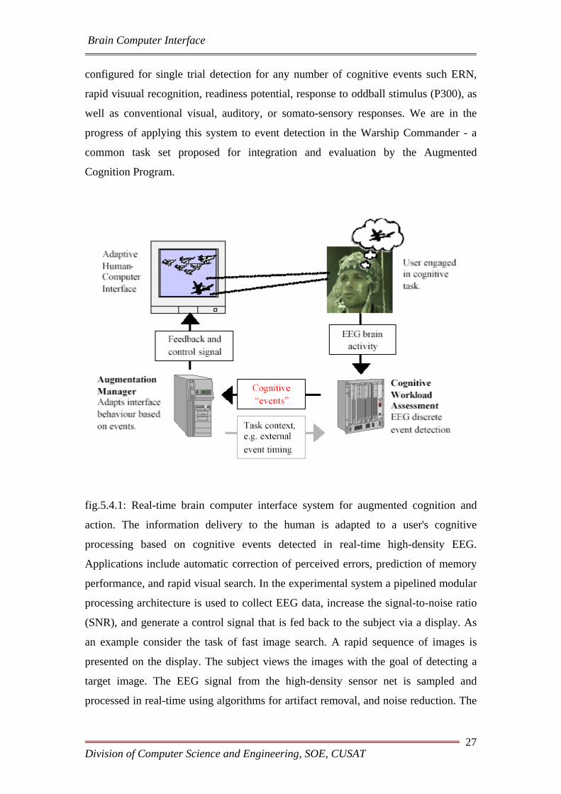
fig.5.4.1: Real-time brain computer interface system for augmented cognition and
action. The information delivery to the human is adapted to a user's cognitive
processing based on cognitive events detected in real-time high-density EEG.
Applications include automatic correction of perceived errors, prediction of memory
performance, and rapid visual search. In the experimental system a pipelined modular
processing architecture is used to collect EEG data, increase the signal-to-noise ratio
(SNR), and generate a control signal that is fed back to the subject via a display. As
an example consider the task of fast image search. A rapid sequence of images is
presented on the display. The subject views the images with the goal of detecting a
target image. The EEG signal from the high-density sensor net is sampled and
processed in real-time using algorithms for artifact removal, and noise reduction. The
Division of Computer Science and Engineering, SOE, CUSAT 27
Brain Computer Interface
signal is analyzed in real-time to identify the cognitive activity associated with visual
target detection. The augmentation manager records the images associated with
recognition events. This information is used to prioritize the large volumes of imagery
that has to be analyzed. The selected images can subsequently be presented for more
careful analysis without interrupting the fast visual processing of the human subjects
in the initial scan. In Phase 1 of the project it has been demonstrated that improved
prioritization performance is obtained as compared to selecting the image with a
manual button push.
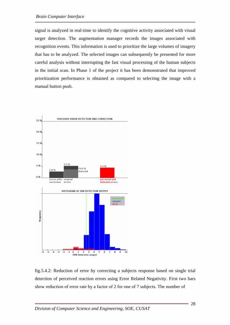
fig.5.4.2: Reduction of error by correcting a subjects response based on single trial
detection of perceived reaction errors using Error Related Negativity. First two bars
show reduction of error rate by a factor of 2 for one of 7 subjects. The number of
Division of Computer Science and Engineering, SOE, CUSAT 28

Brain Computer Interface
perceived and detected errors (right) could be understood as an "gauge" that measures
perceived task difficulty over an extended period of time (minutes).
fig.5.4.3: Increase in target image throughput for detected EEG signatures compared
to the covert responses (button release). Note that the detected EEG signature results
in a larger fraction of the targets to be placed in the front of the image stack, thus
improving image search efficiency.
Division of Computer Science and Engineering, SOE, CUSAT 29

Brain Computer Interface
Chapter6. Experimental Brain Computer Interface Software for the Modular EEG (The ABI software)
6.1. Introduction:
ABI is a simple software for the Modular EEG that implements an
experimental Brain Computer Interface (BCI). Nowadays, BCI research is an highly
active field, but the existing technology is still immature for its use outside of a lab's
settings. The ABI software tries to provide a simple tool for hobbyists to do
experiments on its own with BCI technology.
Screenshot Download
6.2. Work of the software:
The ABI is a BCI based on trials. A trial is a time interval where the user generates
brainwaves to perform an action. The BCI tries to process this signal and to associate
it to a given class. The association is done by feeding a neural net with the
preprocessed EEG data. The neural net's output is then further processed and this final
output corresponds to the given class. The neural net should be trained in order to
learn the association.
Division of Computer Science and Engineering, SOE, CUSAT 30

Brain Computer Interface
The classifier's idea is heavily based on Christin Schäfer's design (winner of the BCI
Competition II, Motor Imaginery Trials).
The ABI software allows you to
• Do simple Biofeedback. You can display raw EEG channels, narrow band
frequency amplitudes and classes.
• Simulate trials.
• Record trials for a number of choice of different classes.
• Train the interface.
6.3. The classification achieved by this software:
The method has been previously applied to the data provided by the BCI
Competition II data (dataset III, Graz University, Motor Imaginary) and compared
against the results obtained by the contributors. The method has outperformed the
results achieved by them, obtaining a higher Mutual Information (which was the
criterion used in the competition) of 0.67 bits (the winner of the competition obtained
0.61 bits).
Of course, it is very important that more people test the software and
report its results to improve the method. Statistical stability can only be guaranteed if
more people try it out.
6.4. Instructions:
By executing ABI, it reads a configuration file called "abi.txt" (which you can edit
with a simple text editor), where the way the BCI should act is specified. ABI tries to
load the trial file defined in the configuration file. The trial file is a text database
containing trials for different classes. Then, the main screen is displayed:
Division of Computer Science and Engineering, SOE, CUSAT 31

Brain Computer Interface
fig.6.4.1:
• a) The EEG channels. The ModularEEG should be turned on. You can choose
the amount of channels by setting the variable NChannels to desired value.
• b) The extracted features. Each color strip indicates the intensity of a given
frequency band. The variable NFeatures indicates the number of features you
want to use. Channels indicates the source channels for the feature extraction.
Frequencies tells ABI what frequencies should be used (in Hertz). Example:
NFeatures = 4, Channels = 0 0 1 1, Frequencies = 10 20 10 20, tells ABI to
use 2 EEG channels, and to extract frequencies 10 Hz and 20 Hz from
channel 0 and channel 1.
Division of Computer Science and Engineering, SOE, CUSAT 32
Brain Computer Interface
• c) Class bar. The variable NClasses tells ABI how many classes it should be
able to discriminate. Each class has an associated bar, and its size (and color)
shows how good the given class has been recognized by the system.
ABI has three operating modes: SIMULATION,
RECORDING and TRAINING. You can switch between operating modes by
pressing F1, F2 or F3 respectively (the software doesn't change its mode instantly,
because a trial shouldn't be interrupted in the middle).
The operation is quite simple. The user records several trials for the
different classes (RECORDING mode). Each class is associated to a different mental
task. After recording a reasonable amount of trials (more than 50 trials for each class),
the user can train the system to learn a way to discriminate between the different
classes (TRAINING mode). This process can be repeated in order to improve the
quality of the recognition. The system can be tested under the SIMULATION mode.
An explanation of the different modes follows.
6.4.1.SIMULATION and RECORDING
These two modes perform single trials. The SIMULATION mode is used to test the
BCI. RECORDING is the same as SIMULATION, with the difference that the EEG
data is recorded and used as training examples. A trial has the following structure:
Division of Computer Science and Engineering, SOE, CUSAT 33

Brain Computer Interface
Table 6.4.1 Simulation mode to test the BCI
Substate Duration Description Screenshot
Preparation TPreparation
seconds
The BCI doesn't
display anything but
the EEG data and
the features. The
user can relax
during this time
(eye blinking, etc.).
Prerecording TPrerecording
seconds
The BCI displays
the target class by
indicating a white
target line. The user
should start to
perform the mental
task associated to
the target class, but
the data isn't
recorded yet.
Recording TrialLength
seconds
The BCI displays
the bars indicating
which classes are
recognized in each
time instant. The
EEG data is
recorded (except in
SIMULATION
mode).
Division of Computer Science and Engineering, SOE, CUSAT 34

Brain Computer Interface
As you can see, a trial is composed of three subintervals, whose duration is defined by
the variables TPreparation, TPrerecording and TrialLength, in the configuration
file.
6.4.2. TRAINING
Table 6.4.2.1 Training
Pressing the F3 key, the system starts to train the
neural net with the available data. The training set
used for this purpose is the set of the last
TrialBuffer recorded trials' features. Example:
Suppose you have recorded 300 trials, and
TrialBuffer = 100. Then the system extracts the
features of the 100 last recorded trials to form the
training set.
Training time depends upon the complexity of the
training data and the amount of recorded data. The
training data is not always separable. If the mental
task for class 1 is too similar to the mental task for
class 2, then the neural net won't be able to do the
separation: this isn't magic :-) .
Division of Computer Science and Engineering, SOE, CUSAT 35

Brain Computer Interface
6.4.3. The Trial Archive:
When exiting ABI, the EEG data recorded so far is saved into the file given by the
parameter Trial Archive. You can open a trial archive with a simple text editor and
see how the trial data has been recorded. Only raw EEG data and the class label is
recorded: the features, which correspond to the real training data, are computed on-
the-fly.
If you start ABI, it will load the trial archive specified in the configuration file if it
exists, or create a new one if not. If the trial archive doesn't match the configuration
file's specifications, then ABI aborts its execution. So you have to be careful to use
correct trial archives and configuration files.
EEG recording between different executions of the ABI system is appended to the
trial archive. This allows you to build your training set in different sessions. Be
careful to use the same electrode settings. Some have reported that the recognition
rate drops between different sessions.
6.4.4 The Configuration File
The configuration file tells ABI where to load the trial data from, how many channels
the system should use, which features it should use, etc. You can open it with your
favourite text editor and edit it. To start ABI with a different configuration file other
than the default "abi.txt", invoce ABI with the following syntax at the command
prompt:
abi <my_configuration_file>
The configuration file basically contains a list of variables. The list of variables is:
Division of Computer Science and Engineering, SOE, CUSAT 36

Brain Computer Interface
Table 6.4.4.1 List of variables in configuration file
Variable
Name Description
Device This is the string that tells ABI how to initialize the ModularEEG.
You shouldn't change it
NChannels The amount of channels to use.
NFeatures The number of features the ABI should extract from the raw EEG
data in order to feed the neural net.
NClasses The number of classes that the system should be able to
discriminate.
TrialArchive The name of the associated trial archive.
Channels The index list of source channels for the feature extraction.
Frequencies The list of frequencies to extract from the EEG data to use as
features.
HiddenUnits The number of hidden units of the neural net.
TrialBuffer The size of the training set used to train the neural net.
TrialLength The length in seconds for each trial.
TPreparation The length in seconds for the preparation time.
TPreRec The length in seconds for the prerecording time.
A variable and its value should be in the same line.
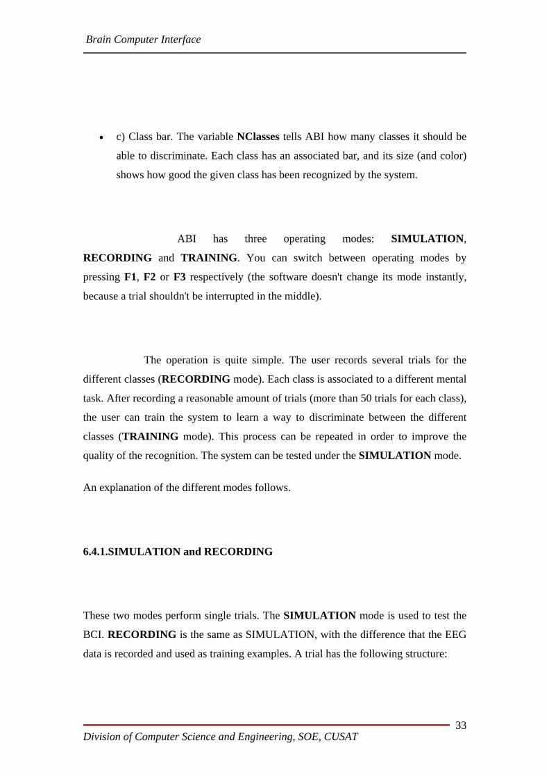
6.4.5 Electrode Positions
As a reference, this is the international 10-20 system:
Division of Computer Science and Engineering, SOE, CUSAT 37
Brain Computer Interface
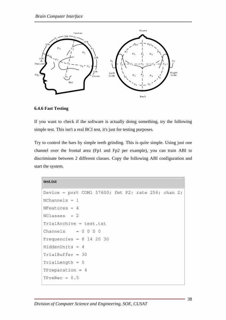
6.4.6 Fast Testing
If you want to check if the software is actually doing something, try the following
simple test. This isn't a real BCI test, it's just for testing purposes.
Try to control the bars by simple teeth grinding. This is quite simple. Using just one
channel over the frontal area (Fp1 and Fp2 per example), you can train ABI to
discriminate between 2 different classes. Copy the following ABI configuration and
start the system.
test.txt
Device = port COM1 57600; fmt P2; rate 256; chan 2;
NChannels = 1
NFeatures = 4
NClasses = 2
TrialArchive = test.txt
Channels = 0 0 0 0
Frequencies = 8 14 20 30
HiddenUnits = 4
TrialBuffer = 30
TrialLength = 5
TPreparation = 4
TPreRec = 0.5
Division of Computer Science and Engineering, SOE, CUSAT 38

Brain Computer Interface
Now, enter the RECORDING mode by pressing [F2]. Grind your teeth when the
system asks you to perform the mental task associated to class 1 (the left bar). Relax
for class 2. After recording 10 trials for each class, train the network by pressing [F3].
Wait until the classification error drops to a reasonable amount (per example, 1.2
bits). Then, enter the SIMULATION node by pressing [F1]. Repeat the same as when
you've been recording. The system should classify the trials correctly: when you grind
your teeth, the left bar should be higher than the right one, and viceversa.
6.4.7. Considerations
First of all, be patient! The system tries, by using a trainable classification method, to
adapt the BCI to the user, and in this way, to simplify the learning process required by
the user. Nevertheless, as any other instrument, it requires a considerable amount of
time to use the BCI in order to get nice results.
BCI technology is still in its infancy, so little is known about which mental tasks are
better than others for BCIs. Also, the electrode placing is important. If your electrode
setting isn't appropiate, then it can happen that they even aren't recording the cortical
areas related to the mental task!
Research has discovered the following changes in electrical activity during mental
tasks (this list isn't complete, I hope that the OpenEEG community will discover some
more):
• Motor Imaginery: Imagination of physical movement produces changes in
the sensorymotor cortex. In example, imagination of left and right middle
finger imagination produces changes, namely (de-)synchronization on
electrode positions around C3 and C4. Good features are around 10 and 20 Hz.
• Rotation of 3D objects: Literature stated that during imagination of rotation
of 3d objects involves frontal and temporal lobe activity. They seem to
sinchronize. Good features are around 10 Hz.
• Mental Letter Composition.
• Others (please report!)
Division of Computer Science and Engineering, SOE, CUSAT 39

Brain Computer Interface
Do not use too many features at the same time, 4-10 features are reasonable. If you
want to change the used features, restart the BCI with the appropiate change in the
configuration file.
6.5.Classifier Design:
The design of the classifier is heavily based on Christin Schäfer's design used for the
Dataset III of the BCI Competition II. Instead of using a Gaussian multivariate
Bayesian classifier, here we use a neural net to obtain the classification for each time
instant t. Those outputs are then integrated in time using a weighted sum. The idea is
simple: outputs with low confusion should have higher weights.
These are the different steps:
• Aquire raw EEG data. Filter the EEG channel using a bandpass filter between
4 and 25 Hz.
• Use Morlet Wavelets to extract local frequency information. Compute their
absolute value. These are the feature channels.
• Feed a two layer feedforward neural net with the frequency information and
an additional time channel (restarts at zero at the begin of every trial). The
neural net has two layers: the first weight layer uses the tanh activation
Division of Computer Science and Engineering, SOE, CUSAT 40
Brain Computer Interface
function, the second a normal logistic activation. The net is trained using the
cross-entropy error as the optimization criterion. The output of the neural net
is the estimated instant classification.
• The final classification is obtained after performing a weighted time
integration of the instant outputs, where individual weights are higher for low
entropy outputs.
Division of Computer Science and Engineering, SOE, CUSAT 41

CONCLUSION
Brain-Computer Interface (BCI) is a method of communication based on
voluntary neural activity generated by the brain and independent of its normal
output pathways of peripheral nerves and muscles.
The neural activity used in BCI can be recorded using invasive or
noninvasive techniques.
We can say as detection techniques and experimental designs improve, the
BCI will improve as well and would provide wealth alternatives for individuals to
interact with their environment.

REFERENCES
1. http://www.nicolelislab.net/NLnet_Load.html
2. http://www.youtube.com/watch?v=7-cpcoIJbOU
3. http://www.en.wikipedia.com/braincomputerinterface