Embed Size (px)
Citation preview
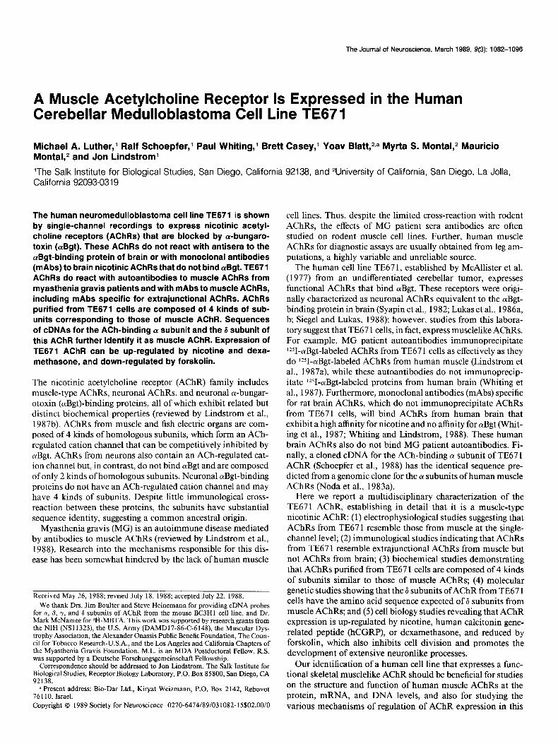
The Journal of Neuroscience, March 1989, g(3): 1082-l 098
A Muscle Acetylcholine Receptor Is Expressed in the Human Cerebellar Medulloblastoma Cell Line TE67 1
Michael A. Luther,’ Ralf Schoepfer,’ Paul Whiting,’ Brett Casey,’ Yoav Blatt,21a Myrta S. Montal,2 Mauricio Montal,2 and Jon Lindstrom’
‘The Salk Institute for Biological Studies, San Diego, California 92138, and 2University of California, San Diego, La Jolla, California 92093-0319
The human neuromedulloblastoma cell line TE671 is shown by single-channel recordings to express nicotinic acetyl- choline receptors (AChRs) that are blocked by cY-bungaro- toxin (crBgt). These AChRs do not react with antisera to the crBgt-binding protein of brain or with monoclonal antibodies (mAbs) to brain nicotinic AChRs that do not bind aBgt. TE671 AChRs do react with autoantibodies to muscle AChRs from myasthenia gravis patients and with mAbs to muscle AChRs, including mAbs specific for extrajunctional AChRs. AChRs purified from TE671 cells are composed of 4 kinds of sub- units corresponding to those of muscle AChR. Sequences of cDNAs for the ACh-binding ar subunit and the 6 subunit of this AChR further identify it as muscle AChR. Expression of TE671 AChR can be up-regulated by nicotine and dexa- methasone, and down-regulated by forskolin.
The nicotinic acetylcholine receptor (AChR) family includes muscle-type AChRs, neuronal AChRs, and neuronal a-bungar- otoxin (otBgt)-binding proteins, all of which exhibit related but distinct biochemical properties (reviewed by Lindstrom et al., 1987b). AChRs from muscle and fish electric organs are com- posed of 4 kinds of homologous subunits, which form an ACh- regulated cation channel that can be competitively inhibited by cuBgt. AChRs from neurons also contain an ACh-regulated cat- ion channel but, in contrast, do not bind otBgt and are composed of only 2 kinds of homologous subunits. Neuronal cuBgt-binding proteins do not have an ACh-regulated cation channel and may have 4 kinds of subunits. Despite little immunological cross- reaction between these proteins, the subunits have substantial sequence identity, suggesting a common ancestral origin.
Myasthenia gravis (MG) is an autoimmune disease mediated by antibodies to muscle AChRs (reviewed by Lindstrom et al., 1988). Research into the mechanisms responsible for this dis- ease has been somewhat hindered by the lack of human muscle
Received May 26, 1988; revised July 18, 1988; accepted July 22, 1988.
We thank Drs. Jim Boulter and Steve Heinemann for providing cDNA probes for cy, 0, y, and 6 subunits of AChR from the mouse BC3Hl cell line, and Dr. Mark McNamee for ‘H-MBTA. This work was supported by research grants from the NIH (NS 11323), the U.S. Army (DAMDl7-86-C-6 148), the Muscular Dys- trophy Association, the Alexander Onassis Public Benefit Foundation, The Coun- cil for Tobacco Research-U.S.A., and the Los Angeles and California Chapters of the Mvasthenia Gravis Foundation. M.L. is an MDA Postdoctoral Fellow. R.S. was supported by a Deutsche Forschungsgemeinschaft Fellowship.
Correspondence should be addressed to Jon Lindstrom, The Salk Institute for Biological Studies, Receptor Biology Laboratory, P.O. Box 85800, San Diego, CA 92138.
B Present address: Bio-Dar Ltd., Kiryat Weizmann, P.O. Box 2142, Rehovot 76 110, Israel. Copyright 0 1989 Society for Neuroscience 0270-6474/89/031082-15$02.00/O
cell lines. Thus, despite the limited cross-reaction with rodent AChRs, the effects of MG patient sera antibodies are often studied on rodent muscle cell lines. Further, human muscle AChRs for diagnostic assays are usually obtained from leg am- putations, a highly variable and unreliable source.
The human cell line TE67 1, established by McAllister et al. (1977) from an undifferentiated cerebellar tumor, expresses functional AChRs that bind otBgt. These receptors were origi- nally characterized as neuronal AChRs equivalent to the aBgt- binding protein in brain (Syapin et al., 1982; Lukas et al., 1986a, b; Siegel and Lukas, 1988); however, studies from this labora- tory suggest that TE67 1 cells, in fact, express musclelike AChRs. For example, MG patient autoantibodies immunoprecipitate Y-otBgt-labeled AChRs from TE67 1 cells as effectively as they do 1251-cuBgt-labeled AChRs from human muscle (Lindstrom et al., 1987a), while these autoantibodies do not immunoprecip- itate 1Z51-aBgt-labeled proteins from human brain (Whiting et al., 1987). Furthermore, monoclonal antibodies (mAbs) specific for rat brain AChRs, which do not immunoprecipitate AChRs from TE671 cells, will bind AChRs from human brain that exhibit a high affinity for nicotine and no affinity for otBgt (Whit- ing et al., 1987; Whiting and Lindstrom, 1988). These human brain AChRs also do not bind MG patient autoantibodies. Fi- nally, a cloned cDNA for the ACh-binding (Y subunit of TE67 1 AChR (Schoepfer et al., 1988) has the identical sequence pre- dicted from a genomic clone for the LY subunits of human muscle AChRs (Noda et al., 1983a).
Here we report a multidisciplinary characterization of the TE671 AChR, establishing in detail that it is a muscle-type nicotinic AChR: (1) electrophysiological studies suggesting that AChRs from TE67 1 resemble those from muscle at the single- channel level; (2) immunological studies indicating that AChRs from TE67 1 resemble extrajunctional AChRs from muscle but not AChRs from brain; (3) biochemical studies demonstrating that AChRs purified from TE67 1 cells are composed of 4 kinds of subunits similar to those of muscle AChRs; (4) molecular genetic studies showing that the 6 subunits of AChR from TE67 1 cells have the amino acid sequence expected of 6 subunits from muscle AChRs; and (5) cell biology studies revealing that AChR expression is up-regulated by nicotine, human calcitonin gene- related peptide (hCGRP), or dexamethasone, and reduced by forskolin, which also inhibits cell division and promotes the development of extensive neuronlike processes.
Our identification of a human cell line that expresses a func- tional skeletal musclelike AChR should be beneficial for studies on the structure and function of human muscle AChRs at the protein, mRNA, and DNA levels, and also for studying the various mechanisms of regulation of AChR expression in this
The Journal of Neuroscience, March 1989, 9(3) 1083
cell line. Further, it should also prove useful for studies of MG, both as a consistent and unli&ed source of antigen for bio: chemical studies and for studying the effects of autoantibodies on AChR function and turnover.
Materials and Methods TE671 cells. Cultures were grown at 37°C in 90% air/lo% CO, in Is- cove’s modified Dulbecco’s medium from Irvine Laboratories supple- mented with either 10% fetal bovine serum or 5% bovine calf serum. For electrophysiological studies, lo4 cells were plated per well in a 24 well plate on 12-mm-diameter glass coverslips in medium with 10% serum. One day later, serum was reduced to O.Ol%, and 2 mM L-glu-
tamine, 10 &ml insulin, and transfenin were added. Electrophysio- logical studies were done in 115 mM NaCl, 5 mM CsCl, 1 mM MgCl,, 25 mM glucose, 25 mM HEPES, pH 7.4, 10 mM TEA, and 0.1 mM anthracene-9-carboxylic acid.
Fluorescent labeling. Cells on coverslips were incubated with 10m7 M
mAb for 30 min, rinsed with 0.15 M NaCl, 15 mM Na phosphate, pH 7.4, incubated with rabbit anti-rat IgG (85 pdml), rinsed, incubated with rhodamine-labeled, affinity-purified goat anti-rabbit IgG (10 ~Lgl ml), and rinsed again. A Zeiss photomicroscope equipped with No- marski optics and a fluorescent attachment (556 nm bandpass excitation filter, 590 nm barrier filter) was used at 63 x magnification.
ringer) and 0.05% SDS, pH 7.5, centrifuged at 140,000 x g for 30 min, and the clarified supematant was retained.
Purification of the TE671 AChR. aBgt was first coupled to Sepharose CL4B at 5.0 mg protein/ml of gel by a modified procedure of Kohn and Wilchek (1982) (D. Shelton, Y. Fujii, W. Knogge, and J. Lindstrom, unpublished data). The clarified, solubilized TE67 1 membrane extract (75-100 ml) from, typically, 12 roller bottles, was applied to a 20 ml column of Sepharose CL4B to adsorb any proteins that may nonspe- cifically adsorb to the column. The eluate was then applied to a 1 ml column of olBgt-affinity gel, and both columns were washed with 200 ml of the extraction buffer. The affinity column was consecutively washed with 200 ml of buffer A containing 1 .O M NaCl, 0.5% Thesit, 0.05% SDS, pH 7.5, followed by 150 ml of 10 mM Tris, 0.1% Thesit, 1 mM NaN,, 10 mM KF, 1 mM IAA, 1 mM aminobenzamidine, 1 mM EDTA, and 1 mM EGTA pH 7.5 (buffer B). The affinity column was then coupled to a hydroxylapatite (HPT) column (1 ml) and the TE67 1 AChR eluted onto the HPT column by recirculating through both columns for 12 hr, 10 ml of buffer B containing 200 mM carbamylcholine, using a peristaltic pump. After displacement of the bound protein, the HPT column was washed with 200 ml of buffer B and then eluted with 150 mM sodium phosphate, 0.5% Thesit, 1 mM NaN,, 1 mM PMSF, 1 mM EDTA, 1 mM EGTA, 1 mM aminobenzamidine, and 1 mM IAA at pH 7.5.
a 40x objective (LWD DL 4OXC, Nikon) equipped with Hoffman
Electrical recordings. Single-channel current electrical recordings were performed as previously described in detail (Sakmann and Neher, 1983).
modulation contrast optics (Modulation Optics, Greenvale, NY). The
Recordings were obtained in both the cell attached and the excised patch
microscope was mounted on a vibration isolation table (Micro g Tech-
configurations. The pipettes were fabricated from Kovar glass (Coming 7052, ID = 1.1 mm, OD = 1.5 mm, 70 mm long) using a vertical pipette puller (David Kopf 700 C, Tujunga, CA). The pipettes were coated with Sylgard- 180 (Dow Coming) within 40 pm from the tip and fire-polished immediately before use under 320x magnification. The tip size was adjusted to-yield 5-l 5 MQ of open pipette resistance when filled and immersed in the buffer described before. The patch pipettes contained the indicated concentration of ACh diluted in the same solution. The cells were observed with an inverted microscope (Nikon -Diaphot) using
Ajinity labeling. TE67 1 AChR was immobilized on cuBgt-Sepharose and then affinity-labeled with )H-MBTA (a gift from Dr. Mark Mc- Namee) as previously described (Whiting and Lindstrom, 1987).
mol) and autoradiography.
Electrophoresis. Electrophoresis was conducted on acrylamide slab
Cloning and sequencing of TE671 AChR 6 subunit cDNA. A cDNA library was prepared as previously described (Schoepfer et al., 1988).
gels in SDS using a Laemmli discontinuous buffer svstem (Laemmli.
The filters were screened under high stringency with the -450 base pair (bn) Eco RI-Ava I fragment of cDNA clone BMD45 1 (a aift of Dr. Jim
1970). Polyacrylamide gels were silver-stained for protein according to the method of Oakley et al. (1980). Polyacrylamide gels of radiolabeled protein were autoradiographed for 4-24 hr at -70°C using preflashed Kodak X-Omat-AR film and an intensifying screen. Autoradiograms were standardized by using Sigma prestained low-molecular-weight standards resolved on the same gel. Electrophoretic transfer of proteins from gels to diazophenylthioether (DPT) paper and subsequent probing with antibodies were as described previously (Gullick and Lindstrom, 1982). After being probed, bound antibodies were detected by incu- bation with 0.5 nM lZSI-labeled mouse anti-rat IgG (l-3 x lOI* cpm/
nical Mfrg. Corp., Waltham, MA). A commercially available extracellular patch-clamp system was used
(LM EPC-5, List Electronics, Darmstadt, FRG, and Medical Systems Corporation, New York). The headstage of the amplifier was mounted on a hydraulic micromanipulator (MO-103N Narishige, Japan). The signal output from the clamp was recorded on FM tape (Racal 4DS, Hythe, Southhampton, England; bandwidth DC-5 kHz). All the records were filtered at 2 kHz on an 8-pole Bassel low-pass filter (Frequency Devices, 9028LPF, Haverhill, MA). The data were digitized at the sam- pling frequency of 10 kHz in an Indec-L-l l/73-70 microcomputer sys- tem (Indec, Sunnyvale, CA). Conductance levels were discriminated as described previously (Labarca et al., 1984). Histograms of dwell times in the open and closed states of the AChR channel were analyzed as described in detail previously (Labarca et al., 1984, 1985; Montal et al., 1986). The results of at least 5 different experiments in each condition are presented. All experiments were done at room temperature (22°C).
Preuaration ofsolubilized TE671 membrane extracts. TE67 1 cell cul- tures were grown in T-flasks for 6 d and then expanded to 2 liter (850 cm*) roller bottles in 5% BCS in Iscove’s modified DMEM medium (Irvine Laboratories) with 2.5 PM dexamethasone. After 10 d in culture the cells were harvested after aspiration of media by first rinsing with cold PBS, pH 7.5, containing 10 mM iodoacetamide (IAA), 10 mM aminobenzamidine, 1 mM phenylmethylsulfonylfluoride (PMSF) to re- move the excess media, and second by shaking in 25 ml per bottle of 50 mM Tris, 150 mM NaCl, 100 mM KF, 5 mM EDTA, 5 mM EGTA, 5 mM IAA, 5 mM aminobenzamidine, 0.5 mM PMSF, bestatin (10 rg/ ml), Trasylol (10 j&ml), soybean trypsin inhibitor (10 &ml), pH 7.5 (buffer A). The bottles were then rinsed with 4 volumes of buffer A to remove any remaining cells. The cells were then pelleted by centrifu- gation at 3000 x g for 30 min. The resulting cell pellet was resuspended in 400 ml of buffer A, lysed by homogenization using a Polytron for 30 set, and centrifuged for 30 min at 10,000 x g. The membrane pellet was resuspended in 250 ml of buffer A, homogenized, and centrifuged as described in the previous step. The resulting pellet was then extracted for 30 min in 4 volumes of buffer A with 1% Thesit detergent (Boeh-
Boulter) coding for the 114 N terminal amino acids of the-mouse AChR 6 subunit. A single positive clone was identified. Plasmid DNA was characterized by restriction enzyme digestion, followed by agarose gel electrophoresis and Southern blot analysis. From the -3 kb insert, the 5’ - 1860 bp Eco-Ava fragment was subcloned into a plasmid vector. Nested deletions were produced by the Exo III/Mung Bean protocol provided by Stratagene. DNA sequencing was performed using a mod- ification of the dideoxynucleotide chain termination method of Sanger et al. (1977).
Regulation of TE671 expression. Cultured cells grown in T-flasks were harvested and 1 x lo5 cells were plated in 6-well tissue culture dishes in Iscove’s medium containing 10% FCS. After 2 d, the media was removed and replaced with this medium containing the indicated con- centrations of forskolin, nicotine, human CGRP (a gift from Dr. Jean Rivier), or dexamethasone. Forskolin and dexamethasone were dis- solved in 95% ethanol, while CGRP was dissolved in PBS. Ethanol or PBS alone had no affect on cell growth or AChR expression. The cells were grown for 2 d and the number of a-Bgt binding sites, AChR function, and RNA encoding the 01, & y, and 6 subunits of the TE67 1 AChR were determined.
The number of olBgt binding sites was determined as follows. After 2 d the medium was removed, and the cells were washed 3 times with 2 ml of Iscove’s media. The cells were then labeled for 1 hr with 0.5 ml of 20 nM 12+aBgt in Iscove’s medium at 37°C. Nonspecific binding was determined by performing the experiments as described, in the presence of 1 mM carbamylcholine. After 1 hr, the cells were again washed 3 times with 2 ml Iscove’s medium. The cells were solubilized with 1.5 ml of 0.5 N NaOH, removed, and bound lZSI-aBgt determined by gamma counting.
AChR function was measured by carbamylcholine-induced influx of 86Rb+ using a modified procedure of Robinson and McGee (1985). Brief- ly, after 2 d of growth in the presence or absence of the various indicated effecters, the media was removed and the cells washed 3 times with 2.0
1064 Luther et al. l TE671 Acetylcholine Receptor
ml Iscove’s. After the third wash, the cells were incubated for 1 hr in 0.5 ml Iscove’s to allow recovery from desensitization of AChRs by the effecters. Media was removed and the cells washed 2 times with 2.0 ml 0.5 M sucrose, 5 mM KCl, 10 mM glucose, 1.8 mM CaCl,, and 15 mM HEPES, pH 7.4. The cells were then washed with 0.5 ml of the same buffer with 2 mM ouabain for 20 set to inhibit Na+-K+ ATPases. The buffer was removed and 86Rb+ uptake was initiated by exposing cells to 0.5 ml of the ouabain buffer containing 5 &i/ml of 86Rb+ with 1 mM carbamylcholine. Control experiments were performed as described, in the absence of carbamvlcholine. Untake was terminated after 30 set bv aspirating the radioacnve solution-and rapidly washing 3 times with 3 ml of 0.3 M NaCl, 5 mM KCl, 1.8 nM CaCl,, 10 mM glucose, and 15 mM HEPES, pH 7.5. The washed cells were solubilized with 1.5 ml 0.5 N NaOH to permit *6Rb+ uptake and protein determination. Radio- activity was determined by liquid scintillation counting of the solubi- lized cells. Results were normalized as described for the determination of olBgt binding sites.
Total RNA was isolated by the guanidine thiocyanate-CsCl procedure of Chirgwin et al. (1979). The amount of RNA isolated was quantitated by ODxo, and equal amounts of RNA from each treatment were size- fractionated by agarose gel electrophoresis containing formaldehyde. The gel was t&&erred te Nylon membranes and probed (Fig. 8) with cloned cDNA inserts (gifts from Dr. Jim Boulter) for the (Y, p, and y subunits of mouse muscle AChR (Heinemann et al., 1986), and the 6 subunit probe was derived from the cDNA clone for TE67 1 b. Hybrid- ization was conducted under hiahlv strinaent conditions: 42°C. 50% formamide, 5 x SSPE, final was&g at 6s;“C, 0.3 x SSPE (where 5 x SSPE is 0.9 M NaCl, 50 mM Na phosphate, pH 7.4, 5 mM EDTA). Autoradiography was performed as described above.
otBgt were also not immune precipitated by a loo-fold molar excess of mAbs 290,293, or 299, which react with AChRs from human brain that have high affinity for nicotine but do not bind LvBgt (Whiting et al., 1987; Whiting and Lindstrom, 1988). Thus, TE67 1 AChRs are also different from AChRs detected in adult human brain.
Functional AChRs are detected on TE671 cells The observation of Syapin et al. (1982) that these AChRs could be blocked by cuBgt by measuring carbamylcholine-induced 86Rb+ influx was confirmed (data not shown). We next used the patch- clamp technique to study AChR activity electrophysiologically at the single-channel level. To record only ACh-activated chan- nels, several other channel types present in these cells are blocked pharmacologically: K+ channels are eliminated by adding tet- raethylammoniumchloride (TEA) and removing K+ from the medium; Ca*+ channels are eliminated by removing Ca2+ from the medium; and Cl- channels are blocked with 0.1 mM an- thracene-9-carboxylic acid.
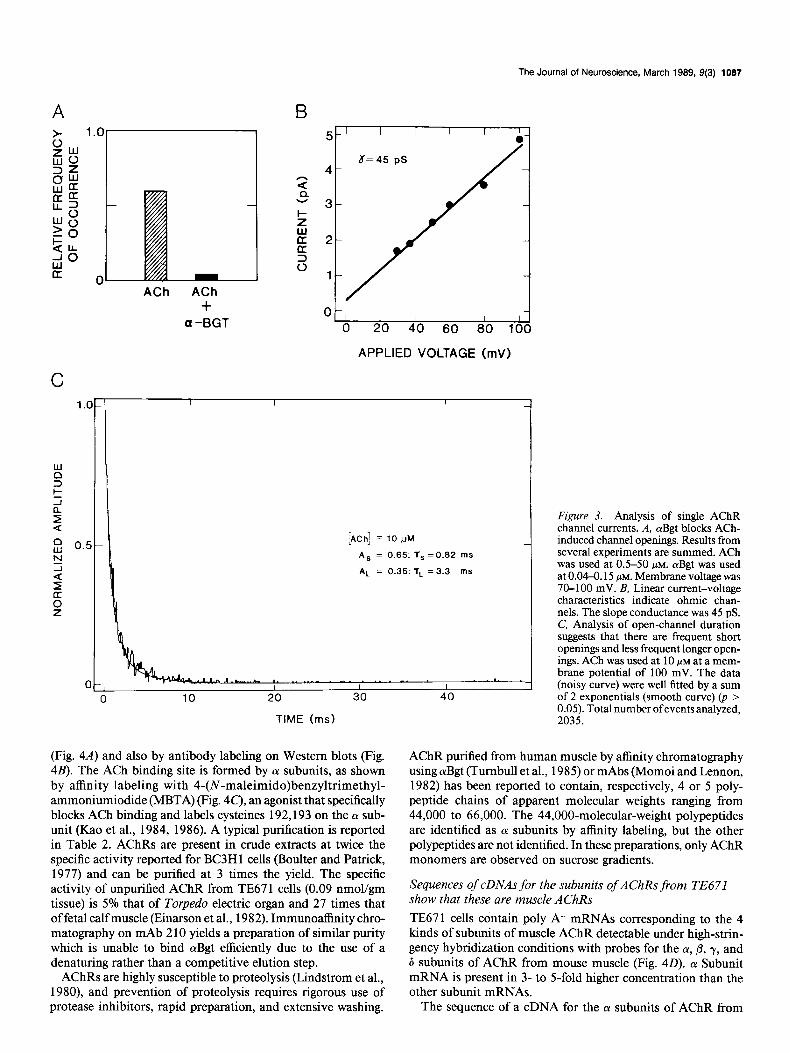
ACh induces bursts of AChR channel openings (Figs. 2, 3). At 0.5 PM ACh the channels are open 3.3% of the time, whereas at 10 /IM ACh this increases to 8.8% (Fig. 2). Opening of TE67 I AChR channels induced by ACh is blocked by aBgt (Fig. 3A). This is expected for muscle AChRs but not for neuronal nico- tinic AChRs (reviewed in Lindstrom et al., 1988). TE67 1 AChR channels exhibit a linear current-voltage relationship in the range 10-100 mV (Fig. 3B) with a single-channel conductance (7) of 44-45 pS (Fig. 2). The majority of channel openings are brief (65% have a time constant [T] of 0.82 msec), whereas a minority of the openings are more prolonged (for 35% 7 = 3.3 msec) (Fig. 3C’). The duration of opening and magnitude of conductance are not affected by the concentration of ACh in the range 0.5- 20 PM or by voltage in the range 50-100 mV. These properties are all consistent with those of muscle AChRs (Neher and Sak- mann, 1976). Blockage of function by cYBgt is the critical char- acteristic distinguishing AChRs on TE67 1 from those on neu- rons, since other electrophysiological properties are similar for neuronal AChRs which do not bind cvBgt (Lipton et al., 1987). Sine (1988) has recently observed electrophysiological proper- ties of AChRs from TE67 1 cells similar to those reported here, and he has characterized their pharmacology in more detail. He finds that AChRs in TE67 1 cells are similar in many respects to those from Torpedo electric organ or mouse BC3H- 1 muscle cells but differ from these most prominently in having longer closed-channel times, slower desensitization onset and offset, symmetry in binding reversible nicotinic antagonists, and much more rapid dissociation of bound cYBgt.
Poly A+ RNA was prepared from total RNA by oligo-dT column chromatography. The mRNA species for LY, 0, y, and 6 was identified as above using mouse muscle cDNA probes (Heinemann et al., 1986).
Results and Discussion AChRs are localized on the surface of some TE671 cells Fluorescent labeling indicates that AChRs are present on the surface of some cells in a serum-starved TE67 1 culture but not others (Fig. 1). AChRs were identified using 3 mAbs directed at the main immunogenic region (MIR) on the extracellular surface of (Y subunits, raised against AChRs from Electrophorus (mAb 35) human muscle (mAb 203), and muscle of mice and cattle(mAb210)(Tzartoset al., 1981,1983,1987). Eachshowed equivalent results. Under the serum starvation conditions used for electrophysiological studies, labeling of only about 60% of cells grown on coverslips is observed. From these results it is not evident whether several clonal types are present, or whether a pluripotent clonal type undergoes partial differentiation under these culture conditions.
AChRs from TE671 cells are not of the neuronal type AChRs solubilized from TE67 1 cells and labeled with lZ51-LuBgt are not immune precipitated by a 400-fold molar excess of antiserum to the cuBgt-binding protein purified from chicken brain (data not shown). This high-titer antiserum precipitates 5 Fmol of cYBgt binding sites from chicken brain per liter of serum and cross-reacts 0.8% with the cYBgt-binding protein from hu- man brain. The antiserum also shows no reaction on Western blots of purified TE67 1 AChR under conditions where antisera to AChR purified from TE671 label corresponding subunits from AChRs of TE67 1 and Torpedo electric organ (M. Luther and J. Lindstrom, unpublished observations). This data, along with the fact that mAbs like 35, 203, and 210, and MG patient autoantibodies bind AChR from TE671 but not cYBgt-binding proteins from human brain (Whiting et al., 1987), suggest that TE671 AChRs are not identical to the common cYBgt-binding proteins from human brain.
AChRs solubilized from TE671 cells and labeled with Y-
AChRs at mature neuromuscular junctions typically have shorter open channel lifetimes and greater conductances than do extrajunctional AChRs (Schuetze and Role, 1987). Analysis of ACh-induced noise at human neuromuscular junctions sug- gested that T = 1.3 msec (Cull-Candy et al., 1979; Albuquerque et al., 198 l), whereas human myotubes in culture, which would be expected to have extrajunctional AChRs, exhibit T = 2.4 msec at room temperature (Bevan et al., 1978; Adams and Bevan, 1985). The observation that TE67 1 AChRs exhibit r = 0.82 and 3.3 msec may suggest that the cells synthesize both junctional and extrajunctional forms of AChRs. However, AChRs from both Torpedo electric organ (Labarca et al., 1984, 1985) and muscle (Sine and Steinbach, 1984; Colquhoun and Sakmann, 1985) open into either a short- or long-duration state.

The Journal of Neuroscience, March 1989, 9(3) 1088
NOMARSKI FLUORESCENCE
Figure I. AChRs are not localized on the surface of all cells in a serum-starved TE671 culture. AChRs were labeled with mAb 210, followed by rabbit anti-rat IgG, and then rhodamine-labeled goat anti-rabbit IgG. The same fields are examined in the pairs A, B and C, D.
Junctional AChRs are thought to contain e subunits instead of the y subunits characteristic of extrajunctional AChRs (Mishina et al., 1986; Witzemann et al., 1987). Determining whether the AChRs from TE67 1 cells are of the junctional or extrajunctional type is relevant not only to their electrophysiological properties and biochemical structure, but also to their antigenic structure, as some MC patient autoantibodies react exclusively with ex- trajunctional AChRs (Weinberg and Hall, 1979; Schuetze et al., 1985).
AChRs on TE671 cells have the antigenic structure of extrajunctional muscle AChRs TE67 1 AChRs cross-react with antisera and mAbs specific for the 4 kinds of subunits in muscle AChR (Table 1). These data, in conjunction with the evidence that MC patient autoantibod- ies bind TE67 1 AChRs equally as well as muscle AChRs (Lind- Strom et al., 1987a; Whiting et al., 1987), indicate that AChRs from TE671 cells appear to have the basic antigenic structure expected of human muscle AChRs.
The AChRs on TE67 1 cells are extrajunctional. This is shown by their ability to react with mAbs specific for extrajunctional AChRs. Two mouse mAbs, C, and F,, raised against AChR from human muscle have previously been shown to react with AChRs extracted from human denervated or fetal muscle but not with junctional AChRs in sections of human muscle (Whit- ing et al., 1986). The titers of mAbs C, and F, are quite similar for AChRs extracted from the muscles of a leg amputated for diabetic neuropathy (5.3 x 1 O-4 and 6.8 x 1 O+ M, respectively) and for AChRs extracted from TE671 cells (6.8 x 1O-4 and 8.3 x 10⁻⁵~, respectively). This suggests that AChRs from TE67 1 cells should have y rather than E subunits.
AChRs purified from TE671 cells have the biochemical structure of muscle AChRs AChRs affinity-purified from TE671 cells on an arBgt affinity column consist of 4 polypeptides corresponding to (Y, p, y, and 6 subunits of AChR from Torpedo electric organ by apparent molecular weights (42,000; 52,500; 55,000; and 62,000)

1088 Luther et al. - TE671 Acetylcholine Receptor
The Journal of Neuroscience, March 1989, 9(3) 1087
+ a -BGT 00
0 20 40 60 80 100
APPLIED VOLTAGE (mV)
C
C
[ACh] = 10 ,uM
A, = 0.65: 1, =0.62 ms
AL = 0.35: ‘fL ~3.3 ms
0 10 20 ;Ju 4u
TIME (ms)
Figure 3. Analysis of single AChR channel currents. A, cuBgt blocks ACh- induced channel openings. Results from several experiments are summed. ACh was used at OS-50 PM. olBgt was used at 0.04-o. 15 PM. Membrane voltage was 70-100 mV. B, Linear current-voltage characteristics indicate ohmic chan- nels. The slope conductance was 45 pS. C, Analysis of open-channel duration suggests that there are frequent short openings and less frequent longer open- ings. ACh was used at 10 PM at a mem- brane potential of 100 mV. The data (noisy curve) were well fitted by a sum of 2 exponentials (smooth curve) (p > 0.05). Total numberofeventsanalyzed, 2035.
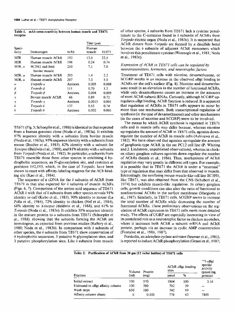
(Fig. 4A) and also by antibody labeling on Western blots (Fig. 4B). The ACh binding site is formed by cx subunits, as shown by affinity labeling with 4-(N-maleimido)benzyltrimethyl- ammoniumiodide (MBTA) (Fig. 40, an agonist that specifically blocks ACh binding and labels cysteines 192,193 on the (Y sub- unit (Kao et al., 1984, 1986). A typical purification is reported in Table 2. AChRs are present in crude extracts at twice the specific activity reported for BC3H 1 cells (Boulter and Patrick, 1977) and can be purified at 3 times the yield. The specific activity of unpurified AChR from TE67 1 cells (0.09 nmol/gm tissue) is 5% that of Torpedo electric organ and 27 times that of fetal calf muscle (Einarson et al., 1982). Immunoaffinity chro- matography on mAb 2 10 yields a preparation of similar purity which is unable to bind aBgt efficiently due to the use of a denaturing rather than a competitive elution step.
AChRs are highly susceptible to proteolysis (Lindstrom et al., 1980), and prevention of proteolysis requires rigorous use of protease inhibitors, rapid preparation, and extensive washing.
AChR purified from human muscle by affinity chromatography using otBgt (Tumbull et al., 1985) or mAbs (Momoi and Lennon, 1982) has been reported to contain, respectively, 4 or 5 poly- peptide chains of apparent molecular weights ranging from 44,000 to 66,000. The 44,000-molecular-weight polypeptides are identified as (Y subunits by affinity labeling, but the other polypeptides are not identified. In these preparations, only AChR monomers are observed on sucrose gradients.
Sequences of cDNAs for the subunits of AChRs from TE671 show that these are muscle AChRs
TE671 cells contain poly A+ mRNAs corresponding to the 4 kinds of subunits of muscle AChR detectable under high-strin- gency hybridization conditions with probes for the (Y, p, y, and 6 subunits of AChR from mouse muscle (Fig. 40). (Y Subunit mRNA is present in 3- to 5-fold higher concentration than the other subunit mRNAs.
The sequence of a cDNA for the a! subunits of AChR from
1088 Luther et al. - TE671 Acetylcholine Receptor
Table 1. mAb cross-reactivity between human muscle and TE671 receptor
Speci- ficity
MIR MIR MIR, CY
Titer (pi)
Human Immunogen mAb muscle TE671
Human muscle AChR 192 12.0 22.5 Human muscle AChR 196 0.24 0.74 BC3H 1 and fetal 210 7.1 7.0
bovine muscle MIR, cr Human muscle AChR 203 1.4 2.2 MIR, CY Human muscle AChR 207 7.3 5.1
; Torpedo a Antisera 0.005 0.008 Torpedo (3 111 0.70 1.2
P Torpedo (3 Antisera 0.004 0.005 Y Bovine muscle AChR 66 0.89 0.72
Y Torpedo y Antisera 0.0015 0.001 6 Torpedo 6 137 0.52 0.74 6 Torpedo 6 Antisera 0.002 0.001
TE67 1 (Fig. 5; Schoepfer et al., 1988) is identical to that expected from a human genomic clone (Noda et al., 1983a). It exhibits 97% sequence identity with (Y subunits from bovine muscle (Noda et al, 1983a), 95% sequence identity with o( subunits from mouse (Boulter et al., 1985) 82% identity with O( subunit for Xenopus (Baldwin et al., 1988) and 8 1% identity with (Y subunits from Torpedo (Noda et al., 1982). In particular, cy subunits from TE671 resemble those from other species in exhibiting 4 hy- drophobic sequences, an N-glycosylation site, and cysteines at positions 192,193, which, in the case of Torpedo, have been shown to react with affinity-labeling reagents for the ACh bind- ing site (Kao et al., 1984).
The sequence of a cDNA for the 6 subunits of AChR from TE671 is that also expected for 6 subunits of muscle AChRs (Figs. 6, 7). Comparison of the amino acid sequence of TE67 1 AChR 6 with that of 6 subunits from other species reveals 9 1% identity to calf (Kubo et al., 1985) 90% identity to mouse (La Polla et al., 1984) 72% identity to chicken (Nef et al., 1984), 68% identity to Xenopus (Baldwin et al., 1988), and 61% to Torpedo (Noda et al., 1983~). It exhibits 30% sequence identity in the mature protein to (Y subunits from TE671 (Schoepfer et al., 1988) showing that the subunits forming the AChR are homologous, as expected from previous studies (Raftery et al., 1980; Noda et al., 1983b). In comparison with 6 subunits of other species, the 6 subunits from TE67 1 show conservation of 4 hydrophobic sequences, 3 putative N-glycosylation sites, and 3 putative phosphorylation sites. Like 6 subunits from muscle
of other species, 6 subunits from TE67 1 lack a cysteine penul- timate to the C-terminus found in 6 subunits of AChRs from Torpedo electric organ (Noda et al., 1983~). It is suspected that AChR dimers from Torpedo are formed by a disulfide bond between the 6 subunits of adjacent AChR monomers which involves this penultimate cysteine (Wennogle et al., 198 1; Noda et al., 1983~).
Expression of AChR in TE671 cells can be regulated by neurotransmitters, hormones, and neurotrophic factors
Treatment of TE671 cells with nicotine, dexamethasone, or hCGRP results in an increase in the observed aBgt binding to AChRs on the cell’s surface (Fig. 8). Nicotine and dexametha- sone result in an elevation in the number of functional AChRs, while only dexamethasone causes an increase in the amounts of most AChR subunit RNAs. Curiously, although hCGRP up- regulates orBgt binding, AChR function is reduced. It is apparent that regulation of AChRs in TE671 cells appears to occur by more than one mechanism. Both transcriptional regulation of synthesis (in the case of dexamethasone) and other mechanisms (in the cases of nicotine and hCGRP) seem to be involved.
The means by which AChR synthesis is regulated in TE67 1 cells and in muscle cells may differ because, whereas nicotine up-regulates the amount of AChR in TE67 1 cells, agonists down- regulate the number of AChR in muscle cells (Ashizawa et al., 1982). We have observed that agonists up-regulate the amount of ganglionic-type AChR in the rat PC12 cell line (P. Whiting and J. Lindstrom, unpublished observations), whereas in chick- en ciliary ganglion cultures agonists down-regulate the number of AChRs (Smith et al., 1986). Thus, mechanisms of AChR regulation may vary greatly in different cell types. For example, it is possible that in TE671 the AChR is under a “neuronal” type of regulation that may differ from that observed in muscle. Interestingly, the nonfusing mouse muscle-like cell line BC3H 1, like TE671, was also obtained from the CNS (Schubert et al., 1974) but exhibits muscle-like regulation. In ciliary ganglion cells, growth conditions can also alter the ratio of functional to nonfunctional AChRs in the surface membrane (Margiotta et al., 1987). Similarly, in TE67 1 cells, hCGRP seems to increase the total number of AChRs while decreasing the number of functional AChRs. These preliminary observations on the reg- ulation of AChR expression in TE67 1 cells merit more detailed study. The effects of CGRP are especially interesting in view of its postulated role as a neurotrophic factor in chicken myotubes, where it increases both AChR o( subunit mRNA and AChR protein, perhaps via an increase in cyclic AMP concentration (Fontaine et al., 1986, 1987).
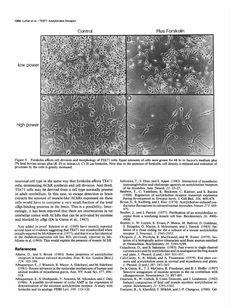
Forskolin, an adenylate cyclase activator (Seaman et al., 198 l), is reported to induce AChR phosphorylation (Grassi et al., 1987;
Table 2. Purification of AChR from 20 gm (12 roller bottles) of TE671 cells
‘=I-aBgt
AChR aBgt binding specific activity
Volume Protein sites (pmohmg Fraction (ml) (mg) pm01 % protein)
Initial extract 100 570 1804 100 3.16 Unbound to olBgt affinity column 100 380 702 39 - Wash steps 650 180 342 19 -
Affinity column eluate 6 0.100 776 43 7800

The Journal of Neuroscience, March 1989, 9(3) 1089
31-
21.5-
21.5-.--.-
C
carbamylcholine +-
Mwx103 .’
21.5-
-28s
-18s
Figure 4. Subunits of AChRs irom TE67 1. A, AChRs athnity-purified from TE67 1 and Torpedo eletric organ have similar subunits. AChRs (10 pg) were resolved into their subunits by electro- phoresis on a 10% acrylamide gel in SDS under reducing conditions and stained with Coomassie blue. B, Sub- units from TE671 AChRs correspond to those of electric organ AChRs by Western blotting. Purified TE67 1 AChR (50 rig/lane) was resolved into subunits by electrophoresis and then blotted onto DPT paper. Bach lane was incubated with the indicated antibody, mAb 61 (Tzartos et al., 1981) and mAb 111 (Tzartos et al., 1986) at 10 nM, and y and 6 anti-subunit sera (Lindstrom et al., 1979) at 1 nM. Bound antibodies were localized by autoradiography us- ing lZ51 mouse anti-rat IgG. C, Affinity labeling with 3H-MBTA identifies the (Y subunits of AChR from TE671 as forming the ACh binding site. D, Poly A+ mRNAs for the 4 subunits of TE67 1 AChR are detected by high-stringency hybridization with cDNAs for mouse muscle AChR (Y, 8, y, and 6 subunits. The cDNA probes used were described by Heinemann et al. (1986).
Miles et al., 1987; Smith et al., 1987) and to enhance AChR not be mediated by increased CAMP and may involve some desensitization (Albuquerque et al., 1986; Huganir et al., 1986; other second messenger. However, a recent report by Wagoner Middleton et al., 1986). Treatment of TE67 1 cells with forskolin and Pallotta (1988) indicates that although forskolin may stim- causes a decrease in the number of functional AChRs to back- ulate CAMP-dependent phosphorylation in intact muscle and ground levels, which is accounted for by the reduction of arBgt facilitate AChR desensitization, nonetheless, in intact muscle binding and AChR subunit mRNAs (Fig. 8). Forskolin also CAMP-dependent phosphorylation does not modulate desen- appears to inhibit cell division and result in the formation of sitization. CAMP-induced phosphorylation and desensitization extensive neuron-like projections (Fig. 9). This effect, how- of AChRs could explain the lower number of active AChRs on ever, is reversible. These results suggest that if the effects of the surface of hCGRP-treated TE67 1 cells. Forskolin treatment forskolin are mediated by an elevation in CAMP concentration, of TE67 1 cells does not induce synthesis of neuronal nicotinic then the increase in AChR induced by hCGRP in TE67 1 must AChRs since treated cells do not bind 3H-nicotine with high

MEPR
PLLL
LLCL
CSA
VLC
SE
B I
MEL
CRVL
LLIFS
AA
ALC
YE
ACL
-
ioo
i20
140
is0
is0
icm
32
0
140
160
380
300
a20
a40
380
Yl-
N4:
4 hy
droph
obic
sequ
ence
r
AChi
N-gly
cosy
lation
sit
s dis
ulfide
-linke
d cy
steine
r ne
ar the
ac
etylch
olins
bin
ding
site
Sequ
ence
re
feren
ces
TE67
1 Sc
hoep
fer
at
al.
‘88
FEBS
Le
tters
226:2
35.
ITlO
USe
Boult
er
et
al.
‘07
PNAS
84
:7763
. ca
lf No
da
et
al.
‘83
Natur
e 30
5:818
. ch
icken
Ne
f et
a
I.,
'88
EYBO
J
7:696
. Xe
nopu
s Ba
ldwin
et
al.
‘88
JCeIl
Bio
460:4
69.
Torp
edo
Noda
et
a
I. '82
Na
ture
299:7
93.
Figu
re
5.
Com
paris
on
of d
educ
ed
amin
o ac
id
sequ
ence
s fo
r AC
hR
CY
subu
nits
am
ong
spec
ies.
Am
ino
acid
s co
nser
ved
in a
ll 5
spec
ies
are
high
light
ed.
MI-M
4 in
dica
te
hydr
opho
bic
sequ
ence
s. T
he
N-g
lyco
syla
tion
site
on
O( su
buni
ts
is in
dica
ted
by th
e do
wnwa
rd
arro
w (
1).
Cys
tein
es 1
92 a
nd
193,
whi
ch
are
the
site
of a
ffini
ty l
abel
ing
by M
BTA
(Kao
et
al.,
198
4),
are
mar
ked
by A
Ch.

The Journal of Neuroscience, March 1989, 9(3) 1091
-21 -1 MetGluClyProValLeuThrLeuGlyLeuLeuAlaAlaL~~AlaValCysGlySerTrpGly
. . . ..TGGGATG~AGGGGCCAG~GCTGACACT~GGGCTGCTG~CTGCCCTGG~GGTGTGTGG~AGCTGGGG~ -60 -40 -20 -1
1 20 LeuAsnGluGluGluArgLeuIleArgHisLeuPheClnCl~LysGlyTy~As~LysGl~L~~A~gP~~V~l FTGAACGAG”AGGAGCGGCTGATCCAFCTCTTTCAAGAGAAGGGCT~CAACAAGGA~CTCCGGCCC~TG 1 20 40 60
40 AlaHisLysCluGluSerValAspValAlaLeuAlaLeuThrLeuSerAsnLe~IleS~rLeuLysGluVal CCACACA?AGAGGAGAGTGTGGACGTT~CCCTGGCCCTtACACTCTC~AACCTCATC?CCCTGAAAG~AGTT
ee 100 120 140 60
GluGluThrLauThrThrAsnValTrpIleCluHisGlyT~pTh~AspAsnArgL~uLysTrpAsnAl~Glu GAGGA~ACCCTCACT~CCAATGTGT~GATAGAGCAGGCTGGACA~ACAACCGGC~GAAGTGGAA~GCTGAA
160 180 200
790 --_ 240 260 260 100 120
AsnAsnAspGlySerPheGlnIleSerTyrSerCysAsnV~lL~~ValTyrHisTyrGlyPheValTyrTrp AACAATGACGGCTCCTTCCAGATCTCCTACTFCTGCAACCT~CTTGTCTAC~ACTACGGCT?CGTGTACTG~
308 328 348 360 140
380 400 420 160
SerLeuLysPheSerSsrLeuLysTyrThrAlaLysGl~Il~ThrL~uS~rLe~LysGlnAspAlaLysGlu TCCCTCAAGTTCAGTTCCCTCAAGTAT?CCGGCCAAAG~GATCACCCT~AGCCTGAAA~AGGATGCCA~GGAG
448 468 480 500 180
AsnArgThrTyrProValGluTrpIleIleIleAspProGluGlyPh~ThrGluAs~GlyGluTrpGluIle AACCGCACCTACCCCCTGGAGTGGA?CATCATTGA~CCTGAAGGC~TCACAGAGA~CGGGGAGTG~GAGATA
520 540 560 200
ValHisArgProAlaArgValAsnValAspProArgAlsProLeuAspSerProSerArgGlnAspIl~Thr GTCcACCGGCCGGcCAGGGTCAA~GTGGACCCC~GAGCCCCTC?GGACAGCCC~AGCCGCCAG~ACATCACC
560 600 620 640 220 240
PheTyrLsuIleIlsArgArgLysProLeuPheTyrIleIleAsnIl~LeuV~lPreCysV~lLeuIleS~r TTCTACCTCATCATCCGCCGCAAGCCCCTCTTCTACATCATCAACATCCTGGTGCCCTGCG~GCTCATCTC~
666 sse 708 728 260
PheMetValAsnLeuVa$heTyrLeuProAlaAspSerGlyGluLysThrSerValAlaIleSerValLeu TTCATGGTC?ACCTGGTCTTCTACCTACC~GCTGACAGT~GTGAGAAGA~ATCAGTGGC~ATCTCGGTG~TC
740 760 760 280
LeuAlaGlnSsrVslPheLeuLeuLsuIleSerLysArgL~uProAl~ThrS~~M~tAlaIl~ProL~uIle CTCGCTC?GTCTGTCTTFCTGCTGCTC~TCTCCAAGC~TCTGCCTGC~ACATCCATG~CCATCCCCC?TATC
800 620 840 860 300
GlyLyoPheLeuLeuPheGlyMatValLeuValThrMetVaiValValIl~CysV~lIl~ValL~uAsnIle GGCAAcTTCCTGCTCTTCGGCATGG?GCTGGTCAC~ATGGTTGTG~TGATCTGTG?CATCGTGCT~AACATC
880 900 920 320
HisPheArgThrProSerThrHisV~lLeuSerCluClyValLysLysL~~Ph~Le~GluThrLeuProGlu CACTTCCGAACACFCAGCACCCA?GTGCTGTCTGACGCCGTCA~GAAGCTCTT~CTGGAGACC~TGCCGGAG
940 980 980 1000 340 360
LsuLeuHisMetSerArgProAlnCluAspGlyProSerProGlyAlaLeuV~lArgArgSerS~rS~rL~u CTCCTGCACATCTCCCGCCCA~CAGAGGATG~ACCCAGCCC?GGGGCCCTG~TGCGGAGGA~CAGCTCCCT~
1020 1040 1060 1060 380
GlyTyrIleSerLysAl~GIuGluTy~Ph~L~~L~~LysS~~ArgSerAspL~~M~tPh~Gl~LysGinSer GGATACATC~CCAAGGCCG?GGAGTACTT~CTGCTCAAG~CCCGCAGTG~CCTCATGTT~GAGAAGCAG?CA
1100 1120 1140 400
GluArgHisGlyLeuAl~ArgArgLeuThrThrAl~A~gA~gP~~ProAl~S~~S~~Gl~Gl~Al~Gl~Gl~ GAGCGGC~TGGGCTGGC~AGGCGCCTC~CCACTGCAC~CCGGCCCCC~GCAAGCTCT~AGCAGGCCC~GCAG
11aa 1160 1200 1220 420
GluLeuPheAsnGluLeuLysP~oAlaVslAspGlyAlaAs~Ph~Il~ValAsnHisM~tArgAspGlnAsn GAACTFTTCAATGAG’TGAAGCCGG~TGTGGATGG~GCAAACTTC~TTGTTAACC~CATGAGGGA~CAGAAC
1240 1260 1280 440
AsnTyrAsnGluGluLy.AspSerTrpAsnArgVslAl~ArgThrValAspArgL~uCysLouPh~ValVal AATTACAATGAGG?GAAAGACAG~TGGAACCGACTCCtCCGCA~AGTGGACCG~CTCTGCCTG~TTGTGGTG 1308 1320 1340 1360
460 460 ThrProValMetV.IValGIyThrAlsTrpIl~PheLeuClnGlyV~lTyrAsnGlnProProProGlnPro ACGCCTGTCATGGTGGTGGGCACAGCCTGGATCTTCCTGCAGGGCGTTTAC~ACCAGCCAC~ACCCCAGCC~
1368 1408 1428 144a
PheProGlyAspProTyrSerTyrAsnValGlnAspLysArgPheIle TTTCCTGGGCACCCCTACTFCTACAACGT~CAGGACAAG~GCTTCATCT~GGGTGGGCC?GTTGGGGAG~CA
1460 1480 1500
GGAGACACCAGGGTCTG?GAGAGGAGC~ACAGTCCCT~ATGACACCC~CTCCTAGCC~TGAGGCTCG~GCCC
1620 1540 1660 1580
CTCAG?CTGGGGAAG?GTCCAAGGA~GGGAGGGAG~AGCCACTCC?CAATGCTCA~TGGCTCCCC?GAAATC 1600 1620 1640
AAG?CAGGGGCCA~CCGAGG.....
1660
Figure 6. Nucleotide sequence and deduced amino acid sequence of a TE671 cDNA clone coding for the AChR 6 subunit. The mature protein starts at position + 1. The cDNA clone 6.4 untranslated region extends 124 nu- cleotides further upstream. This por- tion of the sequence contains unrelated sequences revealed by Northern blot analysis (data not shown).

Lead
er pe
ptide
t
TE67
1 (1
) M
EGPV
LTLG
YO
USE
(2)
MAG
PVLT
LG
XENO
PUS
(6)
TORP
EOO
(8
) M
GN
IHFV
i00
:20
:40
is0
i80
100
120
Yl
u2
u3
Pi
DS
DC
DC
DS
DS
ES
$40
200
$80
3.00
82
0 $4
0 36
0
Pi
Pi
Y4
-Rm
AR
RPPA
S--S
EqAG
GEBN
EIKP
AV~A
~~HM
RD~~
DSL3
NRVA
ROV~
CL~V
WM
VV~
E
ARRP
PAS-
-SEQ
V(.
ARRP
PAG-
-SEQ
AQQE
SE
LKPA
VI
& AR
FAPA
AT-S
EQ---
- DH
LDPT
LI
380
$00
420
440
$60
180
Ml
- u4
: hy
droph
obic
sequ
ence
s Se
quen
ce
refer
ence
s Pi
: po
tentia
l ph
osph
orylat
ion
sites
m
ouse
La
Polla
et
al.
1 ‘8
4 PN
AS
81~7
970.
ti po
tentia
l N-
glyco
sylat
ion
sites
in
TE67
1 ca
lf Ku
bo
et
al.
‘65
EurJB
ioche
m
149:~
. pre
sum
ed
site
of
AChR
ch
icken
N
ef
et
al.
‘84
PNAS
81
:7975
. dim
eriza
tion
in To
rped
o Xe
nopu
s Ba
ldwin
et
al.
‘66
JCeII
Bio
166:4
69.
Torp
edo
Noda
et
al.
‘63
Na
ture
381:2
51.
2-29-8
8
Figu
re
7.
Com
paris
on
of d
educ
ed
amin
o ac
id s
eque
nces
for
AC
hR
6 su
buni
ts
amon
g va
rious
sp
ecie
s. N
umbe
rs
indi
cate
am
ino
acid
pos
ition
with
in
Torp
edo
sequ
ence
. Am
ino
acid
s co
nser
ved
in a
ll 6
spec
ies
are
high
light
ed.
Ml-M
4 in
dica
te
hydr
opho
bic
sequ
ence
s; p
oten
tial
phos
phor
ylat
ion
site
s ar
e in
dica
ted
by P
i; po
tent
ial
N-g
lyco
syla
tion
site
s ar
e in
dica
ted
by
the
down
ward
ar
row
( 1).
The
penu
ltimat
e cy
stei
ne,
which
is
thou
ght
to b
e th
e si
te o
f AC
hR
dim
eriza
tion
in
Torp
edo,
is
poi
nted
ou
t by
the
upwa
rd
arro
w (
t ).

The Journal of Neuroscience, March 1999, 9(3) 1093
Subunit Northern Blot Intensity
Treatment
forskolin
nicotine
CGRP
dexo- nethosone
Control Forskolin Nicotine G9PW (1mM)
e, fm
n.d.
1
Subunit mRNA
cx B Y 6
t t t t
--+I ?-
---c t-
+ t --
AChR Protein
(aBgt/cell)
t
t
t
t
CGRP Dexa- (o.I~M) methasone
(2.3~)
II Q
P
Y
6
AChR Function
(flux/cett)
i
t
t
t
Specific Activity
(flux/aBgt)
-
+
t
t
affinity or mAbs specific for human brain AChRs. Also, hy- bridization signals are not detected when northern blots of TE67 1 RNA are hybridized at high stringency with the cDNA probe (~4 (Goldman et al., 1987); this probe codes for the ACh- binding subunit of the AChR from rat brain, which has high affinity for nicotine. It is possible that forskolin may induce the synthesis of some other neuronal receptors or AChRs, but these have not yet been detected.
Concluding remarks
We have proven that extrajunctional muscle-like AChRs are expressed in relatively large amounts by the human neuronal cell line TE67 1. This cell line is a valuable system for studying the structure of human muscle AChR. AChR expression in these cells can be regulated by several factors, which can be employed to study in detail the mechanisms regulating AChR expression in TE671 cells.
This cell line may prove valuable for studies of MG because (1) it provides a much larger and more uniform source of human
Potential Primary
Regulatory Mechanism
transcription t
post transcription t
post transcription t post translotion +
transcription + post translotion 4
Figure 8. Nicotine, dexamethasone, CGRP, and forskolin affect AChR expression in TE671 cells. First, 1 x lo5 cells were plated in each 3.5 cm dish on day 0. On day 2, media was supple- mented as indicated. On day 4, carba- mylcholine-induced esRb+ influx was measured on sister triplicate cultures. Background was determined for each culture conditions and subtracted to give the values shown (average background, 500 cpm). Northern blots using equal amounts oftotal RNA from other sister cultures were probed successively with )*P-labeled mouse (Y, mouse 8, mouse y, and human 8 cDNAs. 1*51-aBgt bind- ing to cell surfaces was measured in a series of cultures.
AChR for use in diagnostic immunoassays than does amputated leg muscle; (2) it can be used more effectively to study antigenic modulation or inhibition of AChR function by MG autoanti- bodies than can primary cultures of human fetal muscle (which are difficult to obtain) or rodent muscle cell lines (which cross- react poorly); and (3) it is a good source of human muscle AChR subunit cDNAs, which in suitable expression systems may prove valuable for mapping the antigenic structure of human AChR and providing antigen for studies of specific immunosuppres- sion of MG.
Why should muscle-like AChRs be expressed in a neuronal cell line? We have considered 3 possibilities. First, expression of muscle AChR may be an aberration induced by the trans- formation events that produced this tumor line. Second, TE67 1 cells may be derived from a neuronal cell type that transiently expresses muscle-like AChRs during development. This would explain why muscle-like AChRs are not detected in extracts of adult human brains, and one could imagine that some devel- opmental inducer could normally affect the development of this
1094 Luther et al. * TE671 Acetylcholine Receptor
Control Plus Forskolin
low powel
high power
Figure 9. Forskolin affects cell division and morphology of TE671 cells. Equal amounts of cells were grown for 48 hr in Iscove’s medium plus 5% fetal bovine serum plus (B, D) or minus (A. C’) 20 PM forskolin. Note that in the presence of forskolin, cell density is reduced and extension of processes by the cells is greatly increased.
neuronal cell type in the same way that forskolin affects TE67 1 cells, terminating AChR synthesis and cell division. And third, TE67 1 cells may be derived from a cell type normally present in adult cerebellum. In this case, to escape detection in brain extracts the amount of muscle-like AChRs expressed on these cells would have to comprise a very small fraction of the total aBgt-binding proteins in the brain. This is a possibility. Inter- estingly, it has been reported that there are interneurons in rat cerebellar cortex with AChRs that can be activated by nicotine and blocked by olBgt (De la Garza et al., 1987).
Note added in proof Stratton et al. (1989) have recently reported several lines of evidence suggesting that TE67 1 was misidentified when initially reported by McAllister et al. (1977), and that it is in fact identical to the rhabdomyosarcoma cell line RD, previously reported by Mc- Allister et al. (1969). This would explain the presence of muscle AChR.
References Adams, D., and S. Bevan (1985) Some properties of acetylcholine
receptors in human cultured myotubes. Proc. R. Sot. London [Biol.] 224: 183-196.
Albuquerque, E., J. Wamick, R. Mayer, A. Eldefmwi, and M. Eldefrawi (198 1) Recent advances in the molecular mechanisms of human and animal models of myasthenia gravis. Ann. NY Acad. Sci. 377: 496- 518.
Albuquerque, E., S. Deshpande, V. Aracawa, M. Alkondon, and J. Daly (1986) A possible involvement of cyclic AMP in the expression of desensitization of the nicotinic acetylcholine receptor. A study with forskolin and its analogs. FEBS Lett. 199: 113-l 20.
Ashizawa, T., S. Elias, and S. Appel (1982) Interaction of myasthenic immunoglobulins and choline& agonists on acetylcholine receptors of rat myotubes. Ann. Neurol. 11: 22-27.
Baldwin, T., C. Yashihara, K. Blackmer, C. Kintner, and S. Burden (1988) Regulation of acetylcholine receptor transcript expression during development in Xenopus luevis. J. Cell Biol. 106: 469-478.
Bevan, S., R. Kullberg, and J. Rice (1978) Acetylcholine-induced con- ductance fluctuations in cultured human myotubes. Nature 273: 469- 470.
Boulter, J., and J. Patrick (1977) Purification of an acetylcholine re- ceptor from a nonfusing muscle cell line. Biochemistry 16: 4900- 4908.
Boulter, J., W. Luyten, K. Evans, P. Mason, M. Ballivet, D. Goldman, S. Stengelin, G. Martin, S. Heinemann, and J. Patrick (1985) Iso- lation of a clone coding for the 01 subunit of a mouse acetylcholine receptor. J. Neurosci. 5: 2545-2552.
Chirgwin, J., A. Pryzbyla, R. MacDonald, and W. Rutter (1979) Iso- lation of a biologically active ribonucleic acid from sources enriched in ribonuclease. Biochemistry 18: 5294-5299.
Colquhoun, D., and B. Sakmann (1985) Fast events in single channel currents activated by acetylcholine and its analogues at the frog muscle end plate. J. Physiol. (Lond.) 369: 501-557.
Cull-Candy, S., R. Miledi, and A. Trautmann (1979) End plate cur- rents and acetylcholine noise at normal and myasthenia end plates. J. Physiol. (Lond.) 287: 247-265.
De la Garza, R., T. J. McGuire, R. Freedman, and B. J. Hoffer (1987) Selective antagonism of nicotine actions in the rat cerebellum with cYBungarotoxin. Neuroscience 23: 887-89 1.
Einarson, B., W. Gullick, B. Conti-Tronconi, and J. Lindstrom (1982) Subunit composition of fetal calf muscle nicotinic acetylcholine re- ceptor. Biochemistry 21: 5295-5302.
Fontaine, B., A. Klarsfeld, T. H&felt, and J.-P. Changeux (1986) Cal-

The Journal of Neuroscience, March 1989, 9(3) 1095
citonin gene-related peptide, a peptide present in spinal cord motor neurons, increases the number of acetylcholine receptors in primary cultures of chick embryo myotubes. Neurosci. Lett..71: 59-65.
Fontaine. B.. A. Klarsfeld. and J.-P. Chanaeux (1987) Calcitonin aene- related’ peptide and muscle activity regulate acetylcholine receptor a-subunit mRNA levels by distinct intracellular pathways. J. Cell Biol. 105: 1337-1342.
Goldman, D., E. Deneris, W. Luyten, A. Kochar, J. Patrick, and S. Heinemann (1987) Members of a nicotinic acetylcholine receptor gene family are expressed in different regions of the mammalian cen- tral nervous system. Cell 48: 965-973,
Grassi, F., L. Monaco, and F. Eusebi (1987) Acetylcholine receptor channel properties in rat myotubes exposed to forskolin. Biochem. Biophys. Res. Commun. 147: 1000-1007.
Gullick, W., and J. Lindstrom (1982) The antigenic structure of the acetylcholine receptor from Torpedo californicu. J. Cell. Biochem. 19: 223-230.
Heinemann, S., J. Boulter, J. Connolly, D. Goldman, K. Evans, D. Treco, M. Ballivet, and J. Patrick (1986) Molecular biology of mus- cle and neural acetylcholine receptors. In Nicotinic Acetylcholine Re- ceptor Structure and Function, A. Maelicke, ed., pp. 360-387, Spring- er-Verlag, Heidelberg.
Huganir, R., A. Delcour, P. Greengard, and G. Hess (1986) Phos- phorylation of the nicotinic acetylcholine receptor regulates its rate of desensitization. Nature 321: 774-777.
Kao, P., and A. Karlin (1986) Acetylcholine receptor binding site contains a disulfide crosslink between adjacent half-cystinyl residues. J. Biol. Chem. 261: 8085-8088.
Kao, P., A. Dwork, R. Kaldany, M. Silver, J. Wideman, S. Stein, and A. Karlin (1984) Identification of the (Y subunit half cystine specif- ically labeled by an affinity reagent for the acetylcholine receptor bindina site. J. Biol. Chem. 259: 11662-l 1665.
Kohn J.,&rd M. Wilchek (1982) A new approach (cyano-transfer) for cyanogen-bromide activation of sepharose at neutral pH, which yields activated resins free of interfering nitrogen derivatives. Biochem. Bio- whys. Res. Commun. 107: 878-884.
Kubo, T., M. Noda, T. Takai, T. Tanabe, T. Kayano, S. Shimizu, K. Tanaka. H. Takahashi. T. Hirose. S. Inavoma. R. Kikuno. T. Mivata. and S. Numa (1985)’ Primary structure of ‘a subunit precursor of calf muscle acetylcholine receptor deduced from cDNA sequence. Eur. J. Biochem. 149: 5-13.
Labarca, P., J. Lindstrom, and M. Montal (1984) Acetylcholine re- ceptor in planar lipid bilayers: Characterization of the channel prop- e&es of the purified nicotinic acetylcholine receptor from Torpedo californica in reconstituted planar linid bilavers. J. Gen. Phvsiol. 83: 473496.
Labarca, P., J. Rice, D. Fredkin, and M. Montal (1985) Kinetic anal- ysis of channel gating: Application to the choline@ receptor channel and the chloride channel from Torpedo calzjbrnica. Biophys. J. 47: 469-479.
Laemmli, U. (1970) Cleavage of structural proteins during the assem- bly of the head of bacteriophage T4. Nature 227: 680-685.
La Polla, R., K. Mayne, and N. Davidson (1984) Isolation and char- acterization of a cDNA clone for the complete protein coding region of the (Y subunit ofthe mouse acetylcholine receptor. Proc. Natl. Acad. Sci. USA 81: 7970-7974.
Lindstrom, J., B. Walter, and B. Einarson (1979) Immunochemical similarities between subunits ofacetylcholine receptors from Torpedo, Electrophorus, and mammalian muscle. Biochemistry 18: 4470-4480.
Lindstrom. J., W. Gullick. B. Conti-Tronconi. and M. Ellisman (1980) Proteolytic’nicking of the acetylcholine receptor. Biochemistry 16 4791-4795.
Lindstrom, J., M. Criado, M. Ratnam, P. Whiting, S. Ralston, J. Rivier, V. Satin, and P. Sargent (1987a) Using monoclonal antibodies to determine the structures of acetylcholine receptors from electric or- gans, muscles, and neurons. Ann. NY Acad. Sci. 505: 208-225.
Lindstrom, J., R. Schoepfer, and P. Whiting (1987b) Molecular studies of the neuronal nicotinic acetylcholine receptor family. Mol. Neu- robiol. 1: 281-337.
Lindstrom, J., G. D. Shelton, and Y. Fujii (1988) Myasthenia gravis. Adv. Immunol. 42: 233-284.
Lipton, S., E. Aizenman, and R. Loring (1987) Neural nicotinic ace- tylcholine responses in solitary mammalian retinal ganglion cells. Pfluegers Arch. 410: 37-43.
Lukas, R. (1986a) Characterization ofcuraremimetic neurotoxin bind-
ing sites on membrane fractions derived from the human medullo- blastoma clonal line TE67 1. J. Neurochem. 46: 1936-l 94 1.
Lukas, R. (1986b) Immunochemical and pharmacological distinctions between curaremimetic neurotoxin binding sites of central, auto- nomic, and peripheral origin. Proc. Natl. Acad. Sci. USA 83: 574 l- 5745.
Margiotta, J., D. Berg, and V. Dionne (1987) The properties and regulation of functional acetylcholine receptors on chick ciliary gan- glion neurons. J. Neurosci. 7: 3612-3622.
McCallister, R. M., J. Melnyk, J. Z. Finklestein, E. C. Adams, and M. B. Gardner (1969) Cultivation in vitro of cells derived from a human rhabdomyosarcoma. Cancer 24: 520-526.
McAllister, R., H. Isaacs, R. Rongey, M. Peer, W. Au, S. Sonkup, and M. Gardner (1977) Establishment of a human medulloblastoma cell line. Int. J. Cancer 20: 206-212.
Middleton, P., F. Jaramillo, and S. Scheutze (1986) Forskolin increases rate of acetylcholine receptor desensitization at rat soleus end plates. Proc. Natl. Acad. Sci. USA 83: 4967-4971.
Miles, K., D. Anthony, L. Rubin, P. Greengard, and R. Huganir (1987) Regulation of nicotinic acetylcholine receptor phosphorylation in rat myotubes by forskolin and CAMP. Proc. Natl. Acad. Sci. USA 84: 6591-6595.
Mishina, M., T. Takai, K. Imoto, M. Noda, T. Takahashi, S. Numa, C. Methfessel. and B. Sakmann (1986) Molecular distinction be- tween fetal and adult forms of muscle acetylcholine receptor. Nature 321: 406-411.
Momoi, M., and V. Lennon (1982) Purification and biochemical char- acterization of nicotinic acetylcholine receptors of human muscle. J. Biol. Chem. 257: 12757-12764.
Montal. M.. R. Anholt. and P. Labarca (1986) The reconstituted ace- tylcholine receptor. In Zon Channel Re~onstiiution, C. Miller, ed., pp. 157-204, Plenum, New York.
Nef, P., A. Mauron, R. Stalder, C. Alliod, and M. Ballivet (1984) Structure, linkage, and sequence of the two genes encoding the 01 and y subunits of the nicotinic acetylcholine receptor. Proc. Natl. Acad. Sci. USA 81: 7975-7979.
Neher, E., and B. Sakmann (1976) Single-channel currents recorded from membrane of dennervated frog muscle. Nature 260: 799-801.
Noda. M.. H. Takahashi. T. Tanabe. M. Tovosato. Y. Furutani. T. Hirose, M. Asai, S. Inayama, T. Miyata, and S. Numa (1982) Pri- mary structure of a-subunit precursor of Torpedo californica acetyl- choline receptor deduced from cDNA sequence. Nature 299: 793- 797.
Noda, M., Y. Furutani, H. Takahashi, M. Toyosato, T. Tanabe, S. Shimizu, S. Kikyotani, T. Kayano, T. Hirose, S. Inayama, and S. Numa (1983a) Cloning and sequence analysis of calf cDNA and human genomic DNA encoding 01 subunit precursor of muscle ace- tylcholine receptor. Nature 305: 8 18-823.
Noda, M., H. Takahashi, T. Tanabe, M. Toyosato, S. Kikyotani, Y. Furutani, T. Hirose, H. Takashima, S. Inayama, T. Miyata, and S. Numa (1983b) Structural homology of Torpedo californicu acetyl- choline receptor subunits. Nature 302: 528-532.
Noda, M., H. Takahashi, T. Tanabe, M. Toyosato, S. Kikyotani, T. Hirose, M. Asai, H. Takashima, S. Inayama, T. Miyata, and S. Numa ( 1983~) Primarv structures of B- and &subunit precursor of Toroedo >aliforkca acetylcholine receptor deduced from cDNA sequences. Nature 301: 25 l-255.
Oakley, B., D. Drisch, and R. Morris (1980) A simplified ultrasensitive silver stain for detecting proteins in polyacrylamide gels. Anal. Bio- them. 105: 361-363.
Raftery, M., M. Hunkapillar, C. Strader, and L. Hood (1980) Ace- tylcholine receptor: Complex of homologous subunits. Science 208: 1454-1457.
Robinson, D., and R. McGee (1985) Agonist-induced regulation of the neuronal nicotinic acetylcholine receptor of PC1 2 cells. Mol. Phar- macol. 27: 409-4 17.
Sakmann, B., and E. Neher (1983) Single Channel Recording, Plenum, New York.
Sanger, F., S. Nicklen, and A. R. Coulson (1977) DNA sequencing with chain-terminating inhibitors. Proc. Natl. Acad. Sci. USA 74: 5463-5467.
Schoepfer, R., M. Luther, and J. Lindstrom (1988) The human medul- loblastoma cell line TE67 1 expresses a muscle-like acetylcholine re- ceptor: Cloning of the (Y subunit cDNA. FEBS Lett. 226: 235-240.
Schubert, D., A. J. Harris, C. E. Devine, and S. Heinemann (1974)

1096 Luther et al. * TE671 Acetylcholine Receptor
Characterization of a unique muscle cell line. J. Cell Biol. 61: 398- 413.
Schuetze, S., and L. Role (1987) Developmental regulation of nicotine acetylcholine receptors. Annu. Rev. Neurobiol. 10: 403-457.
Schuetze, S., S. Vicini, and Z. Hall (1985) Myasthenic serum selec- tively blocks acetylcholine receptors with long channel open times at developing rat endplates. Proc. Natl. Acad. Sci. USA 82: 2533-2537.
Seaman, K., W. Padgett, and J. Daly (198 1) Forskolin: Unique diter- pene activator of gdenylate cycl&e in membranes and intact cells. Proc. Natl. Acad. Sci. USA 6: 3363-3367.
Siegel, H. N., and R. J. Lukas (1988) Nicotinic agonists regulate a-Bungarotoxin binding sites of TE67 1 human medulloblastoma cells. J. Neurochem. 50: 1272-1278.
Sine, S. (1988) Functional properties of human skeletal muscle ace- tylcholine receptors expressed by the TE67 1 cell line. J. Biol. Chem. 263: 18052-l 8062.
Sine, S., and J. H. Steinbach (1984) Activation of nicotinic acetyl- choline receptor. Biophys. J. 45: 175-l 85.
Smith, M., J. Margiotta, A. France, Jr., J. Lindstrom, and D. Berg (1986) Cholinergic modulation of an acetylcholine receptor-like an- tigen on the surface of chick ciliary ganglion neurons in cell culture. J. Neurosci. 6: 946-953.
Smith, M., J. Merlie, and J. Lawrence (1987) Regulation of phosphor- ylation of nicotinic acetylcholine receptors in mouse BC3H- 1 myo- cytes. Proc. Natl. Acad. Sci. USA 84: 6601-6605.
Stratton, M. R., B. R. Reeves, and C. S. Cooper (1989) (Letter to the Editor) Nature 337: 3 11-312.
Syapin, P., P. Salvaterra, and J. Engelhardt (1982) Neuronal-like fea- tures of TE67 1 cells: Presence of a functioning nicotinic cholinergic receptor. Brain Res. 231: 365-377.
Tumbull, G., R. Harrison, and G. Lunt (1985) Nicotinic acetylcholine receptor from foetal human skeletal muscle. Int. J. Dev. Neurosci. 3: 123-134.
Tzartos, S., D. Rand, B. Einarson, and J. Lindstrom (198 1) Mapping of surface structures on electrophorus acetylcholine receptor using monoclonal antibodies. J. Biol. Chem. 256: 8635-8645.
Tzartos, S., S. Hochschwender, L. Langeberg, and J. Lindstrom (1983) Demonstration of a main immunogenic region on acetylcholine re- ceptors from human muscle using monoclonal antibodies to human receptor. FEBS Lett. 158: 116-l 18.
Tzartos, S., L. Langeberg, S. Hochschwender, L. Swanson, and J. Lind- Strom (1986) Characteristics of monoclonal antibodies to denatured Torpedo and to native calf acetylcholine receptors: Species, subunit, and region specificity. J. Neuroimmunol. IO: 235-253.
Tzartos, S., S. Hochschwender, P. Vasquez, and J. Lindstrom (1987) Passive transfer of experimental autoimmune myasthenia gravis by monoclonal antibodies to the main immunogenic region of the ace- tylcholine receptor. J. Neuroimmunol. 15: 185-l 94.
Wagoner, P., and B. Pallotta (1988) Modulation of acetylcholine re- ceptor desensitization by forskolin is independent of CAMP. Science 240: 1655-1657.
Weinberg, C., and Z. Hall (1979) Antibodies from patients with myas- thenia gravis recognize determinants unique to extraiunctional ace- tylcholine receptors. Proc. Natl. Acad. SC; USA 76: 304-508.
Wennogle. L.. R. Oswald. T. Saitch. and J.-P. Channeux (198 1) Dis- section bf ihe 66,000 dalton sub&it of the acet&holhe receptor. Biochemistry 20: 2492-2497.
Whiting, P., and J. Lindstrom (1987) Affinity labeling of neuronal acetylcholine receptors localizes the neurotransmitter binding site to the 8 subunit. FEkS Lett. 213: 55-60.
Whiting, P., and J. Lindstrom (1988) Characterization of bovine and human neuronal nicotinic acetylcholine receptors using monoclonal antibodies. J. Neurosci. 8: 3393-3404. -
Whiting, P., A. Vincent, M. Schluep, and J. Newsom-Davis (1986) Monoclonal antibodies that distinguish between normal and dener- vated human acetylcholine receptor. J. Neuroimmunol. 1 I: 223-235.
Whiting, P., J. Cooper, and J. Lindstrom (1987) Antibodies in sera from patients with myasthenia gravis do not bind to acetylcholine receptors from human brain. J. Neuroimmunol. 16: 205-2 13.
Witzemann, V., B. Barg, Y. Nishikawa, B. Sakmann, and S. Numa (1987) Differential regulation of muscle acetylcholine receptor y and t subunit mRNAs. FEBS Lett. 223: 104-I 12.