Embed Size (px)
Citation preview

Clinical StudyA Modified Translaminar Osseous Channel-AssistedPercutaneous Endoscopic Lumbar Discectomy forHighly Migrated and Sequestrated Disc Herniations ofthe Upper Lumbar: Clinical Outcomes, Surgical Indications,and Technical Considerations
Zhijun Xin, Wenbo Liao, Jun Ao, Jianpu Qin, Fang Chen, Zhiyuan Ye, and Yuqiang Cai
Department of Spinal Surgery, The First Affiliated Hospital of Zunyi Medical College, Zunyi, China
Correspondence should be addressed to Wenbo Liao; [email protected]
Received 20 January 2017; Accepted 22 February 2017; Published 30 March 2017
Academic Editor: Alessandro Landi
Copyright © 2017 Zhijun Xin et al. This is an open access article distributed under the Creative Commons Attribution License,which permits unrestricted use, distribution, and reproduction in any medium, provided the original work is properly cited.
Objective is to describe a safe and effective percutaneous endoscopic approach for removal of highly migrated and sequestrateddisc herniations of the upper lumbar spine and to report the results, surgical indications, and technical considerations of thenew technique. Eleven patients who had highly migrated and sequestrated disc herniations in the upper lumbar were included inthis study. A retrospective study was performed for all patients after translaminar osseous channel-assisted PELD was performed.Radiologic findings were investigated, and pre-and postoperative visual analog scale (VAS) assessments for back and leg pain andOswestry disability index (ODI) evaluations were performed. Surgical outcomes were evaluated under modified MacNab criteria.All of the patients were followed for more than 1 year. The preoperative and postoperative radiologic findings revealed that thedecompression of the herniated nucleus pulposus (HNP) was complete. After surgery, the mean VAS scores for back and leg painimmediately improved from 8.64 (range, 7–10) and 8.00 (range, 6–10) to 2.91 (range, 2–4) and 2.27 (range, 1–3), respectively. Themean preoperative ODI was 65.58 (range, 52.2–86), which decreased to 7.51 (range, 1.8–18) at the 12-month postoperative follow-up. The MacNab scores at the final follow-up included nine excellent, one good, and one fair. The modified translaminar osseouschannel-assisted PELD could be a safe and effective option for the treatment of highly migrated and sequestrated disc herniationsof the upper lumbar.
1. Introduction
Upper lumbar disc herniation is disc herniation at the L1-L2,L2-L3, or L3-L4 level, accounting for approximately 5% of alllumbar disc herniations [1]. Due to the special anatomicalstructure and functional characteristics of the upper lumbarspine, such as the narrow spinal space and the smaller rangeof motion, the dural sac and nerve structure in the spinalcanal of the upper lumbar are more likely to be compressed,which is more likely to manifest as multiple neurologicaldisorders rather than being limited to the involvement of acertain nerve [2]. Therefore, the clinical therapeutic outcomeof this type of herniation is oftenworse than those of the lowerlumbar [3]. At the same time, since this disease is difficult to
treat conservatively to reach remission, when the diagnosisis clear, surgical treatment is frequently recommended [4].However, due to the special anatomical and functionalcharacteristics of the high lumbar spine, selection of theappropriate surgical treatment strategy is difficult, especiallywhen the disc herniation is highly migrated and sequestrated(i.e., located beneath the pars interarticularis medial to thepedicle). In these cases, the selection of the surgical methodis even more difficult, even for experienced spinal surgeons[5, 6].
Surgical treatments for highly migrated and sequestrateddisc herniation of the upper lumbar include the traditionalposterior laminectomy and the percutaneous endoscopictechnique.However, posterior laminectomy often leads to the
HindawiBioMed Research InternationalVolume 2017, Article ID 3069575, 7 pageshttps://doi.org/10.1155/2017/3069575

2 BioMed Research International
Table 1: Clinical and neuroimaging characteristics in eleven patients with highly migrated and sequestrated disc herniations of the upperlumbar∗.
Case number Age (yrs) Sex Back pain Radicular pain Motor deficit Sensory deficit FNST SLRT Level of CM Level of DH1 65 F + rt leg rt L-1 SD rt L-1, L-2 SD + − L-1 body L1-22 53 F + rt leg rt L-3 SD rt L-3 SD + + L1-2 disc L3-43 31 M + Both legs Both L-2 SD Both L-2 SD + − L-1 body L2-34 45 F + lt leg − lt L-3 SD − − L-1 body L3-45 37 F + rt leg − rt L-1, L-2 SD + − L1-2 disc L1-26 43 M + Both legs − − − − L-1 body L2-37 55 F + rt leg rt L-1 SD rt L-1 SD + − L-1 body L1-28 47 M + Both legs Both L-2 Both L2 SD + − L-1 body L1-29 32 M + lt leg lt L-3 lt L3, L4 SD − + L-1 body L3-410 59 F + Both legs Both L-2 SD Both L-2 SD + − L1-2 disc L2-311 62 F + lt leg − − + − L-1 body L1-2∗No patient had bladder or bowel dysfunction.CM = conus medullaris; DH = disc herniation; FNST = femoral nerve stretch test; SD = sensory dermatome; SLRT = straight leg raising test; − = absent; + =present.
destruction of the stability of the motion segments due to itsresection of the lamina, the isthmus, and the intervertebralfacet joints; further, this procedure can induce postoperativeback pain and complications [5–8]. Although the percu-taneous endoscopic lumbar discectomy (PELD) techniqueis recommended for the treatment of highly migrated andsequestrated disc herniations of the upper lumbar regiondue to its numerous advantages and accelerated patientrecovery [9], currently, most PELD techniques are developedfrom techniques based on Kambin’s transforaminal approachor a translaminar approach. These traditional endoscopicapproaches have many limitations in the treatment of highlymigrated and sequestrated disc herniations of the upperlumbar. Since traditional PELD approaches have the short-comings of insufficient exposure, inevitable damage to theintervertebral joints, and vision limitation so that the surgeoncannot achieve excision of the herniated nucleus pulposus(HNP), some spinal surgeons suggest that this surgicalmethod is not suitable for the treatment of highly migratedand sequestrated disc herniations of the upper lumbar [4, 5,10].
To our knowledge, at present, there are few relevantstudies on PELD techniques for the treatment of highlymigrated and sequestrated disc herniations of the upperlumbar; furthermore, the clinical outcome is also not ideal.Therefore, based on our practical clinical experience in thetreatment of highly migrated and sequestrated disc hernia-tions of the upper lumbar using an improved PELD approach,we present a novel technique: translaminar osseous channel-assisted PELD.Thepresent studywill elaborate this techniquefrom the aspects of clinical efficacy, indications, and technicalreferences.
2. Patients and Methods
2.1. Inclusion and Exclusion Criteria. The inclusion criteriawere patients with highly migrated and sequestrated discherniations, whichwas displaced away from the extrusion site
and greater than the posterior marginal disc height measuredfrom the adjacent endplate level of the upper body [4, 5,10–12], at L1-L2, L2-L3, or L3-L4 level as demonstrated bycomputed tomography (CT) and magnetic resonance imag-ing (MRI); single-segment disc herniation; and unsuccessfulconservative treatment for at least 6 weeks. The exclusioncriteria were the presence of spinal stenosis; clear instabilitiesor deformities; chronic discogenic back pain; painless motorweakness and pyogenic discitis.
11 patients (7 female, 4male) who were operated on at ourhospital between April 2014 and January 2015 and underwenta translaminar osseous channel-assisted PELDwere enrolled.Their ages ranged from 31 to 65 years (mean: 48.1 years). Theduration of pain ranged from 53 to 87 days (mean: 68 days).All patients present with back pain and radiating pain to oneor both legs. Table 1 summarizes the clinical characteristicsand neuroimaging findings in the studied cases.
2.2. Surgical Technique. In these cases, a laminar channel wasperformed in order to assist PELD. The surgeries wereunder epidural anesthesia and radiographic control with thepatient prone by one endoscopic spine surgeon (Liao). Beforesurgery, we used axial MR or CT images to calculate thedistance of skin entry point, and the target point of theneedle had been marked on the skin according to preop-erative radiological information. Confirming the segment,the line of spinal joints and skin entry point were markedunder posterior-anterior radiograph control, positioning skinincision. The needle trajectory aimed at the target positionof the laminar, where the HNP lies hidden by the laminarof vertebra, then blunt insertion of a dilator was done with6.0mm diameter onto the target position of the laminar.Insertion of the trephine (OD: 7.5mm, ID: 6.5mm, Joimax,Germany) was done via the dilator and punching on thelaminar, then clockwise rotation and forwarding about 1.0 cmwere made, with resecting of the lateral bone of the laminar,and the exacted area of the bone was removed with thetrephine. The working sheath used has an inside diameter

BioMed Research International 3
(a) (b)
Figure 1: Intraoperative C-arm images of the procedure. Insertion of the working sheath toward the right lower laminar of the lumbar 1vertebra. Anteroposterior radiograph showing the downward inclination of the working sheath lying at the medial pedicular line (a). Lateralradiograph showing the working sheath piercing the lamina (b).
(a) (b)
Figure 2: Intraoperative endoscopic view before decompression. A circular osteal groove (black arrows) is made with a trepan on the laminaof the vertebra to site a working sheath (a). The exiting nerve root (stars) is shifted laterally by the disc protrusion fragments (black arrows).The dural sac (crosses) is also compressed by the protrusion fragments, and the protruding fragments (long arrow) can be easily accessedunder direct visualization by the endoscope (b).
of 6.5mm and a beveled opening (Figure 1). Then withinsertion of the endoscope through the working canal,further operation is performed under visual control andcontinuous fluid flow with 0.9% saline solution. Preparationof the ligamentum flavum and the fat in canal, resectionof the lateral flavum ligament, and identification of thelateral edge of the dural sac and the exiting root were done.Bipolar radiofrequency coagulation of the venous plexus andpreparation of the spinal nerve under particular attentionwere done. Direct visualization of the HNP can be achievedthrough the endoscope and intraoperative view revealing thehighly migrated and sequestrated fragments located at theaxillary region of the exiting root (Figure 2). Remove theHNP using the endoscopic forceps and complete decom-pression of it (Figure 3). After successful decompression,
complete hemostasis was confirmed. After all instrumentswere removed, direct closure of skin was performed. Nodrainage was required.
2.3. Postoperative Course. The patients had uneventful post-operative recoveries and were discharged at the third postop-erative day. MRI and CT scan were done to ensure successfulremoval of the HNP at the second postoperative day. Visualanalog scale (VAS) evaluation for pain and Oswestry dis-ability index (ODI) analysis were repeated in the immediatepostoperative period and also at months 3, 6, and 12 aftersurgery and subsequently if required. A flexion-extensionradiograph was also required at the second postoperative dayto ensure there was no postoperative instability.

4 BioMed Research International
Figure 3: Intraoperative endoscopic view after resection of theprotruding fragments; the decompressed dural sac (crosses) and thenerve root (stars) are confirmed, and the protruding fragments aredeflected at the axilla of the exiting nerve.
2.4. Reference Index. Radiologic findings can be revealedbefore and after the surgery by the routine lumbar radio-graphs along with MRI and CT scan.
All patients were evaluated by VAS of 0–10 for back andleg pain with ten being rated as maximum discomfort andassessed clinical function on the basis of ODI score beforeoperative and each postoperative time points. Statisticalanalysis of the datawas performed by SPSS statistical software(version 18.0, Chicago, IL, USA) and repeated measures dataof variancewere used for the statistical analysis. Relationshipsbetween each postoperative time point variable and outcomewere analyzed using Bonferroni statistical analysis. Values for𝑃 less than 0.05 were considered statistically significant.
The surgical outcomes were divided into excellent, good,fair, and poor on the basis of modified MacNab criteria.
3. Results
The mean operation time was 82 minutes (range, 65–116).There was no measurable blood loss and the operationwas technically feasible in all patients. After the surgery,preoperative symptoms improved in all patients. Postop-erative instability was not observed on flexion-extensionradiographs. No patient has nerve root injury, cerebrospinalfluid leakage, or major blood vessel and postoperative axialpain at the last follow-up.Themean follow-up periodwas 16.2months (range, 14.6–17.1 months).
3.1. Radiological Findings. MRI examinations at the secondpostoperative day demonstrated complete decompression ofthe HNP and the compressional dural sac and the shiftingnerve root were returned (Figure 4).
On the postoperative reconstructed CT scans, the boneresection of the laminar and the translaminar channel isclearly indicated (Figure 5).
3.2. VAS for Back and Leg Pain and ODI. The average pre-operative VAS scores of back and leg pain and ODI were8.64 ± 0.28 (range, 7–10), 8.00 ± 0.49 (range, 6–10), and
Figure 4: Comparison of magnetic resonance imaging (MRI)findings before and after surgery. Preoperative sagittal plane T2-weighted MRI showing highly upmigrated and sequestrated discherniation fragments at the L1-L2 level. Postoperative sagittal planeT2-weighted MRI of the same patient demonstrating completeremoval of the L1-L2 disc protrusion fragments.
65.58 ± 3.40 (range, 52.2–86), respectively. Compared withpreoperative state, patients had an average VAS of 0.36 ± 0.15(range, 0-1) and 0.73 ± 0.19 (range, 0–2) for back and leg painand an average ODI of 7.51 ± 1.45 (range, 1.8–18) at 12 monthsafter surgery. Table 2 shows the results of theODI and theVASscores for back and leg pain; there is a constant and significant(𝑃 < 0.001) improvement in the pain and activities of dailyliving. Compared with preoperative state, the ODI and VASwere significantly decreased at each postoperative time point(𝑃 < 0.05).
3.3. Clinical Result. The overall success rate was 90.9%.According to modified MacNab criteria, the final follow-upwas excellent in 9 of 11 patients (81.8%) and good in 1 patient(9.1%), and there was only one in fair and none in poor.
4. Discussion
Currently, the efficacy of the clinical surgical treatment ofsequestrated and migrated disc herniations of the upperlumbar is not ideal. When the disc herniation is located atthe upper lumbar level, the selection of the surgical approachis difficult. The particularly special anatomical features of theupper lumbar compared with the lower lumbar, such as thenarrow spinal canal, the short and fixed nerve roots, and thenarrow lamina window, often lead to inefficacy of surgicaltreatment for disc herniations of the upper lumbar; selectionof the surgical treatment strategy for highly migrated andsequestrated disc herniations of the upper lumbar is evenmore difficult [5, 6, 13]. Traditional open surgery includesanterior or posterior surgery; however, to achieve the goalof removing HNP, spinal surgeons often need to removeimportant intervertebral joints or other structures, whichoften leads to postoperative instability and other compli-cations [8]. In addition, the decompression and fixation of

BioMed Research International 5
(a) (b) (c)
Figure 5: Postoperative computed tomography (CT) scans. The bone resection of the lamina is clearly indicated by CT scans in the axial (a)and sagittal (b) views. Reconstructed CT scans demonstrating no injury on the articular process and the pedicle of the vertebral arch (c).
Table 2: Comparison of VAS and ODI in eleven patients before operation and at each postoperative time point (𝑥 ± 𝑆).
Preoperative Immediate 3 months 6 months 12 months 𝐹 valuesVAS of back 8.64 ± 0.28 2.91 ± 0.21
∗2.18 ± 0.12
∗#1.45 ± 0.16
∗#0.36 ± 0.15
∗#𝑃 < 0.001
VAS of leg 8.00 ± 0.49 2.27 ± 0.24∗
1.46 ± 0.28∗#
1.09 ± 0.21∗#
0.73 ± 0.19∗#
𝑃 < 0.001
ODI 65.58 ± 3.40 31.25 ± 2.83∗
18.93 ± 1.78∗#
12.16 ± 1.49∗#
7.51 ± 1.45∗#
𝑃 < 0.001
∗Compared with preoperative, 𝑃 < 0.05.#Compared with immediately after surgery, 𝑃 < 0.05.
open surgery increase the possibility of poor fusion, bonegraft failure, donor site complications, difficult repair, theloss of motion segments, and other issues [14]. Therefore,to reduce the trauma caused by surgery, since Hijikata etal. first reported the percutaneous nucleus pulposus removaltechnique in 1975 [15], an increasing number of differ-ent versions of endoscopic techniques have been reportedby spinal surgeons, such as Kambin et al.’s arthroscopicmicrodiscectomy (AMD) [16], Yeung’s selective endoscopicdiscectomy [17], and Mayer and Brock’s PELD technique[18]. Compared with open surgery, PELD techniques pro-vide identical visualization and exposure of the spine, withreduced incidence of operative morbidity and caused lesspain, cosmetic benefit, and rapid recovery [9, 19]. However,while some scholars have used endoscopic techniques inthe treatment of highly migrated and sequestrated discherniation of the upper lumbar, others are skeptical about itsapplication [4, 11]. The traditional PELD technique often hastwo entry approaches: the transforaminal and interlaminarapproaches. However, occlusions from osseous obstructionof the pedicle and obstruction of the exiting nerve root andother soft tissues cause limitations in the working area ofthe transforaminal PELD technique and result in difficultyachieving a complete resection of HNP, especially whentreating patients with highly migrated and sequestrated discherniations of the upper lumbar. Since the space between theexiting root and the articular process is very limited, evenwhen extreme caution is taken, it is impossible to completely
avoid the risk of damaging the exiting nerve root [13]. It hasbeen reported that approximately 1.0%–6.7% of patients haveexiting nerve root injuries and incomplete HNP removal thatnecessitate reoperation. However, because of these risks, wideapplication of the transforaminal PELD technique is limited,especially when the space between the exiting nerve root andthe intervertebral joint is narrow [9, 10, 17, 20]. Therefore, forpatients with narrow spaces between the nerve root and thefacet, some scholars suggest the application of other surgicalmethods, such as interlaminar PELD. However, for highlymigrated and sequestrated disc herniations of the upperlumbar, the relatively narrow interlaminar window and thelow interlaminar gap of relatively herniated fragments alsolimit the implementation of the interlaminar approach PELD;according to Choi’s opinion, in particular, the interlaminarPELD technique is only applicable to the L5/S1 level [21].Some studies in the literature have reported the use of a duralsac surgical approach for the treatment of herniations of theupper lumbar, but this surgical method increases the riskof cerebrospinal fluid leakage, postoperative infection, andnerve injury [22].
Although the application of the PELD technique forthe treatment of highly migrated and sequestrated discherniation of upper lumbar has limitations, due to thenumerous advantages of this technique, spinal surgeonshave never stopped exploring this technique. Choi et al. [5]and Schubert and Hoogland [23] have reported a modifiedtransforaminal PELD technique, which partially removes the
6 BioMed Research International
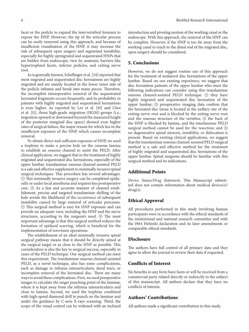
facet or the pedicle to expand the intervertebral foramen toexpose the HNP. However, the tip of the articular processcan be easily removed using this approach, and because ofinsufficient visualization of the HNP, it may increase therisk of subsequent open surgery and segmental instability,especially for highly upmigrated and sequestrated HNPs thatare hidden from endoscopic view by anatomic barriers likehypertrophied facets, inferior pedicles, and exiting nerveroot.
As is generally known, Schellinger et al. [24] reported thatmost migrated and sequestrated disc herniations are highlymigrated and are mainly located in the lower inner side ofthe pedicle isthmus and break into many pieces. Therefore,the incomplete intraoperative removal of the sequestratedherniated fragments occurs frequently, and its probability inpatients with highly migrated and sequestrated herniationsis even higher. As reported by Lee et al. [10] and Choiet al. [11], those high-grade migration (HGM) groups (i.e.,migration upward or downward beyond the measured heightof the posterior marginal disc space) showed even higherrates of surgical failure, the major reason for which lies in theinsufficient exposure of the HNP, which causes incompleteremoval.
To obtain direct and sufficient exposure of HNP, we useda trephine to make a precise hole on the osseous laminato establish an osseous channel to assist the PELD. Afterclinical application, we suggest that in the treatment of highlymigrated and sequestrated disc herniations, especially of theupper lumbar, translaminar osseous channel-assisted PELDis a safe and effective supplement tominimally invasive spinalsurgical techniques. This procedure has several advantages.A This minimally invasive surgery can be completed epidu-rally or under local anesthesia and requires less postoperativecare. B As a fast and accurate manner of channel estab-lishment, precise and targeted translaminar drilling of thehole avoids the likelihood of the occurrence of subsequentinstability caused by large removal of articular processes.C This surgical method is easy for HNP exposure and canprovide an adequate view, including the HNP and the nervestructures, according to the surgeon’s need. D The mostimportant advantage is that this surgical method reduces theformation of epidural scarring, which is beneficial for theimplementation of rerevision operations.
The establishment of an ideal minimally invasive spinalsurgical pathway means that it should be directly aimed atthe surgical target or as close to the HNP as possible. Thisconsideration is also the key to surgical success, especially incases of the PELD technique. Our surgical method can meetthis requirement. The translaminar osseous channel-assistedPELD, as a novel technique, also has some complications,such as damage to isthmus interarticularis, dural tears, orincomplete removal of the herniated disc. There are manyways to avoid these complications. First, we used preoperativeimages to calculate the target punching point of the laminar,where it is kept away from the isthmus interarticularis andclose to lamina. Second, we used the trephine combinedwith high-speed diamond drill to punch on the laminar andunder the guidance by C-arm X-rays scanning. Third, thescope of the visual control can be widened with an inclined
introduction and pivotingmotion of the working canal or theendoscope. With this approach, the removal of the HNP canbe complete. However, if the HNP is too far away from theworking canal to reach to the distal end of the migrated disc,open surgery should be considered.
5. Conclusions
However, we do not suggest routine use of this approachfor the treatment of unilateral disc herniations of the upperlumbar. Based on our existing experience, we suggest thatdisc herniation patients of the upper lumbar who meet thefollowing indications can consider using this translaminarosseous channel-assisted PELD technique: A they havehighly migrated and sequestrated disc herniation of theupper lumbar; B preoperative imaging data confirm thatthe herniated disc tissue is located at the axillary site of theexiting nerve root and is blocked by the exiting nerve rootand the osseous structure of the vertebra; C the back ofthe HNP is blocked by lamina, and the interlaminar PELDsurgical method cannot be used for the resection; and Dno degenerative spinal stenosis, instability, or dislocation ispresent. Based on existing clinical applications, we suggestthat the translaminar osseous channel-assisted PELD surgicalmethod is a safe and effective method for the treatmentof highly migrated and sequestrated disc herniations of theupper lumbar. Spinal surgeons should be familiar with thissurgical method and its indications.
Additional Points
Device Status/Drug Statement. This Manuscript submit-ted does not contain information about medical device(s)/drug(s).
Ethical Approval
All procedures performed in this study involving humanparticipants were in accordance with the ethical standards ofthe institutional and national research committee and withthe 1964 Helsinki declaration and its later amendments orcomparable ethical standards.
Disclosure
The authors have full control of all primary data and theyagree to allow the journal to review their data if requested.
Conflicts of Interest
No benefits in any form have been or will be received from acommercial party related directly or indirectly to the subjectof this manuscript. All authors declare that they have noconflicts of interest.
Authors’ Contributions
All authors made a significant contribution to this study.

BioMed Research International 7
Acknowledgments
The authors thank Yuping Cao for the preparation of illustra-tions, and Fujun Wu, Lin Chen, and Weijun Kong for theirhelp in preparation of the manuscript.
References
[1] H. Saberi and A. V. Isfahani, “Higher preoperative OswestryDisability Index is associated with better surgical outcome inupper lumbar disc herniations,” European Spine Journal, vol. 17,no. 1, pp. 117–121, 2008.
[2] D.-S. Lee, K.-S. Park, andM.-S. Park, “The comparative analysisof clinical characteristics and surgical results between theupper and lower lumbar disc herniations,” Journal of KoreanNeurosurgical Society, vol. 54, no. 5, pp. 379–383, 2013.
[3] S. P. Sanderson, J. Houten, T. Errico, D. Forshaw, J. Bauman, andP. R. Cooper, “The unique characteristics of ‘upper’ lumbar discherniations,” Neurosurgery, vol. 55, no. 2, pp. 385–389, 2004.
[4] S. Lee, S.-K. Kim, S.-H. Lee et al., “Percutaneous endoscopiclumbar discectomy for migrated disc herniation: classificationof disc migration and surgical approaches,” European SpineJournal, vol. 16, no. 3, pp. 431–437, 2007.
[5] G.Choi, S.-H. Lee, P. Lokhande et al., “Percutaneous endoscopicapproach for highly migrated intracanal disc herniations byforaminoplastic technique using rigid working channel endo-scope,” Spine, vol. 33, no. 15, pp. E508–E515, 2008.
[6] J. Ying, K. Huang, M. Zhu et al., “The effect and feasibility studyof transforaminal percutaneous endoscopic lumbar discectomyvia superior border of inferior pedicle approach for down-migrated intracanal disc herniations,” Medicine, vol. 95, no. 8,Article ID e2899, 2016.
[7] J. Y. Cho, S.-H. Lee, and H.-Y. Lee, “Prevention of developmentof postoperative dysesthesia in transforaminal percutaneousendoscopic lumbar discectomy for intracanalicular lumbar discherniation: floating retraction technique,” Minimally InvasiveNeurosurgery, vol. 54, no. 5-6, pp. 214–218, 2011.
[8] S. G. Osman, K. Nibu, M. M. Panjabi, E. B. Marsolais, andR. Chaudhary, “Transforaminal and posterior decompressionsof the lumbar spine: a comparative study of stability andintervertebral foramen area,” Spine, vol. 22, no. 15, pp. 1690–1695, 1997.
[9] Y. Ahn, S.-H. Lee, J. H. Lee, J. U. Kim, and W. C. Liu,“Transforaminal percutaneous endoscopic lumbar discectomyfor upper lumbar disc herniation: clinical outcome, prognosticfactors, and technical consideration,”Acta Neurochirurgica, vol.151, pp. 199–206, 2009.
[10] S.-H. Lee, U. K. Byung, Y. Ahn et al., “Operative failureof percutaneous endoscopic lumbar discectomy: a radiologicanalysis of 55 cases,” Spine, vol. 31, no. 10, pp. E285–E290, 2006.
[11] G. Choi, H. N. Modi, N. Prada et al., “Clinical results ofXMR-assisted percutaneous transforaminal endoscopic lumbardiscectomy,” Journal of Orthopaedic Surgery and Research, vol.8, no. 1, article 14, 2013.
[12] S. Lee, S.-K. Kim, S.-H. Lee et al., “New surgical techniques ofpercutaneous endoscopic lumbar discectomy for migrated discherniation,” Joint Diseases and Related Surgery, vol. 16, pp. 102–110, 2005.
[13] D.-S. Kim, J.-K. Lee, J.-W. Jang, B.-S. Ko, J.-H. Lee, and S.-H.Kim, “Clinical features and treatments of upper lumbar discHerniations,” Journal of Korean Neurosurgical Society, vol. 48,no. 2, pp. 119–124, 2010.
[14] S. Ruetten, M. Komp, H. Merk, and G. Godolias, “Full-endoscopic interlaminar and transforaminal lumbar discec-tomy versus conventional microsurgical technique: A Prospec-tive, Randomized, Controlled Study,” Spine, vol. 33, no. 9, pp.931–939, 2008.
[15] S. Hijikata, M. Yamagishi, and T. Nakayma, “Percutaneous dis-cectomy: a new treatment method for lumbar disc herniation,”Journal of Tokyo Denryoku Hospital, vol. 5, pp. 39–44, 1975.
[16] P. Kambin, E. O’Brien, L. Zhou, and J. L. Schaffer, “Arthro-scopic microdiscectomy and selective fragmentectomy,” Clini-calOrthopaedics andRelatedResearch, no. 347, pp. 150–167, 1998.
[17] A. T. Yeung, “Minimally invasive disc surgery with the Yeungendoscopic spine system (YESS),” Surgical Technology Interna-tional, vol. 8, pp. 267–277, 1999.
[18] H. M. Mayer and M. Brock, “Percutaneous endoscopic discec-tomy: surgical technique and preliminary results compared tomicrosurgical discectomy,” Journal of Neurosurgery, vol. 78, no.2, pp. 216–225, 1993.
[19] S. Ruetten, M. Komp, and G. Godolias, “A new full-endoscopictechnique for the interlaminar operation of lumbar disc hernia-tions using 6-mm endoscopes: prospective 2-year results of 331patients,”Minimally InvasiveNeurosurgery, vol. 49, no. 2, pp. 80–87, 2006.
[20] S. Ruetten, M. Komp, H. Merk, and G. Godolias, “Use ofnewly developed instruments and endoscopes: full-endoscopicresection of lumbar disc herniations via the interlaminar andlateral transforaminal approach,” Journal ofNeurosurgery: Spine,vol. 6, no. 6, pp. 521–530, 2007.
[21] I. Choi, J.-O. Ahn, W.-S. So, S.-J. Lee, I.-J. Choi, and H. Kim,“Exiting root injury in transforaminal endoscopic discectomy:preoperative image considerations for safety,” European SpineJournal, vol. 22, no. 11, pp. 2481–2487, 2013.
[22] J.-W. Choi, J.-K. Lee, K.-S. Moon, H. Hur, Y.-S. Kim, andK. Soo-Han, “Transdural approach for calcified central discherniations of the upper lumbar spine: technical note,” Journalof Neurosurgery: Spine, vol. 7, no. 3, pp. 370–374, 2007.
[23] M. Schubert and T. Hoogland, “Endoscopic transforaminalnucleotomy with foraminoplasty for lumbar disk herniation,”Operative Orthopadie und Traumatologie, vol. 17, no. 6, pp. 641–661, 2005.
[24] D. Schellinger, H. J. Manz, B. Vidic et al., “Disk fragmentmigra-tion,” Radiology, vol. 175, no. 3, pp. 831–836, 1990.

Submit your manuscripts athttps://www.hindawi.com
Stem CellsInternational
Hindawi Publishing Corporationhttp://www.hindawi.com Volume 2014
Hindawi Publishing Corporationhttp://www.hindawi.com Volume 2014
MEDIATORSINFLAMMATION
of
Hindawi Publishing Corporationhttp://www.hindawi.com Volume 2014
Behavioural Neurology
EndocrinologyInternational Journal of
Hindawi Publishing Corporationhttp://www.hindawi.com Volume 2014
Hindawi Publishing Corporationhttp://www.hindawi.com Volume 2014
Disease Markers
Hindawi Publishing Corporationhttp://www.hindawi.com Volume 2014
BioMed Research International
OncologyJournal of
Hindawi Publishing Corporationhttp://www.hindawi.com Volume 2014
Hindawi Publishing Corporationhttp://www.hindawi.com Volume 2014
Oxidative Medicine and Cellular Longevity
Hindawi Publishing Corporationhttp://www.hindawi.com Volume 2014
PPAR Research
The Scientific World JournalHindawi Publishing Corporation http://www.hindawi.com Volume 2014
Immunology ResearchHindawi Publishing Corporationhttp://www.hindawi.com Volume 2014
Journal of
ObesityJournal of
Hindawi Publishing Corporationhttp://www.hindawi.com Volume 2014
Hindawi Publishing Corporationhttp://www.hindawi.com Volume 2014
Computational and Mathematical Methods in Medicine
OphthalmologyJournal of
Hindawi Publishing Corporationhttp://www.hindawi.com Volume 2014
Diabetes ResearchJournal of
Hindawi Publishing Corporationhttp://www.hindawi.com Volume 2014
Hindawi Publishing Corporationhttp://www.hindawi.com Volume 2014
Research and TreatmentAIDS
Hindawi Publishing Corporationhttp://www.hindawi.com Volume 2014
Gastroenterology Research and Practice
Hindawi Publishing Corporationhttp://www.hindawi.com Volume 2014
Parkinson’s Disease
Evidence-Based Complementary and Alternative Medicine
Volume 2014Hindawi Publishing Corporationhttp://www.hindawi.com