Embed Size (px)
Citation preview
A method for including proteinflexibility in protein-ligand docking:Improving tools for database miningand virtual screening
Howard B. Broughton
Merck, Sharp & Dohme Neuroscience Research Centre, Essex, United Kingdom
Second-generation methods for docking ligands into theirbiological receptors, such as FLOG, provide for flexibilityof the ligand but not of the receptor. Molecular dynamicsbased methods, such as free energy perturbation, accountfor flexibility, solvent effects, etc., but are very time con-suming. We combined the use of statistical analysis ofconformational samples from short-run protein moleculardynamics with grid-based docking protocols and demon-strated improved performance in two test cases. Our statis-tical analysis explores the importance of the averagestrength of a potential interaction with the biological targetand optionally applies a weighting depending on the vari-ability in the strength of the interaction seen during dynam-ics simulation. Using these methods, we improved the num-ber of known dihydrofolate reductase ligands found in thetop-ranked 10% of a database of drug-like molecules, insearches based on the three-dimensional structure of theprotein. These methods are able to match the ability ofmanual docking to assess likely inactivity on steric groundsand indeed to rank order ligands from a homologous seriesof cyclooxygenase-2 inhibitors with good correlation totheir true activity. Furthermore, these methods reduce theneed for human intervention in setting up molecular dockingexperiments. © 2000 by Elsevier Science Inc.
Keywords: molecular dynamics, docking, flexible, cycloox-ygenase, prostaglandin H2 synthase, dihydrofolate reduc-tase, database search, rank order
INTRODUCTION
Prediction of the binding geometry and energy (and henceaffinity) of ligands at their biological targets remains a verydifficult problem, but one of great interest in the design ofnovel medicines. In the modeling of protein-ligand interac-tions, it is normal practice to make drastic, simplifying assump-tions: the protein usually is treated as rigid; side-chain orien-tations, tautomeric forms, and protonation states often areselected based on a subjective view of likely hydrogen bondingpatterns; any crystallographically observed water moleculesmay or may not be included (or may be supplemented byprograms that add solvent to the molecule) on a largely arbi-trary basis; and inorganic counterions generally are ignored.Despite such simplifications, the methods remain useful inareas such as database mining (virtual or real), combinatoriallibrary design, and lead optimzsation, leading to considerablerecent interest in the field.1–3 Programs such as DOCK,4
FlexX,5 GOLD,6 and AutoDOCK7 have become regularly usedtools for molecular modelers.
Although the “holy grail” of molecular docking is to repro-duce the experimentally observed “pose” of the ligand in thebinding site, our objective in this study was oriented moredirectly to the problems of drug discovery: we wanted toestablish whether the inclusion of protein flexibility in a dock-ing protocol would improve its utility as a “virtual screen”either in a database mining context or for the prediction ofrank-order activity in a homologous series. There is a clearadvantage in finding lead compounds in the first few thousandcompounds screened from typical corporate sample collectionscomprising a few hundred thousand to a few million com-pounds. Combinatorial libraries are best designed to explorestructure-activity relationships (SAR) fully in interesting areas,not to rediscover conclusions that would have been obviousfrom consideration of the steric capacity of the protein targetsite. Hence, there is a demand for a rapid method to assessautomatically whether a candidate library member is likely to
Color plates for this article are on pages 302–304.
Corresponding author: H.B. Broughton, Merck, Sharp & Dohme Neuro-science Research Centre, Terlings Park, Eastwick Road, Harlow, EssexCM20 2QR, United Kingdom. Tel.: 01279 440000.
E-mail address: [email protected] (H.B. Broughton)
Journal of Molecular Graphics and Modelling 18, 247–257, 2000© 2000 by Elsevier Science Inc. 1093-3263/00/$–see front matter655 Avenue of the Americas, New York, NY 10010 PII S1093-3263(00)00036-X
be active or not. In lead optimization, methods that can suggestwhere a particular drug candidate may interact with otherbiological targets than the intended one will enable efficientuse of those screening resources that are available. Modelsbased on such methods can even help to avoid errors in thedefinition of the generic scope of a chemical invention, byensuring that the compounds covered in a patent, beyond theexperimentally tested examples, are likely to be active againstthat particular biological target. Manual docking of ligands intoproteins often is sufficient for these purposes, and humanintervention can avoid some pitfalls of automated methods.However, automation is necessary for those applications wherea more objective assessment is required or where large num-bers of molecules are to be examined.
The FLOG method for automatically docking multiple di-verse conformations of candidate ligands into a site on aprotein surface is typical of “second-generation” docking pro-grams in that it allows the ligand to be flexible while makingsimplifying assumptions about the protein.8 Recent literaturehas described improvements in methods that allow ligands tobe flexible,9,10 but more significantly has begun to describemethods that provide for some flexibility of the protein, forexample, by considering ensembles of experimentally deter-mined structures11 or by allowing for some limited side-chainflexibility. 12,13 We believed that it would be valuable to try toincorporate protein flexibility and water mobility into theFLOG method in the hopes that this would improve FLOG’sability to select active compounds from databases This viewappears to be supported by a recent publication on the effect ofinduced fit with at least one docking program.14 The objectiveof improved selection from a database would be met if amodified FLOG were able to find more and better actives faster(i.e., would rank the compounds higher following a databasesearch) than FLOG based solely on the crystal structure. Be-cause many of the potential applications of docking in medic-inal chemistry are oriented toward work within a homologousseries of compounds, a modified FLOG also would ideallysurpass the original version in its ability to distinguish inactive,weakly active, and active ligands in such series. It is possibleto make a crude, somewhat subjective assessment of quality offit manually, by docking each compound into the target site andseeing whether there are sufficient unfavorable contacts withthe protein to suggest that it is highly likely that the moleculewill be inactive, and we sought to reproduce this ability todistinguish potentially active from inactive compounds basedon steric fit using FLOG. We also hoped that the enhancementswould be sufficient to lead to an improved rank correlationbetween the FLOG score and the true activity rank of com-pounds in a homologous series.
In order to implement this in practice, we decided to usestatistically weighted descriptors of the target active site insingle FLOG runs, rather than trying to solve the combinatorialproblem of docking multiple conformations of multiple mole-cules into multiple protein conformations. Although this ap-proach has some similarities with literature methods usingensembles of experimentally determined protein conforma-tions,11 we chose to use molecular dynamics simulation as thesource of conformational samples. This is an important choicebecause onlyone crystal structure is available for many pro-teins of pharmacological interest, and because it ensures awider exploration of the accessible conformational space of theprotein than might be expected for the crystallographic case,
where many solid-state constraints may exist. We also used anovel method of producing an “average” model of the activesite into which docking was to be performed, based on statis-tically weighting the mean potential protein-ligand interactionenergies computed from the molecular dynamics runs. Molec-ular dynamics simulations (of much greater length and com-plexity) also are the method of choice in free energy perturba-tion methods of calculating the interaction energy betweenligand and receptor.2 FLOG has been described in detail else-where,8 but the methods used here could be adapted to workwith other grid-based molecule docking programs.
The test beds chosen for each of these objectives weredesigned to be representative of the various levels of structuraldata available in typical cases. Thus, for the database miningproblem, the well-studied and crystallographically refined caseof Lactobacillus caseidihydrofolate reductase/methotrexatecomplex15 (DHFR/MTX, PDB16 code 3DFR, 1.7 Å structure)was used. This enables direct comparison with an extensiveliterature, based on a very good crystal structure complete withwater molecules. The database used was the MINDEX12 flexi-base,17 based on the 12th edition of theMerck Index,18 whichcontains about 7000 drug-like molecules including knownDHFR ligands and substrates. For the rank ordering of a seriesof candidate molecules to a protein target, the murinecyclooxygenase-2 (COX-2, prostaglandin H2 synthase) struc-ture19 (PDB code 1CX2, resolution 3.0 Å) was used in con-junction with a range of structures mostly of thevic-diarylheterocyclic class. This protein was chosen for its medicinalimportance, its membership in the class of membrane-associated proteins, and because its structure has been solved ata poorer level of resolution than was the case for 3DFR.COX-2 also is known to have limited flexibility in one smallpart (a section of helix D, near the CF3 group of SC-558 in the1CX2 crystal structure), whereas the remainder of the activesite is quite rigid and unaffected by ligand binding.20 Thesecomplexities test more fully the ability of the modified FLOGmethods to work with structural data more typical of real-worlddrug discovery. Furthermore, it is known that the COX-2inhibitory activity of members of thevic-diaryl heterocyclicclass of ligands is exquisitely sensitive to structural variation insome parts of the molecule while being tolerant of considerablechanges elsewhere.21---26 Hence, a reliable method for predict-ing rank-order binding for these ligands should be able todistinguish to some degree whether or not the effect of changesof each of these kinds would be expected to have an effect onthe enzyme inhibitory properties of the molecule.
METHODS
Initial Structure Preparation
The crystal structures 3DFR and 1CX2 were retrieved from theBrookhaven database using the WWW interface and read intothe Sybyl molecular modeling package.27 In the case of 1CX2,a single protein chain, with associated ligand and haem, wasobtained by deletion of all atoms in chains B, C, and D. Atomtypes and charges were assigned for 3DFR using the Triposimplementation of the original Kollman AMBER united-atomset,28 with atoms in the ligands being set by hand to the nearestequivalent type. Atom types and charges were assigned for1CX2 using the Tripos implementation of all-atomAMBER95,29 with supplementary parameters and the charges
248 J. Mol. Graphics Mod., 2000, Vol. 18, June
for the haem based on those provided on the AMBER web-site.30 The water model used for 3DFR was TIP3P, using watermolecules produced by adding hydrogen atoms to the oxygenatoms whose coordinates were present in the PDB entry usingSybyl’s “fillvalence” function. No water was present in thePDB entry, and none was added to the model of COX-2.Ligand and cofactor charges were obtained by calculatingMOPAC31 6 AM1 ESP-fit charges on the crystallographicallyobserved geometry of MTX and NADPH (NDP) and MNDOESP-fit charges on the optimized geometry for the COX-2ligand SC-558, after addition of hydrogens in idealized posi-tions where necessary. N-acetylglucosamine was used as amodel system for the NAG groups on COX-2, and the chargeswere scaled after attachment to the protein to maintain overallneutrality.
DHFR Model: Refinement and Simulation
The following sequence, which was chosen for its ability toadequately handle the relaxation of poorly oriented water mol-ecules, was used:
1. Protein, MTX, and cofactor (NDP) frozen2. Molecular dynamics simulation, 1 fs time step, 10 fs non-
bonded reset with velocity scaling
● 100 fs at 50K
● 100 fs at 150K
● 100 fs at 200K
3. 1000 fs without velocity scaling at 300K4. Energy minimization for 100 steps of Powell algorithm5. Side chains, NDP, and MTX “unfrozen,” protein backbone
held rigid6. Energy minimization for 1000 steps of Powell algorithm7. All constraints removed and minimization for another 1000
steps of Powell to a gradient,0.5.
After this process of annealment, the final structure showed aCa fit of 0.55 Å root-mean-square (RMS) and maximum Ca
displacement of 1.34 Å, with respect to 3DFR.Dynamics simulation of DHFR followed a heating regime in
which simulations of 100 fs were carried out at temperatures of50K, 150K, and 250K, followed by a 700 fs interval at 300K,all with velocity scaling and nonbonded update every 10 fs.Velocity scaling then was turned off and simulation continuedat 300K for another 50 ps with a structure written to disk every1 ps.
COX-2 Model: Refinement and Simulation
The COX-2 model was partially refined by energy minimiza-tion, initially 21,000 iterations using the Powell algorithm witha nonbonded cutoff of 8 Å and a dielectric function of 1/4r,followed by a final optimization of 200 iterations with a non-bonded cutoff of 12 Å. The Sybyl “anneal” function was usedto set up “hot,” “warm,” and “cold” regions of the protein,these being:
● Hot: regions in which the atoms are included in the energycalculation and can move in minimizations or dynamicssimulations
● Warm: regions in which interactions are considered forenergy calculation but the atoms are held rigid
● Cold: regions in which the atoms are held rigid and are notconsidered in evaluating the energy of the system.
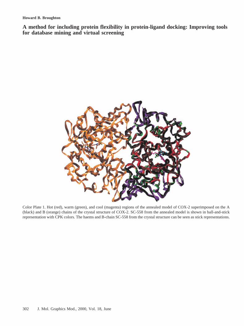
Initially, an “ActiveiiSite” region was defined as any residuewith at least one atom within 8 Å of the ligand SC-558. The“hot” region then was defined as all those residues with at leastone atom within an 8 Å radius of any atom of any Active2Siteresidue, the “warm” region included residues that had at leastone atom within 12 Å of any ActiveiiSite residue but whichwere not in the “hot” region, and the remaining atoms were inthe “cold” region. Minimization of the structure with thesezones defined proceeded to convergence (gradient,0.05),providing the base structure for the dynamics simulation. Thestructure obtained in this way is shown in Color Plate 1,superimposed on the Ca atoms of the A chain of the ho-modimer formed by the A and B chains in the crystal structure,with the annealment regions shown in different colors.
The dynamics simulation on COX-2 started at 10K andproceded for 1000 fs with a 1 fstime step and a nonbondedupdate with velocity scaling and momentum removal every 10fs. The temperature was incremented 10K and the simulationcontinued under otherwise identical conditions another 1000 fs.This cycle was repeated until a temperature of 300K wasreached. Velocity scaling then was turned off and the simula-tion continued for another 76 ps, with the structure beingwritten every 500 fs. After this dynamics simulation, the RMSfit of the Ca atoms to those of the crystal structure was 0.82 Å.
Generation of the FLOG Grids and Match Pointsfor Both Models
The last 40 structures recorded for the DHFR case and the last150 recorded from the COX-2 case were used for the next step.The DHFR structures were aligned on the inhibitor MTX andthe residues and water molecules with atoms within 8 Å of theligand, but excluding the ligand itself, were extracted for thegeneration of FLOG grids. The COX-2 structures were alignedon the inhibitor SC-558 and residues with atoms within 10 Å ofthe ligand were similarly extracted.
Although FLOG has been described elsewhere, a brief sum-mary of the steps involved is included here:
1. Assign atom types to the protein. Normally, the side-chainorientations, decision to keep or exclude water moleculesetc., also are done at this stage.
2. Set up a three-dimensional lattice of equally spaced pointsseparated by a distanced (d 5 0.3 was used for theseexperiments except as noted below).
3. At each point in the lattice, calculate the interaction energywith the protein for each of five (hydrogen-bond acceptor,hydrogen-bond donor, polar, hydrophobic and “other” [van-der-Waals interactions only]) or seven (the above plus cat-ion and anion) prototypical atom types to give five or seven“grids” (respectively).
4. Find the maxima (in FLOG, more positive scores representmore favorable interactions) in each grid. Place a “matchpoint” at each maximum subject to the condition that themaximum must represent an interaction energy of.1.0FLOG score unit and that match points must not be closer
249J. Mol. Graphics Mod., 2000, Vol. 18, June
than 1.2 Å. Each match point acquires the type of the gridin which it represents a maximum.
5. For each conformation of each candidate ligand, find ori-entations such that atom-atom distances in the ligand ap-proximately correspond to match point-match point dis-tances (clique detection method as used in SQ32) betweenatoms and match points of corresponding types.
6. Score each orientation of each conformation of each mole-cule by summing the interaction energy for each atom of theligand, reading the value from the grid of the type corre-sponding to the property-type of the atom (H-bond donor,acceptor, etc.).
7. Optionally apply some optimization, including simplexrigid-body translation and rotation, to the candidate orien-tation, and calculate final best score.
Atom type assignment was carried out on each sample confor-mation from the dynamics runs according to the standardFLOG procedure. For these experiments, the amino-acid side-chain orientations and water positions were not modified, withno human intervention. Generation of the interaction energygrids used by FLOG was done in the standard, fully automatedway, producing five three-dimensional grids for each samplefrom the molecular dynamics run. For comparison purposes,the same techniques also were applied to the original crystalstructures without further refinement except that in the case ofDHFR, for this “control” experiment only, the water moleculeswere all removed from the crystal structure before grid gener-ation, for reasons explained later.
The grids obtained in this way from the structures sampledfrom molecular dynamics were subjected to statistical analysis.Each grid point can be specified by four indices—i, j, and kspecifying the offsets from the grid origin in each dimension,and l specifying the type of the grid (donor, acceptor, etc.)—and each was considered in turn and statistics computed acrossall conformers of the protein sampled by dynamics. The valuescomputed for each grid point were the minimum value, max-imum value, mean value, standard deviation, and a weightedmean. The latter was calculated for a grid point Wijkl , based onthe mean (Mijkl ) and standard deviation (Sijkl ) observed at thatpoint, thus:If
Mijkl 5 0,
Wijkl 5 0
Else
Wijkl 5 Mijkl p exp2 (Sijkl2/Mijkl
2)
This weighted grid emphasizes areas where the “interactionenergy” varies little for a particular atom type between theconformers sampled, giving a good score for those cases wherethere is “reliably” a good interaction for the candidate atomtype across all conformers sampled. It also down-weights theeffect of occasional occupation of a region of space by a part ofthe protein or a water molecule, because, as has been observedby others11, the magnitude of repulsive terms can otherwiseunrealistically dominate the “average” interaction energy.
The grid points calculated in this way were written to gridfiles with the same dimensionality as the original input grids.Color Plate 2c shows the difference between the simple mean
van-der-Waals grid and the corresponding weighted mean grid(both contoured at a level corresponding to a very slightlyunfavorable interaction,20.1) for the DHFR case.
The statistically derived grids were used for comparisonwith those grids directly generated from the crystal structures,all the remaining steps in the FLOG process being those usedas standard, with a relative weighting for van-der-Waals andelectrostatic effects of 0.3 and 1.0, respectively.
Match points for the clique-detection step of FLOG weregenerated as normal from these grids. FLOG normally auto-matically chooses the match point with the largest interactionenergy as an “essential” match point, requiring it to be presentin every clique. For this study, the “essential” match point waschosen manually to ensure that a comparable point was used ineach run; the point type selected was “1” 32 (i.e., any ligandatom could be matched to the point). For the DHFR case, thepoint was chosen (as previously8) to lie close to the binding siteof the 2-amino group of the pteridine of MTX. For the COX-2case, the match point with highest interaction energy that laywithin the part of the binding pocket occupied by both selectiveand nonselective cyclooxygenase inhibitors was used in orderto allow comparison with nonselective compounds such asflurbiprofen.
Databases and Searches
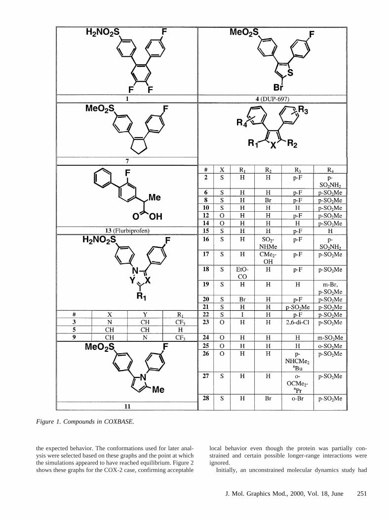
FLOG was used to orient conformations of molecules fromappropriate databases on the match points, followed by scoringusing the corresponding grids. For COX-2, a selection of 28ligands selected from the literature or evaluated against humanCOX-2 in house were chosen, the majority from thevic-diarylheteroaryl class. A flexibase17 (COXBASE) was constructedcontaining 250 diverse conformations of each of these and wassearched using FLOG. The structures of molecules in COX-BASE are given in Figure 1. The choice of compounds inCOXBASE was made to include compounds that would clearlybe expected to be inactive, based on their massively increasedsize over SC-558 and the evidently limited space available inthe enzyme, compounds that contained more subtle effectssuch as halogen substitution, and compounds where conforma-tional effects due to juxtaposition of substituents could beexpected. Compounds also were chosen to reflect the widevariety of core structures from the literature. Thus, COXBASEcontains compounds with large changes in structure that areexpected to remove activity and compounds with more subtlechanges that may vary inhibitory activity up or down withrespect to SC-558. The nature of the bioassays used makes rankorder of potency rather than explicit IC50 values the mostappropriate measure of activity for these compounds.
RESULTS AND DISCUSSION
Dynamics Studies
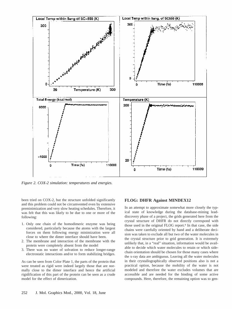
The initial dynamics simulations were routine, short simula-tions. This is in some contrast to alternate methods of predict-ing binding affinity such as free energy perturbation methods,which generally require lengthy and specialized calculationsand very careful choice of force-field parameters for atoms inprosthetic groups and ligands. In both simulations, graphs ofthe local temperature in the region of the ligandvs the overalltemperature, and of energy and temperaturesvs time, showed
250 J. Mol. Graphics Mod., 2000, Vol. 18, June
the expected behavior. The conformations used for later anal-ysis were selected based on these graphs and the point at whichthe simulations appeared to have reached equilibrium. Figure 2shows these graphs for the COX-2 case, confirming acceptable
local behavior even though the protein was partially con-strained and certain possible longer-range interactions wereignored.
Initially, an unconstrained molecular dynamics study had
Figure 1. Compounds in COXBASE.
251J. Mol. Graphics Mod., 2000, Vol. 18, June
been tried on COX-2, but the structure unfolded significantlyand this problem could not be circumvented even by extensivepreminimization and very slow heating schedules. Therefore, itwas felt that this was likely to be due to one or more of thefollowing:
1. Only one chain of the homodimeric enzyme was beingconsidered, particularly because the atoms with the largestforces on them following energy minimization were allclose to where the dimer interface should have been.
2. The membrane and interaction of the membrane with theprotein were completely absent from the model
3. There was no water of solvation to reduce longer-rangeelectrostatic interactions and/or to form stabilizing bridges.
As can be seen from Color Plate 1, the parts of the protein thatwere treated as rigid were indeed largely those that are nor-mally close to the dimer interface and hence the artificialrigidification of this part of the protein can be seen as a crudemodel for the effect of dimerization.
FLOG: DHFR Against MINDEX12
In an attempt to approximate somewhat more closely the typ-ical state of knowledge during the database-mining lead-discovery phase of a project, the grids generated here from thecrystal structure of DHFR do not directly correspond withthose used in the original FLOG report.8 In that case, the sidechains were carefully oriented by hand and a deliberate deci-sion was taken to exclude all but two of the water molecules inthe crystal structure prior to grid generation. It is extremelyunlikely that, in a “real” situation, information would be avail-able to decide which water molecules to retain or which side-chain orientation should be chosen for those many cases wherethe x-ray data are ambiguous. Leaving all the water moleculesin their crystallographically observed positions also is not apractical option, because the mobility of the water is notmodeled and therefore the water excludes volumes that areaccessible and are needed for the binding of some activecompounds. Here, therefore, the remaining option was to gen-
Figure 2. COX-2 simulation: temperatures and energies.
252 J. Mol. Graphics Mod., 2000, Vol. 18, June
erate the grid after simple removal of the water molecules fromthe crystal structure, which is a common approach in suchstudies.14 In turn, this simplification gives rise to other prob-lems: visualization of the grids by contouring reveals a com-plex energy surface (Color Plate 2a), which unsurprisinglycontains many local maxima, and hence gives rise to manymatch points during the match point generation step. Due to thelarge amounts of time required to try every ligand conforma-tion in every orientation on so many match points, several stepshad to be taken to simplify the grids and match points in thiscase. First, the grid resolutiond was set to 0.5 instead of 0.3.Two hundred match points generated further than 5Å awayfrom the crystallographically observed position of the ligandwere removed (this distance was that used in the originalFLOG report8), along with points found to be isolated from theremainder (e.g., in shallow surface depressions). These stepsleft 116 match points. The corresponding grids for the“Weighted Average” case (Color Plate 2b) are notably simplerand the surfaces are smoother, and there was no need to uselower resolution or artificially reduce the number of gridpoints. The “Average” and “Weighted Average” grids show anumber of differences, most clearly in the van-der-Waals grid.Color Plate 2c shows an area close to the carboxylate-bindingarea for MTX where the occasional presence of a water mol-ecule during the dynamics run occludes the space in the “Av-erage” grid case, but not in the “Weighted Average” model.Both these models do handle water mobility, albeit in differentways, and therefore there was no need to remove any of thecrystallographically observed water molecules from these runs.
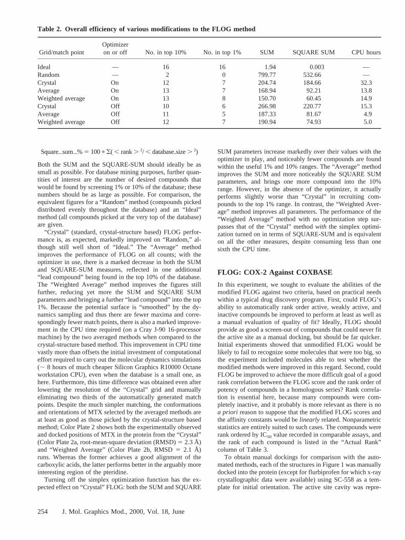
In order to assess the relative capacity of the methods to findactive compounds, after completion of the searches of bothenantiomers of compound conformers in MINDEX12, the listsof molecules oriented successfully by FLOG were ordered byFLOG score and the resulting list searched for 16 compoundsidentified from the Merck Index as having activity at DHFR. In
Table 1 each compound is listed together with the proportion ofthe database (as a percentage) that would have had to bescreened to find it had the FLOG score by that method beenused as a guide to screening order. Results from the crystallo-graphically derived (Crystalii%), mean (Averageii%), andweighted mean (Weightedii%) grids/match points are shown,and the data are given for the “standard” FLOG method withsimplex optimization of the ligand into the receptor cavityturned on.
It is worth noting that all the methods succeeded in findingnot only compounds of the folate/pterin class, but also man-aged to score reasonably highly other DHFR inhibitors such astrimethoprim, brodimoprim, and tetroxoprim. It is possiblewithin FLOG to “turn off” the simplex optimizer (step 7 in theFLOG summary given earlier), reducing search times signifi-cantly. We felt that grids and match points that better repre-sented the receptor might reduce the need for the simplexoptimization step. In order to study the effect of the dynamicssampling on this aspect of FLOG, a more summary form ofresults is needed, and, together with the amount of CPU used,is given in Table 2. Some idea of the overall performance of aparticular variant of the FLOG method can be obtained bysimply summing the percentile positions (as given in Table 1)of these known ligands in the database to give the SUMparameter. A correction factor is applied for the compoundsthat had negative FLOG scores and therefore were not rankordered; these are assumed to be found at the point in thedatabase halfway between the last compound scored by FLOGand the end of the database. Although the simple SUM gives anidea of the utility of a method, it does not adequately reflect thefact that it is often more useful to find one lead after screening5% of the database and a second after screening 35% than tofind two leads at the 20% point. To reflect this, the SQUARE-SUM is calculated as
Table 1. FLOG rank of known DHFR ligands in MINDEX12
NameMonograph
no. Crystalo% Averageo% Weightedo%
Denopterin I2944 0.01 0.47 0.10A-denopterin I151 0.03 1.13 0.27Methotrexate I6065 0.04 0.17 0.41Folinic acid I4254 0.07 29.86 9.19Folic acid I4253 0.13 0.93 0.19Methopterin I6064 0.39 0.07 0.74Aminopterin I493 0.68 0.03 0.07Ninopterin I6647 1.04 0.04 0.14Trimetrexate I9851 2.85 1.70 3.25Piritrexim I7654 4.92 2.43 6.43Rhizopterin I8348 5.05 0.30 0.80Tetroxoprim I9386 6.51 4.15 6.01Brodimoprim I1401 12.83 5.12 11.24Sapropterin I8515 16.15 2.77 6.93Trimethoprim I9840 21.93 36.55 42.70Edatrexate I3553 — — 62.23
FLOG does not output orientations of structures with negative scores (unfavorable interaction expected with the receptor), and so compounds not found arelisted with a dash.
253J. Mol. Graphics Mod., 2000, Vol. 18, June
Squareoisumio% 5 100p (( , rank. 2/ , databaseosize. 2)
Both the SUM and the SQUARE-SUM should ideally be assmall as possible. For database mining purposes, further quan-tities of interest are the number of desired compounds thatwould be found by screening 1% or 10% of the database; thesenumbers should be as large as possible. For comparison, theequivalent figures for a “Random” method (compounds pickeddistributed evenly throughout the database) and an “Ideal”method (all compounds picked at the very top of the database)are given.
“Crystal” (standard, crystal-structure based) FLOG perfor-mance is, as expected, markedly improved on “Random,” al-though still well short of “Ideal.” The “Average” methodimproves the performance of FLOG on all counts; with theoptimizer in use, there is a marked decrease in both the SUMand SQUARE-SUM measures, reflected in one additional“lead compound” being found in the top 10% of the database.The “Weighted Average” method improves the figures stillfurther, reducing yet more the SUM and SQUARE SUMparameters and bringing a further “lead compound” into the top1%. Because the potential surface is “smoothed” by the dy-namics sampling and thus there are fewer maxima and corre-spondingly fewer match points, there is also a marked improve-ment in the CPU time required (on a Cray J-90 16-processormachine) by the two averaged methods when compared to thecrystal-structure based method. This improvement in CPU timevastly more than offsets the initial investment of computationaleffort required to carry out the molecular dynamics simulations(; 8 hours of much cheaper Silicon Graphics R10000 Octaneworkstation CPU), even when the database is a small one, ashere. Furthermore, this time difference was obtained even afterlowering the resolution of the “Crystal” grid and manuallyeliminating two thirds of the automatically generated matchpoints. Despite the much simpler matching, the conformationsand orientations of MTX selected by the averaged methods areat least as good as those picked by the crystal-structure basedmethod; Color Plate 2 shows both the experimentally observedand docked positions of MTX in the protein from the “Crystal”(Color Plate 2a, root-mean-square deviation (RMSD)5 2.3 Å)and “Weighted Average” (Color Plate 2b, RMSD5 2.1 Å)runs. Whereas the former achieves a good alignment of thecarboxylic acids, the latter performs better in the arguably moreinteresting region of the pteridine.
Turning off the simplex optimization function has the ex-pected effect on “Crystal” FLOG: both the SUM and SQUARE
SUM parameters increase markedly over their values with theoptimizer in play, and noticeably fewer compounds are foundwithin the useful 1% and 10% ranges. The “Average” methodimproves the SUM and more noticeably the SQUARE SUMparameters, and brings one more compound into the 10%range. However, in the absence of the optimizer, it actuallyperforms slightly worse than “Crystal” in recruiting com-pounds to the top 1% range. In contrast, the “Weighted Aver-age” method improves all parameters. The performance of the“Weighted Average” method with no optimization step sur-passes that of the “Crystal” methodwith the simplex optimi-zation turned on in terms of SQUARE-SUM and is equivalenton all the other measures, despite consuming less than onesixth the CPU time.
FLOG: COX-2 Against COXBASE
In this experiment, we sought to evaluate the abilities of themodified FLOG against two criteria, based on practical needswithin a typical drug discovery program. First, could FLOG’sability to automatically rank order active, weakly active, andinactive compounds be improved to perform at least as well asa manual evaluation of quality of fit? Ideally, FLOG shouldprovide as good a screen-out of compounds that could never fitthe active site as a manual docking, but should be far quicker.Initial experiments showed that unmodified FLOG would belikely to fail to recognize some molecules that were too big, sothe experiment included molecules able to test whether themodified methods were improved in this regard. Second, couldFLOG be improved to achieve the more difficult goal of a goodrank correlation between the FLOG score and the rank order ofpotency of compounds in a homologous series? Rank correla-tion is essential here, because many compounds were com-pletely inactive, and it probably is more relevant as there is noa priori reason to suppose that the modified FLOG scores andthe affinity constants would belinearly related. Nonparametricstatistics are entirely suited to such cases. The compounds wererank ordered by IC50 value recorded in comparable assays, andthe rank of each compound is listed in the “Actual Rank”column of Table 3.
To obtain manual dockings for comparison with the auto-mated methods, each of the structures in Figure 1 was manuallydocked into the protein (except for flurbiprofen for which x-raycrystallographic data were available) using SC-558 as a tem-plate for initial orientation. The active site cavity was repre-
Table 2. Overall efficiency of various modifications to the FLOG method
Grid/match pointOptimizeron or off No. in top 10% No. in top 1% SUM SQUARE SUM CPU hours
Ideal — 16 16 1.94 0.003 —Random — 2 0 799.77 532.66 —Crystal On 12 7 204.74 184.66 32.3Average On 13 7 168.94 92.21 13.8Weighted average On 13 8 150.70 60.45 14.9Crystal Off 10 6 266.98 220.77 15.3Average Off 11 5 187.33 81.67 4.9Weighted average Off 12 7 190.94 74.93 5.0
254 J. Mol. Graphics Mod., 2000, Vol. 18, June
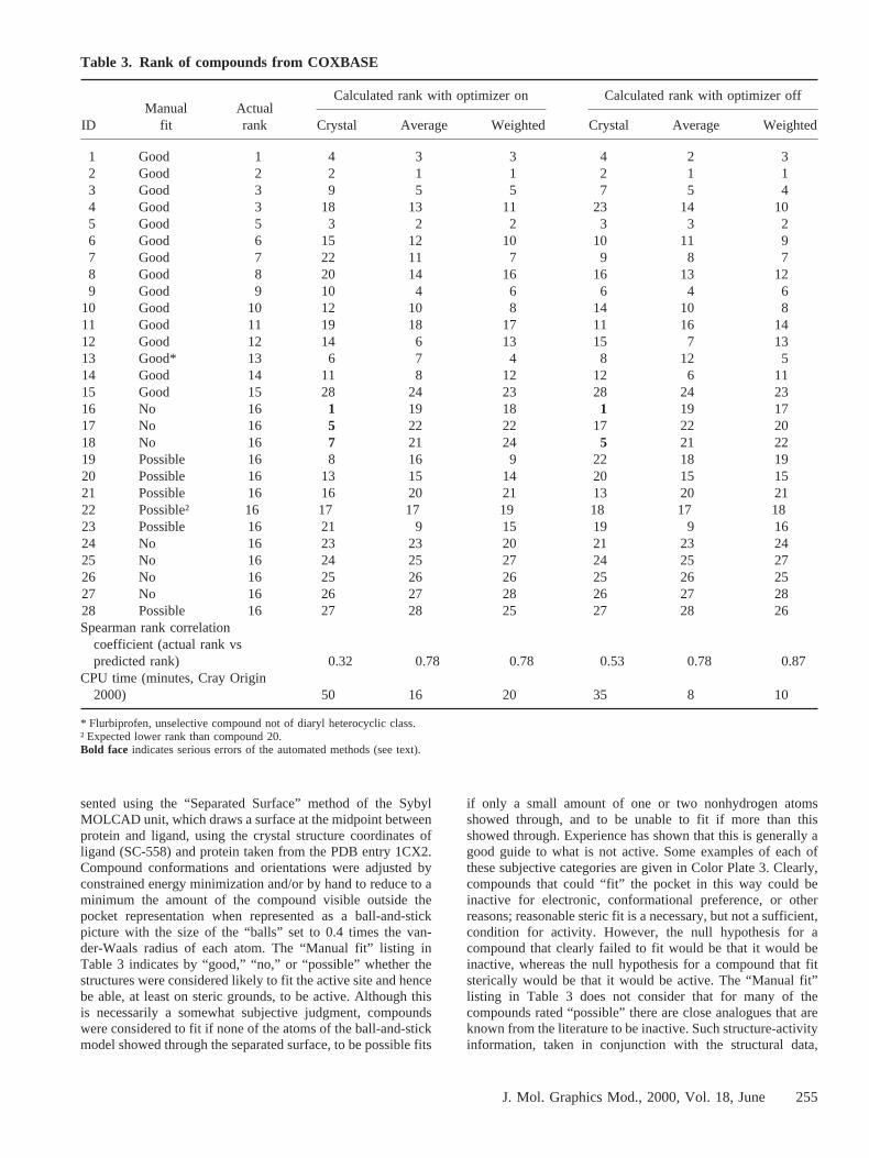
sented using the “Separated Surface” method of the SybylMOLCAD unit, which draws a surface at the midpoint betweenprotein and ligand, using the crystal structure coordinates ofligand (SC-558) and protein taken from the PDB entry 1CX2.Compound conformations and orientations were adjusted byconstrained energy minimization and/or by hand to reduce to aminimum the amount of the compound visible outside thepocket representation when represented as a ball-and-stickpicture with the size of the “balls” set to 0.4 times the van-der-Waals radius of each atom. The “Manual fit” listing inTable 3 indicates by “good,” “no,” or “possible” whether thestructures were considered likely to fit the active site and hencebe able, at least on steric grounds, to be active. Although thisis necessarily a somewhat subjective judgment, compoundswere considered to fit if none of the atoms of the ball-and-stickmodel showed through the separated surface, to be possible fits
if only a small amount of one or two nonhydrogen atomsshowed through, and to be unable to fit if more than thisshowed through. Experience has shown that this is generally agood guide to what is not active. Some examples of each ofthese subjective categories are given in Color Plate 3. Clearly,compounds that could “fit” the pocket in this way could beinactive for electronic, conformational preference, or otherreasons; reasonable steric fit is a necessary, but not a sufficient,condition for activity. However, the null hypothesis for acompound that clearly failed to fit would be that it would beinactive, whereas the null hypothesis for a compound that fitsterically would be that it would be active. The “Manual fit”listing in Table 3 does not consider that for many of thecompounds rated “possible” there are close analogues that areknown from the literature to be inactive. Such structure-activityinformation, taken in conjunction with the structural data,
Table 3. Rank of compounds from COXBASE
IDManual
fitActualrank
Calculated rank with optimizer on Calculated rank with optimizer off
Crystal Average Weighted Crystal Average Weighted
1 Good 1 4 3 3 4 2 32 Good 2 2 1 1 2 1 13 Good 3 9 5 5 7 5 44 Good 3 18 13 11 23 14 105 Good 5 3 2 2 3 3 26 Good 6 15 12 10 10 11 97 Good 7 22 11 7 9 8 78 Good 8 20 14 16 16 13 129 Good 9 10 4 6 6 4 6
10 Good 10 12 10 8 14 10 811 Good 11 19 18 17 11 16 1412 Good 12 14 6 13 15 7 1313 Good* 13 6 7 4 8 12 514 Good 14 11 8 12 12 6 1115 Good 15 28 24 23 28 24 2316 No 16 1 19 18 1 19 1717 No 16 5 22 22 17 22 2018 No 16 7 21 24 5 21 2219 Possible 16 8 16 9 22 18 1920 Possible 16 13 15 14 20 15 1521 Possible 16 16 20 21 13 20 2122 Possible† 16 17 17 19 18 17 1823 Possible 16 21 9 15 19 9 1624 No 16 23 23 20 21 23 2425 No 16 24 25 27 24 25 2726 No 16 25 26 26 25 26 2527 No 16 26 27 28 26 27 2828 Possible 16 27 28 25 27 28 26Spearman rank correlation
coefficient (actual rank vspredicted rank) 0.32 0.78 0.78 0.53 0.78 0.87
CPU time (minutes, Cray Origin2000) 50 16 20 35 8 10
* Flurbiprofen, unselective compound not of diaryl heterocyclic class.† Expected lower rank than compound 20.Bold face indicates serious errors of the automated methods (see text).
255J. Mol. Graphics Mod., 2000, Vol. 18, June
would often permit a clear prediction of “active” or “inactive,”but is often not available in the early stages of a reasearchprogram and so has not been used here.
Grids were generated as before for the unperturbed crystalstructure (“Crystal”), and from the dynamics run an “Average”and “Weighted Average” grid were prepared. Match pointswere generated from each of these grids, providing 101, 26,and 27 match points, respectively, from which the highest-scoring point close to the bromophenyl ring of SC-558 in thecrystal structure was selected as the “essential” point.
FLOG was used as before to orient and score conformationsof each molecule from COXBASE in the active site, with andwithout the simplex optimization step, considering both enan-tiomers of all the conformations in COXBASE. In Table 3, theresults of these searches are given as the rank order in whichcompounds were found, along with the quality of manual fitand the rank of the actual activity from literature reports or asmeasured in equivalent assays in house. To provide an idea ofthe scale, actual ranks 15 and 16 represent compounds with anIC50 of $100mM; the most potent compound (with actual rankof 1) had an IC50 of ;4 nM, whereas compounds ranked 1 to11 all had IC50 values better than 100 nM. A test compoundranked above 11 by any of the FLOG methods therefore wouldbe expected to be active, whereas for one ranking further downthe list than rank 15 the null hypothesis would be that thecompound would be inactive.
The data in Table 3 can be analyzed for important errorsmade by the automated methods. A compound predictednot tofit by the manual method should not be ranked highly by theautomated methods (the contrary is not necessarily true: acompound judged to be a “good” or “possible” fit manuallymay fail to rank highly by the automated methods because ofpoor electrostatic complementarity or other good reasons). Inthe case of the unmodified crystal-structure based runs, threecompounds (16, 17, and 18) failed on this measure, all of whichwere substituted on the central heterocyclic core with moder-ately sized groups. However, the “Average” and “WeightedAverage” runswereable to correctly assign these and all othercompounds to ranks consistent with the manual method andwith experimental results. Similarly, the quality of fit of thepredicted to the actual, measured rank order of potency can beevaluated for each case by the Spearman rank correlationcoefficient provided in the penultimate row of Table 3. Therank correlation coefficients of the method based purely on thecrystal structure are rather poor, mainly due to some particu-larly severe errors such as with compound 16 (ranked first, butreally inactive) and compound 4 (ranked 18th or 23rd, depend-ing on use or otherwise of the optimizer, but really a;10 nMcompound). However, the “Average” and “Weighted Average”methods provide a satisfactory rank ordering of the compoundsin which the actual rank order of activity is well reproduced,with the majority of the errors being with compounds bearingsmall substituents such as halogen atoms, or juxtapositions ofsubstituents likely to distort the conformation (such distortionswere not accounted for in the building of COXBASE, wherethe emphasis was on diversity of conformation rather than lowenergy). These improvements over the “Crystal” method alsowere achieved with significant savings in the computer re-sources required. Flurbiprofen was slightly overrated by mostof the methods, but it is remarkable that the methods are ableto rank order across structural classes even with limited accu-racy. Between classes of compounds, there is none of the
cancellation of errors that can occur between members of ahomologous series, and this is why in the database searchingtest (where cross-class ranking is important) the best that couldbe achieved by these methods was to raise the hit rate in the top10% of the database from;0.3% (“Random” method) to;2.2% (“Weighted Average” method).
CONCLUSION
It is clear that the “Average” and particularly the “WeightedAverage” methods are better than the unmodified “Crystal”method of using FLOG at database searching. With theseimprovements, the automated methods also perform wellenough to be used for the tasks outlined in the Introductionrelating to homologous series, in particular providing a rapidtool for detection of likely inactives that is as good as or betterthan manual alignment of candidate compounds in the bindingsite. Although the modifications require an initial moleculardynamics simulation to be run, the time spent on this is morethan compensated for by the time saved on FLOG itself, evenwith a small database of candidate structures. In addition, themodified methods provide a more objective tool that no longerrequires the user to specify side-chain orientations or electwhether to retain particular crystallographic water moleculesand requires no manual editing of the match points used toalign candidate ligands in the binding pocket. The methodscould be readily adapted to other docking methods and scoringfunctions.
ACKNOWLEDGMENTS
We thank Drs. R. Sheridan, M. Miller, and S. Kearsley forassistance in the use and modification of the FLOG, SQ, andrelated code; Dr. Peter Hunt for helpful discussions; and MarkOeullet and David Percival for biological results.
REFERENCES
1 Dixon, J.S. Evaluation of the CASP2 Docking Section.Prot. Struct. Funct. Genet.1998, Volume date 1997,Suppl. 1, pp. 198–204, and other papers in the specialsupplement
2 Kubinyi, H., Folkers, G., and Martin, YC (eds.). 3-DQSAR in drug design: Ligand-protein interactions andmolecular similarity. In:Perspectives Drug DiscoveryDesign, Volume 9/10/11.Kluwer/Escom, Dordrecht, TheNetherlands, 1998
3 Stahl, M., and Bo¨hm, H.-J. Development of filter func-tions for protein-ligand docking.J. Mol. GraphicsModel. 1998,16, 121–132
4 Kuntz, I.D., Blaney, J.M., Oatley, S.J., Langridge, R.,and Ferrin, T.E. A geometric approach to macromole-cule-ligand interactions.J. Mol. Biol. 1982, 161, 269–288
5 Rarey, M., Kramer, B., Lengauer, T., and Klebe, G.. Afast flexible docking method using an incremental con-struction algorithm.J. Mol. Biol. 1996,261, 470--489
6 Jones, G., Willett, P., Glen, R.C., Leach, A.R., andTaylor, R. Development and validation of a geneticalgorithm for flexible docking.J. Mol. Biol. 1997,267,727–748
7 Goodsell, D.S., Morris, G.M., and Olson, A.J. Auto-
256 J. Mol. Graphics Mod., 2000, Vol. 18, June
mated docking of flexible ligands: Application ofAutoDock. J. Mol. Recognit.1996,9, 1–5
8 Miller, M.D., Kearsley, S.K., Underwood, D.J., andSheridan, R.P. FLOG: A system to select “quasi-flexible” ligands complementary to a receptor of knownthree-dimensional structure.J. Comput. Aided Mol. De-sign 1994,8, 153–174
9 Hoffmann, D., Kramer, B., Washio, T., Steinmetzer, T.,Rarey, M., and Lengauer, T. Two-stage method forprotein-ligand docking.J. Med. Chem.1999,42, 4422–4433
10 Wang, J., Kollman, P.A., and Kuntz, .ID. Flexible liganddocking: A multistep strategy approach.Prot. Struct.Funct. Genet.1999,36, 1–19
11 Knegtel, R.M.A., Kuntz, I.D., and Oshiro, C.M. Molec-ular docking to ensembles of protein structures.J. Mol.Biol. 1997,266, 424–440
12 Wade, R.C., Sobolev, V., Ortiz, A.R., and Peters, G.Computational approaches to modeling receptor flexibil-ity upon ligand binding: Application to interfaciallyactivated enzymes.NATO ASI Ser., Ser. E (Structure-Based Drug Design)1998,352, 223–232
13 Leach, A.R. Ligand docking to proteins with discreteside-chain flexibility.J. Mol. Biol. 1994,235, 345–356
14 Murray, C.W., Baxter, C.A., and Frenkel, A.D. Thesensitivity of the results of molecular docking to inducedfit effects: Application to thrombin, thermolysin andneuraminidase.J. Comput. Aided Mol. Design1999,13,547–562
15 Bolin, J.T., Filman, D.J., Matthews, D.A., Hamlin, R.C.,and Krant, J. Crystal structures of Escherichia coli andLactobacillus casei dihydrofolate reductase refined at1.7 Angstroms resolution I. General features and bindingof methotrexate.J. Biol. Chem.1982,257,13650–13662
16 Bernstein, F.C., Koetzle, T.F., Williams, G.J., Meyer,E.E. Jr., Brice, M.D., Rodgers, J.R., Kennard, O., Shi-manouchi, T., and Tasumi, M. The protein data bank: Acomputer-based archival file for macromolecular struc-tures.J. Mol. Biol. 1977112, 535: http://www.rcsb.org/pdb/
17 Kearsley, S.K., Underwood, D.J., Sheridan, R.P., andMiller, M.D. Flexibases: A way to enhance the use ofmolecular docking methods.J. Comput. Aided Mol. De-sign 1994,8, 565–582
18 Budavari, S (ed.).Merck Index. Merck & Co. Inc.,Whitehouse Station, NJ, 1996
19 Kurumbail, R.G., Stevens, A.M., Gierse, J.K., Mc-Donald, J.J., Stegeman, R.A., Pak, J.Y., Gildehaus, D.,Miyashiro, J.M., Penning, T.D., Seibert, K., Isakson,P.C., and Stallings, W.C. Structural basis for selectiveinhibition of cyclooxygenase-2 by anti-inflammatoryagents.Nature 1996,384, 644--648
20 Luong, C., Miller, A., Barnett, J., Chow, J., Ramesha,C., and Browner, M.F. Flexibility of the NSAID bindingsite in the structure of human cyclooxygenase-2.Nat.Struct. Biol.1996,3, 927–933
21 Khanna, I.K., Weier, R.M., Yu, Y., Collins, P.W., Mi-
yashiro, J.M., Koboldt, C.M., Vennhuizen, A.W., Cur-rie, J.L., Seibert, K., and Isakson, P.C. 1,2-Diarylpyr-roles as potent and selective inhibitors ofcyclooxygenase-2.J. Med. Chem.1997,40, 1619–1633
22 Penning, T.D., Talley, J.J., Bertenshaw, S.R., Carter,J.S., Collins, P.W., Docter, S., Graneto, M.J., Lee, L.F.,Malecha, J.W., Miyashiro, J.M., Rogers, R.S., Rogier,D.J., Yu, S.S., Anderson, G.D., Burton, E.G., Cogburn,J.N., Gregory, S.A., Koboldt, C.M., Perkins, W.E., Seib-ert, K., Veenhuizen, A.W., Zhang, Y.Y., and Isakson,P.C. Synthesis and biological evaluation of the 1,5-diarylpyrazole class of cyclooxygenase-2 inhibitors:Identification of 4-[5-(4-methylphenyl)-3-(trifluoro-methyl)-1H-pyrazol-1-yl[benzenesulfonamide (SC-58635, Celecoxib).J. Med. Chem.1997,40, 1347–1365
23 Reitz, D.B., Li, J.J., Norton, M.B., Reinhard, E.J., Col-lins, J.T., Anderson, G.D., Gregory, S.A., Koboldt,C.M., Perkins, W.E., Seibert, K., and Isakson, P.C. Se-lective cyclooxygenase inhibitors: Novel 1,2-diaryl-cyclopentenes are potent and orally active COX-2 inhib-itors. J. Med. Chem.1994,37, 3878–3881
24 Bertenshaw, S.R., Talley, J.J., Rogier, D.J., Graneto,M.J., Rogers, R.S., Kramer, S.W., Penning, T.D., Ko-boldt, C.M., Veenhuizen, A.W., Zhang, Y., and Perkins,W.E. 3,4-Diarylthiophenes are selective COX-2 inhibi-tors. Bioorg. Med. Chem. Lett.1995,5, 2919–2922
25 Li, J.J., Norton, M.B., Reinhard, E.J., Anderson, G.D.,Gregory, S.A., Isakson, P.C., Koboldt, C.M., Masferrer,J.L., Perkins, W.E., Seibert, K., Zhang, Y., Zweifel,B.S., and Reitz, D.B. Novel terphenyls as selectivecyclooxygenase-2 inhibitors and orally active anti-inflammatory agents.J. Med. Chem.1996,39, 1846
26 Khanna, I.K., Weier, R.M., Yu, Y., Xu, X.D., Koszyk,F.J., Collins, P.W., Koboldt, C.M., Veenhuizen, A.W.,Perkins, W.E., Casler, J.J., Masferrer, J.L., Zhang, Y.Y.,Gregory, S.A., Seibert, K., and Isakson, P.C. 1,2-Diarylimidazoles as potent, cyclooxygenase-2 selective,and orally active antiinflammatory agents.J. Med.Chem.1997,40, 1634–1647
27 Tripos, Inc., St. Louis, MO, USA.http://www.tripos.com28 Weiner, S.J., Kollman, P.A., Case, D.A., Singh, U.,
Ghio, C., Alagona, G., Profeta, S. Jr., and Weiner, P. Anew force field for molecular mechanical simulation ofnucleic acids and proteins.J. Am. Chem. Soc.1984,106,765–784
29 Cornell, W.D., Cieplak, P., Bayly, C.I., Gould, I.R.,Merz, K.M. Jr., Ferguson, D.M., Spellmeyer, D.C., Fox,T., Caldwell, J.W., and Kollman, PA. A second gener-ation force field for the simulation of proteins and nu-cleic acids.J. Am. Chem. Soc.1995,117, 5179–5197
30 http://www.amber.ucsf.edu/amber/amber.html31 Stewart, J.J.P., QCPE program #45532 Miller, M.D. Sheridan, R.P., and Kearsley, S.K. SQ: A
program for rapidly producing pharmacophorically rel-evant molecular superpositions.J. Med. Chem.1999,42,1505–1514
257J. Mol. Graphics Mod., 2000, Vol. 18, June
Howard B. Broughton
A method for including protein flexibility in protein-ligand docking: Improving toolsfor database mining and virtual screening
Color Plate 1. Hot (red), warm (green), and cool (magenta) regions of the annealed model of COX-2 superimposed on the A(black) and B (orange) chains of the crystal structure of COX-2. SC-558 from the annealed model is shown in ball-and-stickrepresentation with CPK colors. The haems and B-chain SC-558 from the crystal structure can be seen as stick representations.
302 J. Mol. Graphics Mod., 2000, Vol. 18, June
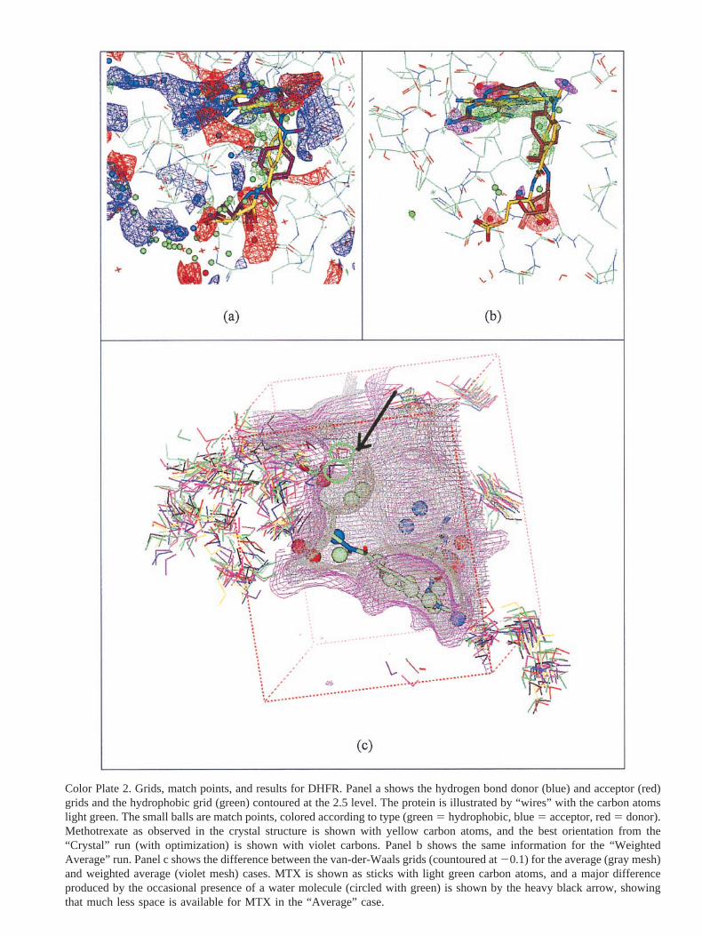
Color Plate 2. Grids, match points, and results for DHFR. Panel a shows the hydrogen bond donor (blue) and acceptor (red)grids and the hydrophobic grid (green) contoured at the 2.5 level. The protein is illustrated by “wires” with the carbon atomslight green. The small balls are match points, colored according to type (green5 hydrophobic, blue5 acceptor, red5 donor).Methotrexate as observed in the crystal structure is shown with yellow carbon atoms, and the best orientation from the“Crystal” run (with optimization) is shown with violet carbons. Panel b shows the same information for the “WeightedAverage” run. Panel c shows the difference between the van-der-Waals grids (countoured at20.1) for the average (gray mesh)and weighted average (violet mesh) cases. MTX is shown as sticks with light green carbon atoms, and a major differenceproduced by the occasional presence of a water molecule (circled with green) is shown by the heavy black arrow, showingthat much less space is available for MTX in the “Average” case.
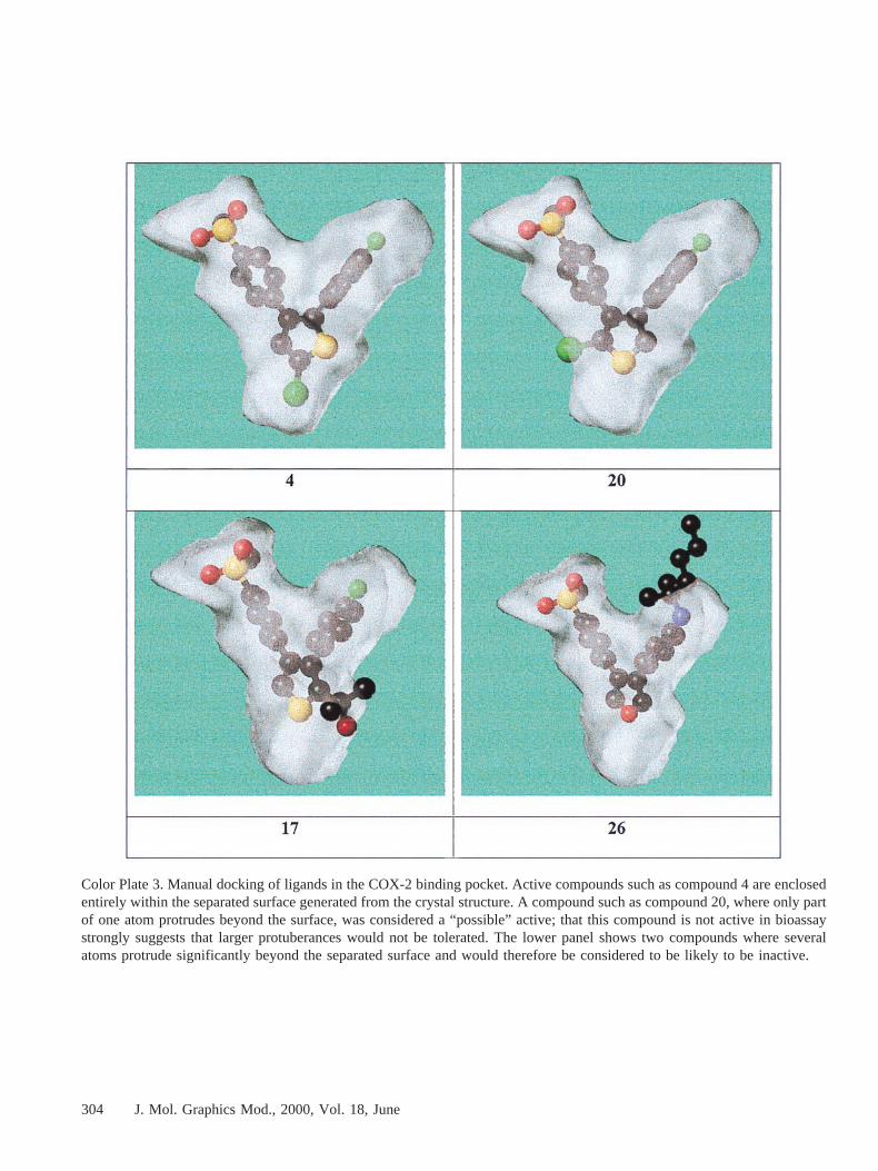
Color Plate 3. Manual docking of ligands in the COX-2 binding pocket. Active compounds such as compound 4 are enclosedentirely within the separated surface generated from the crystal structure. A compound such as compound 20, where only partof one atom protrudes beyond the surface, was considered a “possible” active; that this compound is not active in bioassaystrongly suggests that larger protuberances would not be tolerated. The lower panel shows two compounds where severalatoms protrude significantly beyond the separated surface and would therefore be considered to be likely to be inactive.
304 J. Mol. Graphics Mod., 2000, Vol. 18, June