Embed Size (px)
Citation preview
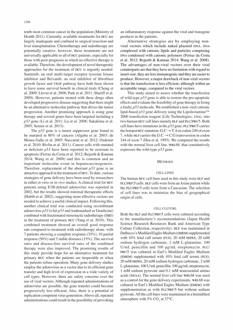
Sains Malaysiana 46(8)(2017): 1289–1297 http://dx.doi.org/10.17576/jsm-2017-4608-15
A Gain of Function p53 Gene Mutant Promotes Growth Suppression in Human Liver Cancer Cells
(Pemulihan Fungsi Mutan Gen p53 Menggalakkan Penindasan Pertumbuhan Sel Kanser Hati Manusia)
NOR ADZIMAH JOHDI*, SITI NURMI NASIR & RAHMAN JAMAL
ABSTRACT
Primary liver cancer is one of the most common cancer in the world with highest cancer mortality rate. The most common type of primary liver cancer is hepatocellular carcinoma (HCC). There are many risk factors for liver cancer and currently available treatments for HCC are largely inadequate. Gene mutation and dysfunction of p53 are common and is recognized as an important molecular event in hepatocarcinogenesis. Therefore, replacement of the aberrant p53 gene is an attractive approach in the treatment of HCC providing an alternative treatment for primary HCC. In this study, we assessed whether the transfection with wild-type p53 gene is able to restore the pro-apoptotic effects and evaluate the feasibility of gene therapy in fixing a faulty p53 molecule. We established a non-viral cationic lipid-based p53 gene delivery into two human HCC cell lines namely HLF and PLC/PRF/5 cells. Both cell lines have mutations in the p53 gene. We compared the results with the normal liver cell line, WRL68, that constitutively expresses the wild-type p53 gene. In this study, the introduction of wild-type p53 gene into HLF and PLC/PRF/5 cells resulted in an increased of p53 gene expression, protein expression and cells growth inhibition shown in MTS reduction cell viability assay, FITC-Annexin V and PI apoptosis assay, western blot and caspase activity assay. In summary, the study provides a promising therapeutic approach for p53 gene delivery into HCC patients. The p53 gene delivery can be instituted together with chemotherapy as a combination treatment to induce apoptosis.
Keywords: Apoptosis; cell lines; hepatocellular carcinoma; p53 gene; transfection
ABSTRAK
Kanser hati utama adalah salah satu kanser umum di dunia dengan kadar kematian kanser tertinggi. Jenis yang paling biasa untuk kanser hati utama adalah karsinoma hepatosel (HCC). Terdapat banyak faktor risiko untuk kanser hati utama dan rawatan untuk HCC sebahagian besarnya tidak mencukupi. Mutasi gen dan kehilangan fungsi gen p53 adalah faktor yang paling biasa dan gen p53 diiktiraf sebagai antara rantaian molekul yang penting dalam proses pembentukan kanser hati. Oleh itu, pemulihan mutan gen p53 yang defektif adalah satu pendekatan yang menarik dalam rawatan HCC sebagai rawatan alternatif untuk HCC utama. Dalam kajian ini, penilaian dilakukan sama ada transfeksi gen p53 normal ke atas titisan sel HCC mampu mengembalikan kesan pro-apoptotik dan potensi terapi gen dalam pemulihan molekul p53 yang defektif. Ini melibatkan transfeksi gen p53 normal menggunakan teknik lipid kationik kepada dua jenis titisan sel HCC manusia iaitu HLF dan PLC/PRF/5. Kedua-dua jenis titisan sel ini mempunyai mutasi dalam gen p53. Keputusan kajian dibandingkan dengan titisan sel hati normal, WRL68, yang mengekspres gen p53 normal. Transfeksi gen p53 normal ke dalam titisan sel HLF dan PLC/PRF/5 menyebabkan peningkatan ekspresi gen p53, pertambahan ekspresi protein p53 dan perencatan pertumbuhan titisan sel-sel yang ditunjukkan dalam assai MTS sel pengurangan daya maju, assai apoptosis FITC-Annexin V & PI, pemendapan barat dan aktiviti caspase. Secara ringkasnya, kajian ini menyediakan pendekatan terapeutik yang boleh digunakan untuk menyalurkan gen p53 ke dalam sel pesakit HCC. Penyampaian gen p53 boleh dimulakan bersama dengan kemoterapi sebagai rawatan kombinasi untuk mendorong aktiviti apoptosis.
Kata kunci: Apoptosis; gen p53; hepatosel karsinoma; titisan sel; transfeksi
INTRODUCTION
Primary liver cancer is the sixth most common cancer in the world with an estimate of 0.8 million people diagnosed annually (C.P 2014). It also accounts for one of the highest cancer mortality rates with 0.7 million deaths (9%) per year. The most common type of primary liver cancer is hepatocellular carcinoma (HCC). The major risk factors for liver cancer include chronic infection with hepatitis B and C (accounting for 54% and 31% of cases worldwide,
respectively), the consumption of food contaminated with aflatoxin (produced by fungi which can contaminate food such as maize and nuts in tropical or sub-tropical countries) and heavy alcohol consumption (Chuang et al. 2009; Morgan et al. 2004; Parkin 2006; Shariff et al. 2009). The developing countries carry the biggest burden of liver cancer, with more than eight out of ten (84%) cases being diagnosed in 2008. In Malaysia, liver cancer is the fifth most common cancer in males and the
1290
tenth most common cancer in the population (Ministry of Health 2011). Currently, available treatments for HCC are largely inadequate and confined to surgical resection and liver transplantation. Chemotherapy and radiotherapy are potentially curative, however, these treatments are not universally applicable to all of HCC patients, especially for those with poor prognosis in which no effective therapy is available. Therefore, the development of novel therapeutic approaches for the treatment of HCC is urgently needed. Sunitinib, an oral multi-target receptor tyrosine kinase inhibitor and Brivanib, an oral inhibitor of fibroblast growth factor and VEGF pathway have both been shown to have some survival benefit in clinical trials (Cheng et al. 2009; Llovet et al. 2008; Park et al. 2011; Shariff et al. 2009). However, patients treated with these drugs often developed progressive disease suggesting that there might be an alternative molecular pathway that drives the tumor progression. Another promising approach is using gene therapy and several genes have been targeted including a p53 gene (Li et al. 2011; Lu et al. 2008; Sakakima et al. 2005; Senzer et al. 2013). The p53 gene is a tumor suppressor gene found to be mutated in 80% of cancers (Algaba et al. 2003; de Moura Gallo et al. 2005; Naccarati et al. 2012; Ozdemir et al. 2010; Rivlin et al. 2011). Cancer cells with mutated or deficient p53 have been reported to be resistant to apoptosis (Ferraz da Costa et al. 2012; Rejeeth & Kannan 2014; Wang et al. 2008) and this is common and an important molecular event in hepatocarcinogenesis. Therefore, replacement of the aberrant p53 gene is an attractive approach in the treatment of HCC. To date, various strategies of gene delivery have been used by researchers in either in vitro or in vivo studies. A clinical trial for HCC patients using E1B-deleted adenovirus was reported in 2002, but the results showed minimal therapeutic effects (Habib et al. 2002), suggesting more effective vectors are needed to achieve a useful clinical impact. Following this, another clinical trial was conducted using recombinant adenovirus-p53 (rAd-p53 and trademarked as Gendicine) combined with fractionated stereotactic radiotherapy (fSRT) in the treatment of primary HCC (Yang et al. 2010). This combined treatment showed an overall good response rate compared to treatment with radiotherapy alone, with 7 patients showing a complete response (35%), 10 partial response (50%) and 3 stable diseases (15%). The survival rates and disease-free survival rates of the combined therapy were also improved. The promising results of this study provide hope for an alternative treatment for primary HCC when the patients are inoperable or when the patients refuse operation. Many gene delivery studies employ the adenovirus as a vector due to its efficient gene transfer and high level of expression in a wide variety of cell types. However, there are safety concerns over the use of viral vectors. Although repeated administrations of adenovirus are possible, the gene transfer could become progressively less efficient. Also, there is a potential of replication competent virus generation. Above all, repeated administrations could result in the possibility of provoking
an inflammatory response against the viral and transgene products in the patients. Alternative strategies are by employing non-viral vectors which include naked plasmid DNA, DNA complexed with cationic lipids and particles comprising DNA condensed with cationic polymers (Ferraz da Costa et al. 2012; Rejeeth & Kannan 2014; Wang et al. 2008). The advantages of non-viral vectors over their viral counterparts are that they have no limitation with regard to insert-size, they are less immunogenic and they are easier to produce. However, a major drawback of non-viral vectors is that the transfection is less efficient, although within an acceptable range, compared to the viral vectors. This study aimed to assess whether the transfection of wild-type p53 gene is able to restore the pro-apoptotic effects and evaluate the feasibility of gene therapy in fixing a faulty p53 molecule. We established a non-viral cationic lipid-based p53 gene delivery using the Lipofectamine™ 2000 transfection reagent (Life Technologies, USA), into two human HCC cell lines namely HLF and PLC/PRF/5. Both cell lines have mutations in the p53 gene. PLC/PRF/5 carries the hotspot HCC mutation: G:C → T:A in codon 249 of exon 7, while HLF carries the G:C → C:G transversion in codon 244 of exon 7 (Hsu et al. 1993). We compared the results with the normal liver cell line, WRL68, that constitutively expresses the wild-type p53 gene.
METHODS
CELL LINES
The human HCC cell lines used in this study were HLF and PLC/PRF/5 cells. HLF cells were from an Asian patient while the PLC/PRF/5 cells were from a Caucasian. The selection of cell lines was to minimize the bias of geographical origin of cells.
CELL CULTURE
Both the HLF and PLC/PRF/5 cells were cultured according to the manufacturer’s recommendations (Japan Health Science Research Resources Bank and American Type Culture Collection, respectively). HLF was maintained in Dulbecco’s Modified Eagles Medium (DMEM) supplemented with 10% fetal calf serum (FCS), 20 mM HEPES, 20 mM sodium hydrogen carbonate, 2 mM L-glutamine, 100 U/mL penicillin and 100 μg/mL streptomycin. PLC/PRF/5 was cultured in Earl’s Modified Eagles Medium (EMEM) supplemented with 10% fetal calf serum (FCS), 20 mM HEPES, 20 mM sodium hydrogen carbonate, 2 mM L-glutamine, 100 U/mL penicillin, 100 μg/mL streptomycin, 1 mM sodium pyruvate and 0.1 mM nonessential amino acids (NEAA). The normal liver cell line WRL68 was used as a control for the gene delivery experiments. WRL68 was cultured in Earl’s Modified Eagles Medium (EMEM) with supplementation as with PLC/PRF/5 but without sodium pyruvate. All the cell lines were maintained in a humidified atmosphere with 5% CO2 at 37°C.

1291
PLASMIDS
The plasmid 11770:p53-GFP containing wild-type p53 gene and tagged with GFP (Addgene, USA) was expanded in DH5α cells (Invitrogen, USA) and purified using the Plasmid Mini Kit (Qiagen, USA). The host plasmid, pEGFP-N1 (Addgene, USA) was used as a control. The plasmid was digested with restriction enzymes SalI and BamHI in reference to the vector map and was visualized by 1% agarose gel.
WILD-TYPE p53 GENE TRANSFECTION
The transient transfections were performed using the Lipofectamine® 2000 Transfection Reagent and according to the manufacturer’s protocol (Life Technologies, USA). Cells were seeded a day before transfection at different densities according to the size of culture vessels and the respective assays; 1.5 × 10⁴ cells for the 96-well plate in MTS assay and Caspase 3/7 assay; 5.0 × 10⁵ cells for the 6-well plate for GFP quantification, FITC-Annexin V and Propidium Iodide (PI) double staining assay, RNA and protein extractions. On the following day, the culture medium was replaced and cells were treated with serum-free media with either plasmid p53-GFP complexed with LipofectamineÔ 2000 transfection reagent or host plasmid complexed with LipofectamineÔ 2000 transfection reagent at a ratio 1:1. Untreated cells and cells treated with LipofectamineÔ 2000 transfection reagent alone served as negative controls. Transfection was allowed to take place for 48 h and the cells were harvested, analyzed and assayed accordingly.
SCORING OF GFP-POSITIVE CELLS
Transfection efficiency was scored and visualized by a confocal microscope (Leica TCS SP5 II, Leica Microsystems) at excitation wavelength 488 nm after 48 h of transfection. Following this, transfected cells were quantified using the FACS Aria II(BD Biosciences, USA) and BD FACSDiva Software (Becton Dickinson, USA).
QUANTITATIVE REAL TIME-POLYMERASE CHAIN REACTION (QRT-PCR)
Total RNA was extracted from the transfected cell lines 48 h post transfection using RNeasy Mini Kit (Qiagen, USA) according to the manufacturer’s instructions. The RNA concentrations were measured using a Bioanalyzer (Agilent 2100, USA). First strand cDNA was synthesized using Reverse Transcription System (Bio-Rad, USA) according to manufacturer’s protocols. The qRT-PCR was performed using Rotor-Gene 600 (Corbett Lifescience, USA) and the p53 gene was amplified using the primer set for exon 7 (171 bp): 5’-GGCCTCATCTTGGGCCTGTG-3’ and 5’-CAGTGTGCAGGGTGGCAAGT-3’. b-actin (395 bp) was used as the housekeeping gene control and the primer set used was: 5’-AGGCTGTGCTGTCCCTGTATGC-3’ and 5’-ACCCAAGAAGGAAGGCTGGAAA-3’. Amplification was performed at an annealing temperature of 66°C. Experiments were done n triplicates.
WESTERN BLOTTING ANALYSIS
All antibodies used in this study were purchased from Santa Cruz Biotechnology, USA unless stated otherwise. Total protein was extracted from the transfected cells 48 h post transfection using the RIPA lysis buffer. Following protein quantitation, proteins were separated on SDS-PAGE and transferred onto polyvinyl difluoride membrane according to standard methods. The membrane was blocked in Tris-buffered saline containing skimmed milk prior to probing with primary antibodies against p53, p21 and b-actin (as a loading control) proteins. After serial washes with Tris-buffered saline, the membrane was incubated with horseradish peroxidase-conjugated secondary antibodies. Immunoreactive proteins were detected using an enhanced chemiluminescence system (ChemiDocTM Imaging System, Bio-Rad).
MTS REDUCTION CELL VIABILITY ASSAY
Cell viability was assessed using MTS CellTiter 96® Nonradioactive Cell Proliferation Assay according to the manufacturer’s protocol (Promega, USA). Briefly, cells were seeded in 96-well cell culture plates at 1 × 10⁴ cells per well. After 48 h post-transfection, the medium was replaced and incubated with 20 uL with 3-(4,5-dimethylthiazol-2-yl)-5-(3-carboxymethoxyphenyl)-2-(4-sulfophenyl)-2H-tetrazolium (MTS) reagent at 37°C for 4 h and absorbance was measured using a spectrophotometer at wavelength 490 nm. All assays were performed in triplicate and repeated in three independent experiments.
FITC-ANNEXIN V AND PI APOPTOSIS ASSAY
48 h post-transfection, both attached and floating cells were collected, washed twice with PBS and stained with FITC-Annexin V Apoptosis Detection Kit (Sigma, USA) according to the manufacturer’s protocol. Basically, 5 μL of FITC-Annexin V conjugate and 10 μL of PI solution were added to each cell suspension, followed by incubation at room temperature for exactly 10 min and protected from light. After double supravital staining, the samples were immediately analyzed using FACS Aria II (BD Biosciences, USA) and BD FACSDiva Software (Becton Dickinson, USA).
CASPASE ACTIVITY ASSAY
Following 48 h post transfection, the activities of caspase 3 and 7 were measured using the Caspase-Glo® 3/7 Assay assay kit according to the manufacturer’s recommendation (Promega, Madison USA). Briefly, the plates containing cells were treated with 100 μL of Caspase-Glo and then gently mixed on a plate shaker at 300-500 rpm for 30 s. The plate was then incubated at room temperature for 2 h. The luminescence of each sample was measured using a plate-reading luminometer (ThermoLabsystems, Thermo Fisher Scientific) with parameters of 1 min lag time and 0.5 s/well of read time. The experiments were performed in triplicates and repeated on two separate-initiated cultures.
1292
STATISTICAL ANALYSIS
All the statistical analyses were performed using GraphPad Prism 7 and statistics significance was calculated by student t-test. A p<0.01 was considered statistically significant.
RESULTS
PLASMID p53-GFP
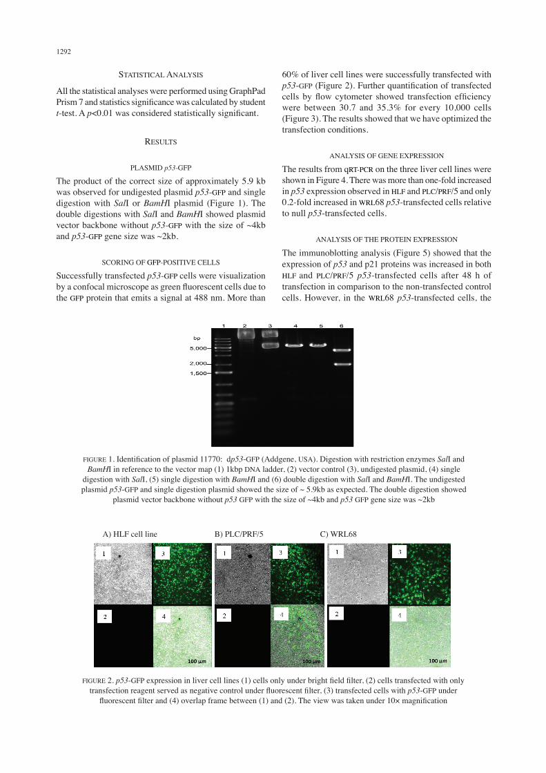
The product of the correct size of approximately 5.9 kb was observed for undigested plasmid p53-GFP and single digestion with SalI or BamHI plasmid (Figure 1). The double digestions with SalI and BamHI showed plasmid vector backbone without p53-GFP with the size of ~4kb and p53-GFP gene size was ~2kb.
SCORING OF GFP-POSITIVE CELLS
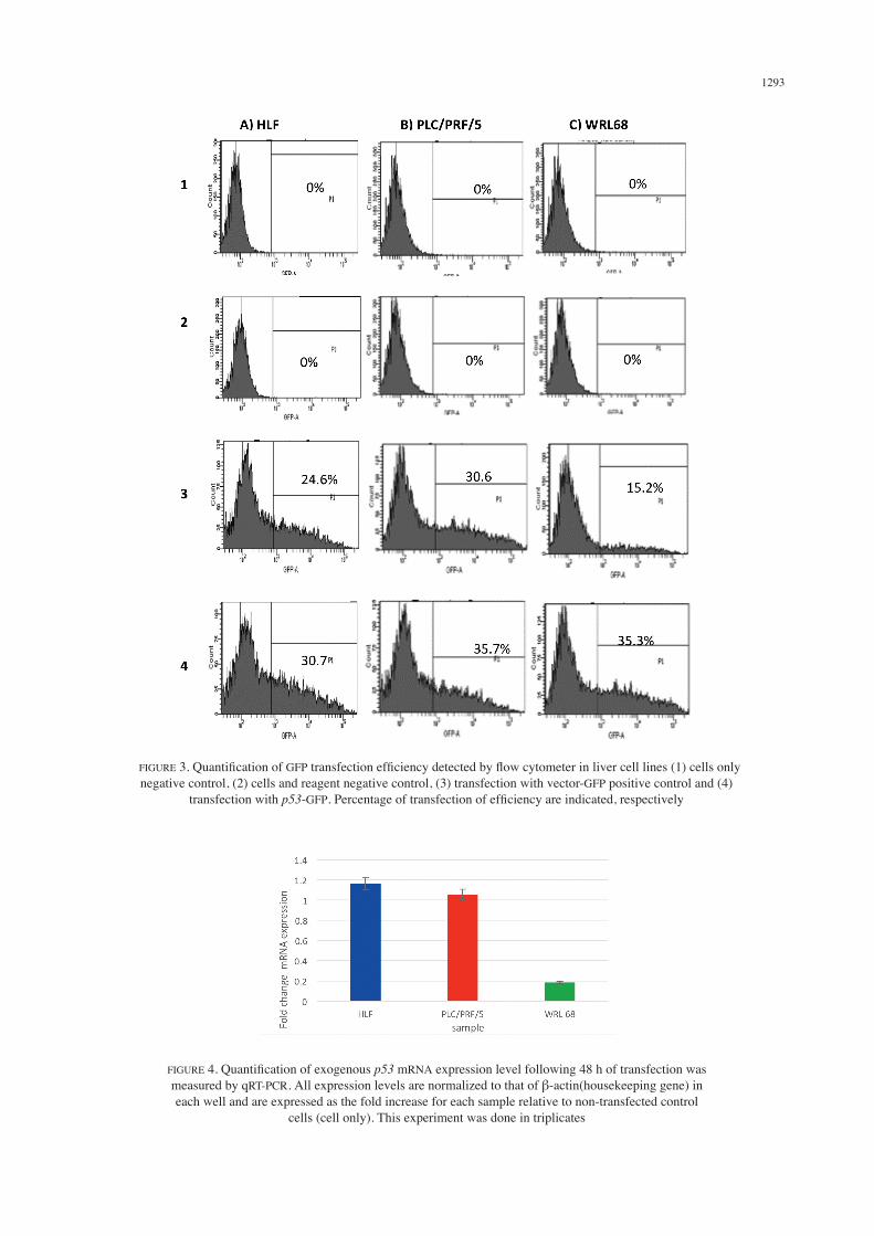
Successfully transfected p53-GFP cells were visualization by a confocal microscope as green fluorescent cells due to the GFP protein that emits a signal at 488 nm. More than
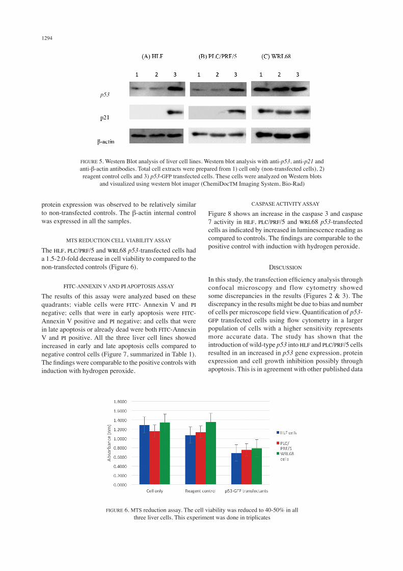
60% of liver cell lines were successfully transfected with p53-GFP (Figure 2). Further quantification of transfected cells by flow cytometer showed transfection efficiency were between 30.7 and 35.3% for every 10,000 cells (Figure 3). The results showed that we have optimized the transfection conditions.
ANALYSIS OF GENE EXPRESSION
The results from qRT-PCR on the three liver cell lines were shown in Figure 4. There was more than one-fold increased in p53 expression observed in HLF and PLC/PRF/5 and only 0.2-fold increased in WRL68 p53-transfected cells relative to null p53-transfected cells.
ANALYSIS OF THE PROTEIN EXPRESSION
The immunoblotting analysis (Figure 5) showed that the expression of p53 and p21 proteins was increased in both HLF and PLC/PRF/5 p53-transfected cells after 48 h of transfection in comparison to the non-transfected control cells. However, in the WRL68 p53-transfected cells, the
FIGURE 1. Identification of plasmid 11770: dp53-GFP (Addgene, USA). Digestion with restriction enzymes SalI and BamHI in reference to the vector map (1) 1kbp DNA ladder, (2) vector control (3), undigested plasmid, (4) single
digestion with SalI, (5) single digestion with BamHI and (6) double digestion with SalI and BamHI. The undigested plasmid p53-GFP and single digestion plasmid showed the size of ~ 5.9kb as expected. The double digestion showed
plasmid vector backbone without p53 GFP with the size of ~4kb and p53 GFP gene size was ~2kb
A) HLF cell line B) PLC/PRF/5 C) WRL68
FIGURE 2. p53-GFP expression in liver cell lines (1) cells only under bright field filter, (2) cells transfected with only transfection reagent served as negative control under fluorescent filter, (3) transfected cells with p53-GFP under
fluorescent filter and (4) overlap frame between (1) and (2). The view was taken under 10× magnification
1293
FIGURE 3. Quantification of GFP transfection efficiency detected by flow cytometer in liver cell lines (1) cells only negative control, (2) cells and reagent negative control, (3) transfection with vector-GFP positive control and (4)
transfection with p53-GFP. Percentage of transfection of efficiency are indicated, respectively
FIGURE 4. Quantification of exogenous p53 mRNA expression level following 48 h of transfection was measured by qRT-PCR. All expression levels are normalized to that of β-actin(housekeeping gene) in each well and are expressed as the fold increase for each sample relative to non-transfected control
cells (cell only). This experiment was done in triplicates
1294
protein expression was observed to be relatively similar to non-transfected controls. The β-actin internal control was expressed in all the samples.
MTS REDUCTION CELL VIABILITY ASSAY
The HLF, PLC/PRF/5 and WRL68 p53-transfected cells had a 1.5-2.0-fold decrease in cell viability to compared to the non-transfected controls (Figure 6).
FITC-ANNEXIN V AND PI APOPTOSIS ASSAY
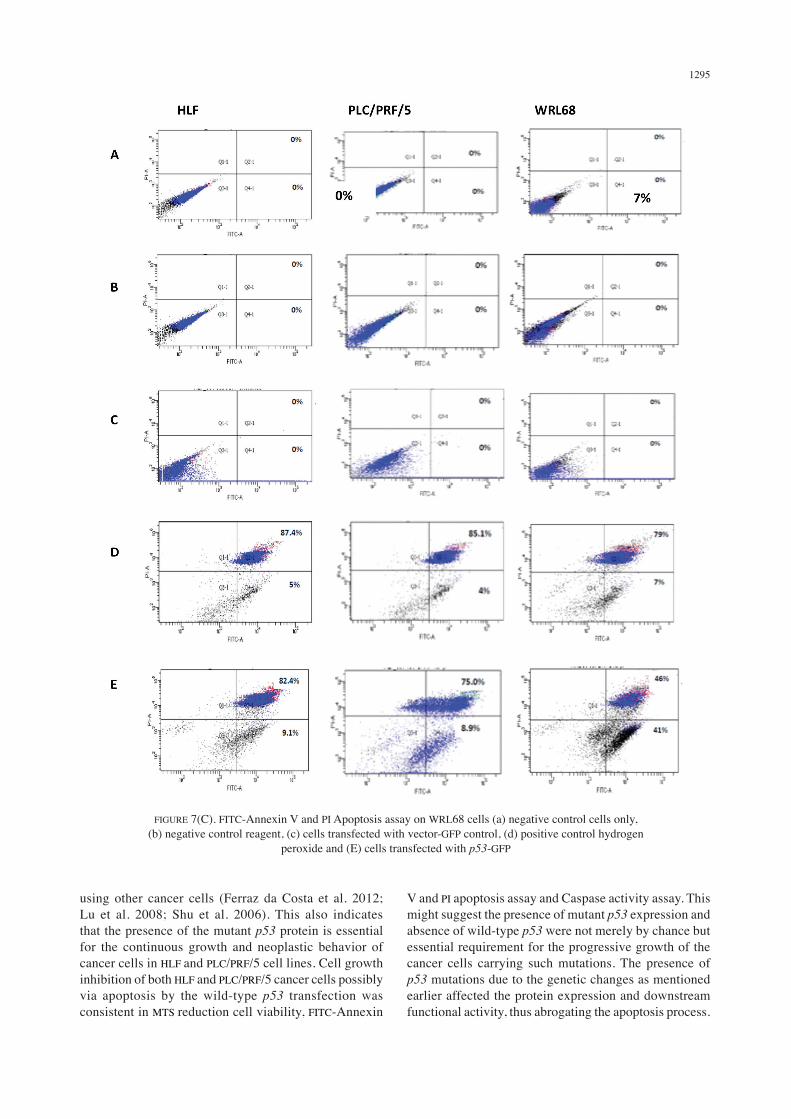
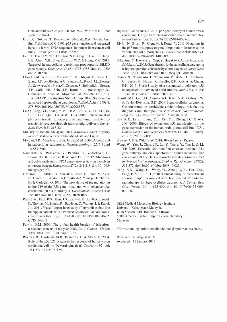
The results of this assay were analyzed based on these quadrants; viable cells were FITC- Annexin V and PI negative; cells that were in early apoptosis were FITC-Annexin V positive and PI negative; and cells that were in late apoptosis or already dead were both FITC-Annexin V and PI positive. All the three liver cell lines showed increased in early and late apoptosis cells compared to negative control cells (Figure 7, summarized in Table 1). The findings were comparable to the positive controls with induction with hydrogen peroxide.
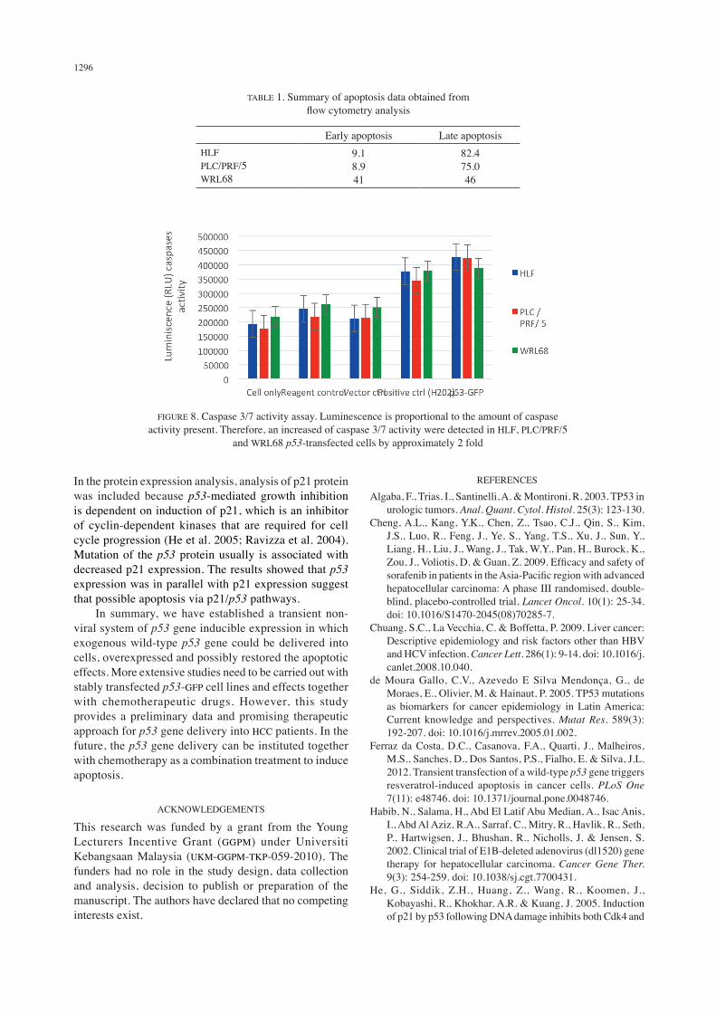
CASPASE ACTIVITY ASSAY
Figure 8 shows an increase in the caspase 3 and caspase 7 activity in HLF, PLC/PRF/5 and WRL68 p53-transfected cells as indicated by increased in luminescence reading as compared to controls. The findings are comparable to the positive control with induction with hydrogen peroxide.
DISCUSSION
In this study, the transfection efficiency analysis through confocal microscopy and flow cytometry showed some discrepancies in the results (Figures 2 & 3). The discrepancy in the results might be due to bias and number of cells per microscope field view. Quantification of p53-GFP transfected cells using flow cytometry in a larger population of cells with a higher sensitivity represents more accurate data. The study has shown that the introduction of wild-type p53 into HLF and PLC/PRF/5 cells resulted in an increased in p53 gene expression, protein expression and cell growth inhibition possibly through apoptosis. This is in agreement with other published data
FIGURE 6. MTS reduction assay. The cell viability was reduced to 40-50% in all three liver cells. This experiment was done in triplicates
FIGURE 5. Western Blot analysis of liver cell lines. Western blot analysis with anti-p53, anti-p21 and anti-β-actin antibodies. Total cell extracts were prepared from 1) cell only (non-transfected cells), 2) reagent control cells and 3) p53-GFP transfected cells. These cells were analyzed on Western blots
and visualized using western blot imager (ChemiDocTM Imaging System, Bio-Rad)
p53
1295
FIGURE 7(C). FITC-Annexin V and PI Apoptosis assay on WRL68 cells (a) negative control cells only, (b) negative control reagent, (c) cells transfected with vector-GFP control, (d) positive control hydrogen
peroxide and (E) cells transfected with p53-GFP
using other cancer cells (Ferraz da Costa et al. 2012; Lu et al. 2008; Shu et al. 2006). This also indicates that the presence of the mutant p53 protein is essential for the continuous growth and neoplastic behavior of cancer cells in HLF and PLC/PRF/5 cell lines. Cell growth inhibition of both HLF and PLC/PRF/5 cancer cells possibly via apoptosis by the wild-type p53 transfection was consistent in MTS reduction cell viability, FITC-Annexin
V and PI apoptosis assay and Caspase activity assay. This might suggest the presence of mutant p53 expression and absence of wild-type p53 were not merely by chance but essential requirement for the progressive growth of the cancer cells carrying such mutations. The presence of p53 mutations due to the genetic changes as mentioned earlier affected the protein expression and downstream functional activity, thus abrogating the apoptosis process.
1296
In the protein expression analysis, analysis of p21 protein was included because p53-mediated growth inhibition is dependent on induction of p21, which is an inhibitor of cyclin-dependent kinases that are required for cell cycle progression (He et al. 2005; Ravizza et al. 2004). Mutation of the p53 protein usually is associated with decreased p21 expression. The results showed that p53 expression was in parallel with p21 expression suggest that possible apoptosis via p21/p53 pathways. In summary, we have established a transient non-viral system of p53 gene inducible expression in which exogenous wild-type p53 gene could be delivered into cells, overexpressed and possibly restored the apoptotic effects. More extensive studies need to be carried out with stably transfected p53-GFP cell lines and effects together with chemotherapeutic drugs. However, this study provides a preliminary data and promising therapeutic approach for p53 gene delivery into HCC patients. In the future, the p53 gene delivery can be instituted together with chemotherapy as a combination treatment to induce apoptosis.
ACKNOWLEDGEMENTS
This research was funded by a grant from the Young Lecturers Incentive Grant (GGPM) under Universiti Kebangsaan Malaysia (UKM-GGPM-TKP-059-2010). The funders had no role in the study design, data collection and analysis, decision to publish or preparation of the manuscript. The authors have declared that no competing interests exist.
REFERENCES
Algaba, F., Trias, I., Santinelli, A. & Montironi, R. 2003. TP53 in urologic tumors. Anal. Quant. Cytol. Histol. 25(3): 123-130.
Cheng, A.L., Kang, Y.K., Chen, Z., Tsao, C.J., Qin, S., Kim, J.S., Luo, R., Feng, J., Ye, S., Yang, T.S., Xu, J., Sun, Y., Liang, H., Liu, J., Wang, J., Tak, W.Y., Pan, H., Burock, K., Zou, J., Voliotis, D. & Guan, Z. 2009. Efficacy and safety of sorafenib in patients in the Asia-Pacific region with advanced hepatocellular carcinoma: A phase III randomised, double-blind, placebo-controlled trial. Lancet Oncol. 10(1): 25-34. doi: 10.1016/S1470-2045(08)70285-7.
Chuang, S.C., La Vecchia, C. & Boffetta, P. 2009. Liver cancer: Descriptive epidemiology and risk factors other than HBV and HCV infection. Cancer Lett. 286(1): 9-14. doi: 10.1016/j.canlet.2008.10.040.
de Moura Gallo, C.V., Azevedo E Silva Mendonça, G., de Moraes, E., Olivier, M. & Hainaut, P. 2005. TP53 mutations as biomarkers for cancer epidemiology in Latin America: Current knowledge and perspectives. Mutat Res. 589(3): 192-207. doi: 10.1016/j.mrrev.2005.01.002.
Ferraz da Costa, D.C., Casanova, F.A., Quarti, J., Malheiros, M.S., Sanches, D., Dos Santos, P.S., Fialho, E. & Silva, J.L. 2012. Transient transfection of a wild-type p53 gene triggers resveratrol-induced apoptosis in cancer cells. PLoS One 7(11): e48746. doi: 10.1371/journal.pone.0048746.
Habib, N., Salama, H., Abd El Latif Abu Median, A., Isac Anis, I., Abd Al Aziz, R.A., Sarraf, C., Mitry, R., Havlik, R., Seth, P., Hartwigsen, J., Bhushan, R., Nicholls, J. & Jensen, S. 2002. Clinical trial of E1B-deleted adenovirus (dl1520) gene therapy for hepatocellular carcinoma. Cancer Gene Ther. 9(3): 254-259. doi: 10.1038/sj.cgt.7700431.
He, G., Siddik, Z.H., Huang, Z., Wang, R., Koomen, J., Kobayashi, R., Khokhar, A.R. & Kuang, J. 2005. Induction of p21 by p53 following DNA damage inhibits both Cdk4 and
TABLE 1. Summary of apoptosis data obtained from flow cytometry analysis
Early apoptosis Late apoptosisHLFPLC/PRF/5WRL68
9.18.941
82.475.046
FIGURE 8. Caspase 3/7 activity assay. Luminescence is proportional to the amount of caspase activity present. Therefore, an increased of caspase 3/7 activity were detected in HLF, PLC/PRF/5
and WRL68 p53-transfected cells by approximately 2 fold
1297
Cdk2 activities. Oncogene 24(18): 2929-2943. doi: 10.1038/sj.onc.1208474.
Hsu, I.C., Tokiwa, T., Bennett, W., Metcalf, R.A., Welsh, J.A., Sun, T. & Harris, C.C. 1993. p53 gene mutation and integrated hepatitis B viral DNA sequences in human liver cancer cell lines. Carcinogenesis 14(5): 987-992.
Li, L.Y., Dai, H.Y., Yeh, F.L., Kan, S.F., Lang, J., Hsu, J.L., Jeng, L.B., Chen, Y.H., Sher, Y.P., Lin, W.C. & Hung, M.C. 2011. Targeted hepatocellular carcinoma proapoptotic BikDD gene therapy. Oncogene 30(15): 1773-1783. doi: 10.1038/onc.2010.558.
Llovet, J.M., Ricci, S., Mazzaferro, V., Hilgard, P., Gane, E., Blanc, J.F., de Oliveira, A.C., Santoro, A., Raoul, J.L., Forner, A., Schwartz, M., Porta, C., Zeuzem, S., Bolondi, L., Greten, T.F., Galle, P.R., Seitz, J.F., Borbath, I., Häussinger, D., Giannaris, T., Shan, M., Moscovici, M., Voliotis, D., Bruix, J. & SHARP Investigators Study Group. 2008. Sorafenib in advanced hepatocellular carcinoma. N. Engl. J. Med. 359(4): 378-390. doi: 10.1056/NEJMoa0708857.
Lu, Q., Teng, G.J., Zhang, Y., Niu, H.Z., Zhu, G.Y., An, Y.L., Yu, H., Li, G.Z., Qiu, D.H. & Wu, C.G. 2008. Enhancement of p53 gene transfer efficiency in hepatic tumor mediated by transferrin receptor through trans-arterial delivery. Cancer Biol. Ther. 7(2): 218-224.
Ministry of Health, Malaysia. 2011. National Cancer Registry Report, Malaysia Cancer Statistics-Data and Figure.
Morgan, T.R., Mandayam, S. & Jamal, M.M. 2004. Alcohol and hepatocellular carcinoma. Gastroenterology 127(5 Suppl 1): S87-S96.
Naccarati, A., Polakova, V., Pardini, B., Vodickova, L., Hemminki, K., Kumar, R. & Vodicka, P. 2012. Mutations and polymorphisms in TP53 gene--an overview on the role in colorectal cancer. Mutagenesis 27(2): 211-218. doi: 10.1093/mutage/ger067.
Ozdemir, F.T., Tiftikci, A., Sancak, S., Eren, F., Tahan, V., Akın, H., Gündüz, F., Kedrah, A.E., Ustündağ, Y., Avşar, E., Tözün, N. & Ozdoğan, O. 2010. The prevalence of the mutation in codon 249 of the P53 gene in patients with hepatocellular carcinoma (HCC) in Turkey. J. Gastrointest. Cancer 41(3): 185-189. doi: 10.1007/s12029-010-9140-5.
Park, J.W., Finn, R.S., Kim, J.S., Karwal, M., Li, R.K., Ismail, F., Thomas, M., Harris, R., Baudelet, C., Walters, I. & Raoul, J.L. 2011. Phase II, open-label study of brivanib as first-line therapy in patients with advanced hepatocellular carcinoma. Clin. Cancer Res. 17(7): 1973-1983. doi: 10.1158/1078-0432.CCR-10-2011.
Parkin, D.M. 2006. The global health burden of infection-associated cancers in the year 2002. Int. J. Cancer 118(12): 3030-3044. doi: 10.1002/ijc.21731.
Ravizza, R., Gariboldi, M.B., Passarelli, L. & Monti, E. 2004. Role of the p53/p21 system in the response of human colon carcinoma cells to Doxorubicin. BMC Cancer 4: 92. doi: 10.1186/1471-2407-4-92.
Rejeeth, C. & Kannan, S. 2014. p53 gene therapy of human breast carcinoma: Using a transferrin-modified silica nanoparticles. Breast Cancer. doi: 10.1007/s12282-014-0537-z.
Rivlin, N., Brosh, R., Oren, M. & Rotter, V. 2011. Mutations in the p53 tumor suppressor gene: Important milestones at the various steps of tumorigenesis. Genes Cancer 2(4): 466-474. doi: 10.1177/1947601911408889.
Sakakima, Y., Hayashi, S., Yagi, Y., Hayakawa, A., Tachibana, K. & Nakao, A. 2005. Gene therapy for hepatocellular carcinoma using sonoporation enhanced by contrast agents. Cancer Gene Ther. 12(11): 884-889. doi: 10.1038/sj.cgt.7700850.
Senzer, N., Nemunaitis, J., Nemunaitis, D., Bedell, C., Edelman, G., Barve, M., Nunan, R., Pirollo, K.F., Rait, A. & Chang, E.H. 2013. Phase I study of a systemically delivered p53 nanoparticle in advanced solid tumors. Mol. Ther. 21(5): 1096-1103. doi: 10.1038/mt.2013.32.
Shariff, M.I., Cox, I.J., Gomaa, A.I., Khan, S.A., Gedroyc, W. & Taylor-Robinson, S.D. 2009. Hepatocellular carcinoma: Current trends in worldwide epidemiology, risk factors, diagnosis and therapeutics. Expert Rev. Gastroenterol. Hepatol. 3(4): 353-367. doi: 10.1586/egh.09.35.
Shu, K.X., Li, B., Liang, Y.L., Xie, Y.F., Zhang, J.C. & Wei, J.M. 2006. Effects of exogenous p53 transfection on the gene expression in the human brain glioma cell line U251. Colloids Surf. B Biointerfaces 47(2): 126-131. doi: 10.1016/j.colsurfb.2005.12.005.
Stewart, C.P. & Wild, B.W. 2014. World Cancer Report.Wang, W., Yao, J., Zhou, J.P., Lu, Y., Wang, Y., Tao, L. & Li,
Y.P. 2008. Urocanic acid-modified chitosan-mediated p53 gene delivery inducing apoptosis of human hepatocellular carcinoma cell line HepG2 is involved in its antitumor effect in vitro and in vivo. Biochem. Biophys. Res. Commun. 377(2): 567-572. doi: 10.1016/j.bbrc.2008.10.023.
Yang, Z.X., Wang, D., Wang, G., Zhang, Q.H., Liu, J.M., Peng, P. & Liu, X.H. 2010. Clinical study of recombinant adenovirus-p53 combined with fractionated stereotactic radiotherapy for hepatocellular carcinoma. J. Cancer Res. Clin. Oncol. 136(4): 625-630. doi: 10.1007/s00432-009-0701-6.
UKM Medical Molecular Biology InstituteUniversiti Kebangsaan MalaysiaJalan Yaacob Latif, Bandar Tun Razak56000 Cheras, Kuala Lumpur, Federal TerritoryMalaysia
*Corresponding author; email: [email protected]
Received: 18 August 2016Accepted: 11 January 2017