Embed Size (px)
Citation preview
Correspondence
REFERENCES
1. Demierre MF, Kerl H, Willemze R. Primary cutaneous B cell lympho-mas: A practical approach. Hematol/Oncol Clin North Am. 2003;17:1333-50.
2. O’Connor WJ, Broadland DG. Merkel cell carcinoma. Dermatol Surg.1996;22:262-7.
3. Panda VB, Conway RM, Taylor SF. Primary cutaneous B cell lymphoma
Fig. 2—Microscopic examination of the eyelid specimen showvesiculated nuclei and prominent nucleoli (H&E, �100) (A). Higchemical stains were positive for CD20 (peroxidase antiperoxidain the neoplastic cells (abiding-biotin-peroxidase complex imm
with the highest total number of excavations, at 4.
e56 CAN J OPHTHALMOL—VOL. 47, NO. 6, DECEMBER 2012
4. Mak ST, Wong ACM, Tse RKK. Diffuse large B-cell lymphoma mas-querading as orbital cellulitis. HK Med J. 2010;16:484-6.
5. Huerva V, Canto LM, Marti M. Primary diffuse large B-cell lymphoma ofthe lower eyelid. Ophthal Plast Reconstr Surg. 2003;19:160-1.
Can J Ophthalmol 2012;47:e55–e560008-4182/11/$-see front matter © 2012 Canadian Ophthalmological Society.
Published by Elsevier Inc. All rights reserved.
numerous mononuclear cells, including large cells with round,magnification of the specimen (H&E, �200) (B). Immunohisto-
, �250) (C). Immunohistochemical stains were positive for Ki-67histochemical technique, �100) (D).
presenting as recurrent eyelid swelling. Clin Experiment Ophthalmol.2008;36:672-4. dx.doi.org/10.1016/j.jcjo.2012.06.006
A case of multiple focal choroidal excavations
Focal choroidal excavation was first described by Jam-pol et al.1 in 2006 under the name choroidal excavation.Since that time, a total of 16 cases have been reported,most recently under the label of focal choroidal excava-tion (FCE).1-4 In all instances, optical coherence to-mography (OCT) has demonstrated 1 or more focalareas of choroidal depression without any evidence ofscleral ectasia or staphyloma. The retina is found eitherto conform to the path of the choroid or to maintain itsnormal course and remain separated from the choroidby a hyporeflective area. In this article, we report thefirst instance of an FCE having both a conforming and anonconforming retina in the same eye, as well as a case
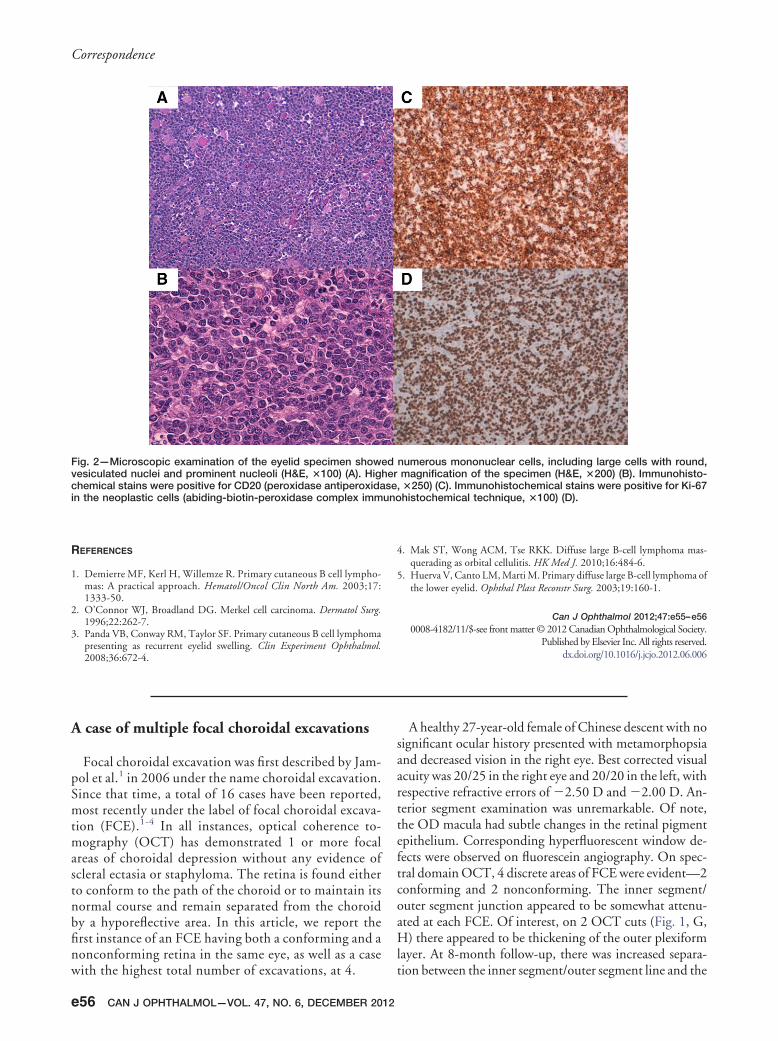
A healthy 27-year-old female of Chinese descent with nosignificant ocular history presented with metamorphopsiaand decreased vision in the right eye. Best corrected visualacuity was 20/25 in the right eye and 20/20 in the left, withrespective refractive errors of �2.50 D and �2.00 D. An-terior segment examination was unremarkable. Of note,the OD macula had subtle changes in the retinal pigmentepithelium. Corresponding hyperfluorescent window de-fects were observed on fluorescein angiography. On spec-tral domain OCT, 4 discrete areas of FCE were evident—2conforming and 2 nonconforming. The inner segment/outer segment junction appeared to be somewhat attenu-ated at each FCE. Of interest, on 2 OCT cuts (Fig. 1, G,H) there appeared to be thickening of the outer plexiformlayer. At 8-month follow-up, there was increased separa-
edherseuno
tion between the inner segment/outer segment line and the
spectral domain optical coherence tomography imaging. Apparentn in g and h. FCE, focal choroidal excavation.
Correspondence
retinal pigment epithelium in the 2 nonconforming le-sions. No case of conforming FCE converting to noncon-forming FCE has been documented, so long-term fol-low-up is needed to determine whether this is the naturalprogression of the disease.
A review of patient characteristics from the literaturereveals certain trends (Table 1). Including our case, themajority are young (mean 41 years old); myopic (76%);female (76%); and with unilateral involvement (94%).There does seem to be a preponderance of Asian patients(53%). Recognizing an obvious selection bias, we foundthat most patients are symptomatic (65%), with either de-creased vision or metamorphopsia. Pigmentary changeshave been observed in all eyes.
As demonstrated in cases reported by Margolis et al.2
and Abe et al.,3 vision may be significantly affected in thiscondition. One patient detailed by Margolis et al. had ahistory of central serous retinopathy and developed a cho-
Fig. 1—A healthy 27-year-old female of Chinese descent preseneye. The colour fundus photograph of the right eye (a) demons(b) appears normal. A red-free image of the right eye (c) highligangiography (d) demonstrates subtle hyperfluorescent areas inand nonconforming FCEs (e and f, respectively) are evident onthickening of the outer plexiform layer is seen in the cuts show
ted with metamorphopsia and slightly decreased vision in the righttrates subtle pigmentary changes in the macula, while the left eyehts the changes in the retinal pigment epithelium, and fluoresceinthe macula, corresponding to window defects. Both conforming
roidal neovascular membrane. The group notes that the
CA
Table 1—Patient characteristics in 17 cases of focal choroidalexcavation
Characteristics
Mean age in years (range) 41 (22–62)Female sex (%) 13 (76)Unilateral presentation (%) 16 (94)Conforming FCE (%)* 11/24 (46)*Race (%)
White 6 (35)Asian 9 (53)Black 1 (6)
Hispanic 1 (6)Symptomatic 11 (65)Myopia 13 (76)
FCE, focal choroidal excavation.*Three eyes have been reported to have 2 FCE lesions (1 patient was affectedbilaterally), and 1 (our case) had 4 lesions. Thus, 24 total lesions have been
reported in 18 eyes of 17 patients.N J OPHTHALMOL—VOL. 47, NO. 6, DECEMBER 2012 e57
Correspondence
choroid in patients with FCE, as in those with centralserous retinopathy, appears to be thickened. One wouldtypically expect myopic individuals to have a thinner cho-roid, so perhaps this relates to the pathophysiology of thedisease or, alternatively, may be a consequence of it.
Although the etiology of FCE is still unknown, in all like-lihood it may represent a congenital malformation. As morecases of this interesting condition are reported, we will de-velop a better understanding of its causes and evolution.
John C. Chen, R. Rishi GuptaDepartment of Ophthalmology, McGill University, Montreal, Que.
Correspondence to:
lesions (c).
e58 CAN J OPHTHALMOL—VOL. 47, NO. 6, DECEMBER 2012
REFERENCES
1. Jampol LM, Shankle J, Schroeder R, et al. Diagnostic and therapeuticchallenges. Retina. 2006;26:1072-6.
2. Margolis R, Mukkamala SK, Jampol LM, et al. The expanded spectrum offocal choroidal excavation. Arch Ophthalmol. 2011;129:1320-5.
3. Abe S, Yamamoto T, Kirii E, Yamashita H. Cup-shaped choroidal exca-vation detected by optical coherence tomography: A case report. RetinCases Brief Rep Falls. 2010;4:373-6.
4. Wakabayashi Y, Nishimura A, Higashide T, et al. Unilateral choroidalexcavation in the macula detected by spectral-domain optical coherencetomography. Acta Ophthalmol. 2010;88:e87-91.
Can J Ophthalmol 2012;47:e56–e580008-4182/11/$-see front matter © 2012 Canadian Ophthalmological Society.
Published by Elsevier Inc. All rights reserved.
John C. Chen, MD: [email protected] dx.doi.org/10.1016/j.jcjo.2012.07.006
Cotrimoxazole-resistant Nocardiasclerokeratitis: effective therapy withfourth-generation fluoroquinolones
Keratitis caused by Nocardia species makes up merely1.42%-1.7% of all microbial keratitis.1 Nocardia sclero-keratitis, though a rare complication, is usually successfullytreated by systemic cotrimoxazole therapy.2
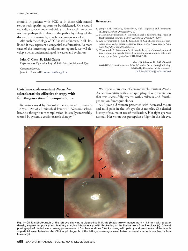
Fig. 1—Clinical photograph of the left eye showing a plaquedensity supero temporally and feathery margins inferonasallphotograph of the left eye showing prominence of 3 scleral nsuperficial vascularization (b). Clinical photograph of the lef
We report a rare case of cotrimoxazole-resistant Nocar-dia sclerokeratitis with a unique plaquelike presentationthat was successfully treated with amikacin and fourth-generation fluoroquinolones.
A 70-year-old woman presented with decreased visionand mild pain in the left eye for 2 months. She deniedhistory of trauma or use of medication. Her right eye wasnormal. Her vision was perception of light in the left eye.
e infiltrate (black arrow) measuring 8 � 7.5 mm with greaterith thickening at the limbus from 5 to 9 o’clock (a). Clinical
ules (black arrows) with patchy and less dense infiltrate withe showing a vascularized corneal scar with resolved sclera
-liky, wod
t ey


![Unilateral Choroidal Osteoma with Choroidal Neovascularization...Surgical evacuation of the choroidal neovascular membrane has been reported [12] but the visual outcome was not favorable](https://img.dokumen.tips/doc/110x75/6053732923e31173be575e28/unilateral-choroidal-osteoma-with-choroidal-neovascularization-surgical-evacuation.jpg)