Embed Size (px)
Citation preview

UNIVERSITATEA DE ŞTIINŢE AGRICOLE
ŞI MEDICINĂ VETERINARĂ A BANATULUI
TIMIŞOARA
FACULTATEA DE MEDICINĂ VETERINARĂ
LUCRĂRI ŞTIINŢIFICE
MEDICINĂ VETERINARĂ
TIMIŞOARA
VOLUMUL LII (4)
SCIENTIFICAL PAPERS
VETERINARY MEDICINE

EDITORIAL BOARD
Prof. VIOREL HERMAN, PhD, DVM - Faculty of Veterinary Medicine BUASVM Timisoara
Prof. ILEANA NICHITA, PhD, DVM – Faculty of Veterinary Medicine BUASVM Timisoara
Prof. SORIN MORARIU, PhD, DVM - Faculty of Veterinary Medicine BUASVM Timisoara
Prof. MARIUS PENTEA, PhD, DVM - Faculty of Veterinary Medicine BUASVM Timisoara
Lecturer DORU MORAR, PhD, DVM - Faculty of Veterinary Medicine BUASVM Timisoara
Prof. ION OPRESCU, PhD, DVM – Faculty of Veterinary Medicine BUASVM Timisoara
Prof. EMIL TIRZIU, PhD, DVM - Faculty of Veterinary Medicine BUASVM Timisoara
EDITOR-IN-CHIEF:
Assoc. Prof. NARCISA MEDERLE, PhD, DVM - Faculty of Veterinary Medicine BUASVM Timisoara
Editorial assistants:
Lecturer LILIANA CĂRPINIȘAN, PhD, DVM - Faculty of Veterinary Medicine BUASVM Timisoara
Lecturer ALINA GHIȘE, PhD, DVM - Faculty of Veterinary Medicine BUASVM Timisoara
Lecturer ADRIANA MORAR, PhD, DVM - Faculty of Veterinary Medicine BUASVM Timisoara
Assistant JELENA SAVICI, PhD, DVM - Faculty of Veterinary Medicine BUASVM Timisoara
Assistant MIHAELA PETCU, PhD, DVM - Faculty of Veterinary Medicine BUASVM Timisoara
Assoc. Prof. CORINA PASCU, PhD, DVM - Faculty of Veterinary Medicine BUASVM Timisoara
Lecturer IONICA IANCU, PhD, DVM - Faculty of Veterinary Medicine BUASVM Timisoara
Assistant IULIA BUCUR, PhD, DVM - Faculty of Veterinary Medicine BUASVM Timisoara
SCIENTIFIC ADVISORY COMMITTEE
Prof. DUSAN ORLIC, PhD, DVM - Scientific Veterinary Institute Novi Sad, Serbia
Prof. JOVAN BOJKOVSKI, PhD, DVM - Faculty of Veterinary Medicine, Belgrade, Serbia
Prof. IVAN PAVLOVIĆ, PhD, DVM - Scientific Veterinary Institute, Belgrade, Serbia
Prof. MANFRED GAREIS, PhD, DVM - Ludwig-Maximilians-Universität München, Germany
Prof. HANS WERNER KRUTSCH, PhD, DVM – Institute of Meet Science, Nurenberg, Germany
Prof. NICOLAE MANOLESCU, PhD, DVM, Dr. HC – Oncologic Institute ”Prof. dr. Al. Trestioreanu” Bucharest,
Corresponding member of Romanian Academy, Titular member of Romanian Academy of Medical Science,
Honorific member of Romanian Academy of Agricultural and Forestry Science
Prof. MIHAI DECUN, PhD, DVM – Faculty of Veterinary Medicine BUASVM Timisoara, Titular member of
Romanian Academy of Agricultural and Forestry Science
Prof. HORIA CERNESCU, PhD, DVM, Dr. HC - Faculty of Veterinary Medicine BUASVM Timisoara, Titular
member of Romanian Academy of Agricultural and Forestry Science, Member of BASeVA
Prof. GHEORGHE DARABUS, PhD, DVM – Faculty of Veterinary Medicine BUASVM Timisoara, Titular member
of Romanian Academy of Agricultural and Forestry Science
Prof. IOAN GROZA, PhD, DVM - Faculty of Veterinary Medicine UASVM Cluj Napoca
Prof. CORNEL CATOI, PhD, DVM - Faculty of Veterinary Medicine UASVM Cluj Napoca
Prof. VASILE COZMA, PhD, DVM - Faculty of V eterinary Medicine UASVM Cluj Napoca
Prof. GABRIEL PREDOI, PhD, DVM - Faculty of Veterinary Medicine UASVM Bucuresti
Prof. LIVIU MIRON, PhD, DVM - Faculty of Veterinary Medicine UASVM Iasi
Prof. GHEORGHE SĂVUTĂ, PhD, DVM - Faculty of Veterinary Medicine UASVM Iasi
Prof. ALEXANDRA TRIF, PhD, DVM, Dr. HC. - Faculty of Veterinary Medicine BUASVM Timisoara
Prof. NICOLAE CĂTANĂ, PhD, DVM - Faculty of Veterinary Medicine BUASVM Timisoara
Prof. ROMEO CRISTINA, PhD, DVM - Faculty of Veterinary Medicine BUASVM Timisoara
Prof. CORNEL IGNA, PhD, DVM - Faculty of Veterinary Medicine BUASVM Timisoara
Prof. MIHAI MAREȘ, PhD, DVM - Faculty of Veterinary Medicine UASVM Iasi
Assoc. Prof. FLORIN BETEG, PhD, DVM - Faculty of Veterinary Medicine UASVM Cluj Napoca
To be cited: LUCRARI STIINTIFICE: MEDICINA VETERINARA TIMISOARA (SCIENTIFICAL PAPERS: VETERINARY MEDICINE TIMISOARA), vol. LII (4), 2019
Available online at: http://www.usab-tm.ro/USAMVBT_Revista_ro_1086.html Indexed and/or abstracted in: CABI Full Text, CAB Abstracts, Ulrich's Periodicals Directory Editor: AGROPRINT TIMISOARA ISSN: 1221-5295 Printed by: IMPRIMERIA MIRTON TIMISOARA

UNIVERSITATEA DE ŞTIINŢE AGRICOLE
ŞI MEDICINĂ VETERINARĂ A BANATULUI
TIMIŞOARA
FACULTATEA DE MEDICINĂ VETERINARĂ
LUCRĂRI ŞTIINŢIFICE
MEDICINĂ VETERINARĂ
TIMIŞOARA
VOLUMUL LII (4)
SCIENTIFICAL PAPERS
VETERINARY MEDICINE
TIMIŞOARA
2019

LUCRĂRI ŞTIINŢIFICE MEDICINĂ VETERINARĂ VOL. LII(4), 2019, TIMIŞOARA
4
CHENOPODIUM QUINOA – AN ALTERNATIVE GRAIN IN FOOD AND FEED PRODUCTS
AHMADI M.
1, MEDERLE N.
1, MILOVANOV C.
1, MORARIU S.
1, NICHITA I.
1,
MORARIU F.1, PASCALAU R.
2, DRONCA D.
3
1Faculty of Veterinary Medicine, Banat’s University of Agricultural Sciences and Veterinary Medicine ”King Michael I of Romania” from Timisoara (BUASVM),
Timisoara – 300645, Calea Aradului, No. 119, Timisoara, Romania
2International relations Office, BUASVM, Timisoara, Romania
3Faculty of Bioengineering of Animal Resources, BUASVM, Timisoara, Romania
E-mail: [email protected]
Summary
The food and feed industry is looking for new resources of high biologic active nutrients, which raises the nutritional value to the final products and also does not contain allergenic substances. Quinoa (Chenopodium quinoa) is a pseudocereal with high nutritional value due to its content of carbohydrates and proteins, being a gluten-free grain – which places this plant in the category of products also for celiac disease and intolerance or sensitivity to gluten. There are more varieties of quinoa (white, red, black) depending also on the origin, but generally the quinoa seed contain high level of nutritional valuable amino acids, fatty acids, vitamins, minerals, dietary fibers and are rich in carbohydrates. The quinoa can be used as dry whole seeds, leaves – fresh or cooked, or even flour both in human and animal nutrition. Keywords: quinoa, nutritional values
Quinoa (known also as quinua and keen-wah) is a pseudocereal that
comes from South America, more precisely from the Andes Mountains from Peru, Bolivia, and Chile. It is also considered “the Inca rice” and “quinua” means “mother grain” on the Incas’ language. Quinoa (Chenopodium quinoa) is an annual plant of goosefoot family, like spinach and red beet, and quinoa is more a seed then a grain. Its leaves are edible and are consumed like spinach leaves, and the seeds are used like cereals. Quinoa seeds are found in different color variations of yellow, orange, red, purple, green, black, and the stems are magenta (7).
The year 2013 was declared the "International Year of Quinoa" by the General Assembly of the United States - as a sign of recognition of the practices of the Andean people that have preserved and promoted the quinoa since ancient times! Quinoa is considered “super food” due to its nutritional properties, regarding the content of essential amino acids, vitamins, and minerals; and also the plant has a very good ability to easy adapt to various climates and ecological environments (12).
Quinoa seed is richer in crude protein content (13.8% dry weight) compared to wheat (13.0), millet – pearl (11.9), rye (11.5), oat (11.1), corn (8.7), rice (7.3), but lower then barley (14.7) and buckwheat (18.5). Lipid content of quinoa is higher (5.0% dry weight) than other crops, buckwheat (4.9), oat (4.6),

LUCRĂRI ŞTIINŢIFICE MEDICINĂ VETERINARĂ VOL. LII(4), 2019, TIMIŞOARA
5
millet – pearl (4.0) and corn (3.9). It has considerable carbohydrates content (59.7% dry weight), more than buckwheat (43.5) and oat (57.6), but less than barley (67.8), millet – pearl (68.6), rye (69.6), wheat (70.0), corn (70.9), and rice (80.4). It is rich in dietary fibers (4.1% dry weight), more than most of the crops except buckwheat; and its ash content (3.4% dry weight) is higher than the other crops except buckwheat and barley (1, 3, 8). Thus, due to these very important nutritional facts, quinoa world production quantity was four times greater in 2016 compared to 2000 year (4).
Quinoa – Nutritional facts The protein content of food and feed is very important due to the presence
of essential amino acids, and also it is important because it could contain some proteins that limit the consumption for some peoples or animals with enzymes deficiency. Thus, quinoa seeds are free of gluten and the lipid content is very low, being a pseudocereal also recommended for those with gluten intolerance or to those with hypo-lipid diet. Even if it does not contain gluten, the essential amino acids are well represented in quinoa seeds - as is presented in table 1(4).
Table 1
Essential amino acids composition of quinoa seeds (g/100g)
Essential Amino Acids Quinoa Durum wheat White rice
Alanine (Ala) 0.59 0.43 0.38
Aspartic acid (Asp) 1.13 0.62 0.62
Arginine (Arg) 1.09 0.48 0.55
Cysteine (Cys) 0.20 0.29 0.14
Glycine (Gly) 0.69 0.50 0.30
Glutamic acid (Glu) 1.87 4.74 1.29
Histidine (His) 0.41 0.32 0.16
Isoleucine (Ile) 0.50 0.53 0.29
Leucine (Leu) 0.84 0.93 0.55
Lysine (Lys) 0.77 0.30 0.24
Methionine (Met) 0.31 0.22 0.16
Phenylalanine (Phe) 0.59 0.68 0.35
Proline (Pro) 0.77 1.46 0.31
Serine (Ser) 0.57 0.67 0.35
Threonine (Thr) 0.42 0.37 0.24
Tryptophan (Trp) 0.17 0.18 0.08
Tyrosine (Tyr) 0.27 0.36 0.22
Valine (Val) 0.59 0.59 0.40
As we can see in the table above, from comparative data of quinoa seeds,
durum wheat and white rice, the quinoa seeds have higher content of alanine, aspartic acid, arginine, glycine, histidine, lysine, methionine, threonine, and the same

LUCRĂRI ŞTIINŢIFICE MEDICINĂ VETERINARĂ VOL. LII(4), 2019, TIMIŞOARA
6
concentration of valine with durum wheat, but higher than in white rice. These nutritional characteristics prove that quinoa seeds are a very good resource of biologic active principles, and proteins with high biologic value.
Another important nutritional fact regarding the quinoa seeds is the composition in minerals and vitamins. Thus, table 2 presents the minerals composition of quinoa seeds compared to durum wheat and white rice (4).
Table 2 Mineral composition of quinoa seeds (mg/100g)
Minerals Quinoa Durum wheat White rice
Calcium (Ca) 47.0 34.0 9.0
Copper (Cu) 0.6 0.6 0.1
Iron (Fe) 4.6 3.5 0.8
Magnesium (Mg) 197.0 144.0 35.0
Manganese (Mn) 2.0 3.0 1.1
Phosphorus (P) 457.0 508.0 108.0
Potassium (K) 563.0 431.0 86.0
Selenium– Se (g/100g) 8.5 89.4 ND*
Sodium (Na) 5.0 5.0 1.0 *ND – not detectable
Quinoa seeds are very good sources for essential minerals for animal and
human organisms. From data presented in the table 2 it can easy observed that quinoa has higher concentration of calcium, iron, magnesium, potassium, and sodium comparative with durum wheat and white rice. Also, quinoa is a much better source of calcium – 47mg/100g compared to banana – 1.35mg/100g; and also is richer in potassium – 563mg/100g then banana – 358-467mg/100g (11).
Liposoluble vitamins from quinoa seeds are very important because are rich in
biologic active components like -carotene, -cryptoxanthin, tocopherols (-, -, -, and
-tocopherol), and also lutein, zeaxanthin and betaine. Quinoa seeds do not contain -carotene, ergocalciferol, cholecalciferol (vitamins D), quinones (vitamins K), and lycopene – as is presented in table 3 (4).
Quinoa seeds are recommended to be consume by females with tendency to skeletal disorders (like osteoporosis) – being a good source of calcium, for individuals with celiac diseases, anemia, obesity, diabetes, and dyslipidemia (10).
Proteins from amaranth, quinoa, and chia seeds are a very good source of proteins that can be used as a valuable protein ingredient to animal feeding diet, having very good solubility, gelling and textural properties (5).
Quinoa protein and chitosan could be an excellent source for edible films as nanoparticles used for reducing the water vapor permeability, being much more effective compared to the films obtained from sunflower oil (6).
Due to its nutritional quality, quinoa can be used as an ingredient in lots of dishes varieties, and could be a very good substitution ingredient for bulgur, white rice,

LUCRĂRI ŞTIINŢIFICE MEDICINĂ VETERINARĂ VOL. LII(4), 2019, TIMIŞOARA
7
durum wheat, and flour (2, 9).
Table 3 Vitamins and bioactive components of quinoa seeds (g/100g)
Nutrient Quinoa
-carotene (Vitamin A) 0.0
-carotene (Vitamin A) 8.0
-cryptoxanthin 1.0
Lycopene 0.0
Lutein and Zeaxanthin 163.0
Ergocalciferol and cholecalciferol (Vitamins D) 0.0
Quinones (Vitamins K) 0.0
-tocopherol (Vitamin E) 2.44
-tocopherol (Vitamin E) 0.08
-tocopherol (Vitamin E) 4.55
-tocopherol (Vitamin E) 0.35
Betaine 630.4
Choline (total) 70.2
Conclusions
Quinoa seeds are important ingredients for food and feed dietary plans. The
seeds are very good sources of essential amino acids, minerals, vitamins, and other biological active principles – being recommended also for individuals with medical disorders.
Quinoa seeds are gluten-free and low lipid pseudocereals – which made this product to be valuable for individuals with celiac problems, with obesity, diabetes, and dyslipidemia.
References 1. Brinegar, C., The seed storage proteins of quinoa. FoodProteins and Lipids, 1997,
109-115. 2. Graf, B.L., Rojas-Silva, P., Rojo, L.E., Delatorre-Herrera, J., Baldeon, M.E.,
Raskin, I., Innovative in Health Value and Functional Food Development of Quinoa (Chenopodium quinoaWilld.), Compr. Rev. Food Sci. Food Saf., 2015, 14(4), 431-445.
3. Gul, M., Tekce, E., Quinoa: a new feedstuff in animal nutrition, YemMagazin, 2016, 76, 29-35.
4. Hernandez-Ledesma, B., Quinoa (Chenopodium quinoa Willd.) as a source of nutrients and bioactive compounds: a review, Bioactive Compounds in Health and Disease, 2019, 2(3), 27-47.
5. Lopez, D.N., Galante, M., Raimundo, G., Spelzini, D., Boeris, V., Functional

LUCRĂRI ŞTIINŢIFICE MEDICINĂ VETERINARĂ VOL. LII(4), 2019, TIMIŞOARA
8
properties of amaranth, quinoa and chia proteins and the biological activities of their hydrolyzates, Food Research International, 2019, 116, 419-429.
6. Medina, E., Caro, N., Abugoch, L., Gamboa, A., Diaz-Dosque, M., Tapia, C., Chitosan thymol nanoparticles improve the antimicrobial effect and the vapour barrier of chitosan-quinoa protein film, Journal of Food Engineering, 2019, 240, 191-198.
7. Oelke, E.A., Putnam, D.H., Teynor, T.M., Oplinger, E.S., Alternative Field Crops Manual – Quinoa, University of Wisconsin and University of Minnesota, 1992.
8. Risi, J., Galwey, N.W., Effects of sowing date and sowing rate on plant development and grain yield of quinoa (Chenopodium quinoa) in a temperate environment, The Journal of Agricultural Science, 1991, 117(03), 325.
9. Sadiq, M.B., Singh, M., Kumar Anal, A., Chapter 5. Application of food by-products in medical and pharmaceutical industries, in Food Processing By-Products and their Utilization, Kumar Anal, A. – Editor, Wiley Blackwell, Thailand, 2008.
10. Sezgin, A.C., Sanlier, N., A new generation plant for the conventional cuisine: quinoa (Chenopodium Quinoa Willd.), Trends in Food Science and Technology, 2019, 86, 51-58.
11. Siji, S., Nandini, P.V., Chemical and nutrient composition of selected banana varieties of Kerala, International Journal of Advanced Engineering, Management and Science, 2017, 3(4), 401-404.
12. ***Food and Agriculture Organization of the United Nations, Quinoa, 2019 (http://www.fao.org/quinoa/en/; http://www.fao.org/quinoa-2013/press-room/news/ detail/en/).

LUCRĂRI ŞTIINŢIFICE MEDICINĂ VETERINARĂ VOL. LII(4), 2019, TIMIŞOARA
9
THE INCIDENCE OF DYSTOCIA IN COWS AND ITS EFFECTS ON POSTPARTUM REPRODUCTIVE PERFORMANCE AND MILK
PRODUCTION
BEREAN D., BLAGA PETREAN A., CENARIU M., GIURGIU O., NADĂŞ G.,
PALL E., BOGDAN L.M.
University of Agricultural Sciences and Veterinary Medicine, 400372, Calea Mănăştur, no. 3-5, Cluj-Napoca, Romania, Faculty of Veterinary Medicine
E-mail: [email protected]
Summary
The purpose of this research was to assess the incidence of dystocia in relation with postpartum reproductive performance and milk production in Sacuieu area (Cluj county). In the current study were evaluated 3160 animals that were calving during 2015-2017. During this period 93 dystocia were recorded (35 in 2015, 27 in 2016 and 31 in 2017). Depending on the treatment applied in 78.12% of the cases, a conservative treatment was established and 21.88% of the animals were subjected to surgical treatment. The reproductive performances and the milk yield were significantly lower at the animals with dystocia. In the same time the service interval, the service period and the calving interval were significantly longer in cows affected with dystocia compared to normal ones. The conception rate was lower, but the number of artificial inseminations per conception was higher in cows diagnosed with dystocia. Keywords: dystocia, milk production, reproductive performances
The object of every farmer is to have maximum exploitation of the animals without endangering their physiological regulatory mechanisms expressed in the form of estrus cycles within the optimum postpartum period, body weight and body condition score and milk production levels (10). Milk and dairy products play an essential role in human nutrition. The microbial and chemical contamination of raw milk during animal feed production, dairy processing or packaging is of great concern for public health, especially for products purchased from local producers (8).
The parturition is an important moment in the health of the animals and in the good management of reproduction. Dystocia can be defined as the inability of the cow to expel neonates through the birth canal from the uterus. This condition occurs as a result of problems with the dam’s uterus or birth canal, or with the foetus. It can occur in conditions such as pelvic canal abnormalities, uterine inertia and neoplasm of the vagina, fetal oversize, incomplete cervical dilation and maldispositions of the foetus. Improper cervical dilation appears to be more frequent maternal cause of dystocia in cattle (9). The causes of dystocia are multiple: maternal causes (the constriction of the birth canal and the deficiency of the expulsive forces, feto-materrnal disproportion, incomplete cervical dilatation,

LUCRĂRI ŞTIINŢIFICE MEDICINĂ VETERINARĂ VOL. LII(4), 2019, TIMIŞOARA
10
uterine torsion, uterine inertia, hernia of the gravid uterus) fetal causes (fetal oversize, fetal maldispositions, twinning, fetal diseases) (9).
The diagnosis of dystocia is based on history and physical examination
(10). Dystocia is many more common in primiparous than in multiparous. The incidence of dystocia is greater in pregnancies that terminate early due to uterine disease, fetal death and twinning or that terminates after a prolonged gestation period due to excessive size of the foetus. Its incidence in various species is not satisfactorily recorded. Roberts indicated that the incidence in cattle is about 3.3% (13).
The purpose of this research was to assess the incidence of dystocia in relation with postpartum reproductive performance and milk production in Sacuieu area (Cluj county).
Materials and methods
The research was carried during a period from January 2015 - December
2017 in Sacuieu area from Cluj county. A number of 3160 animals were enclosed in this study, during this interval, the parturition has been monitored, and in case of dystocia some kind of treatments were applied. The diagnosis of dystocia was based on history and clinical examination. Before the examination, the owner was asked about the: expected calving date (gestation length), information about the past, if the cattle is at the first parturition or if she is multiparous, if previous calving has evolved slightly, for how long is the cow in labor, if there was any progress in calving, if some assistance has been made so far and witch measures were undertaken. The clinical examination was performed transvaginal. In this way the condition of the cervix was evaluated. If the cervix was closed the examination continued with a transrectal examination for diagnosis of the uterine torsion. If the cervix was open the transvaginal examination continued by determining the size of the birth canal, the dimension and the posture of the foetus.
The treatments applied were in many part conservatives by in some cases this kind of treatments were not possible and the surgical treatment was performed. The cesarean was made by the standing left paralumbar celiotomy. The incision was made vertically in the middle of the paralumbar fossa, starting approximately 10 cm ventral to the transverse processes of the lumbar vertebrae and continuing ventrally, far enough to allow removal of the calf. The uterus was exteriorized and the foetus was pulled out by a longitudinal incision on the gravid horn. After the foetus was pulled out the uterus was closed with a double absorbable suture. Closure of the abdominal wall was made in 3 layers: the peritoneum, the muscle layer and the skin. The treatment with antibiotics and analgesics substances was made for 6 days. For conservative treatments were used obstetrical methods: traction, repulsion, torsion and reposition. The animals who had no complication after the treatment were followed up for determinate some parameters of reproduction, the rest of the animals were removed from the study.

LUCRĂRI ŞTIINŢIFICE MEDICINĂ VETERINARĂ VOL. LII(4), 2019, TIMIŞOARA
11
Results and discussions
Of 3160 births, 97.06% required no assistance, while 2.94% required
assistance of some sort, the incidence in every year is present in Table 1.
Table 1 Incidence of dystocia
Year Number of cases Percentage
2015 35 3.42%
2016 27 2.36%
2017 31 3.11%
Total 94 2.94%
Of 3160 births, 3157 were singleton and 3 were twin. All twins births were
from multiparous cows. From 3 twin births in 1 case was necessary the intervention, so, this was a dystocia. In 52 cases, the dystocia was determinate by the dystocic position, presentation or posture of the foetus, in 34 cases, the retention of the lambs or of the head was the cause of dystocia. In 8 cases the dystocia was caused by the maternal causes, in 1 case the persistence of the precervical bride, in 2 cases the cervix stenosis and in 5 cases the lack of uterine contractions (Table 2).
Table 2
Cause of dystocia
Cause of dystocia Percent of dystocia
Twin births 0.94%
Fetal maldisposition 48.88%
Maternal 7.52%
Retention 31.96%
In 85.54% the treatment for dystocia was performed by obstetrical
methods: traction, rotation, repulsion and torsion, and in 14.46% surgery treatment was applied by the cesarean intervention. Of the animals who were treated by obstetrical methods in 95.86% of the cases the calves were extracted alive, in 4.16% the calves were dead. From the animals with cesarean in 85% the calf was born alive and in 15% the calf was lost. The cows that were treated by obstetrical methods were recuperated in proportion of 94.40%, 6.60% of the animals were out of study and from the cows who were treated by surgical method 80% of the

LUCRĂRI ŞTIINŢIFICE MEDICINĂ VETERINARĂ VOL. LII(4), 2019, TIMIŞOARA
12
animals were recuperated, in 20% the animals were out of study. The results of treatment are presented in Table 3.
Table 3
Results of treatment
Treatment Number of
cows Live
calves Dead
calves Recovered
cows Cows out of study
Obstetrical methods
72 71 3 68 4
Cesarean section
20 17 3 16 4
The reproductive performances and the milk yield were significantly lower
at the animals with dystocia. The average of service period was 112 days at the animals with dystocia more with 43 days that the average service period of the animals with eutocic birth where the average of service period was 69 days. The average of calving interval was also longer with 39 days at the animals with dystocia. Average daily milk yield was for cows with dystocia compared to normal cows. The average of the milk production was lower in the first 15 days for the cows with dystocia, after that the production was the same but the interval of production was longer for the cows with dystocia.
The average incidence of dystocia was 2.94%, and varied from 2.36% to 3.42% in 2016, 2017, respectively 2015. The incidence of dystocia observed in the present study is lower than in other international (3, 6) where was 10.08%. We put the good results on the fact that the animals are house in free system most time of the year. The general causes of dystocia in cows are fetal attitude, fetal - maternal size and maternal related cause (2). In the present study, the major causes of dystocia were fetal maldispositions 48.88%, twin births 0.94%, maternal causes 7.52% and retention of the head or of the lambs 31.96%. The situation was a little bit different in a study on 25 cows where the major cause of dystocia was the results of fetal causes which were fetal maldispositions 15 (60%), fetal abnormalities 6 (24%) and fetal emphysema 4 (16%) (10). In this study were lost 8 cows and 5 calves. The percentage of dead calves was 6.38%, lower than in a similar study (1) where was 16.38%. Dystocia cause prolonged hypoxia and acidosis, which, if not resulting in the death of the full-term foetus, may result in weakness and prolonged recumbence after delivery. This may reduce colostral immunoglobulin intake, resulting in an increased short to medium-term mortality rate (7).
The percentage rate of conservative treatment of dystocia in the present study was 78.12%, more than 43.78% and 48.4%, values mentioned in literature (4, 5). Based on the results of the study, it appears that the treatment of dystocia in cows by cesarean section was 21.88%, lower than Arthur et al. (10) which was 64% and higher than Goyache and Gutierrez (5) which was 1.9%. The goals of

LUCRĂRI ŞTIINŢIFICE MEDICINĂ VETERINARĂ VOL. LII(4), 2019, TIMIŞOARA
13
cesarean section are preserved of the dam and calf and the future reproductive efficiency of the dam (12).
Conclusions
The average incidence of dystocia was 2.94%, and varied from 2.36% to
3.42% in 2016, 2017, respectively 2015. The average of service period at the animals with dystocia was 112 days,
more with 43 days that the average service period of the animals with eutocic birth where the average of service period was 69 days.
The average of the milk production at the animals with dystocia is lower in the first 15 days but the global production is the same because the interval of production was longer for the cows with dystocia.
References 1. Anderson, P., Minimizing calving difficulty in beef cattle, Proceedings:
Minnesota Beef Cattle Improvement, MN, USA, Association Annual Beef Cattle Conference, 2012, 21, 1-15.
2. Arthur, P.F., Makarechian, M., Price, M.A., Incidence of dystocia and perinatal calf mortality resulting from reciprocal crossing of double muscled and normal cattle, Can. Vet. J., 1988, 29,163-167.
3. Berry, D.P., Lee, J.M., Macdonald, K.A., Roche, J.R., Body condition score and body weight effects on dystocia and stillbirths and consequent effects on post calving performance, J Dairy Sci, 2007, 90, 4201–4211.
4. Citek, J., Hardecka, E. Rehout, V., Hanusova, L., Obstetrical problems and stillbirth in beef cattle, J. Anim. Sci., 2011, 29(2), 109-118.
5. Goyache, A., Gutierrez, J.P., Heritability of reproductive trait in Austriana di loss Valles beef cattle breed, Arch. Tierz., 2001, 44(5), 489-496.
6. Hadi, A., Abdolmohammadi, M.A., Dadpasand, M., Asaadi, A., Prevalence, Risk Factors and Consequent Effect of Dystocia in Holstein Dairy Cows, Iran, Asian-Australas J Anim Sci., 2012, 25(4), 447–451.
7. Lombard, J.E., Garry, F.B., Tomlinson, S.M., Garber, L.P., Impacts of dystocia on health and survival of dairy calves, Journal of Dairy Science, 2007, 90, 1751-1760.
8. Miclean, M., Cadar, O., Levei, E.A., Todea, D.A., Human health risk assessment of organochlorine compounds associated with raw milk consumption in a Romanian industrial area, Ital. J. Food Sci., 2018, 30.
9. Mekonnen, M., Moges, N., A Review on Dystocia in Cows, European Journal of Biological Sciences, 2016, 8(3), 91-100.
10. Momont, H., Bovine reproductive emergencies. Veterinary Clinic, North America, Food Animal Practices, 2005, 21(3), 711-27.

LUCRĂRI ŞTIINŢIFICE MEDICINĂ VETERINARĂ VOL. LII(4), 2019, TIMIŞOARA
14
11. Mwaanga, E.S., Janowski S., Anoestrus in Dairy Cows: Causes, Prevalence and Clinical Forms, Reproduction in Domestic Animals Volume, 2001, 35(5).
12. Newman, K.D., Anderson, D.E., Cesarean section in cows, Vet. Clin. Food. Anim., 2005, 21, 73-100.
13. Roberts, S.J., Veterinary Obstetrics and Genital disease, 2ed
, New Delhi: CBS Publishers and Distributors, 2004.

LUCRĂRI ŞTIINŢIFICE MEDICINĂ VETERINARĂ VOL. LII(4), 2019, TIMIŞOARA
15
DESCRIPTIVE ANALYSIS OF SOMATIC CELL COUNT USING STATISTICAL TOOLS
CĂRUNTA A.
1, CHIȘ M.1, ILIE D.E.2, MIOK K.
1, MOLERIU R.
1, MUREȘAN R.1, ZAHARIA C.
1, ZAHARIE D.
1
1West University of Timișoara, Faculty of Mathematics and Computer Science,
300223, blvd. Vasile Pârvan, No. 4, Timisoara, Romania
2Research and Development Station for Bovine, 310059, Calea Bodrogului 32,
Arad, Romania E-mail: [email protected]
Summary
(SCC) can be used as an indicator of subclinical mastitis and its analysis in relation with the milk composition can provide useful information on the existence of some correlations or patterns. Based on milk production data recorded during 5 years (2012-2015, 2017) at the Research and Development Station for Bovine Arad we conducted a statistical analysis aiming to identify correlations between SCC and milk characteristics (protein and fat content, lactose, nonfat solids, milk quantity, pH, casein) and to find potential profiles of SCC evolution. The correlation analysis was based on 226 lactating cows for which at least 20 measurements were available. Both classical correlation coefficient (i.e. Pearson) and correlation coefficient for repeated measurements (i.e. Bland-Altman) have been computed. In both cases, a moderate negative correlation between SCC and the lactose level has been identified while no significant correlation between SCC and the other milk characteristics has been detected. However, a more accurate description of the relation between SCC and lactose was obtained using a linear mixed model. Aiming to analyze SCC profiles, an additional attribute has been added to the data based on the following encoding rule: the attribute has value 0 if SCC is smaller than 2x10
5 cells/ml, 1 if it is larger than 2x10
5 cells/mL
and 2 if the value is missing. In this way, data vectors containing 13 values per year have been constructed for 175 cows and a dissimilarity matrix has been constructed as a first step for cluster analysis. Overall, the results have shown that lactose and SCC were negatively correlated. Keywords: Somatic cell count, mastitis, correlation analysis, similarity measures, clustering
Mastitis is one of the most frequently occurring and costly diseases in dairy industry worldwide (14, 16) with an incidence ranging from 12% to 40%. The disease represents one of the major reasons for involuntary culling and productive loss. Economical estimations indicated an average cost per mastitis case ranging from 95 to 211 USD, depending on the pathogen and status of the cow (11). According to (6), the average cost of a clinical mastitis case was 179 USD (composed of 115 USD for of milk yield losses, 14 USD for increased mortality and 50 USD for treatment-associated costs). Mastitis can have detrimental animal health and economic consequences and therefore it is essential to monitor intramammary infections in dairy cows. Various methods for intramammary infections diagnosis are available. The diagnosis of mastitis is usually based on

LUCRĂRI ŞTIINŢIFICE MEDICINĂ VETERINARĂ VOL. LII(4), 2019, TIMIŞOARA
16
clinical observations and measures of the inflammatory response to infection, whereas the diagnosis of an intramammary infection is based on the identification of the infectious agent (1). However, somatic cell count (SCC) is frequently utilized to control the intramammary infections status at both herd and cow levels. The SCC measurements are extensively available to dairy farmers and are used more often as a result of low cost comparing to microbiological culture (21).
Milk somatic cells are secreted in milk during the normal course of milking and are a mixture of milk-producing cells and immune cells (2). The SCC represents the cell count of somatic cells in milk and is used as an indicator for estimating mammary health and milk quality of dairy animals. The SCC is quantified as the number of somatic cells per milliliter and within Europe, the limit is 4x10
5 cells/ml according to Council Directive 92/46/EEC of 1992. In the USA, the
legal maximum SCC is 7.5 x10⁵ cells/ml and Canada has a limit of 5x10
5 cells/ml of
raw milk (22). Generally, a level of SCC below 105 cells/ml represents a healthy
quarter and a level larger than 2x10⁵ cells/ml indicates mastitis (13, 18). Many
countries employ SCC as an indicator in order to monitor the mastitis frequency in dairy cows and also as a marker of raw milk quality because of wide availability and use throughout the dairy industry in the world. The number of somatic cells in milk can be significantly increased by poor management practices, stressful conditions and any environmental conditions changes (2). However, the number of somatic cells in milk is influenced also by other factors such as parity, lactation stage, cow productivity, health and breed. Increased SCC is correlated with decreases in casein, milk fat, and lactose; increased enzymatic activity; and implicitly reduced quality and yield of milk (5, 16). The inflammatory process in the mammary gland was observed at cow and heard level using the SCC as diagnostic tool.
The aim of the current research was to analyze the applicability of several statistical approaches (correlation analysis, mixed linear models, cluster analysis) in the identification of correlations between SCC and milk characteristics (protein and fat content, lactose, nonfat solids, milk quantity, pH) and of some potential profiles of SCC evolution.
Materials and methods
Animals All research activities involved in the present study were performed in accordance with the European Union’s Directive for animal experimentation (Directive 2010/63/EU). The study was carried out in a farm situated in the West part of Romania at the Research and Development Station for Bovine Arad, where Romanian Spotted and Romanian Brown cows, managed under identical conditions, were included in the research herd. The cattle involved in the study were primiparous and multiparous (between 1th and 11th lactation) and were

LUCRĂRI ŞTIINŢIFICE MEDICINĂ VETERINARĂ VOL. LII(4), 2019, TIMIŞOARA
17
included in the Official Performance and Recording Scheme. A dataset of 264 cows (215 Romanian Spotted and 49 Romanian Brown) was analyzed.
All cattle were milked twice per day in a ‘herringbone’ milking parlour (2 sides x 14 units). The milking parlour was equipped with AfiMilk 3.076 A-DU® software. Furthermore, all cattle were fitted with AfiTag® pedometers. During the study, cattle were kept on deep straw bedding, with a space allowance of 9 m
2 in
the resting area and free access to water and outside paddocks. Cattle were housed in groups of 40 to 50 animals, according to lactation stage and productivity, regardless of their breed. Data collection Milk yield per milking session and milk conductivity were recorded and collected daily using AfiMilk 3.076 A-DU
® software and hardware fitted in the milking parlour
during 5 years (2012-2015, 2017). Production and milk quality data (milk production, protein and fat content, lactose, nonfat solids, pH and the somatic cell count) were taken from the results of the official performance recordings. Statistical analysis Aiming to identify statistical methods appropriate for the analysis of the collected data, characterized by a temporal but also hierarchical structure due to repeated measurements, several methods have been investigated. The analyzed statistical techniques are shortly described in the following. (a)Pearson correlation analysis. Primary statistical tools to study correlations between two quantitative variables X and Y are the Pearson correlation coefficient r (quantifying linear association) and the Spearman correlation coefficient ρ (used to investigate monotonic associations that may not necessarily be linear). (b)Intra-class correlation coefficient. An important characteristic of nested data is that observations on the same subject tend to be more alike than observations on different subjects. The extent of this homogeneity is quantified by the intra-class correlation coefficient (ICC), defined, for a given quantitative variable, as the ratio of the between-subject variance and the total variance. A high ICC indicates that groups of observations are homogeneous and/or very different from each other. An ICC equal to 0 means that there is no between-subject variance. In this case, in the context of a correlation analysis, the grouping of observations has no impact on the relation between the variables of interest and can be ignored in further analyses. (c)Correlation coefficient for repeated measurements. An alternative that can be used when there are several measurements on the same variable for each subject is the repeated measurements correlation coefficient (rmcorr) of Bland and Altman

LUCRĂRI ŞTIINŢIFICE MEDICINĂ VETERINARĂ VOL. LII(4), 2019, TIMIŞOARA
18
(7, 8), which quantifies the common within-individual linear association for paired measures assessed on two or more occasions for multiple subjects. Namely, it can be used to analyze if, for a subject, an increase in the values of the first variable is associated with an increase or decrease in values of the second variable.
The determination of rmcorr between X and Y involves defining a linear model with Y as the response variable and X and the subject as predictors by which the variation of Y is partitioned in the variation explained by X (SSX), the variation explained by subjects (SSB), and the residual variation (SSres). Removing the variation due to subjects, rmcorr is defined as
having the same sign as X in the regression model. The value of rmcorr remains the same if X and Y are interchanged. A limitation of rmcorr is that, as one can see from the definition, it assumes that the impact of X on Y is the same for all individuals (the slopes of the regression lines for all subjects are the same). More details about this statistical analysis technique can be found in (3). (d) Linear mixed models. A more general framework which allows the modelling of differing within-subject behaviors is represented by linear mixed models. Here the slopes of individual regression lines are allowed to vary, as well as the intercepts. They are treated as random coefficients and we may be interested in their mean, their variance and their covariance. Namely, in our case the model can be stated as
where and denote the values of X and Y for observation i on subject j,
and are subject – specific intercepts and slopes, and represents
residual random error, assumed to be normally distributed, with homogeneous variance across subjects. For a detailed technical discussion on linear mixed models we refer the reader to (18) and (20). (e) Cluster analysis. This technique relies on the usage of the dissimilarity between data instances (e.g. consisting of values recorded for one subject) in order to identify natural groups (clusters) in the data. In the context of this paper both hierarchical agglomerative algorithms and partitional algorithms (PAM – partitioning around medoids) have been used to identify atypical behavior (outliers) and prototypes (e.g. behavior profiles), respectively. Software tools The statistical analyses were carried out in R, version 3.5.2. The computation of rmcorr was performed using the package rmcorr (4). The linear mixed model was

LUCRĂRI ŞTIINŢIFICE MEDICINĂ VETERINARĂ VOL. LII(4), 2019, TIMIŞOARA
19
fit using the facilities of the package multilevel (9). The cluster analysis is based on clust and TSclust packages (13).
Results and discussions
(a)Results of the analysis based on Pearson correlation coefficient. By computing the Pearson correlation coefficient between SCC and the other milk characteristics (see Table 1), it follows that the strongest association with SCC was found for lactose (r=-0.30). This would imply that an increase of SCC (i.e. a potential infection) is associated with a significant decrease of lactose. However, the definition of the correlation coefficient assumes that the observations are independent. Therefore, the Pearson correlation coefficient may not be appropriate in the analysis of our data because of their inherent nested structure: there are multiple observations for the same subject (cow), and as such, the assumption of independence is violated, making the conclusions obtained by using this coefficient unreliable. Same remark is true for the Spearman correlation coefficient. In the following, we will analyze the association between SCC and lactose by using approaches which take into account the nested structure of data.
Table 1
Pearson Correlation Coefficient between SCC and milk characteristics
Characteristic C1 C2 C3 C4 C5 C6 C7 C8 C9
r -0.08 -0.01 0.05 -0.30 -0.14 0.06 0.02 -0.09 0.01
Milk characteristics: C1 – quantity, C2 – casein, C3 – fat level, C4 – lactose, C5 – pH, C6 – protein level, C7 – fat per protein level, C8 – non-fat solids, C9 – urea (b)Results of the analysis based on ICC. Table 2 shows the intra-class correlation coefficients for the investigated variables. The highest ICC was found for lactose. This is a strong argument against the validity of the Pearson correlation coefficient when analyzing the relation between lactose and SCC suggesting that other techniques should be investigated.
Table 2
Intraclass Correlation Coefficient for SCC and milk characteristics
Characteristic SCC C1 C2 C3 C4 C5 C6 C7 C8 C9
ICC 0.17 0.13 0.01 0.15 0.34 0.23 0.15 0.11 0.24 0.05
Milk characteristics: C1 – quantity, C2 – casein, C3 – fat level, C4 – lactose, C5 – pH, C6 – protein level, C7 – fat per protein level, C8 – non-fat solids, C9 – urea
(c) Results of the analysis based on the correlation coefficient for repeated measurements. The value of rmcorr between SCC and lactose for our data set is -0.26, which is only a slight difference from the Pearson correlation coefficient found earlier. However, this result should be treated with caution as rmcorr assumes, as

LUCRĂRI ŞTIINŢIFICE MEDICINĂ VETERINARĂ VOL. LII(4), 2019, TIMIŞOARA
20
mentioned before, the impact of X on Y is the same for all individuals. When this is not the case, i.e. when we suspect different subjects exhibit different relations between X and Y, rmcorr may no longer be appropriate. To investigate this possibility, we computed the Pearson correlation coefficient between SCC and lactose for each subject. These coefficients were found to have a great variability, ranging between -0.85 and 0.22, with a mean of -0.30. The distribution of the correlation coefficients is presented in (Fig. 1(a)). There are subjects displaying a positive association between SCC and lactose and subjects for which the association is negative, pointing to a clear difference in individual responses. This could be explained by the existence of various individual traits, such as age, breed, season, diet, environment, which have an influence on SCC and lactose and which are not taken into account in this model.
For further emphasis of this behavior, the two cows with the most extreme values of the Pearson correlation coefficients, -0.85 and 0.22, respectively, were considered separately. In (Fig. 1(b)) is illustrated that the relation between SCC and lactose is very different for these two subjects, the correlation coefficient of r=-0.65 found for their joint data being representative for neither subject. For the first one (dashed line), there is a clear decrease in lactose as SCC increases, whereas for the second one (dotted line), lactose remains elevated even as SCC increases. Consequently, the determination of rmcorr is not a suitable analysis technique in this case.
(d) Results of the analysis based on linear mixed models. We considered a linear mixed model with lactose as the response variable and SCC and subject as the random effects. The estimated model coefficients can be found in Table 3 and the regression lines per subject are displayed in (Fig. 2). A likelihood ratio test confirmed that the model with random slopes and intercepts is significantly better than one with random intercepts alone (p<0.0001). It can be seen that, on average, lactose decreases as SCC increases, but significant variability exists between subjects. This variability can also be seen in (Fig. 3), which displays the estimated regression lines for 10 arbitrarily selected cows. Moreover, the slopes and intercepts appear to be uncorrelated, therefore a greater base lactose value does not imply a stronger relation with SCC. (e)Results of the cluster analysis. The aim of the cluster analysis was to investigate the time series of events corresponding subclinical mastitis occurrence (SCC value larger than 2x10
5 cells/ml). Therefore, in a first approach, an additional attribute
has been added to the data, based on the following encoding rule: the attribute has value 0 if SCC is smaller than 2x10
5 cells/ml, 1 if it is larger than 2 x10
5 cells/mL
and 2 if the value is missing.

LUCRĂRI ŞTIINŢIFICE MEDICINĂ VETERINARĂ VOL. LII(4), 2019, TIMIŞOARA
21
(a) (b) Fig. 1. (a) The distribution of individual Pearson correlation coefficients for the 226 cows. (b) Scatterplot of SCC vs lactose for two cows (dashed line: cow 1 and dotted line: cow 2) and corresponding regression lines
Table 3 Coefficients of the linear mixed model describing the relation between
lactose and SCC, accounting for differences between cows
Fixed effects: lactose ~ SCC
Value Std. Error DF t-value p-value
Intercept 4.747125 0.008641002 5619 549.3720 0
SCC -0.000087 0.000006991 5619 -12.3842 0
Random effects: ~ SCC | subject
Std. Dev. Correlation
Intercept 0.1235293 Intercept
SCC 0.00006533 0.051
Residual 0.1751804

LUCRĂRI ŞTIINŢIFICE MEDICINĂ VETERINARĂ VOL. LII(4), 2019, TIMIŞOARA
22
Fig. 2. Individual regression lines for the 226 animals, estimated by the linear
mixed model
Fig. 3. Individual regression lines estimated by the linear mixed model (for 10 arbitrarily selected animals)
Only the animals for which there have been recorded values in 2012-2015
have been taken into account. In this way, data vectors containing 13 values per year have been constructed for 175 cows. Based on the dissimilarity values computed for these 52-dimensional data using the CORT measure (an adaptive dissimilarity measure for time series which quantifies both the difference on values and on the temporal correlation behavior) provided in the TSclust R package (17),

LUCRĂRI ŞTIINŢIFICE MEDICINĂ VETERINARĂ VOL. LII(4), 2019, TIMIŞOARA
23
five clusters have been identified, as suggested by the intra-class variance illustrated in the first graph of (Fig. 4) where a minimum is obtained for five clusters. The five prototypes presented in the other graphs of (Fig. 4) provide several evolution profiles which might be useful in identifying specific behaviors. For instance, the first and the third prototypes represent clusters where for most measurements the SCC level is either missing or larger than 2x10
5, while the last
prototype corresponds to a cluster for which in most cases SCC is either missing or smaller than 2x10
5.
Fig. 4. Influence of the number of clusters on the average cluster width and prototypes obtained by applying a PAM (Partitioning Around Medoids)
algorithm

LUCRĂRI ŞTIINŢIFICE MEDICINĂ VETERINARĂ VOL. LII(4), 2019, TIMIŞOARA
24
The second approach aimed to analyze patterns of behavior based on the percentage of cases when SCC is smaller than 2x10
5, when it is between 2x10
5
and 4x10⁵
and when it is larger than 4x10⁵. The percentages have been computed
using the values corresponding to 10 measurements in each year. By applying a single linkage agglomerative algorithm (based on the Euclidean distance), for 10-dimensional vectors corresponding to four years (2012-2015), the dendrograms illustrated in (Fig. 5) have been obtained. Such dendrograms allow the identification of atypical cases, as is the case of values corresponding to 2014 year, which was consistently identified by clustering algorithms as corresponding to a different behavior. These results illustrate different types of information which can be extracted from data by applying clustering algorithms.
Fig. 5. Results of agglomerative clustering applied to the 10-dimensional
vectors containing the percentage of cases with SCC in the specified range Studies conducted by other authors with respect to the relations between
SCC and milk characteristics revealed a negative correlation between SSC and lactose. The authors of (10) found a negative correlation between SSC and milk yield and lactose. The results reported in (12) indicate that SCC was significantly correlated with a decrease in milk constituents only under conditions of average SCC in milk above 1x10
6 cells/ml. However, in the same study (12) correlations
obtained between SCC and lactose were not significant in one farm comparing with another farm where the author found a negative correlation between SCC and total solids and also lactose contents.
The lactose is a sensitive parameter in milk that reports disorders in secretory tissues and biosynthesis of this component is decreased due to infection of mammary gland (19). In the present study, the overall results have shown that lactose and SCC were negatively correlated which confirms the statements of other authors.
LUCRĂRI ŞTIINŢIFICE MEDICINĂ VETERINARĂ VOL. LII(4), 2019, TIMIŞOARA
25
Conclusions
When using statistical tools to analyze somatic cell count and its relation
with other milk characteristics based on multiple measurements obtained from the same lot of animals, it is important to acknowledge that these data are not independent, and there may be various characteristics of each individual that influence the intensity and even the type of the relation. A high intra-class correlation coefficient indicates that appropriate measures must be taken to account for the lack of independence, and among these, the use of linear mixed models appears as most suitable. On the other hand in the cluster analysis, if the dissimilarity measure is adequately chosen, then useful information on atypical behaviors or prototypes corresponding to clusters grouping common behaviors can be extracted from data which describe the evolution of SCC levels.
References 1. Adkins, P.R.F., Middleton, J.R., Methods for Diagnosing Mastitis, Veterinary
Clinics of North America: Food Animal Practice, 2018, 34, 3, 479–491. 2. Alhussien, M.N, Dang, A.K., Milk somatic cells, factors influencing their
release, future prospects, and practical utility in dairy animals: An overview, Vet World, 2018, 11, 5, 562-577.
3. Bakdash, J.Z., Marusich, L.R., Repeated Measures Correlation, Frontiers in Psychology, 2017, 8, Article 456, 1-13.
4. Bakdash, J.Z., Marusich, L.R., rmcorr: Repeated Measures Correlation, R package version 0.3.0, 2018, https://CRAN.R-project.org/package=rmcorr.
5. Ballou, L.U., Pasquini, M., Bremel, R.D., Everson, T., Sommer D., Factors affecting herd milk composition and milk plasmin at four levels of somatic cell counts, J. Dairy Sci., 1995, 78, 2186–2195.
6. Bar, D., Tauer, L.W., Bennett, G., Gonzalez, R.N., Hertl, J.A., Schukken, Y.H., Schulte, H.F., Welcome, F.L., Grohn, Y.T., The Cost of Generic Clinical Mastitis in Dairy Cows as Estimated by Using Dynamic Programming, J. Dairy Sci., 2008, 91, 6, 2205-2214.
7. Bland, J.M., Altman, D.G., Calculating correlation coefficients with repeated observations: part 1 - Correlation within subjects, BMJ, 1995, 310, 446.
8. Bland, J.M., Altman, D.G., Calculating correlation coefficients with repeated observations: part 2 - Correlation between subjects, BMJ, 1995, 310, 633.
9. Bliese, P., multilevel: Multilevel Functions, R package version 2.6, 2016, https://CRAN.R-project.org/package=multilevel.
10. Cinar, M., Serbester, U., Ceyhan, A., Gorgulu, M., Effect of Somatic Cell Count on Milk Yield and Composition of First and Second Lactation Dairy Cows, Italian Journal of Animal Science, 2015, 14, 1, 3646, 105-108.

LUCRĂRI ŞTIINŢIFICE MEDICINĂ VETERINARĂ VOL. LII(4), 2019, TIMIŞOARA
26
11. Cha, E., Bar, D., Hertl, J.A., Tauer, L.W., Bennett, G., González, R.N., Schukken, Y.H., Welcome, F.L., Gröhn, Y.T., The cost and management of different types of clinical mastitis in dairy cows estimated by dynamic programming, J. Dairy Sci., 2011, 94, 9, 4476-4487.
12. Fernandes, A.M., Oliveira, C.A.F., Tavolaro, P., Relationship between somatic cell counts and composition of milk from individual Holstein cows, Arq. Inst. Biol. Sao Paulo, 2004, 71, 163-166.
13. Flynt, A., Dean, N., A Survey of Popular R Packages for Cluster Analysis, Journal of Educational and Behavioral Statistics, 2016, 41, 2, 205-225.
14. Halasa, T., Huijps, K., Osteras, O., Hogeveen, H., Economic effects of bovine mastitis and mastitis management: A review, Vet Q, 2007, 29, 18–31.
15. International Dairy Federation, Recommendations for presenting of mastitis related data, IDF Bulletin 321, 1997, Brussels, Belgium.
16. Malek dos Reis, C.B., Barreiro, J.R., Mestieri, L., Porcionato, M.A., dos Santos, M.V., Effect of somatic cell count and mastitis pathogens on milk composition in Gyr cows, BMC Vet Res, 2013, 9(67), 1-7.
17. Mori, U., Mendiburu, A., Lozano, J.A., Distance Measures for Time Series in R: the TSdist Package, The R Journal, 2016, 8, 2, 451-459.
18. Pinheiro, J.C., Bates, D.M., Mixed-Effects Models in S and S-PLUS, Springer, 2000.
19. Rajčević, M., Potočnik, K., Levstek, J., Correlations between somatic cells count and milk composition with regard to the season, Agriculturae Conspectus Scientificus, 2003, 68(3), 221-226.
20. Raudenbush, S.W., Bryk, A.S., Hierarchical linear models, Applications and data analysis methods (2
nd Ed.), Sage Publications, 2002.
21. Schukken, Y.H., Wilson, D.J., Welcome, F., Garrison-Tinofsky, L., Gonzales, R.N., Monitoring udder health and milk quality using somatic cell counts, Vet Res, 2003, 34, 579–596.
22. Sharma, N., Singh, N.K., Bhadwal, M.S., Relationship of Somatic Cell Count and Mastitis: An Overview, Asian-Aust. J. Anim. Sci, 2011, 24, 3, 429-438.

LUCRĂRI ŞTIINŢIFICE MEDICINĂ VETERINARĂ VOL. LII(4), 2019, TIMIŞOARA
27
RESEARCH OF METHICILLIN RESISTANCE STAPHYLOCOCCI IN A PIG’S FARM
DÉGI J., CRISTINA R.T., DÉGI D.M., MUSELIN F., DUMITRESCU C., IANCU I.
Faculty of Veterinary Medicine, Banat’s University of Agricultural Sciences and Veterinary Medicine ”King Michael I of Romania” from Timisoara (BUASVM),
Timisoara – 300645, Calea Aradului, No. 119, Timisoara, Romania
E-mail: [email protected]
Summary In pigs, staphylococci are important opportunistic pathogens often found in the microflora of skin and mucosal surfaces of the upper respiratory tract. Since the introduction of antibiotics into human clinical use staphylococci have shown rapid acquisition of resistance to almost all major classes of antibiotics, particularly in those strains associated with nosocomial infections in humans. Little is known about the development and spread of antimicrobial resistance in staphylococci in pigs. In this study, investigated a total 187 samples from healthy pigs, different skin areas (nipples, per vulvar, ear and abdominal) were examined for the presence of Staphylococcus through standard methods. The antibiotic susceptibility of the isolated strains was tested using the Vitek 2 system. The microorganism was found in 48 pigs (25.67%), colonized in the nipples skin (9/38; 23.68%) and per vulvar skin (3/25; 12.00%) from sows with piglets; nipples skin (3/23; 13.04%) and per vulvar skin (2/22; 9.09%) from pregnant sows; ear skin (13/38; 34.21%) from weaned piglets and abdominal skin (18/41; 43.90%) from fat pigs. Antimicrobial susceptibility testing revealed a remarkably susceptible population, all of isolates, to nine drugs tested, and resistant to benzyl penicillin (50.00%; 24/48), tetracycline (37.5%; 18/48), gentamicin (35.41%; 17/48), erythromycin (25.00%; 12/48), ampicillin (22.91%; 11/48), and kanamycin (20.83%; 10/48). Eighth methicillin resistant isolates (oxacillin, respectively cefoxitin) were identified. Although 12.5% (6/48) of isolates were chloramphenicol resistant, 10.41% (5/48) trimethoprim/sulfamethoxazole resistant, and 4.1% (2/48) nitrofurantoin resistant. No inducible clindamycin resistance was found. Correct identification of staphylococcal isolates is very important for the accurate management of staphylococcal infections, but it is also essential for a better understanding of the pathophysiological factors affecting the clinical outcome and for epidemiological surveillance and the distribution these bacteria in pigs and people. Our results showed the presence of non-host-specific staphylococcal species with multidrug resistance, including that to methicillin (oxacillin and cefoxitin). Keywords: Staphylococcus, swine, methicillin, resistance
Staphylococci are important opportunistic pathogens often found in the
microflora of skin and mucosal surfaces of the upper respiratory tract of man and animals.
One year after the introduction of methicillin in clinical practice (1961), have been described methicillin resistant Staphylococcus aureus (MRSA) strains (10). Since then, MRSA has become a major human pathogen, responsible for considerable mortality, morbidity and healthcare expenditure in both nosocomial and community settings (1, 15).

LUCRĂRI ŞTIINŢIFICE MEDICINĂ VETERINARĂ VOL. LII(4), 2019, TIMIŞOARA
28
Although rarely reported in the past, the prevalence of MRSA in pigs, along with cases of possible pig-to-human transmission and vice versa, have been the subject of considerable and increasing interest over the past few years (2, 21).
MRSA has become a major nosocomial pathogen, highly prevalent in many European countries and throughout the world (1).
S. aureus are normal inhabitants of pigs, and occur in all herds (2). Prevalence of MRSA in pig herds varies widely (0 to 50%) among European countries (1). The pig herd prevalence of MRSA in North America is uncertain, but appears lower than in many European countries (15, 24). MRSA prevalence is high (>50%) in pigs in positive herds, but has minimal effect on swine health (15).
The capacity of S. aureus/MRSA strains of livestock origin to colonize spread, and cause disease in humans remains uncertain (15).
Since the introduction of antibiotics into human clinical use staphylococci have shown rapid acquisition of resistance to almost all the major classes of antibiotics, particularly in those strains associated with nosocomial infections in humans.
Little is known about the development and spread of antimicrobial resistance in staphylococci in swine.
Thus, the current study was undertaken to investigate the occurrence of staphylococcal flora from pig’s farm in healthy animals and to provide new data about their antibiotic resistance.
Materials and methods
This research was conducted in the Laboratory of Bacterial infectious diseases, Department of Infectious Diseases and Preventive Medicine, of the Faculty of Veterinary Medicine Timisoara, Western Romania. Samples were taken from clinically healthy pigs from a swine farm in a village in the Arad County, in Western Romania, during July-December 2018.
Samples required bacteriological examinations and were collected from a total of 187 clinically healthy pigs (sows with piglets, pregnant sows, weaners and fat pigs), representing samples from different anatomical areas (skin): nipples, per vulvar, ear and abdomen. Samples were collected using sterile cotton wool pads secured to the plastic rod and placed in sterile tubes (standard product), which were further processed for bacterial isolation.
Pathological materials were plated on 5% sheep blood agar (Biomedics, Madrid, Spain). Staphylococci strains isolated and purified were tested on biochemical and pathogenic characters.
Strains were sub-cultured twice, and then grown for 18–24 h at 37°C on 5% sheep blood agar (Biomedics, Madrid, Spain) in air. Suspensions of these cultures were made in 0.45% saline, adjusted to the turbidity of a 0.6 McFarland Standard, and used to load the test cards for VITEK 2 Systems, which was used in accordance with the manufacturer’s directions.

LUCRĂRI ŞTIINŢIFICE MEDICINĂ VETERINARĂ VOL. LII(4), 2019, TIMIŞOARA
29
Isolates were further differentiated to species level on the basis of their biochemical properties using the Vitek 2 Systems (bioMérieux, Marcy-I'Etoile, France), and Vitek GP ID card (bioMérieux, France). All characterized isolates have shown very good and excellent (consistence) confidence level (96-99%).
Pathogenic factors controlled were haemolysins and the presence of the two types of coagulase. Highlighting coagulase was made by the two techniques used for this purpose. To highlight coagulase Prolex STAPH Latex rapid kit (Pro-Lab Diagnostics, United Kingdom) was used. Free diffusible coagulase was highlighted from technique in tubes using rabbit plasma with EDTA (Bactident Coagulase, Merk, Canada).
Subsequently, bacterial resistance of all isolated Staphylococcus strains were tested for susceptibility to nineteen commonly used antibiotics through Vitek 2 AST GP69 card (bioMérieux, France).
The following classes of antibiotics were used: beta lactams (benzyl penicillin, ampicillin, oxacillin, and imipenem), cephalosporin (cefoxitin), monobactam (ampicillin/sulbactan), aminoglycoside (gentamicin, kanamycin), fluoroquinolone (enrofloxacin, marbofloxacin), macrolide (erythromycin), lincosamide (clindamycin), glycopeptide (vancomycin), tetracycline (tetracycline), other antimicrobials (fusidic acid), monoxycarbolic acid (mupirocin), amphenicol (chloramphenicol), rifamycin (rifampicin) and sulphonamides (trimethoprim/sulfamethoxazole, nitrofurantoin). Tests for inducible clindamycin resistance were used. Oxacillin susceptibility test was used to predict mecA-mediated resistance in S. aureus. The mecA gene responsible for methicillin resistance was detected by PCR.
All strains with high resistance to oxacillin and cefoxitin tested for the presence of S. aureus specific DNA element, such as the mecA gene (533 bp), in accordance with the methods of Reischl et al. (14). Amplification products were analyzed on 1.5% agarose gel stained with ethidium bromide and a UV transilluminator.
Statistical analyses were performed using the online version of VassarStats software (26).
Results and discussions
Overall, 48 (25.7%; 95% confidence interval [CI] 19.7-32.66%) of the 187 collected samples were found to be Staphylococcus positive through standard examination methods. All oxacillin and cefoxitin resistant isolated bacterial strains were successfully amplified targeting the mecA gene, confirming the results of the antibiotics sensitivity test. The obtained PCR products showed typical profiles for methicillin resistance in the 1.5% agarose gel.
In this study 48 strains of staphylococci were isolated, including 30 coagulase positive strains, represented by S. hyicus and S. aureus, respectively 18 of coagulase negative strains represented by S. haemolyticus, S. sciuri and S.

LUCRĂRI ŞTIINŢIFICE MEDICINĂ VETERINARĂ VOL. LII(4), 2019, TIMIŞOARA
30
epidermidis, isolated from pigs in different anatomical areas. The VITEK 2 system correctly identified to the species level of the 48 strains (Table 1), 19 S. aureus (99% accurate), 11 of S. hycus (98,5% accurate), 7 of S. sciuri (96.9% accurate), 7 of S. epidermidis (92.7% accurate), and 5 of S. haemolyticus (96.5% accurate).
Table 1
Distribution of staphylococci strains of isolated from healthy pigs according to skin areas
Age category/skin
areas
Number of processed samples
No. of Staphylococcus
positive samples
95% CI
Strains of staphylococci isolated
S. aureus
S. hycus
S. epidermidis
S. sciuri
S. haemolyticus
Sows with piglets
nipples skin 38 9 (23.7%) 12.02-40.61
3 2 2 1 1
per vulvar skin 25 3 (12.0%) 3.15-32.34
1 1 - 1 -
Pregnant sows
per vulvar skin 22 2 (9.0%) 1.59-30.62
- 1 - 1 1
nipples skin 23 3 (13.0%) 3.43-34.66
2 1 - - -
Weaned piglets
ears skin 38 13 (34.2%) 20.14-51.42
5 2 3 2 1
Fat pigs
abdominal skin 41 18 (43.9%) 28.82-60.11
8 4 2 2 2
Total 187 48 (25.7%) 19.7-32.66
19 11 7 7 5
Strains of staphylococci unexposed to the pressure of antibiotics are
sensitive to these substances; however, isolates from pigs with various conditions under pressure due to antibiotic therapy may show multiple resistances’ phenomenon.
The results of antibiotic susceptibility testing of staphylococci strains isolated from pigs, using Vitek 2 ASTGP69 card are presented in Table 2.
The used Vitek 2 system has proved an accurate, rapid (18h), and relatively easy to use determination of antimicrobial susceptibility of the bacterial strains.During the study the phenomenon of multiple resistances and the resistance type from oxacillin and cefoxitin (methicillin) were monitored, for all staphylococci strains isolated from pigs (Table 3).

LUCRĂRI ŞTIINŢIFICE MEDICINĂ VETERINARĂ VOL. LII(4), 2019, TIMIŞOARA
31
Table 2 Results of the sensitivity to antibiotics of Staphylococcus strains isolated
from healthy pigs
Name of antimicrobial substance
Number of susceptible staphylococcal strains
S. aureus (n=19)
S. hyicus (n=11)
S. epidermidis (n=7)
S. sciuri (n=7)
S. haemolyticus
(n=5)
Benzyl penicillin (P) 9 7 4 3 3
Ampicillin (AM) 14 9 6 5 4
ampicillin/sulbactan (SAM) 19 11 7 7 5
Oxacillin (Ox) 15 7 7 7 5
Imipenem (IPM) 19 11 7 7 5
Gentamicin (GM) 11 10 5 4 3
Kanamycin (K) 15 8 6 6 2
Enrofloxacin (ENR) 19 11 7 7 5
Marbofloxacin (MAR) 19 11 7 7 5
Erythromycin (E) 15 9 4 6 4
Clindamycin (CM) 19 11 7 7 5
Vancomycin (VA) 19 11 7 7 5
Tetracycline (TE) 16 6 3 3 2
Fusidic acid (FA) 19 11 7 7 5
Mupirocin (MUP) 19 11 7 7 5
Chloramphenicol (C) 17 9 5 7 5
Rifampicin (RA) 19 11 7 7 5
Trimethoprim/sulfamethoxazole (SXT)
16 10 6 7 5
Nitrofurantoin (NIF) 17 9 7 7 5
Cefoxitin (FOX) 15 7 7 7 5
Table 3 Resistance type of the species of staphylococci isolated from healthy pigs
Antimicrobials /Staphylococcus
spp. (n=48)
S. aureus
S. hycus
S. epidermidis
S. sciuri
S. haemolyticus
Total resistant strains (n/%)
95% CI
Benzyl penicillin (P) 10 4 3 4 3 24 (50.0) 35.43-64.57
Ampicillin (AM) 5 2 1 2 1 11 (22.9) 12.52-37.67
Oxacillin (Ox) 4 4 0 0 0 8 (16.7) 7.97-30.77
Gentamicin (GM) 8 2 2 3 2 17 (35.4) 22.55-50.61
Kanamycin (K) 4 1 1 1 3 10 (20.8) 10.95-35.4
Erythromycin (E) 4 3 3 1 1 12 (25.0) 14.11-39.89
Tetracycline (TE) 3 4 4 4 3 18 (37.5) 24.32-52.67
Chloramphenicol (C) 2 2 2 0 0 6 (12.5) 5.19-25.94
Trimethoprim /sulfamethoxazole
(SXT) 3 1 1 0 0 5 (10.4) 3.9-23.45
Nitrofurantoin (NIF) 2 0 0 0 0 2 (4.2) 0.73-15.43
Cefoxitin (FOX) 4 4 0 0 0 8 (16.7) 7.97-30.77

LUCRĂRI ŞTIINŢIFICE MEDICINĂ VETERINARĂ VOL. LII(4), 2019, TIMIŞOARA
32
Antimicrobial susceptibility testing revealed a remarkably susceptible population, all of isolates, to nine drugs tested, and resistant to benzyl penicillin (50.0%; 24/48; CI 35.43-64.57), tetracycline (37.5%; 18/48; CI 24.32-52.67), gentamicin (35.4%; 17/48; CI 22.55-50.61), erythromycin (25.0%; 12/48; CI 14.11-39.89), ampicillin (22.9%; 11/48; CI 12.52-37.67), and kanamycin (20.8%; 10/48; CI 10.95-35.4). Eighth methicillin resistant isolates (oxacillin, respectively cefoxitin) were identified. Although 12.5% (6/48; CI 5.19-25.94) of isolates were chloramphenicol resistant, 10.4% (5/48; CI 3.9-23.45) trimethoprim/sulfamethoxazole resistant, 4.1% (2/48; CI 0.73-15.43) nitrofurantoin resistant, no inducible clindamycin resistance was found (Table 2).
Sensitivity to tetracycline was reduced, 18 strains were resistant to this group of antibiotics (Table 2).
All strains tested were susceptible to enrofloxacin and marbofloxacin, even if fluoroquinolones are used in the therapy of swine.
Analysing results from the table it can be seen that the sensitivity to antibiotics was variable depending on the group and classes of antibiotics.
In the case of antibiotics, ampicillin/sulbactan, imipenem, enrofloxacin, marbofloxacin, clindamycin, vancomycin, fusidic acid, mupirocin and rifampicin, considered the drug of choice for staphylococci, the number of sensitive strains was 100% (Table 2). This suggests that isolates tested came from pigs to which these antibiotics were not used. Also, it can be said that all of these antibiotics are part of the kit for staphylococcal infections, typically used in humans and in the treatment of these infections in animals, respectively.
When compared β-lactams (benzyl penicillin, ampicillin, oxacillin, and imipenem) and monobactam (ampicillin/sulbactan), sensitivity was highest, except Staphylococcus aureus and S. hyicus, for which eight oxacillin and cefoxitin resistant strains were isolated. Of these, three strains of S. hyicus and five strains of S. aureus (Table 3). The strains tested were mostly sensitive to β-lactams, a result of previous treatments done correctly.
The phenomenon of antibiotic resistance in the case of β-lactam is based on the type of genetic determinants of plasmid and chromosomal governing the synthesis of β-lactamase, broad spectrum, which provides the resistance of staphylococci. Resistance to methicillin is transmitted by plasmids (R factor) having a pattern common to other β-lactams. For this reason, methicillin-resistant staphylococcal strains are considered zoonotic risk strains of staphylococci, particularly with a complex circuit human- animal - human, respectively (12, 13, 19, 24, 26).
The development of staphylococci resistance to different antibiotics, it is a consequence of wasteful use in the treatment of diseases in pigs. An antibiotic used irrationally creates a selection pressure, that is, selected and transmitted genetic determinants of plasmid and chromosomal type. Consequently, the phenomenon of multiple resistance that is transmitted intra and interspecific. It is important particularly because the resistance to methicillin can be associated with

LUCRĂRI ŞTIINŢIFICE MEDICINĂ VETERINARĂ VOL. LII(4), 2019, TIMIŞOARA
33
resistance to β-lactams and other groups of antibiotics (7, 10, 15). After testing staphylococci strains isolated from pigs, against different
classes of antibiotics, oxacillin and cefoxitin resistant strains, and more type of resistances, against β-lactams, tetracyclines were identified.
The data on methicillin resistance and type of resistances identified are similar to the results communicated by other authors on the phenomenon of resistance to antibiotics (7, 10, 15).
S. aureus is an epiphyte, a normal microorganism in pigs, and occurs in all herds (2). The prevalence of MRSA strains in pig herds varies widely (0-50%) among European countries (1). Actual prevalence of MRSA in pigs in North America is uncertain, but appears to be lower than in many European countries (15, 25). The prevalence of MRSA is high (> 50%) in pigs from herds positive but has little effect on the health of pigs.
S. aureus is found in dust and air in the pigs farms (7), and healthy people working in these farms and shelters, often are carriers of S. aureus from pig nasal mucosa (2, 11, 15, 22, 23). MRSA can be detected in the case of 20-80% of clinically healthy workers operating in MRSA positive pig herds, much more than other categories of people (1.5% in the US; <0.11% in Netherlands) (4, 8).
The risk of exposure to MRSA from animals is largely restricted to persons who have direct contact with animals and their families respectively (3, 5, 18).
The ability of S. aureus strains/animal MRSA to colonize, to spread and cause disease in humans remains uncertain. It seems that the line ST398 persists only for a short time (hours or days) to most people, but some can colonize humans months or years without developing infections (6, 9, 16, 17, 20). In Dutch hospitals ST398 line spread between people was identified, and was four times more common than MRSA strains of human origin. There have been described outbreaks of infection of MRSA ST398 line data so far. Other lines of MRSA may also occur in pigs (eg., ST9 in Asia, North America ST5), but public health implications are unknown.
Conclusions
Massive growth of Gram positive haemolytic, catalase positive and negative cocci was observed in samples from clinically healthy pigs. The strains was identified as Staphylococcus aureus, Staphylococcus hyicus, Staphylococcus epidermidis, Staphylococcus sciuri and Staphylococcus haemolyticus by Vitek GP ID card biochemical tests (bioMérieux® SA, France) and this result was confirmed by latex agglutination test and free diffusible coagulase test (Prolex STAPH Latex rapid kit (Pro-Lab Diagnostics, United Kingdom and Bactident Coagulase, Merk, Canada). From pig farm 48 strains of staphylococci were isolated, including 30 coagulase positive strains (S. hyicus and S. aureus) and 18 of coagulase negative strains (S. haemolyticus, S. epidermidis, respectively S. sciuri), from clinically healthy pigs in different anatomical areas.

LUCRĂRI ŞTIINŢIFICE MEDICINĂ VETERINARĂ VOL. LII(4), 2019, TIMIŞOARA
34
All strains of staphylococci isolated from pigs showed sensitivity of 100% for antibiotics: ampicillin/sulbactan, imipenem, enrofloxacine, marbofloxacin, clindamycin, vancomycin, fusidic acid, rifampicin, and mupirocin, considered the drug of choice for these bacteria.
When compared β-lactams (benzyl penicillin, ampicillin, oxacillin, and imipenem) and monobactam (ampicillin/sulbactan) sensitivity was highest, except Staphylococcus aureus and S. hycus, which were isolated with eight oxacillin and cefoxitin resistant strains. Most staphylococcal strains isolate from healthy pigs have developed multidrug resistance. According to the concentration gradient (E-Test) methods (Vitek 2 AST GP69 card, bioMérieux, France), the isolated oxacillin and cefoxitin resistant S. aureus and S. hyicus, confirmed by isolation of plasmid and amplification of mecA gene, is responsible for methicillin resistance, by PCR. Phenotypically, all staphylococcal isolates were resistant to 11 antibiotics of the 20 tested, but sensitive to β-lactams (imipenem), monobactam (ampicillin/sulbactan), fluroquinolone (enrofloxacine, marbofloxacin), lincosamide (clindamycin), glico peptide (vancomycin), acid monocarboxylic (Mupirocin), rapamycin (rifampicin), and other antimicrobial (fusidic acid).
In conclusion, correct identification of staphylococcal isolates (coagulase positive and coagulase negative) is very important for the accurate management of staphylococcal infections, but it is also essential for a better understanding of the pathophysiological factors affecting the clinical outcome and for epidemiological surveillance and the distribution these bacteria in pigs and people.
Our results showed the presence of non-host-specific staphylococcal species with multidrug resistance, including that to methicillin (oxacillin and cefoxitin).
Acknowledgements
Activities under this work were carried out in the Research Laboratory Complex ″Horia Cernescu″ - Research Laboratory in bacterial infectious diseases
(B.6.e),inside the project: A bio-economical approach of the antimicrobial agents - use and resistance, code: PN-III P1-1.2-FPRD-2017.
References
1. Anon, Analysis of the baseline survey on the prevalence of methicillin-resistant
Staphylococcus aureus (MRSA) in holdings with breeding pigs, in the EU, 2008 [1] - Part A: MRSA prevalence estimates EFSA J, 2009, 7(11), 1376.
2. Armand-Lefevre, L., Ruimy, R., Andremont, A., Clonal comparison of Staphylococcus aureus isolates from healthy pig farmers, human controls, and pigs, Emerg. Infect. Dis., 2005, 11(5), 711-714.

LUCRĂRI ŞTIINŢIFICE MEDICINĂ VETERINARĂ VOL. LII(4), 2019, TIMIŞOARA
35
3. Bisdorff, B., Scholhölter, J.L., Claussen, K., Pulz, M., Nowak, D., Radon, K., MRSA-ST398 in livestock farmers and neighboring residents in a rural area in Germany, Epidemiol Infect, 2012, 140(10), 1800-8.
4. Bode, L.G., Wertheim, H.F., Kluytmans, J.A., Bogaers-Hofman, D., Vandenbroucke-Grauls, C.M., Sustained low prevalence of methicillin-resistant Staphylococcus aureus upon admission to hospital in The Netherlands, J. Hosp. Infect., 2011, 79, 198-201.
5. Cuny, C., Nathaus, R., Layer, F., Strommenger, B., Altmann, D., Witte, W., Nasal colonization of humans with methicillin-resistant Staphylococcus aureus (MRSA) CC398 with and without exposure to pigs, PLoS ONE, 2009, 4(8), e6800.
6. Frana, T.S., Beahm, A.R., Hanson, B.M., Kinyon, J.M., Layman, L.L., Karriker, L.A., Ramirez, A., Smith, T.C., Isolation and characterization of methicillin-resistant Staphylococcus aureus from pork farms and visiting veterinary students, PLoS One, 2013, 8(1), e53738.
7. Gibbs, S.G., Green, C.F., Tarwater, P.M., Mota, L.C., Mena, K.D., Scarpino, P.V., Isolation of antibiotic-resistant bacteria from the air plume downwind of a swine confined or concentrated animal feeding operation, Environ Health Perspect, 2006, 114, 1032-1037.
8. Gorwitz, R.J., Kruszon-Moran, D., McAllister, S.K., McQuillan, G., McDougal, L.K., Fosheim, G.E., Jensen, B.J., Killgore, G., Tenover, F.C., Kuehnert, M.J., Changes in the prevalence of nasal colonization with Staphylococcus aureus in the United States, 2001-2004, J. Infect. Dis., 2008, 197(9), 1226-1234.
9. Graveland, H., Wagenaar, J.A., Bergs, K., Heesterbeek, H., Heederik, D., Persistence of livestock associated MRSA CC398 in humans is dependent on intensity of animal contact, PLoS One, 2011, 6, e16830.
10. Guardabassi, L., Courvalin, P., Modes of antimicrobial action and mechanisms of bacterial resistance, In: Antimicrobial resistance in bacteria of animal origin Ed ASM Press Washington, D.C., 2006.
11. Khanna, T., Friendship, R., Dewey, C., Weese, J.S., Methicillin resistant Staphylococcus aureus colonization in pigs and pig farmers, Vet. Microbiol., 2008,128, 298-303.
12. King, M.D., Humphrey, B.J., Wang, Y.F., Kourbatova, E.V., Ray, S.M., Blumberg, H.M., Emergence of community-acquired methicillin-resistant Staphylococcus aureus USA 300 clone as the predominant cause of skin and soft-tissue infections, Ann. Inter. Med., 2006, 144, 309–317.
13. Kluytmans-Vandenbergh, M.F., Kluytmans, J.A., Community acquired methicillin-resistant Staphylococcus aureus: current perspectives, Clin. Microbiol. Infect., 2006, 12(1), 9–15.
14. Reischl, U., Linde, H.J., Metz, M., Leppmeier, B., Lehn, N., Rapid identification of methicillin-resistant Staphylococcus aureus and simultaneous

LUCRĂRI ŞTIINŢIFICE MEDICINĂ VETERINARĂ VOL. LII(4), 2019, TIMIŞOARA
36
species confirmation using real-time fluorescence PCR, J. Clin. Microbiol., 2008, 38, 2429–33.
15. Smith, T.A., Gebreyes, W.A., Abley, M.J., Bayleyegn, M., Harper-Maples, A., Forshey, B.M., Male, M.J., Martin, H.W., Sreevatsan, S., Thakur, S., Thiruvengadam, M., Davies, P.R., Methicillin-resistant Staphylococcus aureus in pigs and farm workers on conventional and antibiotic-free swine farms in the USA, PLoS One, 2013, 8(5), e63704.
16. Sun, J., Linhares, L., Yang, M., Sreevatsan, S., Davies, P., Longitudinal study of Staphylococcus aureus and MRSA colonization of US swine veterinarians Proceedings, 10th International Conference on the Epidemiology and Control of Foodborne Pathogens in Pork (SafePork), Portland ME Sept 10-12, 2013.
17. Van Cleef, B.A., Graveland, H., Haenen, A.P., Van De Giessen, A.W., Heederik, D., Wagenaar, J.A., Kluytmans, J.A., Persistence of livestock-associated methicillin-resistant Staphylococcus aureus in field workers after short-term occupational exposure to pigs and veal calves, J. Clin. Microbiol., 2011, 49, 1030-1033.
18. Van Cleef, B.A., Verkade, E.J., Wulf, M., Buiting, A.G., Voss, A., Huijsdens, X.W., Prevalence of livestock-associated MRSA in communities with high pig-densities in The Netherlands, PLoS ONE, 2010, 5, e9385.
19. Vandenesch, F., Naimi, T., Enright, M.C., Community acquired methicillin-resistant Staphylococcus aureus carrying the Panton–Valentine leucocidin genes: worldwide emergence, Emerg. Infect. Dis., 2003, 9, 978–984.
20. Verkade, E., Van Benthem, B., Den Bergh, M.K., Van Cleef, B., Van Rijen, M., Bosch, T., Kluytmans, A., Dynamics and determinants of Staphylococcus aureus carriage in livestock veterinarians: a prospective cohort study, J. Clin. Infect. Dis., 2013, 57(2), e11-7.
21. Voss, A., Loeffen, F., Bakker, J., Wulf, M., Klaassen, C., Methicillin-resistant Staphylococcus aureus in pig farming, Emerg. Infect. Dis., 2005, 11, 1965–1966.
22. Voss, A., Loeffen, F., Bakker, J., Wulf, M.W., Methicillin-resistant Staphylococcus aureus in pig farming, Vet. Microbiol., 2006, 14, 65-68.
23. Wan M.T., Lauderdale, T.L., Chou C.C., Characteristics and virulence factors of livestock associated ST9 methicillin-resistant Staphylococcus aureus with a novel recombinant staphylocoagulase type, Vet. Microbiol., 2013, 162(24), 779-784.
24. Weese, J.S., Rousseau, J., Deckert, A., Gow, S., Reid-Smith, R.J., Clostridium difficile and methicillin-resistant Staphylococcus aureus shedding by slaughter-age pigs, BMC Vet. Res., 2011, 7, 41.
25. Wertheim, H.F., Vos, M.C., Boelens, H.A., Low prevalence of methicillin resistant Staphylococcus aureus (MRSA) at hospital admission in the Netherlands: the value of search and destroy and restrictive antibiotic use, J. Hosp. Infect., 2004, 56, 321–325.
26. ***http://vassarstats.net/prop1.html

LUCRĂRI ŞTIINŢIFICE MEDICINĂ VETERINARĂ VOL. LII(4), 2019, TIMIŞOARA
39
DEPLOYMENT OF THE CARVER PLUS SHOCK VULNERABILITY ASSESSMENT METHOD IN A FOOD PROCESSING FACILITY - A
CASE STUDY
GEORGESCU M., RAITA Ș.M.
University of Agronomic Sciences and Veterinary Medicine of Bucharest, Veterinary Medicine College of Bucharest, 011464, Blvd. Mărăşti no. 59,
Sector 1, Romania E-mail: [email protected]
Summary
Food safety plans have been increasingly implemented by food business operators, mainly due to requirements of the food regulations, but also due to market competitiveness which set continuously growing standards for these programs. However, food defense is still an overlooked issue, leaving significant unattended risk associated with the food industry activities. This study reveals the steps to be followed for achieving a threat vulnerability assessment using the CARVER plus shock method, in order to put together an effective food defense plan. The CARVER plus shock analysis was conducted in a food processing facility, in 5 steps: establishment of parameters, assembling experts, detailing food supply chain, assigning scores and ranking critical nodes depending on the vulnerability relative to each other. The completed analysisis the most relevant item in the development of a complete facility defense program. It must be reviewed on a regular basis to ensure all changes in risk levels are covered at all times. The model revealed in the present case study can be easily tailored for other particular food business industries. Keywords:CARVER plus shock, vulnerability assessment, food processing facility.
Since September 11th, 2001, security regulations on several industries were
radically changed and increased awareness of the vulnerability of the food chain lead to the development of food defense system recommendations to be applied in the food sector (9). USA was the first nation to prepare guidelines and regulations on this matter, such as the Public Health Security and Preparedness and Response Act, issued in 2002 by the FDA (14) and Food Safety Modernization Act, signed by the Obama Administration in 2011 (15). Unfortunately, the incidents related to intentional contamination of food products increasingly become a huge problem in the international food supply chain and food defense systems need to get adopted worldwide (3, 4, 7, 8). Therefore, it is necessary to raise awareness of food defense (2) at every level of the food chain, from farm to fork (11, 12), from government and academic institutions (9) to retail chains, the media and consumers. The principles of food defense, first applied within the US (10), should be implemented at every level of the food chain, all over the world.
Provided that not enough communications have addressed this topic so far (5, 6), this study aims to increase awareness on the food defense strategies and system implementation process in Romania by revealing a detailed application of a

LUCRĂRI ŞTIINŢIFICE MEDICINĂ VETERINARĂ VOL. LII(4), 2019, TIMIŞOARA
40
vulnerability assessment using CARVER plus shock methodology in a food processing facility. The target readers are food safety specialists who may further develop and implement food defense tailored plans for Food Business Operators (FBOs).
Materials and methods
The method of choice for this study was CARVER plus shock, which is considered a prioritization tool, used especially in the food industry, for an efficient identification of the most attractive targets within a food company. By determining the most vulnerable points in the company’s infrastructure, focusing the defense measures becomes easier. The most recent CARVER tool was applied for this study, including the most recent added attribute, namely the ”shock”:
1. Criticality - which is a measure of public health and economic impacts of a possible attack.
2. Accessibility – implies the ability to physically access and do harm to the company.
3. Recoverability – ability of the food business operation to recover from an attack.
4. Vulnerability – refers to the ease of accomplishing an attack. 5. Effect – the amount of direct loss from an attack as measured by loss in
production, but not limited to that. 6. Recognizability – the ease of identifying target. 7. Shock - is the seventh attribute of a target, which combines the health
economic and psychological impacts of an attack, into a more comprehensive attribute of the target.
The attractiveness of the target was ranked on a scale from one to ten on the basis of specific scales that have been developed for each of the seven attributes, by USDA-FSIS and HHS. Conditions that are associated with lower attractiveness (or lower vulnerability) are assigned lower values (e.g., 1 or 2), whereas, those cases associated with higher attractiveness (vulnerability) are assigned higher values (e.g., 9 or 10). Evaluation by scoring of the various elements of the food establishment of interest for each of the CARVER-Shock attributes was used to help identify where within that infrastructure an attack is most likely to occur.
The survey was carried out, under the privacy policy terms of the cooperation agreement with the representatives of management, in a food industry facility, approved by veterinary certification for catering type activities, coded CAEN 5552. The facility under investigation was built and designed specifically for food production and processing. The company has over 200 employees, working in four shifts to ensure continuous activity.

LUCRĂRI ŞTIINŢIFICE MEDICINĂ VETERINARĂ VOL. LII(4), 2019, TIMIŞOARA
41
Results and discussions
The CARVER plus shock analysis was conducted by following 4 out of 5 described steps:
1.Step 1 - Establishing parameters. 2.Step 2 - Assembling experts 3.Step 3 - Detailing food establishment and processing flowchart. 4.Assigning scores for each CARVER attribute and identifying the critical
nodes. The fifth step was described by USDA-FSIS, as the stage when what has been
learned is being applied. This step is not the subject of the present paper, since it consists in development of the food defense plan, based on the vulnerability assessment described in the present study.
CARVER plus shock attributes and scales. The attributes and scales used for the current case study were mostly those described by USDA-FSIS. Criticality and shock scales were reduced to a lesser extent (Table 1) (1), as to reflect more appropriately the nature of the food business operation under investigation. To assure consideration for any potential intentional food contamination which could also have major psychological and economic impacts on the FBO (food business operator), the criticality was calculated using the method described by Yadav et al. (13), by following a worksheet which includes the agent, batch size, serving size, serving per batch, dose required per serving, total amount required per batch, distribution unit, units produced, % of units sold before warning, units for potential consumption, consumers per distribution unit, number of potential exposures, % units consumed before warning, number of exposures, morbidity/mortality rates, number of illnesses/deaths. This criticality calculation worksheet was filled in by a team consisted of veterinary medicine specialist with expertise in food science and epidemiology, production and sales representatives, an external medical doctor and an external risk management expert.
Table1
CARVER plus shockadaptedattributesandscales
Attribute Description Criteria Scale
Criticality A target is critical when introduction of threat agents
into food at this location would have significant
health or economic impact.
Loss of > 90 % of the total economic value (or loss of human lives)
9-10
Loss of between 61% and 90 % of the total economic value (or
hospitalization longer than 10 days)
7-8
Loss of between 31% and 60% of the total economic value (hospitalization
of 5-10 days)
5-6

LUCRĂRI ŞTIINŢIFICE MEDICINĂ VETERINARĂ VOL. LII(4), 2019, TIMIŞOARA
42
Attribute Description Criteria Scale
Loss of between 10% and 30% of the total economic value (hospitalization
of up to 5 days)
3-4
Loss of < 10% of the total economic (consumer complaint, or food borne infection/poisoning outbreak, with no
hospitalization required)
1-2
Accessibility A target is accessible when an attacker can reach the
target to conduct the attack and go undetected. It may
be seen as the openness of the target to the threat.
Accessibility is independent of the probability of
successful introduction of threat agents.
Easily Accessible (e.g., target is outside building and no perimeter fence). Limited physical or
human barriers or observation. Attacker has relatively unlimited
access to the target. Attack can be carried out using medium or large volumes of contaminant without
undue concern of detection. Multiple sources of information concerning the
facility and the target are easily available.
9-10
Accessible (e.g., target is inside building, but in unsecured part of facility). Human observation and physical barriers limited. Attacker has access to the
target for an hour or less. Attack can be carried out with moderate to large volumes of contaminant, but requires
the use of stealth. Only limited specific information is available on the facility
and the target.
7-8
Partially Accessible (e.g. inside building, but in a relatively unsecured,
but busy, part of facility). Under constant possible human observation.
Some physical barriers may be present. Contaminant must be
disguised, and time limitations are significant. Only general, non-specific information is available on the facility
and the target.
5-6

LUCRĂRI ŞTIINŢIFICE MEDICINĂ VETERINARĂ VOL. LII(4), 2019, TIMIŞOARA
43
Attribute Description Criteria Scale
Hardly Accessible (e.g., inside building in a secured part
of facility). Human observation and physical barriers with an established means of detection. Access generally restricted to operators or authorized
persons. Contaminant must be disguised and time limitations are
extreme. Limited general information available on the facility and the target.
3-4
Not Accessible. Physical barriers, alarms, and human
observation. Defined means of intervention in place. Attacker can
access target for less than 5 minutes with all equipment carried in pockets.
No useful publicly available information concerning the target.
1-2
Recoverability A target’s recuperability is measured in the time it will take for the specific system to recover productivity. The
effect of a possible decrease in demand is
considered in this criterion
> 1 year 9-10
6 months to 1 year 7-8
3-6 months 5-6
1-3 months 3-4
< 1 month 1-2
Vulnerability A measure of the ease with which threat agents can be
introduced in quantities sufficient to achieve the
attacker’s purpose once the target has been reached.
Vulnerability is determined both by the characteristics of the target (e.g., ease of
introducing agents, ability to uniformly mix agents into
target) and the characteristics of the
surrounding environment
easy introduction of sufficient agents to achieve aim.
9-10
almost always allow for introduction of sufficient agents to achieve aim.
7-8
Target characteristics allow 30 to 60% probability that sufficient agents can
be added to achieve aim.
5-6
Target characteristics allow moderate probability (10 to 30 %) that sufficient agents can be added to achieve aim.
3-4

LUCRĂRI ŞTIINŢIFICE MEDICINĂ VETERINARĂ VOL. LII(4), 2019, TIMIŞOARA
44
Attribute Description Criteria Scale
(ability to work unobserved, time available for
introduction of agents). It is also important to consider
what interventions are already in place that might
thwart an attack.
Target characteristics allow low probability (less than 10%) sufficient agents can be added to achieve aim.
1-2
Effect Effect is a measure of the percentage of system
productivity damaged by an attack at a single facility. Thus, effect is inversely
related to the total number of facilities producing the
same product.
Greater than 50% of the system’s production impacted
9-10
25-50% of the system’s production impacted
7-8
10-25% of the system’s production impacted
5-6
1-10% of the system’s production impacted
3-4
Less than 1% of system’s production impacted
1-2
Recognizability A target’s recognizability is the degree to which it can be identified by an attacker without confusion with other
targets or components.
The target is clearly recognizable and requires little or no training for
recognition
9-10
The target is easily recognizable and requires only a small amount of
training for recognition
7-8
The target is difficult to recognize or might be confused with other targets or target components and requires
some training for recognition
5-6
The target is difficult to recognize. It is easily confused with other targets or components and requires extensive
training for recognition
3-4
The target cannot be recognized under any conditions, except by
experts.
1-2
Shock Shock is the combined measure of the health,
psychological, and collateral national economic impacts of a successful attack on
the target system. Shock is
Target has major historical, cultural, religious, or other symbolic
importance. Loss of lives. Major impact on sensitive subpopulations,
e.g., children or elderly. National economic impact.
9-10

LUCRĂRI ŞTIINŢIFICE MEDICINĂ VETERINARĂ VOL. LII(4), 2019, TIMIŞOARA
45
Attribute Description Criteria Scale
considered on a national level. The psychological
impact will be increased if there are a large number of
deaths or the target has historical, cultural, religious
or other symbolic significance. Mass
casualties are not required to achieve widespread
economic loss or psychological damage.
Collateral economic damage includes such
items as decreased national economic activity, increased unemployment in collateral
industries, etc. Psychological impact will be
increased if victims are members of sensitive
subpopulations such as children or the elderly.
Target has high historical, cultural, religious, or other symbolic
importance. Hospitalization longer than 10 days. Significant impact on
sensitive subpopulations, e.g., children or elderly. National economic
impact.
7-8
Target has moderate historical, cultural, religious, or other symbolic importance. Hospitalization between
5-10 days. Moderate impact on sensitive subpopulations, e.g.,
children or elderly. Economic burden may lead company to bankruptcy.
5-6
Target has little historical, cultural, religious, or other symbolic
importance. Hospitalization 1-5 days Small impact on sensitive
subpopulations, e.g., children or elderly. Difficult economic burden on
company.
3-4
Target has no historical, cultural, religious, or other symbolic
importance. Food-borne illness with no hospitalization. No impact on sensitive subpopulations, e.g.,
children or elderly. Small economic burden on company.
1-2
1. Step 1 - Establishing parameters. The establishment has a surface of more than 5000 m
2, with 8 separate
entries/exits, used as follows: - Reception food products - raw materials; - Delivery area foodstuffs, packed finished products; - Input from the beneficiaries; food debris, specific equipment - return of recipients; - Delivery specific equipment and beverage to beneficiaries; - Delivery of waste - Return from beneficiaries, towastedisposalcompanies; - Input/Output personnel; - Input/Output vehicles toward and then away from an engineering workshop.
The unit is connected to the city source of drinking water and sewerage system. The atmosphere inside is conditioned, and work spaces are artificially illuminated; in certain areas there are also windows for natural lighting and fluorescent lamps protected against breakage. The locations are divided as to ensure the separation of the “dirty" and “clean"areas, in order to minimize the risk of cross-contamination of foodstuffs. Preliminary preparation areas are completely separate from
LUCRĂRI ŞTIINŢIFICE MEDICINĂ VETERINARĂ VOL. LII(4), 2019, TIMIŞOARA
46
the areas of assembly of the finished products and foodstuffs intended for direct human consumption (RTE). The unit is provided with chilling areas (chilled, frozen, thaw) suitably divided and with storage areas on the categories of stored products. There are two alternatives for waste management and the company has its own accredited pest-control unit as well as and internal accredited sanitation and disinfection team. All chemicals are stored separately. The establishment has 10 restrooms, two locker rooms, equipped with sufficient cabinets for staff. The production is carried out under the self-control to a laboratory for sensory and microbiological analysis performed by specialized staff.
Once the facility is described, before any scoring can begin, the scenarios and assumptions used in the analysis were established: - the food process subject to assessment should include all final products produced by the company; - the endpoint of concern includes food-borne illness and death and to a lesser extent economic impact; - for the specific company, the attackers could range anywhere from disgruntled employees or competitors to terrorist organizations. Consideration must be given to the fact that different attackers have different capabilities and different goals; - for the considered establishment, the type of agent(s) which might be used include biological or chemical agents. As potential agents may have different properties—potency, heat stability, pH stability, half-life, pathogenicity/toxicity — the impact of an intentional contamination incident was considered the highest possible throughout the study, mostly because no sustainable prediction could be favorable for one agent or another.
2. Step 2 - Assembling experts Usually, a team of subject matter experts should is tasked with conducting the
assessment. The team should consist, at a minimum, of experts in food production (specifically for the food process being evaluated), food science, toxicology, epidemiology, microbiology, medicine (human and veterinarian) and risk assessment. The team must apply the CARVER-Shock method to each element of food system infrastructure and come to a consensus on the value from one to ten for each attribute, using the scenario and assumptions established in Step 1.
For the purpose of this study, the initial team included a veterinary medicine specialist with expertise in food science and epidemiology, production and sales managers and one additional representative of each compartment, the human resources manager, an external medical doctor and an external risk management expert. In total, the team included 11 members, all highly specialized in their area or expertise and having over 10 years experience in their jobs. The employees must be considered the first line of defense as they are conducting activities in the production line, in the warehouse and are typically aware of issues and potential hazards long before management and supervisors. This is why compartment representatives were included in the initial team. The team was provided with a short training on food defense essentials, on the vulnerability assessment method they were going to apply and on

LUCRĂRI ŞTIINŢIFICE MEDICINĂ VETERINARĂ VOL. LII(4), 2019, TIMIŞOARA
47
essential issues such as communication and confidentiality among the team. 3. Step 3 - Detailing food establishment and processing flowchart. Considering the type of activities certificated for this establishment, it was
reasonable for the food defense vulnerability assessment team, to be presented with a high number of technological process flows, for a significant number of food product types produced. Since no management system would be effective for managing such a tremendous variety of products at once, the total number of products was classified to 46 groups of items. However, the reduced number of process flow to be considered was still high for an efficient vulnerability assessment. Consequently, a diagram for the universal operational process flow was used as input for the vulnerability assessment method (Fig. 1).
Fig. 1. Universal operational process flow covering 46 product groups
4. Assigning scores for each CARVER attribute and identifying the critical nodes.
Vulnerability assessment was conducted for the inspected facility by the assembled team, through evaluation of a number of nodes (elements of

LUCRĂRI ŞTIINŢIFICE MEDICINĂ VETERINARĂ VOL. LII(4), 2019, TIMIŞOARA
48
foodsystem infrastructure and processing flow), using the CARVER plus shock attributes for each nodes. The described scale was used for scoring each node, with the purpose of ranking the facility nodes, in order to identify the critical nodes. For the purpose of this work, critical nodes were considered those nodes scored higher than 7. The evaluated nodes and a short description of the findings is given in Table 2. The vulnerability assessment by CARVER plus shock method revealed 6 critical nodes, with CARVER plus shock score higher than 7: Chilled storage / thawing, Preparation / assemble / plating, Delivery of foodstuffs, packed finished products, Delivery of non-food to beneficiaries (specific equipment and beverages), transportantion and Input from the beneficiaries (food debris, specific equipment - return of recipients). Individual CARVER plus shock attributes scores are highlighted in Table 2. The description of findings for each node allows for recommendations of improvement of defense measures which can be used for improvement of the vulnerability.
Table 2 Facility nodes vulnerability assessment by CARVER plus shockmethod
Facility node Description of findings CARVER plus shock score
C A R V E R Sh
Avg
Fences and gates
- security fences are installed right on the line of property and there are areas outside the fence with trees and shrubs which cannot be maintained and which block the view to the outside of the fenced area
- gate guard personnel available but clearing of visitors is only done by documentation presentation;
- No initial inspection of the truck performed by guards
- facility clearly recognizable and commercial signs present
5 5 9 4 7 7 2 5,57
Reception of food products - raw materials.
- security of premises of reception area is guaranteed by appropriate lighting and a CCTV system;
- however, on a closer look, there is no continuous monitoring of the CCTV and historical monitoring is used By only capturing images on tape, not enough support is provided in case of a security event occurrence.
6 4 4 4 5 NA 2 4,16
Environmental storage
- no internal CCTV system in place - no control access and easy access by
all employees
4 9 7 8 9 NA 2 6,5

LUCRĂRI ŞTIINŢIFICE MEDICINĂ VETERINARĂ VOL. LII(4), 2019, TIMIŞOARA
49
Facility node Description of findings CARVER plus shock score
C A R V E R Sh
Avg
Chilled storage / thawing
- no internal CCTV system in place - no control access and easy access by
all employees - Controls for refrigeration systems
located in a easy access, unsecured area
- Many employees provided with password for computerized systems
9 9 9 7 8 NA 2 7,33
Raw food preparation
- no internal CCTV system in place - uniform access policy procedure in
place - no control access and easy access by
all employees - No logs for access to raw material
storage
7 8 7 6 7 NA 2 6,16
Cooking / reheating
- no internal CCTV system in place - no control access and easy access by
all - uncontrolled access to records
8 8 7 8 7 NA 2 6,67
Preparation / assemble /
plating
- no internal CCTV system in place - unauthorized personnel can be seen in
preparation areas
9 9 9 9 9 NA 2 7,83
Storage of RTE products
- no internal CCTV system in place - control access in place - adequate records in place
4 2 2 2 2 NA 2 2,33
Delivery of foodstuffs,
packed finished products.
- CCTV system in place with historical monitoring
- truck drivers may enter the facility with no restriction
9 8 8 9 8 NA 2 7,33
Delivery of non-food to
beneficiaries (specific
equipment and beverages).
- CCTV system in place with historical monitoring
- truck drivers may enter the facility with no restriction
9 8 8 8 8 NA 2 7,16
Office areas - clearly marked - authorized access only - control access
1 1 1 1 1 NA 2 1,16

LUCRĂRI ŞTIINŢIFICE MEDICINĂ VETERINARĂ VOL. LII(4), 2019, TIMIŞOARA
50
Facility node Description of findings CARVER plus shock score
C A R V E R Sh
Avg
Transportation - truck drivers may enter the facility with no restriction
- unlocked and even open trucks are seen unsupervised
9 9 8 8 8 NA 2 7,33
Input from the beneficiaries (food debris,
specific equipment -
return of recipients).
- no CCTV coverage of the area - no restricted access - easy access in and out of the building - truck drivers may enter the facility with
no restriction
9 9 8 9 9 NA 2 7,67
Delivery of waste - (Return
from beneficiaries, to waste disposal companies).
- no internal CCTV system in place - no restricted access - easy access in and out of the area - truck drivers may enter the facility with
no restriction - no access inside the facility from this
area
6 5 5 6 5 NA 2 4,83
Input/Output personnel.
- CCTV system in place with historical monitoring
- control access
2 2 2 2 2 NA 2 2
Input/Output vehicles toward and then away
from an engineering workshop.
- parking lots for vehicles not subject to control;
- easy access to and from the vehicles engineering area
- truck drivers may enter the facility with no restriction
- no access inside the facility from this area
8 7 7 7 7 NA 2 6,33
C, A, R, V, E, R - CARVER plus shockattributes; sh-shock; NA-notapplicable; AVG - average.
Until very recently (March 2019), the FDA site provided a free access
software, named ”CARVER + Shock”, copyrighted by Sandia National Laboratories and FDA. As described by other authors (13) CARVER + Shock software simulated the thought processes in play during a face-to-face CARVER + Shock session by allowing the user to build a process flow diagram and to answer a series of questions for each of the seven CARVER + Shock attributes for each process flow diagram node. This way, FBOs could easily access the knowhow and perform adequate vulnerability assessments for their facilities in order to develop an efficient food defense plan. However, this software is no longer available on the FDA site, due to the fact that FDA just released in the beginning of March 2019, a second installment of the draft guidance and will soon release un updated version

LUCRĂRI ŞTIINŢIFICE MEDICINĂ VETERINARĂ VOL. LII(4), 2019, TIMIŞOARA
51
of the software (16). Therefore, availability of model vulnerability assessments in the food industry may help FBOs to show less reluctance to additional system implementation and investigations conducted in their facility and provide a better insight on the benefits of considering food defense measures and plan development.
Conclusions
Even though food defense systems are not yet regulated, nor standardized within the EU, international awareness of the potential threat of intentional contamination and adulteration is drawing more attention of non-US countries on the vulnerability assessment of the food chain and food business industries. The vulnerability assessment conducted in a large food industry facility, approved by veterinary certification for catering type activities, using CARVER plus shock method, revealed 6 critical nods in the food business system, with average scores higher than 7, which included facility premises as well as processing technology stages. The description of the findings pointed mostly towards accessibility to highly sensitive areas of the company systems, lack of security measures of active surveillance and monitoring and unsecured access to computerized systems, database and registers. The availability of vulnerability assessments case studies in the food industry may help FBOs to show less reluctance to additional system implementation and investigations conducted in their facility. Clear description of the vulnerability assessment methods and ranking of the critical nodes may provide a better insight on the benefits of considering food defense measures and plan development.
References 1. AIB International, The AIB International Guide to Food Defense for Food
Processing, Packaging and Distributing Facilities. Written by Reeve L and Stevenson P., AIB International, 2007.
2. Applebaum, R.S. Terrorism and the nation’s food supply perspectives of the food industry: where we are, what we have, and what we need, J Food Sci., 2014, doi: 10.1111/j.1365-2621.2004.tb15493.x.
3. Barzegar, A., Abdollahi, M., Malek, A., Evaluating Persian Adoption of FDA Food Defense Assessment Checklist for Dairy Producing Units in Northeastern of Iran. Journal of Nutrition Fasting and Health, 2018, 6(2), 97-106.
4. Bogadi, N.P., Banovic, M., Babic, I., Food defense system in food industry: perspective of the EU countries. Journal fur Verbraucherschutz und Lebensmittelsicherheit, 2016, 11(3), 217-26.
5. Georgescu, M., Savu, C., Food defense management key role in strategic planning and design of food business operations – ORM case study, Scientific Works, Series C, Veterinary Medicine, Bucharest, 2013a, LIX(2), 262.
LUCRĂRI ŞTIINŢIFICE MEDICINĂ VETERINARĂ VOL. LII(4), 2019, TIMIŞOARA
52
6. Georgescu, M., Savu, C., Dobrea, M., Dobrea (Popescu), A.A., Păunescu, I., Comparative assessment of food safety and food defense management systems in food business operations, Scientific Works, Series C, Veterinary Medicine, Bucharest, 2013b, LIX(2), 261.
7. Kanagawa, Y., Developing a national food defense 300 guideline based on a vulnerability assessment of intentional food contamination in Japanese food factories using the carver + shock vulnerability assessment tool, Foodborne Pathog Dis, 2014a, 11(12), 953-9.
8. Kanagawa, Y., Tentative food defense guidelines for food producers and processors in Japan, Nihon KoshuEiseiZasshi, 2014b, 61(2), 100-9.
9. Klitzke, C.J., Food defense practices of school districts in northern US states, Iowa State University, 2013.
10. Sobel, J., Khan, A.S., Swerdlow, D.L., Threat of biological terrorist attack on the US food supply: The CDC perspective, Lancet 2002, 9(359), 874-81.
11. Tăpăloagă, D., Tăpăloagă, P.R., Study regarding animal organic farming in Romania - current status and trends, Scientific Papers. Series D, Animal Science, 2017, Vol. LX.
12. Tapaloaga, D., Tapaloaga, P.R., Ilie, L.I., Georgescu, M., Georgescu, D., From Conventional To Organic Agriculture - Romanian Past And Future Perspectives - Scientific Papers-Series D-Animal Science, 2018, 61(1), 239-244.
13. Yadav, V., Sharma, A., A Free Software for Food Industries to Ensure Food Safety: CARVER + Shock, Comprehensive Reviews in Food Science and Food Safety, 2011, 10, 109 - 117.
14. ***US Food and Drug Administration 2002. Bioterrorism act. http://www.fda.gov/RegulatoryInformation/Legislation/ucm148797.html.
15. ***US Food and Drug Administration 2011. Food Safety Modernization Act. Available at https://www.govinfo.gov/content/pkg/PLAW-111publ353/pdf/PLAW-111publ353.pdf.
16. ***US Food and Drug Administration 2019. Mitigation Strategies to Protect Food Against Intentional Adulteration: Guidance for Industry. Revised Draft Guidance. U.S. Department of Health and Human Services Food and Drug Administration. Center for Food Safety and Applied Nutrition. March 2019. Available at https://www.fda.gov/downloads/Food/GuidanceRegulation/ GuidanceDocumentsRegulatoryInformation/UCM611043.pdf

LUCRĂRI ŞTIINŢIFICE MEDICINĂ VETERINARĂ VOL. LII(4), 2019, TIMIŞOARA
53
RESEARCH ON THE EVOLUTION OF ENZOOTIC BOVINE LEUKOSIS IN SOUTHWEST ROMANIA BETWEEN 2013-2017
IANCU I., BEJINARU C.V., PASCU C., DÉGI J., CĂTANĂ N., HERMAN V.
Banat’s University of Agricultural Sciences and Veterinary Medicine ”King Michael I
of Romania” from Timisoara, Faculty of Veterinary Medicine, 300645, Calea Aradului, No. 119, Timisoara, Romania
E-mail: [email protected]
Summary In the following are presented the results obtained, through the serological tests regarding enzootic bovine leucosis, in South-West Romania, between 2013-2017. In the studied time period, the total number of bovines fluctuated. All the bovines above 6 months have been examined as follows: 32011 in 2013, 31453 in 2014, 29584 in 2015, 28759 in 2016 and 25701 in 2017. After the serological tests, positive serological bovines have been found as follows: 3 bovines in 2014, 5 bovines in 2015, 3 bovines in 2016 located in one or more households. Between 2013-2017, 8 facilities were sanitized, from which 7 were non-professional and 1 was commercial. Therefore, in 2017 no enzootic bovine leucosis has been found. Keywords: bovine enzootic leukosis, cattle, ELISA
Enzootic bovine leukosis (EBL) is an infectious and contagious disease produced by the bovine enzootic leukosis virus (BELV) which targets bovines and buffalos. The disease has a predominantly chronical evolution and is characterised through the malignant proliferation of lymphocytes in lymphohematopoietic organs, their metastasis and the development of lymphosarcoma type tumours in tissues and organs (1, 5).
EBL is one of the most important types of manifestations of leukosis, it being described as an oncogenic virosis, that manifests through the hyperplasia of the active mesenchyme (5, 7, 9).
The importance of the disease consists of low milk and meat production, the slaughter of necessity, the high cost of prophylaxis and control measures and, in its sanitary importance (2, 4, 8).
In the countries where this disease was diagnosed were have introduced a drastic veterinary procedure to control EBL (3, 10).
The result of these measures was the eradication of EBL in some countries.
Materials and methods
The epidemiological study was done between 2013-2017, on the existent
bovine population in farms and households from the southwest of Romania. EBL controls in the farms were made in accordance with The Actions

LUCRĂRI ŞTIINŢIFICE MEDICINĂ VETERINARĂ VOL. LII(4), 2019, TIMIŞOARA
54
Program of Surveillance, Prevention, Control and Eradication of animal diseases. Along with the primary data obtained from the regional laboratories there
were studied: exam results given after lab tests, the results from control-slaughters, the results from epidemiological trials, sanitizing plans, disinfection acts and from the results of the sanitation tests.
We point out that in the archives of the veterinary agencies there was no data about the number of slaughtered or dead animals with macroscopical lesions that are EBL specific
For the serological test, the Enzootic Bovine Leukosis Virus Antibody Test Kit was used, produced by IDEXX used as advised by the manufacturer (11).
Results and discussions
The serological tests were made between 2013-2017 in bovines from the farms and households from the South West of Romania.
After the research that has taken place, the data from was refined and interpreted (Table 1).
Table 1
Evolution of EBL between 2013-2017 taking into account the total populations and the ones under control
Year Total number Bovines>24
months Total number of tested animals
Number of positive samples
2013 51021 32017 32011 0 2014 51415 31476 31453 3 2015 51699 29587 29584 5 2016 49109 28762 28759 3 2017 42470 25709 25701 0
From the analysis of that data, it results that between 2013-2017 the total
number of studied bovines had fluctuations, thus in 01.01.2013 there were 51.021 bovines, in 01.01.2014 there were 51.415 bovines, in 01.01.2015 there were 51.699 bovines, in 01.01.2016 there were 49.109 bovines and in 01.01.2017 there were 42.470.
A similar evolution was registered in the number of households which owned bovines. There numbers were situated between 21789, 21567, 21034, 20543, 20484.
The average bovine number per household was between 2.42 and 2.84, whilst in Europe, the average number of bovines in a farm is 70. Such a small dimension of the Romanian bovine farm has major implications in the prophylaxis

LUCRĂRI ŞTIINŢIFICE MEDICINĂ VETERINARĂ VOL. LII(4), 2019, TIMIŞOARA
55
and control of this disease because the general prophylaxis guidelines can't be followed strictly which are very important against this disease, because the bovines in Romania from contaminated and un-contaminated households graze together.
As a result of the implementation of the Surveillance program, the serological test was undergone. During the studied period, there was a variable number, 32.011 test in 2013, 31453 tests in 2014, 29584 tests in 2015, 28759 tests in 2016, 25701 tests in 2017.
After the serological tests, serological positive bovines were identified as follows: in 2014, 3 bovines, in 2015 5 bovines, in 2016 3 bovines. These bovines, in 2014, were situated in one household, in 2015 in 3 households and in 2016 in 2 households.
Against the results found in those tests, elimination procedures were implemented, thus: in 2014 3 animals were eliminated, in 2015 5 animals were eliminated, in 2016 3 animals were eliminated.
Regarding the evolution of the number of disease outbreaks, the dynamic is presented as follows:
31st December 2014-3 outbreaks, 31st December 2015- 3 outbreaks, 31st December- 2 outbreaks.
From the analysis of the data about the control of EBL in old outbreaks, we can conclude that the old EBL outbreaks are on a descending slope. In those outbreaks, there is a variable number of contaminated households: 3 in 2014, 3 in 2015 and 2 in 2016.
In the case of the outbreaks, there have been healthy bovines as follows: In 2015, 80 animals, in 2016, 110. At the moment of the serological test done on those animals, there was only one positive case in 2015. From the bovines found in the old outbreaks, there were 3 bovines eliminated in 2014, 5 bovines in 2015, 3 bovines in 2016. There weren't any mortality losses and in 2013 and 2017 there were also no positive cases.
In the populations which had to undergo the control regulations as stated in the veterinary laws, there were no positive bovines left.
From the old disease outbreaks, 3 were sanitised in 2014, 3 in 2015, 2 in 2016. These disease outbreaks had 3 households in them in 2014, 3 in 2015 and 2 in 2016.
In literature, it is mentioned that in an individual farm with a number of 27 cows and 7 heifers, the number of animals varied from year to year. The first positive animals were detected during the routine spring tests, after that, measures to eliminate positive animals were taken. In the following years, through the extraction measures and serological tests there were no more cases recorded (3, 6).
The results are similar to the ones obtained by other researchers which mention a variable number of bovines, from year to year, with oscillations regarding the numbers of positive animals (2, 8).

LUCRĂRI ŞTIINŢIFICE MEDICINĂ VETERINARĂ VOL. LII(4), 2019, TIMIŞOARA
56
The usage of the extraction measures for positive animals, surveillance and population control may lead, in a couple of years to populations free from EBL (1, 2).
Conclusions
From the analysis of the evolution of EBL in SouthWest Romania, between
2013-2017, it has been noticed that the cattle population was decreasing, therefore at the beginning of 2013 there were 51021 and at the end of 2017 there were 42470, 8551 were missing.
Not taking into account the decrease in the total number of bovines, the number of households stays relatively high, in 2017 there were 20484 households with bovines. This great number of owners translates to the fact that the average dimension of the bovine farm is situated around a value of 2.45, which makes it hard to fight against EBL.
In the time frame between 2013 and 2017, in the households owning bovines, the fight against EBL has registered a favourable evolution, the infection percentage being very low, reaching to the point that in 2017 there was no positive sample for EBL.
During the period of the study, there were 8 facilities that were sanitised, 7 of which were non-professional and a commercial one. The applied method was through extraction.
References
1. Acaite, J., Tamosiunas, V., Lukauskas, K., Milius, J., Pieskus, J., The eradication experience of enzootic bovine leukosis from Lithuania, Preventive Veterinary Medicine, 2007, 82, 83-89.
2. Afonso, A., Matas, R.G., Maggiore, A., Merten, C., Agnes Rortais, A., Huang, T., Robinson, T., EFSA's activities on emerging risks in 2017, EFSA Supporting Publications, 2019, 16, 1.
3. Felmer, R., Zúñiga, J., López, A., Miranda, H., Prevalence and space distribution of brucellosis, bovine leukaemia, bovine viral diarrhea and infectious bovine rhinotracheitis by using bulk milk ELISA test in dairy herds of the IX Region, Chile., Archivos de Medicina Veterinaria, 2009, 41, 17-26.
4. Herman, V., Moga Mânzat, R., Rămneanţu, M., Diagnosticul în bolile infecţioase ale animalelor, Ed. Mirton, Timişoara, 2008.
5. Moga Mânzat, R., Boli virotice și prionice ale animalelor, Editura Brumar, Timișoara, 2005.
6. Pascu, C., Costinar, L., Dragoș, V., Herman, V., Describing of an enzootic bovine leukosis outbreak, Lucrari Stiintifice-Universitatea de Ştiinţe Agricole a Banatului Timişoara, Medicină Veterinară, 2015, 48, 3, 134-139.

LUCRĂRI ŞTIINŢIFICE MEDICINĂ VETERINARĂ VOL. LII(4), 2019, TIMIŞOARA
57
7. Tizard, I.R., Veterinary Immunology, An introduction, 8th edition, Ed. Saunders Elsevier, Missouri, USA, 2009.
8. Trainin, Z., Ungar-Waron, H., Meirom, R., Barnea, A., Sela, M., The direct and indirect economic impacts of bovine leukemia virus infection on dairy cattle, Israel Journal of Veterinary Medicine, 1996, 60, 4, 121-130.
9. ***Enzootic Bovine Leukosis, OIE Terrestrial Animal Health Code 2017 (Terrestrial Code), vol 2, cap. 11.9.
10. ***https://efsa.onlinelibrary.wiley.com/doi/pdf/10.2903/j.efsa.2015.4188 11. ***https://www.idexx.com/en/livestock/livestock-tests/ruminant-tests/idexx-
leukosis-serum-x2-ab-test/

LUCRĂRI ŞTIINŢIFICE MEDICINĂ VETERINARĂ VOL. LII(4), 2019, TIMIŞOARA
58
EFFECT OF DEUTERIUM-DEPLETED WATER BASED ACTIVATORS ON SPERM MOTILITY IN PIKEPERCH (SANDER LUCIOPERCA), STERLET (ACIPENSER RUTHENUS), RUSSIAN STURGEON (ACIPENSER GUELDENSTAEDTII) AND CATFISH
(SILURUS GLANIS)
IGNA V.1, MATHIU T.M.
1, TELEA A.
2, GROZEA A.
2
1Banat’s University of Agricultural Sciences and Veterinary Medicine ”King Michael
I of Romania” from Timisoara, Faculty of Veterinary Medicine, 300645, Calea Aradului, No. 119, Timisoara, Romania
2Faculty of Animal Science and Biotechnologies, BUASVM, Timisoara, Romania
E-mail: [email protected]
Summary
In aquaculture reproductive biotechnologies, semen quality indicators, especially spermatozoa motility, are key points for the success of egg fertilization. The activating solution that provides sperm motility for a longer period of time can be effective in case of low quality of fresh semen or in the use of cryopreserved semen. The aim of the present study was to determine the effects of different activators, based on deuterium-depleted water, on sperm motility duration and sperm velocity, in four species of fish: pikeperch (Sander lucioperca), starlet (Acipenser ruthenus), russian sturgeon (Acipenser gueldenstaedtii) and catfish (Silurus glanis).Deuterium-depleted water (DDW) at 60 and 90 ppm, used either alone, or as basis for an activation medium, determined an increase in the velocity and duration of sperm motility in pikeperch, sterlet and russian sturgeon.In catfish, deuterium-depleted water had no effect on sperm motility parameters. Keywords: Deuterium depleted water (DDW), sperm motility, activators, fish
Deuterium-depleted water, also known as "light water", is water with a
deuterium content below 144 ppm. To date, several studies in the medical field have shown its effects, including: increased vascular reactivity (4), stimulation of non-specific immunity (1, 10), increased resistance of the body to doses of sublethal and lethal radiation, antioxidant properties and more anti-tumor properties (11). The demonstrated beneficial effects of deuterium-depleted water on living organisms has led to an extension of research in different fields.
There is relatively little research on the effects of DDW on reproduction. A study conducted by Kirkina in 2014 (6) shows that decrease or increase in deuterium concentrations in water may cause activation or inhibition of biological function. An increased percentage of hatching of rainbow trout eggs has been reported when used a 1:1 DDW:fresh water media (9).
Aquaculture breeding biotechnologies has been developing in recent decades due to the threat upon aquatic diversity, mainly through overfishing and environmental pollution. Semen quality, especially spermatozoa motility, is a key point in the success of fish artificial reproduction. Different activating media may

LUCRĂRI ŞTIINŢIFICE MEDICINĂ VETERINARĂ VOL. LII(4), 2019, TIMIŞOARA
59
increase the spermatozoa motility parameters, such as the total motility, velocity or motility duration. Whether it is raw or preserved semen, a good activation of the spermatozoa with a high percentage of motility and long duration of motility, ensures a uniform and efficient fertilization of the eggs. There are opinions that an activating solution that ensures long-term spermatozoa motility can be effective when sperm quality is lower, as in the case of cryopreservation.
Materials and methods
Fish and Semen collection Twenty-eight males from three fish families (Percidae, Acipenseridae and
Siluridae) and of four species (Sander lucioperca, Acipenser ruthenus, Acipenser gueldenstaedtii and Silurus glanis), were included in this study (Table1).
The fish were reared their entire life indoors, in a recirculating aquaculture system (RAS), in the Aquaculture Research Facility of Banat`s University of Agricultural Sciences and Veterinary Medicine „King Michael I of Romania”, from Timisoara (BUASVM).
Table 1 The fish used in the study
Family Species Number of males
included in the study Age of fish
(years)
Percidae Pikeperch (Sander lucioperca) 18 6
Acipenseridae Sterlet (Acipenser ruthenus) 5 3-7
Russian sturgeon (Acipenser gueldenstaedtii)
3 10
Siluridae Catfish (Silurus glanis) 2 3
Spermiation was hormonally induced, by intraperitoneal administration of:
200 UI kg-1
body weight, human chorionic gonadotropin – hCG (Chorulon, MSD Animal Health, Netherlands) in pikeperch; 20 μg kg-1
body weight LHRH (LH-RH ethylamide acetate salt hydrate, SIGMA-ALDRICH, Germany) in sterlet and russian sturgeon and two doses of carp pituitary suspension at 12 hour interval (D1- 0.2 mg kg
-1 and D2- 1.8 mg kg
-1) in catfish.
The sterlet, russian sturgeon and catfish males were anesthetized in a clove oil bath (0.30 mg/l) and then placed on a V-shaped bracket to avoid the fish movements and to facilitate the semen collection procedure. A plastic pipette or catheter was inserted through the urogenital papilla and an abdominal massage in craniocaudal direction was performed, in order to facilitate semen collection.

LUCRĂRI ŞTIINŢIFICE MEDICINĂ VETERINARĂ VOL. LII(4), 2019, TIMIŞOARA
60
The collected semen samples were placed in the refrigerator, at a temperature of 3ºC and kept until the sperm sampling was completed from all the males included in the experimental group. The sperm samples were then transported to the Reproduction Laboratory of the Faculty of Veterinary Medicine Timisoara, using a refrigerating equipment to ensure a transport temperature range between 0-4ºC.
Sperm motility analysis The contamination of the semen with urine or water from the basin was
verified by microscopic examination of a drop from each semen sample. Six activating solutions were used: Deuterium Depleted Water in three
different concentrations: 30, 60, 90 ppm (I, II, III); freshwater (IV); modified Kowalsky activator (8), containing DDM (V) and Kowalsky activator (VI) containing 10 mMTris, 20 mM NaCl, 2 mM CaCl2, pH 8.5 (5) supplemented with 0.5% BSA (Table 2).
Table 2 Sperm activating solutions
Sperm activating solutions
I II III IV V VI DDW 30
ppm D/D+H
DDW 60 ppm
D/D+H
DDW 90 ppm
D/D+H
Freshwater Activator Kowalsky modified
10 mM Tris-HCl, 20 mM NaCl, 2 mM CaCl2,(pH 8.5) supplemented with 0.5% BSA, DDW
Activator Kowalsky 10 mM Tris-HCl 20 mM NaCl 2 mM CaCl2(pH 8.5) supplemented with 0.5% BSA, sterile water
For activation and evaluation of sperm motility, 5 µl semen of each sample was mixed with 125 µl activating solution. The duration of spermatozoa motility was established by sperm monitoring in light microscopy (Olympus BX51), from initiation of movement, immediately post-activation, until 100% spermatozoa stopped the movement. The velocity was subjectively evaluated by microscopy.
This procedure was performed for all sperm samples, with each of the six motility activators.
Results and discussions
The semen was collected at different time intervals after hormone administration, depending on the species: at 70-80 hours in Pikeperch; at 30-36 hours in Sterlet and Russian Sturgeon; at 28 hours after administration of the first dose (DI), and at 16 hours after administration of the second dose (DII) of carp

LUCRĂRI ŞTIINŢIFICE MEDICINĂ VETERINARĂ VOL. LII(4), 2019, TIMIŞOARA
61
pituitary suspension in Catfish. The evaluation of spermatozoa motility duration after activation, using six
different activating solutions, led to the following results. In Pikeperch, the longest period of sperm motility was recorded when using
the activating solution V, containing DDW: 21.29 ± 5.04 minutes (Fig. 1). When using freshwater, spermatozoa motility was kept for 8.06 ± 4.12 minutes and in case of DDW - 90 ppm (activator III) the sperm motility was 9.33± 4.02 minutes. The average motility duration obtained using the basin water is in accordance with the data reported by Korbuly et. al. (7).
Fig. 1. The average time of Pikeperch spermatozoa motility duration, post-activation, with the six activators (I-VI)
In Sterlet, the longest duration of spermatozoa motility was recorded when using the activating solutions V (32.14 ± 8.31) and VI (32.11 ±19.25) (Fig. 2). The results obtained after activation with DDW-based solution in concentrations of 30 ppm, 60 ppm and 90 ppm are higher compared to the value obtained when activating with fresh water (IV activator) and above the maximum value given in the literature, over 5-6 minutes respectively (2).

LUCRĂRI ŞTIINŢIFICE MEDICINĂ VETERINARĂ VOL. LII(4), 2019, TIMIŞOARA
62
Fig. 2. The average time of Sterlet spermatozoa motility duration, post-activation, with the six activators (I-VI)
The activator V (modified Kowalsky + DDW) used to activate Russian
Sturgeon spermatozoa led to the longest duration of motility: 30 ± 2.12 minutes. In case of freshwater activator (IV), the average value recorded was 18.45 ± 5.76 minutes. From the three different concentrations of DDW, the 90 ppm solution (III) generated the longest motility duration: 17.25 ± 5.12 minutes (Fig. 3). Different studies have reported motility of post activating spermatozoa for 5-13 minutes (2, 3).
Fig. 3. The average time of the Russian sturgeon spermatozoa motility duration, post-activation, with the six activators (I-VI)

LUCRĂRI ŞTIINŢIFICE MEDICINĂ VETERINARĂ VOL. LII(4), 2019, TIMIŞOARA
63
In Catfish, the average values of spermatozoa motility duration did not reveal differences following the use of the six activating solutions. Post-activation spermatozoa motility was 2 minutes, with limits between 1.5 and 2.5 minutes (Fig. 4).
Fig. 4. The average time of Catfish spermatozoa motility duration, post-activation, with the six activators (I-VI)
The evaluation of effect of the activating solutions on the spermatozoa
velocity revealed an obvious increase of this parameters when used the activating solutions V (modified Kowalsky media + DDW) and III (DDW 90 ppm) in all species.
Conclusions
Deuterium-depleted water, at 60 ppm and 90 ppm concentration, used as
an activating solution, leads to an increase of the spermatozoa motility duration, post-activation, compared to freshwater, in Pikeperch and Sterlet.
Replacing the sterile water with depleted deuterium water in the activating media, resulted in an increase of the duration of spermatozoa motility in Pikeperch and Russian Sturgeon.
No effect of deuterium-depleted water based activators on sperm motility was registered in Catfish.
Acknowledgments
This study was realized using the infrastructure project ”Dezvoltarea
infrastructurii de cercetare, educaţie şi servicii în domeniile medicinei veterinare şi tehnologiilor innovative pentru RO 05”, cod SMIS-CSNR 2669.

LUCRĂRI ŞTIINŢIFICE MEDICINĂ VETERINARĂ VOL. LII(4), 2019, TIMIŞOARA
64
References
1. Bild, W., Ștefănescu, I., Hăulică, I., Lupușoru, C., Tițescu, G., Iliescu, R., Năstasă, V., Research Concerning The Radioprotective And Immunostimulating Effects Of Deuterium-Depleted Water, Romanian Journal of Physiology: Physiological Sciences, 1999, 36(3-4), 205-218.
2. HadiAlavia, S.M., Cosson, J., Sperm motility in fishes. I. Effects of temperature and pH: a review, Cell Biology International, 2005, 29, 101-110.
3. HadiAlavia, S.M., Hatef, A., Psenicka, M., Kaspar, V., Boryshpolets, S., Dzyuba, B., Cosson, J., Bondarenko, V., Rodina, M., Gela, D., Linhart, O., Sperm biology and control of reproduction in sturgeon: (II) sperm morphology, acrosome reaction, motility and cryopreservation, Reviews In Fish Biology And Fisheries, 2012, 22, 861–886.
4. Hăulică, I., Peculea, M., Ştefănescu, I., Tiţescu, Gh., Todiraş, M., Bild, W., Effects of Heavy and Deuterium-Depleted Water on Vascular Reactivity, Romanian Journal of Physiology (Physiological Sciences), 1998, 35(1-2), 25-32.
5. Jähnichen, H.D., Warnecke, E., Trölsch, K., Kohlmann, Bergler, H., Pluta, H.J., Motility and fertilizing capability of cryopreserved Acipenserruthenus L. Sperm, Journal of Applied Ichthyology, 1999, 15, 2004-206.
6. Kirkina, A.A., Lobyshev, V.I., Lopina, O.D., Doronin, Yu.K., Burdeinaya, T. N., Chernopyatko, A.S., Isotopic Effects of Low Concentration of Deuterium in Water on Biological Systems, Biofizika, 2014, 59, 2, 399–407.
7. Korbuly, B., Grozea, A., Cean, A., Bănăţean-Dunea, I., Păcală, N., Vălean, A., Diluarea lapţilor de şalău (Sander lucioperca) pentru creşterea eficienţei inactivării adn-ului spermatozoizilor, Lucrări ştiinţifice Zootehnie şi Biotehnologii, 2009, 42, 2.
8. Kowalski, R.K., Cejko, B.I., Irnazarow, I., Szczepkowski, M., Dobosz, S., Glogowski, J., Short-Term Storage of Diluted Fish Sperm in Air Versus Oxygen, Turk. J. Fish. Aquat. Sci., 2014, 14, 831-834.
9. Pricope, F.C., Ștefănescu, I., Tițescu, Gh., Efectele utilizării apei sărăcite în deuterium asupra reproducerii la păstrăvul curcubeu, INC-DTC-ICSI, Rm. Vâlcea, 1997.
10. Ștefănescu, I., Manolescu, N., Comișel, V., Tamaian, R., Feurdean, V., Studies Concerning DDW Use For Deuterium Depletion In The Organism Of Laboratory Animals, Environmental Engineering and Management Journal, 2010, 9, 11, 1473-1476.
11. Ștefănescu, I., Manolescu, N., Balint, E., Comișel, V., Tamaian, R., Mladin, C., O recenzie asupra abordărilor științifice privitoare la efectele biologice ale apei sărăcite în deuteriu, Revista Română de Oncologie Comparată, 2011, 21, 1392-1409.

LUCRĂRI ŞTIINŢIFICE MEDICINĂ VETERINARĂ VOL. LII(4), 2019, TIMIŞOARA
65
DEUTERIUM DEPLETED WATER - BIOMEDICAL IMPLICATIONS
MATHIU T. M., IGNA V.
Banat’s University of Agricultural Sciences and Veterinary Medicine ”King Michael I of Romania” from Timisoara, Faculty of Veterinary Medicine, 300645, Calea
Aradului, No. 119, Timisoara, Romania
E-mail: [email protected]
Summary Deuterium depleted water (DDW), also known as “light water”, is the water with low deuterium (2H) content (less than 144ppm). In nature, water has approximately 144 ppm 2H, with variations influenced by temperature or altitude, across the entire Earth. DDW can be obtained based on physical and chemical differences between normal water (H2O) and heavy water (D2O). Using deuterium depleted water as part of cellular medium or as drinking water for organisms leads to modifications in 2H content of the cells, plasma and tissues. This paper aims to review the studies investigating the use of deuterium depleted water in different medical fields like oncology, toxicology, immunology, cardiology and reproduction. The main bioactive effects that were observed and noted in the reviewed papers focus on remission of certain tumoral cells, antioxidative and antitoxic effects, increase of the vascular resistance, immunostimulatory effects, anti-aging effects and increasing resistance in some dermatological pathologies. Although the conclusions may reveal beneficial effects of light water on cells and living organisms, the biological mechanisms underlying these results need further research. Keywords: deuterium, deuterium depleted water, anti-tumoral, antioxidative
Ever since the discovery of deuterium (D/H), the stable and heavy isotope of hydrogen, many scientific researches tried to find various effects that it may have on the living matter (4, 5, 6, 7, 10, 15, 25, 29, 32, 33, 34). Most of them concern the effect of lowering the concentration of this heavy isotope within the living systems (4, 33).
Deuterium depleted water (DDW) or “light water” has a lower content of deuterium (less than 144 ppm, according to the International Atomic Energy Agency) compared to natural water. The concentration of deuterium in natural water varies across the Earth, from one pole to another, and it can also be influenced by the temperature and altitude (12, 33). Based on the physical and chemical differences between normal and heavy water (water with D/H>144ppm), deuterium depleted water can be usually obtained through artificial methods like distillation or electrolysis (33).
The living matter is sensitive to changes in the deuterium content. The consumption of DDW as well as its use as a cellular medium represents a simple method of decreasing the D/H in individual cells and organisms. As some studies reveal, the D/H level in the body is influenced by the D/H level in drinking water. Also, the gradient of D/H in tissues versus plasma tends to be CD organs< CD blood.
This is due to some isotopic exchange reactions in the macromolecules (1, 5, 6,

LUCRĂRI ŞTIINŢIFICE MEDICINĂ VETERINARĂ VOL. LII(4), 2019, TIMIŞOARA
66
10, 14, 17, 20, 22, 24, 33). Deuterium depleted water effect in oncology Many experiments on DDW focus on its anti-tumoral effects. Some in vivo,
as well as in vitro studies reflect an inhibitory effect on various cancerous cell lines, while the healthy cells are not affected. Researchers tend to suppose this may be due to an increased sensitivity for D/H depletion of the genes that promote the tumoral growth. Among these cell lines are PC-3 (prostatic cancer), L929 (fibroblastic cancer), MCF7 (breast cancer) and M14 (melanoma) (7, 13, 14, 19, 20, 28, 29, 30, 31, 33, 34).
Clinical studies regarding the effect of drinking DDW on laboratory animals (Wistar rats) promote the possibility of using it as an adjuvant in anti-cancer therapy, as the results show tumoral regression in a third of the subjects, while the rest of the subjects show cancer stagnation (3, 13, 30). The comparative studies conducted by Balint et al. (3) using different concentrations of D/H in drinking water, starting from 25 ppm to 105 ppm, demonstrated the highest biological effect at a content of 60ppm D/H of the water administrated ad libitum.
A major drawback in oncology is represented by the unwanted side-effects of chemotherapy and radiotherapy. It is fully acknowledged that DDW cannot be used alone in anti-cancer therapy, but along with chemotherapy it can improve the effect of the cytostatic. Some clinical studies conducted on tumorized mice show that not only the antitumoral effect of the cytostatic was improved, but also an attenuation of side effects could be seen, all of which increase the comfort and life quality of the subjects (14, 28, 33, 34).
Romanian researchers Manolescu et al. and Pop et al. cit. by Stefanescu (33) revealed that in histopathological examination of rats treated with cytostatic agents and fed with DDW, the intensity of degenerative effects on liver cells and renal cells was lower than in the control group. Moreover, in DDW 60 ppm test group, the results showed a reduction of cytochrome P450 enzyme activity and a decrease of gluthation S-transferases, both implied in cytostatic metabolization, resulting a more protective effect against oxidative processes (33).
In oncology, radiotherapy plays an important role. A new direction of study for DDW showed a possible radio-protective effect. The experiment conducted by Bild et al. (5) concluded that irradiated mice fed with DDW had a survival rate of 61% compared to the irradiated control group with a survival rate of only 25%. The side-effects of irradiation are leucopenia, body-weight loss, gastro-intestinal hemorrhage and lesions in the hematopoietic organs. The DDW tested groups maintained within the normal limits the number of red and white blood cells and slightly decreased the body-weight loss (5, 35).
In 2005, Corneanu et al. cit. by Stefanescu (33) studied the effect of DDW administered with a bioactive extract from Nigella sativa at subjects irradiated with X rays and discovered that “light” water may be active only on cells with intense mitotic activity (8, 33).

LUCRĂRI ŞTIINŢIFICE MEDICINĂ VETERINARĂ VOL. LII(4), 2019, TIMIŞOARA
67
Deuterium depleted water effect in immunology Studies have shown that DDW has the capacity to stimulate the immune
response in organisms (1, 5, 11, 33). In previous studies, authors reported an increased immune-stimulatory
effect in groups with DDW feeding, after an induced inflammation (16, 33, 34). The immunologic response was marked by an increase in polymorphonucleates and their phagocytic capacity, along with increased lymphocytes in the peripheral blood (5, 33).
During the oncologic experiment of Manolescu et al. cit. by Stefanescu (33) using deuterium depleted water in dogs, the researchersconcluded that subjects with various types of cancer, treated continuously with DDW 60ppm D/H, had a long-term immune response due to a modulation of immune cells, making them stronger in front of diseases.
A recent Russian study made on presenile rats revealed that consumption of DDW (46 ppm) over a period of 20-22 months resulted in a shinier and denser fur coat, with an improved bactericidal power of the skin in aged subjects (10).
Deuterium depleted water effect in toxicology Preventive administration of DDW in various intoxications leads to
diminished side effects and anti-oxidative stimulation (1, 21, 23, 24, 26, 33). A study made on experimental cadmium intoxication (increased toxicity in
blood) in rats revealed some positive effects of light water (30ppm D/H) on the hemoglobin values after it was administered on a long-term (26).
Experiments conducted by Olariu et al. (23) revealed that DDW administered on a short term may have a pro-oxidant effect, but on a long-term administration it may counteract part of the lesions made by the toxic agent. In chromium intoxication, prolonged administration of DDW tends to increase the power of the anti-oxidative system, leading to faster elimination of Cr from the cells (24, 26).
Avila et al. (2) study followed the effect of DDW at 90 ppm and 120 ppm concentration on C. elegans nematode intoxicated with manganese. The results revealed a protection of the organism, probably explained by the inhibition of reactive oxygen generation.
More recent studies on rats with hepatorenal toxicity, conducted by Russian researchers, found out that consumption of DDW for 42 days leads to modification in biochemical parameters in plasma. Results revealed that bilirubin, creatinine, ASAT and ALAT were decreased in tested groups and that the integral index of chronic intoxication did not increase (9).
Deuterium depleted water effect on the vascular system The in vitro experiment made by Hăulică et al. (16) focused on identifying
modifications of the basal tone of the isolated aorta ring after being treated with vasodilating and vasoconstrictor substances (phenyl epinephrine, norepinephrine and angiotensin II), with mediums containing very low and very high deuterium

LUCRĂRI ŞTIINŢIFICE MEDICINĂ VETERINARĂ VOL. LII(4), 2019, TIMIŞOARA
68
concentration. The modification of deuterium content, especially in heavy water (274 ppm D/H) led to a significant decrease in the basal tone of the aorta, even if it was treated with phenyl epinephrine (causes vasoconstriction). At the same time, the deuterium depleted water relaxing effect was not shown, and the endothelium had a tendency to increase the basal tone, starting from a concentration of 25-30 ppm D/H. The addition of vasoconstrictor substances increased the vascular reactivity even more, regardless if the aortic ring was intact or was de-endothelized (15).
Deuterium depleted water effect in reproduction There are a few studies about implications of deuterium in the reproduction
field. A research made by Kirkina et al. (18) followed the survival rate of loach larvae and 3-month old great ramshorns in DDW. The test revealed opposing results. While the survivability of the snails was decreased in light water compared to the control group, the loach larvae rate of survival was reliably higher than in the control group. This also shows how different the response to deuterium depletion may be, depending on the biological system and its function.
A recent experiment carried out by Dzhimak et al. (9) revealed that geriatric female rats that consumed DDW (46 ± 2ppm D/H) for a period of 20 to 22 months presented estrous cycle (diestrous, proestrous, metestrous) and the ratio between the cycle phases was similar to young female rats. Authors concluded it may be due to the restoration of neuroendocrine regulation, which is disturbed in geriatric organisms. These results make it possible to consider introduction of DDW in the nutrition of presenile subjects (10).
Studies made by romanian researchers Pricope et al. (27) with fecundation solutions in rainbow trout (Oncorhynchus mykiss) reproduction concluded that a solution of 1:1 proportion of deuterium depleted water with normal water, used as a fecundation medium, had a benefic impact on embryos and their survival rate.
Conclusions
The experimental studies conducted until now have shown some benefic effects on the living matter, by decreasing deuterium in cells and organisms. Further research is necessary to explain the physical and chemical mechanisms underlying the effects of deuterium - depleted water in organisms.
References 1. Artsybasheva, O., Barysheva, E., Shashkov, D., Vlasov, R., Tekutskaya, E.,
Changes of oxidation during use the food diet with deuterium depleted water in laboratory animals with purulent inflammation, Russian Open Medical Journal, 2014, 3, 201.
2. Ávila, D.S., Somlyai, G., Somlyai, I., Aschner, M., Anti-aging effects of deuterium depletion on Mn-induced toxicity in a C. elegans model, Toxicology

LUCRĂRI ŞTIINŢIFICE MEDICINĂ VETERINARĂ VOL. LII(4), 2019, TIMIŞOARA
69
Letters, 2012, 211(3), 319-324. 3. Balint, E., Manolescu, N., Crînganu, D., Militaru, M., Pop, A., Codreanu, M.,
Panait, M., Lastofka, D., Folosirea apei sărăcite în deuteriu de 60 ppm la animalele de companie, Lucrări științifice U.S.A.M.V.B, 2003-2004, Seria C, XLVI-XLVII.
4. Basov, A.A., Anatolyevna, E.A., Alexandrovich, A., Nikolaevich, V.N., Victorovich, A.M., Viacheslavovna, F.L., Gennadievich, M.B., Sergeevich, S.D., Influence of Deuterium-Depleted Water on the Isotope D/H Composition of Liver Tissue and Morphological Development of Rats at Different Periods of Ontogenesis, Iranian Biomedical Journal, 2019, 23(2), 129-141.
5. Bild, W., Ștefănescu, I., Hăulică, I., Lupușoru, C., Tițescu, G., Iliescu, R., Năstasă, V., Research Concerning The Radioprotective And Immunostimulating Effects Of Deuterium - Depleted Water, Romanian Journal of Physiology: Physiological Sciences, 1999, 36(3-4), 205-218.
6. Bykov, M.I., Chikhladze, M., Shchava, V.V., Anatolyevna, E.A., Bykov, I.M., Popov, K.A., Moiseev, A.V., Baryshev, M.G., Changes in Rabbits’ Liver Morphological Parameters in Simulation of Hypoxia Corrected by the Use of The Modified Isotopic D/H Composition in The Water, RUDN Journal Of Medicine, 2018, 22(1), 120-126.
7. Cong, F.S., Zhang, Y., Sheng, H., Ao, Z.H., Zhang, S., Wang, J., Deuterium-depleted water inhibits human lung carcinoma cell growth by apoptosis, Experimental and Therapeutic Medicine 2010, 1, 277-283.
8. Corneanu, C.G., Corneanu, M., Crăciun, C., The Adaptogenic Features Of The Nigella Sativa L. Species (Ranunculaceae), Oltenia. Studii și comunicări, Științele Naturii, 2013, XXIX, No. 2.
9. Dzhimak, S.S, Basov, A.A., Anatolyevna, E.A., Viacheslavovna, F.L., Alexandrovna, K.E., Romanovna, V.E., Mikhailovna, L.O., Baryshev, M.G., Influence of Deuterium-Depleted Water on Hepatorenal Toxicity, Jundishapur J. Nat. Pharm. Prod., 2018, 13(2):e69557.
10. Dzhimak, S.S., Ivanovna, S.A., Vitalievna, Z.G., Basov, A.A, Kit, O.I, Viacheslavovna, F.L., Anatolievna, K.T., Alekseevna, S.E., Panteleevna, P.T., Baryshev, M.G., Timakov, A.A., Some Systemic Effects of Deuterium Depleted Water on Presenile Female Rats, Jundishapur Journal Nat. Pharm. Prod., 2018, 13(3):e83494.
11. Editoiu, C., Popescu, C., Ispas, G., Corneanu, G.C., Zagnat, M., Ștefănescu, I., The effect of biologically active substances of aralia mandshurica and deuterium depleted water on the structure of spleen in musmuscullus, Annals of RSCB, 2010, XV, 2, 212-216.
12. Forisz, I., Deuterium content of natural materials focusing on water: an overview, Abstract, The 2nd International Congress on Deuterium Depletion, Budapesta, Ungaria, 2012.
13. Gyongy, Z., Somlyai, G., Deuterium Depletion can Decrease the Expression of C-myc, Ha-ras and p53 Gene in Carcinogen -Treated Mice, In Vivo, 2000,

LUCRĂRI ŞTIINŢIFICE MEDICINĂ VETERINARĂ VOL. LII(4), 2019, TIMIŞOARA
70
14(3), 437-440. 14. Gyongyi, Z., Budan, F., Szabo, I., Ember, I., Kiss, I., Krempels, K., Somlya,
I., Somlyai, G., Deuterium Depleted Water Effects on Survival of Lung Cancer Patients and Expression of Kras, Bcl2, and Myc Genes in Mouse Lung, Nutrition and Cancer, 2013, 65(2), 240–246.
15. Hăulică, I., Peculea, M., Ştefănescu, I., Tiţescu, Gh., Todiraş, M., Bild, W., Effects of Heavy and Deuterium-Depleted Water on Vascular Reactivity, Romanian Journal of Physiology (Physiological Sciences), 1998, 35, 1-2, 25-32.
16. Hăulică, I., Ştefănescu, I., Bild, W., Tiţescu, Gh., Tamaian, R., Năstasă, V., Ioniță, T., Research concerning physiological effects of deuterium depleted water on unspecific immunity, Conferința de inginerie biomedicală ”INGIMED 2002”, București, România, I, 5, 1-8.
17. Kirk, B.G., In Search of the Fountain of Youth- Preliminary Analysis of Deuterium’s Role in DNA Degradation, The Official Newsletter of the American Academy of Anti-Aging Medicine, 2003.
18. Kirkina, A.A., Lobyshev, V.I., Lopina, O.D., Doronin, Yu, K., Burdeinaya, T.N., Chernopyatko, A.S., Isotopic Effects of Low Concentration of Deuterium in Water on Biological Systems, Biofizika, 2014, 59, 2, 399–407.
19. Kovács, A., Guller, I., Krempels, K., Somlya, I., Jánosi, I., Gyöngyi, Z., Szabó, I., Ember, I., Somlyai, G., Deuterium Depletion May Delay the Progression of Prostate Cancer, Journal of Cancer Therapy, 2011, 2, 548-556.
20. Krempels, K., Somlya, I., Gyöngyi, Z., Ember, I., Balog, K., Abonyi, O., Somlyai, G., A retrospective study of survival in breast cancer patients undergoing deuterium depletion in addition to conventional therapies, Journal of Cancer Research and Therapy, 2013, Vol. 8,194–200.
21. Lisicin, A.B., Barishev, M.G., Basov, A.A., Barisheva, E.V., Bikov, I.M., Didikin, A.S, Tekutskaya, E.E., Timakov, A.A., Fedulova, L.V., Chernuha, I.M., Dzhimak, S.S., Influence of deuterium depleted water on the organism of laboratory animals in various functional conditions of nonspecific protective systems, Biophysics, 2014, 59, 4, 620-627.
22. Mladin, C., Ciobica, A., Lefter, R., Popescu, A., Bild, W., Deuterium depleted water has stimulating effects on long-term memory in rats, Neuroscience Letters, 2014, 583, 154–158.
23. Olariu, L., Petcu, M., Tulcan, C., Chis-Buiga, I., Pup, M., Florin, M., Brudiu, I., Deuterium Depleted Water- Antioxidant Or Prooxidant?, Lucrări Științifice Medicină Veterinară Timișoara, 2007, XL, 265-269.
24. Olariu, L., Petcu, M., Cuna, S., Scurtu, M., Tulcan, C., Brudiu, I., The Role Of Deuterium Depleted Water (Ddw) Administration In Blood Deuterium Concentration In Cr (Vi) Intoxicated Rats, Lucrări Științifice Medicină Veterinară Timișoara, 2010, XIII(2), 193-196.
25. Pangman, M.J., Effects of deuterium - depleted water on cell membrane integrity and overall health, as measured by heart rate variability, bioelectrical impedance analysis and galvanic skin response, Abstract, Water conference,

LUCRĂRI ŞTIINŢIFICE MEDICINĂ VETERINARĂ VOL. LII(4), 2019, TIMIŞOARA
71
2016. 26. Petcu, M.D., Olariu, L., Cună, S., Berdea, P., Muselin, F., Crețescu, I., Pup,
M., Brudiu, I., Stana, L., Variation of the deuterium concentration in rats blood after deuterium depleted water administration and intoxication with cadmium, Buletin USAMV-CN, 2008, 65(1-2), 418-423.
27. Pricope, F.C., Ștefănescu, I., Tițescu, G., Efectele utilizării apei sărăcite în deuterium asupra reproducerii la păstrăvul curcubeu, rb 1997, INC-DTC-ICSI Rm. Vâlcea.
28. Soleyman-Jahi, S., Zendehdel, K., Akbarzadeh, K., Haddadi, M., Amanpour, S., Muhammadnejad, S., In vitro Assessment of Antineoplastic Effects of Deuterium Depleted Water, Pacific Asia Journal of Cancer Prevention, 2014, 15, 5, 2179-2183.
29. Somlyai, G., Jancso, G., Jakli, G., Vass, K., Barna, B., Lakics, V., Tamas, G., Naturally occurring deuterium is essential for the normal growth rate of cells, Federation of European Biochemical Societies, 1993, 317, 1, 2, 1-4.
30. Somlyai, G., Lasaky, G., Berkenyi, T., Galbacs, Z., Galbacs, G., Kiss, S.A., Jakli, G., Jankso, G., The Biological Effects Of Deuterium- Depleted Water, A Possible New Tool In Cancer Therapy, Journal of Oncology, 1998, 30, 4, 91-94.
31. Somlyai, G., Să Învingem Cancerul! Efectele Biologice Ale Reducerii De Deuteriu, Ed. Conphys, Râmnicu-Vâlcea, 2001.
32. Ștefănescu, I., Manolescu, N., Comișel, V., Tamaian, R., Feurdean, V., Studies Concerning DDW Use For Deuterium Depletion In The Organism Of Laboratory Animals, Environmental Engineering and Management Journal, 2010, 9, 11, 1473-1476.
33. Ștefănescu, I., Manolescu, N., Balint, E., Comișel, V., Tamaian, R., Mladin, C., O recenzie asupra abordărilor științifice privitoare la efectele biologice ale apei sărăcite în deuteriu, Revista Română de Oncologie Comparată, 2011, 21, 1392-1409.
34. Tamaian, R., Ștefănescu, I., Tițescu, Gh., Deuterium’s Effects In Cancer Biology, The 12th ICIT International Conference “Progress in Cryogenics and Isotopes Separation”, 2006, Calimanesti – Caciulata, România.
35. Virág, V., Varjas, T., Gyöngyi, Z., Somlyai, G., Ember, I., Nádasi, E., The possible role of natural products in the dietotherapy of cancer-related weight loss: an animal model, ActaAlimentaria, 2007, 36(2), 249–256.

LUCRĂRI ŞTIINŢIFICE MEDICINĂ VETERINARĂ VOL. LII(4), 2019, TIMIŞOARA
72
EPIDEMIOLOGICAL RESEARCH IN CANINE DIROFILARIOSIS IN THESSALONIKI AREA OF GREECE
MEDERLE N.¹, OPRUŢI D.A.¹, DIAKOU A.²
¹University of Agricultural Sciences and Veterinary Medicine King Michael I of Romania from Timişoara, Faculty of Veterinary Medicine, Calea Aradului, 119,
Timişoara-300645, Romania ²Aristotle University of Thessaloniki, Faculty of Veterinary Medicine, University
Campus, 541 24 Thessaloniki, Greece E-mail: [email protected]
Summary
Dirofilariosis is a zoonotic parasitic disease in dogs which is caused by the filarial nematode parasites Dirofilaria immitis and Dirofilaria repens, both transmitted by the bite of infected mosquitoes. Both D. immitis and D. repens are enzootic in Greece. The aim of the present study was to bring the new epidemiological data on the Dirofilaria infections in dogs in Thessaloniki area (Greece) and to evaluate the risk factors of heartworm infection. The samples were collected over a period of 9 months (July - March) from 100 dogs. The blood samples were examined by blood smear, the modified Knott’s method and by serology (Pet Check IDEXX kit). The obtained data were analyzed by Kruskal Wallis statistical software. We diagnosed dirofilariosis in the Thessaloniki region (Greece) with a prevalence of 7% by the blood smear and Knott’s modified methods, respectively by 14% by the serology. The risk groups, diagnosed with the highest percentage of prevalence, were hunting dogs (26.47%) and males (57.14%). High percentages of the prevalence of canine dirofilariosis diagnosed in Thessaloniki (Axiou Delta) indicate this region as one of the most favorable for the development of vector hosts. Keywords: dirofilariosis, dogs, prevalence, diagnosis, Thessaloniki
Canine vector-borne diseases (CVBDs) represent a wide group of
diseases of major significance for canine health. In addition to their veterinary importance, many of these diseases are of great zoonotic concern, adding a risk of potential transmission to humans.
Dirofilariosis is a zoonotic parasitic disease. Dirofilaria spp. infections in dogs are caused by the filarial nematode parasites Dirofilaria immitis and Dirofilaria repens, both transmitted by the bite of infected mosquitoes. Dirofilaria immitis, also known as “heartworm”, is the causative agent of dirofilariosis (heartworm disease), one of the most serious parasitic diseases affecting dogs and some other carnivores, while D. repens parasitizes the subcutaneous and intramuscular connective tissue. Moreover, these parasites, and in particular D. repens, have zoonotic implications: D. immitis is the causative agent of pneumonic dirofilariosis and D. repens can cause subcutaneous or ocular dirofilariosis in humans. Both D. immitis and D. repens are enzootic in Greece (1, 2, 9, 11).
Many European countries are enzootic for infections with Dirofilaria immitis. Some studies have shown a higher prevalence of Dirofilaria immitis in Greece, Italy, Spain, as well as in the countries of South America (2, 3, 4, 6, 7, 8,

LUCRĂRI ŞTIINŢIFICE MEDICINĂ VETERINARĂ VOL. LII(4), 2019, TIMIŞOARA
73
10, 12). The expansion of the distribution of these filarial parasites is due to the
climatic changes, the increasing abundance of mosquitoes and the extended movement of the main hosts, that is, the microfilaremic dogs between different areas of the world, but also the abundance of the wild reservoirs (3, 6).
In the light of evidence of dirofilariosis spreading to new areas around the world, it is very important to evaluate the prevalence and risk factors of filarial infections in both enzootic and non-enzootic areas, in order to ensure minimal surveillance of these pathogens, both veterinary and human (11).
In this context, the aim of the present study was to bring the new epidemiological data on the Dirofilaria infections in dogs in Thessaloniki area (Greece) and to evaluate the risk factors of heartworm infection.
Materials and methods
The Thessaloniki (Greece) is an endemic area in terms of vector-borne
diseases, due to its geographical position. Thessaloniki is the second city after Athens, with a population of 325,182, where most families (3-4 members) own at least one dog. Thessaloniki is located in Thermaikos Bay, where the largest rivers in Greece, Aliakmonas, Loudias and Axios form the Axiou delta. The presence of marshlands and rice crops in this area led to an increase in the population of mosquitoes and of course the risk of transmission of dirofilariosis (13).
The present study was performed on three groups of dogs from the Thessaloniki region (100 dogs). Possible risk factors were assessed including dogs' lifestyle and climatological parameters of the area.
Group 1 (28 dogs) – House dogs - Dogs that had owners, lived in the yard and had limited access to rivers and contact with other dogs. The group consisted of males and females of different races and half-breeds, ranging in age from 1 to 7 years, asymptomatic or presenting clinical signs (cough, ocular secretions, apathy).
Group 2 (38 dogs) - Stray dogs - they came from 4 different regions of the city, males and females of different ages, had unlimited access to areas near the water and contact with other dogs, with and without clinical signs of illness.
Group 3 (34 dogs) - Hunting dogs - dogs of different breeds, between the ages of 1-8 years, male and female, with and without clinical signs. They are in hunting service for a period of between 6 months and 4 years, but with the same service areas in central Macedonia, Thermaikos Bay, with periodic access to rivers, swamps and forests; possible contact with other dogs and wild canids.
The samples were collected over a period of 9 months (July - March). The blood samples were examined by blood smear, the modified Knott’s method and by serology (Pet Check IDEXX kit) (Fig. 1, 2). The obtained data were analyzed by Kruskal Wallis statistical software (5).

LUCRĂRI ŞTIINŢIFICE MEDICINĂ VETERINARĂ VOL. LII(4), 2019, TIMIŞOARA
74
Fig. 1. Samples examination by serology
Fig. 2. Samples examination by blood smear

LUCRĂRI ŞTIINŢIFICE MEDICINĂ VETERINARĂ VOL. LII(4), 2019, TIMIŞOARA
75
Results and discussions
Following the study, seven (7%) dogs out of 100 dogs examined were diagnosed with Dirofilaria immitis by the blood smear method, 7 dogs out of 100 (7%) by the modified Knott’s method and 14 (14%) dogs by serology method (Fig. 3, 4).
Fig. 3. Dirofilaria immitis (microfilariae) by the blood smear method

LUCRĂRI ŞTIINŢIFICE MEDICINĂ VETERINARĂ VOL. LII(4), 2019, TIMIŞOARA
76
Fig. 4. Dirofilaria immitis (microfilariae) by the modified Knott’s method
In group 1 – House dogs: in 28 samples examined, we did not highlight the
presence of microfilariae by the methods of blood smear and modified Knott’s; only one sample was positive by the serological test.
In group 2 - 38 Stray Dogs: three positive samples by the blood smear method, three positive samples by the modified Knott’s method and four positive samples by the serological test.
In group 3 - Hunting dogs: From 34 dogs, we obtained four positive samples by blood drop method, four positive samples by the modified Knott method, nine positive samples by serological test.
The prevalence of the disease according to the groups was the following: group 3 - Hunting dogs (26.47%), followed by group 2 - Stray dogs (10.52%) and then group 1 – House dogs (3.4 %).
The results of the present study can be compared to those of researchers in the Greece and in Europe and United States (4, 7, 8, 9, 10, 11, 12). The high prevalence of dirofilariosis diagnosed in hunting dogs that did not show clinical signs joins the results of the study performed by Genchi et al. (3, 4, 6).
Papazahariadou et al. highlighted a high percentage of the prevalence of dirofilariosis in hunting dogs used service for periods between 2 months and 6 years compared to the prevalence identified in dogs who had a different lifestyle (9).

LUCRĂRI ŞTIINŢIFICE MEDICINĂ VETERINARĂ VOL. LII(4), 2019, TIMIŞOARA
77
The studies performed by Diakou et al. revealed the prevalence of Dirofilaria infections in dogs in Greece and the risk factors of heartworm infection. This study initiated the drawing of the epizootological map of canine dirofilariosis after investigating Dirofilaria infections in five locations along the north-south axis of the country, i.e. municipalities of Thessaloniki, Larissa, Achaia, Attica and Heraklion, associated with the five largest urban centres of Greece. In total, 31 (4.1%) out of 750 examined animals were found positive for D. immitis by any of the tests applied (Knott’s method and serological examination). At the municipality level, the prevalence of infection was 14, 7, 5.3, 0.7 and 0 % for D. immitis, 1, 2, 8.7, 0.3 and 0 % for D. repens, In addition, in three dogs (one each in Thessaloniki, Achaia and Attica) mixed D. immitis - D. repens infections were detected by the Knott’s method. The area of the country, dog’s usage and age were determined as risk factors for heartworm infection (1, 2).
Conclusions
We diagnosed dirofilariosis in the Thessaloniki region (Greece) with a
prevalence of 7% by the blood smear and Knott’s modified methods, respectively by 14% by the serology.
The risk groups, diagnosed with the highest percentage of prevalence, were hunting dogs (26.47%) and males (57.14%).
High percentage of the prevalence of canine dirofilariosis diagnosed in Thessaloniki (Axiou Delta) indicates this region as one of the most favorable for the development of vector hosts.
Acknowledgement
The study was conducted during the Erasmus program with the support of the Professors from the Parasitology Department of the Aristotle Thessaloniki University, Faculty of Veterinary Medicine (Greece).
References
1. Diakou, A., The prevalence of canine dirofilariosis in the region of Attiki, J. Hell.
Med. Vet. Soc., 2001, 52, 152-6. 2. Diakou, A., Kapantaidakis, E., Tamvakis, A., Giannakis, V., Strus, N.,
Dirofilaria infections in dogs in different areas of Greece, Parasit Vectors, 2016, 9, 508.
3. Genchi, C., Rinaldi, I., Mortarino, M., Genchi, M., Cringoli, G., Climate and Dirofilaria infection in Europe, Vet Parasitol., 2005, 133, 137-48.
4. Genchi, C., Rinaldi, L., Cascone, C., Mortarino, M., Cringoli, G., Is heartworm disease really spreading in Europe?, Vet. Parasitol., 2005, 133, 137–48.

LUCRĂRI ŞTIINŢIFICE MEDICINĂ VETERINARĂ VOL. LII(4), 2019, TIMIŞOARA
78
5. Genchi, C., Venco, L., Genchi, M., Guideline for the laboratory diagnosis of canine and feline Dirofilaria infections, Ed. Rolando, Italia, 2005.
6. Genchi, C., Mortarino, M., Rinaldi, l., Cringoli, G., Traldi, G., Genchi, M., Changing climate and changing vector-borne disease distribution: the example of Dirofilaria in Europe, Vet. Parasitol., 2011, 176, 295-9.
7. Montoya-Alonso, J., Morchon, R., Falcon-Cordon, Y., Falcon-Cordon, S., Simon, F., Carreton, E., Prevalence of Heartworm in dogs and cats of Madrid, Spain, Parasitol. Vectors., 2017, 26, 10, 1, 354.
8. Morchon, R., Carreton, E., Gonzales-Miguel, J., Mellado-Hernandez, I., Heartworm disease (Dirofilaria immitis) and their vectors in Europe–new distribution trends, Front Physiol., 2012, 3, 196.
9. Papazahariadou, M.G., Koutinas, A.F., Rallis, T.S., Haralabidis, S.T., Prevalence of Microfilaraemia in episodic weakness and clinically normal dogs belonging to hunting breeds, J. Helminthol., 1994, 68, 243-5.
10. Tasic, A., Tasic, S., Gabrielli, S., Miladinovic-Tasic, N., Ignjatovic, A., Dordevic, J., Canine Dirofilaria infections in two uninvestigated areas of Serbia: epidemiological and genetic aspects, Vector Borne Zoonotic Dis., 2012, 12, 1031–5.
11. Vakalis, N., Himonas, C., Human and canine dirofilariasis in Greece, Parasitologica, 1997, 39, 389–91.
12. Vezzani, D., Fontanarrosa, M.F., Eiras, D.F., Are antigen test kits efficient for detecting heartworm-infected dogs at the southern distribution limit of the parasite in South America? Preliminary results., Research in Vet. Science, 2008, 85, 113–115.
13. ***https://www.ncbi.nim.nih.gov. PMC5029097

LUCRĂRI ŞTIINŢIFICE MEDICINĂ VETERINARĂ VOL. LII(4), 2019, TIMIŞOARA
79
THE IDENTIFICATION AND ANTIMICROBIAL SUSCEPTIBILITY PROFILE OF CONJUNCTIVAL FLORA FROM DOGS
NADĂȘ G.C., FILIPOI C.D., BOUARI C.M., BUZURA-MATEI I.A., CHIRILĂ F.,
NOVAC C.Ș., FIȚ N.I.
University of Agricultural Sciences and Veterinary Medicine Cluj-Napoca, Faculty of Veterinary Medicine, 400372, Calea Mănăștur, No. 3-5, Cluj-Napoca, Romania
E-mail: [email protected]
Summary The microbial flora from the conjunctival sac is represented by mainly Gram-positive species, with Gram-negative bacteria usually isolated when conjunctivitis is diagnosed. A total of thirty-seven samples from the same number of dogs with clinical signs of conjunctivitis were collected from a private practice in North Rhine Westphalia, Germany. Microbial identification was performed using MALDI-TOF mass spectroscopy technique. The evaluation of antimicrobial susceptibility was performed in the same laboratory using disk diffusion test. The results revealed that the most frequently isolated microorganism was represented by Staphylococcus pseudintermedius, present in 19 (51.35%) samples, followed by Streptococcus pyogenes in seven (18.91%), Staphylococcus haemolyticus in five (13.51%), and Escherichia coli, Bacillus pumilus and Pseudomonas aeruginosa, each in one (2.7%) sample. A total number of six (16.21%) samples were negative for the presence of bacterial species. The most efficient antimicrobials were doxycycline and tobramycin, while the least recommended is Polymyxin B. Keywords: conjunctivitis, antimicrobial susceptibility, MALDI-TOF
Conjunctivitis is a common problem in some dog breeds (5), with major
impact on eye health status, mainly associated to eyelid conformation and morphology (1). This problem is frequently described in Schnauzer, Pekingese, Pug and Saint Bernard. The microbial flora from the conjunctival sac is represented by mainly Gram-positive species, with the predominance for Staphylococcus spp., most of them nonpathogenic, with Gram-negative bacteria usually isolated when conjunctivitis is diagnosed (6, 8).
Conformational abnormalities are generally represented by entropion and ectropion, with the potential to change the opportunistic character of microbiota from nonpathogenic to opportunistic pathogenic species (9). The occurrence of dog conjunctivitis is also related to age, breed, climate, geology and geography (10).
The diagnosis and treatment of ocular diseases in dogs are influenced by the identification of the ocular microbial flora and the isolation methods available for most laboratories (3). The treatment protocols should consider the administration of antibiotics after the evaluation of antimicrobial susceptibility in order to prevent antimicrobial resistance phenomenon. The therapeutic approach usually associates an anti-inflammatory drug with two broad spectrum antibiotics, such as neomycin and polymyxin B (7). Such products, due to their long term use in the

LUCRĂRI ŞTIINŢIFICE MEDICINĂ VETERINARĂ VOL. LII(4), 2019, TIMIŞOARA
80
treatment of bacterial conjunctivitis developed resistance, and new alternatives using aminoglycosides such as gentamicin or tobramycin and fluoroquinolones (levofloxacin, moxifloxacin, gatifloxacin or ciprofloxacin) are commonly used by practitioners in the treatment of bacterial conjunctivitis (2).
Considering the importance of bacterial conjunctivitis in dogs, the aim of our study was to identify the microorganisms present in the ocular secretion of dogs with ocular lesions and evaluate the antimicrobial susceptibility to antibiotics of the identified bacterial species.
Materials and methods
A total of thirty-seven samples from the same number of dogs with clinical signs
of conjunctivitis were collected from a private practice in North Rhine Westphalia, Germany. The sampling involved moistening the cotton swabs in sterile saline and uses them to roll the cornea and also swipe the inner area of the lower eyelid. The swabs were then sent within 2 hours to a private laboratory for microbiological analysis. The inoculation was performed on Mueller-Hinton and MacConkey agar. 24 hours colonies were used for microbial identification in Matrix-Assisted Laser Desorption Ionization (MALDI-TOF) mass spectroscopy machine. The method is a rapid, accurate and cost-effective technique for the identification of microorganism (bacteria, fungi and viruses). In this machine samples are fixed in a crystalline matrix and are bombarded by a laser. The sample molecules vaporize into the vacuum while being ionized at the same time without fragmenting or decomposing.
The evaluation of antimicrobial susceptibility was performed in the same laboratory using disk diffusion test. The results of the inhibition areas were determined and the average of this inhibition zone calculated. The antibiotics chosen were represented by molecules which are commercially available as topical ophthalmic preparations, such as gentamicin, neomycin, kanamycin, tobramycin, doxycycline, chloramphenicol and Polymyxin B.
Results and discussions
The age of the dogs taken into the study was between seven months and 13 years, with eleven common breed dogs and other breeds represented by French bulldog, Labrador retriever, Cocker Spaniel, Shi Tzu etc. The results revealed that the most frequently isolated microorganism was represented by Staphylococcus pseudintermedius, present in 19 (51.35%) samples, followed by Streptococcus pyogenes in seven (18.91%), Staphylococcus haemolyticus in five (13.51%), and Escherichia coli, Bacillus pumilus and Pseudomonas aeruginosa, each in one (2.7%) sample. A total number of six (16.21%) samples were negative for the presence of bacterial species.
Bacterial associations were identified in 11 samples (29.72%); the most common was represented by the association between Staphylococcus

LUCRĂRI ŞTIINŢIFICE MEDICINĂ VETERINARĂ VOL. LII(4), 2019, TIMIŞOARA
81
pseudintermedius and Staphylococcus haemolyticus, in a total of five (13.51%) samples.
Regarding the antimicrobial susceptibility testing the most efficient antibiotic was represented by doxycycline, with the average of the inhibition area diameter of 28.36 mm, followed by tobramycin with 28.18 mm, chloramphenicol with 25.14 mm, kanamycin with 23.93, neomycin with 21.53, gentamicin with 19.11 and Polymyxin B with 12.19 mm.
The references in the field are discussing that the presence of Gram positive species is normal for ocular microbiota and the possibility of this flora to become pathogenic is represented by the association with Gram negative bacteria (7, 8). This was not demonstrated in our study, since the isolation of Gram negative species only occurred in two cases.
Regarding the antimicrobial susceptibility, the efficiency of doxycycline and tobramycin for the treatment of ocular diseases are well documented, at the same time mentioning the resistance to Polymyxin B (2, 4).
Conclusions
The study of bacterial species isolated from the conjunctival sac of dogs with ocular lesions and their susceptibility to antibiotics resulted in the following conclusions:
Staphylococcus pseudintermedius is the most frequently isolated microorganism, present in more than 50% of the samples.
Bacterial associations are important, representing about 30% of the investigated samples.
Gram positive species predominate, isolated from 31 (83.78%) samples, with a minor importance for Gram negative bacteria, only present in two samples.
The most efficient antimicrobials were doxycycline and tobramycin, while the least recommended is Polymyxin B.
References 1. Bedford, P.G.C., Conditions of the eyelids in the dog. Journal of small animal
practice, 1988, 29(7), 416-428. 2. D’Arienzo, P.A., Wagner, R.S., Jamison, T., Bell, B., Dajcs, J.J., Stroman,
D.W., Comparison of fluoroquinolone kinetics of kill in susceptible and resistant gram-positive conjunctival pathogens, Advances in therapy, 2010, 27(1), 39-47.
3. Garbacz, K., Żarnowska, S., Piechowicz, L., Haras, K., Pathogenicity potential of Staphylococcus pseudintermedius strains isolated from canine carriers and from dogs with infection signs, Virulence, 2013, 4(3), 255-259.
4. Leigue, L., Montiani-Ferreira, F., Moore, B.A., Antimicrobial susceptibility and minimal inhibitory concentration of Pseudomonas aeruginosa isolated from septic ocular surface disease in different animal species, Open veterinary journal, 2016, 6(3), 215-222.

LUCRĂRI ŞTIINŢIFICE MEDICINĂ VETERINARĂ VOL. LII(4), 2019, TIMIŞOARA
82
5. Miller, P.E., Breed predisposition to eye disorders. Slatter's Fundamentals of Veterinary Ophthalmology-E-Book, 2007.
6. Prado, M.R., Rocha, M.F., Brito, É.H., Girão, M.D., Monteiro, A.J., Teixeira, M.F., Sidrim, J.J., Survey of bacterial microorganisms in the conjunctival sac of
clinically normal dogs and dogs with ulcerative keratitis in Fortaleza, Ceará,
Brazil. Veterinary Ophthalmology, 2005, 8(1), 33-37.
7. Tolar, E.L., Hendrix, D.V., Rohrbach, B.W., Plummer, C.E., Brooks, D.E., Gelatt, K.N., Evaluation of clinical characteristics and bacterial isolates in dogs
with bacterial keratitis: 97 cases (1993–2003), Journal of the American
Veterinary Medical Association, 2006, 228(1), 80-85.
8. Verneuil, M., Durand, B., Marcon, C., Guillot, J., Conjunctival and cutaneous fungal flora in clinically normal dogs in southern France, Journal de Mycologie Médicale/Journal of Medical Mycology, 2014, 24(1), 25-28.
9. Wang, L., Pan, Q., Zhang, L., Xue, Q., Cui, J., Qi, C., Investigation of bacterial microorganisms in the conjunctival sac of clinically normal dogs and dogs with ulcerative keratitis in Beijing, China, Veterinary Ophthalmology, 2008, 11(3), 145-149.
10. Willcox, M.D., Characterization of the normal microbiota of the ocular surface,
Experimental eye research, 2013, 117, 99-105.

LUCRĂRI ŞTIINŢIFICE MEDICINĂ VETERINARĂ VOL. LII(4), 2019, TIMIŞOARA
83
DATA INTERPRETATION FROM A DNA FINGERPRINTING EXPERIMENT
NICULA A.G., TULCAN C., BOLDURA O.M.
Banat’s University of Agricultural Sciences and Veterinary Medicine ” King Michael
I of Romania” from Timisoara, Faculty of Veterinary Medicine, 300645, Calea Aradului, No. 119, Timisoara, Romania
E-mail: [email protected]
Summary
DNA fingerprinting, also called genotyping, or identity testing, in genetics, is a method of isolating and identifying variable elements within the base-pair sequence of DNA (deoxyribonucleic acid). The technique was developed in 1984 by British geneticist Alec Jeffreys, after he noticed that certain sequences of highly variable DNA (known as minisatellites), which do not contribute to the functions of genes, are repeated within genes. Later, after the event of PCR method development, certain type of molecular markers from the group of VNTR were developed having the ability of revealing a unique molecular pattern of an individual. A fingerprinting experiment starts with DNA isolation followed by several PCR amplifications. The obtained amplicons are migrated in agarose gel and so a DNA fingerprint for a particular set of molecular markers is obtained. After the laboratory work all the data are analyzed in silico. This paper describes the procedure of analysis and interpretation of data obtained from the DNA fingerprinting experiments. The image analysis software is presented as well as similarity and genetic diversity matrixes and dendrogram development. Keywords: DNA fingerprinting, VNTR molecular markers systems, PCR amplification, molecular data analysis
The DNA fingerprinting or genotyping as it is also called, was first discovered by Alec Jeffreys which was a British geneticist. In 1984, he was studying the DNA using the x-ray procedure, when he discovered that in the DNA chain there are to be found sequences of DNA which have no particular contribution to the genes and which are repeated many times within the DNA chain of an individual. In other words, the DNA fingerprinting experiment is used to identify individuals based on the characteristics of their DNA structure (4).
Within the DNA molecule gene sequences which are 99.7% common to all organisms are found. Those sequences they have the role in maintaining the genes functionality and are called coding sequences (2), they determine the amino acids sequences in coded proteins or the gene expression in the tissues (7). The other 0.3% sequences are located between genes and are fragments of base pairs which are repeated many times in the DNA strand. Those repetitive genes location and number vary from an individual to another and they have no discovered role in the gene functionality thus, being called the non-coding DNA, representing the genetic material that makes every individual unique (Fig. 1). When it comes to the individuals that are genetically paired, these sequences are more alike, and by using them the difference between species and individuals can be noticed (7).

LUCRĂRI ŞTIINŢIFICE MEDICINĂ VETERINARĂ VOL. LII(4), 2019, TIMIŞOARA
84
Fig. 1. Repeated portions of base pairs between the genes (4) The sequences discovered by Jeffreys, 1987, in the DNA are called
molecular markers and they come under different names. They can be either specific or unspecific. For the specific molecular markers, it is necessary to know from which species they are developed, because in the DNA fingerprinting study, they will show only specie specific sequences (3). The unspecific markers contain characteristics that can be found in the universal genome so they are more likely to be used in a DNA fingerprinting because the sequences can be found in a large area of species. The repetitive DNA is not random in the genome, but rather is ordinated. The telomeres of the chromosomes contain these sequences that are why they are called macro or mini satellites. One of the most used markers are the VNTR – Variable Number of Tandem Repeats. These are DNA sequences which are repeated by a variable number of times end-to-end at single location, they consist in 10 to 60 base pairs (2). They are located in the telomeres regions of the chromosomes and usually their location is different from an individual to another. For example, an individual can have in the genome the sequence GATAGATA and this repeats 10 times and another can have the same sequence but only in 5 repeats (4). From this category we can highlight the ISSR markers – Inter Simple Sequence Repeats which are microsatellites formed by 2-3 base pairs which are repeated and another category is the DAMD markers which are minisatellites formed by 12-15 base pairs. Those sequences can be genetically inherited from parent to child so they can be used in practice for the phylogeny reveling tests, especially when the study is conducted within a population of individuals (2).
Materials and methods
In the following part the process of a DNA fingerprinting experiment will described.
A DNA fingerprinting experiment is summed up to 3 stages: DNA isolation, DNA purification and PCR amplification and last but not least electrophoresis. Before this, the markers that are to be used in the experiment need to be tested, so an initial screening of the markers it is made. The primers specific to the molecular markers, are mixed with the purified DNA and then the target sequences are amplified and migrated by electrophoresis methods (5). The results vary from one

LUCRĂRI ŞTIINŢIFICE MEDICINĂ VETERINARĂ VOL. LII(4), 2019, TIMIŞOARA
85
marker to another (2) the markers which have more migrated DNA bands on the electrophoresis (the fourth column in the image) will be used in the experiment because in the presented particular case this marker will provide more polymorphic data (Fig. 2).
Fig. 2. The screening process for selecting the markers in the experiment
The DNA isolation is made by extracting DNA from tissues or cells, blood for instance or hair. After the fragment of DNA is isolated it has to be washed from all the residuals that can interfere with the process of amplifying the DNA and interpreting the results, this is called the purification stage and it means that the DNA which interests the study is purified from other soluble proteins and cell debris using organic extraction wit phenols, ethanol precipitation, followed by centrifugation, there are special kits created which help us do the purification in a short time (1). After the purification the DNA is amplified using the PCR – Polymerase Chain Reaction which basically it is a technique which makes more copies of a DNA region using cycles of temperature changes, which allows the DNA strands to be denaturized and then to bind the primers at their ends by cooling the DNA and then rise the temperature again in order to synthetize new strands of DNA. After the DNA fragment is amplified, an electrophoresis gel is prepared. Usually this is an agarose gel which has good electrical conductibility for the DNA molecules that are migrated on this support. Beside the DNA, a molecular marker is used, which has a preset weight and number of DNA fragments, e.g. 50-1000 bp or 100-2500 bp bands (2). This molecular marker helps us determine the molecular size of the DNA fragments which migrate in the current experiment (Fig. 3). The DNA is charged electrically negative so in the electrophoretic field it will migrate to the positive pole. By the technique of electrophoresis are separated the large molecular fragments from the small ones based on their molecular weight and their ability to migrate in the gel. The larger the fragments are the slower they will migrate in the electrical field, the smaller they are the quicker they will get to the positive pole. Thus, the result will be that the DNA fragments will occupy different position and realize specific migration for every individual. The migrated bands on the gel can be seen by using the UV light (4).

LUCRĂRI ŞTIINŢIFICE MEDICINĂ VETERINARĂ VOL. LII(4), 2019, TIMIŞOARA
86
Fig. 3. Example of a DNA amplicons migrated on agarose gel
Results and discussions
After the DNA fingerprint of the individuals included in the experiment is obtained, the interpreting of this fingerprint is pursued. First of all, the individual DNA in columns is identified (Fig. 4).
Fig. 4. Columns with individual DNA fingerprint Afterwards, the migrated bands are identified in the electrophoretic field.
Every band is a fragment of DNA of a variable weight which occupies a different position and its weight can be determined using the molecular marker (Fig. 5).
Once these steps are taken, the program will measure the weight of all the identified DNA bands and we get a table with raw data (Fig. 6). The bands with similar weights will be distributed so they are in the same area of the table (5). Based on those molecular weight values, a matrix using the binary model: ,,1” for the present allele (visible band) in the gel image and ,,0” for the positions where no visible amplicon exists for the analyzed individual.
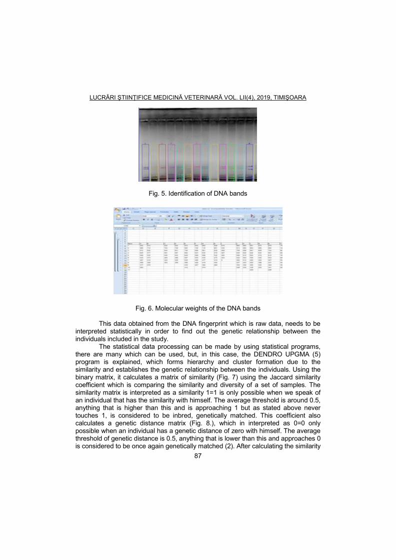
LUCRĂRI ŞTIINŢIFICE MEDICINĂ VETERINARĂ VOL. LII(4), 2019, TIMIŞOARA
87
Fig. 5. Identification of DNA bands
Fig. 6. Molecular weights of the DNA bands
This data obtained from the DNA fingerprint which is raw data, needs to be interpreted statistically in order to find out the genetic relationship between the individuals included in the study.
The statistical data processing can be made by using statistical programs, there are many which can be used, but, in this case, the DENDRO UPGMA (5) program is explained, which forms hierarchy and cluster formation due to the similarity and establishes the genetic relationship between the individuals. Using the binary matrix, it calculates a matrix of similarity (Fig. 7) using the Jaccard similarity coefficient which is comparing the similarity and diversity of a set of samples. The similarity matrix is interpreted as a similarity 1=1 is only possible when we speak of an individual that has the similarity with himself. The average threshold is around 0.5, anything that is higher than this and is approaching 1 but as stated above never touches 1, is considered to be inbred, genetically matched. This coefficient also calculates a genetic distance matrix (Fig. 8.), which in interpreted as 0=0 only possible when an individual has a genetic distance of zero with himself. The average threshold of genetic distance is 0.5, anything that is lower than this and approaches 0 is considered to be once again genetically matched (2). After calculating the similarity

LUCRĂRI ŞTIINŢIFICE MEDICINĂ VETERINARĂ VOL. LII(4), 2019, TIMIŞOARA
88
and distance matrix, the program will execute a dendrogram (9) which is a diagram formed by hierarchical clustering.
Fig. 7. Example of similarity matrix with Jaccard coefficient The clusters are formed using the similarity matrix, according to the similarity
levels that were found between the individuals of the study. The genetic distance matrix will reveal the length of the clusters, the more genetically close the individuals are the shortest are the cluster, and if the distance between them is high, the clusters will be longer.
Fig. 8. Dendrogram based on the similarity and genetic distance matrix (5)
Practical application of the DNA fingerprinting Since its discovery in 1984, this method has been started to be used in
variable domains. In medicine for example, it is used to identify a good genetic

LUCRĂRI ŞTIINŢIFICE MEDICINĂ VETERINARĂ VOL. LII(4), 2019, TIMIŞOARA
89
similarity in order to make an organ transplant or narrow transplant, also is beginning to be used to personalize treatments for cancer patients (6). It is also used in forensics, where a sample of DNA, from blood or hair found at the crime scene can incriminate someone or exonerate them for a crime depending on the similarity of the DNA samples. It is used in the phylogeny tests, identifying the parents of a child, it was used to determine the relationship in case of inheritance or to help people separated by negative consequences to rebound with their children or parents. It is used on animals too, it can be used in a farm to determine how closed are the animals that are to be used to the reproduction line in order to avoid negative consequences in the following generations (6). It can be used to verify the pedigree of an animal, like a racehorse, who's provenience is very important in order to appreciate his value. It can be used to study isolated populations of animals which are separated geographically and compare the similarities and differences that appear genetically between them. Minisatellite DNA fingerprinting was applied to other avian breeding systems, to measure genetic variation, and to assess bird population structure to identify ‘source’ and ‘sink’ populations. Minisatellite DNA fingerprinting was also used to examine population variation in other wild animals and in fish, examples include the California Channel Island fox, the humpback whale (3). It is also used on plants to identify the genetically modified plants in agriculture. It was even used on insects, the migratory ones to determine the causes of their migration and the path they are taking and how to stop them from destroying the agriculture (6).
Conclusions
In this this paper are presented and explained the key points of a DNA fingerprinting experiment and data interpretation from this type of study. Here were included the methods and the stages of preparing the DNA and the interpretation of the data using statistical methods. DNA fingerprinting it is a very useful tool in medicine, paternity tests, crime, population dynamics and agriculture.
Acknowledgements
This work was conducted in Laboratories A1c and A2 from Horia Cernescu Research Unit of Banat’s University of Agricultural Sciences and Veterinary Medicine „ King Michael I of Romania”, Timisoara, Romania.
References
1. Belgrader, P., Automated DNA purification and amplification from blood-stained cards using a robotic workstation, BioTechniques, 1995, 19, 427–432.
2. Boldura, O.M., Popa, G., Mircu, C., Ahmadi, M., Tulcan, C., Hutu, I., Popescu, S., Molecular fingerprinting based on DNA markers, a method of brown bear (Ursus arctos) exemplars identification - a case study, Lucrări Ştiinţifice Medicină

LUCRĂRI ŞTIINŢIFICE MEDICINĂ VETERINARĂ VOL. LII(4), 2019, TIMIŞOARA
90
Veterinară Timişoara, 2015, XLVIII(2), 14-22. 3. Boldura, O., Popescu, S., Bruznican, S., Hutu, I., Applicability of a multiplex
PCR assay for the identification of animal species in feedstuffs, Animal Science and Biotechnologies, 2011, 68, 179-188.
4. Chambers, G.K., Curtis, C., Millar, C.D., Huynen, L., Lambert, D.M., DNA fingerprinting in zoology: past, present, future, Investigative Genetics, 2014, 5, 3.
5. Claes, R., Mini- and Microsatellites, Environmental Health Perspectives, 1997, 105(4), 781-789.
6. Garcia, D., Miño, K., DNA fingerprinting - Noticias Y Opiniones/ News And Views, Binatura, 2004, 2(4), 477-480.
7. Jeffreys, A.J., Highly variable minisatellites and DNA fingerprints Biochemical Society Transactions, 1987, 15(3), 309-317.
8. Saad, R., Discovery, development, and current applications of DNA identity testing - Bumc Proceedings, 2005, 18, 130–133.
9. Vasavi, K., Manonj, R., Understanding the basics of DNA fingerprinting in forensic science, Journal of Indian academy of oral medicine and radiology, 2011, 23(4), 613-616.

LUCRĂRI ŞTIINŢIFICE MEDICINĂ VETERINARĂ VOL. LII(4), 2019, TIMIŞOARA
91
MORPHOPATHOLOGICAL RESEARCH IN CANINE INFECTIOUS HEPATITIS AND ASPECTS REGARDING THE
HISTOPATHOLOGICAL DIAGNOSIS OF THE CONDITION
OLARIU-JURCA A.1, CIULAN V.
1, AVRAM E.
2, BORCHESCU G.
1, TESLICI L.E.
1
1 Banat’s University of Agricultural Sciences and Veterinary Medicine „King Michael I of Romania” from Timisoara, 119 Calea Aradului, 300645,Timişoara, Romania
2. LSVSA Satu Mare, Romania
E-mail: [email protected]
Summary The research of the present study has been conducted during the period October 2017-May 2018 through anatomopathological examination of the liver and organs from 5 dog corpses, of various ages, sexes and breeds, coming from private owners, kennels and pet rescue associations. The corpses were necropsied in the Forensics department of the Faculty of Veterinary Medicine Timisoara. Samples (tissue fragments of 2/1.5 cm) were collected following detailed macroscopic examination of the studies organs from the stomach, intestine, lungs, heart, liver, kidneys, lymph nodes and encephalon for histopathological examination. The tissue and organic lesions identified in the studied case, which play a role in the diagnosis of canine infectious hepatitis are represented by specific lesions such as: focal hemorrhagic gastritis, focal hemorrhagic-necrotic hepatitis with evidence of viral hepatocellular intranuclear inclusions, serous, edematous cholangitis, jaundice-like hepatosis and nephrosis, hepatocellular intranuclear inclusions; lymphomonocytic nephritis with viral intranuclear inclusions in the glomerular mesangial cells of the urinipherous tubules; lymphomonocytic meningoencephalitis and non-specific lesions such as: inflammatory pulmonary edema, myocardial hemorrhage in the shape of ecchymosis and sub-epicardial and intraparenchymal suffusions, cardiac, hepatic and renal protein-lipid dystrophies. Keywords: infectious hepatitis, dog, lesions, histopathological exam
Canine infectious hepatitis is a viral, infectious and contagious diseases, with an acute evolution, endemic or epidemic-endemic character, seen in animals belonging to the Canidae family. The causing agent is Canine Adenovirus 1 (CAV-1), belonging to the Adenoviridae family, Mastadenovirus genus. It has cubic, symmetry and 70-80 nm diameter (3, 8, 10) The entrances of the virus are the digestive tract (ingestion of contaminated water and food or by licking contaminated objects), respiratory (inhalation of aerosols loaded with viral particles from coughing, sneezing or barking) and cutaneous through bites or external parasites as well as through various procedures involving the serum and the use of non-sterilized needles or instruments. After reaching the organism through digestive, respiratory or transcutaneous (hematophagous insects, needles, surgical lesions) paths, the virus multiplies at the entrance gate and then via the lymph, it reaches the blood stream causing viraemia, which corresponds to the fever period. Then, the virus diffuses throughout the organism, locating itself in parenchymal

LUCRĂRI ŞTIINŢIFICE MEDICINĂ VETERINARĂ VOL. LII(4), 2019, TIMIŞOARA
92
organs, especially the liver, where it produces acute inflammation or necrosis, eventually leading to other disorders and severe hypoglycemia. Exudates, edema and hemorrhage in various tissues are the consequence of the affection of the vascular endothelium (1, 5, 7, 11). Primary infection sources are sick animals, convalescent or healed animals, as well as animals with unapparent infections. In Romania, the disease was described by Surdan et. al in 1956. Presently, the disease is spread worldwide, causing prejudice in kennels due to mortality and costs implied by prophylaxis and treatment (4, 6, 10, 12).
The prognosis is always critical due to organic alteration which might lead to death (2, 10, 12).
Materials and methods
The research of the present study has been conducted in the period October 2017-May 2018. It consists of morphopathological examination of the liver and organs from 15 dog corpses of various ages, sexes and breeds, from private owners, kennels and pet rescue organizations. The bodies were necropsied in the Forensics department of the Faculty of Veterinary Medicine in Timisoara.
A detailed macroscopic examination of the studied organs was performed following evisceration. Samples for the histopathological exam were collected from the stomach, intestines, lung, heart, liver, kidneys, lymph nodes and encephalon. The macroscopic exam aimed to identify the structural alteration particularities (shape, size, colour, aspect, lobulation, consistency and section exam) and to identify the areas in the organs that show lesions in order to collect tissue samples (tissue fragments of 2/1.5 cm) for microscopic exams. The samples were fixed using 10% formaldehyde solution for 24 hours. They were processed using the paraffin method, sectioned with a microtome at 5-6 micrometers and stained using HEA and HE and finally examined under a microscope with increasing objectives x10, x20, x40.
Subsequently, the pieces were prepared for the paraffin method. The obtained blocks were sectioned using a microtome at 6 micrometers and the sections were stained using the trichromatic method, hematoxylin eosin methylene blue (HEA) in order to enhance the modified structures. After staining, the sections were dehydrated and mounted in an anhydrous environment using Canada balm (9).
The histopathological preparations were examined using an Olympus CX41 microscope (acquired through POS CCE, DICES-MVT 2669-145), with increasing objectives. They were then interpreted and microphotographed.
Results and discussions
The research was based on a corroboration between the necropsic and morphopathological lesions from 5 dog corpses of various breeds, sexes and aged 2-13 months with a presumptive necropsic diagnosis of canine infectious hepatitis (Fig. 1).

LUCRĂRI ŞTIINŢIFICE MEDICINĂ VETERINARĂ VOL. LII(4), 2019, TIMIŞOARA
93
Fig. 1. Dog corpse, 11 months with canine infectious hepatitis External exam: The bodies were in mediocre towards good condition,
apparent mucosae (gums and conjunctiva) were pale, greyish, with a faint jaundice-like aspect and discrete ecchymosis. The subcutaneous conjunctive tissue in the lower cervical region showed infiltration with serous exudate. The submaxillary and retropharyngeal lymph nodes were enlarged, grey colour and moist aspect in two cases (serous lymph node inflammation). The other three cases showed reddish spots (focal haemorrhagic lymph node inflammation) on a grey-whitish background. The cervical and thoracic thymus was swollen, congested and showing small ecchymosis.
External exam. A bright red fluid was present in the thoracic and abdominal cavities (serro-haemorrhagic peritonitis). The macroscopic exam of the organs showed the following aspects: the lungs- in three cases, they were swollen, with passive congestion in one case, catarrhal bronchopneumonia in one of the cases. The heart liver and kidneys were grey-yellowish, with friable consistency, cardio-hepato-renal protein-lipid dystrophies, biliary bladder filled with bile, with a slightly distended wall and mild thickenings. The spleen was slightly enlarged, dark-red colour with a greyish shade on both inspection and section. The lymphoid follicles/red pulp ratio was in favour of the red pulp-pulp hyperplasia. The stomach and small intestine showed a congested wall, with discrete ecchymosis and suffusions in the subserosa. The inner lining of the stomach showed haemorrhagic infiltration on a congested background that remains after washing-focal haemorrhagic gastritis (Fig. 2). The mucosa of the small intestine, on a swollen and congested background, showed small ecchymosis areas covered by white, greyish mucus, slightly adherent to the mucosa.
Lung exam: In three of five cases, following macroscopic exam, the lungs appeared increased in volume, the pleura was transparent and tensed. The pulmonary parenchyma had a dark red colour. On section it had the same colour and the presence of a dark red foam was also noticed (serous exudate) in the

LUCRĂRI ŞTIINŢIFICE MEDICINĂ VETERINARĂ VOL. LII(4), 2019, TIMIŞOARA
94
parenchyma, bronchi and trachea. The lung floatation test (docimasia) was between waters - inflammatory pulmonary oedema (serous bronchopneumonia).
Fig. 2. Haemorrhagic gastritis Microscopically, there were signs of an oxyphillic mass in the interstitial
space of the pulmonary lobules- oedema fluid. Leukocytes comprised in the oedema fluid were present in the lumen of the alveoli and bronchia, in the shape of a homogenous oxyphilic mass. The morphological modifications define the inflammatory pulmonary oedema from a histopathological perspective.
Heart exam. Macroscopically, in three cases, the heart was slightly increased in volume, red-brownish colour upon inspection and section, of a bright shade and semi-elastic consistency- congestive myocardium. The pericardium is transparent, allowing the observation of red foci of various shapes and sizes (subepicardial and myocardial) (Fig. 3) in the myocardium.
Fig. 3. Myocardial and subepicardial haemorrhages

LUCRĂRI ŞTIINŢIFICE MEDICINĂ VETERINARĂ VOL. LII(4), 2019, TIMIŞOARA
95
Microscopically, there were numerous microscopic fields showing dilated capillaries, filled with blood in the interstitial spaces- vascular ectasia (Fig. 4).
Fig. 4. Myocardium, transverse section, capillary ecstasies. HEA staining m. x 40 Liver exam. Macroscopically- the liver in three cases was enlarged, with
round margins, yellowish colour, obvious lobular pattern, yellow lobe periphery, dirt-like aspect, residual fat on the knife blade and friable consistency. Small yellow-greyish necrosis and ecchymosis spots are visible on a yellow background but with difficulty. The gall bladder had a thick, distended wall due to the bile excessive accumulation in the cholecyst cavity-cholecystomegaly. These physical-structural particularities of the hepatic parenchyma and of the cholecyst define hepatic steatosis from a morphopathological point of view (Fig. 5).
Fig. 5. Perilobular hepatic steatosis and cholecystomegaly

LUCRĂRI ŞTIINŢIFICE MEDICINĂ VETERINARĂ VOL. LII(4), 2019, TIMIŞOARA
96
Microscopic exam- all the histological sections of the steatosis livers showed the following in most of the macroscopic fields: congestion of the centrolobular vein and ectasis of the sinusoid capillaries, optically-empty vacuoles in the cytoplasm of hepatocytes found at the periphery of the hepatic lobes, affected nucleus- perilobular hepatic steatosis (Fig. 6); dissociations, fragmentations and focal necrosis of the hepatic cords, accompanied by erythrocyte infiltration on a necrosis background (Fig. 7). Intranuclear viral inclusion are visible in some hepatocytes, in the shape of basophilic spheres when stained with HEA and delimited by a white halo, faintly visible in the nuclear mass- hepatocyte intranuclear inclussions (Fig. 8); serous exudate and predominantly neutrophilic cellular, inflammatory infiltrate around the biiary duct and portal space- edematous cholangitis (Fig. 9).
Fig. 6. Perilobular hepatic steatosis HEA staining m x 10
Fig. 7. Haemorrhagic-necrotic hepatitis: dissociations, fragmentations and focal necrosis of the hepatic cords along with erythrocytic infiltration on a necrosis
background HEA staining m. x 20

LUCRĂRI ŞTIINŢIFICE MEDICINĂ VETERINARĂ VOL. LII(4), 2019, TIMIŞOARA
97
Fig. 8. Liver – intranuclear viral inclusions in the hepatocytes, in the shape of basophilic spheres. HEA staining m. x 40
Fig. 9. Oedematous cholangitis: serous exudate around the biliary ducts. HEA staining m. x 20
Two cases, showing a dystrophic liver background with a mild jaundice-like
shade, presented slightly yellowish biliary pigments in the Kupffer cells cytoplasm from the sinusoid capillaries. The latter appeared hypertrophic due to excessive accumulation of bilirubin. The Kupffer cell nuclei are faded by the pigment granules; hepatocytes had vacuoles due to bilirubin-induced cellular hypoxia- hepatic jaundice (Fig. 10).

LUCRĂRI ŞTIINŢIFICE MEDICINĂ VETERINARĂ VOL. LII(4), 2019, TIMIŞOARA
98
Fig. 10. Liver jaundice: Kupffer cells loaded with biliary pigments. HEA staining m. x 40
Kidney exam-macroscopic In three cases, the kidneys were mildly enlarged and had pale greyish
colour upon inspection and section, with friable consistency-renal jaundice. Microscopically, there were signs of bilirubin granules in the cytoplasm the
renal tubules epithelium that faded the nuclei and agglomerations of bilirubin in the lumen of the uriniferous tubes- bilirubin cylinders-renal jaundice (Fig. 11). In two cases, the kidneys, upon nspection were grey-whitish and on section they had the same colour as on surface as well as fine white, parallel bands, hard to distinguish, greasy aspect and increased consistency- diffuse, lymph histiocytic nephritis. The sections obtained from the nephritic kidneys showed peri-tubular, mesangial and peri-glomerular, lymph histiocytic hyperplasia, hyalinosis of the glomerular ansae and nuclear infusions in the mesangial cells (Fig. 12, 13).
Fig. 11. Renal jaundice: bilirubin granules in the cytoplasm of the renal epithelium. HEA staining m. x 20

LUCRĂRI ŞTIINŢIFICE MEDICINĂ VETERINARĂ VOL. LII(4), 2019, TIMIŞOARA
99
Fig. 12. Lymph histiocytic nephritis: peri-tubular, mesangial and peri-glomerular, lymph histiocytic hyperplasia, hyalinosis of the glomerular ansae and nuclear
infusions in the mesangial cells. HEA staining m. x 40
Fig. 13. Lymph histiocytic nephritis: glomerular ansae hyalinosis nuclear inclusion in the mesangial cells. HEA staining m. x 40
Central nervous system exam Macroscopically the lesions were represented by congestion, small
haemorrhages and meningeal oedema. Microscopically there were neuron dystrophies, peri-neuronal oedema and neuron lysis, lymphomonocytic peri-vascularitis and glial nodules- lymph monocytic meningoencephalitis (Fig. 14, 15).

LUCRĂRI ŞTIINŢIFICE MEDICINĂ VETERINARĂ VOL. LII(4), 2019, TIMIŞOARA
100
Fig. 14. Lymph monocytic meningoencephalitis: perivascular oedema and endo and peri- lymph monocytic peri-vasculitis. HEA x 40
Fig. 15. Lymph monocytic meningoencephalitis – glial nodules: glial cell conglomerations over phagocyted neurons, Col HEA x 40

LUCRĂRI ŞTIINŢIFICE MEDICINĂ VETERINARĂ VOL. LII(4), 2019, TIMIŞOARA
101
Conclusions
The tissular and organic lesions identified in the studied cases with a role in establishing the diagnosis of canine infectious hepatitis are:
Specific lesions: focal haemorrhagic gastritis; focal haemorrhagic-necrotic hepatitis with signs of hepatocytic, intranuclear viral inclusions; serous, edematous cholangitis; bilirubin hepatosis and nephrosis (jaundice like), hepatocytic intranuclear inclusions; lymph monocytic nephritis with intranuclear viral; inclusions in glomerular mesangial cells and in the cells of the uriniferous tubes epithelium; lymph monocytic meningoencephalitis.
Non-specific lesions: inflammatory pulmonaryoedema; myocardial haemorrhages in the shape ofecchymosis and sub-epicardial or intraparenchymal suffusions; cardiac- renal-hepatic protein-lipid dystrophies.
References 1. Greene, C.E., Infectious canine hepatitia and canine acidophil cell hepatitis,
Ed.Greene, Philadelphia, USA, 1998. 2. Greene, C.E., Infectious disease of the dog and cat, Sounders, Missouri, 2006. 3. Herman, V., Sicoe, O., Boli produse de virusuri din familia Adenoviridae. În
"Boli virotice și prionice ale animalelor", Ed. Brumar, 2005. 4. Filea, I.I., Popescu, I.S., Ghid canin, Ed. Ceres, București, 1992. 5. Jubb, K.V.F., Kennedy, P.C., Palmer, N., Pathology of domestic animals,
fourth edition, vol. 2, Academic Press, San Diego, California, SUA, 1993. 6. Koptopoulus, G., Cornwell, J.C., Canine adenovirus: a review, Veterinary
bulletin, 1981, 51, 135-142. 7. Mc Gavin, M.D., Cartlon, W.W., Jac Hary, T.F., Special veterinary pathology,
Ed. Mosby, Inc., Missouri, USA, 2001. 8. Moga Mânzat, R., Boli infecțioase ale animalelor, Ed. Brumar, Timișoara,
2001. 9. Olariu-Jurca, A., Coman, M., Olariu-Jurca, I., Stancu, A., Compendium de
anatomie patologică veterinară practică, româno-englez, Ed. Eurobit, Timișoara, 2015.
10. Perianu, T., Tratat de boli infecțioase ale animalelor. Viroze și boli prionice, vol II, Universitas XXI, Iași, 2016.
11. Pop, M., Vasiu, C., Olariu-Jurca, I., Olariu-Jurca, A., Diagnostic epidemiologic și morfoclinic în boli infecțioase la animale, Ed. Eurobit, Timișoara, 2011.
12. Vasiu, C., Virusuri, viroze și boli prionice la animale, Ed. Mega, Cluj-Napoca, 2006.

LUCRĂRI ŞTIINŢIFICE MEDICINĂ VETERINARĂ VOL. LII(4), 2019, TIMIŞOARA
102
THE POTENTIAL LOCAL PATHOGENIC ROLE OF CIRCULATING IMMUNE COMPLEXES IN CONTAGIOUS AGALACTIA OF SHEEP ȘANDRU C.D.1, NICULAE M.1, BRUDAȘCĂ G.F.1, PALL E.1, VASIU A.1, CERBU
C.1, SUĂTEAN M.I.1, SPÎNU M.1, KRASOVSKAIA I.2
1University of Agricultural Sciences and Veterinary Medicine, Faculty of Veterinary
Medicine, 3-5 Mănăştur street, 400372, Cluj-Napoca, Romania 2 Kazan State Academy of Veterinary Medicine named after N.E. Bauman, Sibirskiy
Trakt, no. 35, 420029, Kazan, Tatarstan, Russia E-mail: [email protected]
Summary
Contagious agalactia of sheep and goats, caused by M. agalactiae, included on the OIE list of notifiable disease, is clinically expressed by septicemia, arthritis and eye lesions. The immune response plays an important role in the clinical outcome of the infection, extent of lesions and survival. The research aimed to place the circulating immune complexes (CICs) in the pathological framework of the disease, by correlating their levels in vaccinated or non-vaccinated sheep showing clinical signs of the disease with the severity of the lesions. Eighteen Ţigaie rams and ewes, aged 2 to 8 years, with clinical illness, from a flock vaccinated against contagious agalactia two years ahead the trial, were sampled twice at 18 days intervals, before and after therapy and circulating immune complexes levels were estimated by 4.2% PEG precipitation method. The location and magnitude of the lesions and vaccinal status were quantified by scores (1 for joint lesions, 2 for ocular lesions and 3 for mixed lesions and 1 for vaccinated, 2 for non-vaccinated, respectively). Excel program was used to correlate the results and evaluate their statistical significance. There was a positive but not significant correlation of CIC levels and vaccination status after the therapy. Albeit no significant difference between CIC levels (0.0089±0.004 and 0.0086±0.009, respectively), there was a decrease in correlation with the lesion score (0.272 and 0.213 respectively) before and after therapy, suggesting the potential pathogenic role of CIC. Keywords: contagious agalactia, circulating immune complexes, lesion score, vaccination, correlations
Contagious agalactia of sheep and goats is a disease well-known for
centuries, nevertheless it occurs currently nowadays and sometimes has an enzootic character (6) and could be endemic, as it is the case of numerous Mediterranean countries, producing a serious economic impact (2, 3). In lactating female animals, it is usually clinically expressed as mastitis, but joint, respiratory distress and ocular lesions are also found in rams, lambs or other categories of animals (7). The diagnosis relies on the identification of the agent, M. agalactiae (4, 5) or on serological methods (8), PCR or identification of various mollecules involved in pathogenesis of the disease (4).
Less attention is attributed to the details of the immune involvement in the pathogenesis of the localized lesions. This study aimed at investigating the

LUCRĂRI ŞTIINŢIFICE MEDICINĂ VETERINARĂ VOL. LII(4), 2019, TIMIŞOARA
103
potential role of circulating immune complexes in the development of lesions by establishing correlations in adult and young animals.
Materials and methods
The study was carried out on eighteen Ţigaie rams and ewes, aged 2 to 8 years, with clinical illness, from a flock vaccinated against contagious agalactia two years ahead the trial, were sampled twice at 18 days intervals, before and after therapy. The research aimed to place the circulating immune complexes (CICs) in the pathological framework of the disease, by correlating their levels in vaccinated or non-vaccinated sheep showing clinical signs of the disease with the severity of the lesions.
Quantification of circulating immune complexes (CIC) Serum immune complexes were evaluated by precipitation with 4.2% PEG,
as described by Yancey and Lawley (4). The blood sampled on coagulant gel was allowed to coagulate at 37°C for
1 hour, when it was stored in the refrigerator for 30 minutes. The resulting serum was centrifuged at 2500 rpm for 10 minutes. Sera were separated, passed into plastic tubules and kept in the freezer at - 20°C until testing.
Dosage of circulating immune complexes was done using the micromethod, by precipitation with 4.2% polyethylene glycol (PEG). The precipitation took place in 60 minutes, at room temperature. The reading was done spectrophotometrically, compared to the buffer solution at 450 nm wavelength. The results are expressed in units and calculated according to the formula:
(U) CICs = (PEG extinction + sample) - (buffer extinction + sample) x 1000
Statistical calculation was performed using the Excel program. Scores were given to the lesional aspects according to their location (articular location = 1, ocular location = 2, mixed location = 3), to the use or absence of vaccination (unvaccinated = 1, vaccinated = 2), respectively to the use or absence of the treatment (untreated = 1, treated = 2). These scores were correlated with the levels of circulating immune complexes recorded in each sampling, to estimate the intervention of immune complexes synthesis in the pathogenetic process.
Results and discussions
The predominance of the articular and ocular forms of the disease, with the presence of mixed form in only one individual (6%) is rendered in Fig. 1.
Fig. 2 illustrates the distribution of lesions by sex and location. Most of the affected animals were females (due to the lower number of males in the herd) with mainly ocular lesions, whereas the males presented exclusively articular lesions.

LUCRĂRI ŞTIINŢIFICE MEDICINĂ VETERINARĂ VOL. LII(4), 2019, TIMIŞOARA
104
The lesion scores granted and the CICs levels determined must also becorrelated with the nutritional status of the animals tested, acknowledging that environmental factors influence the response to mycoplasma with different intensity, with nutritional factors in the foreground (for example, aflatoxin, which increases the serum concentration of gammaglobulin) (5).
The individual values obtained in the first sampling (Table 1) ranged between 0.002 and 0.016 UDO and between 0 and 0.029 UDO in the second sampling.
Changes in average values expressed by a slight increase was observed in the second sampling (0.0089 UDO) when compared to the first (0.0086 UDO). Nonetheless, close average values were observed between the two samplings.
The distribution of the lesions by site and gender were presented in Fig. 1 and 2.
Table 1 Average values and statistical indicators for CIC in sheep with contagious
agalaxia
Sampling I Sampling II
PEG Borate buffer
CICs Sample
no. PEG
Borate buffer
CICs
Average 0.0384 0.0297 0.0086 Average 0.0406 0.0316 0.0089
St. dev. 0.0082 0.0079 0.0036 St. dev. 0.0168 0.0178 0.0090
Var. 6.726 6.365 1.355 Var. 0.0003 0.00031 8.265
Fig. 1. The prevalence of various lesion types/locations

LUCRĂRI ŞTIINŢIFICE MEDICINĂ VETERINARĂ VOL. LII(4), 2019, TIMIŞOARA
105
Fig. 2. Distribution of lesion sites according by gender of the animals
Given that the presence of pathogenic mycoplasmas in the auricular duct of healthy goats was demonstrated even when these animals coexisted with sheep, in extensive farming, their pathogenetic role can be relatively easy to demonstrate and the emergence of an immune response is of no surprise (7).
The correlation coefficient calculation between the lesional scores and the CICs levels shows that the dependence of the two rows studied is direct but not very close, lacking the statistical assurance. Deduction can be made that although the appearance of the lesions may be influenced by the synthesis of circulating immune complexes, this influence is not major or obligatory (Table 2).
Table 2
The ranges of values used to evaluate the correlation between the lesional scores awarded and the levels of circulating immune complexes in each
sampling
Sampling 1 Sampling 2 CIC Score CIC Score
r 0.27227 1.55 0.213351 1.56
The effect of different adjuvants in increasing protection against mycoplasmic mastitis (Mycoplasma agalactiae) is demonstrated by numerous studies. Immunization against contagious agalactia specifically involves the Hsp60 protein (7, 8, 9). Thus, for example, polyinosinic-polycythylic acid is known to lead to the conversion of IgG2a synthesis to IgG3 by modifying isotype B in an appropriate cytokine environment (1). The effect of vaccination per se, by increasing the antibody titer, can increase the concentration of circulating immune
0
2
4
6
8
Joint Ocular Mixed
Males
Females

LUCRĂRI ŞTIINŢIFICE MEDICINĂ VETERINARĂ VOL. LII(4), 2019, TIMIŞOARA
106
complexes. Sometimes these can have undesirable effects, such as worsening of the lesions installed during the course of the disease, when the animals are insufficiently protected due to the long time elapsed from the date of vaccination.
Table 3
The ranges of values used to evaluate the correlation between the levels of immunization and the concentrations of circulating immune complexes in
each sampling
Sample no.
Sampling 1 Sampling 2 CIC Vacc. CIC Vacc.
r -0.24275 1.72 0.272895 1.72
Table 4 The ranges of values used to evaluate the correlation between the therapeutic scores granted and the levels of circulating immune
complexes in each sampling
Sample no.
Sampling 1 Sampling 2 CIC Vacc. CIC Vacc.
r 0.242748 1.27 -0.2729 1.27
The similar estimation made in relation to the vaccination status of the
investigated animals showed an inverse correlation between immunization against agalaxia and CICs levels at first sampling. This correlation turned positive at the second vaccination. The values of r were not statistically assured for this parameter either. Interestingly, at the first vaccination, CICs concentrations decreased with vaccination while at the second harvest, which was performed at an interval of 17 days, CICs levels increased relative to the increase in the time interval elapsed from vaccination and worsening of lesions.
The situation reversed in the case of investigating the correlation between the severity of the lesions and the establishment or the absence of therapy (Table 4). The long-lasting treatment determined the decrease of the circulating immune complex levels. Without going into details about the mechanism of CIC decrease (reducing the synthesis or intensifying their elimination from the circulation), the beneficial influence of the therapy on the healing of the lesions is evident, probably due to the reduction of CICs concentrations.

LUCRĂRI ŞTIINŢIFICE MEDICINĂ VETERINARĂ VOL. LII(4), 2019, TIMIŞOARA
107
Conclusions
The levels of circulating immune complexes in animals with clinical contagious agalactia do not differ significantly before and after the initiation of therapy.
The calculation of the correlation coefficient between the lesion scores and the CICs levels shows that the dependence of the two rows studied is direct but not very close, lacking the statistical assurance.
The correlation of CICs levels with the vaccination status of the investigated animals showed an inverse correlation between immunization against agalactia and the levels of complexes at first sampling. This correlation turned positive at the second vaccination, without statistical assurance of its values. CICs levels increased in relation to the increase in past time from vaccination and worsening of lesions.
Long-term treatment results in decreased levels of circulating immune complexes, beneficially affecting lesion healing, probably due to reduced CICs concentrations.
References 1. Avramidis, N., Victoratos, P., Yiangou, M., Hadjipetrou-Kourounakis, L.,
Adjuvant regulation of cytokine profile and antibody isotype of immune responses to Mycoplasma agalactiae in mice, VetMicrobiol., 2002, 88(4), 325-38.
2. Cottew, G.S., Yeats, F.R., Mycoplasmas and mites in the ears of clinically normal goats, Aust Vet J., 1982, 59(3), 77-81.
3. Glew, M.D., Papazisi, L., Poumarat, F., Bergonier, D., Rosengarten, R., Citti, C., Characterization of a Multigene Family Undergoing High-Frequency DNA Rearrangements and Coding for Abundant Variable Surface Proteins in Mycoplasma agalactiae, Infect Immun., 2000, 68(8), 4539–4548.
4. Gómez-Martín, Á., Amores, J., Paterna, A., De la Fe, C., Contagious agalactia due to Mycoplasma spp. in small dairy ruminants: Epidemiology and prospects for diagnosis and control, The Veterinary Journal., 2013, 198(1), 48–56.
5. Kumar, A., Rahal, A., Chakraborty, S., Verma, A.K., Dhama, K., Mycoplasma
agalactiae, an Etiological Agent of Contagious Agalactia in Small Ruminants: A Review, Veterinary Medicine International, 2014, 2014, 1–13.
6. Madanat, A., Zendulková, D., Pospísil, Z., Contagious agalactia of sheep and goats. A Review, Acta Vet. Brno, 2001, 70, 403–412.
7. Marin, D.E., Taranu, I., Bunaciu, R.P., Pascale, F., Tudor, D.S., Avram, N., Sarca, M., Cureu, I., Criste, R.D., Suta, V., Oswald, I.P., Changes in performance, blood parameters, humoral and cellular immune responses in

LUCRĂRI ŞTIINŢIFICE MEDICINĂ VETERINARĂ VOL. LII(4), 2019, TIMIŞOARA
108
weanling piglets exposed to low doses of aflatoxin, J Anim Sci., 2002, 80(5), 1250-7.
8. Ron, Y., Flitman-Tene, R., Dybvig, K., Yogev, D., Identification and characterization of a site-specific tyrosine recombinase within the variable loci of Mycoplasma bovis, Mycoplasma pulmonis and Mycoplasma agalactiae, Gene, 2002, 292(1-2), 205-11.
9. Scherm, B., Gerlach, G.F., Runge, M., Analysis of heat shock protein 60 encoding mycoplasmas genes and investigations concerning their role in immunity and infection, Vet. Microbiol., 2002, 89(2-3), 141-50.
10. Yancey, K.B., Lawley, T.J., Circulating immune complexes: Their immunochemistry, biology, and detection in selected dermatologic and systemic diseases, Journal of the American Academy of Dermatology, 1984, 10(5), 711–731.

LUCRĂRI ŞTIINŢIFICE MEDICINĂ VETERINARĂ VOL. LII(4), 2019, TIMIŞOARA
109
DISSIMILAR STRESSES INFLUENCE DIFFERENTLY THE IMMUNE RESPONSES IN BROILER CHICKENS
ȘANDRU C.D.1, NICULAE M.1, BRUDAȘCĂ G.F.1, PALL E.1, VASIU A.1, CERBU
C.1, POTÎRNICHE A.1, SPÎNU M.1, KRASOVSKAIA I.2
1University of Agricultural Sciences and Veterinary Medicine, Faculty of Veterinary
Medicine, 3-5 Mănăştur street, 400372, Cluj-Napoca, Romania 2 Kazan State Academy of Veterinary Medicine named after N.E. Bauman, Sibirskiy
Trakt, no. 35, 420029, Kazan, Tatarstan, Russia E-mail: [email protected]
Summary
The sharp increase in intensive farming of broilers is inductive of stress of various origins, with release of corticosterone and subsequent detrimental influence on production and immune performance and thus diminished resistance to disease. The aim of this study was to compare the influence of two different types of stress (relocation and vaccination) on non-specific and adaptive immunity in two equal (n=20) groups of boiler chickens 19 days of age, by monitoring the dynamics of circulating immune complexes (CIC) (PEG 4.2% precipitation method) and adaptive cell mediated immunity (in vitro blast transformation test), through blood sampling prior, and 4 days after the onset of the stressors. The relocation stress led to a significant decrease (0.039±0.003 and 0.0188±0.003, p<0.05) of CIC, while the values were increased for the vaccination stress (0.013±0.002 and 0.039+0.010, p<0.05). The spontaneous blastogenic index decreased in broilers subject to adaptation stress (19.01±13.10% to 12.37±11.11%) but remained unchanged in vaccinated chickens. Furthermore, the response to PHA mitogen was present in vaccinated chickens (28.98±17.71% and 32.49±7.14%) and absent (24.47±10.22% and 24.78±10.47%) in relocated chickens. A comparison of the influences of two different types of stress based on the immunological results indicated that 4 days after the onset of stress, the adaptation was more impacting than the vaccination and that humoral immunity was more influenced than the adaptive cell mediated immunity. Keywords: broiler chickens, adaptation stress, vaccination stress, immunity
In Romania, poultry farming is one of the most intensively growing agri-
food sectors. The poultry sector is continuously growing due to increasing of human population; improve in purchasing power and urbanization (2, 12). The prognosis related to poultry meat production for the year 2020 according to the FAO/OECD, will reach 140 million tons of meat (5). The breeding of chickens in the intensive system represents a true industry, which no longer holds the traditional rural method of breeding. Although obtained after laborious genetic selections and naturally after long improvements, broilers are living organisms, more susceptible to any environmental change than the birds raised in the traditional system. To maximize the growth rate, broilers are subjected to a continuous light (24 hours light, 0 hours dark), or almost continuous (23 hours light, 1 hour dark) to consume

LUCRĂRI ŞTIINŢIFICE MEDICINĂ VETERINARĂ VOL. LII(4), 2019, TIMIŞOARA
110
as much feed as possible and to achieve optimum slaughter weight in the shortest time possible. In order to prevent material losses, as well as to control common diseases, broilers are subjected to prophylactic treatments with antimicrobials and periodic vaccinations, activities with high stressor potential (9), which compromise homeostasis with negative effects on immune status of animals (1, 3). In this way, the monitoring of the immune status, as an indicator of the susceptibility to infectious diseases, can represent a form of control of the potential losses that are finally found in the economic balance of the farmer (10, 11).
In this context, the purpose of this study was to compare the influence of two different types of stress (relocation and vaccination) on non-specific and adaptive immunity in two equal (n=20) groups of broiler chickens 19 days of age, by monitoring the dynamics of circulating immune complexes (CIC) (PEG 4.2% precipitation method) and adaptive cell mediated immunity (in vitro blast transformation test), through blood sampling prior, and 4 days after the onset of the stresses.
Materials and methods
Animals A total of 40 (n=40), 19 day-old broiler chickens from disease free herds
were used in this experiment. The study design was carried out in agreement with the rules and standards set out by the Bioethics Commission of the Faculty of Veterinary Medicine, Cluj-Napoca. The animals were subjected to differentiated testing protocols, as follows: a) Protocol A - the influence of stressors (vaccination) on the immune system was evaluated. The animals were vaccinated with one dose of inactivated LaSota vaccine, on the technological line. Blood samples were collected 4 days before vaccination and 4 days post-vaccination.
b) Protocol B - the influence of accommodation stress to a new living environment, an intervention factor, especially in birds raised for eggs or as a replacement, on the immune system and implicitly the resistance to diseases was investigated. Blood samples were collected at the home farm, four days before relocation and 7 days after the transfer.
Circulating immune complex (CIC) assay Polyethylene glycol precipitation microtest was used to quantify the
circulating immune complexes’ levels. A total volume of 196.7·µl of borate buffer and PEG solution, were added to 3.3·µl serum samples. The plate was then incubated for 1h at room temperature. The optical densities of each well were read at 450 nm wavelength with a multichannel spectrophotometer SUMAL PE2. CIC concentrations were expressed in optical density units (ODU). Calculation of CIC concentrations was performed by the difference between the sample treated with PEG and that treated with borate buffer: CIC (U) = (PEG precipitation value - buffer precipitation value) x1000.

LUCRĂRI ŞTIINŢIFICE MEDICINĂ VETERINARĂ VOL. LII(4), 2019, TIMIŞOARA
111
In vitro blast transformation test The samples were diluted with RPMI 1640 (Sigma-Aldrich, USA) medium
(1:4) with 5% FCS (Gibco) and penicillin and streptomycin (Sigma-Aldrich). The
diluted samples were added 96-well plate, 100 l / well, 6 variants: (1) untreated control culture, (2) phytohemagglutinin-M (PHA-M) (1 µl/well) (3) concanavalin A (Con A) (1 µl/ well). The cultures were incubated for 48h at 37°C and 5% CO2. Glucose concentrations were measured in the initial medium and in all variants, using ortho-toluidine colorimetric test. 12.5 µl of the cultural medium were transferred to 0.5 ml of ortho-toluidine reagent, heated for 8 minutes, added in cold water and evaluated using a spectrophotometer at 610 nm wavelength (Sumal PE2, Karl Zeiss, Germany), using the reagent as a blank. For transformation index (TI) the following formula was used: TI %=[(MG–SG)/MG] ´100, where TI, blast transformation index, MG, glucose concentration in the initial culture medium and SG, glucose concentration in the sample after incubation.
Statistical analysis For statistical interpretation Minitab 16.0 were used. Results were
expressed as average ±standard deviation.
Results and discussions
The relocation stress led to a significant decrease (0.039±0.003 and 0.0188±0.003, p<0.05) of CIC, while the values were increased for the vaccination stress (0.013±0.002 and 0.038±0.010, p<0.05).
Comparing the two types of stress, stress induced by vaccination and relocation-induced stress, it can be observed that the intervention on CIC is different, in the first case the level of complexation is low and the one of elimination increased, whereas in the second case, to one the high level of complexation corresponds to a low elimination of CIC (Fig. 1).
This phenomenon, although it cannot be fully explained under the conditions of the experiments carried out, leads to the conclusion that the stress of relocation is more intense than the vaccination induced stress (8, 10, 11).
The spontaneous blastogenic index decreased in broilers subject to adaptation stress (19.01±13.10% to 12.37±11.11%) but remained unchanged in vaccinated chickens. Furthermore, the response to PHA mitogen was present in vaccinated chickens (28.98±17.71% and 32.49±7.14%) and absent (24.47±10.22% and 24.78±10.47%) in relocated chickens.
The most intense stimulation index was observed in the PHA treated variant in the vaccinated group, followed by the control sample in the relocated birds.

LUCRĂRI ŞTIINŢIFICE MEDICINĂ VETERINARĂ VOL. LII(4), 2019, TIMIŞOARA
112
Fig. 1. Levels of CIC in experimental chicken groups before and after being subjected to relocation and vaccination stress
The differences between the two stressors in the blastogenic response
indicated that the lymphocytes were more susceptible to the vaccination than to the relocation stress (6, 7). This result is supported by the lymphocyte behavior after the stressor’s intervention, an increase being observed in stimulation indices in the relocated group (Fig. 2), while a decrease (p<0.05) was noted in the vaccinated group.
Fig. 2. In vitro stimulation/inhibition indices in experimental chicken groups before
and after being subjected to relocation and vaccination stress

LUCRĂRI ŞTIINŢIFICE MEDICINĂ VETERINARĂ VOL. LII(4), 2019, TIMIŞOARA
113
More intense stimulation was also exerted in the relocated chickens by ConA, the values staying almost unchanged (p non-significant), while subsequent to vaccination, the Con A activity was statistically non significantly inhibiting.
Conclusions
A comparison of the influences of two different types of stress based on the
immunological results indicated that 4 days after the onset of the stress, the adaptation was more impacting than the vaccination, and that humoral immunity was more influenced than the adaptive cell mediated immunity.
References 1. Landers, T.F., Cohen, B., Wittum, T.E., Larson, E.L., A Review of Antibiotic
Use in Food Animals: Perspective, Policy, and Potential, Public Health Rep., 2012. 2. Mbuza, F., Manishimwe, R., Mahoro, J., Simbankabo, T., Nishimwe, K.,
Characterization of broiler poultry production system in Rwanda, Trop Anim Health Prod., 2017, 49(1), 71–77.
3. Mehdi, Y., Létourneau-Montminy, M.P., Gaucher, M.L., Chorfi, Y., Suresh, G., Rouissi, T., Brar, S.K., Côté, C., Ramirez, A.A., Godbout, S., Use of antibiotics in broiler production: Global impacts and alternatives, Anim Nutr., 2018, 4(2), 170–178.
4. Qureshi, M.A., Miller, L., Comparison of macrophage function in several commercial broiler genetic lines, Poult. Sci., 1991, 70, 1094–1101.
5. Sell-Kubiak, E., Wimmers, K., Reyer, H., Szwaczkowski, T., Genetic aspects of feed efficiency and reduction of environmental footprint in broilers: a review, J Appl Genet., 2017, 58(4), 487–498.
6. Srabler, J.G., Avian heterophils and monocytes: phagocytic and bactericidal activities against Salmonella enteritidis, Vet. Microbiol., 1994, 38, 293–305.
7. Styrt, B., Species variation in neutrophil biochemistry and function, J. Leukocyte Biol., 1989, 46, 63–74.
8. Thunlod, S., Sshauenstein, K., Wolf, H., Wick, G., Localization of IgGFc and complement receptors in chicken lymphoid tissue, Scand J Immunol., 1981, 14(2), 145-52.
9. Toth, T.E., Siegel, P.B., Cellular defense of the avian respiratory tract: paucity of free-residing macrophages in the normal chicken, Avian Dis., 1986, 30, 67–75.
10. Zarnea, C., Imunologie Clinică, Editura Medicală, Bucureşti, 1990. 11. Ziegler, H.K., The role of gamma/delta T cells in immunity to infection and
regulation of inflammation, Immunol. Res., 2004, 29, 293–302. 12. ***Food and Agriculture Organization of the United Nations (Faostat),
Livestock production, poultry meat production, 2014, http://faostat.fao.org/site/569/default.aspx#ancor

LUCRĂRI ŞTIINŢIFICE MEDICINĂ VETERINARĂ VOL. LII(4), 2019, TIMIŞOARA
114
MICROBIOGICAL INVESTIGATIONS IN A SEVERE HEMORRHAGIC SYNDROME IN FARMED OSTRICHES
VASIU A.1, TURCU B.1, KICSID Z.1, NICULAE M.1, PALL E.1, OLAH D.I.1,
BRUDAȘCĂ G.F.1, SPÎNU M.1, KRASOVSKAIA I.2
1University of Agricultural Sciences and Veterinary Medicine, Faculty of Veterinary
Medicine, 3-5 Mănăştur street, 400372, Cluj-Napoca, Romania 2Kazan State Academy of Veterinary Medicine named after N.E. Bauman, Sibirskiy
Trakt, no. 35, 420029, Kazan, Tatarstan, Russia E-mail: [email protected]
Summary
Farmed ratites, frequently kept outside their countries of origin, commonly suffer of infectious, bacterial or viral, economically highly impacting diseases. This study aimed at investigating the etiology of two episodes of hemorrhagic enteritis in South African ostriches (Struthio camelus) farmed in NW of Romania. Both episodes (n1=11, n2=45) were recorded in young birds (9-16 month of age), with sudden onset and peracute (sudden death) to sub-acute (6-7 days) clinical course. The clinical picture included depression, anorexia, ataxia, convulsions, oedema and pain in the limbs, followed by death in all cases. The necropsy revealed cahexia, dehydration, anemia and congestion of conjunctive mucosa, diffuse subcutaneous, intramuscular and splenic hemorrhages, necrotic myositis, hemorrhagic to necrotic enteritis and proventriculitis, necrotic hepatitis, congestion of the kidneys, lungs and tracheal mucosa,ecchymosis in the ventricular mucosa. Classical microbiological methods (simple broth, Mueller Hinton agar, McConkey agar) and anaerobic environment (TSC agar) cultivation were performed. The results indicated the presence of Clostridium perfringens and E. coli in the internal organs of the birds and also the bone marrow. It was considered that an imbalance in feeding the birds created favorable conditions for the insertion of these widespread bacteria. In short term, broad spectrum antibiotic treatment was attempted with promising results. Keywords: Struthio camelus, hemorrhagic syndrome, C. perfringens, E. coli
The ostrich is the largest bird on Earth, with a waist up to 2.75 m and a
weight up to 150 kg. Its name, Struthio camelus dated back to Linnae (1758) and the there are 4 subspecies: S. c. syriacus Rothshield, S. c. camelus Linnaeus, S. c. massaicus Neumann, and S. c. australis Gurney (5, 7, 15).
Ostrich farming is nowadays widespread in the world and some ostrich farms of small size could also be found in Romania. Ostrich meat and meat products are priced 5-6 times higher than those of other poultry, since the cholesterollevel in meat is 50% lower than in ruminants, and therefore the demand on the EC market is much higher than farmerscan supply. The by-products, such as skin, feathers, egg shells, can be used very advantageously.
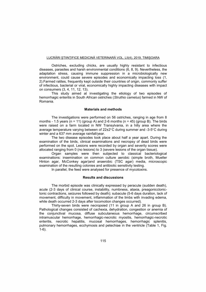
LUCRĂRI ŞTIINŢIFICE MEDICINĂ VETERINARĂ VOL. LII(4), 2019, TIMIŞOARA
115
Ostriches, excluding chicks, are usually highly resistant to infectious diseases, parasites and harsh environmental conditions (6, 8, 9). Nevertheless, the adaptation stress, causing immune suppression in a microbiologically new environment, could cause severe episodes and economically impacting loss (1, 2).Farmed ratites, frequently kept outside their countries of origin, commonly suffer of infectious, bacterial or viral, economically highly impacting diseases with impact on consumers (3, 4, 11, 12, 13).
This study aimed at investigating the etiology of two episodes of hemorrhagic enteritis in South African ostriches (Struthio camelus) farmed in NW of Romania.
Materials and methods
The investigations were performed on 56 ostriches, ranging in age from 8 months - 1.5 years (n = 11) (group A) and 2-6 months (n = 45) (group B). The birds were raised on a farm located in NW Transylvania, in a hilly area where the
average temperature varying between of 22±2C during summer and -3-5C during winter and a 637 mm average rainfall/year.
The two disease episodes took place about half a year apart. During the examination of the birds, clinical examinations and necropsy of dead birds were performed on the spot. Lesions were recorded by organ and severity scores were allocated ranging from 0 (no lesions) to 3 (severe lesions of the organ tissue).
Organ samples were then subjected to classical bacteriological examinations: insemination on common culture aerobic (simple broth, Mueller Hinton agar, McConkey agar)and anaerobic (TSC agar) media, microscopic examination of the resulting colonies and antibiotic sensitivity testing.
In parallel, the feed were analysed for presence of mycotoxins.
Results and discussions
The morbid episode was clinically expressed by peracute (sudden death), acute (2-3 days of clinical course, instability, numbness, ataxia, preagonicclonic-tonic contractions, seizures followed by death); subacute (5-6 days duration, lack of movement, difficulty in movement, inflammation of the limbs with invading edema, while death occurred 2-3 days after locomotion changes occurred).
Thirty-seven birds were necropsied (11 in group A and 26 in group B). Pathological changes consisted of cachexia, dehydration, congestion or anemia of the conjunctival mucosa, diffuse subcutaneous hemorrhage, circumscribed intramuscular hemorrhage, hemorrhagic-necrotic myositis, hemorrhagic-necrotic enteritis, necrotic hepatitis, mucosal hemorrhages, hemorrhagic splenitis, pulmonary hemorrhages, ecchymosis and petechiae in the ventricle (Table 1, Fig. 1-6).

LUCRĂRI ŞTIINŢIFICE MEDICINĂ VETERINARĂ VOL. LII(4), 2019, TIMIŞOARA
116
As shown in the table, the highest average for cumulative scores was recorded in case of 0 and 3, with no statistical differences between the two. Scores 1 and 2 were almost identical in cumulative average.
The most numerous and severe lesions were recorded in the digestive system (ceccum, jejunum, duodenum with scores of 27, 20 and 15 respectively). Similarly, high indices were recorded for the heart and lungs, with either congestion or severe hemorrhages.
In spite of severe locomotion impairment, very severe bone and joint lesions did not prevail, therefore the cause of these clinical signs was considered the toxin’s effect rather than the local lesions.
Table 1
Distribution of the lesions/lesional scores by organ (individual, average and stdev values)
Organ/Score 0 1 2 3 Spleen 14 12 4 7
Liver 10 6 9 12
Heart 0 7 10 20
Duodenum 0 12 10 15
Jejunum 0 9 8 20
Ceccum 0 5 5 27
Cloaca 37 0 0 0
Kidneys 32 2 1 2
Stomac 6 10 12 9
Gizzard 0 12 10 15
CNS 30 5 1 1
Muscle 25 5 1 6
Connective tissue 35 1 1 0
Lungs 6 1 10 20
Bones, joints 30 1 1 5
Cumulative average 14,06 5,56 5,31 10,13
St.dev 14,71 4,37 4,38 8,52

LUCRĂRI ŞTIINŢIFICE MEDICINĂ VETERINARĂ VOL. LII(4), 2019, TIMIŞOARA
117
Fig. 1 Haemorrhagic lesions in the thoraco-abdominal cavity (original, Bianca Turcu)
Fig. 2 Severe haemorrhagic enteristis
(original, Bianca Turcu)
The microbiological results indicated the presence of Clostridium
perfringens A, C and D and E. coli in the internal organs of the birds and also the bone marrow. It was considered that an imbalance in feeding ofthe birds, mainly the exceeding protein, created favorable conditions for the excessive development of these widespread bacteria (10, 14). Along with their virulence, toxic effects of both were widely observed.
Fig. 3. Intestines filled with gas, liver congestion (original, Bianca Turcu)
Fig. 4. Ulcers and haemorrhages in the stomach(original, Bianca Turcu)

LUCRĂRI ŞTIINŢIFICE MEDICINĂ VETERINARĂ VOL. LII(4), 2019, TIMIŞOARA
118
Fig. 5. Enlarged heart and serous atrophy of the fat
(original, Bianca Turcu)
Fig. 6. Enlarged and congestive liver
(original, Bianca Turcu)
The therapy was applied immediately, consisting in short term, of
Doxycyclineadministration with promising results.Emergency antitoxic treatment (5% glucose), supportive treatment (vitamin B1 and C) were also administered. The antimicrobial treatment (Doxycycline 2.5g/10l water) was continued for 4 weeks.
Control measures aimed at protecting the birds from further infections with these agents, therefore vaccination was applied using combined anti-Clostridiumvaccine (strains A, B, C, D), with the dosage calculated based on weight and extrapolation from cattle.
It could be concluded that the necropsy diagnosis supported by microbiological investigations was conclusive of the etiology of the diseases in both situations and served as directory for implementing efficient control measures.
It has to be considered that a correctly implemented technology, with strict control of the diet and hygiene of the farms, along with a well-founded vaccination scheme could prevent important economic losses in farmed ostriches.
References 1. Bello, A., Frei, S., Peters, M., Balkema-Buschmann, A., Baumgartner, W.,
Wohlsein, P., Spontaneous diseases in captive ratites (Struthioniformes) in northwestern Germany: A retrospective study, PLoS ONE, 2017,12(4), e0173873.
2. Buckles, E.L., Eaton, K.A., Swayne, D.E., Cases of spirochete-associated necrotizing typhlitis in captive common rheas (Rhea americana), Avian diseases, 1997, 41(1), 144-8.
3. Huchzermeyer, F.W., Animal health risks associated with ostrich products, Rev Sci Tech., 1997,16(1), 111-6.

LUCRĂRI ŞTIINŢIFICE MEDICINĂ VETERINARĂ VOL. LII(4), 2019, TIMIŞOARA
119
4. Huchzermeyer, F.W., Diseases of ostriches and other ratites, Agricultural research Council, Pretoria, South Africa, 1998.
5. Kummrow, M.S., Ratites or Struthioniformes: Struthiones, Rheae, Cassuarii, Apteryges (Ostriches, Rheas, Emus, Cassowaries, and Kiwis), and Tinamiformes (Tinamous), In: Miller R.E., Fowler M.E., editors. Fowler’s Zoo and Wild Animal Medicine, 8th ed. St. Louis, MO: Saunders Elsevier, 2015.
6. Levy, A., Perelman, B., Warner, T., van Gravenbroek, M., van Creveld, C., Yagil, R., Reference blood chemical values in ostriches (Struthio camelus), Am. J. Vet. Res., 1989, 50(9), 1548-1550.
7. Palomeque, J., Pinto, D., Viscor, G., Hematologic and blood chemistry values of the Masai ostrich (Struthio camellus), J. of Wildlife Dis., 1991, 27(1), 34-40.
8. Perelman, B., Levy, A., Waner, T., Creveld, C., Grevenbroek, M., Yagi, R., Hematology and Blood Chemistry in Ostriches Maintained under Intensive Farming Conditions, Isr. J.Vet.Med., 1989, 45, 1, 49.
9. Pocknell, A.M., Miller, B.J., Neufeld, J.L., Grahn, B.H., Conjunctival mycobacteriosis in two emus (Dromaius novae hollandiae), Veterinary Pathology, 1996, 33(3), 346–8.
10. Sagartz, J.E., Swayne, D.E., Eaton, K.A., Hayes, J.R., Amass, K.D., Wack, R., Necrotizing typhlocolitis associated with a spirochete in rheas (Rhea americana), Avian Diseases, 1992, 36(2), 282–9.
11. Stewart, J., Ratites, In: Ritchie B W, Harrison G J, R HL, editors, Avian Medicine: principles and application, Lake Worth, FL: Wingers Publishing, 1994.
12. Tully, T.N., Ratites, In: Tully TN, Dorrestein GM, Jones AK, editors, Handbook of Avian Medicine, 2nd ed. London: Saunders Elsevier, 2009.
13. Verwoerd, D.J., Ostrich diseases, Rev. sci. tehn. Off. Int. Epiz., 1999, 19(2), 639-661.
14. Zakeri, A., Kashefi, P., Necrotic Enteritis by Clostridium perfringens in Ostrich (Struthio camelus), World Applied Sciences Journal, 2012, 16(6), 842-845.
15. ***IUCN. The IUCN Red List of Threatened Species: International Union for Conservation of Nature and Natural Resources, 2016, http://www.iucnredlist.org.

LUCRĂRI ŞTIINŢIFICE MEDICINĂ VETERINARĂ VOL. LII(4), 2019, TIMIŞOARA
120
TEMPORAL DYNAMICS OF POST-VACCINATIONHUMORAL IMMUNE RESPONSEIN OSTRICHES PRIMED AGAINST
ANTHRAX
VASIU A.1, ȘANDRU C.D.1, SZALO-MUREȘAN D.1, NICULAE M.1, CERBU C.1, SEICHELI R.1, BRUDAȘCĂ G.F.1, KRASOVSKAIA I.2, SPÎNU M.1
1University of Agricultural Sciences and Veterinary Medicine, Faculty of Veterinary
Medicine, 3-5 Mănăştur street, 400372, Cluj-Napoca, Romania 2 Kazan State Academy of Veterinary Medicine named after N.E. Bauman, Sibirskiy
Trakt, no. 35, 420029, Kazan, Tatarstan, Russia E-mail: [email protected]
Summary
Ostrich farming became lately an alternative for small producers allowing access to external markets by meat and skin exports. The paucity of scientifica information on diseases and diagnostic tests, most of the poultry methods being unvalidated, supports the researches regarding enhancement of disease resistance in this peculiar group of birds. Furthermore, semi-intensive raising could become a source for variable pathology, unless fit exploitation technologies are applied, inlcuding veterinary medical prevention. This study aimed at evaluating the fitness of various vaccination protocols in preventing anthrax, one of the most severly impacting zoonoses encountered in ostriches on Romanian farms. Two groups of South African ostriches (Struthio camelus) were subjected to vaccination with the live anti-anthrax vaccine strain R1190, using a single dose (sc injection) of 0.2 ml (group I) and 0.3 ml (group 2). Blood was sampled 3 weeks and 5 month after the priming. Total Ig (24%o zinc sulphate test) and antibody levels (precipitation test) were quantified at both samplings.After three weeks, all birds vaccinated with 0.3 ml versus 0.2 ml showed incresed Ig levels (0.023±0.003 and 0.018±0.004 ODU, respectively). The precipitation test was intensely positive in all birds from group 2 and in significantly lesser numbers (16.66%, p<0.01) in group 1. An almost complete decline of the humoral immune response was encountered after 5 month in both groups.Both methods indicated betters results for an immunizing dose of 0.3 ml of vaccine, but also a compulsory booster before 5 month after the first vaccination. Key words: Strutiocamelus, anthrax, vaccine dosage, antibodies
Ostrich farming was considered a good self-sustainable alternative to other species small-scale farming, due to the increasing demand of hides, meat, egg-shells and feathers overseas as well as a species of interest for zoos (2, 15). As an exotic species to numerous countries where farmed, ostriches encountered a verydiversified pathology, including metabolic infectious and parasitic diseases, technopathies, or metabolic and behavior disorders (1, 3, 4, 8, 11, 12).
In the last few years, ostrich farming started in Romania as a possibility for Romanian small farmersto earn more with limited resources, therefore extensive raising conditions were applied. This system is less controllable than the intensive

LUCRĂRI ŞTIINŢIFICE MEDICINĂ VETERINARĂ VOL. LII(4), 2019, TIMIŞOARA
121
one, thus the animals are exposed to sometimes sharply changing environment and to the insufficient or lack of experience of the farmers. The adaptation stress in the birds is cause to immune suppression and increased susceptibility to various pathogens present on the farm or premises (6), extensive raisingalso favoring/causing diseases and severe losses. Under these circumstances, veterinarians have a difficult task of diagnosing particular diseases, uncommon in the species, ie. anthrax. The relatively small number of imported birds and their relatively high price exerts a pressure in finding preventive means applicable in small farms and tailored to the exotic species (10).
There is little scientific information on ostrich diseases, spontaneous reports being generated “on the way”, not only in Romania but also in other ostrich farming countries. Since the epidemiological pressure varies, the preventive measures should also apply to peculiar epidemiological situations in a way which interrupts the disease cycle.
Sallmonelosis, fowl pox, campylobacteriosis, chlamydiosis, Crimean-Congo haemorrhagic fever (14) and other diseases were reported but very scarce information is available on vaccination of these birds.
Anthrax represents an infectious disease which occasionally occurs in Romania mainly in ruminants, in spite of properly designed vaccination schemes, thus creating sometimes new infectious sources (6) and posing supplementary risk to consumers.
The study aimed at monitoring the results of anti-anthrax vaccination with differentiated doses on adaptive humoral immune response (total immune globulin and antibody levels) humoral factors of the immune system in adult ostriches, primed with different doses of antigen.
Materials and methods
The study was conducted on a private farm, located in a hilly region, with
an average temperature varying between of 22±2C during summer and -3-5C during winter and a 637 mm average rainfall. The ostrich farm consisted of 5 families of adult birds (a total of 19 individuals, 7 males and 12 females), and a fluctuating number of chicks of various ages. Each age category is fed accordingly to its nutritional requirements, with imported industrial food. Vitamin and mineral supplements are administered to the youngsters. The imported birds adapted to the new habitat quite well, except one case of anthrax in the very beginning. No other cases were recorded for about five years, but another bird was then euthanized with similar signs and laboratory tests confirmed anthrax.
To avoid further cases, since the area seemed prone to be a source, a tailored vaccination protocol had to be designed for the farm. Extrapolating the dosage used for other farmed species, the Romanian strainR1190 vaccine was used, with a well-recognized immunizing capacity. 0.2 ml of the Romanian vaccine, was injected to nine of the adult birds (group I, n=3 males, 6 females) while other

LUCRĂRI ŞTIINŢIFICE MEDICINĂ VETERINARĂ VOL. LII(4), 2019, TIMIŞOARA
122
10 (4 males, 6 females) were inoculated with 0.3 ml (group II) subcutaneously. Blood from the wing vein was sampled after three weeks and 5 month subsequent to vaccination (group II).
The sera were separated by centrifugation (Hettich, Germany) subjected to detection of antibodies by a modified radial diffusion (Mancini) test and to total immune globulin quantification by Serb test. For the modified Mancini test, the antigen was represented by the ultrasonicated vaccine and the diffusion substrate was embodied by 1% Noble agar gel, while the sera were disposed in the wells. Unlike in the classical method, the precipitation took place within the well and the intensity of the reaction was graded with an increasing number of pluses. Total gammaglobulin levels were quantified by Serb method, using a classical 24
0/00 zinc
solution and the precipitation taking place in a liquid environment. For statistical interpretation Minitab 16.0 were used. Results were
expressed as average ±standard deviation.
Results and discussions
Anthrax represents a sporadical infectious disease, still causing economic loss in animal communities and it is a serious threat to humans as both contacts and consumers (5, 6). Thus, the importance of preventive measures is utmost and the success of the control strategy depends on appropriate disease surveillance and vaccination programs (10,14).
Table 1 presents the readings obtained in the Mancini test. Four out of the seventeen samples showed no precipitation. The intensity of the precipitation ranged from high (+++) in 8, medium (++) in 3 and week (+) in other 3.
Three weeks after the vaccination with the higher dose, the results were maximal. In time, the response decreased, being almost absent after 5 month. The lower dose induced a lower response in the precipitation test.
The zinc sulphate precipitation test supports the results obtained by radial diffusion (Fig. 1). A dose dependent manner was observed in the increase of the total Ig levels, but there was no statistical significance of the differences. The total Ig levels decreased after five month to levels undetectable by precipitation in some of the birds, which stands for very low protective response in the same individuals. Considering the physiological levels of total Ig in Aves, between 0.02 and 0,05 optical density units, the encountered decrease was statistically significant (p<0.05).
The scarceness of data in evaluating immune activity in ostrichesand similarly in other wild avian species, imposes comparisons by extrapolation with other species. Clinical haematology and blood chemistry as several authors mention (7, 9, 13), but mainly innate and adaptive humoral immune parameters represent, by their crucial role, a useful aid in the diagnosing infectious diseases.

LUCRĂRI ŞTIINŢIFICE MEDICINĂ VETERINARĂ VOL. LII(4), 2019, TIMIŞOARA
123
Table 1 Mancini test results subsequent to vaccination against anthrax in
experimental groups
Sample Group I Sample Group II Sample Group III
1 No
precipitation 2 +++ 14 No precipitation
3 + 5 +++ 15 No precipitation
4 ++ 6 ++ 16 +
9 No precipitation
7 +++ 17 +
10 ++ 8 +++
12 +++ 11 +++
14 + 13 +++
16 +
Ostriches are the only birds known to be naturally susceptible to anthrax,
probably because of lower body temperature (5). Both “sudden death” and “anthrax fever” can occur simultaneously in an unprotected flock (14). Reports on positive clinical outcome subsequent to vaccination were presented, with no details on the immunizing protocol, vaccine type or dosage.
Fig. 1. Total Ig levels in anti-anthrax vaccinated birds (mean values and standard deviations)
Before After Before After Before After
Group I Group IIGroup II, 5 month
later
Stdev 0.002 0.009 0.007 0.003 0.007 0.001
Mean 0.0125 0.018 0.015 0.023 0.015 0.009
0
0.005
0.01
0.015
0.02
0.025
0.03
Op
tica
l d
en
sity
un
its

LUCRĂRI ŞTIINŢIFICE MEDICINĂ VETERINARĂ VOL. LII(4), 2019, TIMIŞOARA
124
In the present study, two different doses were used to immunize separate groups of birds. Both the vaccine and the subcutaneous administration route were well tolerated.
The antibodies were qualitatively detected. In the radial diffusion test, the results were opposite to those expected, the precipitation taking place within the well, not outside it, which was probably due to the insufficient diffusion of the antigen used in the test. Nevertheless, the results were interpretable and could be ranked by intensity of the precipitation, allowing an estimate of the extent of protective immune response. The results proved that a higher dose of vaccine used in this experiment induced higher titers of antibodies, with no secondary effects for the birds. Still, these titers decreased therefore, the protective effect of the immunity lasted less than 5 month. Such results suggest supplementary investigations on the dose-response in anti-anthrax priming, to serve as fundament for appropriate vaccination protocols.
Total Ig levels, quantified by zinc precipitation, supported these results. Both investigated parameters showed the same developmental pattern, 3 as well as 5 month after the vaccination, increasing initially and decreasing afterwards. There was a positive correlation between (r=0.824, p<0.05) the two tests. The research did not aim at establishing the Ig class involved in the post-vaccination immune response, but it could be assumed mainly IgM was present, since no booster was performed. The protective dosage in ostriches was positioned between those recommended for bovine (0.5 ml) and that recommended for equine, sheepandswine.
Nevertheless, the experiment did not prove that the dose of 0.3 ml was the optimal one. Special attention has to be paid to general preventive measures along with the vaccination to avoid disease and mortality, due to the short history of this species in Romania.
Conclusions
The efficacy ofanti-anthrax vaccination in ostriches induced by the use of R1190 vaccine could bebetter monitored by Mancini and zinc sulphate precipitation tests, at a 0.3 ml dose/adult bird, 3 weeks after a single injection. Nevertheless, in order to preserve the duration and intensity of the humoral immune responseand continuously protect the birds, a booster injection has to be given at least 4 month after the previous vaccination.
References 1. Alexander, D.J., A review of avian influenza in different bird species, Veterinary
microbiology, 2000, 74(1-2), 3-13. 2. Bello, A., Frei, S., Peters, M., Balkema-Buschmann, A., Baumgartner, W.,
Wohlsein, P., Spontaneous diseases in captive ratites (Struthioniformes) in

LUCRĂRI ŞTIINŢIFICE MEDICINĂ VETERINARĂ VOL. LII(4), 2019, TIMIŞOARA
125
northwestern Germany: A retrospective study, PLoS ONE, 2017, 12(4), e0173873.
3. Buckles, E.L., Eaton, K.A., Swayne, D.E., Cases of spirochete-associated necrotizing typhlitis in captive common rheas (Rhea americana), Avian diseases, 1997, 41(1), 144–8.
4. Garcia, A., LeClear, C.T., Gaskin, J.M., Mycobacterium avium infection in an ostrich (Struthio camelus), Journal of Zoo and Wildlife Medicine, 2001, 32(1), 96–100.
5. Huchzermeyer, F.W., Animal health risks associated with ostrich products, Rev Sci Tech., 1997, 16(1), 111-6.
6. Huchzermeyer, F.W., Diseases of ostriches and other ratites, Agricultural research Council, Pretoria, South Africa, 1998.
7. Levy, A., Perelman, B., Warner, T., van Gravenbroek, M., van Creveld, C., Yagil, R., Reference blood chemical values in ostriches (Struthio camelus), Am. J. Vet. Res., 1989, 50(9), 1548-1550.
8. Mappley, L.J., La Ragione, R.M., Woodward, M.J., Brachyspira and its role in avian intestinal spirochaetosis, Veterinary Microbiology, 2014, 168(2-4), 245-60.
9. Palomeque, J., Pinto, D., Viscor, G., Hematologic and blood chemistry values of the Masai ostrich (Struthio camellus), J. of Wildlife Dis., 1991, 27(1), 34-40.
10. Perelman, B., Levy, A., Waner, T., Creveld, C., Grevenbroe,. M., Yagi, R., Hematology and Blood Chemistry in Ostriches Maintained under Intensive Farming Conditions, Isr. J.Vet.Med., 1989, 45, 1, 49.
11. Pocknell, A.M., Miller, B.J., Neufeld, J.L., Grahn, B.H., Conjunctival mycobacteriosis in two emus (Dromaius novae hollandiae), Veterinary Pathology, 1996, 33(3), 346-8.
12. Sagartz, J.E., Swayne, D.E., Eaton, K.A., Hayes, J.R., Amass, K.D., Wack, R., Necrotizing typhlocolitis associated with a spirochete in rheas (Rhea americana), Avian Diseases, 1992, 36(2), 282–9.
13. Van heerden, J., Dauth, J., Jarvis, M.J.F., Keffen, R.H., Denny, J.E.F.M., Dreyer, M.J., Kriek, N.P.J., Blood chemical and electrolyte concentrations in the ostrich Struthiocamelus, Journ. South Afr.Vet.Assoc., 1985, 56(2), 75-79.
14. Verwoerd, D.J., Ostrich diseases, Rev. sci. tehn. Off. Int. Epiz., 1999, 19(2), 639-661.
15. ***IUCN, The IUCN Red List of Threatened Species: International Union for Conservation of Nature and Natural Resources 2016,http://www.iucnredlist.org.

LUCRĂRI ŞTIINŢIFICE MEDICINĂ VETERINARĂ VOL. LII(4), 2019, TIMIŞOARA
126
MORPHOLOGY OF THE SKULLS IN BADGER (MELES MELES) AND OTTER (LUTRA LUTRA) – COMPARATIVE ASPECTS
ZAGRAI G.
1, ȘEICARU A.
1, LIXANDRU D.
1, ZAGRAI (MĂIEREAN) A.M.
2
1Faculty of Veterinary Medicine, University of Agricultural Sciences and
Veterinary Medicine Bucharest – 050097, Splaiul Independentei 105, Bucharest, Romania
2Faculty of Veterinary Medicine, University of Agricultural Sciences and
Veterinary Medicine of Cluj-Napoca E-mail: [email protected]
Summary
In some cases it is necessary to establish, in a short time, the species from which a number of bones belong. One example is that from badger (species that are hunted) and otter (protected species). There are some morphometrical study regarding the skulls in these species but comparative aspects missing. For this purpose we conducted a study on six skulls from badger (Meles meles) and five skulls from otter (Lutra lutra). Following an overall analysis of the two skulls, we found the flattening aspect of the otter skull, compared to the badger skull, and a development of neurocranium in the first species. In dorsal aspect were found differences regarding the angles between sagittal ridge and nuchal ridges and between the ratio of the width of the viscerocranium, measured at the level of canine’s alveoli, and the length of viscerocranium, measured between the zygomatic process line and rostral extremity of incisive bone. On the lateral side it is observed the reduction of jugal and retrotimpanic processes in otter. In this two species the holes from the base of the skull and the hard palate look totally different. Establishing the species can be made easier if exist the possibility to study the whole skull. Our study surprised the details that helps to identify the species even if we rely on bone fragments. Keywords: morphology, badger, otter, skull
The two species are carnivores and belong to the Mustelidae order. Both
are part of Romania's fauna and have a similar size. Badger (Meles meles) is a hunting animal of interest to which hunting is permitted, while the otter (Lutra lutra) is currently included on the Red List as a strictly protected species (1, 2, 3).
Many times, veterinarians, biologists, or forestry specialists are in a position to determine the species from which bones, bone fragments or even trophies of brute animals originate (4, 5).
The paper aims to present morphological details to make the distinguish between the skulls of the two species, even when they are not complete.
We take in this study this species for two reasons: the possibilities of confusion with other species of the same order (marten, ferret, hermelin, and weasel) are practically null, and secondly, most of the literature data treats the bones of head only morphometrically.

LUCRĂRI ŞTIINŢIFICE MEDICINĂ VETERINARĂ VOL. LII(4), 2019, TIMIŞOARA
127
Materials and methods
The research was carried out on 6 badger (Meles meles) skulls and 5 otter (Lutra lutra) skulls. The skulls belong to private collections. The otters were made available for study by specialists of the Natural Sciences Museum “Grigore Antipa”, in collaboration with the Anatomy Department of the Faculty of Veterinary Medicine of Bucharest.
The identification, description and homologation of the formations was made in correlation with Nomina Anatomica Veterinaria, 2017.
Results and discussions
On the dorsal face of the skull, it is noted that in the otter the neurocranium
is much more developed than viscerocranium. The nuchal crests (Fig. 1) are oriented laterally at the otter and ventrocranial on the badger. The external sagittal crest is much better represented in the badger comparing to the otter.
A B
Fig. 1. The dorsal face of the skull in the otter (A) and the badger (B) 1- Nuchal crests, 2- External sagital crest
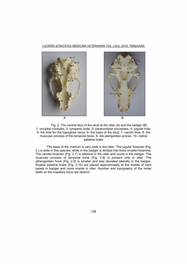
LUCRĂRI ŞTIINŢIFICE MEDICINĂ VETERINARĂ VOL. LII(4), 2019, TIMIŞOARA
128
A B
Fig. 2. The ventral face of the skull at the otter (A) and the badger (B)
1- occipital condyles, 2- tympanic bulla, 3- paracondylar processes, 4- jugular hole, 5- the hole for the hypogloss nerve, 6- the basis of the skull, 7- carotic hole, 8- the
muscular process of the temporal bone, 9- the pterigoidian proces, 10- rostral palatine holes
The base of the cranium is very wide in the otter. The jugular foramen (Fig.
2.) is wide in this species, while in the badger is divided into three smaller foramina. The carotid foramen (Fig. 2.7) is elliptical in the otter and round in the badger. The muscular process of temporal bone (Fig. 2.8) is present only in otter. The ptherigoidian hook (Fig. 2.9) is smaller and less deviated laterally in the badger. Rostral palatine holes (Fig. 2.10) are placed approximately at the middle of hard palate in badger and more cranial in otter. Number and topography of the molar teeth on the maxillary bone are distinct.

LUCRĂRI ŞTIINŢIFICE MEDICINĂ VETERINARĂ VOL. LII(4), 2019, TIMIŞOARA
129
A - OTTER
B - BADGER
Fig. 3. The lateral face of the skull at the otter (A) and the badger (B)
1-temporal fossa, 2- orbital fossa, 3- infraorbital hole, 4- the internal auditive duct, 5- tympanic bulla, 6- pterigoidian hook, 7- external sagital crest, 8- orbito-
temporalcrest, 9- the process of the frontal bone In lateral view, the skull at the otter appears long, with flattened cranial
portion. In badger is higher, with convex dorsal profile. The temporal fossa (Fig. 3.1) is shorter and is delimited from the orbital wall by a very obvious orbito-temporal crest (Fig. 3.8) (low in the otter). The rostro-dorsal edge of the orbit (red arrow) is prominent in otter.
From the dorsal edge of the zygomatic arcade in otter, an obvious frontal process (Fig. 3.9) is raised, absent in badger.

LUCRĂRI ŞTIINŢIFICE MEDICINĂ VETERINARĂ VOL. LII(4), 2019, TIMIŞOARA
130
A B
Fig. 4. The aboral face of the skull at the otter (A) and the badger (B):
1- occipital condyles, 2- occipital hole, 3- nuchal crest, 4- external occipital protuberance, 5- paracondylar processes.
The nuchal surface is taller in badger, with the external occipital
protuberance being easily identified (Fig. 4).
A B
Fig. 5. The mandible in otter (A) and badger (B) Dorsolateral view: 1- the carnassial alveola, 2- maseterine fossa, 3- pterigoidian
fossa The mandibles can be differentiated according to the elongated aspect of
the carnassial alveoli (Fig. 4.1) and the presence of a sharp crest (red arrow) that delimits in the ventral part the masseter in fossa (Fig. 4.2) in otter. It can also identify a bony crest (blue arrow) located above the mandibular hole, limiting in ventral part the pterygoid fossa (Fig. 4.3). These crests are extremely low, very small in badger.
LUCRĂRI ŞTIINŢIFICE MEDICINĂ VETERINARĂ VOL. LII(4), 2019, TIMIŞOARA
131
Our contribution
In the literature, most papers analyze skulls separately and not comparatively. The authors mainly address to the morphometric aspects, aiming to determine age for these species, morphometric variations between genders as well as variations between individuals in different geographic areas.
As mentioned above, all images are original photos made according to the studied material. The results represent the interpretation of the real morphological aspects observed in the skulls of the two species.
The work is a premiere in terms of details of the description and attached iconographic material.
Conclusions
At first sight, the skulls of the two mustelid species show a multitude of
common aspects that make it difficult to determine which species they come from, especially when dealing with incomplete skulls or even fragments of them.
It was found, however, that there is sufficient detail to allow undoubted recognition of the provenance of study material, even in the last two situations, without the need for further exams.
The morphometric differences do not allow confusions between the skulls of these two species and the other mustelids, but we consider that a comparative study between the skull of the badger and the one of the raccoon (Nyctereutes procyonoides) species present in the fauna of Romania, similar to the size, included in the Order Carnivora.
References
1. Abramov, A., Puzachenko, A.Y., Wiig, Y., Cranial variation in the European
badger (Meles meles) (Carnivora, Mustelidae) in Scandinavia, Zoological Journal of the Linnean Society, 157(2), 433–450, 2009.
2. Cotta, V., Bodea, M., Micu, I., Vânatul și vânătoarea din Romania, Ed. Ceres București, 2008.
3. Pertoldi, C., Madsen, A.B., Randi, E., Braun, A., Loeschcke, V., Variation of skull morphometry of Eurasian otters (Lutra lutra) in Dannark and Germany, Ann. Zool. Fennici, 1998, 35, 87-94.
4. Predoi, G., Belu, C., Dumitrescu, I., Georgescu, B., Șeicaru, A., Roșu, P., Bărbuceanu, F., Macroscopic anatomy of the skull of the otter (Lutra lutra), Anatomia, Histologia, Embryologia, 2016, 45, 62.
5. Predoi, G., Georgescu, B., Belu, C., Dumitrescu, I., Anatomia comparată și topografică a animalelor domestice, Vol. II Lito. A.M.C.-USAMV, București, 2002.

LUCRARI STIINTIFICE MEDICINA VETERINARA VOL. LII(4), 2019, TIMISOARA
132
AUTHORS INDEX
A Ahmadi M. 4 Avram E. 91 B Berean D. 9 Bejinaru C.V. 53 Blaga Petrean A. 9 Bogdan L.M. 9 Boldura O.M. 83 Borchescu G. 91 Bouari C.M. 79 Brudașcă G.F. 102, 109, 114, 120 Buzura-Matei I.A. 79 C Cărunta A. 15 Cătană N. 53 Cenariu M. 9 Cerbu C. 102, 109, 114 Chirilă F. 79 Chiș M. 15 Ciulan V. 91 Cristina R.T. 27 D Dégi D.M. 27 Dégi J. 27, 53 Diakou A. 72 Dronca D. 4 Dumitrescu C. 27 F Filipoi C.D. 79 Fiț N.I. 79
G Georgescu M. 39 Giurgiu O. 9 Grozea A. 58 H Herman V. 53 I Iancu I. 27, 53 Igna V. 58, 65 Ilie D.E. 15 K Kicsid Z. 120 Krasovskaia I. 102, 109, 114, 120 L Lixandru D. 126 M Mathiu T.M. 58, 65 Mederle N. 4, 72 Milovanov C. 4 Miok K. 15 Moleriu R.15 Morariu F. 4 Morariu S. 4 Mureșan R. 15 Muselin F. 27 N Nadaș G.C. 79 Nadăş G. 9 Nichita I. 4 Nicula A.G. 83 Niculae M. 102, 109, 114, 120

LUCRARI STIINTIFICE MEDICINA VETERINARA VOL. LII(4), 2019, TIMISOARA
133
Novac C.Ș. 79 O Olah D.I. 120 Olariu-Jurca A. 91 Opruţi D.A. 72 P Pall E. 9, 102, 109, 120 Pascalau R. 4 Pascu C. 53 Potîrniche A. 109 R Raita Ș.M. 39 S Seicaru A. 126 Seicheli R. 114 Spînu M. 102, 109, 114, 120 Suătean M.I. 102 Szalo-Mureșan D. 114 Ș Șandru C.D. 102, 109, 114 T Telea A. 58 Teslici L.E. 91 Tulcan C. 83 Turcu B. 120 V Vasiu A. 102, 109, 114, 120 Z Zagrai (Maierean) A.M. 126 Zagrai G. 126 Zaharia C. 15
Zaharie D. 15

LUCRARI STIINTIFICE MEDICINA VETERINARA VOL. LII(4), 2019, TIMISOARA
134
CONTENT
Ahmadi M., Mederle N., Milovanov C., Morariu S., Nichita I., Morariu F., Pascalau R., Dronca D.
Chenopodium quinoa – an alternative grain in food and feed products
4
Berean D., Blaga Petrean A., Cenariu M., Giurgiu O., Nadăş G., Pall E., Bogdan L.M.
The incidence of dystocia in cows and its effects on postpartum reproductive performance and milk production
9
Cărunta A., Chiș M., Ilie D.E., Miok K., Moleriu R., Mureșan R., Zaharia C., Zaharie D.
Descriptive analysis of somatic cell countusing statistical tools
15
Dégi J., Cristina R.T., Dégi D.M., Muselin F., Dumitrescu C., Iancu I.
Research of methicillin resistance Staphylococci in a pig’s farm
27
Georgescu M., Raita Ș.M. Deployment of the carver plus shock
vulnerability assessment method in a food processing facility - a case study
39
Iancu I., Bejinaru C.V., Pascu C., Dégi J., Cătană N., Herman V.
Research on the evolution of Enzootic Bovine Leukosis in southwest Romania between 2013-2017
53
Igna V., Mathiu T.M., Telea A., Grozea A.
Effect of deuterium-depleted water based activators on sperm motility in pikeperch (Sander lucioperca), sterlet (Acipenser ruthenus), russian sturgeon (Acipenser gueldenstaedtii) and catfish (Silurus glanis)
58
Mathiu T.M., Igna V. Deuterium depleted water - biomedical
implications 65
Mederle N., Opruţi D.A., Diakou A.
Epidemiological research in canine dirofilariosis in Thessaloniki area of Greece
72
Nadaș G.C., Filipoi C.D., Bouari The identification and antimicrobial 79

LUCRARI STIINTIFICE MEDICINA VETERINARA VOL. LII(4), 2019, TIMISOARA
135
C.M., Buzura-Matei I.A., Chirilă F., Novac C.Ș., Fiț N.I.
susceptibility profile of conjunctival flora from dogs
Nicula A.G., Tulcan C., Boldura O.M.
Data interpretation from a DNA fingerprinting experiment
83
Olariu-Jurca A., Ciulan V., Avram E., Borchescu G., Teslici L.E.
Morphopathological research in canine infectious hepatitis and aspects regarding the histopathological diagnosis of the condition
91
Șandru C.D., Niculae M., Brudașcă G.F., Pall E., Vasiu A., Cerbu C., Suătean M.I., Spînu M., Krasovskaia I.
The potential local pathogenic role of circulating immune complexes in contagious agalactia of sheep
102
Șandru C.D., Niculae M., Brudașcă G.F., Pall E., Vasiu A., Cerbu C., Potîrniche A., Spînu M., Krasovskaia I.
Dissimilar stresses influence differently the immune responses in broiler chickens
109
Vasiu A., Șandru C.D., Szalo-Mureșan D., Niculae M., Cerbu C., Seicheli R., Brudașcă G.F., Krasovskaia I., Spînu M.
Temporal dynamics of post-vaccinationhumoral immune responsein ostriches primed against anthrax
114
Vasiu A., Turcu B., Kicsid Z., Niculae M., Pall E., Olah D.I., Brudașcă G.F., Spînu M., Krasovskaia I.
Microbiogical investigations in a severe hemorrhagic syndrome in farmed ostriches
120
Zagrai G., Seicaru A., Lixandru D., Zagrai (Maierean) A.M.
Morphology of the skulls in badger (Meles meles) and otter (Lutra lutra) –comparative aspects
126