Embed Size (px)
Citation preview
72 Deep Tendon ReflexesH. KENNETH WALKER
Definition
In a normal person, when a muscle tendon is tapped briskly,the muscle immediately contracts due to a two-neuron re-flex arc involving the spinal or brainstem segment that in-nervates the muscle . The afferent neuron whose cell bodylies in a dorsal root ganglion innervates the muscle or Golgitendon organ associated with the muscles ; the efferent neu-ron is an alpha motoneuron in the anterior horn of thecord. The cerebral cortex and a number of brainstem nucleiexert influence over the sensory input of the muscle spindlesby means of the gamma motoneurons that are located inthe anterior horn ; these neurons supply a set of musclefibers that control the length of the muscle spindle itself .
Hyporeflexia is an absent or diminished response to tap-ping. It usually indicates a disease that involves one or moreof the components of the two-neuron reflex arc itself .
Hyperreflexia refers to hyperactive or repeating (clonic)reflexes . These usually indicate an interruption of corti-cospinal and other descending pathways that influence thereflex arc due to a suprasegmental lesion, that is, a lesionabove the level of the spinal reflex pathways .
By convention the deep tendon reflexes are graded asfollows :
0 = no response; always abnormal
1 + = a slight but definitely present response ; may or maynot be normal
2 + = a brisk response ; normal
3 + = a very brisk response ; may or may not be normal
4+ = a tap elicits a repeating reflex (clonus) ; always ab-normal
Whether the 1 + and 3 + responses are normal depends onwhat they were previously, that is, the patient's reflex his-tory; what the other reflexes are ; and analysis of associatedfindings such as muscle tone, muscle strength, or otherevidence of disease . Asymmetry of reflexes suggests ab-normality .
Technique
All of the commonly used deep tendon reflexes are pre-sented here in a group. In a screening examination you willusually find it more convenient to integrate the reflex ex-amination into the rest of the examination of that part ofthe body ; that is, do the upper extremity reflexes whenexamining the rest of the upper extremity . When an ab-normality of the reflexes is suspected or discovered, how-ever, the reflexes should be examined as a group with carefulottaT,t ;~A t- tho
.,f rho
Valid test results are best obtained when the patient isrelaxed and not thinking about what you are doing . Aftera general explanation, mingle the specific instructions withquestions or comments designed to get the patient to speakat some length about some other topic . If you cannot getany response with a specific reflex-ankle jerks are usuallythe most difficult-then try the following :
•
Several different positions of the limb .•
Get the patient to put slight tension on the musclebeing tested . One method of achieving this is to havethe patient strongly contract a muscle not being tested .
•
In the upper extremity, have the patient make a fistwith one hand while the opposite extremity is beingtested .
•
If the reflex being tested is the knee jerk or ankle jerk,have the patient perform the `Jendrassik maneuver,"a reinforcement of the reflex (see Gassel, 1964) . Thepatient's fingers of each hand are hooked together soeach arm can forcefully pull against the other . Thesplit second before you are ready to tap the tendon,say "pull ."
•
In general, any way to distract the patient from whatyou are doing will enhance the chances of obtainingthe reflex . Having the patient count or give the namesof children are examples .
The best position is for the patient to be sitting on theside of the bed or examining table . The Babinski reflexhammer (Figure 72 .1) is very good . Use a brisk but notpainful tap. Use your wrist, not your arm, for the action .In an extremity a useful maneuver is to elicit the reflexfrom several different positions, rapidly shifting the limband performing the test. Use varying force and note anyvariance in response.
Note the following features of the reflex response :
•
Amount of hammer force necessary to obtain con-traction
•
Velocity of contraction•
Strength of contraction•
Duration of contraction•
Duration of relaxation phase•
Response of other muscles that were not tested. Whena reflex is hyperactive, that muscle often will respondto the testing of a nearby muscle . A good example isreflex activity of a hyperactive biceps or finger reflex
Figure 72 .1The Babinski reflex hammer .
365
366
IV . THE NEUROLOGIC SYSTEM
when the brachioradialis tendon is tapped . This istermed "overflowing" of a reflex .
After obtaining the reflex on one side, always go im-mediately to the opposite side for the same reflex so thatyou can compare them .
Jaw jerk
Place the tip of your index finger on a relaxed jaw, one thatis about one-third open . Tap briskly on your index fingerand note the speed as the mandible is flexed (see Chapter61 on the trigeminal nerve) .
Biceps Reflex
The forearm should be supported, either resting on thepatient's thighs or resting on the forearm of the examiner .The arm is midway between flexion and extension . Placeyour thumb firmly over the biceps tendon, with your fingerscurling around the elbow, and tap briskly. The forearm willflex at the elbow .
Triceps Reflex
Support the patient's forearm by cradling it with yours orby placing it on the thigh, with the arm midway betweenflexion and extension. Identify the triceps tendon at itsinsertion on the olecranon, and tap just above the insertion .There is extension of the forearm .
Brachioradialis Reflex
The patient's arm should be supported . Identify the bra-chioradialis tendon at the wrist. It inserts at the base of thestyloid process of the radius, usually about 1 cm lateral tothe radial artery. If in doubt, ask the patient to hold thearm as if in a sling-flexed at the elbow and halfway be-tween pronation and supination-and then flex the fore-arm at the elbow against resistance from you . Thebrachioradialis and its tendon will then stand out .
Place the thumb of the hand supporting the patient'selbow on the biceps tendon while tapping the brachiora-dialis tendon with the other hand . Observe three potentialreflexes as you tap.
1. Brachioradialis reflex : flexion and supination of theforearm .
2. Biceps reflex : flexion of the forearm. You will feelthe biceps tendon contract if the biceps reflex is stim-ulated by the tap on the brachioradialis tendon .
3. Finger jerk: flexion of the fingers .
The usual pattern is for only the brachioradialis reflex tobe stimulated . But in the presence of a hyperactive bicepsor finger jerk reflex, these reflexes may be stimulated also .
Finger Jerk
Have the patient gently curl his fingers over your indexfinger, much as a bird curls its claws around the branch of
a tree. Then raise your hand, with the patient's hand nowbeing supported by the curled fingers . Tap briskly on yourfingers so that the force will transmit to the patient's curledfingers. The response is a flexion of the patient's fingers .
Knee Jerk
Let the knees swing free by the side of the bed, and placeone hand on the quadriceps so you can feel its contraction .If the patient is in bed, slightly flex the knee by placingyour forearm under both knees by contraction of the quad-riceps with extension of the lower leg. If the reflex is hy-peractive there is sometimes concomitant adduction of theipsilateral thigh. Adduction of the opposite thigh and ex-tension of the opposite lower leg also can occur simulta-neously if those reflexes are hyperactive . Note that this so-called crossed thigh adduction or leg extension tells youthat the reflexes in the opposite leg are hyperactive . Theytell you nothing about the state of the reflex in the leg beingtested. Use the Jendrassik maneuver if there is no response .
Ankle Jerk
With the patient sitting, place one hand underneath the soleand dorsiflex the foot slightly . Then tap on the Achillestendon just above its insertion on the calcaneus . If the pa-tient is in bed, flex the knee and invert or evert the footsomewhat, cradling the foot and lower leg in your arm .Then tap on the tendon .
If no response is obtained, have the patient face a chairand kneel on it with the knees resting against the back ofthe chair, the elbows on the top of the back, and the feetprojecting over the seat . First dorsiflex the foot slightly andtap on the tendon . Use the Jendrassik maneuver if thisdoesn't work. This position is well suited to observing therelaxation phase of the reflex in patients with suspectedthyroid disease .
See Dejong (1967) for a description of numerous otherreflexes that are useful in certain situations .
Basic Science
A stretch reflex is the contraction of a muscle in responseto stretching of muscle spindles, which are receptors thatlie in parallel with extrafusal muscle fibers. The reflex iscomposed of a two-neuron arc . The afferent neuron, whosecell body is in a sensory ganglion, innervates the spindle .When the muscle spindle is stretched, this neuron fires andmonosynaptically excites alpha motoneurons in the anteriorhorn of the spinal cord . This alpha motoneuron is the sec-ond neuron ; it supplies the muscle that is being tapped ortransiently stretched . The detailed mechanisms underlyingthe operation of the spindle are quite complex, but consid-erable knowledge about them is now available in the liter-ature, and new details are added constantly . The musclespindle is a slender, spindle-shaped structure that is inter-mingled with the usual muscle fibers . Each spindle is com-posed of two types of elongated, poorly staining fibers :nuclear bag fibers and nuclear chain fibers . Each containsmultiple nuclei . Six to ten of these fibers lie within thespindle's connective tissue sheath . They are called "intra-fusal" muscle fibers, since they lie inside the fusiform struc-

Lure, in contrast to the surrounding "extrafusal" fibers thatmake up the contractile element of muscle .
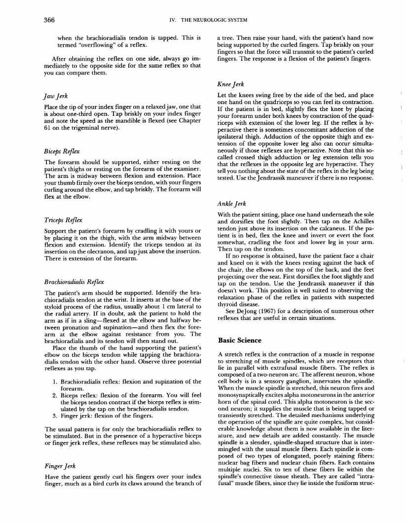
Afferent sensory terminals that innervate the spindlefibers are of two types : primary and secondary (Figure 72 .2) .The spindles fire according to the velocity and amount ofstretch placed upon the central nuclear regions of the in-trafusal fibers . The degree of stretch communicated to thecentral portion of the fibers is determined by two factors :the length and change in length of the surrounding extra-fusal fibers (see Figure 72 .2) and the degree of contractionof the intrafusal fibers (see below) .
Impulses from the spindle receptors enter the dorsalhorn where the information takes four routes : (1) to thecortex; (2) to synapse directly on an alpha motoneuron,which causes immediate contraction of the muscle inner-vated by the spindle, the agonist ; (3) to synapse on an in-hibitory neuron which in turn synapses on an alphamotoneuron that goes to a muscle antagonistic to the oneinnervated by the spindle-thus there is concomitant re-laxation of the antagonist as the agonist contracts ; and (4)to the cerebellum via the dorsal spinocerebellar tracts .
The previous paragraph describes the course taken bythe afferent impulses from the sensory nuclei of the musclespindles. Recall now that the second component of the spin-dle was a contractile element, the intrafusal fibers . The fir-ing of the spindle afferents is dependent upon the lengthof the extrafusal fibers (as outlined above) and the length
72 . DEEP TENDON REFLEXES
367
of the intrafusal fibers . The contraction of the ends of in-trafusal fibers and thus the strength of the central portionsare controlled by gamma motoneurons : these small neuronsare located in the anterior horn and are influenced by thecerebellum, the cortex, and various brainstem nuclei. Theprobable function of this motor innervation of a sensorystructure is to enable these supraspinal structures to "set"and thus ultimately regulate the sensitivity of the spindle .The higher centers and, in particular, the cortex therebyget sensory information from the muscle spindles and, inturn, through the gamma motoneuron, control the amountand quality of information received .
The Golgi tendon organ, which is the second major mus-cle receptor, is attached between the extrafusal fibers andthe tendon . Thus the tendon organ is in series with theextrafusal fibers and will fire as the muscle contracts . Thespindles, in contrast, are parallel with (i .e ., alongside) theextrafusal fibers and so fire when the extrafusal fibers relax(i .e., are stretched). The impulses from the tendon orgango through the dorsal horn and synapse on an inhibitoryinterneuron which in turn synapses on an alpha motoneu-ron that goes to the agonist. Therefore the tendon organultimately causes relaxation of the agonist and, by way ofinterneurons, a facilitation of the antagonist . Informationis also conveyed from these receptors to the cerebellum andcortex .
The spinal reflexes that are set up by the mechanisms
Figure 72.2Summary of muscle spindles and tendon organs .
MuscleReceptors
Fiber
Gamma Motoneuronstypes
Pathways
Sensory endings
(efferents to spindles)
Musclespindle
Nuclearbagfiber
Musclespindle:
Cerebellum Cortex -
- Lie in parallel with extrafusal fibersSensory endingsGroup la "primary ending"Synonym: annulospiral ending
Gives information about length and ve-locity of extension of muscle :
Dynamic firing: spindle firing isgreatest while muscle is lengthen-ing, firing ceases with contractionand is reduced with steady lengthsbelow that seen during the stretch-ing process
Small anterior horn motoneurons that re-ceive descending cortical impulses andthen send efferent axons to the musclespindles (see diagram under Path-ways) . As they fire there is a contrac-tion of the intrafusal fibers of the mus-cle spindle . This causes a stretching ofthe central part of the spindle wherethe sensory fibers are, and con-sequently the sensory fibers fire. Thissetup probably allows supraspinalstructures to regulate the sensitivity ofthe muscle spindles, or their back-ground firing levels .
Gamma dynamic motoneurons supplygroup I primary endings and increasetheir responsiveness to velocity ofspindle elongation .
Gamma static motoneurons supplygroup 1 and group 2 endings and in-crease their levels of firing in responseto steady stretches
Afferentcomponent
31 Dorsal hornVentral horn :
I Alpha
Fmotoneuron
Inhibitory F_interneuron
1k
FAlphamotoneuron
Efferentcomponent
Agonist.muscle
Antagonistmuscle
3. Gammamotoneuron
Nuclearchainfiber
As above, with thecitatory reflex pathwayone or more interneurons .
exception that the ex-may involve
Lie in parallel with extrafusal fibersSensory endingsGroup la as aboveGroup 11 "secondary ending"Synonym . flower-spray ending
Gives information about length and ve-locity of extension of muscle :
Static firing : response greatest whenstretch is constant after contractionhas ceased .
Golgitendonorgan
Tendon
Dorsal horn Lie in series with extrafusal fibers, sincethey are attached to tendons .
Sensory endings : Group lbGives information about muscle tension :
fires briskly during contraction, but lit-tle during passive elongation of themuscle.
organ -3PVentral horn
Facilitatoryinterneuron
J,Antagonist
Alphamuscle
motoneuron
Inhibitoryinterferon
yAgonist
Alphamuscle
motoneuron
368
IV . THE NEUROLOGIC SYSTEM
described above serve the function of keeping the musclefibers adjusted to a certain length and to a certain tension,thereby maintaining muscle tone and ultimately limb pos-ture .
Clinical Significance
Absent stretch reflexes indicate a lesion in the reflex arcitself. Associated symptoms and signs usually make locali-zation possible :
1 . Absent reflexes and sensory loss in the distributionof the nerve supplying the reflex : the lesion involvesthe afferent arc of the reflex-either nerve or dorsalhorn .
2 . Absent reflex with paralysis, muscle atrophy, and fas-ciculations: the lesion involves the efferent arc-an-terior horn cells or efferent nerve, or both .
Peripheral neuropathy is today the most common causeof absent reflexes. The causes include diseases such as di-abetes, alcoholism, amyloidosis, uremia ; vitamin deficien-cies such as pellagra, beriberi, pernicious anemia ; remotecancer; toxins including lead, arsenic, isoniazid, vincristine,diphenylhydantoin . Neuropathies can be predominantlysensory, motor, or mixed and therefore can affect any orall components of the reflex arc (see Adams and Asbury,1970, for a good discussion). Muscle diseases do not producea disturbance of the stretch reflex unless the muscle is ren-dered too weak to contract . This occasionally occurs in dis-eases such as polymyositis and muscular dystrophy.
Hyperactive stretch reflexes are seen when there is in-terruption of the cortical supply to the lower motor neuron,an "upper motor neuron lesion ." The interruption can beanywhere above the segment of the reflex arc . Analysis ofassociated findings enables localization of the lesion .
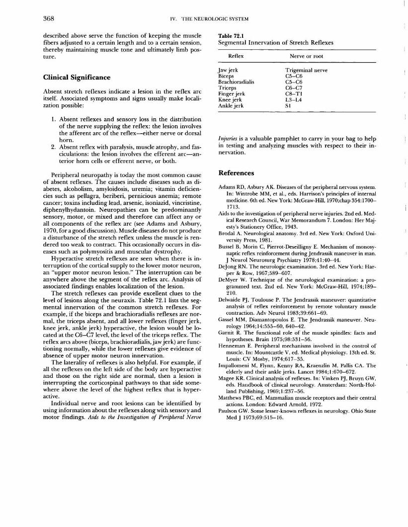
The stretch reflexes can provide excellent clues to thelevel of lesions along the neuraxis . Table 72.1 lists the seg-mental innervation of the common stretch reflexes . Forexample, if the biceps and brachioradialis reflexes are nor-mal, the triceps absent, and all lower reflexes (finger jerk,knee jerk, ankle jerk) hyperactive, the lesion would be lo-cated at the C6-C7 level, the level of the triceps reflex . Thereflex arcs above (biceps, brachioradialis, jaw jerk) are func-tioning normally, while the lower reflexes give evidence ofabsence of upper motor neuron innervation .
The laterality of reflexes is also helpful . For example, ifall the reflexes on the left side of the body are hyperactiveand those on the right side are normal, then a lesion isinterrupting the corticospinal pathways to that side some-where above the level of the highest reflex that is hyper-active .
Individual nerve and root lesions can be identified byusing information about the reflexes along with sensory andmotor findings . Aids to the Investigation of Peripheral Nerve
Table 72 .1Segmental Innervation of Stretch Reflexes
Injuries is a valuable pamphlet to carry in your bag to helpin testing and analyzing muscles with respect to their in-nervation .
References
Adams RD, Asbury AK . Diseases of the peripheral nervous system .In: Wintrobe MM, et al ., eds . Harrison's principles of internalmedicine . 6th ed . New York : McGraw-Hill, 1970 ;chap 354 :1700-1713.
Aids to the investigation of peripheral nerve injuries . 2nd ed . Med-ical Research Council, War Memorandum 7. London: Her Maj-esty's Stationery Office, 1943 .
Brodal A . Neurological anatomy . 3rd ed . New York: Oxford Uni-versity Press, 1981 .
Bussel B, Morin C, Pierrot-Deseilligny E. Mechanism of monosy-naptic reflex reinforcement during Jendrassik maneuver in man .J Neurol Neurosurg Psychiatry 1978 ;41 :40-44 .
Dejong RN . The neurologic examination . 3rd ed . New York : Har-per & Row, 1967 ;589-607 .
DeMyer W. Technique of the neurological examination: a pro-grammed text. 2nd ed. New York : McGraw-Hill, 1974 ;189-210 .
Delwaide PJ, Toulouse P. The Jendrassik maneuver : quantitativeanalysis of reflex reinforcement by remote voluntary musclecontraction. Adv Neurol 1983;39:661-69 .
Gassel MM, Diamantopoulos E . The Jendrassik maneuver. Neu-rology 1964;14:555-60, 640-42 .
Garnit R. The functional role of the muscle spindles : facts andhypotheses . Brain 1975 ;98:531-56 .
Henneman E. Peripheral mechanisms involved in the control ofmuscle. In : Mountcastle V, ed. Medical physiology. 13th ed . St .Louis : CV Mosby, 1974 ;617-35 .
Impallomeni M, Flynn, Kenny RA, Kraenzlin M, Pallis CA . Theelderly and their ankle jerks . Lancet 1984 ; 1 :670-672 .
Magee KR . Clinical analysis of reflexes . In : Vinken PJ, Bruyn GW,eds . Handbook of clinical neurology . Amsterdam : North-Hol-land Publishing, 1969 ;1 :237-56 .
Matthews PBC, ed. Mammalian muscle receptors and their centralactions . London : Edward Arnold, 1972 .
Paulson GW . Some lesser-known reflexes in neurology . Ohio StateMed J 1973;69:515-16 .
Reflex Nerve or root
Jaw jerk Trigeminal nerveBiceps C5-C6Brachioradialis C5-C6Triceps C6-C7Finger jerk C8-T1Knee jerk L3-L4Ankle jerk S 1