Embed Size (px)
Citation preview

40th Annual Mallory-Coleman Resident Research Day
Friday, June 8 , 2012
T H E O H I O S TAT E U N I V E R S I T Y
B l a c k w e l l H o t e l a n d C o n f e r e n c e C e n t e r
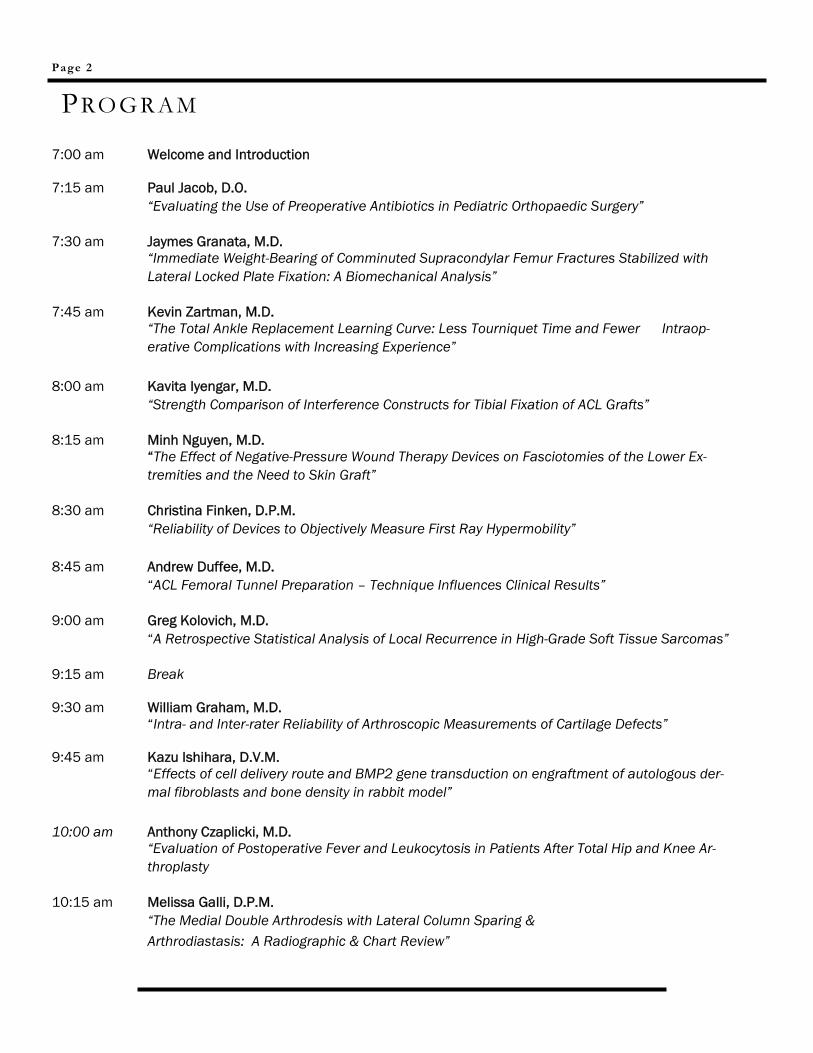
7:00 am Welcome and Introduction 7:15 am Paul Jacob, D.O.
“Evaluating the Use of Preoperative Antibiotics in Pediatric Orthopaedic Surgery” 7:30 am Jaymes Granata, M.D.
“Immediate Weight-Bearing of Comminuted Supracondylar Femur Fractures Stabilized with Lateral Locked Plate Fixation: A Biomechanical Analysis”
7:45 am Kevin Zartman, M.D.
“The Total Ankle Replacement Learning Curve: Less Tourniquet Time and Fewer Intraop-erative Complications with Increasing Experience”
8:00 am Kavita Iyengar, M.D.
“Strength Comparison of Interference Constructs for Tibial Fixation of ACL Grafts” 8:15 am Minh Nguyen, M.D.
“The Effect of Negative-Pressure Wound Therapy Devices on Fasciotomies of the Lower Ex-tremities and the Need to Skin Graft”
8:30 am Christina Finken, D.P.M.
“Reliability of Devices to Objectively Measure First Ray Hypermobility” 8:45 am Andrew Duffee, M.D.
“ACL Femoral Tunnel Preparation – Technique Influences Clinical Results” 9:00 am Greg Kolovich, M.D.
“A Retrospective Statistical Analysis of Local Recurrence in High-Grade Soft Tissue Sarcomas” 9:15 am Break 9:30 am William Graham, M.D.
“Intra- and Inter-rater Reliability of Arthroscopic Measurements of Cartilage Defects” 9:45 am Kazu Ishihara, D.V.M.
“Effects of cell delivery route and BMP2 gene transduction on engraftment of autologous der-mal fibroblasts and bone density in rabbit model”
10:00 am Anthony Czaplicki, M.D.
“Evaluation of Postoperative Fever and Leukocytosis in Patients After Total Hip and Knee Ar-throplasty
10:15 am Melissa Galli, D.P.M.
“The Medial Double Arthrodesis with Lateral Column Sparing & Arthrodiastasis: A Radiographic & Chart Review”
Page 2
PRO G R A M

The Ohio State Univers i ty
10:30 am Mark Jacobson, M.D.
“Open Capsular Shift and Arthroscopic Capsular Plication for Treatment of Multidi-rectional Instability: A Systematic Review”
10:45 am Shah Dodwad, M.D. “Independent Validation of the Thoracolumbar Injury Classification and Severity (TLICS) Score: A Retrospective Analysis of Clinical Management of Thoracolumbar Injuries”
11:00 am Scott Stephens, M.D.
“Repair of Medial Collateral Ligament Injury During Total Knee Arthoplasty” 11:15 am Break 11:30 am Regis O’Keefe, M.D., Visiting Professor and Moderator “Improving Orthopaedic Care Through Translational and Clinical Research: Opportunities and Challenges” 12:30 pm Lunch 1:30 pm Julie Samora, M.D.
“Outcomes Following Injury to the Thumb Ulnar Collateral Ligament – A Systematic Review”
1:45 pm Jaymes Granata, M.D.
“Talonavicular Joint Fixation: A Biomechanical Comparison of Locking Compression Plates and Lag Screws”
2:00 pm Doug Lucas, D.O. “Type II Supracondylar Humerus Fractures: Factors Predictive of Failure for Closed Reduction and Immobilization. ” 2:15 pm William Graham, M.D.
Clinical and Radiographic Outcomes following Surgical Treatment of Discoid Lateral Meniscus in Adolescent and Pediatric Patients
2:30 pm Carmen Quatman, M.D.
“Novel Methods to Practice Personalized Medicine in an Evidence Based Orthopaedic Setting”
2:45 pm John Ryan, M.D. “ACL Outcomes by Sex, A Systematic Review”
3:00 pm End of Day
Page 3

MALLORY-COLEMAN
DAY
Mallory-Coleman resident research day was established by Drs. Thomas Mallory and Carl Coleman in 1972 in memory of Katherine Virginia Mallory and Sally Jo Coleman. This research day was established in order to encourage the development of ideas related to research in orthopaedic surgery and related basic sciences. Each year, a distinguished visiting professor from an outside institution is invited to moderate and analyze the resident presentations and provide constructive criticism and commentary.
Past Visiting Professors: 2011 Henrik Malchau, M.D. 2010 Freddie Fu, M.D. 2009 James Heckman, M.D. 2008 Cato Laurencin, M.D. 2007 William Garrett, M.D. 2006 Peter Stern, M.D. 2005 James Goulet, M.D. 2004 Steven Arnoczky, D.V.M. 2003 Joseph Buckwalter, M.D. 2002 Victor Goldberg, M.D. 2001 James Urbaniak, M.D. 2000 Douglas Jackson, M.D. 1999 Douglas Dennis, MD 1998 Thomas Einhorn, MD 1997 Larry S. Matthews, MD 1996 Gary Friedlander, MD 1995 James Herndon, MD 1994 Clement B. Sledge, MD 1993 Eric L. Radin, MD
2 0 1 2 M A L L O R Y - C O L E M A N
V I S I T I N G P R O F E S S O R A N D M O D E R A T O R :
R E G I S O ’ K E E F E , M . D .
Regis J. O’Keefe, MD, PhD is the Marjorie Strong Wehle Professor and Chair, Department of Orthopaedics and Rehabilitation at the University of Rochester’s School of Medicine and Dentistry in Rochester, New York. He is also the Director of the Center for Musculoskeletal Re-search at the University of Rochester. Dr. O’Keefe is the Associate Dean of Clinical Affairs at the University of Rochester School of Medicine and Dentistry. Dr. O’Keefe earned his BA in philosophy and religious stud-ies and graduated magna cum laude at Yale University in New Haven, Connecticut. After earning his medical de-gree from Harvard Medical School in Boston, Massachu-setts, he completed a Ph.D. in biochemistry and biophys-
ics from the University of Rochester School of Medicine and Dentistry. Dr. O’Keefe served his internship in surgery at New England Deaconess Hospital in Boston, his residency in orthopaedics at the University of Rochester Medical Center and completed an oncology fellowship at the Massachusetts General Hos-pital. In 1993, he joined the faculty at the University of Rochester.
Dr. O’Keefe has authored or coauthored over 230 articles, more than 300 ab-stracts, 14 book chapters and reviews concerning bone repair and development, cancer, inflammatory diseases of bone, genetics, and related topics. Most of his research has been supported by NIH grants, and his NIH funding has consis-tently placed him among the most highly funded orthopaedic surgeon-clinician scientists in the United States.
He is an Associate Editor of the Journal of Bone and Mineral Research. Dr. O’Keefe has served in numerous leadership roles in national orthopaedic organi-zations. Dr. O’Keefe is a member of the Board of Directors of the American Board of Orthopaedic Surgery (ABOS). He served for more 6 years as a mem-ber of the Orthopaedic Research and Education Foundation, including service in the role of Secretary of that organization. Dr. O’Keefe is a past-President of the Orthopaedic Research Society (ORS). He also served the ORS as Treasurer and as a member of the Program Committee. He is the past-President of the United States Bone and Joint Decade, an international coalition of healthcare organiza-tions aimed at decreasing the incidence of bone and joint disorders. Dr. O’Keefe is also the past-Chair of the Skeletal Biology and Skeletal Regeneration Study Section for the National Institutes of Health’s Center for Scientific Review. Dr. O’Keefe is currently serving on the Advisory Council of the National Institutes of Health (NIH) National Arthritis and Musculoskeletal and Skin Diseases (NIAMS). He represents NIAMS on the National Institutes of Health Council of Councils (COC) Advisory Council that reviews trans-NIH initiatives. Dr. O’Keefe has served as the Chair of the American Academy of Orthopaedic American Academy of Orthopaedic Surgeons’ Clinician Scientist Committee. He also has directed the Orthopaedic Research and Education Foundation’s Grant Writing Workshop, a program that mentors young scientists in the critical skill of grant writing. He is a member of the American Association of Physi-cians (AAP).
Dr. O’Keefe has received a variety of teaching and scientific awards including the prestigious ABC Traveling Fellowship from the American Orthopaedic As-sociation and the Kappa Delta Award, recognizing excellence in orthopaedic research. Dr. O’Keefe has worked diligently in his career to promote and ad-vance the basic understanding of musculoskeletal diseases and to translate these discoveries into therapies designed to improve the care of orthopaedic patients.
Page 4
Page 5
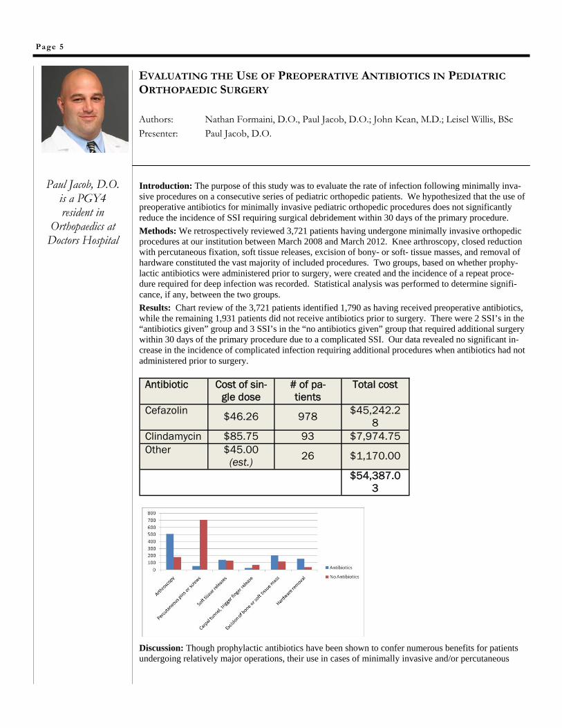
EVALUATING THE USE OF PREOPERATIVE ANTIBIOTICS IN PEDIATRIC ORTHOPAEDIC SURGERY Authors: Nathan Formaini, D.O., Paul Jacob, D.O.; John Kean, M.D.; Leisel Willis, BSc Presenter: Paul Jacob, D.O.
Introduction: The purpose of this study was to evaluate the rate of infection following minimally inva-sive procedures on a consecutive series of pediatric orthopedic patients. We hypothesized that the use of preoperative antibiotics for minimally invasive pediatric orthopedic procedures does not significantly reduce the incidence of SSI requiring surgical debridement within 30 days of the primary procedure.
Methods: We retrospectively reviewed 3,721 patients having undergone minimally invasive orthopedic procedures at our institution between March 2008 and March 2012. Knee arthroscopy, closed reduction with percutaneous fixation, soft tissue releases, excision of bony- or soft- tissue masses, and removal of hardware constituted the vast majority of included procedures. Two groups, based on whether prophy-lactic antibiotics were administered prior to surgery, were created and the incidence of a repeat proce-dure required for deep infection was recorded. Statistical analysis was performed to determine signifi-cance, if any, between the two groups.
Results: Chart review of the 3,721 patients identified 1,790 as having received preoperative antibiotics, while the remaining 1,931 patients did not receive antibiotics prior to surgery. There were 2 SSI’s in the “antibiotics given” group and 3 SSI’s in the “no antibiotics given” group that required additional surgery within 30 days of the primary procedure due to a complicated SSI. Our data revealed no significant in-crease in the incidence of complicated infection requiring additional procedures when antibiotics had not administered prior to surgery.
Discussion: Though prophylactic antibiotics have been shown to confer numerous benefits for patients undergoing relatively major operations, their use in cases of minimally invasive and/or percutaneous
Antibiotic Cost of sin-gle dose
# of pa-tients
Total cost
Cefazolin $46.26 978 $45,242.2
8
Clindamycin $85.75 93 $7,974.75
Other $45.00 (est.)
26 $1,170.00
$54,387.0
3
Paul Jacob, D.O. is a PGY4 resident in
Orthopaedics at Doctors Hospital
INTRODUCTION:
Comminuted supracondylar femur fractures (AO-OTA 33A3) are commonly treated with locking stainless steel plates and screws. Weight bearing is generally restricted for 6-12 weeks until there is radiologic evidence of sufficient callous to support weight bearing. Recent clinical studies have re-ported high nonunion rates with distal femur locked plates.1 One speculated cause of this disruption in fracture healing has been high construct stiffness. In an attempt to induce beneficial motion across the fracture site, some have made recommendations for earlier weight bearing. The purpose of this study was to determine the biomechanical feasibility of an immediate full weight bearing rehabilitation proto-col in distal femur fractures treated with lateral locked plate fixation.
METHODS:
Sixteen cadaveric femora were procured for this study. The donor demographics were chosen to maxi-mize the bone quality and radiographs were taken to exclude samples with defects. A 2.5 centimeter gap osteotomy was made to represent a comminuted supracondylar femur fracture. Ten hole 4.5 mm distal femur locking plates (Smith and Nephew Peri-Loc) were used throughout the study. A standard-ized construct was used in all the specimens: in the distal fragment, five 5.7 mm locking screws were used; in the diaphyseal region, four 4.5 mm non-locking cortical screws were used, placed in the first four proximal holes. The specimens were placed in a servohydraulic testing machine and axially loaded at 1 cycle per second.
The Staircase Method was used to determine the fatigue limit of the construct.2 The run-out limit (200,000 cycles) was chosen to simulate walking two miles per day, seven days per week for ten weeks. Failure was defined as one centimeter of deflection across the osteotomy site.
DATA AND RESULTS:
The average age of the donor was 36 years old, including 12 males and 3 females. The bone quality allowed for excellent purchase of the non-locking diaphyseal screws in all the samples. A single load to failure was estimated to be 2000 N. The fatigue testing loads were started at 80% of the single load to failure.
Nine constructs withstood the full 200,000 cycles at loads ranging from 800N to 1400 N. Six con-structs failed, with cycles to failure ranging from 4,000 to 124,000, and loads ranging from 1000 N to 1600 N. Using the staircase method, the fatigue (endurance) limit of the implant was calculated to be 1329 N± 106 N.
No constructs failed through the screws or screw plate interface. Plastic deformation, when noted, oc-curred at the metaphyseal flare of the plate.
Page 6
IMMEDIATE WEIGHT-BEARING OF COMMINUTED SUPRACONDYLAR FEMUR FRACTURES STABILIZED WITH LATERAL LOCKED PLATE FIXATION: A BIOMECHANICAL ANALYSIS
Authors: Jaymes Granata, M.D.; Alan Litsky, M.D., Sc.D.; David Lustenberger; Robert Probe, M.D., Thomas Ellis, M.D. Presenter: Jaymes Granata, M.D.
Jaymes Granata, M.D. is a resident in Orthopaedics
at The Ohio State University
The Ohio State Univers i ty
DISCUSSION: The high stiffness of stainless steel locking plates has been cited as a primary cause of healing difficulities in up to 32% of all distal femur fractures.1 To decrease the stiffness and encourage interfragmentary motion, a number of implant and technique modifications have been proposed, including increased bridge length across the fracture site, longer plates with less screws, far corti-cal locking diaphyseal screws, and titanium instead of stainless steel plates. Immediate full weight bearing is another option to encourage interfragmentary motion and callous formation. The fatigue limit calculated in this study (1329 N) is approximately 1.9 times body weight for an average 70-kg patient, over a simulated 10-week postoperative course. Given that distal femoral loads during gait have been estimated to be more than two times body weight, the data from this study does not support immediate full weight bearing. A limited immediate weight bearing rehabilitation protocol may be reasonable as an attempt to encourage callous formation. Further clinical studies are needed to determine the effect of early partial weight bearing on heal-ing rates after locked plated fixation of supracondylar femur fractures. REFERENCES: 1. Henderson, CE et al. Locking Plates for Distal Femur Fractures: Is There a Problem With Fracture Healing? J Orthop Trauma 2011; 25:S8-S14. 2. Collins JA. Failure of Materials in Mechanical Design: Analysis, Prediction, Prevention. 2nd Ed. John Wiley & Sons, 383-388, 1993. ACKNOWLEDGEMENTS: Funding Sources:
Orthopedic Trauma Association -- Resident Research Award Musculoskeletal Transplant Foundation -- Materials grant Smith and Nephew -- Materials grant.
DISCLOSURES: The authors have no financial conflict of interest with respect to this study.
Page 7
IMMEDIATE WEIGHT-BEARING OF COMMINUTED SUPRACONDYLAR FEMUR FRACTURES STABILIZED WITH LATERAL LOCKED PLATE FIXATION: A BIOMECHANICAL ANALYSIS, CONTD. Authors: Jaymes Granata, M.D.; Alan Litsky, M.D., Sc.D.; David Lustenberger; Thomas Ellis, M.D.
Jaymes Granata, M.D. is a resident in Orthopaedics
at The Ohio State University

INTRODUCTION: Total ankle replacements have become more prevalent in orthopaedic practice as implants and instrumentation have evolved. Well-trained surgeons who wish to add total ankle replace-ment into their scope of practice will benefit from the data of surgeons with high-volume experience. This study examines a consecutive series of 131 patients from a single orthopaedic practice of 3 Foot and Ankle fellowship trained surgeons from January 2007 to July 2010 with utilization of 3 different implants (Salto Talaris, STAR, and Inbone). Significant learning curves have been demonstrated in the literature3,4. This study evaluates two data sets directly related to technical intraoperative experience with TAR: intraoperative complications and procedure time. Learning curves were evaluated across TAR as a whole and within each specific implant.
METHODS: Institutional review board approval was obtained for this study. Current Procedural Termi-nology (CPT) codes were used to retrospectively identify patients who underwent a total ankle replace-ment (TAR) over a 3-year period (2007-2010) at a single-institution. Records from 4 foot and ankle fel-lowship trained surgeons were reviewed. The implant type, tourniquet time, and intraoperative compli-cation rate was recorded from anesthesia records and dictated operative reports. Tourniquet time is directly related to procedure time. Concomitant procedures (n=18 pts) were subtracted from the over-all time to give the specific TAR time.
DATA AND RESULTS: One hundred thirty-one consecutive total ankle replacements were identified dur-ing the study period. Four were excluded because of revision status, which left 127 primary TAR (43 Salto Talaris, 71 Inbone, 13 STAR). One hundred fifteen patients had documented tourniquet/tourniquet equivalent times, and thirty-five of these had greater than or equal to 120 minutes of tourni-quet time. The three implants included in this study vary greatly in their design and operative tech-nique. The early phase of the study included mostly Salto Talaris implants with the initial 32 TAR im-plants in the series. Tourniquet times were recorded for 31 of the total 43 implants. Eight of the 31 (25.8%) were in the group of >/= 120 minuts. Seventy-one Inbone ankle replacements were implanted and evenly spread along the timeline after the initial Salto-Talaris period. Sixteen of the initial 33 (48%) had tourniquet times of >/=120 minutes. Eight of the remaining 38 (21%) had tourniquet times of >/=120 minutes. Both senior authors decreased the rate of utilizing 120 minutes of tourniquet time mid-way through the study, GB at the 13th and TL at the 20th replacement. Thirteen STAR implants were included, all performed by a single surgeon (GB). Average tourniquet time was 90.6 minutes. Six of the initial 8 (75%) had tourniquet times of greater than 90 minutes. The subsequent 5 replacements were completed each with less than 90 minutes of tourniquet time.
Page 8
THE TOTAL ANKLE REPLACEMENT LEARNING CURVE: LESS TOURNIQUET TIME AND FEWER INTRAOPERATIVE COMPLICATIONS WITH INCREASING EXPERIENCE Authors: Kevin Zartman, M.D.,; Jaymes Granata, M.D.; Scott Ekroth, M.D.; Christopher Hyer, D.P.M.; Terrance Philbin, D.O.; Gregory Berlet, M.D.; Thomas Lee, M.D. Presenter: Kevin Zartman, M.D.
Kevin Zartman,, M.D. is a resident in Orthopaedics at The Ohio State
University
The Ohio State Univers i ty
The most common intraoperative complication was malleolar compromise. Eight of the 43 Salto-Talaris TARs required some form of fixation. Six of the eight (75%) were within the first 18 replace-ments. Four of 71 (6%) Inbone implants had malleolar compromise requiring fixation, and no STAR implants required fixation.
DISCUSSION: This series characterizes the intraoperative learning curve of TAR. Tourniquet times decreased as individual surgeon experience increased within each individual implant and across the series as a whole. The most common intraoperative complication was malleolar disruption (12 out of 127, 9.4%). This was most common early in the Salto group which was the intial phase of the series and served as the initial phase of the learning curve. Specific design features and instrumen-tation also likely contributed, and resulted in an overall rate of 19% for the Salto Talaris. The In-bone group demonstrated a shift from the likelihood of tourniquet time being >/= 120 minutes to being <120 minutes when surgeon experience with the implant exceeded 13 and 20 replacements respectively. We believe that familiarity with the implant along with previous experience with the Salto-talaris replacement are responsible for the shift. The 13 STAR implants had a much lower tourniquet time average compared to the other two implants. We believe this is an effect of cumula-tive surgeon experience with TAR and avoidance of intraoperative complications. Within this group, a learning curve occurred after the initial 8 replacements. The learning curve presented reinforces that TAR is a difficult procedure and should be performed by highly trained surgeons with a high volume foot and ankle practice. REFERENCES: 1. Saltzman et al. FAI. 30(7): 579-596, July 2009. 2. Conti et al. Foot Ankle Clin. 7(4): 791-807, 2002. 3. Haskell et al. FAI. 25(5): 283-289. May 2004. 4. Saltzman et al. FAI. 24(6): 514-518. Jun 2003. Disclosures: None
Page 9
THE TOTAL ANKLE REPLACEMENT LEARNING CURVE: LESS TOURNIQUET TIME AND FEWER INTRAOPERATIVE COMPLICATIONS WITH INCREASING EXPERIENCE, CONTD. Authors: Kevin Zartman, M.D.,; Jaymes Granata, M.D.; Scott Ekroth, M.D.; Christopher Hyer, D.P.M.; Terrance Philbin, D.O.; Gregory Berlet, M.D.; Thomas Lee, M
Kevin Zartman,, M.D. is a resident in Orthopaedics at The Ohio State
University

The Ohio State Univers i ty
INTRODUCTION: Tibial fixation of ACL allografts is most vulnerable early in the post-operative period. Failure occurs most commonly at the tibial tunnel, which has often been called the “weak link” in ACL reconstruc-tion1. Many different devices have been used for tibial fixation during reconstruction and the opti-mal fixation for the tibial side of the graft continues to be an area of concern. The interference screw has been the most commonly used means of fixation. A disadvantage to interference screws is risk of graft slippage around the screw2. More recently, designs have been developed that use a sheath to hold the graft against the osseous tunnel and protect it from screw damage. For this study, we used the interference screw from two manufacturers and compared it to the newer screw and sheath design from the same manufacturers. We hypothesize that the pull-out strength of the fixation devices would be statistically equivalent in a bovine tibia model. METHODS: Matched pairs of fresh-frozen cadaveric bovine knees were obtained and used to com-pare the biomechanical properties of different tibial fixation implants for human hamstring ACL re-construction. Each knee was randomly assigned to receive one of the tibial fixation devices. Tibial tunnels were drilled in the posterior foot-print of the resected ACL at a 55 degree angle to the tibial plateau creating a 45 mm tunnel. Paired hamstring allografts obtained from the Musculo-skeletal Tissue Foundation (MTF) were randomly assigned to groups. A prepared hamstring tendon allograft approximately 8-9 mm in diameter was fixed in the tibial tunnel in accordance with device specifica-tions. Mitek Milagro, Mitek Bio-Intrafix, Smith + Nephew Biosure Sync, and Smith + Nephew Biosure PK fixation systems were tested. After each implant was used as described by the manu-facturer, the tibia was positioned and mounted in the materials testing frame [Bionix 858, MTS Corp. Eden Prairie, MN] so that the line of force was collinear with the graft. Specimens were pre-loaded to 20N for 30 seconds and then pulled in tension under load control at 20N/sec until fail-ure. Six (6) specimens of each fixation devices were tested. Comparisons were by Student’s t-test and ANOVA. DATA AND RESULTS: The mean load to failure for Milagro, Intrafix, Biosure PK, Biosure Sync were 453N, 660N, 524N, and 777N, respectively. There was considerable scatter in the data resulting is substantial standard deviations. ANOVA analysis found no statistically significant difference among the groups (p=.054). Student’s t-test analysis found no statistically significant difference between the Milagro and Intrafix (p=.099) but did find a statistically significant difference between the Smith + Nephew groups of Biosure PK and Biosure Sync (p=.005). There was no significant differences when comparing the Intrafix and Biosure Sync sheath designs (p=.21) and no significant difference between the two traditional interference screws (p=.21). DISCUSSION: Bovine bone was used in this study as it more closely matches the strength and stiffness of the healthy, young patients who typically have ACL reconstructions than does the available cadaveric specimens which tend to be older and more osteoporotic. Our study showed that implementation of a sheath can improve load to failure. Clinically, addition of these devices may decrease the possibil-ity of early failure (before any healing response) where the fixation is heavily relied upon. The rigid fixation of the hamstring graft against the tunnel via the sheath may help in graft incorporation. In conclusion, this study showed that screw and sheath designs appear to have superior initial strength compared to traditional interference screws for tibial fixation in ACL reconstruction.
Page 10
STRENGTH COMPARISON OF INTERFACE CONSTRUCTS FOR TIBIAL FIXATION OF ACL GRAFTS Authors: Kavita Iyengar, M.D.; Kate Heinlein, B.S.; Alan Litsky, M.D., Sc.D.; David Flanigan, M.D. Presenter: Kavita Iyengar, M.D.
Kavita Iyengar,
M.D. is a resident in Orthopaedics at The Ohio State
University
INTRODUCTION: Compartment syndrome is a serious true orthopaedic emergency requiring skin and fascial decompression to release pressure and save the extremity from hypoperfusion and cell death. Fasciotomy-associated morbidities include chronic pain, cosmesis, iatrogenic nerve damage. Fasciotomy incisions frequently require staged management with either delayed pri-mary closure or soft tissue coverage procedure such as skin grafting. There is little information revealing the effects of VAC therapy on the need for split-thickness skin grafting (STSG) versus delayed primary closure (DPC). Skin grafting has its own morbidities, and if VAC use increases the need for skin grafting over delayed primary closure, then the utility of the wound VAC utility may be decreased in certain settings. The purpose of the study is to determine if VAC therapy for lower-extremity fasciotomy wounds results in an increase incidence of skin grafting compared to fasciotomy wounds treated without VAC therapy.
METHODS: A retrospective review of lower-extremity fasciotomies was performed at two level one trauma institutions. Patients were identified using billing inquiry of CPT (Current Procedural Termi-nology) codes for lower-extremity fasciotomies at both institutions between 2006 and 2011. Pa-tient records were reviewed for demographics, fasciotomy location and technique, use of vacuum-assisted closure device (V.A.C. KCI, San Antonio, Texas), the need for STSG versus DPC, time to definitive wound closure or coverage, and development of late infection. The primary outcome of interest was the frequency of delayed primary closure versus the need for split-thickness skin grafting with respect to the use of VAC therapy after fasciotomy. Fischer’s exact tests were calcu-lated with a level of significance of P < 0.05. DATA AND RESULTS: Seventy-three patients met inclusion criteria. Thirty-eight patients under-went application of a VAC dressing after undergoing lower-extremity fasciotomy. Average time to closure or coverage of the fasciotomy was 8.7 days. Ten underwent single-incision fasciotomy, while 28 underwent a two-incision technique. Sixteen patients (42%) in the VAC group required STSG, with the remaining 22 (58 %) undergoing DPC of their fasciotomy incisions. (Table 1). hirty five patients were treated with wound packing or elastic vessel loop assisted closure . There were 13 single-incision and 22 dual-incision fasciotomies performed. Average time to closure or coverage was 6.9 days. Seven (20%) required STSG. Twenty-eight (80%) underwent DPC. (Table 2). The rates of fasciotomy coverage with STSG versus DPC between the two groups, with wound VAC and without, were found to be statistically significant (p = 0.0485).
DISCUSSION: This series demonstrated a statistically significant increased incidence of STSG with the use of wound VAC therapy on fasciotomy wounds compared to fasciotomy wounds treated with saline gauze or elastic wound closure techniques. Clinically this may have an impact on patient out-comes and influence surgical treatment plans as the need for STSG results in additional surgery, donor site morbidity and altered cosmesis.
The Ohio State Univers i ty Page 11
THE EFFECT OF NEGATIVE-PRESSURE WOUND THERAPY DEVICES ON FASCIOTOMIES OF THE LOWER EXTREMITIES AND THE NEED TO SKIN GRAFT
Authors: Minh Nguyen, M.D.; Thomas Ellis, M.D.; T. Groat; Maurice Manring, Ph.D.; A. Mirza Presenter: Minh Nguyen, M.D.
Minh Nguyen, M.D. is a resident in Orthopaedics at The Ohio State
University
Table 1: Wound Vac Group 38 Total Patients Fasciotomy (Single Vs 2 incision)
Single (10)
Vessel Loops (0)
No Vessel Loops (10)
STSG 0 5 DPC 0 5 2 Incision (28)
Vessel Loops (5)
No Vessel Loops (23)
STSG 1 10 DPC 4 13
Table 2: Non Wound VAC Group 35 Total Patients Fasciotomy (Single Vs 2 Incision)
Single (13) Vessel Loops (6) No Vessel Loops (7) STSG 1 1 DPC 5 6 2 Incision (22)
Vessel Loops (13) No Vessel Loops (9)
STSG 2 3
DPC 11 6

The Ohio State Univers i ty
INTRODUCTION:
Hallux abducto valugus (HAV) or bunion is one of the most common boney deformities in the foot. Hypermobility of the first ray is known to play a role in the etiology and severity of HAV but its role in surgical decision making is widely debated. 1 Many argue that if hypermobility is present, then it necessistates a first metatarsal-cuneiform arthodesis to prevent recurrence of the deformity. Hypermobility is usually measured subjectively and there is no evidence based guildlines as to when to correct HAV using a first metatarsal-cuneiform arthodesis. Different methods for measurement of the 1st ray mobility have been described. 2,3,4,5,6 The aim of this study was to test interobserver and intraobserver reliablity for objective measurement of first ray mobility using the Klaue device and the ruler device. Our hypothesis is that the Klaue device will have better interobserver and intraobserver reliability when compared to the ruler device.
METHODS:
Klaue device is a modified ankle foot orthosis (AFO) with gauge over 1st ray to measure amount of dorsal and plantar translocation of the 1st ray in mm while holding the rest of the ankle at 90 degrees and foot in neutral position.5 The ruler device uses 2 blocks of wood with ruler markings in mm on each side. The ankle and STJ were kept at neutral and one hand held one block under 2-5th rays at the metatarsal heads and the other hand dorsiflexed the first ray with the other block held at the plantar first metatarsal head to measure the difference in excursion between the blocks.3 Three separate observers measured 16 feet using the Klaue device twice and the ruler device once.
DATA AND RESULTS:
The average for observer one with the Klaue device was 5.35 mm with range of 2.90 to 7.95 mm and standard deviation of 1.46 mm. The average for observer two with the Klaue device was 8.23 mm with range of 5.13 to 10.49 mm and standard deviation of 1.67 mm. The average for ob-server three with the Klaue device was 5.66 mm with range of 3.83 to 8.31 mm and standard de-viation of 1.24 mm.
The average for observer one with the ruler device was 6.06 mm with range of 4 to 9 mm and stan-dard deviation of 1.48 mm. The average for observer two with the ruler device was 6.25 mm with range of 4 to 9 mm and standard deviation of 1.34 mm. The average for observer three with the ruler device was 5.47 mm with range of 4 to 8 mm and standard deviation of 1.15 mm.
All the data was analyzied using Pearson correlation for interobserver and intraobserver reliabitiy. Klaue device intraobserver ranged from 0.186 to 0.517 and average was 0.299 and interobserver ranged from -0.134 to 0.345 and average was 0.180. The interobeserver range for the ruler de-vice was 0.114 to 0.495 and average of 0.295. The intraobserver range for the Klaue device to ruler device was 0.001 to 0.502 and average was 0.183 and interobserver ranged from -0.497 to 0.444 and average of 0.017.
Page 12
RELIABILITY OF DEVICES TO OBJECTIVELY MEASURE FIRT RAY HYPERMOBILITY
Authors: Christina Finken, D.P.M.; Said Atway, D.P.M. Presenter: Chrsitina Finken, D.P.M.
Christina Finken, D.P.M. is a Resident in
Podiatry at The Ohio State University

The Ohio State Univers i ty
Klaue device intraobserver p-values (<0.05) ranged from 0.040 to 0.491 and averaged 0.335 and interobserver p-values ranged from 0.141 to 0.903 and averaged 0.540. Ruler device interob-server p-values ranged from 0.052 to 0.675 and averaged 0.343. Klaue device to ruler device intraobserver p-values were 0.048 to 0.995 and averaged 0.493 and interoberver p-values were 0.050 to 0.909 and averaged 0.579.
DISCUSSION:
Based on this data neither method for testing first ray hypermobility showed intraobserver or interobserver reliablity. This is likely related to the different amounts of force applied by each observer. We feel future studies should be directed to symptomatic patients with HAV and reevaluate the Klaue device using a load cell with output recorder to measure the amount of force applied by the observer. In previous studies, 55 N was found be best to get maximum dorsiflexion without causing the patient discomfort.7 Other weakness of the study are the small subject number and potential observer bias by the observer being able to see subject and having knowlegde of their prior measurements. Once a reliable objective device to measure mobility of the first ray ahs been estabilished we will look at different methods of bunion correction and each procedures effects on patient satisfaction, correction, and reduction of hypermobility.
REFERENCES:
Glasoe WM, et al. FAI. 22:98-101, 2001 Glasoe WM, et al. Arch Phys Med Rehab. 80:122-24, 1999 Lee KT, et al. FAI. 22: 960-64, 2001 Voellmicke KV, et al. FAI. 23:1040-41, 2002 Jones CP, et al. FAI. 26:951-56, 2005 Glasoe WM, et al. FAI. 23:248-52, 2002 Glasoe WM, et al. FAI. 21:240-46, 2000
ACKNOWLEDGEMENTS:
Dave DeLuccia – American Orthopedics
Mahmoud Abdel-Rasoul, MS, MPH
Darren Woodruff, DPM
DISCLOSURES:
No disclosures
Page 13
RELIABILITY OF DEVICES TO OBJECTIVELY MEASURE FIRT RAY HYPERMOBILITY, CONTD. Authors: Christina Finken, D.P.M.; Said Atway, D.P.M.
Christina Finken, D.P.M. is a Resident in
Podiatry at The Ohio State University

The Ohio State Univers i ty
INTRODUCTION:
Anterior cruciate ligament reconstruction has been largely successful in returning athletes to the play-ing field, yet re-injury, re-operation, and suboptimal outcomes are noted in some populations. Recent efforts to improve results have focused on restoring native ligament anatomy by placing the femoral tunnel lower on the lateral wall of the notch.
While good clinical results have been reported using the trans-tibial technique, anatomic and biome-chanical studies have shown that this technique frequently yields more vertical graft placement that is associated with inferior control of tibial rotation. Independent femoral tunnel techniques (drilling via an accessory anteromedial portal or with an outside-in technique) have been shown to facilitate drilling of a more anatomic femoral tunnel. We hypothesize that the trans-tibial technique is associated with de-creased Knee injury and Osteoarthritis Outcome Scores (KOOS) and increased risk of repeat surgery in the ipsilateral knee.
METHODS:
The study utilized the 2002-2003 Multicenter Orthopaedics Outcomes Network (MOON) dataset. This prospective longitudinal cohort includes pre-operative and six-year follow-up data on patients undergo-ing ACL reconstruction, Available data include surgical technique, patient demographic information, the incidence of associated chondral and meniscal injuries, patient reported outcome scores (Knee In-jury and Osteoarthritis Outcomes Scores - KOOS) and activity level, and the incidence of repeat sur-gery including revision ACL reconstruction
610 patients undergoing primary autograft reconstruction following isolated ACL injury were identified in the cohort. A multiple linear regression model was utilized to determine whether surgical technique (trans-tibial versus independent) was a significant predictor of KOOS scores at six years post-operative, controlling for pre-operative KOOS score, patient age, sex, activity level, BMI, smoking status, and the presence of associated intra-articular pathology (meniscus and cartilage injury).
A multiple logistic regression model was utilized to determine whether surgical technique was a signifi-cant predictor of repeat ipsilateral knee surgery, controlling for patient age, sex, activity level, BMI, smoking status, and the presence of associated intra-articular pathology. Statistical significance was defined as p < 0.05.
DATA AND RESULTS:
Post-operative KOOS scores were available for 535 patients (87.7%) Femoral tunnel preparation tech-nique was not a predictor of KOOS quality of life score at 6 year follow up (p=0.78). Post-reconstruction activity level and body mass index were noted to be significant predictors of KOOS quality of life scores.
Data regarding the incidence of repeat surgery was available for 575 patients (94.3%) Femoral tunnel technique was found to be a significant predictor of subsequent ipsilateral knee surgery (OR = 2.53, 95% CI: (1.47, 4.34), p < 0.0001) The table included details the subsequent ipsilateral knee surgery broken down by surgical technique. Some patients underwent multiple operations.
Page 14
ACL FEMORAL TUNNEL PREPARATION - TECHNIQUE INFLUENCES CLINICAL RESULTS
Authors: Andrew Duffee, M.D.,; Robert Magnussen, M.D.; Angela Pedroza, M.S.; Christopher Kaeding, M.D. Presenter: Andrew Duffee, M.D.
Andrew Duffee, M.D. is a fellow in Orthopaedic Sports Medicine at The
Ohio State University

The Ohio State Univers i ty
DISCUSSION:
The most important finding of this study is that patients who underwent ACL reconstruction utiliz-ing a trans-tibial technique to drill the femoral tunnel had a significantly higher odds of undergoing repeat ipsilateral knee surgery relative to those who in whom the femoral tunnel was drilled with an-other technique. Most of the increased odds of undergoing recurrent surgery in the trans-tibial group are due to the higher incidence of meniscal procedures, articular cartilage procedures, and anterior debride-ments.
Although this study does not explore the reason for this association, it is possible that the decreased rotational control in patients who underwent trans-tibial ACL reconstruction placed these patient at higher risk for articular and meniscal injury. Further the vertical tunnel position that often results from a trans-tibial technique may predispose to graft impingement and an increased incidence of anterior de-bridement surgery to address subsequent Cyclops lesions. Further work is required to confirm these potential contributors to increased odds of revision surgery.
REFERENCES:
1. Alentorn-Geli E, et al. Int Orthop. 34:747-54,2010
2. Steiner M, et al. Am J Sports Med. 37:1912-19,2009
3. Heming J, et al. Am J Sports Med. 35:1708-15,2007
4. Arnold M, et al. Knee Surg Sports Traumatol Arthrosc. 9:194-99,2001
ACKNOWLEDGEMENTS:
The authors would like to acknowledge the MOON staff and contributing authors.
DISCLOSURES:
We have no pertinent disclosures.
Tran-stibial n =
190
Independ-ent n =
420 Articular Cartilage 6 (3.2%) 3 (0.7%) Menis-cus
Me- 7 (3.7%) 4 (1.0%)
Lat- 9 (4.7%) 15 (3.6%)
Total 16 (8.4%) 19 (4.6%) Anterior Debridement 15 (7.9%) 12 (2.9%) Infection 1 (0.5%) 1 (0.2%) Other 9 (4.7%) 6 (1.4%) Unknown procedure 4 (2.2%) 10 (2.4%)
Page 15
ACL FEMORAL TUNNEL PREPARATION - TECHNIQUE INFLUENCES CLINICAL RESULTS
Authors: Andrew Duffee, M.D.,; Robert Magnussen, M.D.; Angela Pedroza, M.S.; Christopher Kaeding, M.D.
Andrew Duffee, M.D. is a fellow in Orthopaedic Sports Medicine at The
Ohio State University

The Ohio State Univers i ty Page 16
Abstract Soft-tissue sarcomas have a mortality rate ranging from 40-60% for high-grade lesions. Prior identi-fied risk factors for post-surgical local recurrence include tumor size, lesion histology, and margin status at resection. Application of 3% non-diluted hydrogen peroxide to the open surgical wound after excision has been hypothesized to decrease surgical infection and tumor local recurrence but has never been analyzed. A better understanding of prognostic factors as well as analysis of the effectiveness of hydrogen peroxide application is needed to guide patient counseling and treatment. Data were collected from 129 patients surgically treated for high-grade soft tissue sarcomas during 2002-2010. The primary endpoint was local recurrence after resection of primary high-grade soft tissue sarcoma. Thirteen vari-ables were investigated: age, gender, race, tumor size, margin status, location, estimated blood loss, operative blood transfusions, pre-operative metastatic disease, pre-operative radiation, post-operative radiation, pre-operative chemotherapy, and post-operative chemotherapy. Means were compared using two sample t-tests while categorical variables were compared using Chi-Squared and Fischer’s exact analysis to determine the best predictors of local recurrence. A one-sample test of proportion was used to compare our local recurrence rate after hydrogen peroxide administration to the lowest published recurrence rate devoid of hydrogen peroxide use. Of the thirteen variables analyzed, only estimated blood loss proved to be a statistically significant (p = 0.039) predictor of local recurrence rate. The odds ratio for estimated blood loss was 1.001185 [1.000059, 1.002313]. For a one milliliter increase in sur-gical blood loss, we expect to see about 0.1185% increase in the odds of having a local recurrence. Our reported local recurrence rate using 3% hydrogen peroxide was 22.03% which was lower than the low-est reported local recurrence rate devoid of hydrogen peroxide use (26%), however, this difference was not statistically significant. These findings reinforce the importance of meticulous hemostasis during surgical resection and demonstrate the effectiveness of hydrogen peroxide in preventing local recur-rence after resection of high grade soft tissue sarcomas.
A RETROSPECTIVE STATISTICAL ANALYSIS OF LOCAL RECURRENCE IN HIGH-GRADE SOFT TISSUE SARCOMAS Authors: Gregory Kolovich, M.D., Adam Wooldridge, M.D., Martha Crist, Joel Mayerson, M.D., Thomas Scharschmidt, M.D. Presenter: Gregory Kolovich, M.D.
Greg Kolovich, M.D. is a resident in Orthopaedics at The Ohio State
University

The Ohio State Univers i ty Page 17
Background and Purpose: Most current surgical algorithms for the treatment of chondral defects use 2 to 3 cm2 as the threshold between techniques. The purpose of this study was to determine if arthroscopic evaluation of chondral defects reliably estimates the true size of the defect and if there is good inter- and intra-observer reliability of measuring cartilage defects. Methods: The experiments were preformed on ten (10) fresh cadaveric knee specimens. Four (4) discrete le-sions were created in each knee. In each location, lesions were randomly assigned a shape and made of various sizes. A plastic mold was created of each defect and the true area of the lesion was deter-mined. Three (3) orthopedic surgeons estimated the area of the cartilage defects using four (4) tech-niques -- pure visualization, and 3 different instruments. Visualization estimations were done first, then surgeons sized the defects in a single knee using one of three various measurement devices in random order and on separate occasions for each knee in order to prevent measurement bias between instruments. A variability analysis was preformed. Results: Overall, surgeons underestimated the size of the defect by arthroscopic measurements. The underesti-mation was more pronounced as the size of the defect increased. Measurements of the trochlea had the highest underestimation per location of defects. There was high intra-rater reliability with meas-urements but poor inter-rater reliability. In terms of variability of the raters, there was not an interac-tive effect. Surgeon precision did not depend on the method used for the different locations. Of the three tools, a metal ruler had the least variability in measurements given by the surgeons. Conclusions: In this study, size of the defect, rater, location, method all affect the measurements. Generally sur-geons underestimate the size of the defect by arthroscopic measurements and bias increases with in-crease in the size of the defect and is affected by the rater, location, and method used.
INTRA– AND INTER-RATER RELIABILITY OF ARTHROSCOPIC MEASUREMENTS OF CARTILAGE DEFECTS Authors: David Flanigan, M.D., Robert Brophy, M.D., J. Carey Presenter: William Graham, M.D.
Greg Kolovich, M.D. is a resident in Orthopaedics at
the Ohio State University

The Ohio State Univers i ty Page 18
Background and Purpose: Most current surgical algorithms for the treatment of chondral defects use 2 to 3 cm2 as the threshold between techniques. The purpose of this study was to determine if arthroscopic evaluation of chondral defects reliably estimates the true size of the defect and if there is good inter- and intra-observer reliability of measuring cartilage defects. Methods: The experiments were preformed on ten (10) fresh cadaveric knee specimens. Four (4) discrete le-sions were created in each knee. In each location, lesions were randomly assigned a shape and made of various sizes. A plastic mold was created of each defect and the true area of the lesion was deter-mined. Three (3) orthopedic surgeons estimated the area of the cartilage defects using four (4) tech-niques -- pure visualization, and 3 different instruments. Visualization estimations were done first, then surgeons sized the defects in a single knee using one of three various measurement devices in random order and on separate occasions for each knee in order to prevent measurement bias between instruments. A variability analysis was preformed. Results: Overall, surgeons underestimated the size of the defect by arthroscopic measurements. The underesti-mation was more pronounced as the size of the defect increased. Measurements of the trochlea had the highest underestimation per location of defects. There was high intra-rater reliability with meas-urements but poor inter-rater reliability. In terms of variability of the raters, there was not an interac-tive effect. Surgeon precision did not depend on the method used for the different locations. Of the three tools, a metal ruler had the least variability in measurements given by the surgeons. Conclusions: In this study, size of the defect, rater, location, method all affect the measurements. Generally sur-geons underestimate the size of the defect by arthroscopic measurements and bias increases with in-crease in the size of the defect and is affected by the rater, location, and method used.
INTRA– AND INTER-RATER RELIABILITY OF ARTHROSCOPIC MEASUREMENTS OF CARTILAGE DEFECTS
Authors: David Flanigan, M.D., Robert Brophy, M.D., J. Carey, M.D., J. Mitchell, B.S., William Graham, M.D., D. Hamilton, M.D., Robert Siston, Ph.D., H. Nagaraja, Ph,.D., C. Latterman, M.D. Presenter: William Graham, M.D.
William Graham, M.D. is a resident in Orthopaedics at The Ohio State
University

The Ohio State Univers i ty Page 19
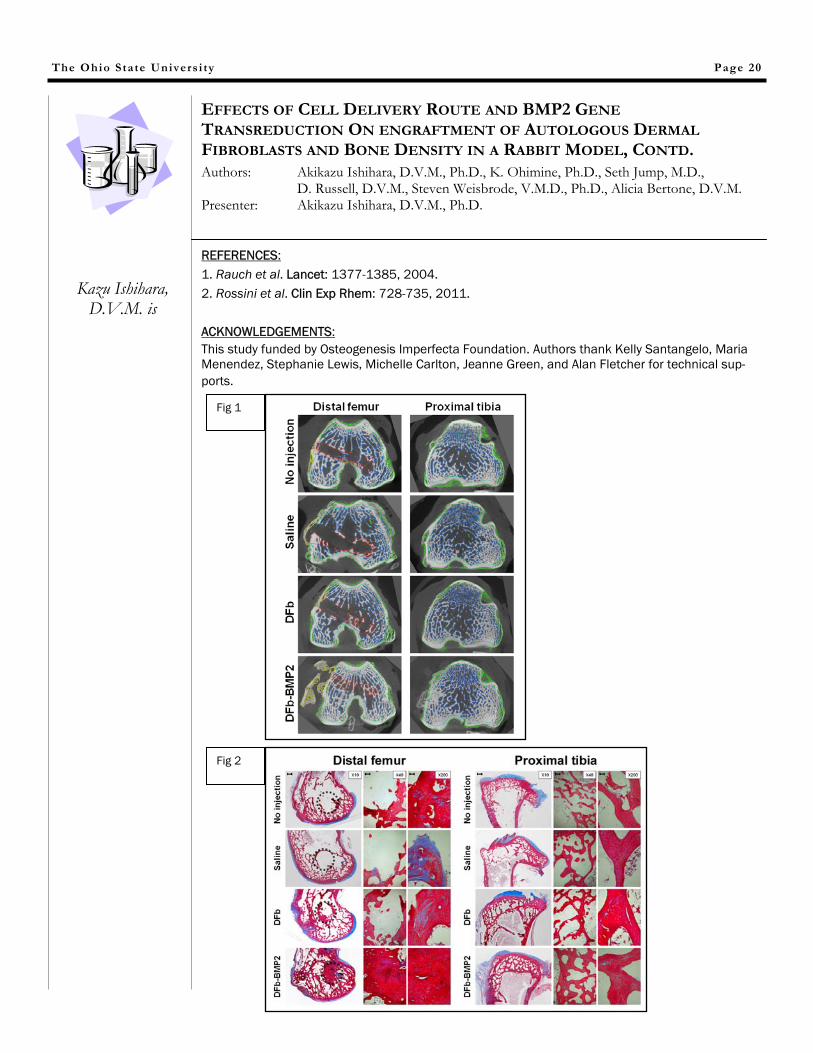
:INTRODUCTION: Bone fragility disorders are challenging orthopedic conditions, such as osteogenesis imperfecta (OI) and osteoporosis, resulting in severe disability with frequent painful fractures.(Rauch 2004; Rossini 2011) Cell-mediated gene therapy with various progenitor cells, including mesenchymal stem cells (MSC), can have a great potential to treat bone fragility disorders; however, optimal methods to increase cell engraftment in target bone tissues need to be established. Although MSC have been extensively studied for cell therapy, dermal fibroblasts (DFb) may be an alternative cell source because of their excellent plasticity and rapid cell yield.
METHODS: In a pilot experiment, an intra-venous and intra-medullar infusion of autologous MSC into rabbit femurs and tibiae showed modest cell engraftment and no measurable changes in bone mineral density and microarchitecture. This could suggest that an alternative cell delivery route or secre-tion of osteogenic growth factor from the engrafted cells may be needed to improve the density and structure of target bones. Therefore, after we confirmed a comparable osteogenic capacity between MSC and DFb with bone morphogenetic protein-2 (BMP2) gene transduction, an intra-osseous injection and intra-medullar infusion of BMP2-expressing autologous DFb into rabbit fe-murs and tibiae were compared. DATA AND RESULTS: Both delivery methods of DFb-BMP2 resulted in increased bone volume and mineral density and improved bone microarchitecture in trabecular bone (Fig 1 and 2). The intra-osseous DFb-BMP2 injection also induced greater bone defect filling, external callus formation, and trabecular surface area. Cell engraftment within trabecular bone and bone marrow tissue was most efficiently achieved by intra-osseous injection of DFb-BMP2. Systemic biodistribution of the locally injected/infused DFb-BMP2 was not evident in distant organs or contralateral hindlimb. Our results sug-gested that BMP2-expressing autologous DFb may be efficiently engrafted in target bones by intra-osseous injection and intra-medullar infusion and improve bone mineral density and bone microar-chitecture. DISCUSSION: Fibroblast-mediated gene therapy using a single intra-osseous injection or intar-medullar infusion of DFb genetically-modified to secrete BMP2 can improve bone mineral density and trabecular mi-croarchitecture in rabbit long bone model. It is of great interest to demonstrate an efficacy of this DFb-mediated BMP2 gene therapy to increase bone density and strength on animal models of bone fragility disorders such as osteogenesis imperfecta and osteoporosis. Use of rapidly growing DFb may induce greater therapeutic and practical benefits over MSC for an application of cell ther-apy in clinical setting.
Continued on Next Page
EFFECTS OF CELL DELIVERY ROUTE AND BMP2 GENE TRANSREDUCTION ON ENGRAFTMENT OF AUTOLOGOUS DERMAL FIBROBLASTS AND BONE DENSITY IN A RABBIT MODEL Authors: Akikazu Ishihara, D.V.M., Ph.D., K. Ohimine, Ph.D., Seth Jump, M.D., D. Russell, D.V.M., Steven Weisbrode, V.M.D., Ph.D., Alicia Bertone, D.V.M. Presenter: Akikazu Ishihara, D.V.M., Ph.D.
Kazu Ishihara, D.V.M. is
REFERENCES: 1. Rauch et al. Lancet: 1377-1385, 2004. 2. Rossini et al. Clin Exp Rhem: 728-735, 2011. ACKNOWLEDGEMENTS: This study funded by Osteogenesis Imperfecta Foundation. Authors thank Kelly Santangelo, Maria Menendez, Stephanie Lewis, Michelle Carlton, Jeanne Green, and Alan Fletcher for technical sup-ports.
EFFECTS OF CELL DELIVERY ROUTE AND BMP2 GENE TRANSREDUCTION ON ENGRAFTMENT OF AUTOLOGOUS DERMAL FIBROBLASTS AND BONE DENSITY IN A RABBIT MODEL, CONTD. Authors: Akikazu Ishihara, D.V.M., Ph.D., K. Ohimine, Ph.D., Seth Jump, M.D., D. Russell, D.V.M., Steven Weisbrode, V.M.D., Ph.D., Alicia Bertone, D.V.M. Presenter: Akikazu Ishihara, D.V.M., Ph.D.
The Ohio State Univers i ty Page 20
Fig 1
Fig 2
Kazu Ishihara, D.V.M. is
The Ohio State Univers i ty Page 21
INTRODUCTION: Total joint arthroplasty (TJA) is among the most common orthopedic procedures performed in the United States. Common findings in the postoperative period include fever and leukocytosis. Often, the physician being notified may be unaware that such abnormal values can commonly occur in the postoperative period. Many times, this leads to additional laboratory and radiographic testing that may not be needed. The authors are unaware of any study evaluating fever and leukocytosis in regards to predicting postoperative infections and the use of additional laboratory studies to evaluate these findings in TKA and THA patients. The purpose of this study was to determine the incidence of fever and leukocytosis in this patient population, the use of commonly ordered tests, and any correlation to future development of total joint infection. METHODS: Institutional review board approval was obtained to retrospectively review medical records and analyze pertinent data. A retrospective review of 426 patient charts, including 214 primary TKAs and 212 primary THAs performed by 2 surgeons at our institution, was completed. Data related to the occurrences and timing of patients' maximum temperature and postoperative leukocytosis were recorded. We reviewed the laboratory tests ordered for those patients with fever and/or leukocytosis to determine the type and results of tests ordered. Finally, we reviewed the medical records to determine if any of these patients had developed postoperative joint infections. DATA AND RESULTS: The TKA group (n = 214) included 67 men and 147 women with a mean age of 66.9 years (range, 33-89 years). Eighteen (8.4%) TKA patients had postoperative fever; 123 (57.5%) had leukocytosis, and 13 (6.1%) had fever and leukocytosis. Postoperative day 2 had the highest number of patients with fever (n = 9; 4.2%), and postoperative day 1 had the highest incidence of leukocytosis (n = 100; 46.7%). These findings led to 21 additional laboratory tests, of which 6 (28.6%) were positive or elevated. One TKA patient (0.5%) had a joint infection at follow-up to this point (range, 23-48 months). The THA group (n = 212) included 68 men and 144 women with a mean age of 61.3 years (range, 15-94 years). Forty-nine (23.1%) THA patients had postoperative fever, 122 (57.5%) had leukocytosis, and 22 (10.4%) had leukocytosis and fever. Postoperative day 1 had the highest number of patients with fever (n = 25; 11.8%), and postoperative day 2 had the highest incidence of leukocytosis (n = 92; 43%). Thirty-nine additional laboratory tests were performed, with positive or elevated results in 8 tests (20.5%).One THA patient (.5%) had joint infections at follow-up.
Continued on Next Page
EVALUATION OF POST-OPERATIVE FEVER AND LEUKOCYTOSIS IN PATIENTS AFTER TOTAL HIP AND KNEE ARTHROPLASTY Authors: Anthony Czaplicki, M.D., Joel Borger, M.D., Joel Politi, M.D., Bryan Chambers, M.D., Benjamin Taylor, M.D. Presenter: Anthony Czaplicki, M.D.
Tony Czaplicki,
M.D. is a resident in Orthoapedics at
Mt. Carmel

DISCUSSION: Postoperative fever and leukocytosis after TJA are common findings. The question of how to determine the importance of these nonspecific findings arises for all physicians who manage our patients postoperatively.
Our study found that postoperative leukocytosis is much more common than fever in TJA patients (57.5% vs 15.8%, respectively). The incidence of fever was highest on postoperative day 1 in THA patients and day 2 in TKA patients, with both trending downward after postoperative day 2. Leukocytosis was found in 57.5% of both TKA and THA patients, but the incidence was highest on postoperative day 1 in TKA patients and postoperative day 2 in THA patients, both again trending downward after postoperative day 2.
Overall, only 6 (10%) of 60 of laboratory tests ordered to evaluate postoperative fever and leukocytosis yielded clinically relevant data. Two patients developed postoperative total joint infection, but notably, neither demonstrated fever, and only 1 of the 2 developed leukocytosis in the immediate postoperative period.
This analysis shows that the overall use of testing for fever and leukocytosis without specific physical examination findings is low and should not be performed routinely. Understanding these findings potentially can help eliminate unnecessary tests, prevent possible delays in discharge, and decrease patient pain and anxiety as well as lower the overall cost of the hospital stay. REFERENCES: 1. Wilson N, Schneller ES, Montgomery K, et al. Hip and knee implants: current trends and policy considerations. Health Aff 2008;27:1587. 2. Andres BM, Taub DD, Gurkan I, et al. Postoperative fever after total knee arthroplasty: the role of cytokines. Clin Orthop Relat Res 2003;221. 3. Kennedy JG, Rodgers WB, Zurakowski D, et al. Pyrexia after total knee replacement. A cause for concern. Am J Orthop 1997;26:549. 4. Guinn S, Castro Jr FP, Garcia R, et al. Fever following total knee arthroplasty. Am J Knee Surg 1999;12:161. 5. Ghosh S, Charity RM, Haidar SG, et al. Pyrexia following total knee replacement. Knee 2006;13:324. 6. Shaw JA, Chung R. Febrile response after knee and hip arthroplasty. Clin Orthop Relat Res 1999;367:181. 7. Bindelglass DF, Pellegrino J. The role of blood cultures in the acute evaluation of postoperative fever in arthroplasty patients. J Arthroplasty 2007;22:701. ACKNOWLEDGEMENTS: Janet L. Tremaine, ELS, DISCLOSURES:None
EVALUATION OF POST-OPERATIVE FEVER AND LEUKOCYTOSIS IN PATIENTS AFTER TOTAL HIP AND KNEE ARTHROPLASTY Authors: Anthony Czaplicki, M.D., Joel Borger, M.D., Joel Politi, M.D., Bryan Chambers, M.D., Benjamin Taylor, M.D.
The Ohio State Univers i ty Page 22
Tony Czaplicki,
M.D. is a resident in Orthoapedics at
Mt. Carmel

The Ohio State Univers i ty
INTRODUCTION:
A strictly medial approach to hindfoot fusion lessens concerns over lateral soft tissue and bony healing in the standard triple1 approach, has been shown to allow for adequate joint preparation,2 provide excel-lent deformity correction, and provide good rates of fusion3 with less soft tissue complications.4 The goal of this retrospective study was to investigate the radiographic changes in the CCJ following fusion of the subtalar (STJ) and talonavicular joints (TNJ) and identify the frequency of CCJ distraction as a result of the medial double.
METHODS:
A radiographic review of consecutive patients after medial double hindfoot arthrodesis with >6 months follow-up was performed after IRB approval. Exclusion criteria included revision surgery, use of exter-nal fixation, and Charcot osteoarthropathy.5 Radiographs were reviewed by two foot and ankle sur-geons. Standard flatfoot angles were noted as well as the degenerative state of the CCJ and surrounding non-operative joints. Grading was done by consensus with “0” = no arthritic changes; “1” = narrowing of the joint space with no other radiographic findings; “2” = moderate sclerosis with osteophytes; “3” = severe sclerosis of the joint with narrowing and significant osteophytes. Radiographic union, defined as the bridging across three cortices, was identified on final radiograph.
RESULTS:
A total of 46 patients (47 feet) were identified by CPT codes. Upon radiographic and chart review, 20 patients (20 feet) with mean follow-up of 9.2 ± 4.1 months (6 - 21); mean age at surgery 61.9 ± 11.2 years (37 - 82); 5 men, 15 women. Average BMI was 35.06 ± 7.64 (23.05 – 49.91). Normal joint con-tour to slightly distracted CCJ was seen in 50% patients postoperatively (10 patients). There was a 50% incidence of CCJ arthrosis improvement from a mean of 1.21 pre-operatively to a mean of 0.65 post-operatively (Table 1). Severe, grade 2 and 3, arthrosis gradients decreased from 35% to 15% at final follow-up. There was only one circumstance of progressive arthritic changes in the CC joint. The aver-age abduction pre-operatively was 150.3° ± 12.5 (126 - 172); the average corrected position 168.1° ± 9.2 (140 - 180). This was an average correction of 17.8° in the transverse plane. Sagittal realignment exhibited a mean improvement of 10.5° from a talo-1st MT angle of 17.7° ± 14.8 (0 - 45) to 7.2°±6.7 (0 - 30). TN coverage increased from 56.3% ± 18.9 (20 - 90) to 96.7% ± 8.4(70 - 100), an increase of 171%. All but one of the 40 joints fused (97.5%) and there no increase in adjacent joint arthritis post-operatively (Table 1).
DISCUSSION:
Astion et al6 found that fusion of the TNJ limited motion in the CCJ to about 2 degrees while Leland et al7 found that the dorsal and plantar calcaneocuboid ligaments provide significant stability of the CCJ. While arthrodesis has provided reproducible results in the hands of many, it inherently disrupts liga-mentous anatomy in preparation and is often riddled with complications such as lateral wound dehis-cence, delayed union, nonunion, prolongs surgical times & increase arthrosis of adjacent joints over time.8,9 Good to excellent short-term results of the medial double fusion exist yet no study existed look-ing at the effects of the medial double on the positioning and arthrosis of the CCJ.9 In the study popula-tion, the radiographic degenerative changes at the CCJ were improved in 50% of patients and only one case progressed. This represents a 95% rate of positive improvement at the CCJ with the medial double.
Continued on Next Page
Page 23
THE MEDIAL DOUBLE ARTHRODESIS WITH LATERAL COLUMN SPARING AND ARTHRODIASTASIS: A RADIOGRAPHIC AND CHART REVIEW
Authors: Gregory Berlet, M.D., Christopher Hyer, D.P.M., Ryan Scott, D.P.M., Melissa Galli, D.P.M. Presenter: Melissa Galli, D.P.M.
Melissa Galli, D.P.M. is a resident in
Podiatry at The Ohio State University

The Ohio State Univers i ty
The outcomes of this study involving 20 patients with a mean follow-up of 9.2 months provide promising results of the medial double arthrodesis in the short-term. Only 2 patients (10%) com-plained of any lateral pain. The current study has shown that the CCJ can be left mobile which can help improve or stabilize arthrosis changes at the CCJ where mild to moderate preoperative arthri-tis exists. It is postulated that the mechanism of improvement is arthrodiastasis of the CCJ follow-ing realignment of the STJ and TNJ. The authors believe maintaining a mobile lateral column is one asset of the medial double arthrodesis. DISCLOSURES: None
REFERENCES: Myerson, et al. JBJS [Br]. 2005; 87-B: 369. Jeng, et al. Foot Ankle Int. 2006; 27(12): 1122-5. Jeng, et al. Foot Ankle Clin. 2005; 10: 515-21. Jackson, et al. JBJS (Br). 2007; 89-B (7): 925-7. Stevens, et al. J of Pedi Ortho. 1999; 19(4): 515-7. Astion DJ, et al. JBJS (Am). 1997; 79-A(2): 241-6. Leland, et al. Foot Ankle Int. 2001; 22 (11): 880-4 Lee MS. Clin Podiatric Med Surg. 2007; 24: 735-44. Weinraub, et al. J Foot Ankle Surg. 2010; 49: 326-30. Table 1: Arthrosis Gradient Given to Surrounding Arthrodesis Joints Pre and Post-Operatively
0 1 2 3 N/A CCJ- Pre 4 9 6 1 0 CCJ- Post 10 7 3 0 0 Cuboid-4th/5th
6 13 0 0 1
Cuboid-4th/5th 5 15 0 0 0
Ankle- Pre 8 7 5 0 0 Ankle- Post 7 9 4 0 0 Nav-Cun- Pre 10 8 2 0 0 Nav-Cun- Post 9 9 1 0 1 Cun-1st Met- Pre 17 3 0 0 0 Cun-1st Met- Post 17 2 1 0 0 Summation of Joints
45/ 48 40/ 42 13/ 9 1/ 0
1/ 1
Page 24
THE MEDIAL DOUBLE ARTHRODESIS WITH LATERAL COLUMN SPARING AND ARTHRODIASTASIS: A RADIOGRAPHIC AND CHART REVIEW
Authors: Gregory Berlet, M.D., Christopher Hyer, D.P.M., Ryan Scott, D.P.M., Melissa Galli, D.P.M. Presenter: Melissa Galli, D.P.M.
Melissa Galli, D.P.M. is a resident in
Podiatry at The Ohio State University
The Ohio State Univers i ty Page 25
OPEN CAPSULAR SHIFT AND ARTHROSCOPIC CAPSULAR PLICATION FOR TREATMENT OF MULTIDIRECTIONAL INSTABILITY: A SYSTEMATIC REVIEW
Authors: Mark Jacobson, M.D., Michael Riggenbach, M.D., Adam Wooldridge, M.D., Julie Bishop, M.D. Presenter: Mark Jacobson, M.D.
Mark Jacobson, M.D. is a resident in Orthopaedics at The Ohio State
University
Conti INTRODUCTION:
The purpose of this study is to review the literature systematically and compare the results of open inferior capsular shift with arthroscopic capsular plication for multidirectional instability in patients without a Bankart lesion. We hypothesized that there is no difference with regard to the specific clinical outcomes evaluated, including recurrent instability, range of motion, return to sport, and com-plications.
METHODS:
We conducted a comprehensive literature search. Databases searched included PubMed from 1966 to 2010, the Cochrane Database of Systematic Reviews and Controlled Trials, CINAHL (Cumulative Index to Nursing and Allied Health Literature) from 1982 to 2010, and SPORTDiscus from 1975 to 2010. Limits included English language, human subjects, and title. Each database was searched for the terms “multidirectional instability,” “inferior instability,” “capsular shift,” “capsular plication,” and “capsulorrhaphy.” Inclusion criteria consisted of the following: MDI defined as instability in at least 2 directions, results with a minimum of 2-year follow up, and surgical treatment consisting of either open inferior capsular shift or arthroscopic capsular plication. We excluded review articles, articles reporting results of patients with Bankart lesions, and articles including patients in whom dislocation occurred voluntarily. After thorough review of each article, the following data were ex-tracted: patient demographics, definition of MDI, surgical technique, failure rate, change in range of motion from preoperatively to postoperatively,
return to sport, and complications.
DATA AND RESULTS:
After reviewing the abstracts of all search results, we identified 33 articles with relevance to this re-view. After application of our inclusion and exclusion criteria, 7 articles remained1-7. No study with a level of evidence higher than Level IV was identified, and all studies consisted of case series, 5 of which were retrospective. There were 197 patients included in all studies, of whom 22 underwent bilateral procedures. A lower rate of recurrent instability in the studies using an open technique as compared with an arthroscopic technique was reported: 11.7% (16 of 137) versus 20% (11 of 55). There was a trend toward increased return to preoperative level of sports participation for patients treated arthroscopically versus those treated with open
capsular shift (86% v 80%). Reporting of preoperative and postoperative range of motion was highly variable among included studies, making interpretation of this outcome difficult. Complications from MDI repair (outside of recurrent instability) were infrequent in the included studies.
DISCUSSION:
This review shows that arthroscopic capsular plication is a reasonable alternative to open capsular shift to reliably decrease the incidence of recurrent instability in patients with MDI. Whereas the gold standard for this pathology has classically been thought to be an open capsular shift, this review does not clearly show open treatment to be superior to arthroscopic treatment. Loss of range of mo-tion is a concern in any capsular tightening procedure; however, no study reported a consistent loss of greater than 40° of external rotation (which was defined as a failure according to the early Neer crite-ria)8. Neither technique was shown to be superior with respect to postoperative range of motion.
Continued on Next Page
The Ohio State Univers i ty
Although there was a slight trend toward increased return to sport for patients treated arthroscopi-cally, no clear conclusion can be drawn given the variability of reporting in the reviewed studies. Analysis of complications shows that both procedures are reliably safe with minimal complications. Although this report does show a similar efficacy with regard to the reported outcomes, the poten-tial benefits of an all-arthroscopic approach that have not been addressed should not be ignored. These include avoidance of subscapular or infraspinatus takedown and the associated complica-tions, limited scarring, and ability to address associated pathology, which may be inaccessible with an open approach9. REFERENCES: 1. Bak, et al. Am J Sports Med. 28:466-471. 2. Choi, et al. Br J Sports Med. 36:290-294. 3. Marquardt, et al. Am J Sports Med. 33:1011-1015. 4. Steinbeck ,et al. Acta Orthop Scand. 68:447-450. 5. Baker, et al. Am J Sports Med. 37:1712-1720. 6. Treacy, et al. J Shoulder Elbow Surg. 8:345-350. 7. Wichman, et al. Oper Tech Sport Med. 5:238-243. 8. Neer, et al. J Bone Joint Surg Am. 62:897-908. 9. Tjoumakaris, et al. Arthroscopy. 27:1422-1433. DISCLOSURES: No author has a disclosure related to the content of this manuscript.
Page 26
OPEN CAPSULAR SHIFT AND ARTHROSCOPIC CAPSULAR PLICATION FOR TREATMENT OF MULTIDIRECTIONAL INSTABILITY: A SYSTEMATIC REVIW, CONTD. Authors: Mark Jacobson, M.D., Michael Riggenbach, M.D., Adam Wooldridge, M.D., Julie Bishop, M.D.
Mark Jacobson, M.D. is a resident in Orthopaedics at The Ohio State
University

The Ohio State Univers i ty
INTRODUCTION: The aim of this study was to validate the Thoracolumbar Injury Classification and Severity (TLICS) system through a retrospective analysis of clinical management of thoracolumbar injuries treated at the Wexner Medical Center at The Ohio State University. We hypothesized that the strategies outlined in the TLICS system would be in congruence with the care of the majority of patients treated at the Wexner Medical Center from 1/1/2006 and 3/31/2011. In addition, we predicted that the ASIA class in the majority of patients would not change whether they had surgical or non-surgical intervention. We also looked at demographics, average hospital length of stay, and average injury severity score (ISS) of ptatients treated surgically vs non-surgically. METHODS: We initially searched the Trauma database at the Wexner Medical Center at The Ohio State Univer-sity for ICD-9 codes involving thoracic and lumbar fractures with and without neurologic deficit. 734 patients were identified. This identified all thoracic and lumbar fractures. We then localized T10-L1 fractures and applied our exclusion criteria, resulting in 201 patients in our study. We then analyzed the electronic medical record and radiographic imaging system to retrospectivelyl assign a TLICS score. The actual treatment course was then compared to the TLICS management protocol regarding surgical or non-surgical intervention. Neurologic outcomes, using the ASIA class was then analyzed pre- and post-treatment to assess any change in ASIA class based on surgical or non-surgical intervention. The end time point for each patient varied and was defined as the date of most recent follow up. DATA AND RESULTS: 201 pts were identified that met our inclusion and exclusion criteria. 137 males and 64 females were included. 59 patients (29.4%) underwent surgical intervention.142 patients (70.6%) underwent non-surgical intervention. Average age was 45.5 with a range from 16-94. Average hospital days of those patients that underwent surgical intervention were 12.1 days. Average hospital days of those patients that underwent non-surgical intervention were 5.9 days. The average injury severity score was 14.1 in those patients that underwent surgical intervention. The average injury severity score was 9.9 in those patients that underwent non-surgical intervention. We found that the care of thoracolumbar injuries at OSUMC was in strong agreement with that proposed by the TLICS system. Indeed, 92% of patients were treated in agreement to TLICS, while 8% had treatment that disagreed with TLICS. Of the patients with an intermediate score of 4, 12 of 16 patients underwent surgical intervention. 20 patients were identified with abnormal ASIA class. 8 of 17 patients with abnormal ASIA class underwent surgical intervention and improved ASIA class. 1 of 3 patients with abnormal ASIA class underwent non-operative management and had improvement of ASIA class. Overall, the number of patients with ASIA class change after intervention was 9 (4.5%). There were 16 patients with an intermediate score of 4 that TLICS recommends surgical or non-surgical management. In this subset of patients, 12 of 16 (75%) of patients underwent surgical intervention.
Continued On Next Page
Page 27
INDEPENDENT VALIDATION OF THE THORACOLUMBAR INJURY CLASSIFICATION AND SEVERITY (TLICS) SCORE: A RETROSPECTIVE ANALYSIS OF CLINICAL MANAGEMENT OF THORACOLUMBAR INJURIES
Authors: Shah Dodwad, M.D., Ronald Wisneski, M.D. Presenter: Shah Dodwad, M.D.
Shah Dodwad, M.D. is a Resident in
Orthopaedics at The Ohio State
University

The Ohio State Univers i ty
DISCUSSION: Despite a myriad of classification systems of thoracolumbar injuries, it has been difficult to identify one universal classification system. A proper classification system should be reliable, reproducible and possess clinical validity. We found that classification of the integrity of the posterior ligamentous complex, injury morphology, and neurologic status, as dictated by the TLICS system, results in a comprehensive assessment of thoracolumbar injuries. The TLICS system represents an easily used, objective and reliable scheme for optimizing clinical management of these injuries.
The TLICS management recommendation corresponds 92% of the time with current spine surgeon thora-columbar injury management suggesting it is a reliable classification system to help guide patient manage-ment. The data shows an increase in hospital days and ISS with those patients that required surgical interven-tion. Interestingly, when patients could be treated surgically or non-surgically, with a TLICS score of 4, most patients underwent surgical intervention, suggesting surgeons tended to operate when in given the option. The TLICS is a classification system to aid in management, but does not supersede sound clinical judgment by the operative surgeon.
REFERENCES: See article for list of references. ACKNOWLEDGEMENTS: The authors wish to thank Dustin Donnelly, PhD for his contributions, as well as, Julia Panzo and Elizabeth Sheridan for their support. Funding for this work was provided by the Department of Or-thopaedic Surgery at OSUMC. DISCLOSURES: No financial or other conflicts of interest.
Page 28
INDEPENDENT VALIDATION OF THE THORACOLUMBAR INJURY CLASSIFICATION AND SEVERITY (TLICS) SCORE: A RETROSPECTIVE ANALYSIS OF CLINICAL MANAGEMENT OF THORACOLUMBAR INJURIES, CONTD. Authors: Shah Dodwad, M.D., Ronald Wisneski, M.D.
Shah Dodwad, M.D. is a Resident in
Orthopaedics at The Ohio State
University
The Ohio State Univers i ty
INTRODUCTION:
Disruption of the medial collateral ligament during total knee arthroplasty can lead to valgus instability and may affect patient outcome and implant survival. The purpose of this paper is to report the results of direct repair of the superficial MCL after iatrogenic mid-substance laceration and the subsequent outcomes.
METHODS:
We retrospectively reviewed all primary TKA over a 5 year for an iatrogenic mid-substance medial collateral ligament laceration. The injury was identified at the time of surgery and was the result of being cut by the oscillating saw in all nine patients. Direct ligament repair was performed with a #5 ethibond suture utilizing a modified becker stitch configuration. The sutures were initially applied and trial implants were then inserted and appropriate tension was determined. Once it was established that the repair could obtain good opposition with appropriate tension, the final components were inserted and sutures were tied with the knee in 30 degrees of flexion. The same implant was used for all patients without the use of any additional constraint. Postoperatively, no bracing was utilized and patients were allowed to be full weight bearing and standard postoperative protocol was followed. Patients were followed at regular intervals and Knee society scores were obtained for pain and function. A physical exam was performed that focused on range of motion and instability. Patients were asked to rate their satisfaction. Finally, radiographs were obtained and reviewed to determine if any loosening or radiographic changes were present.
DATA AND RESULTS:
There were nine MCL lacerations in nine patients, consisting of seven women and two men with an average BMI of 43.3 (range 29.1 to 55.7). The minimum follow-up was 22 months. One patient was too ill to return for a follow-up appointment but was contacted for a phone interview. All patients had a diagnosis of Osteoarthritis. The KSS Pain and Function score averages were 91.5 and 73.3 respectively. The physical exam demonstrated a range of motion of 0 to 120.5 degrees and had stable ligamentous exams to varus, valgus and anterior-posterior stress testing. The radiographs showed no signs of changes in component position, loosening or malalignment. Four of the patients were extremely satisfied with their results and two were very satisfied. Three patients reported mild/occasional knee pain. Two out of the nine patients underwent revision procedures. The first patient obtained an infection one year out of his index procedure while the second revision was a result of patella avascular necrosis. Both patients had a stable exam to ligamentous testing on follow-up. One patient was too ill to follow-up but through a phone interview described his knee as asymptomatic and was satisfied with the procedure and would have it again.
Continued on Next Page
Page 29
REPAIR OF MEDIAL COLLATERAL LIGAMENT INJURY DURING TOTAL KNEE ARTHROPLASTY Authors: Scott Stephens. M.D., Joel Politi, M.D., Jeff Backes, M.D., Tony Czaplicki, M.D. Presenter: Scott Stephens, M.D.
Scott Stephens, M.D. is a resident in Orthopaedics at
Mt. Carmel
The Ohio State Univers i ty
DISCUSSION:
We focused our study on the retrospective results of an iatrogenic mid-substance MCL disruption and found that at 2 year follow-up the patients had comparable functional scores to those found in the litera-ture involving primary total knee arthoplasty. The KSS scores we obtained for pain and function were higher than those reported by Lee et al for their managment of MCL disruption that was identified during arthroplasty procedures. Patients were subjectively satisfied with their results and did not note any instability and this was confirmed with physical exams that showed no signs of instability and full range of motion. The radiographs did not show any signs of component loosening or malposition. There were no complications that were directly related to this treatment and no further surgeries for instability or stiffness were necessary in any patient. The two patients who underwent revisions were not the result of the MCL disruption but instead were the result of a late infection and avascular necrosis of the patella. We were also able to show that this technique allows accurate ligament balancing without the need to increase the size of the polyethene, which had an average PE thickness of 10.4.
Our study did provide some limitations, beginning with its retrospective nature. This is also a very small patient population but taking into account the rarity of this complication and the fact that most studies include MCL avulsions, we feel this sample allows us to focus on a specific type of MCL injury. It would also be beneficial to see longer term follow-up as loosening of either the bone cement or cement prosthesis interface typically is not demonstrated in the short term.
In conclusion, we feel that direct repair of a transected mid-substance superficial MCL, without the use of condylar constrained implants or postoperative protocol alterations such as protected weight bearing and bracing, can provide a stable reconstruction with good results. We feel though that this should be taken into consideration for intra-substance lacerations and cannot be extrapolated to avulsions and knees with vaglus alignment.
REFERENCES:
1. Lee G, et al. CORR. 469: 64-68, 2011
2. Leopold S, et al. JBJS. 83: 86-91, 2001
3. Koo M, et al. J Arthoplasty. 24: 1249-1253, 2009
Page 30
REPAIR OF MEDIAL COLLATERAL LIGAMENT INJURY DURING TOTAL KNEE ARTHROPLASTY, CONTD. Authors: Scott Stephens. M.D., Joel Politi, M.D., Jeff Backes, M.D., Tony Czaplicki, M.D.
Scott Stephens, M.D. is a resident in Orthopaedics at
Mt. Carmel

The Ohio State Univers i ty
INTRODUCTION: Rupture of the thumb ulnar collateral ligament (UCL) is a frequent injury of the hand, commonly caused by sports injuries as well as falls onto an outstretched hand. Disruption of the UCL leads to decreased pinch strength, pain, instability, and ultimately OA. The evidence regarding operative and non-operative treatment of acute and chronic thumb UCL insufficiency is limited to Level IV retrospective case series and Level V expert opinion. Therefore, the primary purpose of this systematic review was to compare non-operative treatment versus surgical repair versus surgical reconstruction of thumb UCL injuries. The authors hypothesized that no difference exists in clinical outcomes between repair and reconstruction for acute injury. Thumb UCL reconstruction was hypothesized to be significantly better than repair for chronic UCL injury. In addition, operative management was hypothesized to result in greater patient satisfaction versus non-operative treatment, and that no difference exists between different types of graft used for thumb UCL reconstruction.
METHODS: A systematic review of multiple medical databases, including Pubmed, SportDiscus, CINAH, and Cochrane Central Register of Controlled Trials, was performed using PRISMA guidelines. Inclusion criteria included English language studies following non-operative or operative treatment of thumb UCL injuries with clinical outcomes and a minimum of two years mean follow-up. The following clinical outcome parameters from each article were extracted, compared, summated and statistically analysed: pain, ROM, key-pinch strength, and stability testing.
Continued on Next Page
Page 31
OUTCOMES FOLLOWING INJURY TO THE THUMB ULNAR COLLATERAL LIGAMENT - A SYSTEMATIC REVIEW Authors: Julie Samora, M.D., Joshua Harris, M.D., Michael Greisser, M.D., Hisham Awan, M.D. Presenter: Julie Samora, M.D.
Julie Samora, M.D. is a resident in Orthopaedics at The Ohio State
University
The Ohio State Univers i ty
DATA AND RESULTS: After reviewing 262 studies, 14 articles were included and analyzed. All articles were level IV evi-dence. There were 30 subjects treated non-operatively, and 199 subjects treated operatively. Pa-tients after operative treatment of either acute or chronic UCL injury appear to have similar grip strength and pinch strength compared to the uninjured thumb, and have overall similar subjective assessments. Comparing pull-out sutures and bone anchor techniques in the acute repair of UCL injuries, suture anchors allowed for accelerated rehabilitation protocol, improved ROM and pinch strength. Suture anchors do appear to be more cost effective as well.
DISCUSSION: After operative treatment of either acute or chronic UCL injury, patients have similar grip strength and pinch strength compared to the uninjured thumb, and have overall similar subjective assessments. Suture anchors and pull-out sutures both demonstrate improved range of motion and pinch strength following surgical treatment of acute UCL injury. Chronic injury is most often treated with reconstruction using palmaris longus autograft with good clinical outcomes. Lack of high level evidence in the form of prospective randomized trials limits our abil-ity to recommend one method of treatment over another.
REFERENCES: 1. Bostock et al. JHS. 18:71-711, 1993 2. Dinowitz, et al. JHS Am. 22:1057-1063, 1997 3. Downey, et al. AJSM. 23:222-226, 1995 4. Fusetti, et al. Chir Main. 24: 217-221, 2005 5. Glickel, et al. JHS Am. 18: 930-941, 1993 6. Katolik, et al. Plas Rec Surg. 122: 1451-1456, 2008 7. Lohman, et al. Skeletal Radiol. 39: 1081-86, 2010 8. Wong, et al. JHS Am. 34: 304-308, 2009
ACKNOWLEDGEMENTS and DISCLOSURES: None.
Page 32
OUTCOMES FOLLOWING INJURY TO THE THUMB ULNAR COLLATERAL LIGAMENT - A SYSTEMATIC REVIEW, CONTD. Authors: Julie Samora, M.D., Joshua Harris, M.D., Michael Greisser, M.D., Hisham Awan, M.D.
Parameter n (%) Number of subjects Male* Female
295 138 (63%) 80 (37%)
Number of thumbsΨ Dominantα Non-dominant Rightβ Left
293 113 (67%) 55 (33%) 19 (37%) 32 (63%)
Injury chronicity¥ Acute Chronic
211 (70%) 92 (30%)
Mean subject age (years) 33.9 (range 12 to 76) Mean follow-up (years) 3.26 (range 2 to 6.3) UCL injury locationρ Proximal Mid-substance Distal Avulsion fracture Ligamentous
0 (0%) 16 (59%) 11 (41%) 54 (46%) 63 (54%)
Stener lesionκ Present Absent
78 (54%) 67 (46%)
Treatment Non-operative Repair Autograft recon Allograft recon
43 (14.7%) 173 (59.0%) 81 (27.7%) 0 (0%)
Julie Samora, M.D. is a resident in Orthopaedics at The Ohio State
University
The Ohio State Univers i ty
INTRODUCTION:
The talonavicular (TN) joint is a key component of the hindfoot joint complex. With the TN joint im-mobilized, over 90% of motion in the adjacent talocalcaneal and calcaneocuboid joints is eliminated.1-3 The ball-and-socket type configuration of the TN joint allows for translation and rotational movements in multiple planes. This relatively unconstrained motion must be controlled with rigid internal fixation constructs to maximize healing in fusion procedures.
Fusion of the talonavicular joint is an effective treatment for hindfoot pain and deformity. Nonunion in hindfoot fusion procedures is most common in the TN joint.2,3 The purpose of this study was to evaluate and compare the mechanical stability of two different forms of fixation for isolated fusion of the talonavicular joint.
METHODS:
Thirteen fresh frozen cadaver foot and ankle specimens were procured for this study. Each specimen was mounted vertically in a robotic biomechanical testing platform and tested before and after surgical fixation. A standardized surgical approach and joint preparation technique was used. Seven specimens received two retrograde lag screws (2S). Six specimens received a locked compression plate with one retrograde lag screw (PS). A constant Achilles tendon load was initially applied followed by internal or external rotation of the foot and axial compression through the tibia. The relative motion of the talus and navicular bones was tracked using three-dimensional optoelectric targets. Motion data was recorded in the traditional anatomic reference frame and then transformed into the talonavicular joint frame.
Relative motion between the talus and navicular was statistically compared at common load limits using a one-way repeated measures ANOVA, followed by Student Neuman-Kuels for multiple comparisons (p= 0.05). For translation data, a three-way repeated measures ANOVA was used to determine the ef-fects of state of the specimen (harvested vs. instrumented), treatment (2S vs PS), and loading condi-tions. The X,Y,Z dimensions were analyzed separately. For rotational data, a two-way repeated meas-ures ANOVA was used to determine the effects of treatment and loading conditions. The X,Y,Z dimen-sions and states (harvested vs instrumented) were analyzed separately. The MIXED procedure of SAS version 9.1.3 was used for analyzing translation and rotation data. Since the results were analyzed in both the camera frame and the joint frame, it was possible to observe the difference between the two frames.
DATA AND RESULTS:
In the talonavicular joint frame, relative to the intact (presurgical) state, the plate fixation group PS showed a significant reduction in separation along the long axis of the talus in three out of the four loading scenarios (P values: 0.007, 0.02 and 0.05). The PS construct also significantly restricted the flexion-extension motion at the joint, compared to the 2S construct, during the external rotation tests (P values: 0.002 and 0.001). When the results were analyzed in the standard anatomic reference frame, no statistically significant differences were detected in the translations and the restriction of flexion-extension was overestimated (P values: 0.0002 and less than 0.0001).
Continued on Next Page
Page 33
TALONAVICULAR JOINT FIXATION: A BIOMECHANICAL COMPARISON OF LOCKING COMPRESSION PLATES AND LAG SCREWS Authors: Jaymes Granata, M.D., Gregory Berlet, M.D., Rahul Gotge, M.D., M.S., Yuan Li, Ph.D., Brian Kelly, M.D., Denis DiAngelo, Ph.D. Presenter: Jaymes Granata, M.D.
Jaymes Granata, M.D. is a resident in Orthopaedics at The Ohio State
University
The Ohio State Univers i ty
DISCUSSION:
In our simulated weight-bearing model, a dorsal locked compression plate with one retrograde screw was more effective at limiting the 3D motion across the talonavicular joint, compared to the traditional construct of two retrograde screws. Motion data transformation to the joint specific reference frame provided a better understanding of the true local joint mechanics. This biomechanical model and data transformation method can be used to evaluate and compare any variety of fixation constructs and is particularly useful for the multi-planar joint orientations in the foot and ankle.
REFERENCES:
1. Mann R.A.: Surgical implications of biomechanics of the foot and ankle. Clin. Orthop. 146:111-118, 1980.
2. Fogel, G., et al. Talonavicular arthrodesis for isolated arthrosis: 9.5-year results and gait analysis. Foot Ankle, 3:105-113, 1982.
3. Mann, R.A., and Beaman, D.N.: Double arthrodesis in the adult. Clin. Orthop. 365:74-80, 1999.
ACKNOWLEDGEMENTS:
Special thanks to Elizabeth Tolley, PhD, for assistance with the statistical analysis. Research funding and support was provided by Wright Medical Technologies.
DISCLOSURES:
The presenting author has no financial conflict of interest with this study. Senior authors (GB,DD) are paid consultants or have received research funding from Wright Medical Technologies.
Page 34
TALONAVICULAR JOINT FIXATION: A BIOMECHANICAL COMPARISON OF LOCKING COMPRESSION PLATES AND LAG SCREWS, CONTD. Authors: Jaymes Granata, M.D., Gregory Berlet, M.D., Rahul Gotge, M.D., M.S., Yuan Li, Ph.D., Brian Kelly, M.D., Denis DiAngelo, Ph.D.
Jaymes Granata, M.D. is a resident in Orthopaedics at The Ohio State
University
The Ohio State Univers i ty
Introduction: Supracondylar humerus fractures account for 60% of all pediatric elbow fractures1–4,8. The peak age for this injury is between 4 and 8 years of age and it is the most common fracture in the 0-7 age group 2,5,6. Despite the frequency of this injury, treatment for Gartland type II injuries remains controversial 7. Sev-eral authors recommend closed reduction and percutaneous pinning for all type II injuries, while others advocate closed reduction and casting as an effective and safe treatment option 4. Unfortunately the literature provides little guidance on which factors identify fractures that will likely fail non-operative management. The purpose of this study was to identify predictive factors for failure of closed reduction and immobilization of displaced, extension type II supracondylar humerus fractures. Methods: After Institutional Review Board (IRB) approval, medical records were queried for all patients treated and released following closed reduction and immobilization of a supracondylar humerus fracture be-tween January 2002 and September 2010. A total of 49 patients fit our inclusion criteria and were di-vided into two groups. Group A lost reduction in follow up and group B did not. Anteroposterior (AP) and lateral radiographs were evaluated for all patients at time of presentation, initial reduction, and at latest follow-up. Measurements obtained included: Baumann’s angle, lateral capitellar-humeral angle (LCHA), anterior humeral line index (AHLI) and the lateral humeroulnar angle. Age, gender and type of immobilization (long arm cast vs. splint) were documented. Change in radiographic measurements obtained at injury (pre-reduction), post reduction and final follow up films were calculated and docu-mented. Statistical analysis was performed utilizing a paired t test or t test for continuous variables, and Chi-square test or Fisher’s exact test for categorical variables in SAS 9.2 (SAS Institute Inc., Cary, NC, USA.) Data and Results: Twenty-nine patients (62% female, 38% male) underwent closed reduction and immobilization with an average age of 4.5 years (range 1-10). Group A and B were reduced 17 degrees and 14 degrees respec-tively. 62% were initially immobilized in a long arm cast, the remainder in a long arm posterior splint. The average position for immobilization (ulnohumeral angle) was 94 and 86 degrees for groups A and B respectively. Group A lost an average of 12 degrees of reduction. Age, gender, position of elbow im-mobilization, and type of immobilization showed no statistical significance between the two groups. Discussion: Type II supracondylar humerus fractures present a treatment dilemma to a treating surgeon. Our data demonstrates that 50% of type II fractures that are significantly displaced will lose on average 70% of their reduction. The method of immobilization was not a statistically significant factor and a posterior splint can replace long arm cast immobilization in the presence of significant swelling. No other factors examined in this study were found predictive of failure. We recommend close radiographic follow-up following closed reduction and immobilization and strong consideration of additional percutaneous pinning if loss of 70% of the reduction obtained would put the distal humerus in unacceptable align-ment. References: 1. Simanovsky N, et al. JPO V. 28: 463-465, 2008 2. Omid R, et al. JBJS V. 90:1121-32, 2008. 3. Pirone AM, et al JBJS V. 70:641-50, 1988. 4. Spencer HT, et al. JBJS V. 92:904-10, 2010. 5. Camus T, et al. JPO V. 31:366-71, 2011. 6. Skaggs D. JAAOS V. 5:303-312, 1997. 7. Fitzgibbons PG, JPO V 4:372-6, 2011 8. Shank C, et al. JPO V. 31:266-71, 2011. Acknowledgements: We would like to thank Leisel Willis, CCRC and Wei Wang, MS, MAS for their contribution.
Page 35
TYPE II SUPRACONDYLAR HUMERUS FRACTURES: FACTORS PREDICTIVE OF FAILURE FOR CLOSED REDUCTION AND IMMOBILIZATION
Authors: Douglas Lucas, D.O., Kevin Klingele, M.D. Presenter: Douglas Lucas, D.O.
Doug Lucas, D.O. is a resident in Orthopaedics at Doctors Hospital
The Ohio State Univers i ty
Background and Purpose: Discoid lateral meniscus (DLM) is the most common anatomic variant of the lateral meniscus. A DLM differs from a normal crescentic lateral meniscus both in regards to size and shape, and also in lacking an organized homogenous collagen network. It is hypothesized that this abnormal collagen network predisposes the DLM to tearing. Historically, symptomatic DLM were treated with total or subtotal meniscectomy. The present understanding of the importance of a functional healthy meniscus for the long term health of the knee has, as with all meniscal pathology, driven the management of symptomatic DLM towards meniscal preservation. Partial meniscectomy, or saucerization of a DLM, is advocated as a means of creating a lateral compartment that more closely resembles the gross anatomy of the lateral tibiofemoral articulation with a normally shaped lateral meniscus. In patients with symptomatic DLM, there is at present limited outcomes data to support or refute the role of partial meniscectomy in minimizing de-generative changes in the knee. The purpose of this study was to compare the outcomes of partial me-niscectomy/saucerization for DLM with subtotal/total meniscectomy for DLM in adolescent and pediat-ric patients, and to identify trends over time. Methods: A systematic review was conducted, evaluating all English language studies that reported outcomes of surgical treatment of DLM in pediatric and adolescent patients. Studies of patients 18 years of age and younger were included. Clinical outcomes were reported using the Ikeuchi grading system and the presence or absence of degenerative changes were documented by radiographs at final follow-up. Results: Eleven studies (306 knees) were included that documented clinical outcomes per the Ikeuchi scale, and 8 studies (171 knees) were included which reported results of radiographs at final follow-up. Follow-up duration ranged from 1 year to 28 years, with 189 knees treated with total/subtotal meniscectomy and 117 knees treated with partial meniscectomy/saucerization. Clinical and radiographic outcomes were equivalent for both short-term and long-term follow-up. Conclusions: Neither the Ikeuchi knee scale, nor plain radiographs at final follow-up demonstrated a difference in long-term outcomes of pediatric or adolescent patients with symptomatic DLM managed with either meniscal sacrificing or meniscal preserving techniques. Further investigation is needed to evaluate whether preserving any of the histomorphologically abnormal DLM provides protection for the lateral compartment of the knee against long term degenerative changes.
Page 36
CLINICAL AND RADIOGRAPHIC OUTCOMES FOLLOWING SURGICAL TREATMENT OF DISCOID LATERAL MENISCUS IN ADOLESCENT AND PEDIATRIC PATIENTS
Authors: William Graham, M.D., Robert Brophy, M.D., David Flanigan, M.D. Presenter: William Graham, M.D.
William Graham, M.D. is a resident in Orthopaedics at The Ohio State
University

The Ohio State Univers i ty
INTRODUCTION:
Current models for evidence generation in the field of orthopaedic surgery often appear to be in conflict with direct application of evidence to individual patients. Conventional evidence generation and evi-dence based practice hierarchies encourage and value studies with strict inclusion/exclusion criteria meant to represent the “average” patient. However, it is difficult to know how to apply population-based predictions of outcomes to a single patient who may not be similar to the sample used to create the population estimates. Recent advances in complex systems analysis approaches and the generation of large outcomes datasets have created new and greater opportunities to explore how to remedy this challenge. 1
Purpose: The overall purpose of this study was to explore how a complex systems analytical approach applied to a clinical outcomes dataset can improve clinicians’ abilities to provide more individualized, specific prognoses for patients. More specifically, the purpose was to compare and contrast a conven-tional analysis approach (variable-centered) with a complex systems analysis strategy (patient-centered).
Hypothesis: Patient-centered analyses would allow for improved identification of how an individual patient relates to a large surgical outcome database.
Clinical Significance: A variable-centered strategy describes associations between variables, while patient-centered analysis compiles patient profiles that retain all the information about the variables for each patient.
METHODS:
A single surgeon’s outcomes database (n = 276) regarding the incidence of graft failure 2 years status post anterior cruciate ligament reconstruction (ACLR) was analyzed with both a variable-centered ap-proach (previously reported in Kaeding et al).2 and a patient-centered approach. Data available for each patient included: sex, age, graft type (allograft versus autograft), Marx Activity Scale Score and graft failure/no failure at two years post-reconstruction.
The dataset was previously analyzed with a variable-centered approach that used multivariate logistic regression to create a parsimonious prediction model with the variables that best accounted for the vari-ance in graft failures.2 For the current study, the dataset was re-analyzed using a patient-centered ap-proach that created patient profile characterizations representing all subjects that matched a particular combination of the variables. The results of each method are described in the results for comparisons to demonstrate the differences in clinical utility for each method.
Page 37
NOVEL METHODS TO PRACTICE PERSONALIZED MEDICINE IN AN EVIDENCE BASED ORTHOPAEDIC SETTING Authors: Catherine Quatman-Yates, DPT, Ph.D., Carmen Quatman, M.D., Angela Pedroza, M.P.H, Timothy Hewett, Ph.D., Christopher Kaeding, M.D. Presenter: Carmen Quatman, M.D.
Carmen Quatman, M.D. is a Resident in Orthopaedics at The Ohio State
University
The Ohio State Univers i ty
DATA AND RESULTS: Variable-Centered Approach:
Conventional statistical results of this dataset were previously reported in Kaeding et al.2 Age, graft type and Marx activity score were all found to be predictive of graft failure. However, due to high collinearity with age, Marx activity score was dropped from the final model. The model indicated that the odds of graft failure for a patient with an allograft was 2.84 times higher than a patient with an autograft, regardless of age (P=0.04). When graft type is held constant, a 10 year increase in age reduces the odds of ACL graft rupture by 43% (P <0.001). No significant differences were observed between sexes in terms of graft failure.
Patient-Centered Approach: The results of the patient-centered approach yielded an array of patient-characteristic profiles that allowed for calculation of graft failure ratios for all patients that fall within a particular profile. Many of the profiles were too small to achieve statistical power that would enable direct comparisons of odds ratios with the variable-centered approach. However, the graft failure ratios were consistent with age and graft type playing a large role in failure across the profiles. In addition, the ratios pro-vided some indications for how the dataset could be analyzed in the future that might yield differ-ences between sexes in terms of graft failure. While the specifics of the patient-profile groupings are too extensive to report here, an example of how it might be used would be: Of all female pa-tients that this surgeon operated on that were age 25 years or younger with an allograft and went on to participate in high-level cutting and pivoting activities, 4 out of 22 (approximately 18%) ex-perienced graft failure within two years. In addition, these analyses identified the patient profile types with very limited data available. DISCUSSION AND CLINICAL IMPLICATIONS: To our knowledge, this is the first report of patient-centered surgical outcomes analyses for ortho-paedic outcome data. The results from this study demonstrate that patient-centered analyses may complement and even extend conventional statistical results by providing richer profiles that char-acterize the similarities and dissimilarities among patients within large clinical outcome datasets. Development of clinician-friendly tools that utilize patient-centered analytical techniques may pro-vide a platform for a more comprehensive understanding and enhanced ability to practice EBM and “personalized health care.” REFERENCES: 1. Quatman et al. Br J Sports Med. 43:1100-7, 2009. 2. Kaeding et al. Sports Health. 3:73-81, 2011. 3. Laursen et al. Merrill-Palmer Quarterly, 23:377-89, 2006. ACKNOWLEDGEMENTS AND DISCLOSURES: The authors acknowledge the MOON ACL study group participants for the use of the surgical out-comes database for performing these analyses. This work was funded by grants from the Ohio State University Sports Health and Performance Institute and R01-AR053684.
Page 38
NOVEL METHODS TO PRACTICE PERSONALIZED MEDICINE IN AN EVIDENCE BASED ORTHOPAEDIC SETTING Authors: Catherine Quatman-Yates, DPT, Ph.D., Carmen Quatman, M.D., Angela Pedroza, M.S., Timothy Hewett, Ph.D., Christopher Kaeding, M.D.
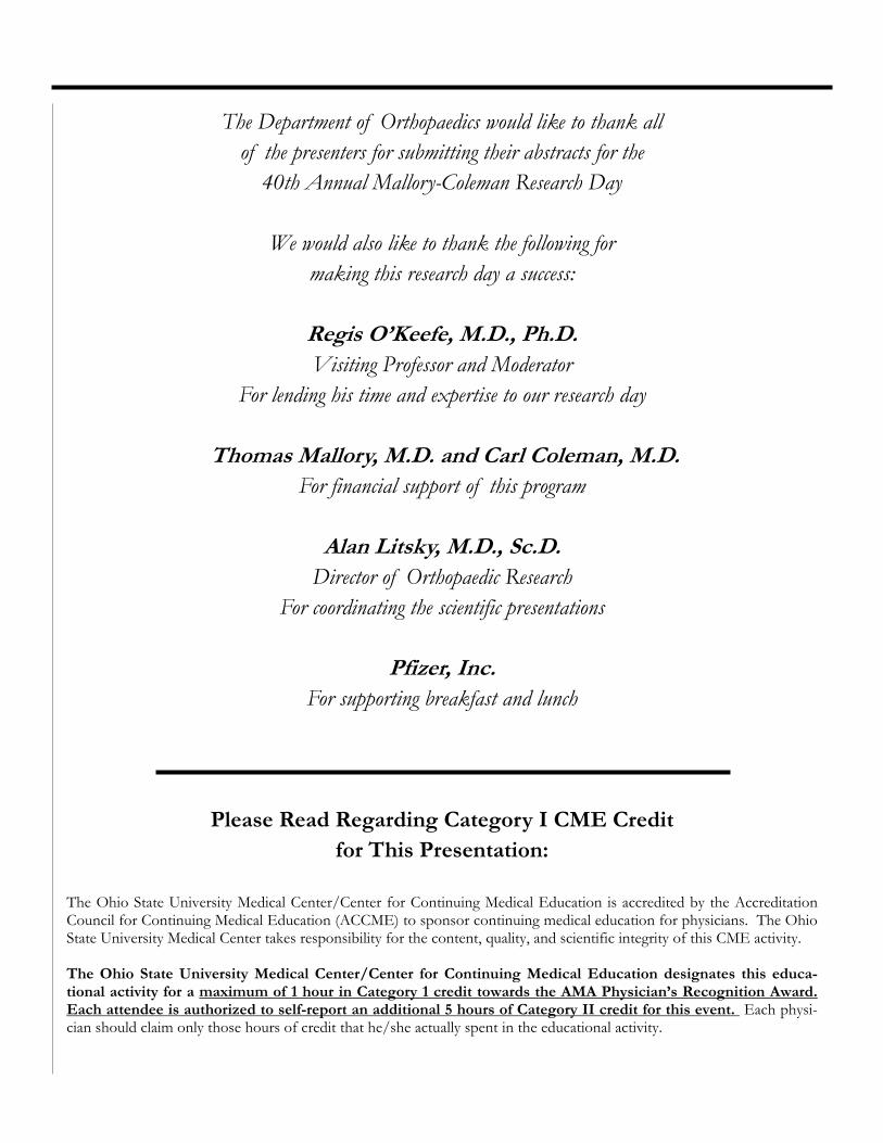
The Department of Orthopaedics would like to thank all of the presenters for submitting their abstracts for the
40th Annual Mallory-Coleman Research Day
We would also like to thank the following for making this research day a success:
Regis O’Keefe, M.D., Ph.D. Visiting Professor and Moderator
For lending his time and expertise to our research day
Thomas Mallory, M.D. and Carl Coleman, M.D. For financial support of this program
Alan Litsky, M.D., Sc.D.
Director of Orthopaedic Research For coordinating the scientific presentations
Pfizer, Inc.
For supporting breakfast and lunch
Please Read Regarding Category I CME Credit for This Presentation:
The Ohio State University Medical Center/Center for Continuing Medical Education is accredited by the Accreditation Council for Continuing Medical Education (ACCME) to sponsor continuing medical education for physicians. The Ohio State University Medical Center takes responsibility for the content, quality, and scientific integrity of this CME activity. The Ohio State University Medical Center/Center for Continuing Medical Education designates this educa-tional activity for a maximum of 1 hour in Category 1 credit towards the AMA Physician’s Recognition Award. Each attendee is authorized to self-report an additional 5 hours of Category II credit for this event. Each physi-cian should claim only those hours of credit that he/she actually spent in the educational activity.