Embed Size (px)
Citation preview
304 JOURNAL OF MICROELECTROMECHANICAL SYSTEMS, VOL. 25, NO. 2, APRIL 2016
Remote Axial Tuning in Microscopy UtilizingHydrogel-Driven Tunable Liquid Lens
Aditi Kanhere, Guangyun Lin, and Hongrui Jiang, Senior Member, IEEE
Abstract— This paper presents the use of a tunable-focusthermoresponsive hydrogel-based liquid lens in combination withan objective lens to achieve remote axial focusing in conventionalmicroscopy. The goal of this design is to eliminate imagedistortion due to sample vibrations caused by mechanical stagescanning. This approach reduces the mechanical complexityand power consumption due to the use of electrically tunablelenses, while achieving a twofold increase in the axial scanningrange. The merits of the proposed design were demonstratedby characterizing a customized microscope system over a scan-ning range of 1700 µm. A lateral resolution of 2 µm wasobtained consistently throughout the scanning range. HealthySpodoptera frugiperda Sf21 insect cells imaging was used to verifythe depth scanning ability and the resolution of our remotefocusing microscope system. [2015-0282]
Index Terms— Tunable lenses, microscopy, axial tuning.
I. INTRODUCTION
CONVENTIONAL wide-field microscopy is one of themost widely used optical microscopy technique. This
technique typically captures a two-dimensional image of aspecimen. However, many applications such as diagnosticmicrofluidic applications [1], tissue sampling [2] or multi-layered microfluidic samples [3] require a volumetric scan.To enable a volumetric visualization of the sample it isnecessary to move the sample, relative to the fixed focalplane of the microscope objective, along the axial direc-tion. For this purpose, a mechanical z-scanning stage istypically employed to enable linear translation along thedepth of the sample. The stage enables a controlled shiftof the focal plane through different layers of the sample.Typical approaches used to achieve such axial scanningemploy a motorized stepper stage or a piezoelectric stageto move the sample along the z-axis. While stepper motorsoffer the advantage of unlimited travel distance, they sufferfrom hysteresis. Piezoelectric stages on the other hand, helpeliminate hysteresis at the cost of the travel distance which is
Manuscript received October 13, 2015; revised January 4, 2016; acceptedJanuary 14, 2016. Date of publication February 3, 2016; date of current versionMarch 31, 2016. This work was supported by the U.S. National Institutes ofHealth under Grant 1DP2OD008678-01. Subject Editor J. A. Yeh.
A. Kanhere is with Digilens Inc., Sunnyvale, CA 94089 USA (e-mail:[email protected]).
G. Lin is with the Department of Electrical and Computer Engineer-ing, University of Wisconsin–Madison, Madison, WI 53706 USA (e-mail:[email protected]).
H. Jiang is with the Department of Electrical and Computer Engi-neering, Department of Materials Science and Engineering, Departmentof Biomedical Engineering, and the McPherson Eye Research Institute,University of Wisconsin-Madison, Madison, WI 53706 USA (e-mail:[email protected]).
Color versions of one or more of the figures in this paper are availableonline at http://ieeexplore.ieee.org.
Digital Object Identifier 10.1109/JMEMS.2016.2518922
reduced to 100-200 µm. Both types of stages, however, arebulky and cause vibrations and wobble in the sample due tohigh inertia. Additional care is required to avoid mechanicalovershoots and backlash from the tip touching the sample [4].Additionally, for water or oil-immersion lenses, vibration ofthe sample stage can cause disturbance or ripples in the immer-sion media that can lead to significant distortion in the images.
A robust alternative to the use of mechanical scanningstages is a remote axial focusing system that allows boththe objective and the sample to be stationary. One way toachieve this is the employment of a tunable-focus lens inthe imaging path to achieve shift of the axial position of thenative focal plane of the microscope through different depthsof the specimen being imaged. Several groups have reportedthe implementation of tunable-focus lenses for depth scanningin different microscopy techniques, including fast axial focus-ing in two-photon microscopy [5], optical coherence micro-scopy [6], optical tweezers [7] and light-sheet microscopy [8].Koukourakis et al. previously presented their proof-of-principle work showing the utility of adaptive lenses foraxial focusing in confocal microscopy [9]. Other researchgroups have used electrically tunable lenses based on polymermembrane-liquid interface as well as electro-wetting based liq-uid lenses to achieve volumetric scanning [10]–[12]. However,most of these techniques either require high driving voltagesor can achieve a unidirectional axial focal shift and need tobe combined with an offset lens to focus symmetrically aboveand below the native focal plane. Adding optical componentsrequires a higher precision in alignment as well as contributesto optical aberrations. Using the aforementioned solutions, anaxial tuning range of 700 µm or less can be achieved.
We propose a compact, energy and cost-efficient solutionfor remote axial focusing using thermo-responsive hydrogelbased liquid lenses that can achieve scanning over 1700 µm,which is a two-fold increase in the previously reported valuesfor axial scan in conventional microscopy. Hydrogel-basedliquid lenses offer a high degree of flexibility in terms oftuning temperature range, direction and magnitude of the axialfocus scan. We characterized the system using optical modelsin ZEMAX and imaging microspheres suspended in stackedmicrofluidic channels. A resolution of 2 µm or better wasobserved across all values of attainable focal scans. HealthySpodoptera frugiperda Sf21 insect cells were imaged to verifythe axial focusing functionality of the optical system.
II. EXPERIMENTAL
A. Principle of Operation
The work presented here is developed on the key idea that afocus-tunable liquid lens when introduced in the optical path
1057-7157 © 2016 IEEE. Personal use is permitted, but republication/redistribution requires IEEE permission.See http://www.ieee.org/publications_standards/publications/rights/index.html for more information.
KANHERE et al.: REMOTE AXIAL TUNING IN MICROSCOPY UTILIZING HYDROGEL-DRIVEN TUNABLE LIQUID LENS 305
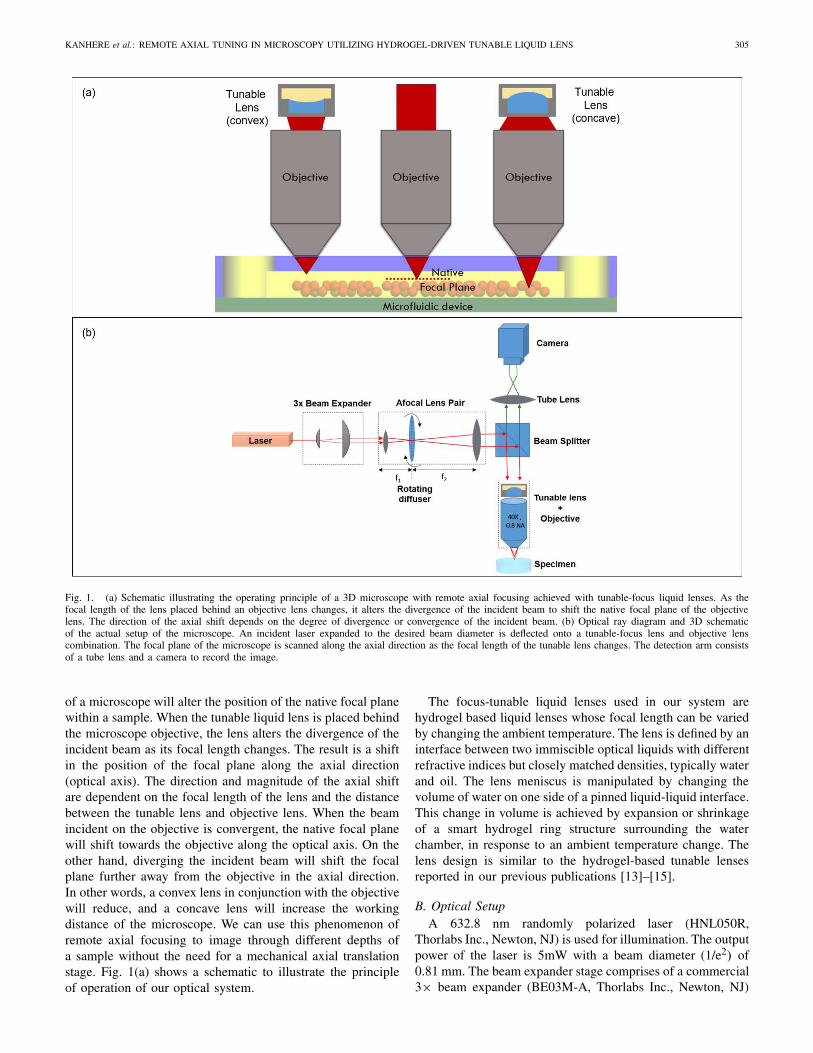
Fig. 1. (a) Schematic illustrating the operating principle of a 3D microscope with remote axial focusing achieved with tunable-focus liquid lenses. As thefocal length of the lens placed behind an objective lens changes, it alters the divergence of the incident beam to shift the native focal plane of the objectivelens. The direction of the axial shift depends on the degree of divergence or convergence of the incident beam. (b) Optical ray diagram and 3D schematicof the actual setup of the microscope. An incident laser expanded to the desired beam diameter is deflected onto a tunable-focus lens and objective lenscombination. The focal plane of the microscope is scanned along the axial direction as the focal length of the tunable lens changes. The detection arm consistsof a tube lens and a camera to record the image.
of a microscope will alter the position of the native focal planewithin a sample. When the tunable liquid lens is placed behindthe microscope objective, the lens alters the divergence of theincident beam as its focal length changes. The result is a shiftin the position of the focal plane along the axial direction(optical axis). The direction and magnitude of the axial shiftare dependent on the focal length of the lens and the distancebetween the tunable lens and objective lens. When the beamincident on the objective is convergent, the native focal planewill shift towards the objective along the optical axis. On theother hand, diverging the incident beam will shift the focalplane further away from the objective in the axial direction.In other words, a convex lens in conjunction with the objectivewill reduce, and a concave lens will increase the workingdistance of the microscope. We can use this phenomenon ofremote axial focusing to image through different depths ofa sample without the need for a mechanical axial translationstage. Fig. 1(a) shows a schematic to illustrate the principleof operation of our optical system.
The focus-tunable liquid lenses used in our system arehydrogel based liquid lenses whose focal length can be variedby changing the ambient temperature. The lens is defined by aninterface between two immiscible optical liquids with differentrefractive indices but closely matched densities, typically waterand oil. The lens meniscus is manipulated by changing thevolume of water on one side of a pinned liquid-liquid interface.This change in volume is achieved by expansion or shrinkageof a smart hydrogel ring structure surrounding the waterchamber, in response to an ambient temperature change. Thelens design is similar to the hydrogel-based tunable lensesreported in our previous publications [13]–[15].
B. Optical SetupA 632.8 nm randomly polarized laser (HNL050R,
Thorlabs Inc., Newton, NJ) is used for illumination. The outputpower of the laser is 5mW with a beam diameter (1/e2) of0.81 mm. The beam expander stage comprises of a commercial3× beam expander (BE03M-A, Thorlabs Inc., Newton, NJ)
306 JOURNAL OF MICROELECTROMECHANICAL SYSTEMS, VOL. 25, NO. 2, APRIL 2016
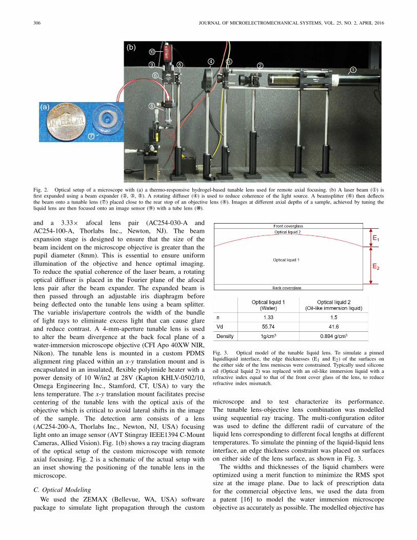
Fig. 2. Optical setup of a microscope with (a) a thermo-responsive hydrogel-based tunable lens used for remote axial focusing. (b) A laser beam (①) isfirst expanded using a beam expander (②, ③, ⑤). A rotating diffuser (④) is used to reduce coherence of the light source. A beamsplitter (⑥) then deflectsthe beam onto a tunable lens (⑦) placed close to the rear stop of an objective lens (⑧). Images at different axial depths of a sample, achieved by tuning theliquid lens are then focused onto an image sensor (⑨) with a tube lens (⑩).
and a 3.33× afocal lens pair (AC254-030-A andAC254-100-A, Thorlabs Inc., Newton, NJ). The beamexpansion stage is designed to ensure that the size of thebeam incident on the microscope objective is greater than thepupil diameter (8mm). This is essential to ensure uniformillumination of the objective and hence optimal imaging.To reduce the spatial coherence of the laser beam, a rotatingoptical diffuser is placed in the Fourier plane of the afocallens pair after the beam expander. The expanded beam isthen passed through an adjustable iris diaphragm beforebeing deflected onto the tunable lens using a beam splitter.The variable iris/aperture controls the width of the bundleof light rays to eliminate excess light that can cause glareand reduce contrast. A 4-mm-aperture tunable lens is usedto alter the beam divergence at the back focal plane of awater-immersion microscope objective (CFI Apo 40XW NIR,Nikon). The tunable lens is mounted in a custom PDMSalignment ring placed within an x-y translation mount and isencapsulated in an insulated, flexible polyimide heater with apower density of 10 W/in2 at 28V (Kapton KHLV-0502/10,Omega Engineering Inc., Stamford, CT, USA) to vary thelens temperature. The x-y translation mount facilitates precisecentering of the tunable lens with the optical axis of theobjective which is critical to avoid lateral shifts in the imageof the sample. The detection arm consists of a lens(AC254-200-A, Thorlabs Inc., Newton, NJ, USA) focusinglight onto an image sensor (AVT Stingray IEEE1394 C-MountCameras, Allied Vision). Fig. 1(b) shows a ray tracing diagramof the optical setup of the custom microscope with remoteaxial focusing. Fig. 2 is a schematic of the actual setup withan inset showing the positioning of the tunable lens in themicroscope.
C. Optical Modeling
We used the ZEMAX (Bellevue, WA, USA) softwarepackage to simulate light propagation through the custom
Fig. 3. Optical model of the tunable liquid lens. To simulate a pinnedliquidliquid interface, the edge thicknesses (E1 and E2) of the surfaces onthe either side of the lens meniscus were constrained. Typically used siliconeoil (Optical liquid 2) was replaced with an oil-like immersion liquid with arefractive index equal to that of the front cover glass of the lens, to reducerefractive index mismatch.
microscope and to test characterize its performance.The tunable lens-objective lens combination was modelledusing sequential ray tracing. The multi-configuration editorwas used to define the different radii of curvature of theliquid lens corresponding to different focal lengths at differenttemperatures. To simulate the pinning of the liquid-liquid lensinterface, an edge thickness constraint was placed on surfaceson either side of the lens surface, as shown in Fig. 3.
The widths and thicknesses of the liquid chambers wereoptimized using a merit function to minimize the RMS spotsize at the image plane. Due to lack of prescription datafor the commercial objective lens, we used the data froma patent [16] to model the water immersion microscopeobjective as accurately as possible. The modelled objective has
KANHERE et al.: REMOTE AXIAL TUNING IN MICROSCOPY UTILIZING HYDROGEL-DRIVEN TUNABLE LIQUID LENS 307
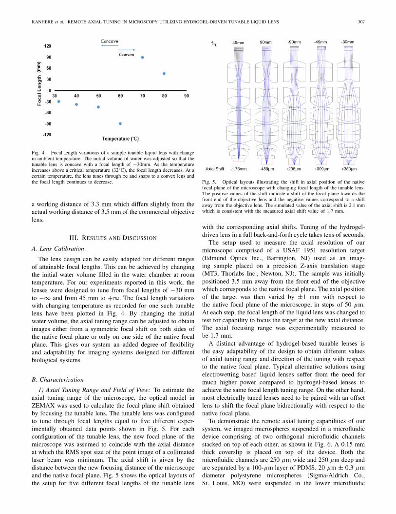
Fig. 4. Focal length variations of a sample tunable liquid lens with changein ambient temperature. The initial volume of water was adjusted so that thetunable lens is concave with a focal length of −30mm. As the temperatureincreases above a critical temperature (32°C), the focal length decreases. At acertain temperature, the lens tunes through ∞ and snaps to a convex lens andthe focal length continues to decrease.
a working distance of 3.3 mm which differs slightly from theactual working distance of 3.5 mm of the commercial objectivelens.
III. RESULTS AND DISCUSSION
A. Lens Calibration
The lens design can be easily adapted for different rangesof attainable focal lengths. This can be achieved by changingthe initial water volume filled in the water chamber at roomtemperature. For our experiments reported in this work, thelenses were designed to tune from focal lengths of −30 mmto −∞ and from 45 mm to +∞. The focal length variationswith changing temperature as recorded for one such tunablelens have been plotted in Fig. 4. By changing the initialwater volume, the axial tuning range can be adjusted to obtainimages either from a symmetric focal shift on both sides ofthe native focal plane or only on one side of the native focalplane. This gives our system an added degree of flexibilityand adaptability for imaging systems designed for differentbiological systems.
B. Characterization
1) Axial Tuning Range and Field of View: To estimate theaxial tuning range of the microscope, the optical model inZEMAX was used to calculate the focal plane shift obtainedby focusing the tunable lens. The tunable lens was configuredto tune through focal lengths equal to five different exper-imentally obtained data points shown in Fig. 5. For eachconfiguration of the tunable lens, the new focal plane of themicroscope was assumed to coincide with the axial distanceat which the RMS spot size of the point image of a collimatedlaser beam was minimum. The axial shift is given by thedistance between the new focusing distance of the microscopeand the native focal plane. Fig. 5 shows the optical layouts ofthe setup for five different focal lengths of the tunable lens
Fig. 5. Optical layouts illustrating the shift in axial position of the nativefocal plane of the microscope with changing focal length of the tunable lens.The positive values of the shift indicate a shift of the focal plane towards thefront end of the objective lens and the negative values correspond to a shiftaway from the objective lens. The simulated value of the axial shift is 2.1 mmwhich is consistent with the measured axial shift value of 1.7 mm.
with the corresponding axial shifts. Tuning of the hydrogel-driven lens in a full back-and-forth cycle takes tens of seconds.
The setup used to measure the axial resolution of ourmicroscope comprised of a USAF 1951 resolution target(Edmund Optics Inc., Barrington, NJ) used as an imag-ing sample placed on a precision Z-axis translation stage(MT3, Thorlabs Inc., Newton, NJ). The sample was initiallypositioned 3.5 mm away from the front end of the objectivewhich corresponds to the native focal plane. The axial positionof the target was then varied by ±1 mm with respect tothe native focal plane of the microscope, in steps of 50 µm.At each step, the focal length of the liquid lens was changed totest for capability to focus the target at the new axial distance.The axial focusing range was experimentally measured tobe 1.7 mm.
A distinct advantage of hydrogel-based tunable lenses isthe easy adaptability of the design to obtain different valuesof axial tuning range and direction of the tuning with respectto the native focal plane. Typical alternative solutions usingelectrowetting based liquid lenses suffer from the need formuch higher power compared to hydrogel-based lenses toachieve the same focal length tuning range. On the other hand,most electrically tuned lenses need to be paired with an offsetlens to shift the focal plane bidrectionally with respect to thenative focal plane.
To demonstrate the remote axial tuning capabilities of oursystem, we imaged microspheres suspended in a microfluidicdevice comprising of two orthogonal microfluidic channelsstacked on top of each other, as shown in Fig. 6. A 0.15 mmthick coverslip is placed on top of the device. Both themicrofluidic channels are 250 µm wide and 250 µm deep andare separated by a 100-µm layer of PDMS. 20 µm ± 0.3 µmdiameter polystyrene microspheres (Sigma-Aldrich Co.,St. Louis, MO) were suspended in the lower microfluidic

308 JOURNAL OF MICROELECTROMECHANICAL SYSTEMS, VOL. 25, NO. 2, APRIL 2016
Fig. 6. Demonstration of remote axial focusing in the 3D microscope usingimages of a microfluidic device. (a) Schematic of the microfluidic device madeup of two stacked orthogonal microfluidic channels, each 250 µm wide and250 µm deep. 20 µm and 40 µm microspheres are suspended in lower andupper channels respectively. At the start, the focal plane of the microscopeis aligned with the base of the device, then axial focusing is implemented toimage upwards through different depths of the device. (b) The focal plane firstshifts through the lower microfluidic channels to obtain images of the 20 µmbeads. (c)-(e) As the focal plane continues to shift upwards, the microscope isable to focus on different layers of 40 µm beads within the upper microfluidicchannel. The focus gradually shifts from a lower layer of beads in (c) to ahighlighted bead sitting atop this layer in (e). All images have been croppedto the same field of view for ease of comparison of images captured atdifferent axial dChange in field of view with varying axial depths due tonon-telecentricity of the optical setup.
TABLE I
CHANGE IN FIELD OF VIEW WITH VARYING AXIAL DEPTHS
DUE TO NONTELECENTRICITY OF THE OPTICAL SETUP
channel whereas the upper microfluidic channel consistsof 40 µm ± 0.3 µm diameter polymethacrylate microspheres(Sigma-Aldrich Co., St. Louis, MO). To image the entiremicrofluidic sample, we needed to achieve an axial focusingshift of 750 µm from the top of the specimen. For this,temperature of the tunable lens was varied from 55°C to 80°Cto obtain focal lengths between 90 mm and −30 mm.
In our setup, the tunable lens is placed at a finite distance(5 mm) behind the rearstop of the microscope objective whichlies inside the mounting shoulder and is inaccessible. As aresult, the system has an angular (non-constant) field of view.In other words, the system is non-telecentric and exhibitsdifferent fields of view at different axial positions. Anotherinterpretation of this is that the numerical aperture of thesystem changes as the axial position of the focal plane ischanged. To characterize the effects of non-telecentricity inour imaging setup, we measured the fields of view bothexperimentally and in ZEMAX for different curvatures of theliquid lens.
Table 1 summarizes the simulated and experimental field ofview variations with respect to various axial focus positions.
Fig. 7. (a) MTF plots of the optical system at different focal lengths of thetunable lens. The performance of the system deteriorates with increasing focallengths. However objects with 200 lp/mm spatial frequency can be imagedwith a reasonable contrast throughout the focusing range. (b) An image ofa 1951 USAF resolution target as seen by the microscope. The smallestelement discernible by the system has 228.1 lp/mm, which is close to theestimated resolution limit of the system.
2) Aberrations and Resolution: A widely accepted measureto characterize the resolution and performance of a microscopeis the Modulation Transfer Function (MTF). MTF is a measureof the ability of a microscope to transfer contrast fromthe specimen to the intermediate image plane at a specificresolution. Fig. 7(a) shows the diffraction MTF plots of ourmicroscope at different focal lengths of the tunable lens, assimulated in ZEMAX.
An object imaged with a 20% contrast (MTF20) canbe recovered with reasonable fidelity with standard imageprocessing techniques. Throughout the axial scanning range,our optical microscope can image objects with a spatialfrequency of 200 line pairs /mm (lp/mm) at 20% contrastor more. To compare the simulated results with experimentalobservations, an image of the 1951 USAF resolution targetwas captured with the tunable lens tuned to the focal lengthof +45 mm. Element 6 in Group 7 with a resolutionof 2.19 µm or a spatial frequency of 238.1 lp/mm is clearlydiscernible with a reasonable contrast.
Spherical aberrations as estimated by the optical model inZEMAX are summarized in Table 2. As the focal plane shiftscloser to the front end of the objective lens, the numerical
KANHERE et al.: REMOTE AXIAL TUNING IN MICROSCOPY UTILIZING HYDROGEL-DRIVEN TUNABLE LIQUID LENS 309
TABLE II
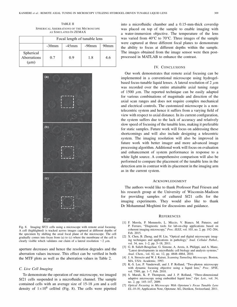
SPHERICAL ABERRATIONS OF THE MICROSCOPEAS SIMULATED IN ZEMAX
Fig. 8. Imaging SF21 cells using a microscope with remote axial focusing.A cell (highlighted) is tracked across images captured at different depths ofthe specimen by shifting the axial focal plane of the microscope. The cellgradually comes into focus from (a) to (c) where the membrane of the cell isclearly visible which validates our claim of a lateral resolution ∼2 µm.
aperture decreases and hence the resolution degrades and theaberration values increase. This effect can be verified in boththe MTF plots as well as the aberration values in Table 2.
C. Live Cell Imaging
To demonstrate the operation of our microscope, we imagedSf21 cells suspended in a microfluidic channel. The samplecontained cells with an average size of 15-18 µm and a celldensity of 1×10⁶ cell/ml (Fig. 8). The cells were pipetted
into a microfluidic chamber and a 0.15-mm-thick coverslipwas placed on top of the sample to enable imaging witha water-immersion objective. The temperature of the lenswas varied from 40°C to 70°C. Three images of the samplewere captured at three different focal planes to demonstratethe ability to focus at different depths within the sample.The images obtained from the image sensor were then post-processed in MATLAB to enhance the contrast.
IV. CONCLUSIONS
Our work demonstrates that remote axial focusing can beimplemented in a conventional microscope using hydrogel-based focus-tunable liquid lenses. A lateral resolution of 2 µmwas recorded over the entire attainable axial tuning rangeof 1500 µm. The reported technique can be easily adaptedfor various combinations of magnitude and direction of theaxial scan ranges and does not require complex mechanicaland electrical controls. The customized microscope is a non-telecentric system and hence it suffers from a varying field ofview with respect to axial distance. In its current configuration,the system suffers due to the lack of accuracy and relativelyslow speed of focusing of the tunable lens, making it preferablefor static samples. Future work will focus on addressing theseshortcomings and will also include designing a telecentricsystem. The imaging resolution will also be improved infuture work with better imager and more advanced imageprocessing algorithm. Additional work will focus on evaluationand enhancement of system performance in response to awhite light source. A comprehensive comparison will also beperformed to compare the placement of the tunable lens in thedetection arm in contrast with its placement in the imaging armas in the current system.
ACKNOWLEDGMENT
The authors would like to thank Professor Paul Friesen andhis research group at the University of Wisconsin-Madisonfor providing samples of cultured Sf21 cells for theimaging experiments. They would also like to thankDr Mohammad Moghimi for discussions and guidance.
REFERENCES
[1] F. Merola, P. Memmolo, L. Miccio, V. Bianco, M. Paturzo, andP. Ferraro, “Diagnostic tools for lab-on-chip applications based oncoherent imaging microscopy,” Proc. IEEE, vol. 103, no. 2, pp. 192–204,Feb. 2015.
[2] X. Chen, B. Zheng, and H. Liu, “Optical and digital microscopic imag-ing techniques and applications in pathology,” Anal. Cellular Pathol.,vol. 34, nos. 1–2, pp. 5–18, 2011.
[3] G. B. Salieb-Beugelaar, G. Simone, A. Arora, A. Philippi, and A. Manz,“Latest developments in microfluidic cell biology and analysis systems,”Anal. Chem., vol. 82, no. 12, pp. 4848–4864, 2010.
[4] J. A. Stroscio and W. J. Kaiser, Scanning Tunneling Microscopy. Boston,MA, USA: Academic, 1993.
[5] K.-S. Lee, P. Vanderwall, and J. P. Rolland, “Two-photon microscopywith dynamic focusing objective using a liquid lens,” Proc. SPIE,vol. 7569, pp. 1–7, Feb. 2010.
[6] S. Murali, K. P. Thompson, and J. P. Rolland, “Three-dimensionaladaptive microscopy using embedded liquid lens,” Opt. Lett., vol. 34,no. 2, pp. 145–147, 2009.
[7] Optical Focusing in Microscopy With Optotune’s Focus Tunable LensEL-10-30, Application Note, Optotune AG, Dietikon, Switzerland, 2011.
310 JOURNAL OF MICROELECTROMECHANICAL SYSTEMS, VOL. 25, NO. 2, APRIL 2016
[8] F. O. Fahrbach, F. F. Voigt, B. Schmid, F. Helmchen, and J. Huisken,“Rapid 3D light-sheet microscopy with a tunable lens,” Opt. Exp.,vol. 21, no. 18, pp. 21010–21026, 2013.
[9] N. Koukourakis et al., “Effects of axial scanning in confocal microscopyemploying adaptive lenses (CAL),” Proc. SPIE, vol. 9132, p. 91320P,May 2014.
[10] S. Liu and H. Hua, “Extended depth-of-field microscopic imaging witha variable focus microscope objective,” Opt. Exp., vol. 19, no. 1,pp. 353–362, 2011.
[11] Y. Nakai et al., “High-speed microscopy with an electrically tunablelens to image the dynamics of in vivo molecular complexes,” Rev. Sci.Instrum., vol. 86, no. 1, p. 013707, 2015.
[12] C. Zuo, Q. Chen, W. Qu, and A. Asundi, “High-speed transport-of-intensity phase microscopy with an electrically tunable lens,” Opt. Exp.,vol. 21, no. 20, pp. 24060–24075, 2013.
[13] L. Dong, A. K. Agarwal, D. J. Beebe, and H. Jiang, “Adaptive liquidmicrolenses activated by stimuli-responsive hydrogels,” Nature, vol. 442,no. 7102, pp. 551–554, 2006.
[14] L. Dong, A. K. Agarwal, D. J. Beebe, and H. Jiang, “Variable-focusliquid microlenses and microlens arrays actuated by thermoresponsivehydrogels,” Adv. Mater., vol. 19, no. 3, pp. 401–405, 2007.
[15] X. Zeng, C. Li, D. Zhu, H. J. Cho, and H. Jiang, “Tunable microlensarrays actuated by various thermo-responsive hydrogel structures,”J. Micromech. Microeng., vol. 20, no. 11, p. 115035, 2010.
[16] A. Katsuyuki, “Embodiment 1,” Japanese Patent 8–292 374,Nov. 5, 1996.
Aditi Kanhere received the B.S. degree inelectronics and telecommunication engineering fromthe University of Pune, Maharashtra, India, in 2009,and the Ph.D. degree in electrical engineeringfrom the University of Wisconsin–Madison, USA,in 2015. She is currently with Digilens Inc., CA,USA. Her other research interests include opti-cal imaging, lens design, camera calibration, andholography.
Guangyun Lin received the Ph.D. degree from Zhongshan University,Guangzhou, China, in 1997. She was a Faculty Member with ZhongshanUniversity, Guangzhou, China, from 1992 to 1998. She was a PostdoctoralResearch Associate with the Boyce Thompson Institute, Cornell University,Ithaca, NY, USA, from 1998 to 2001. She was a Scientist with InvitrogenLife Technology, Carlsbad, CA, USA, from 2001 to 2004. She is currently anAssociate Scientist with the Department of Bacteriology, and the Departmentof Electrical and Computer Engineering, University of Wisconsin–Madison,WI, USA. She has been with the University of Wisconsin–Madison since2005. Her research interests focus on molecular biology study of C. botulinumtoxins and their complex, and biosensing technologies.
Hongrui Jiang received the B.S. degree in physicsfrom Peking University, Beijing, China, and theM.S. and Ph.D. degrees in electrical engineeringfrom Cornell University, Ithaca, NY, in 1999 and2001, respectively. From 2001 to 2002, he was aPostdoctoral Researcher with the Berkeley Sensorand Actuator Center, University of California atBerkeley. He is currently the Vilas DistinguishedAchievement Professor and a Lynn H. MatthiasProfessor in Engineering with the Department ofElectrical and Computer Engineering, a Faculty
Affiliate with the Department of Biomedical Engineering, and the Departmentof Materials Science and Engineering, and a Member of the McPhersonEye Research Institute with the University of Wisconsin–Madison. Hisresearch interests are in microfabrication technology, biological and chemicalmicrosensors, microactuators, optical microelectromechanical systems, smartmaterials and micro-/nanostructures, lab-on-chip, and biomimetics and bioin-spiration. He is a Member of the Editorial Board of the IEEE/ASME JOURNAL
OF MICROELECTROMECHANICAL SYSTEMS. He is a Fellow of the Instituteof Physics, the Royal Society of Chemistry, and the American Institute forMedical and Biological Engineering. He was a recipient of the NationalScience Foundation CAREER Award and the Defense Advanced ResearchProjects Agency Young Faculty Award in 2008, the H. I. Romnes FacultyFellowship of the University of Wisconsin–Madison in 2011, the NationalInstitutes of Health Director’s New Innovator Award in 2011, and the VilasAssociate Award of the University of Wisconsin in 2013.

![Liquid Encapsulation Technology for Microelectromechanical ... · Liquid Encapsulation Technology for Microelectromechanical Systems Norihisa Miki ... [27]. Therefore, sealing with](https://img.dokumen.tips/doc/110x75/5ebd6745ad290220a7044b42/liquid-encapsulation-technology-for-microelectromechanical-liquid-encapsulation.jpg)