Embed Size (px)
Citation preview

30 Uveal Melanoma Richard Pötter, Erik Van Limbergen 1 Introduction Uveal melanoma account for about 75% of all primary malignant intraocular tumours (20% retinoblastoma). Eye melanoma is a rare malignancy with a mean incidence of less than one new case per 100.000 inhabitants and year (from 0.1 to 1.5), thus representing about 0.003% of all new cancers, with a slight male predominance. Eye melanoma is extremely rare in black people and in the Asian population. Most eye melanoma are diagnosed in the sixth and seventh decade of life (22). Uveal melanomas do not present symptoms in the beginning. At a later stage of the disease patients most frequently complain about impairment, loss of vision, loss of the visual field, and only in advanced disease about pain (e.g. due to secondary glaucoma). These clinical symptoms are dependent on the extent and the location of the tumour and on the accompanying retinal detachment, for example with loss of central vision in case of macula involvement. The most important prognostic factor for local control and survival is tumour size, followed by some other factors, among which are the epitheloid cell type, vascularisation pattern, tumour location (anterior to the equator), and extrascleral growth. There is evidence that immunology plays a major part in particular in induction of metastatic spread. Enucleation has been the standard treatment until the 80ies, when radiotherapy was found to offer a therapeutic alternative being at least equally effective in controlling tumour growth (about 90%) and at the same time eye- and vision-sparing. Ophthalmic plaque brachytherapy and proton radiotherapy at dedicated facilities have played a major role in this breakthrough. Nowadays, for the vast majority of uveal melanomas radiotherapy is the treatment of choice. The differential indication for the various types of radiotherapy has not yet been finally clarified and is depending on various factors, such as tumour thickness and diameter, tumour location, and availability of different types of radiotherapy. As eye plaque brachytherapy (mainly based on Ru-106 or I-125) has been proven to be highly efficient in small and medium-sized tumours in many locations in the globe (isoeffective to proton radiotherapy (19)) and as it is easily accessible and not expensive, eye plaque brachytherapy today represents the method of choice for these tumours. Often, brachytherapy is combined with some form of local ophthalmologic treatment, which is mostly laser coagulation. Local resection is rarely used, and should be restricted to specific situations and experienced centres. 2 Ocular Anatomy The globe is composed of three tunicae. The outer coat consists of the opaque sclera with a thickness of 0.3 to 1.0 mm and a diameter of 24 mm (which tolerates extremely high radiation doses), on which the six ocular muscles controlling eye movement insert, and the clear cornea. The uvea (middle coat) is formed by the choroid (thickness 0.1 - 0.3 mm), the ciliary body (thickness 2 mm), and the iris (thickness 0.5 - 3 mm), which represents the origin of different types of uveal melanoma. The inner coat consists of the retina including the retinal pigment epithelium (thickness 0.1 mm). The vascular supply for the retina is derived from the central retinal artery entering the globe through the optic nerve. The vitreous body filling the inner part of the globe does not contain

592 Uveal Melanoma
any vital cells. The optic nerve enters the globe at the optic disc (papilla of the optic nerve). The three tunicae of the globe stop at the border of the optic disc. The macula represents the point of the sharpest sight and is therefore most important for the overall function of the eye and is located temporal to the optic disc (3 mm). (3,22,36) 3 Pathology Many uveal melanomas develop on the basis of a uveal nevus. Congenital ocular melanosis may also precede uveal melanoma as some other diseases like the dysplastic nevus syndrome and neurofibromatosis. No correlation has been found with skin melanoma and sun exposure. Uveal melanomas are solitary tumours arising in one eye, taking their origin from the vascular eye wall. As the overlying retinal vessels rarely feed the tumour, ocular melanomas are primarily dependent on basal uveal blood vessels. Choroidal melanoma usually presents as a well defined pigmented nodular tumour with a round, elliptic, or lengthy basis. The major growth pattern (75%) is towards the inner part of the globe resulting in a dome shaped configuration. At the same time, the tumour frequently spreads along the choroid or along the inner part of the sclera (up to 50%). The overlying retina is most often damaged for various reasons (e.g. through impaired blood supply). Nodular tumours erode in more than 25% through Bruch`s membrane, which covers the choroid. Because of its elasticity this membrane cuts into the tumour, leading to a typical mushroom configuration, in which the surface of the mushroom may sometimes be larger than its basis. Especially in this configuration, different forms of retinal tumour invasion are seen as well as cell spread into the vitreous body. Nearly all choroidal melanoma lead to a serous retinal detachment in its direct vicinity and/or far away at the caudal part of the fundus following gravity. The optic nerve is almost never infiltrated, not even in juxtapapillary tumours (22). Rarely, extrascleral extension is observed. Diffuse infiltrating flat choroidal tumours are rare. The amount of pigmentation in choroidal melanoma varies significantly. Beside its nodular growth, melanoma of the ciliary body can also extend into the posterior part of the globe along the uvea and/or the sclera and into the anterior part towards the iris. The iridocorneal angle can be infiltrated rather early, which may lead to a secondary glaucoma. On the other hand, the lens may become displaced because of tumour growth (22). Iris melanoma is usually presented as flat pigmented lesion and because of its clinical appearance is detected at an early stage. The majority of uveal melanomas are located in the choroid (70%), the rest in the ciliary body (25%), and in the iris (5%). Histopathology reveals different types of uveal melanoma with different prognosis: spindle cell melanoma (better prognosis), melanoma with a significant amount of epitheloid cells (intermediate prognosis), epitheloid cell melanoma (poor prognosis). Prior to radiotherapy no biopsy is taken for histopathological specification, this subdivision is only of theoretical interest within the frame of radiotherapy (22). As local tumour control is high, prognosis is strongly correlated to distant metastases, which are most frequently observed in the liver (90%). Distant metastases can occur during a long time period after

Uveal Melanoma 593
successful local treatment (up to more than 20 years). Tumour re-growth is a significant risk factor for metastatic disease. Metastases at first presentation are uncommon. 4 Work Up The major part of the work up is performed by the ophthalmologist experienced in diagnosis and management of uveal melanoma (3,22,36). This fact has to be underlined, as diagnosis and decisions for management are established without histopathology, through a certain number of non-invasive methods. The final diagnosis is based on the ophthalmologic expert’s comprehensive judgement taking into account results from the different diagnostic methods applied. For experienced ophthalmologists the rate of false positive findings is nowadays reported to be below 1 - 2% (6). Fig 30.1: Classification system for uveal melanoma (modified from TNM-Atlas (37)) A: T1A: Tumour extension up to 7 mm, thickness up to 2 mm B: T1B: Tumour extension >7 up to 10 mm, thickness >2 up to 3 mm; C: T2: Tumour extension >10 up to 15 mm, thickness >3 up to 5 mm D: T3: Tumour extension > 15 mm; thickness > 5 mm.

594 Uveal Melanoma
The following examinations are to be performed: Clinical examinations: patient history of cancer, indirect binocular ophthalmoscopy with photodocumentation (morphology, topography, tumour size) and schematic drawing; slit lamp examination, transillumination.
Fluorescence angiography;
Sonography (standardised A and B): tumour dimensions (thickness, diameter) and topography, differential diagnosis (uveal metastases, hemangioma, melanocytoma and hemorrhagic lesions), prognostic factors (location, morphology, tumour volume, vascularization).
CT and/or MRI are not mandatory. Intraocular needle biopsy is rarely used. Metastatic disease is to be excluded for example by imaging of the liver. A T system for the staging of ocular melanoma is available, but until recently the use of this system has been very limited. This classification systems takes into account tumour thickness and the maximal diameter of the tumour basis, on which recommendations for management are based (22) (Fig 30.1): T1 (Small melanoma): Tumour thickness between 1 - 3 mm, greatest tumour dimension (basis) up to 10 mm; T2 (Medium-sized melanoma): tumour thickness between >3 mm and 5 mm, greatest tumour dimension (basis) >10 up to 15 mm; T3 (large melanoma): tumour thickness >5 mm, greatest tumour dimension (basis) >15 mm. 5 Indications, Contra-indications The main indications for eye sparing brachytherapy are small and medium-sized tumours (1,22). Several factors have a major impact on the decision on the type of management, the most important are tumour size, location and growth. Very small tumours (up to 2 - 3 mm thickness (T1)) without any tendency of growth should first be observed and only treated in case of growth. These tumours then become candidates for eye plaque brachytherapy (Ru-106/I-125), which may be combined with some form of laser coagulation. Medium-sized tumours with a thickness of up to 5 mm (T2) need active treatment and are candidates for eye plaque brachytherapy (electrons/photons). Eye plaque brachytherapy is also appropriate for a tumour thickness larger than 5 mm (T3) up to a maximum of 8 (10) mm, but for these large tumours brachytherapy then is preferably to be performed with a gamma emitter (I-125). Large tumours with a thickness >8 - 10 mm should not be treated with brachytherapy, as the results are poor in terms of local control and radiation related morbidity. The best candidates for eye plaque brachytherapy are posterior uveal melanoma not extending too far beyond the equator. Tumours near the optic disc and the macula (within 1 - 2 mm) are not ideal candidates for brachytherapy, as an underdosage at the tumour margin can hardly be avoided. On the other hand, significant brachytherapy related morbidity at the optic disc and/or at the macula is to be expected. Exceptions from these principles can be considered in specific situations, where issues are to be taken into account reflecting mainly quality of life aspects of the patient. For example, in case of only one remaining functioning eye carrying an uveal melanoma, this eye is to be conserved as long as

Uveal Melanoma 595
possible with as much vision as possible. In case of patients with a poor general health status with a limited life expectancy and in aged patients, the diseased eye should also be conserved as long as possible for obvious reasons. In such situations, the indication for brachytherapy should be taken more generously, for example thick tumours (>8 mm) or juxtapapillary tumours may be included in the indication list for eye plaque brachytherapy. However, the potential of external stereotactic photon beam therapy should also be taken into account in such specific situations, if available (7,38). 6 Target Volume The Clinical Target Volume (CTV) is determined based on findings from sonography, ophthalmoscopy, and finally on the tumour`s transillumination shadow indicating the macroscopic tumour extension during ophthalmologic intervention. The tumour dimensions in the preoperative planning phase are mainly based on findings from ultrasonography (tumour basis diameter and thickness). The basis diameter of the CTV is determined by the tumour basis diameter (Fig 30.2). A safety margin of 1 - 2 mm for subclinical disease is added in all directions, which accounts for microscopic spread along the ocular tunicae, mainly the uvea and the sclera. For a given tumour basis diameter of 11 mm, the CTV basis diameter would add up to ~13 mm. In some situations, uncertainties in tumour delineation (e.g. in the exact drawing of the tumour shadow during transillumination) and/or in plaque localization (e.g. in posterior pole locations) may be observed, which can be taken into account by adding an extra safety margin (PTV). The amount of such a safety margin should be determined individually according to the given situation. No safety margin for eye ball movements is taken into consideration, as the eye plaque is sutured tightly onto the outer sclera. Safety margin considerations have to be especially taken into account if the tumour is located in the vicinity of critical structures, such as the optic disc, the macula, or the ciliary body. In certain situations (amelanotic melanoma, specific mushroom configurations) specific care has to be taken. The thickness of the CTV is determined by the tumour thickness. The thickness is usually taken from sonography findings (B-scan) with 1 mm added for the sclera. The thickness is measured at the most prominent point of the dome shaped tumour (tumour apex) with a line drawn towards the uvea which has to be perpendicular to its basis (Fig 30.2).
Fig 30.2: Tumour thickness (orthogonal to the sclera) and tumour diameter measured by ophthalmologic ultrasound (B-scan) in eye melanoma. The arrow indicates the tumour apex including the retina and the outer border of the choroid. In order to arrive at the thickness of the CTV one mm is added taking into account the thickness of the adjacent sclera.
Therefore, in the dome shaped tumours these dimensions usually represent the maximum tumour thickness. Uncertainties in the determination of tumour target thickness are (only) related to the (sonographic) measurement of the tumour thickness itself.

596 Uveal Melanoma
Corresponding to the specific growth pattern of uveal melanoma, there are some debates about the target cells in uveal melanoma, which need to be treated with a certain dose: the tumour cells at the tumour apex and/or at the uveal layer, which is regarded as carrying the main blood supply. The whole tumour volume and its microscopic extensions form the basis of the CTV, therefore the apex tumour dose represents the minimum target dose (for the apex tumour cells). In contrast, the uveal layer forms the tumour basis, from which the tumour growth takes its origin and is continuously fed via the uveal blood supply. This layer receives a significantly higher dose, which is close to the scleral (surface) dose. At present, there is no consensus about the required dose to the target. However, the minimum target dose and the scleral surface dose should be recorded and reported. As the critical structures to be spared from excessively high radiation doses (fovea, optic disc, choroid, retina) are located at the level of the tumour basis, the doses at the tumour basis are most relevant for the assessment of brachytherapy related morbidity. Fig 30.3 Determination of GTV and CTV in eye melanoma (T2) with retinal detachment. 7 Technique The application can be done under local or general anaesthesia. The conjunctiva is opened at the conjunctival ring and the sclera is exposed in the quadrant where the tumour is located. If necessary, a muscle is snared with a sling or even dissected at its insertion to refix it after removal of the applicator. The tumour is localized by transscleral illumination and the GTV and/or the CTV is accurately marked on the overlying sclera, for example with a crayon (avoid errors due to parallax) (Fig 30.4 A,B,C). The marking can be controlled by pushing the sclera along the marked line which can be controlled by fundoscopy. Based on this marking, which indicates the outer border of GTV or the CTV, the adequate plaque is chosen, taking into account the findings from ultrasound.

Uveal Melanoma 597
Fig 30.4: Technique of implantation of a Ru-106 eye plaque: A: Transillumination showing the shadow of the uveal melanoma on the sclera, which serves as a basis for the delineation of GTV and CTV. B: Control diaphanoscopy with a transparent acrylic dummy applicator in place (black edges) shows the relation between GTV (black), CTV (black points) and the black applicator edge (generous safety margin). C: Part of the CTV on the sclera as drawn during transillumination; Ru-106 applicator being forwarded onto the sclera. D: Ru-106 applicator in place fixed with two episcleral sutures covering adequately the GTV and CTV. The selection of the size of the plaque (15.5/20/25 mm diameter for Ruthenium-106) is based on the dimensions of the clinical target volume, which usually exceeds the GTV at each side by 1 - 2 mm. Furthermore, it has to be taken into account, that there is an inactive edge at the outer margin of the plaque of 0.6 - 0.7 mm. Following the example given for CTV determination, a Ruthenium-106 eye plaque with a diameter of 15 mm should be chosen for a tumour basis of 11 mm, allowing 1 mm for microscopic spread on each side (13 mm) and 1 mm for dosimetric uncertainties (e.g. inactive edge) (compare Fig 30.8). The preplanning for iodine-125 eye plaques must be so precise, that the manufactured plaque fulfils the demands for dimensions as outlined during the brachytherapy procedure. A dummy plaque without radioactive seeds is firstly attached to the scleral area overlying the tumour. The plaque location is re-confirmed with point-source transillumination and indirect ophthalmoscopy. This procedure allows sutures to be placed which hold the actual plaque, and minimises hence radiation exposure to the surgical team.
A B
C D

598 Uveal Melanoma
Such a dummy applicator is almost routinely used for iodine-125 eye plaque placement, and in difficult cases for Ruthenium-106 eye plaque treatment. Prior to treatment the selected Ru-106 eye applicator is removed from the safety container and put for a certain time period into a glass with destilled water to clean it. Afterwards it is dried. These procedures should be done behind a PMMA shield for radiation protection reasons. When forwarding the applicator to the ophthalmologist performing the surgical procedure, care has to be taken that the concave irradiating surface of the applicator is always directed towards radiation absorbing material. The applicator is finally fixed by 2 episcleral sutures (Fig 30.2 D), maybe one additional suture crossing the applicator for pushing it tightly against the sclera. Most important is to reassure that there is no space at all or fluid between the concave surface of the applicator and the sclera because this would lead to major underdosage in the respective area. Finally, the conjunctiva is closed by a few sutures to prevent infection. The removal of the plaque follows general surgical principles. Care has to be taken to avoid diplopia, if a muscle had been dissected at its insertion. During the removal of the applicator, the general rules of radiation protection have to be taken into consideration (safety container). Patients can also be treated with a high dose rate Strontium eye plaque (26). The eye plaques are adjusted from normal Strontium eye applicators (e.g. diameter of 16 mm, 2 mm inactive edge, active diameter of 12 mm) (see Fig 30.11). Because this is a high dose rate plaque, the plaque fixation technique has to be adapted. An application ring is first sutured to the sclera in the correct treatment position. After transillumination control of the position of the tumour within the ring, the plaque is quickly brought in place and fixed by a crossed silk threat with a not over the posterior side of the plaque. This procedure can be done under local retrobulbar anaesthesia, a long acting anaestheticum, so that the implant, the treatment and the removal can be done under the same anaesthesia. 8/9 Dosimetry, Dose, Dose Rate Accuracy of dose measurement has been modest until recently, in particular the uncertainty of dose measurement for Ruthenium-106 eye applicators has been indicated to be +/- 30%. New developments in dosimetry (extrapolation chamber, film dosimetry, scintillation dosimetry) seem to make it possible that this uncertainty is reduced to about +/- 10-15%. Eye plaque dosimetry has been mainly based on manual dose calculation until a few years ago, when computer assisted dose calculation became available. Dependent on the eye plaque different types of dose calculation methods have been used with different degrees of accuracy. The use of high energy gamma rays like Co-60 result in high exposure for the operating team and to higher radiation induced vascular complications in both the eye and the orbital structures. Such isotopes have been abandoned for eye plaque therapy for many years. As eye plaque brachytherapy with Ruthenium-106 and Iodine-125 are the most popular at present, dosimetry for these two isotopes is given here in more detail.

Uveal Melanoma 599
Fig 30.5: Depth dose curves for Ru-106, I-125 and Sr-90
8.1 Ru-106 eye plaque brachytherapy The isotope Ruthenium-106 has a half life of 367 days and decays to Rhodium 106 while emitting beta rays with a maximum energy of 39 keV. Rhodium-106 with a half life of 36 seconds, decays to Pd-106 with a maximum energy of 3.5 MeV and a mean energy of 1.5 MeV providing the effective therapeutic irradiation. 8.1.1 Description of ruthenium-106 applicators Ruthenium-106 applicators have been developed by Lommatzsch and Vollmar in the 60’s in East Berlin (1966) (22). The Ru-106 is almost equally distributed on the concave surface of the shell shaped applicator. Fig 30.6: Ruthenium-106 eye plaque applicators (BEBIG) in different size and shape for the treatment of various types of uveal melanoma, including iris melanoma. The applicator types CCB and CCA are the most often used for small and medium-sized melanoma. The use of cut out applicators for tumours near the optic nerve (COB, COC) is more critical as the dose distribution may result in under-dosage in the juxtapapillary area.

600 Uveal Melanoma
Towards the eye ball a thin (0.1 mm) silver foil covers the Ru-106; this coverage has almost no absorption effect on the beta ray emission. On the other hand the convex surface is covered by a 0.7 mm silver foil, which absorbs more than 95% of all radioactivity. This fact is important for radiation protection of the personnel in the operating theatre with regard to the handling of the applicator, as only minor safety procedures have to be undertaken. The concave irradiating side of the applicator should always be protected as electrons have a limited range only in tissue and not in air. There is an inactive edge at the peripheral margin of the applicator which is reported to be about 0.7 - 0.8 mm. The external diameter varies between 15 and 20 mm, the spherical radius between 12 and 14 mm. There are usually two lugs at each applicator, by which it can be sutured to the sclera. Small grooves can be put onto the convex side of the applicator, enabling a fixation of the applicator against the sclera by a suture across the applicator. There are applicators with notches, enabling the positioning near the optic nerve and applicators for the brachytherapy of ciliary body melanoma. The nominal activity varies (dependent on the size of the applicator) between 13 and 26 MBq or 0.35 and 0.7 mCi. 8.1.2 Dosimetry of ruthenium-106 eye plaque brachytherapy The total dose of Ru-106 brachytherapy is usually recommended to be about 100 Gy, specified at the apex of the tumour which represents the CTV. This dose is calculated along the central axis of the applicator at the apex of the CTV. This minimum target dose is also to be given to the whole basis of the CTV, taking carefully into account the dimensions of the applicator, of the active layer of Ruthenium-106 on its concave surface, and of the corresponding isodose distribution at the peripheral margins (inactive edge ~0.8 mm). The dose at the sclera varies significantly, somewhere between 200 and 1000 Gy in tumours which are 2 - 5 mm thick. This variation is due to the different tumour thickness treated. In case of larger tumours, the scleral dose may even go beyond 1000 Gy, which is usually well tolerated. However, if scleral doses significantly beyond 1000 Gy should be applied, this should not be done within one session.
The dose rate at the day of application is dependent on the actual activity of the Ru-106 applicator and the prescribed dose to the CTV, taking also into account the dose to the sclera/uvea and/or to critical structures nearby the CTV, for example at the optical disc. The dose rate at the beginning of an active Ru-106 eye plaque is specified at the concave surface and is usually around 12 - 20 cGy per minute or 720 - 1200 cGy per hour (MDR). According to the rapid dose fall off, the dose rate changes with depth to 5 - 9 cGy per minute or 300 - 540 cGy per hour at 3 mm (43%) and to 2.4 - 4 cGy per minute or 150 - 240 cGy per hour at 5 mm (20%) at the beginning of a Ru-106 eye plaque (MDR/LDR). As the half life time of Ru-106 is about 367 days, there is a significant change in dose rate over time towards lower dose rates. For a given applicator the dose rate at the beginning is specified by the manufacturer. The maximum tumour prominence as measured by ultrasound is taken and 1 mm is added for the sclera thickness to arrive at the target depth. The dose rate per hour at the target depth (Minimum Target Dosetimeapp/hour) (MTD) is calculated based on the reference dose rate per minute at the target depth given in the calibration certificate of the manufacturer for the relative depth dose curve for a given Ru-106 applicator (MTDtime0/minute). This MTDtime0 is to be multiplied with a factor representing the decay of the Ruthenium-106 since the time of production (factordecay) and multiplied with 60. The decay factor is looked up in a table (Table 30.1) or diagram indicating the decay of Ruthenium-106 over time: MTDtimeapp/hour = MTDtime0 /minute x Factordecay x 60.
Uveal Melanoma 601
Decay factors for Ruthenium-106
Weeks 0 1 2 3 4 5 6 7 8 9 0 1 0.987 0.974 0.961 0.949 0.936 0.924 0.912 0.900 0.888
10 0.877 0.865 0.854 0.843 0.832 0.821 0.810 0.800 0.789 0.779 20 0.769 0.759 0.749 0.739 0.729 0.720 0.710 0.701 0.692 0.683 30 0.674 0.665 0.657 0.648 0.639 0.631 0.623 0.615 0.607 0.599 40 0.591 0.583 0.576 0.568 0.561 0.553 0.546 0.539 0.532 0.525 50 0.518 0.511 0.505 0.498 0.492 0.485 0.479 0.473 0.466 0.460
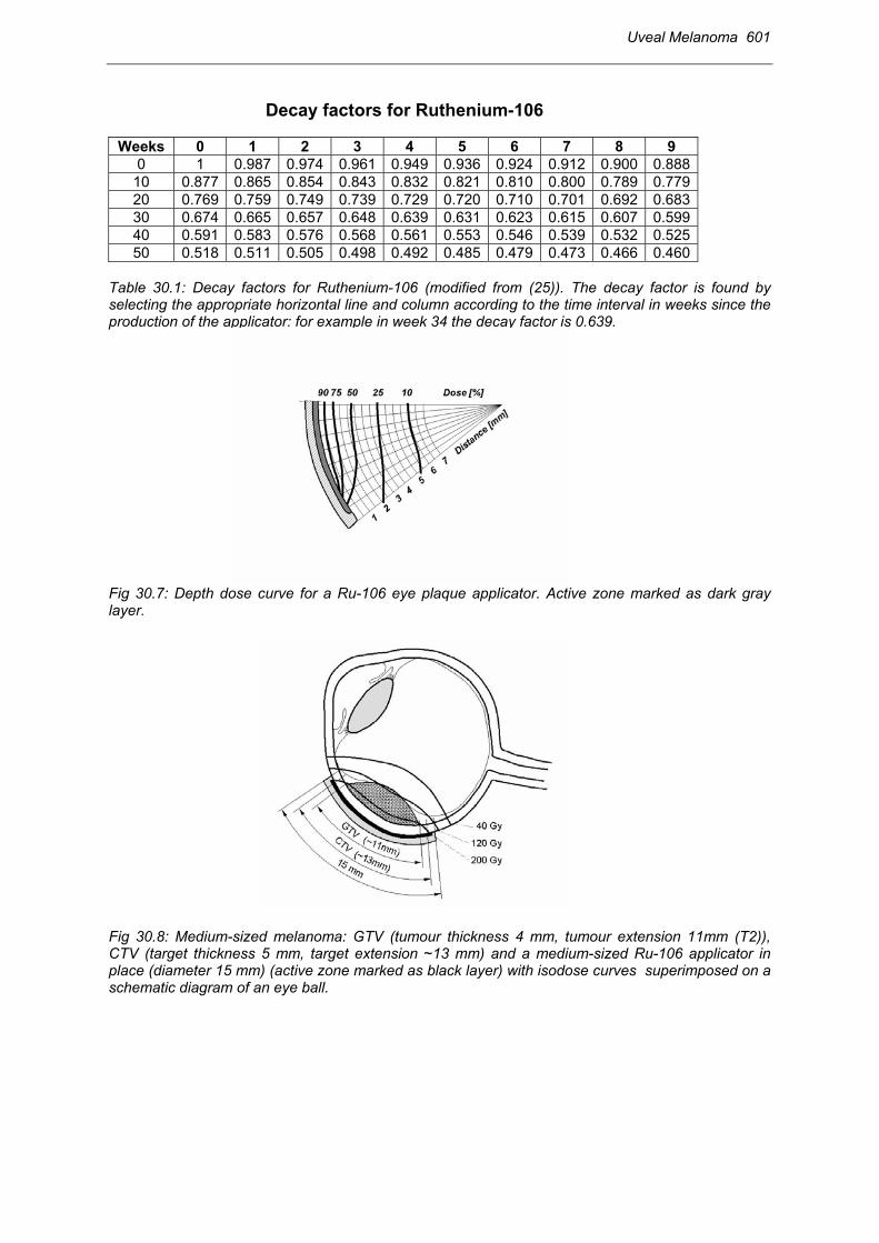
Table 30.1: Decay factors for Ruthenium-106 (modified from (25)). The decay factor is found by selecting the appropriate horizontal line and column according to the time interval in weeks since the production of the applicator: for example in week 34 the decay factor is 0.639. Fig 30.7: Depth dose curve for a Ru-106 eye plaque applicator. Active zone marked as dark gray layer. Fig 30.8: Medium-sized melanoma: GTV (tumour thickness 4 mm, tumour extension 11mm (T2)), CTV (target thickness 5 mm, target extension ~13 mm) and a medium-sized Ru-106 applicator in place (diameter 15 mm) (active zone marked as black layer) with isodose curves superimposed on a schematic diagram of an eye ball.

602 Uveal Melanoma
The overall time of application (hours) is calculated by dividing the prescribed target dose (Gy) by the minimum target dose rate (Gy/hour): Prescribed MTD (Gy) Timeapp (hours) = ----------------------------
MTDtimeapp (Gy/hour). The scleral dose is calculated in the same way. For a prescribed dose of 100 Gy at the tumour apex, the duration of a Ru-106 application is somewhere between 1 and 7 days depending on the various factors mentioned. 8.2 Iodine-125 eye plaque brachytherapy Iodine-125 is a gamma emitter with a half life of 60 days. I-125 decays exclusively by electron capture to an excited state of Te-125 which spontaneously decays to the ground state with the emission of 35.5 kV gamma photons. Characteristic x-rays in the range of 27-35 kV are produced also due to electron capture and internal conversion. The half value layer for Gold is only 0.025 mm, hence 0.5 mm of gold is sufficient to absorb 99.95% of the incident gamma rays. The low energy of I-125 results also in a smaller radiation safety problem than using other gamma emitting sources, such as Co-60 or Ir-192. 8.2.1 Description of the applicators Iodine-125 was introduced to treat ocular tumours at the end of the 70-ies (30). Presently I-125 is predominantly used in the US to treat uveal melanoma. Fig 30.9: Iodine-125 applicator with acrylic plaque insert for seven I-125 seeds (20). In many centres Iodine-125 plaques are individually fabricated using multiple seeds imbedded in a concave gold plaque. Iodine seeds are usually delivered in Titanium encapsulation (0.05 mm) containing between 0.5 – 20 mCi of I-125, the (outer) seed dimension is about 5 x 1 mm. Because of the presence of titanium and end welds the dose distribution around iodine seeds is highly anisotropic. The seeds are adhered to the concave portion of the plaque with an adhesive and at completion of treatment they are removed by dissolving the adhesive, and are re-used. Instead of using an adhesive silicon acrylic plaque inserts are also used to accommodate seeds (4 - 18, depending on

Uveal Melanoma 603
plaque size) at fixed positions. The major difficulties in the design of Iodine-125 seed eye plaques result from the need to have thin device to slip over the surface of the eye and the relatively bulky physical dimensions of the seeds. The gold layer in the concave plaque absorbs radiation and hence minimises doses to healthy structures in dorsal direction. The ability to attenuate I-125 with a thin layer of gold has allowed the production of rimmed plaques. The rim further reduces the lateral dose contributions to healthy structures of the eye and helps therefore to reduce ocular morbidity. On the other hand, higher plaque failures have been observed in thin tumours in close proximity of the optic nerve when using rimmed plaques. Similarly as for Ru-106 applicators plaque diameters vary according to the dimension of the tumour (15 - 20 mm). Cutouts are used for tumours next to the optic nerve. 8.2.2 Dosimetry of iodine-125 applicators Patients are treated with dose rates between 50 - 100 cGy/hr and apical doses between 70 - 150 Gy are given, doses at the tumour base are in the range between 200 - 700 Gy. Depending on tumour size treatment times vary between 30 and 300 hours (33,15). Fig 30.10: Isodose depth dose distribution in relation to tumour configuration for eye plaque brachytherapy (applicator in place) for Ruthenium-106 (target thickness 5 mm) and Iodine 125 (target thickness 6 mm) (modified from 22) Due to the individual source seed arrangements in the gold plaque treatment planning for Iodine-125 is more complex. Treatment planning is usually performed using dedicated software. Model calculations take into account the active length of the seeds, scatter within the phantom and anisotropy of dose distribution from a single seed. However, approximations are made such as the seed is simulated by an unfiltered line source located at the geometrical centre of the seed (24). Other models used for dosimetric calculations are based on a point source assumption (13). These simplified models are sufficiently accurate for clinical calculations, deviations of a few percent between TLD measurements and calculations have been reported in literature (24). The resulting isodose distribution when using multiple seed arrangements in rimmed or un-rimmed plaques can be irregular or asymmetric. When performing an individual plaque construction the radiation physicist should be consulted. In order to avoid misalignment of the plaque, especially when using custom made plaques with asymmetric seed configuration, it is recommended that the physicist is present at the time of surgery in the operating room with isodose curves.

604 Uveal Melanoma
8.3 Miscellaneous (SR-90, IR-192, PD-103) With Strontium-90 (beta emitter, usually used as a Strontium/Yttrium source, half life 28 years, Eßmax = 2.28 MeV, average 0.9 MeV) the delivery of 600 Gy in 1-3 hours (HDR) has been safely carried out without serious adverse side effects. More recently, this dose has been reduced to 450 Gy. Dose is referred to the sclera (600 Gy) and also recorded at the apex of the tumour (from 20 Gy to about 300 Gy). In case of tumours larger than 5 mm height, a second application is foreseen. If tumour persistence is observed after 6 months a second application is performed. Fig 30.11: Strontium-90 eye plaque (Amersham) and application ring. A: HDR strontium-90 eye plaque and application ring for quick and safe insertion of the HDR plaque. B: Fixation of the applicator ring to the sclera for insertion of a HDR strontium-90 eye plaque. The higher gamma energy of Ir-192 (average energy 0.37 MV, half life 74 days) leads to more shallow dose decrease with distance from the source as compared to I-125. The dose distribution from Ir-192 sources is not optimal since the penetrating radiation from this isotope may deliver considerable dose to uninvolved structures such as the lens, retina, optic nerve. For distances larger than 8 mm from the plaque the dose rate decreases more rapidly for I-125. Some investigators have used Palladium-103 for eye plaque therapy (13). Pd-103 has a short half life of 17 days and decays by electron capture with the emission of characteristic x-rays in the range of 20-23 kV. Because of their lower energy, Pd-103 generated photons are even more rapidly absorbed in tissue. This can be an advantage for thin tumours but a big disadvantage for prominent tumours. The combination of Ruthenium-106 and Iodine-125 has been reported, resulting in an improved dose distribution (14). However, this advantage is outweighed by possible confusion that could occur when several nuclides are used, as there is a higher risk of treatment planning errors. 10 Monitoring During the treatment time (applicator in place) only general rules of radioprotection have to be taken into consideration. For the beta emitter Ruthenium-106, the electrons are almost completely absorbed within the eye ball. There is an extremely small amount of bremsstrahlung background near the patient, which
A B

Uveal Melanoma 605
usually can be ignored. However, each person in contact with such patient has to be allowed to come into contact with radiation and has to be monitored for radiation protection (personal dosimetry). For iodine-125, the low energy photons are not completely absorbed within the eye ball. Half value layers in water (or tissue) for Iodine-125 and Pd-103 are less than 2 cm. Half value layer for I-125 in lead is 0.025 mm, hence common lead accessories provide a sufficient shielding. Packer et al (30) reported a maximum dose of 2.5 µGy for the surgical team during a I-125 application. However, for eye plaque brachytherapy the general radiation protection rules should always be taken into consideration: minimising exposure time to personal and patient, maximising distance (e.g. by using tweezers), using protection where possible (e.g. by wearing lead aprons). Specific acute side effects like swelling of the conjunctiva, acute exudative retinal detachment are transient and require symptomatic treatment. Prophylactic treatment with oral and non-steroid agents may be indicated. Local treatment after surgery includes atropine, steroid and non-steroid medication, eventually in combination with a local antibiotic. 11 Results Treatment results and morbidity are specific for the respective method. In the following subsections each method is addressed in a separate paragraph. Specific patterns of tumour regression are typical for different tumour types and also vary with the treatment method. A rapid complete remission with a sharply delineated flat white scar may be achieved in some cases. More often, tumour regression takes place slowly resulting in a white scar at the margin and a prominent grey mass in the centre (“grey mouse”). Sometimes there is (almost) no change in tumour extension and height for years. Therapeutic decisions are usually taken if a tumour regrowth is suspected because of tumour enlargement (fundoscopy, sonography) or because of decrease in sonographic reflexivity. Follow-up after eye-conserving treatment is usually based on ophthalmoscopy, ultrasound and fluoroscence angiography. 11.1 Results of surgery Results of enucleation are not given in detail here. From a few retrospective series it has been reported that radiotherapy is at least as good in terms of survival as compared to enucleation (21,8). There is no increased risk of early distant metastases after radiotherapy as it has been reported after classical forms of enucleation (39). The results from the large randomised trial performed by the co-operative malignant melanoma study group in North America (COMS) comparing enucleation and iodine-125 eye plaque radiotherapy showed that 5 year survival rates after enucleation (n=660) were almost identical with 81% to conservative treatment with I-125 eye plaque brachytherapy (see below) (9). The results of enucleation seem to be better in large tumours (>> 10 mm height) in terms of improved local control and less ocular morbidity. In these cases, enucleation represents a straightforward procedure without significant morbidity (the eyeball is removed), whereas radiation therapy (external beam or brachytherapy) has a considerable local failure rate and a significant morbidity rate (e.g. retinal detachment, neovascular glaucoma), often making a secondary enucleation inevitable.

606 Uveal Melanoma
In selected cases a surgical alternative to enucleation seems to be conservative local resection (e.g. large tumours), which is to be performed by a highly specialized ophthalmologist. This method has been reported to lead to similar results (16). 11.2. Results of external beam radiotherapy Hadron therapy is the treatment of choice, if external beam therapy is to be used. The major experience has been reported from proton radiotherapy (helium). However, recently some promising results have also been obtained with stereotactic photon beam radiotherapy (7,38). 11.2.1 Proton radiotherapy (Helium) A large amount of data has been collected at a few dedicated centres in the world (e.g. Boston, San Francisco, Lausanne/Villigen, Nice, Liverpool). Local control has been reported in more than 95% of the patients after 5 years (5,10,29). Survival at 5 years is between 73% (5), 81% (29) and 85% (10,11). The melanoma related mortality rate varies from 6% to 19% at 5 years (5,10,11). Radiation related morbidity is more often found in the anterior part of the eye (atrophy of the eyelid, loss of eyelash, keratitis, dry eye, cataract), but also in the posterior part, in particular in large tumours located near the macula and/or the optic disc (maculopathy, optic nerve atrophy). Neovascular glaucoma is reported from 7% up to 31% (4,5) in particular in large tumours (10). Because of this significant morbidity, proton radiotherapy is not recommended any more for large tumours exceeding 10 mm in height. Salvage enucleation had to be performed in these series in 8% to 22%. 11.2.2 Stereotactic photon radiotherapy Stereotactic photon radiotherapy is at present experimental, but seems to be promising. In preliminary results from few dedicated centres, local tumour control seems to be comparable to hadron beam therapy, whereas morbidity seems to be less pronounced, in particular in the anterior part of the eye (38). Dieckmann et al. (7) presented recently a linac based stereotactic irradiation technique for uveal melanomas allowing for fractionated photon radiotherapy. 11.3 Results of brachytherapy Eye plaque brachytherapy is nowadays mainly based on Ruthenium-106 and Iodine-125. Although the results with Co-60 eye plaque brachytherapy have been excellent with regard to tumour control, they have been poor with regard to radiation related morbidity. Because of this unfavourable therapeutic window, Co-60 eye plaque brachytherapy has been abandoned. The overall local recurrence rate after eye plaque therapy goes up to approximately 15% at 5 years and is dependent on various factors. Metastatic disease is reported up to >30% within 15 years (17,23). Enucleation is required in about 6% of all patients, in the majority during the first years (35). 11.3.1 Ruthenium-106 eye plaque brachytherapy There is a considerable amount of data available today, reflecting the broad experience with Ru-106 eye plaque brachytherapy (Table 30.2). Overall survival at 5 years is 73 to 91% (2,18,28,32) and mainly dependent on patient selection. In a recent meta-analysis of five large series (n=1066), the melanoma-related 5-year and 10-year mortality rates were found to be 14% and 22%, respectively (34). At 5 years, the mortality rates were

Uveal Melanoma 607
6% for small and medium-sized (T1/T2) and 26% for large tumours (T3). After 15 years overall melanoma related mortality rates were 33.3% (23). Table 30.2: RUTHENIUM 106 EYE PLAQUE BRACHYTHERAPY (n=1241 patients)
Leipzig
(Lommatzsch 1986)
Hamburg (Kleineidam et
al. 1993)
Helsinki (Summanen et al. 1996)
Lyon (Grange et al.
1995)
Stockholm (Seregard et
al. 1997)
Münster (Pötter et al.
1997) Time period
1964-1984 1975-1986 1981-1991 1983-1993 1979-1995 1981-1989
Patients (n)
309 184 100 207 266 175
Age (years)
52 58 59 60 63 60.5
Mean Follow-up (years) (range)
6.7 (1.0-21.0)
6.1 (0.1.-13.3)
3.3 (1.1.-10.1)
(0.1.-10.0)
3.6 (0.5.-12.5)
5.0 (1.0.-10.0)
T1 (n) 71
45 7 6 15 48
T2 (n)
8 40 37 40 48 56
T3 (n)
21 15 56 53 37 66
LTD > 10 mm (%)
29 55 93 91 85 NA
Tumour apex dose
> 100 Gy 250 Gy (median)
100 Gy (median)
> 60 Gy 100 Gy (median)
150 Gy (140-160)
Scleral dose (Applicator surface)
< 1500 Gy 747 Gy (median)
< 1200 Gy < 1500 Gy 543 Gy (median)
< 1500 Gy
5-yr mortality rate (%) melanoma (all causes)
11 (16) NA (16) 22 17 (20) 14 11 (27)
10-yr mortality rate (%) melanoma (all causes)
20 (34) NA (27) 34 19 (29) 24 16 (40)
5-yr mortality rate (%) melanoma: T1,T2 vs. T3
NA NA 3 vs 39 9 vs 24 5 vs 21 9 vs 13
Table 30.2: Results from Ru-106 eye plaque brachytherapy from different study groups in Europe (modified from Seregard et al. (34)) The main adverse prognostic factors are tumour size (tumour diameter larger than tumour height), tumour site (anterior location with involvement of the ciliary body), and age (34). Tumour control, which is rarely reported, is at 3 years 88% for small tumours and 85% for large tumours (32). In the old series of Lommatzsch tumour control at 15 years was 63.2% (23). A useful vision can be obtained in the majority of patients, if the tumour is not located too near the macula or the optic nerve, or if it is not too large. Long term radiation related morbidity is strongly correlated with the tumour site and the corresponding position of the plaque. It is most pronounced in large tumours near the macula and/or the optic disc with degeneration or destruction of the macula and atrophy of the optic nerve. Radiation retinopathy, neovascular glaucoma and cataract are not common and are seen in this order in decreasing frequency. Salvage enucleation, mainly for tumour regrowth and rarely for neovascular glaucoma, was performed in 8% to 21% in the series reported (see above).

608 Uveal Melanoma
11.3.2 Iodine-125 eye plaque brachytherapy Fontanesi et al. (15) presented results of 144 patients treated with I-125 plaques and reported 90% local control while Quivey et al. (33) reported 81% local control for 150 patients treated between 1982 and 1990. Patients enrolled in the randomised COMS trial in North America (enucleation vs. iodine-125 brachytherapy) and treated with Iodine 125 (n=657) had a 5 year survival rate of 81%, with a 9% mortality rate from melanoma. These rates were almost identical to those in the treatment arm in which patients were assigned to enucleation (see above) (9). A substantial impairment in visual acuity (20/200 or worse) was seen in 43% of the treated eyes after three years compared to the pre-treatment level in the COMS trial (26). 11.3.3 Strontium-90 eye plaque brachytherapy Strontium-90 brachytherapy has been reported to result in favourable outcome in small and medium-sized eye melanoma (27). In a series of 46 eye melanoma treated with Sr-90 tumour diameter ranged from 4.5 to 15 mm, tumour thickness from 1.5 to7 mm. Mean surface dose was 558 Gy. With a mean follow up of 4.1 years 30 tumours (65%) regressed to a flat scar, 13 (28%) were in regression but still present and 3 eyes (6.5%) had to be enucleated for recurrence (eye preservation rate 93.5%). No serious radiogenic complications were reported. Large lesions could be treated in two fractions with a long interval allowing for regression (~6 months). 12 References 1. Alberti WE, Sagermann RH. Radiotherapy of introcular tumours. Med Radiol 1993; Springer,
Berlin Heidelberg New York. 2. Bornfeld N, Lommatzsch PK, Hirche H, et al. Metastases after brachytherapy of uveal
melanomas with Ru-106/Rh-106 plaues. In: Bornfeld N et al. (eds): Tumors of the Eye. Kugler Publications Amsterdam-New York 1991; 419-423.
3. Char DH. Clin Ocul Oncol 1989 Churchill Livingstone, 2nd edition, New York. 4. Char DH, Quivey JM, Castro JR, et al. Helium ions versus I-125 brachytherapy in the
management of uveal melanoma. A prospective, randomized, dynamically balanced trial. Ophthalmology 1993; 100: 1547-555.
5. Char DH, Kroll SM, Castro J. Ten-year follow-up of Helium ion therapy for uveal melanoma. Am J Ophthalmology, 1998; 125: 81-9.
6. COMS (Collaborative Ocular Melanoma Study Group) Accuracy of diagnosis of choroidal melanomas in the Collaborative Ocular Melanoma Study. COMS report no. 1. Arch Ophthalmology 1990; 108:1268-73.
7. Dieckmann K, Bogner J, Georg D, et al. A linac based stereotactic irradiation technique of uveal melanoma. Radioth Oncol 2001; 61: 49-56.
8. Diener-West M, Hawkins BS, Markowitz JM. A review of mortality from choroidal melanoma. II. A meta-analysis of 5-year mortality rates following enucleation, 1966 through 1988. Arch Ophthalmol 1992; 110: 245-50.
9. Diener-West M, Earle JD, Fine SL, et al. The COMS randomized trial of iodine 125 brachytherapy for choroidal melanoma, III: initial mortality findings. COMS Report No. 18. Arch Ophthalmol 2001; 119: 1067-8.
10. Egger E, Zografos L, Munkel, et al. Results of proton radiotherapy for uveal melanomas. In Wiegel et al. (eds): Radiotherapy of ocular disease. Front Radiat Ther Oncol 1997; 30: 111-22.
11. Egger E, Schalenbourg A, Zografos L. Maximizing local tumor control and survival after proton beam radiotherapy of uveal melanoma. Int J Radiat Oncol Biol Phys 2001; 51:138-47.
12. Finger PT. Radiation therapy for choroidal melanoma. Surv Ophthalmol 1997; 42, 215-32.

Uveal Melanoma 609
13. Finger PT, Lu D, Buffa A, et al. Palladium-103 versus Iodine-125 for ophthalmologic plaque radiotherapy. Int J Rad Oncol Biol Phys 1993; 27: 849-54.
14. Flühs D, Bambynek M, Heintz M, et al. Dosimetry and design of radioactive eye plaques. In Wiegel et al. (eds): Radiotherapy of ocular disease. Radiat Ther Oncol 1997; 30: 26-38.
15. Fontanesi J, Meyer D, Shihao X, et al. Treatment of choroidal melanoma with I-125 plaque. Int J Rad Oncol Biol Phys 1993; 26: 619-23.
16. Foulds WS, Damato BE, Burton RL. Local resection in the management of choroidal melanoma. In Bornfeld et al. (eds): Tumours of the Eye. Kugler, Amsterdam, 1991; 553-60.
17. Freire JE, De-Potter P, Brady LW, et al. Brachytherapy in primary ocular tumors. Semin Surg Oncol 1997; 13: 167-76.
18. Grange JD, Joshi G, Gerard JP, et al. (1995) Bilan de dix années d’expérience de la beta-curiethérapie de contact par applicateur de ruthénium-106, pour mélanome de l’uvée. Revue de 207 cas et hommage à P.K. Lommatzsch. Ophthalmologie, 1995; 9: 317-23.
19. Grange JD, Gerard JP, Kodjikian L, et al. Qinze ans d`expérience dans le traitement des mélanomes de l’uvée postérieure par la radiothérapie. Cancer Radiother 1999; 3 (Suppl 1), 89-97.
20. Karolis C, Frost RB, Billson FA. A thin I-125 seed eye plaque to treat intraocular tumors using an acrylic insert to precisly position the sources. Int J Rad Oncol Biol Phys 1990; 18: 1209-213.
21. Lommatzsch PK. ß-irradiation of choroidal melanoma with 106 Ru/106 Rh- Applicators.16 years' experience. Arch Ophthalmol 1983; 101: 713-17.
22. Lommatzsch PK. Opthalmologische Onkologie. 1999 Enke, Stuttgart. 23. Lommatzsch PK, Werschnik C, Schuster E. Long-term follow-up of Ru-106/Rh-106
brachytherapy for posterior uveal melanoma. Graefes Arch Clin Exp Ophthalmol 2000; 238: 129-37.
24. Luxton G, Astrahan MA, Liggett PE. Dosimetric calculations and measurements of gold plaque ophthalmic irradiations using iridium-192 and iodine-125 seeds. Int J Rad Oncol Biol Phys 1989; 15: 167-76.
25. Pierquin B, Marinello G. A practical manual of brachytherapy. Madison, Wisconsin: Medical Physics Publishing, 1997.
26. Melia BM, Abramson DH, Albert DM, et al. The COMS randomized trial of iodine-125 brachytherapy for choroidal melanoma. I: Visual acuity after 3 years. COMS Report No. 16. Ophthalmol 2001; 108: 348-66.
27. Missotten R, Van Limbergen E. Treatment results after Sr-90 eye plaque brachytherapy. Ophthalmol 1998; 104: 125-30.
28. Müller RP, Busse H, Pötter R, et al. Results of high dose Ruthenium-106 irradiation of choroidal meanomas. Int J Radiat Oncol Biol Phys 1986; 12: 1749-53.
29. Munzenrider JE. Proton Therapy for uveal meanomas and other eye lesions. In Pötter R et al. (eds): Hadrons – A challenge for high precision radiotherapy. Strahlenther Onkol 1999; 175(Suppl II): 68-74.
30. Packer S, Rotman M. Radiotherapy of choroidal melanoma with iodine-125. Ophthalmology 1980; 87: 582-90.
31. Packer S, Rotman M, Fairchild RG. Irradiation of choroidal melanoma with iodine-125 ophthalmic plaque. Arch Ophthalmol 1980; 98: 1453-57.
32. Pötter R, Janssen K, Prott, FJ, et al. Ruthenium-106 eye plaque brachytherapy in the conservative treatment of uveal melanoma: evaluation of 175 patients treated with 150 Gy from 1981-1989. In Wiegel et al. (eds): Radiotherapy of ocular disease. Front Radiat Ther Oncol 1997; 30: 143-9.
33. Quivey JM, Augsburger J, Snelling L, et al. Iodine-125 plaque therapy for uveal melanoma. Analysis of the impact of time and dose factors on local control. Cancer 1996; 77: 2356-62.
34. Seregard S. Long-term survival after ruthenium plaque radiotherapy for uveal melanoma. A meta-analysis including 1.066 patients. Acta Opthalmol Scandinavia 1999; 77: 414-17.
35. Shields CL, Shields JA, Karlsson U, et al. Reasons for enucleation after plaque radiotherapy for posterior uveal melanoma. Clinical findings. Ophthalmology 1989; 96: 919-23.

610 Uveal Melanoma
36. Shields JA, Shields C. Intraocular Tumors. A Text and Atlas. Saunders, Philadelphia, 1992. 37. TNM-Atlas International Union Against Cancer. Spiessl B, Beahrs OH, Hermanek P, et al.
Springer Berlin New York, 1998; 300-306. 38. Zehetmayer M, Kitz K, Menapace R, et al. Local tumor control and morbidity after one to three
fractions of stereotactic external beam irradiation for uveal melanoma. Radioth Oncol 2000; 55:135-44.
39. Zimmermann LE, Mc Lean IW, Foster WD. Does enucleation of the eye containing a malignant melanoma prevent or accelerate the dissemination of tumour cells? Brit J Ophthal 1978; 62: 420.
Acknowledgements: We are grateful for advice and support to Karin Dieckmann, Dietmar Georg and Martin Zehetmayer from Vienna University and Uwe Haverkamp from Münster in Germany.