Embed Size (px)
Citation preview

ABSTRACTS
29th Annual Meeting of the Children’s Orthopaedics, Frankfurtam Main, 24–25 April 2015
� The Author(s) 2015. This article is published with open access at Springerlink.com
Contents
Axial Deviations............................................................................ 59
Electromagnetic Tracking to Improve Accuracy of Femoral
Derotation Osteotomy ............................................................. 59
Gait Deviations do not Match Clinical Observations in Patients
with Increased Femoral Torsion and Healthy Subjects ......... 59
Guiding Growth in Fibular Hemimelia; Rebound of Valgus
Deformity Following Temporary Hemiepiphyseodesis ......... 60
Description and Evaluation of Operative Deformity Correction
in Calcium-Deficiency Rickets in North Nigeria................... 60
Guiding Growth in Preschooler is Safe.................................. 61
Comparison of FlexTack and Eight-Plate for Temporary Hemiepi-
physiodesis............................................................................... 61
Hip Dysplasia ................................................................................ 62
Mismatch of the Femoral Head and the Acetabulum After Open
Reduction of the Developmental Dislocated Hip: Does the Use
of Currently Available Classification Systems of Osteonecrosis
Underestimate the Outcome? .................................................. 62
Effect of Surgical Treatment on Gait Performance and Dynamic
Knee Joint Loading in Patients with Legg–Calve–Perthes
Disease ..................................................................................... 62
Decreased Anterior Femoral Neck Off-Set’’ After In Situ Pinning
for Slipped Capital Femoral Epiphysis (SCFE): Correlation
with Clinical Symptoms and Range of Motion of the Hip
Joint.......................................................................................... 63
Results of Percutaneous Musculotendinous Release in Children
with Hip Dysplasia Secondary to Cerebral Palsy Aged Under
6 Years..................................................................................... 63
Subtrochanteric Femur Fracture as Serious Complication After
Operative Stabilization of Slipped Capital Femoral Epiphysis:
A Reason Not to Operate the Non-Affected Hip?................. 64
Hip Morphology in MPS 1-H Patients After Hematopoietic Stem
Cell Transplantation ................................................................ 64
Gait Deviations After Perthes Disease in Dependence of the Radi-
ological Outcome .................................................................... 65
Best Lectures ................................................................................. 65
Treatment of Congenital Wry–Neck (Torticollis) by Dreiss’
Method and Computer Aided Analysis of the Face
Asymmetry............................................................................... 65
Criteria for Successful Arm-Prosthetic Supply in Childhood
and Adolescence...................................................................... 65
Experiences with Clubfoot Therapy in Older Children in
Germany................................................................................... 66
A Critical View on In-Patient Treated Hip Luxation in the Era
of Ultrasound Screening.......................................................... 66
Axial Deviations/Limb Lengthening ............................................ 66
Principle, Indication and Midterm Results of the Femoral Inter-
trochanteric Valgus Osteotomy in Legg–Calve–Perthes
Disease ..................................................................................... 66
Guided Growth of the Infant’s Skull: A Pediatric Orthopedic
Method? ................................................................................... 67
Correction of Static Axial Alignment in Children with Knee
Varus or Valgus Deformities Through Growth Guidance: Does It
Also Correct Dynamic Frontal Plane Moments During Walk-
ing?........................................................................................... 67
Guiding Growth Supports Correction of Equinus in Residual
Paediatric Clubfoot.................................................................. 68
Lengthening and Correction of Axial Deviations in Children 68
Limb Lengthening by External Fixation Techniques in 41 Patients
Affected by Proximal Femoral Focal Deficiency and Fibular
Hemimelia................................................................................ 68
The Correction in Complex Deformities of the Femur as in Prox-
imal Focal Femur Deficiency (PFFD) Performing Double
Osteotomy of the Femur and Stabilization with an External
Fixator ...................................................................................... 69
Disturbed Growth of Spine/Deformities of the Feet.................... 69
3-D-Gait Analysis and 4-D-Body-Surface Changes in the Effect
of Hypercorrection Braces (‘‘Mirror-Braces’’) in Left Lumbar
Scoliosis (LENKE Type V/VI AIS) ....................................... 69
Rib to Pelvis VEPTR Construct Treatment Duplicates Normal
Height Gain of Lumbar Vertebral Growth in Neuromuscular
Scoliosis ................................................................................... 69
Increased Risk of Pathological Fractures Due to Vitamin D
Deficiency in Handicapped Adolescent Patients: Clinical Fea-
tures, Diagnostic and Substitution .......................................... 70
Radiologic Results and Quality of Life Following Scoliosis
Surgery in Patients with Severe Cerebral Palsy .................... 70
Ventral Epiphyseodesis of the Distal Tibia for Treatment of Lim-
itation of Passive Dorsalextension.......................................... 71
Hallux Varus Congenitus and Hallux Varus Duplicatus ....... 71
123
J Child Orthop (2016) 10:57–92
DOI 10.1007/s11832-015-0701-9
Surgical Treatment of Rigid Cavovarus Deformity in Children
and Adolescents....................................................................... 71
Lengthening in Brachymetatarsia with an Internal Fixator ... 72
Correction of Adduction and Supination Residual Deformity
in Clubfoot............................................................................... 72
Infantile Tumour Surgery/Varia.................................................... 73
The Delayed Diagnosis in Osteo- und Ewing Sarcoma: A Face-
book-Based Survey.................................................................. 73
Primary Malignant Bone and Soft Tissue Sarcoma in Children
and Adolescent: An Analysis of a Nationwide Sarcoma
Center....................................................................................... 73
Influence of Pathological Fractures on the Prognosis of Primary
Malignant Bone Tumors ......................................................... 73
Morbus Trevor: Clinical Manifestation and Therapy of Dysplasia
Epiphysealis Hemimelica ........................................................ 74
Osteomyelitis of the Patella: Rare Disease and Therefore Impor-
tant Not to be Missed.............................................................. 74
Poster Presentation ........................................................................ 74
3 Entities: 1 Goal: Temporary Hemi-Epiphyseodesis to Stop
Progression of Severe Varus Deformity. Reports and Review
of the Literature for Morbus Blount, Multiple Epiphyseal
Dysplasia and Focal Fibrocartilaginous Dysplasia ................ 74
3-Level en Bloc Spondylectomy T4-6 of a Spinal NOS Sarcoma
G2 in a 7 Year Old Boy ......................................................... 75
A Limping Adolescent: Der Intressante Fall ......................... 75
Age-Dependent 3D Foot Analysis During Heel-Raise of Paediatric
Flatfeet ..................................................................................... 76
Altered Rotational Leg Alignment in Flatfeet Predisposes
for Knee Overloadings ............................................................ 76
Analysis of the Quality of Life in Children with Longitudinal
Deficiency of the Lower Extremities, Arthrogryposis Multiplex
Congenita or Skeletal Dysplasia ............................................. 77
Anatomic Reconstruction of the Medial Patellofemoral Ligament
in Children and Adolescents Using a Pedicled Quadriceps Tendon
Graft ......................................................................................... 77
Anterior Knee Pain in a 15 Year Old Girl with a Hyperplastic
Trochlear Groove and Femoral Neck Rotation Deformity.... 78
Arthroscopic Reduction and Primary Osteoplasty for Severe
and Moderate Slipped Capital Femoral Epiphysis: Surgical
Technique................................................................................. 78
Biomechanical Evaluation of Two Tension-Band-Principle Based
Tools for Guided Growth: FlexTack vs. Eight-Plate............. 78
Camptodactyly-Arthropathy-Coxa Vara-Pericarditis (CACP): Is
There a Right Time for Surgical Treatment?......................... 79
Correction of Gait After Derotation Osteotomies in Cerebral
Palsy: Are The Effects Predictable?....................................... 79
Correction of Procurvation Deformity near the Knee Joint
in Children by Means of Guiding Growth ............................. 79
Correction of Recurrent Equinus Deformity in Surgical Treated
Clubfeet by Anterior Distal Tibia Epiphyseodesis ................ 80
Deformity Correction and Lengthening of the Lower Limb, 45
Surgeries in 24 Patients with Various Skeletal-Dysplasia..... 80
Delayed Severe Case of Perthes Disease ............................... 81
Different Gait Patterns in Patients with Femoral Rotational
Malalignment: Influence of Gait Analysis on Treatment
Decisions.................................................................................. 81
Differential Therapy in Infants with Positional Skull Defor-
mity .......................................................................................... 81
Disabling Muscular Weakness and Massive Genua Valga Due
to Rickets Secondary to Coeliac Disease............................... 82
Femoral Torsion in Children with PFFD (Proximal Focal Femur
Deficiency)............................................................................... 82
Gait Changes in Boys with Duchenne Muscular Dystrophy After
Rideau’s Multilevel Soft Tissue Release................................ 82
How to Treat-Necrosis of Femoral Head and Neck Caused
by Infantile Septic Coxitis ...................................................... 83
Hyperthyreosis: Uncommon Reason for Gait Disturbance
in Orthopaedic Practice ........................................................... 83
If the Nutcracker Becomes a Brainteaser: Case Presentation
of a Cuboid Fracture and Lisfranc Joint Injury in a 6 Year Old
Boy........................................................................................... 83
Kniest Dysplasia ...................................................................... 84
Large Osteochondromas of the Femoral Neck: Results of Resec-
tion by an Intertrochanteric Osteotomy.................................. 84
Magnetic Controlled Growing Rods: First Experience in 12
Patients..................................................................................... 85
Metamorphosis of Normal Human Lumbar Vertebrae
to Quadruped-Like Shape by VEPTR Induced Growth Modula-
tion ........................................................................................... 85
Midfoot Joints Compensate the Decreased Dorsal Extension
of Ankle Joint Following Clubfoot Correction ...................... 85
Mobility Without Wheelchair Despite Complex Dysmorphia-
Syndrome ................................................................................. 86
Multiple Osteochondromata (exostoses) in the Forearm
with Carpal Deviation ............................................................. 86
Musculoskeletal Manifestations in Mucopolysaccharidosis Type I
(Hurler Syndrome) Following Hematopoietic Stem Cell Trans-
plantation ................................................................................. 86
Postradiation Acetabular Dysplasia in Rhabdomyosarcoma . 87
Proximal Epiphyseal Slip Following Proximal Varus Derotation
Osteotomy of Femur ............................................................... 87
Reconstruction of a Traumatic Joint Defect in the Forefoot
by Autologous Iliac Apophysis Transplantation in a Child .. 87
Spinal Flexibility in AIS: A Non-Invasive, Pre-Operative and Pa-
tient-Specific Method .............................................................. 88
The Indication of Genu Valgum Correction by Guided
Growth ..................................................................................... 88
The Late Presentation of Knock Knees: A Case Report ....... 88
The Results of Preoperative Halo-Gravity Traction for the Treat-
ment of Severe Spinal Deformity in Children ....................... 89
Treatment of Congenital Pseudarthrosis of the Tibia Using BMP-
2: Report of Two Cases .......................................................... 89
Treatment of Severe Infantile Blount Disease (Langenskiold Type
5) in a 5-Year-Old Female: A Case Report ........................... 90
Treatment of Steroid-Induced Avascular Osteonecrosis Through
Core Decompression and Application of Mesenchymal Stromal
Cells ......................................................................................... 90
Unilateral Idiopathic Massive Hypertrophy of Intrinsic Foot
Muscles .................................................................................... 91
Valgusdeformity and Malalignement oft the Ankle Caused
by Multiple Enchondroma in a Child Suffering From M.
Ollier ........................................................................................ 91
Vanishing Bone Disease of the Right Lower Leg................. 91
58 J Child Orthop (2016) 10:57–92
123
Axial Deviations
Axial deviations V1.2
Electromagnetic Tracking to Improve Accuracy of Femoral
Derotation Osteotomy
T. Dreher*1, C. Auer2, M. Niklasch1, J. Block2, F. Braatz3, H.
Dickhaus2, A. Geisbusch1
1Universitatsklinik Heidelberg, Klinik fur Orthopadie und Unfall-
chirurgie, Heidelberg, Deutschland, 2Universitatsklinik Heidelberg,
Medizinische Informatik, Heidelberg, Deutschland, 3Univer-
sitatsklinik Gottingen, Orthopadie, Gottingen, Deutschland
Question Rotational abnormalities are common among children with
cerebral palsy and treated with femoral derotation osteotomy (FDO).
Different authors reported variable outcome including over- or under-
correction [1–3]. It could be shown that only 60 % of the derotation
performed during FDO can be found in after surgery although
K-wires were used to control derotation. The purpose of this study
was to introduce an electromagnetic tracking system to improve
accuracy of FDO.
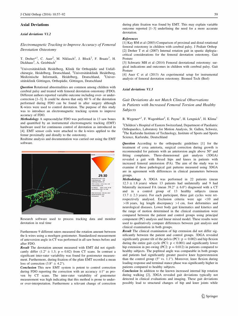
Methodology A supracondylar FDO was performed in 13 saw bones
and quantified by an instrumental electromagnetic tracking (EMT)
hardware used for continuous control of derotation as introduced in
[4]. EMT sensor coils were attached to the k-wires applied to the
femur proximally and distally to the osteotomy.
Realtime analysis and documentation was carried out using the EMT
software.
Research software used to process tracking data and monitor
derotation in real time
Furthermore 9 different raters measured the rotation amount between
the k-wires using a moeltgen goniometer. Standardized measurement
of anteversion angle in CT was performed in all saw bones before and
after FDO.
Result The derotation amount measured with EMT did not signifi-
cantly differ (1.2� ± 1.3; p = 0.82) from CT scans. In contrast a
significant inter-rater variability was found for goniometer measure-
ment. Furthermore, during fixation of the plate EMT recorded a mean
loss of correction (3.8� ± 4.2�).Conclusion This new EMT system is potent to control correction
during FDO reporting the correction with an accuracy ±1� as pro-
ven by CT scans. The inter-rater variability of goniometer
measurement was high indicating that this method is prone to under-
or over-interpretation. Furthermore a relevant change of correction
during plate fixation was found by EMT. This may explain variable
outcome reported [1–3] underlining the need for a more accurate
derotation.
References[1] Kay RM et al (2003) Comparison of proximal and distal rotational
femoral osteotomy in children with cerebral palsy. J Pediatr Orthop
[2] Dreher T et al (2007) Internal rotation gait in spastic diplegia–
critical considerations for the femoral derotation osteotomy. Gait
Posture
[3] Schwartz MH et al (2014) Femoral derotational osteotomy: sur-
gical indications and outcomes in children with cerebral palsy. Gait
Posture
[4] Auer C et al (2013) An experimental setup for instrumental
analysis of femoral derotation osteotomy. Biomed Tech (Berl)
Axial deviations V1.3
Gait Deviations do not Match Clinical Observations
in Patients with Increased Femoral Torsion and Healthy
Subjects
R. Wegener*1, F. Wagenblast2, E. Payne1, H. Lengnick1, H. Klima1
1Children’s Hospital of Eastern Switzerland, Department of Paediatric
Orthopaedics, Laboratory for Motion Analysis, St. Gallen, Schweiz,2The Karlsruhe Institute of Technology, Institute of Sports and Sports
Science, Karlsruhe, Deutschland
Question According to the orthopaedic guidelines [1] for the
treatment of coxa antetorta, surgical correction during growth is
recommended for patients with an antetorsion angle above 50� and
fitting complaints. Three-dimensional gait analysis (3DGA)
revealed a gait with flexed hips and knees in patients with
increased femoral antetorsion (FA). The aim of the study was to
examine if these pathological gait patterns measured using 3DGA
are in agreement with differences in clinical parameters between
groups.
Methodology A 3DGA was performed in 22 patients (mean
12.3 ± 1.8 years) where 13 patients had unilaterally and 9 had
bilaterally increased FA (mean 39.2� ± 6.0�) diagnosed with a CT
and in a control group of 13 healthy subjects (mean
13.7 ± 2.3 years). For each participant, three gait cycles were ret-
rospectively analysed. Exclusion criteria were age \10 and
[18 years, leg length discrepancy [1 cm, foot deformities and
neurological diseases. Lower body gait kinematics and kinetics and
the range of motion determined in the clinical examination were
compared between the patient and control groups using principal
component (PC) analysis and linear mixed model. These results were
used to qualitatively compare differences between gait analysis and
clinical examination in both groups.
Result The clinical examination of hip extension did not differ sig-
nificantly between the patient and control groups. 3DGA revealed
significantly greater tilt of the pelvis (PC1: p = 0.002) and hip flexion
during the entire gait cycle (PC1: p\ 0.001) and significantly lower
hip extension in pre-swing (PC2: p = 0.012) in patients compared to
healthy subjects. The popliteal angle was comparable in both groups
and patients had significantly greater passive knee hyperextension
than the control group (5� vs. 1.1�). Moreover, knee flexion during
loading response and terminal stance phase was significantly higher in
patients compared to healthy subjects.
Conclusion In addition to the known increased internal hip rotation
during walking [2], 3DGA revealed gait deviations typically not
detected in clinical evaluations and imaging. These gait deviations
possibly lead to structural changes of hip and knee joints while
J Child Orthop (2016) 10:57–92 59
123
walking with permanent internal hip rotation. Hence, 3DGA facili-
tates a differentiated decision-making concerning conservative or
surgical therapy and should be regarded as an essential diagnostic tool
in the orthopaedics guidelines.
References[1] Dt. Ges F (2002) Orthopadie und orthop. Chirurgie & BV d. Arzte
f. Orthopadie (Hrsg.), Leitlinien der Orthopadie, Dt. Arzte-
Verlag, Koln. http://www.leitliniensekretariat.de/files/MyLayout/pdf/
idiopathische_coxa_antetorta.pdf. Accessed 31 Dec 2014
[2] Bruderer-Hofstetter M, Fenner V, Payne E, Zdenek K, Klima H,
Wegener R (2015) Gait deviations and compensations in pediatric
patients with increased femoral torsion, J Orthop Res 155–62
Axial deviations V1.4
Guiding Growth in Fibular Hemimelia; Rebound of Valgus
Deformity Following Temporary Hemiepiphyseodesis
S. Marx*, S. Nader
Schon Klinik, Klinik fur Kinderorthopadie, Vogtareuth, Deutschland
Question Fibular hemimelia is a rare congenital defect of the lower
limb. Due to hypoplasia or aplasia of the fibular bone the patient
reveals numerous deformities of the limb such as shortening of the
tibia, knee instability and foot deformity. One of the major problems
is the valgus deformity of the knee. Guiding growth with temporary
hemiepiphyseodesis of the proximal tibia and/or distal femur is an
appropriate and easy way to cope with this deformity and has been
applied for many years. Radler et al. examined in 2011 recurrence
rates in fibular hemimelia after deformity correction, but did not
focus on recurrence after temporary hemiepiphyseodesis. The goal
of this study was to reveal the exact recurrence rates of valgus
deformity in children with fibular hemimelia depending on the
extent of congenital defect, degree of valgus deformity and age at
treatment.
Methodology Between January 2009 and January 2015 we monitored
28 patients aged 3–19 years with fibular hemimelia and deformity in
the knee which required correction by temporary hemiepiphyseodesis.
26 of these patients could be evaluated for this study. All of them
were evaluated with standing a.p. radiographs of the leg before
hemiepiphyseodesis, before explantation of 8-plates and at the point
of clinically obvious rebound. The X-rays were measured with the
TraumaCad program. We documented the development of MAD, the
mLDFA, the mMPTA and height of tibial epiphysis before
hemiepipyseodesis, at the point of explantation and at the point of
clinically obvious rebound.
All patients were treated with hemiepiphyseodesis by 8-plate
implantation either only at the proximal tibia and/or distal femur.
Result As we expected, the patients with higher severity of defect
such as aplasia showed a higher rate of rebound then the patients with
fibular hypoplasia. The younger the child at the point of first
implantation, the faster the rebound occurred. The amount of over-
correction did not influence the reoccurrence of deformity itself.
Conclusion Guiding growth by hemiepiphyseodesis is a well estab-
lished procedure to correct valgus deformity in patients with fibular
hemimelia. Patients and parents can be informed prior to treatment
that the deformity will reoccur dependent on the age and amount of
deformity.
In time of increasing lengthenings with intramedullary nails, this
information could also be of a value for obtaining the optimal con-
ditions before such intervention.
Axial deviations V1.5
Description and Evaluation of Operative Deformity
Correction in Calcium-Deficiency Rickets in North Nigeria
V. Wesselsky*1, P. Raab2
1Ev. Waldkrankenhaus Spandau, Abteilung fur Orthopadie und
Unfallchirurgie, Berlin, Deutschland, 2Konig-Ludwig-Haus, Lehr-
stuhl fur Orthopadie der Universitat, Wurzburg, Deutschland
Question Rickets is a recurrent disease especially in countries with
limited resources all around the world. Medical therapy including oral
calcium substitution are shown to improve a patients clinical symp-
toms as well as have an impact on deformities especially in the lower
extremity. In a literature review no existing reports about operative
deformity correction and its point of intervention in calcium defi-
ciency rickets could be found.
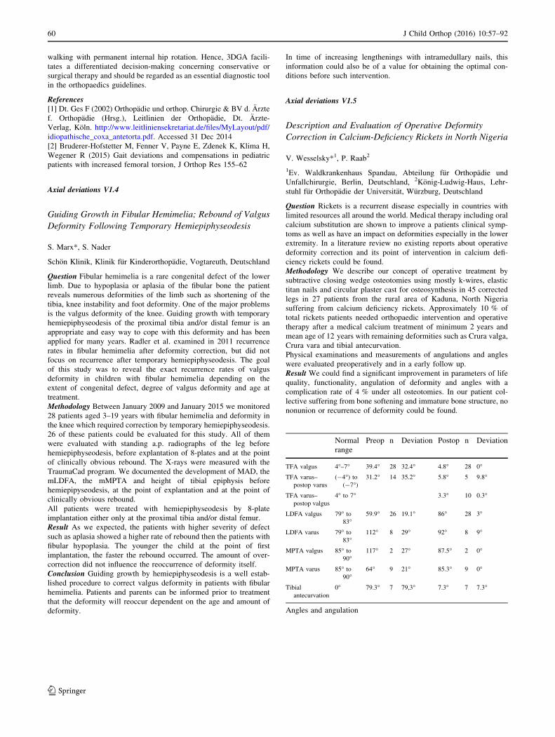
Methodology We describe our concept of operative treatment by
subtractive closing wedge osteotomies using mostly k-wires, elastic
titan nails and circular plaster cast for osteosynthesis in 45 corrected
legs in 27 patients from the rural area of Kaduna, North Nigeria
suffering from calcium deficiency rickets. Approximately 10 % of
total rickets patients needed orthopaedic intervention and operative
therapy after a medical calcium treatment of minimum 2 years and
mean age of 12 years with remaining deformities such as Crura valga,
Crura vara and tibial antecurvation.
Physical examinations and measurements of angulations and angles
were evaluated preoperatively and in a early follow up.
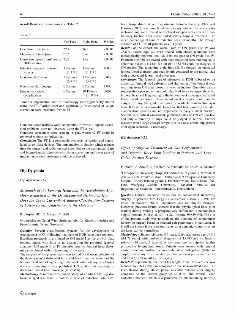
Result We could find a significant improvement in parameters of life
quality, functionality, angulation of deformity and angles with a
complication rate of 4 % under all osteotomies. In our patient col-
lective suffering from bone softening and immature bone structure, no
nonunion or recurrence of deformity could be found.
Normal
range
Preop n Deviation Postop n Deviation
TFA valgus 4�–7� 39.4� 28 32.4� 4.8� 28 0�TFA varus–
postop varus
(-4�) to
(-7�)31.2� 14 35.2� 5.8� 5 9.8�
TFA varus–
postop valgus
4� to 7� 3.3� 10 0.3�
LDFA valgus 79� to
83�59.9� 26 19.1� 86� 28 3�
LDFA varus 79� to
83�112� 8 29� 92� 8 9�
MPTA valgus 85� to
90�117� 2 27� 87.5� 2 0�
MPTA varus 85� to
90�64� 9 21� 85.3� 9 0�
Tibial
antecurvation
0� 79.3� 7 79,3� 7.3� 7 7.3�
Angles and angulation
60 J Child Orthop (2016) 10:57–92
123
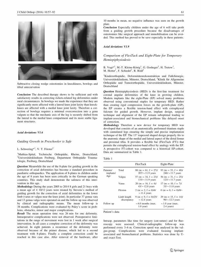
Subtractive closing wedge osteotomies in knockknees, bowlegs and
tibial antecurvation
Conclusion The described therapy shows to be sufficient and with
satisfactory results in correcting rickets-related leg deformities under
rural circumstances. In bowlegs we made the experience that they are
significantly more affected with a lateral knee joint laxity than knock-
knees are affected with a medial knee joint laxity. Therefore a cor-
rection of bowlegs requires a minimal overcorrection into a genu
valgum so that the mechanic axis of the leg is securely shifted from
the lateral to the medial knee compartment and its more stable liga-
ment structures.
Axial deviations V1.6
Guiding Growth in Preschooler is Safe
L. Schwering*1, V. T. Forster2
1Mathias-Spital, Technische Orthopadie, Rheine, Deutschland,2Universitatsklinikum Freiburg, Department Orthopadie Trauma-
tologie, Freiburg, Deutschland
Question Meanwhile the use of the 8-plate for guiding growth in the
correction of axial deformities has become a standard procedure in
paediatric orthopaedics. The application of 8-plates in children under
the age of 8 years has been seen critically in the German speaking
countries. This study shall demonstrate the safeness of this inter-
vention in this age.
Methodology During the years 2005 to 2014 8 girls and 21 boys with
a mean age of 4 10/12 years were treated by Stevens’s method of
guiding growth for the correction of axial deformities in the lower
limb (varus or valgus near the knee joint). In particular 37 genua vara
and 13 genua valga were operated on and the follow up was observed
by clinical and radiographic means. The mean follow-up is
38 months. Complications were evaluated by Paley’s score in prob-
lems, obstacles, minor and major complications.
Result The mean operation time was 26 min for one deformity.
Intraoperative complications were not observed. Postoperative limi-
tations in the range of movement were lost in 1 week after surgical
intervention. In all cases a complete correction of the deformity was
achieved. In eight patients a recurrence of the deformity were
observed because of the primer disease, which led to a second
treatment with 8-plates. Finally a complete correction could be
reached in this case also. After removal of the hardware, after
10 months in mean, no negative influence was seen on the growth
plate.
Conclusion Especially children under the age of 6 will take profit
from a guiding growth procedure because the disadvantages of
osteotomies like surgical approach and immobilisation can be avoi-
ded. This method has proved to be save especially in these patients.
Axial deviations V1.9
Comparison of FlexTack and Eight-Plate for Temporary
Hemiepiphysiodesis
B. Vogt*1, M.-T. Kleine-Konig1, G. Gosheger2, H. Tretow1,
M. Horter1, F. Schiedel1, R. Rodl1
1Kinderorthopadie, Deformitatenrekonstruktion und Fußchirurgie,
Universitatsklinikum, Munster, Deutschland, 2Klinik fur Allgemeine
Orthopadie und Tumororthopadie, Universitatsklinikum, Munster,
Deutschland
Question Hemiepiphysiodesis (HED) is the first-line treatment for
coronal angular deformities of the knee in growing children.
Modern implants like the eight-Plate (EP) solved many problems
observed using conventional staples for temporary HED. Rather
than creating rigid compression forces on the growth-plate (GP),
the EP creates a flexible tension-band effect with extraphyseal
fulcrum for guided growth. However, implant design, surgical
technique and alignment of the EP remain suboptimal leading to
implant-associated and biomechanical problems like delayed onset
of correction.
Methodology Therefore a new device for temporary HED was
developed that consists of an anatomically preformed titanium staple
with cannulated legs ensuring the simple and precise implantation
technique of the EP. The 13� trapezoid shaped design properly fits to
the anatomic shape of the medial and lateral aspect of the distal femur
and proximal tibia. It provides a flexible bar (FlexTack (FT)) that
permits the extraphyseal tension-band effect by analogy with the EP.
A prospective FT-cohort was compared to a historical EP-cohort.
Data are summarized in Table 1.
Table 1
FlexTack Eight-Plate
Patients/implants/Age
Total 88 (m = 61, f = 27)/207/*11.9 years
93 (m = 53, f = 40)/246/*11.7 years
Valgus 57 (m = 34, f = 23)/133/*11.9 years
56 (m = 31, f = 25)/123/*11.7 years
Varus 20 (m = 16, f = 4)/43/*12.0 years
17 (m = 10, f = 7)/33/*11.0 years
Flexion 2 (m = 2, f = 0)/6/*11.4 years
0 (m = 0, f = 0)/0/–
Limb lengthdiscrepancy
9 (m = 9, f = 0)/25/*12.4 years
29 (m = 15, f = 14)/90/*12.3 years
Follow up *6.0 months (max.1.0 year)
*1.0 year (max.2.4 years)
Patient’s data
Intraop. parameters like time for surgery (cut-suture) and for fluo-
roscopy were assessed. Clinical-radiographic follow-up was
performed every 3–6 m. Correction speed was analysed in the val-
gus-group. Complications were evaluated focusing implant-
associated and biomechanical problems. Statistics was done by U-
and exact-Test.
J Child Orthop (2016) 10:57–92 61
123
Result Results are summarized in Table 2.
Table 2
FlexTack Eight-Plate P value
Operation time [min] 22.4 36.8 \0.001
Fluoroscopy time [min] 0.26 0.42 \0.001
Correction speed [mm/month/
HED-location]
3.27 1.59 \0.001
Infection required revision
surgery
1 Patient
(1.1 %)
1 Patient
(1.1 %)
1000
Hematoma/effusion 5 Patients
(5.7 %)
3 Patients
(3.2 %)
0.488
Neurovascular damage 0 Patient 0 Patient 1.000
Implant-associated
complication
0 Patient 10 Patients
(10.8 %)
0.006
Time for implantation and for fluoroscopy were significantly shorter
using the FT. Earlier onset and significantly faster speed of valgus
correction were measured for the FT
Common complications were comparable. However, implant-associ-
ated problems were not observed using the FT as yet.
Complete corrections were seen in 16 pat., whose 47 FT could be
removed without complications.
Conclusion The FT is a reasonable synthesis of staples and cannu-
lated screw-plate-devices. The implantation is simpler which reduces
time for surgery and radiation exposure. Due to the anatomical shape
and biomechanical improvements faster correction and lower rates of
implant-associated problems could be achieved.
Hip Dysplasia
Hip dysplasia V2.1
Mismatch of the Femoral Head and the Acetabulum After
Open Reduction of the Developmental Dislocated Hip:
Does the Use of Currently Available Classification Systems
of Osteonecrosis Underestimate the Outcome?
R. Pospischill*, R. Ganger, F. Grill
Orthopadisches Spital Wien Speising, Abt. fur Kinderorthopadie und
Fusschirurgie, Wien, Osterreich
Question Several classification systems for the development of
osteonecrosis (ON) following treatment of DDH have been reported.
Excellent prognosis is attributed to ON grade I as the growth plate
remains intact, with little or no changes on the proximal femoral
anatomy. ON grade II to IV describe specific femoral head defor-
mities combined with a shortening of the neck.
The purpose of the present study was to find out if open reduction of
the developmental dislocated hip could lead to an overgrowth of the
femoral head and a lengthening of the neck with radiological changes
not corresponding to any published ON grades but resulting in
decreased lateral head coverage (mismatch).
Methodology A retrospective cohort study of children with hip dis-
location aged less than 12 months at time of reduction, who have
been hospitalized at our department between January 1998 and
February 2007, was completed. 49 patients satisfied the criteria for
inclusion and were treated with closed or open reduction with pre-
liminary traction after initial failed Pavlik harness treatment. The
mean patient age at time of reduction was 3.8 months. The average
duration of FU for all patients was 7.4 years.
Result For the cohort, the overall rate of ON grade I to IV was
25.8 %. Seven hips (29.2 %) treated with closed reduction were
radiologically abnormal and could be assigned to ON grade I to IV.
Fourteen hips (56 %) treated with open reduction were radiologically
abnormal but only six (24 %) out of 14 (52 %) could be assigned to
ON grades. The remaining eight hips (32 %) showed an increased
femoral head diameter and neck length compared to the normal side
with a decreased lateral head coverage.
Conclusion The femoral part of mismatch in DDH is based on an
aspherical femoral head deformity and shortening of the femoral neck
resulting from ON after closed or open reduction. Our observation
suggest that open reduction could also lead to an overgrowth of the
femoral head and lengthening of the femoral neck causing a decreased
lateral head coverage. These radiological changes could not be
assigned to any ON grades of currently available classification sys-
tems. It therefore is reasonable to assume that first, currently available
classification systems are not applicable in daily clinical practice.
Second, in a critical assessment, published rates of ON are too low
and only a minority of hips could be judged as normal. Further
research with a large-enough sample size to assess normal hip growth
after open reduction is necessary.
Hip dysplasia V2.2
Effect of Surgical Treatment on Gait Performance
and Dynamic Knee Joint Loading in Patients with Legg–
Calve–Perthes Disease
F. Stief*1, S. Adolf2, L. Kremer2, A. Schmidt3, M. Brkic2, A. Meurer2
1Orthopaedic University Hospital Friedrichsheim gGmbH, Movement
Analysis Lab, Frankfurt/Main, Deutschland, 2Orthopaedic University
Hospital Friedrichsheim gGmbH, Frankfurt/Main, Deutschland, 3Jo-
hann Wolfgang Goethe University, Frankfurt Initiative for
Regenerative Medicine, Frankfurt/Main, Deutschland
Question Current outcome evaluations of containment improving
surgery in patients with Legg–Calve–Perthes disease (LCPD) are
based on standard clinical parameters and radiological changes.
However, previous results showed that the physiological knee joint
loading during walking is preoperatively shifted into a pathological
valgus moment [Stief et al. (2014) Gait Posture 39:859–64]. The aim
of the present study was to evaluate the outcome of containment
improving surgery based on selected gait parameters. In particular, it
is still not known if the preoperative existing dynamic valgus thrust of
the knee can be normalised.
Methodology Sixteen children [14 male, 2 female; mean age of 6.1
(±1.7) years] with unilateral diagnosis of LCPD and 19 healthy
subjects (14 male, 5 female) at the same age participated in this
prospective longitudinal study. Patients were treated with femoral
varus osteotomy, isolated or in combination with pelvic (Salter or
Triple) osteotomy. Instrumented gait analysis was performed before
and 13.4 (±2.5) months after surgery.
Result Postoperatively, the mean leg length of the involved side was
reduced by 0.6 (±0.8) cm compared to the non-involved side. The
knee flexion during stance phase was still reduced after surgery
compared to the control group (p\ 0.001). The external knee
adduction moment, which is a parameter for characterizing intrinsic
62 J Child Orthop (2016) 10:57–92
123

compressive load in the knee joint, was postoperatively still different
in comparison to healthy subjects at the same age (p = 0.049).
Conclusion Despite predominantly good results shown by the stan-
dard clinical and radiographic examinations for the hip joint, gait
analysis detected various functional deficits at the level of the knee
after surgery. Patients displayed stiff knees on the affected side,
probably to compensate for leg length discrepancy. This altered gait
pattern might be one explanation for the valgus thrust of the knee,
which could be sufficient to deform the lateral compartment or
influence the remaining growth plate and the physiological develop-
ment of the mechanical axis of the leg in young patients with LCPD.
A further explanation for this pathological knee joint loading may be
the result of the surgery due to a change of the coronal plane align-
ment of the proximal femur. In conclusion, lower limb alignment and
dynamic knee joint loading should be controlled during the process of
the disease and the postoperative rehabilitation period to avoid
degenerative changes in the lateral knee compartment in young
patients with LCPD.
Hip dysplasia V2.3
Decreased Anterior Femoral Neck Off-Set’’ After In Situ
Pinning for Slipped Capital Femoral Epiphysis (SCFE):
Correlation with Clinical Symptoms and Range of Motion
of the Hip Joint
K. Mladenov*, K. Vedder, A. Protzel, U. von Deimling
Asklepios Kinderklinik, Sankt Augustin, Deutschland
Question: It has been suggested that decreased anterior femoral neck
off-set may produce ‘‘cam’’ like impingement and cause hip joint pain
and arthrosis. However the correlation between radiologic findings
and clinical range of motion of the hip joint has been very sparsely
discussed in the literature.
Purpose: To study the clinical correlation between radiologically
decreased anterior femoral neck off-set and clinical findings with
focus on hip pain and range of motion.
Methodology: Retrospective single center study.
The inclusion criteria were: patients with mild to moderate SCFE,
treated with ‘‘in situ’’ pinning and followed up for a minimum of
2 years after surgery. The anterior femoral neck off set was evaluated
on standardized lateral hip X-rays by means of the ‘‘alpha’’ angle as
described by Notzli. Values of over 50� were considered as patho-
logical. Clinical data for hip complaints, function and range of motion
were collected from the patient charts. The Harris Hip score was
calculated for every patient. Hips having normal anterior off set
(Group I) were compared with the unaffected side and with those
having decreased off set (Group II). Focus was placed on Hip flexion,
internal rotation and patients complaints.
Result Forty six patients met the inclusion criteria, (22 Group I, 24—
Group II). Harris hip score was identical in both groups (99.8 vs. 98.7
points). Hip flexion of affected hips with decreased anterior off set, of
affected hips with normal anterior off set and of the unaffected hips
was also identical (127.6� vs. 129.7� vs. 130.6�, respectively). Internal
rotation of affected hips with decreased anterior neck off set was
significantly lower compared to affected hips with normal anterior off
set and to unaffected hips (23.3� vs. 36.9� vs. 37�, respectively) and
this was independent from the degree oft he initial slip.
Conclusion: Decreased anterior femoral neck off set leads to limited
internal rotation of the hip joint. On the short term there are no
negative effects on hip function and complaints. The correlation
between decreased anterior neck off set, decreased internal rotation,
femuro-acetabular impingement and hip osteoarthrosis should be
studied on the long term, since it make take many years for symptoms
to develop.
Hip dysplasia V2.5
Results of Percutaneous Musculotendinous Release
in Children with Hip Dysplasia Secondary to Cerebral
Palsy Aged Under 6 Years
V. Gattung*, P. Bernius, M. Poschmann
Schon Klinikum Munchen Harlaching, Zentrum fur Kinder- und
Neuroorthopadie, Munchen, Deutschland
Question Beside other factors elevated muscle tonus can lead to hip
dysplasia with subsequent luxation in cerebral palsy. Especially hip
adducting, hip flexing and medial knee bending muscles play a main
role in developing and maintaining a hip dislocation. Due to the
maturation potential of the hip, in children younger than 6 years
conservative methods like botulinum toxin therapy, physiotherapy or
special orthoses may improve centering of the hip. But hips with a
Reimers migration index (RMI) more than 40 % require surgical
treatment.
In children with cerebral palsy we prefer percutaneous muscle release
because we consider it a minimal invasive method with low risks and
good results. Therefore we studied the influence of this method on the
RMI.
Methodology We retrospectively examined 43 patients suffering from
cerebral palsy from 2 to 6 years (mean 4.04 years) and 55 hips. The
severity of cerebral palsy was classified according to gross motor
function classification system (GMFCS). Patient’s GMFCS ranged
from 3 to 5. Inclusion criteria were children aged under 6 years with
an RMI more than 25 %. RMI was measured in X-rays pre- and
postoperatively in a follow up of 11.9 months (0–36 months).
Depending on clinical examination of the patients being under
anaesthetic immediately before operation, we performed percuta-
neous release of hip adducting, superficial hip flexing or medial knee
bending muscles or combinations of these.
Result Altogether RMI could be improved from 42.5 to 37.8 %.
Concerning hips with a RMI from 25 to 39 % (group A, n = 22) RMI
could be improved only slightly (from 30.8 to 29.8 %), whereas hips
with a RMI over 40 % (group B, n = 33) RMI could be improved
from 50.2 to 43.1 %.
In group A the RMI worsened in 5 hips (22.7 %), stayed equal in 11
hips (50 %) and improved in 6 hips (27.3 %).
In group B the RMI worsened in 4 hips (12.1 %), stayed equal in 12
hips (36.4 %) and improved in 17 hips (51.5 %).
In GMFCS 3 patients, RMI could be improved from 37.1 to 33.6 %,
in GMFCS 4 patients from 42.1 to 33.8 % and in GMFCS 5 patients
from 46.9 to 45.8 %.
J Child Orthop (2016) 10:57–92 63
123

Conclusion Soft tissue balancing is an efficient possibility to avoid
further hip migration in children with cerebral palsy aged under
6 years. Although operative hip reconstruction might be necessary in
future, early percutaneous muscle release can reach a better situation
for further reconstructive surgery.
Hip dysplasia V2.6
Subtrochanteric Femur Fracture as Serious Complication
After Operative Stabilization of Slipped Capital Femoral
Epiphysis: A Reason Not to Operate the Non-Affected Hip?
H. Lengnick*, B. Schultz, P. Gerhard, R. Wegener, H. Klima,
E. Payne
Ostschweizer Kinderspital, Abteilung Kinderchirurgie, St. Gallen,
Schweiz
Question Slipped capital femoral epiphysis (SCFE) is a common
problem in adolescents making operative stabilization most often
necessary. Slipping of the contralateral (cl) side is described in
20–60 %, while operative treatment is unclear and controversially
discussed 1, 2.
Reported complications after operative treatment in SCFE are chon-
drolysis, femur head necrosis, premature closure of the epiphysis,
infection, implant failure, but not subtrochanteric femur fracture 3.
We present the data of a patient cohort to show complication risk after
operative treatment in SCFE and discuss necessity of operative sta-
bilization of the contralateral side.
Methodology Operative treatment of SCFE was performed in 22 hips
of 17 patients (male n = 10, female n = 7) by four different ortho-
paedic surgeons. On average patients were 12.7 (±1.6) years old, 14
showed SCFE acuta, four chronic and four acute on chronic SCFE.
Prophylactic stabilization of the contralateral side was done in 12 hip
joints. Implants for osteosynthesis after closed reduction (slipping
\40�) were Hansson-Pins (SCFE n = 7, cl n = 6), K-wires (SCFE
n = 10, cl n = 4) and single screw (SCFE n = 5, cl n = 2). Open
reduction ([40�) was necessary in two patients.
Result Overall nine complications (23 %) have been shown: sub-
trochanteric fractures after adequate trauma in five (13 %), K-wire
overlength in three (8 %) and loosening in one patient (3 %). Within
the group with subtrochanteric fractures three patients (60 %) showed
fracturing of the contralateral side. Concerning subtrochanteric femur
fractures Hansson Pins showed the highest rate of fractures in three
patients (60 %). Fracture risk was highest within the first 2 months
postoperatively.
Conclusion Subtrochanteric femur fracture is a serious complication
after operative stabilization of SCFE. In our study fracture risk after
stabilization of the contralateral side is comparable with the SCFE
side. Possible reasons for this are inadequate operation technique with
impairment of the lateral corticales by multiple drilling, patient’s
constitution and lacking of compliance.
Due to fracture risk for the proximal femur after contralateral stabi-
lization we recommend to carefully evaluate the indication for
operation of the contralateral hip taking pain, patient compliance,
surgeons’ experience and available implant into account.
References[1] Jerre R et al (1994) JBJS 76:563–7
[2] Koczewski P (2001) Chir Ortop Pol 66:357–64
[3] Hefti F (2006) Springer Verlag, 2.Auflage, S.219–26
Hip dysplasia V2.8
Hip Morphology in MPS 1-H Patients After Hematopoietic
Stem Cell Transplantation
S. Breyer*1, N. Muschol2, M. Schmidt2, K. Babin1, M. Rupprecht1,
K. Ridderbusch1, R. Stucker1
1Altonaer Kinderkrankenhaus, Kinderorthopadie, Hamburg,
Deutschland, 2UKE, Padiatrie, Hamburg, Deutschland
Question The purpose of this study was to determine the morphology
of the acetabulum and the head of the femur in MPS-1H patients
using magnetic resonance imaging (MRI) and plain radiographs of the
pelvis. Information should help in the preoperative decision making
process.
Methodology A retrospective review of patients with MPS-1H was
performed. 32 hips were analyzed. Radiographic evaluation of the
pelvis was performed on all patients, MRI in eight cases. Hip dys-
plasia was quantified by acetabular index (AI) as described by Tonnis
(Tonnis 1976). As an index of hip stability, the migration percentage
of Reimers (MP) was used. (Reimers 1980). The measurements
include AI for the labrum, cartilaginous and bony landmarks and MP
for the bony and soft tissue coverage. Bone AI on MRI was compared
with the same angles on the radiographs.
Result The average age at the time of radiography was 5.0 years. The
mean AI in radiography was 36.1�. The mean migration percentage in
the radiographic view was 60.6 %. In the MRI group the average age
was 6.4 years. The mean radiological AI was confirmed in MRI
measurements. The average C-AI was 20.6�. It decreased even more
in consideration of the labrum. No difference was seen in the
migration percentage of Reimers. As a sign of instability the MP of
Reimers in plain radiography, was increased in all hips. The soft
tissue coverage was measured in the MP and showed a decreased
level of instability. The mean MP corresponding to the cartilage
coverage was 45.3 %. In consideration of the labrum, the MP
decreased down to levels of 27.5 %.
Conclusion Our results establish that patients with MPS-1H have a
three times greater cartilaginous coverage then healthy kids. Only
radiological criteria can leads to misunderstanding the morphology.
MRI measurements can document the cartilaginous coverage and help
in decision making processes. Our findings suggest that MRI pro-
motes more accurate selection of children for pelvic reconstruction.
Further multicentric studies have to confirm this.
Hipmorphology
64 J Child Orthop (2016) 10:57–92
123

Hip dysplasia V2.9
Gait Deviations After Perthes Disease in Dependence
of the Radiological Outcome
B. Westhoff*, A. Muller-Reinartz, L. Hegemann, C. Zilkens,
D. Rosenthal, R. Krauspe
University of Duesseldorf, Medical Faculty Department of Ortho-
paedics, Duesseldorf, Deutschland
Question Depending on the final extent of hip deformity Perthes
disease will lead to early osteoarthritis. The aim was to analyze the
functional outcome and to correlate the results with the radiological
outcome.
Methodology 30 adults (mean age 31 + 11 years) treated for unilat-
eral LCPD were investigated clinically, radiographically and by 3D-
gait analysis. The clinical result was rated by the Harris Hip Score
(HHS). According to the Stulberg-rating system patients were divided
in 2 subgroups: group 1 = spherical joint (Stulberg type 1 and 2,
n = 16), group 2 = aspherical joint (Stulberg type 3–5, n = 14). 3D-
gait-analysis was performed with a VICON 512 system. Patients
walked at a self-selected speed–barefoot. Spatiotemporal, kinematic
and kinetic parameters were evaluated and compared to a group of
normal adults (n = 40, average age 28.0 years). Statistical analysis
was performed by the Mann–Whitney-U test for independent sam-
ples; the correlation within in the patients group was performed using
an ANOVA-analysis and the Kruskal–Wallis-test for non-parametric
samples; a p value of less than 0.05 was considered to indicate sta-
tistical significance.
Result Patients with a spherical joint showed a significantly better
clinical results (HHS 89 vs. 78 p., p = 0.019) and less degenerative
changes. Gait deviations were more severe in group 2: asymmetry
related to the duration of stance phase was more pronounced; in
sagittal plane kinematics ROM of the hip and knee joint were
reduced, asymmetry between involved and non-involved side was
more pronounced too; in frontal plane ROM of the pelvis and trunk
was reduced as well; power generation at the hip joint was impaired
as well.
Conclusion Gait analysis after LCPD showed significant deviations
of the gait pattern in comparison to the controls with loss of sym-
metry. These are part of an unloading mechanism. The deviations are
more pronounced in case of a poor radiological outcome. Further
studies are necessary to determine functional predictors for the
development of secondary osteoarthritis which may than be influ-
enced by conservative or surgical treatment options.
Compared to early follow-up examinations after LCPD (Westhoff, Int
Orthop 2011) gait deviations are more pronounced in early adulthood.
Best Lectures
Best lectures V-Top
Treatment of Congenital Wry–Neck (Torticollis) by Dreiss’
Method and Computer Aided Analysis of the Face
Asymmetry
L. Schwering*1, V. T. Forster2
1Mathias-Spital, Technische Orthopadie, Rheine, Deutschland,2Universitatsklinikum Freiburg, Department Orthopadie Trauma-
tologie, Freiburg, Deutschland
Question The treatment of wry neck changed from the first operative
intervention by Isaac Minnius 1642 to modern pediatric orthopedic
concepts. In allusion to Foederl’s approach G. Dreiss developed a
concept for the treatment of this deformity in the early seventies of
the last century. The purpose of this investigation is to demonstrate
the efficiency of the procedure and the control of the scoliosis of the
skull by graphic programs (Corel Draw 12 and Adobe Photoshop
CS4).
Methodology In the pediatric orthopedic practice wry neck is pri-
mary treated by conservative means like physiotherapy. The success
rate is reported to be 90 % and more (H. Binder 1987). During the
last 10 years 14 children (9 boys and 5 girls) had to be treated
operatively finally. The mean age at operation was 6 years and
4 months. The Dreiss’ procedure consists from triterminal tenotomy
of the sternocleidomastoid muscle in a special way. Postoperative a
removable diadem thermocast was applied for four to 6 weeks
depending on the patients age and the severity of the deformity. A
digital picture of the patients face was documented pre- and post-
operatively. After dividing the face in the midline two new ‘‘faces’’
from two left and two right parts were created. Using this graphic
controlled method the improvement of the asymmetry of the face
was detected.
Result The mean follow up was 7 years and 10 months. During the
operation no complication (especially damage to the facial nerve) was
observed. Range of movement to the restricted side improved but
could not reach the same range of motion as on the other side in
children older than 6 years at the intervention. The results of com-
puter aided analysis illustrated a harmonization between the two
artificial faces.
Conclusion The Dreiss’ procedure in the treatment of wry neck
proved to be an excellent method compared with the existing litera-
ture. With the number of patients it might be useful to operate
children with wry neck under the age of six to reach a complete cure.
The computer aided analysis is a noninvasive tool that helps to show
the improvement of facial asymmetry.
Best lectures V-Top
Criteria for Successful Arm-Prosthetic Supply
in Childhood and Adolescence
M. Horter*1, J. Thormann2, G. Gosheger3, H. Kirch2, R. Rodl1,
H. H. Wetz2
1Kinderorthopadie, Deformitatenrekonstruktion und Fußchirurgie,
Universitatsklinikum, Munster, Deutschland, 2Klinik fur Technische
Orthopadie und Rehabilitation, Universitatsklinikum, Munster,
Deutschland, 3Klinik fur Allgemeine Orthopadie und Tumor-
orthopadie, Universitatsklinikum, Munster, Deutschland
Question Which criteria have to be considered to achieve a successful
arm-prosthetic supply? What do acceptance and compliance of
prosthetic devices in childhood and adolescence depend on? Does
family influence the children’s attitude towards disability and
acceptance of the prosthesis and how could this effect be used for
therapy in a positive way?
Do children even need prosthetic devices?
Methodology We examined 51 patients (0–16 years at first contact,
21 girls, 30 boys) based on a retrospective data analysis and a self-
made questionnaire for children and their parents. Defined criteria
were: sex, diagnosis, kind of prescription, age of child at the date of
first contact with the clinic and of first prosthetic supply, wearing
behaviour and wearing time in hours per day, reasons for rejection,
stigmatisation of parents and their children, selective wearing of the
J Child Orthop (2016) 10:57–92 65
123
prosthesis, the effect of group behaviour on other children, and pro-
posals for improvement.
Result The patient’s sex shows influence of the arm-prosthetic
acceptance: 75 % of the female patients use their prostheses, but
only 33 % of the male patients. Amputated patients wear their
prostheses more often than patients with congenital deformities. The
average age at the date of first prosthetic supply was 4.6 years. The
success rate decreases mainly at the male patients with increasing
age of first prosthetic supply. Besides the type of prescription,
stigmatisation of the parents as well as the child’s environment
shows a latent impact on compliance and consequent acceptance of
the prosthetic device. The results of this study are congruent with
current literature.
Conclusion Orthopaedic specialists, parents and the children should
decide together about best time and need of prosthetic supplies. This
study reveals the difficulty in finding Methodology criteria, which
promote the acceptance. The environment and the education of the
child show major effects. It is not possible to generalize if the supply
of a child with a prosthetic device is absolutely necessary, but the
child should have the opportunity to decide on its own. Current lit-
erature especially emphasizes the amenities of prostheses concerning
the physical and psychological development. Apparently the practical
experience differs: Half of the children do not even wear the pros-
thetic device.
Even tests published in preceding studies that intend to set up a
classification system for the prosthesis wearing behaviour do not
legitimise the rejection to supply a child with prosthetic device.
Best lectures V-Top
Experiences with Clubfoot Therapy in Older Children
in Germany
U. Bruckner*1, A. Reeg2, B. Schnuck1, M. Schulte1
1Agaplesion Diakonieklinikum Rotenburg, Kinderorthopadie, Roten-
burg/Wumme, Deutschland, 2Stiftung Feuerkinder, Berlin,
Deutschland
Question In 2012 the Deutsche Gesellschaft fur Orthopadie und
Orthopadische Chirurgie (DGOOC) introduced revised guidelines for
the treatment of clubfoot (cf). These guidelines include the Ponseti
therapy as a standard for newborns but also focuses on the treatments
of relapses in children up to the age of 3 years.
However, in German-speaking countries there exist no guidelines for
the therapy of untreated clubfoot cases and relapses for older children
([3 years of age). As a result, clubfoot therapy is applied on a case-
by-case basis.
Methodology The aim of this paper is to identify and adopt a national
guideline for the therapy of clubfeet in children older than 3 years of
age. In order to gain insights on the current practices, standards as
well as the short- and long-term treatment results of clubfoot therapy
in Germany we carried out expert interviews in several orthopaedic
departments specialised on children.
Result The best way of treatment seems to be the initial Ponseti
therapy combined with minimal surgery, depending from the result of
the casting.
Conclusion In line with international experiences our data suggests
that the standard Ponseti therapy offers the most promising treat-
ment results in cases of older children. We therefore recommend
the DGOOC guidelines to be extended to children older than
3 years.
Best lectures V-Top
A Critical View on In-Patient Treated Hip Luxation
in the Era of Ultrasound Screening
A. Postler*, K.-P. Gunther, F. Thielemann
UniversitatsCentrum fur Orthopadie und Unfallchirurgie, Dresden,
Deutschland
Question In spite of the dutifully performing ultrasound screening in
Germany there have been repeated cases of dislocated hips, which
aren’t be detected, even treated on time. The aim of the current study
was therefore to investigate the courses of those children, who need
an in-patient treatment.
Methodology In this retrospective cohort study 74 children (62 %
female, 12 % male) with hip dislocation, who needed an in-patient
treatment between January 2001 and February 2014 were included.
Children with neuropathic, myopathic or teratological luxation of the
hip were excluded.
Main risk factors, date of first ultrasound screening, kind of pre-
treatment and the subsequent procedures were recorded.
Result Four children got their first ultrasound screening delayed after
the 6th week of life and six children had no screening at all.
54 Children (73.0 %) were advised after the 6th week of life. 43 patients
had a previous treatment for the hip dislocation, three in foreign
countries. Prior treatment involves therapy by various bandages
(41.9 %), overhead-extension (5.4 %), spica cast (1.4 %), multiple
forms of noninvasive therapy (12.2 %) or even frustrating surgical
procedures (4.1 %).
In 60.8 % (n = 45) an initial closed reduction and spica cast immobi-
lization for 12 weeks was successful. Five of those children (6.8 %)
needed a two-step closed reduction. In 39.2 % (n = 29) of the children an
open reduction and postoperative immobilization was performed.
Because of persistent dysplasia of the acetabular roof and instability
24 children (12 patients from the closed reduction group, 12 patients
from the open reduction group) needed further operative procedures.
Those secondary operative procedures were usually performed after a
period of prolonged conservative treatment at an average age of
27.6 months (range 10–74 months).
Conclusion The results of the current study emphasize the importance
of the early ultrasound screening and adequate therapy in cases of a
positive family history and breech position. Delayed detection of the
hip dislocation without any prior treatment (32.4 %, n = 24) was the
main factor for open reduction (75.9 % of all open reduction cases),
respectively secondary surgical procedures to stabilize the hip.
It should be assumed that a frustrating previous treatment will be a
further reason for any necessary in-patient management. An experi-
enced out- and in-patient treatment is therefore mandatory.
Axial Deviations/Limb Lengthening
Axial deviations/limb lengthening V4.2
Principle, Indication and Midterm Results of the Femoral
Intertrochanteric Valgus Osteotomy in Legg–Calve–
Perthes Disease
B. Heimkes*, B. Weiß, C. M. Ziegler, S. Utzschneider
Klinikum der Universitat (LMU), Campus Großhadern, Klinik fur
Orthopadie, Physikalische Medizin und Rehabilitation, Munchen,
Deutschland
66 J Child Orthop (2016) 10:57–92
123
Question Occurrence of hinge abduction is a negative prognostic
factor in Legg–Calve–Perthes disease. For this problem Catterall
proposed femoral valgus (extension) osteotomy as a salvage
procedure.
Purpose of the retrospective study was to evaluate the indications and
results of this procedure in our patients with Legg–Calve–Perthes
disease.
Methodology 28 patients who underwent the procedure between 2004
and 2013 were examinated clinically and radiologically at an average
of 5.5 years (range 1.0–10.5 years) after surgery.
Result Indication. The mean age at surgery was 10.7 years (range
6.2–16.3 years). 96.4 % of the patients were pretreated conserva-
tively, 10.7 % additionally by pelvic osteotomy, 25.0 % by varus
femoral osteotomy and 3.6 % by tissue release. Staging at time of
procedure was: 10.7 % condensation, 50.0 % fragmentation, 14.3 %
reossification and 25.0 % remodelling stage. Grading: 0 % were
classified Herring A, 50.0 % Herring B and 25 % Herring C. 25.0 %
could not be classified. Follow up: (1) The hip abduction increased
30.0� (range -2.5� to 75.0�) from preoperative 15.0� (range -30.0�to 50.0�) to postoperative 40.0� (range 15.0� to 65.0�). (2) The
average neck-shaft angle changed from preoperative 129.5� (range
107.8�–149.6�) to postoperative 149.0� (range 131.6�–168.1�). (3)
Final radiographic outcome according to Stulberg: Type II deformity
7.1 %, Type III 39.3 %, Type IV 21.4 %, Type V 7.1 %. 25.0 % of
the patients could not be classified yet.
Conclusion According to the results femoral valgisation is recom-
mended both as a salvage procedure after failed previous osteotomies
as well as a first procedure for hips with hinge abduction.
Axial deviations/limb lengthening V4.3
Guided Growth of the Infant’s Skull: A Pediatric
Orthopedic Method?
H. Willenborg*, S. Martin
Orthopadie der MHH im Annastift, Kinderorthopadie, Hannover,
Deutschland
Question Guided growth of the infants skull—a pediatric orthopedic
method?
How long should we observe the development of an asymmetrical
development of the infants skull? When should we intervene by
guiding growth? In which cases is the conservative approach not
indicated? When should we proceed to surgery?
Methodology Infants are impeded in their healthy development by
asymmetry. This deformity may lead to life-long problems.
Asymmetry in infants is often multifactorial and can involve the
entire body.
Individual aspects can aggravate each other: asymmetrical positioning
of the feet, impeded hip abduction (with or without DDH), asym-
metrical hip rotation, reversible blockages of the pelvis, the sacro-
iliac joint or the upper cervical spine with or without wry neck etc.
If the infant is denied the variation of positioning during the first
months of life and is constantly kept on its back (not only during sleep
as advised by the German association for sleep medicine) a defor-
mation of the soft skull may result. Non-orthotic treatment can be of
help up to the age of 6 months. Because of the hardening of the skull
there is no realistic chance of achieving symmetry ‘‘spontaneously’’
beyond this age.
Depending on the severity of the skull deformity, guided growth
through helmet therapy can be initiated as early as an age of 4 months
(or later) to achieve symmetry for the skull, the face and the axis of
the ears.
Result Different methods are needed for the treatment of the vicious
circle of ‘‘asymmetry syndrome’’ (positioning, chiropractic, physio-
therapy, bandaging, casting etc.). Pediatric orthopedists should be
enabled to perform a holistic assessment and examination of the
infant. Helmet therapy does not only lead to a symmetrical shape of
the skull but often also contributes to a resolution of the more com-
plex ‘‘asymmetry syndrome’’.
On principle a cranio synostosis cannot be treated by helmet therapy.
In severe cases this malformation is an indication for surgery. If
synostosis and positioning problems occur together, both: conserva-
tive and surgical approaches, can be combined.
Conclusion Helmet therapy is a very valuable contribution to pedi-
atric orthopedic treatment.
Axial deviations/limb lengthening V4.4
Correction of Static Axial Alignment in Children with Knee
Varus or Valgus Deformities Through Growth Guidance:
Does It Also Correct Dynamic Frontal Plane Moments
During Walking?
H. Bohm*1, F. Stief2, K. Sander3, C. Multerer1, L. Doderlein1,
M. Hosl1
1Orthopadische Kinderklinik, Kinderorthopadie, Aschau, Deutsch-
land, 2Orthopaedic University Hospital Friedrichsheim gGmbH,
Movement Analysis Lab, Frankfurt/Main, Deutschland, 3Universitat
Jena, Waldkrankenhaus GmbH, Eisenberg, Deutschland
Question Malaligned knees are predisposed to the development and
progression of unicompartmental problems because of the excessive
load placed on one side of the knee. Therefore guided growth in
skeletally immature patients is recommended. Indication for correc-
tion of varus/valgus deformities are based on static radiographs.
However the internal knee abduction moment, a valid marker of
mechanical wear at the knee joint during walking, showed only weak
correlation to malalignment determined by static radiographs. So far,
none of the studies about guided growth reported the effect on the
loading situation during walking. This is astonishing since the pro-
cedure aim to prevent excessive load placed on one side of the knee.
Therefore, the aim of the study was to measure the effects of growth
guidance on the normalization of frontal plane knee joint moments
during walking. The hypothesis was that the change in dynamic
moments and the change in static alignment are closely related.
Methodology 8 patients (11–15 years) with idiopathic axial varus or
valgus malalignment participated. 16 typically developed peers
served as controls. Gait analysis and clinical assessment were per-
formed the day before implantation and explantation of eight plates.
The static mechanical tibiofemoral axis angle (MAA) was calculated
from a captured standing trials and radiographs. The dynamic frontal
plane knee moments were calculated as the average over the stance
phase of gait. A total of 15 legs was individually analyzed pre and
postoperatively with respect to controls. Correlation between static
MAA and dynamic frontal plane knee joint moments and their change
by guiding growth were performed.
Result The changes in dynamic knee moment in the frontal plane
following guiding growth showed excellent and significant correlation
to the changes in static MAA (R = 0.97, p\ 0.001), so that the
hypothesis of the study turned out to be correct. Contrary to the
correlation of the changes, there was no correlation between static and
dynamic measures in both sessions. This can be negative when a
natural loading situation before treatment turned into a pathological
one after treatment.
J Child Orthop (2016) 10:57–92 67
123
Conclusion In conclusion guiding growth has a predictable effect on
the dynamic load situation during walking. Gait analysis might be
useful to assess the preoperative load distribution during walking; it
might reveal individual gait pathologies that might further affect
dynamic joint moments in cases where the static situation has been
already corrected.
Axial deviations/limb lengthening V4.5
Guiding Growth Supports Correction of Equinus
in Residual Paediatric Clubfoot
V. T. Forster*1, L. Schwering2
1Klinik fur Orthopadie und Unfallchirurgie, Universitatsklinikum,
Freiburg, Deutschland, 2Mathias-Spital Rheine, Technische Ortho-
padie, Rheine, Deutschland
Question After clubfoot correction pathologic ankle joint angles may
remain and result in unphysiologic gait with extended forefoot load.
Neither a new release nor tendon lengthening can solve this problem.
The purpose of this clinical trial is the answer to the question if
guiding growth can be useful in the correction of this pathology of the
tibial pilon.
Methodology Eight boys and two girls with 16 feet in equinus posi-
tion because of pathologic joint surface angles of the distal tibia
between 95� and 100� were treated by the means of guiding growth.
Five boys presented seven previous treated idiopathic clubfeet, one
boy after achilles tendon lengthening (ATL) due to muscular dys-
trophy in both feet and one girl also after ATL because of congenital
arthrogryposis multiplex. To improve gait and make the wear of
splints easier one 8-plate was applied strictly in ventral position of the
distal tibial growth plate. Patients files and radiographs were
evaluated.
Result During a mean follow up of 2 years an 17 months and a mean
duration of the 8-plate in situ of 20 months a relevant improvement of
the distal tibial joint surface angle near 80� (Paley) was observed. In
one boy the correction was incomplete in two feet because of maturity
ahead of time. In one boy’s foot a moderate overcorrection was
achieved voluntarily. Neither during the implantation of the 8-plate
nor during the hardware removal any complication was detected. The
mean operation time from incision to suture lasts 33 min/8 plate.
Conclusion Guiding growth seems to be a useful tool in the correc-
tion of enhanced distal tibial joint surface angles in children. Because
of the minimal invasive approach and immediate full weight bearing
postoperative this seems to be suitable for children. This intervention
can restore foot function by normalization of tibial pilon. Meticulous
preparation and exact application of the 8-plate are prerequisites for a
trial free from complications.
Axial deviations/limb lengthening V4.6
Lengthening and Correction of Axial Deviations
in Children
M. Schmidt*, G. Salameh
Waldhofzentrum, Kinderorthopadie, Kronberg, Deutschland
Question Limb lengthening and deformity correction in children has
in most cases obstacles and complications during treatment like axial
deviations s, knee flexion contracture, drop foot and deformity in
addition to congenital and acquired malalignment. This need a special
method and strategy for successful treatment
Methodology The external fixation hinge system (Salahmehfix) is an
arch hinge system with various diameters to assemble the shape of the
extremity with special connecting hinges to correct axial and rota-
tional deviations.
The stability of the simple functional arc system is high, it has a good
tolerance by the young patients.
It can be modified during the treatment if needed to achieve full
restoration of the extremity.
Result From 1995 to 2013 this system was used in 548 children with
different indications in the lower and upper limbs. They presented
with axial deviations, limb length discrepancies and combined
deformities.
Results were excellent in 312 cases, good in 164 cases, fair in 84
cases and 6 had poor outcome.
Complications were mainly superficial pin infection, pin breakage,
but no nerve or vascular complication was seen.
Conclusion The new developed external fixation system allows the
correction of Axial deviations, length discrepancy, contractures and
even combined complex deformities.
The new system provide high stability, high tolerance by the children,
fast weight bearing and easy handling by the surgeon.
The treatment is not easy and experience is essential to get a suc-
cessful result.
Axial deviations/limb lengthening V4.7
Limb Lengthening by External Fixation Techniques in 41
Patients Affected by Proximal Femoral Focal Deficiency
and Fibular Hemimelia
H. Bonnyai*, M. Klein, S. Nader
Schon Klinik, Klinik fur Kinderorthopadie, Vogtareuth, Deutschland
Question The limb lengthening technique introduced by Gavril Ili-
zarov and further developed over the past three decades has
increasingly become an alternative to amputation for patients affected
by Proximal Femoral Focal Deficiency (PFFD) and/or Fibular
Hemimelia (FH). It is challenging, causes complications, but may be
termed successful after all.
The aim of this study was to examine the lengthening distance and the
lengthening index of the limb lengthening technique of the lower
extremity in our department, as well as to analyze its complications.
Methodology 41 patients with PFFD and/or FH who had all together
43 limb lengthening procedures with an external fixator, either a
Taylor Spatial Frame or a Mono Rail System, were studied. Surgery
reports, discharge letters and X-rays of the patients dating from
between 2008 and 2013 were evaluated and examined, including
major and minor complications, as well as the therapy thereof.
Result The average lengthening distance was 87 mm (Femur: 74 mm,
Tibia: 93 mm) over an average period of 366 days. Thus, the average
rate of lengthening was 0.27 mm/days. In other words, the average
healing index was 3.76 days/mm. Major and/or minor complications
were observed during almost all lengthening procedures. Complica-
tions included a pin or a wire failure, loosening or local infection,
traumatic fractures of the bone to be lengthened, reduction in range of
motion in the knee, premature bone healing, and subluxation. Most
complications were temporarily and could easily be treated. Therapy
was either conservative or operative, depending on the extent of the
complication.
Conclusion Limb lengthening is a viable therapy in children with
PFFD and/or FH. Patients and parents have to be aware of the
complications though; complications are common but manageable
and mostly preliminary. Regular physiotherapy and orthesis are
essential for the success of limb lengthening with the external fixator.
68 J Child Orthop (2016) 10:57–92
123
Axial deviations/limb lengthening V4.9
The Correction in Complex Deformities of the Femur
as in Proximal Focal Femur Deficiency (PFFD)
Performing Double Osteotomy of the Femur
and Stabilization with an External Fixator
H. Forkl*, S. Nader
Schon Klinik, Klinik fur Kinderorthopadie, Vogtareuth, Deutschland
Question PFFD is a rare congenital deformity of the femur associated
with leg length discrepancy, deformity of axis and abnormity of
femoral torsion. After analyzing the axis of the limb double osteot-
omy of the femur is often required to correct the deformities. The
correction is aimed on the predefined position to achieve physiolog-
ical axis and torsion and the same length of legs.
Methodology This study includes 6 patients with PFFD requiring
double osteotomy of the femur and stabilization with an external
fixator. All cases showed a multilevel deformity of the femur that had
to be corrected.
Preoperative radiological diagnostic like X-ray of the femur (a.p. and
lateral), MRI measurement of the torsion and X-ray of the hand to
determine the skeletal age were done followed by digital analysis of
the deformity and definition of the CORAs. The present discrepancy
of the length and the expected discrepancy at maturity were calcu-
lated by the multiplier method. The complexity of the deformities
required double osteotomy of the femur and application of an external
fixator. The osteotomy was performed following the plan and the
external fixator was attached to the bone on predefined position. The
achieved correction of the axis, the distance of lengthening and
complications were recorded.
Result In all patients double osteotomy of the femur was performed.
The age of patients was 9.5 (±3.9) years. The radiological outcome
showed in a.p. view an average deformity of 28� proximal and 18�distal. In the lateral view it was 16� (±9). The AT of the femoral neck
was reduced by 24� compared to the other side. The proximal
deformity in a.p. view could be corrected to an NSA of 124� (±12.3).
The mLDFA could be corrected to 87� (±1.2). The mean lengthening
resulted in 50 mm. The fixator was removed after 277 days in aver-
age. There were no intraoperative complications. During lengthening
process infections of the pins and reduction of the ROM of the knee
joint were recognized.
Conclusion Double osteotomy of the femur and use of an external
fixator to correct deformities in cases of PFFD represents a save
method of correction with the option for limb lengthening. Accurate
preoperative planning is important. By using this method the defor-
mities can be corrected on different levels simultaneous causing only
low soft tissue trauma. Postoperative the axis and torsion can be
adjusted during the limb lengthening to achieve the best result. The
rate of complications intraoperative and postoperative is low.
Disturbed Growth of Spine/Deformities of the Feet
Disturbed growth of spine/deformities of the feet V5.1
3-D-Gait Analysis and 4-D-Body-Surface Changes
in the Effect of Hypercorrection Braces (‘‘Mirror-Braces’’)
in Left Lumbar Scoliosis (LENKE Type V/VI AIS)
J. Matussek*, E. Dingeldey, J. Wolfsteiner
Orthopadische Klinik Universitat Regensburg, Kinderorthopadie,
Regensburg, Deutschland
Question To avoid overblown expectations of patients and their
families in the conservative AIS treatment, a realistic prognosis of the
proposed treatment path has to be defined after about 4 months of in-
brace treatment and to be followed up over the whole treatment
period. X-Ray alone is not capable to describe the postural problems
in stance and gait that occur in left lumbar scoliosis (LENKE V/VI or
King I) which is mechanically very different from typical thoracic
curvatures in AIS. Active correction of the childs postural asymmetry
of the spino-pelvic-complex -as it has to be addressed to during brace
treatment and learned in physiotherapy programmes- is a great
challenge for the individual and the medical and orthotists team. 3-D-
Gait data, 4-D surface.
Methodology 30 female IS patients with defined lumbar curvatures:
Lenke 5C/6C were treated with braces of a straightforward hyper-
correction design
– Risser 0–2
– Bracing for an average of 16–21 h a day
– 3 D-gait analysis at initial contact and after 12 months and surface
stereometric measurements with the 4D-formetric system were
performed initially and at every clinical control date.
– X-ray control at 4(6)/12/(24) months in the brace in two planes.
– Initial Cobb angles ranged between 20� and 45� lumbally.
– Typical physiotherapy programme (Schroth) (29/week).
– 3 D gait data of the spino-pelvic complex were obtained.
Result Once the primary 3 D-correction after 4 months of brace
treatment and physiotherapy programme valued more than 40 % ([1/
3) (n:30) King I/II curvatures), these improvements could be main-
tained or even improved during the next 20 months. Hypercorrective
braces yielded a significantly better primary Cobb angle improvement
and a better surface scan- and 3-D-gait symmetry.
The active correction without the brace, documented with the 4D-
Formetric-System did not immediately correspond with changes of
the Cobb angle, though usually after a delay of 6 months of repeated
training, active improvements could be documented in 30 patients.
In 7 patients, stereometric measurements were not immediately
conclusive. With a primary correction \40 % (n:7)in the brace, we
observed a further loss of achived improvements of the Cobb angle at
start towards the end of skeletal growth. 3 D Gait data showed sig-
nificant changes of the spinopelvic complex after 12 months of
effective treatment.
Conclusion The typical gait and optometric changes of LENKE V/VI
curves have to be sufficiently addressed in the proposed brace
designs.
Disturbed growth of spine/deformities of the feet V5.2
Rib to Pelvis VEPTR Construct Treatment Duplicates
Normal Height Gain of Lumbar Vertebral Growth
in Neuromuscular Scoliosis
N. Floter*1, C. Hagemann2, K. Ridderbusch1, M. Rupprecht1, R.
Stucker1, P. Kunkel2
1Altonaer Kinderkrankenhaus, Kinderorthopadie, Hamburg,
Deutschland, 2Altonaer Kinderkrankenhaus, Padiatrische Neu-
rochirurgie, Hamburg, Deutschland
Question Neuromuscular scoliosis frequently emerges at a young age
and fast progression rate. In non-ambulatory patients vertical
expandable prosthetic titanium rib (VEPTR) constructs attached
J Child Orthop (2016) 10:57–92 69
123
bilaterally between upper thoracic ribs and iliac crest are growth
preserving and thus often the treatment of choice.
In this study we want to evaluate the impact on growth patterns of
vertebrae within the length of distraction exerted by this devices.
Methodology Twenty-six children (16 male, 10 female; mean age at
index surgery 8.6 years) were treated between 2008 and 2014 with
bilateral VEPTR constructs. 6 patients had scoliosis due to spasticity,
7 had myopathy or muscular dystrophy, 5 spinal muscular atrophy
and 8 patients syndromic diseases. Patients with spina bifida were
excluded because of their altered lumbar vertebral anatomy. Vertebral
height and width of the topmost thoracic vertebra within instrumen-
tation level (usually T3) and height and width of L5 were measured in
post-index and post-final follow-up X-rays. The amount and velocity
of growth was calculated for each vertebra and compared to normal
growth data from the literature.
Result The average follow-up period was 41 month (range
6–78 months). Lengthening was performed regularly every 6 months.
The mean height gain per year was 1.9 mm in L5 and 0.5 mm in the
thoracic vertebrae. The mean width gain per year was 0.2 mm in L5
and 0.7 mm in the thoracic vertebrae. The difference between height
gain per year of L5 and the thoracic vertebrae was highly significant
(p = 0.0002). No significant difference could be shown between the
width gain of lumbar and thoracic vertebrae.
Conclusion Normal height and width gain of each lumbar vertebra is
approximately 1 mm per year for children between 5 and 15 years of
age. The thoracic vertebrae only increase their size at a rate of about
0.5 mm in height and width.
In this study the growth velocity in height of L5 under vertical
traction was twice as much as normal whereas width gain was
reduced. This effect could not be shown in the thoracic spine
within the traction length. The Hueter–Volkmann principle states
that appendicular growth plates kept under traction tend to grow
faster because of load reduction. In the thoracic vertebrae stabi-
lized by the rib cage this unloading effect seems to be of less
impact.
It can be concluded that the longitudinal growth of the lumbar ver-
tebrae is not only preserved but also stimulated in neuromuscular
patients treated with bilateral rip to pelvis VEPTR constructs.
Disturbed growth of spine/deformities of the feet V5.3
Increased Risk of Pathological Fractures Due to Vitamin D
Deficiency in Handicapped Adolescent Patients: Clinical
Features, Diagnostic and Substitution
E. Payne*1, H. Lengnick1, H. Klima1, U. Probst-Scheidegger2, D.
l’Allemand-Jander2
1Children’s Hospital of Eastern Switzerland, Department of Paediatric
Orthopaedics, Laboratory for Motion Analysis, St. Gallen, Schweiz,2Children’s Hospital of Eastern Switzerland, Department of Paediatric
Endocrinology and Diabetology, St. Gallen, Schweiz
Question Bone fractures in paediatric orthopaedic patients with
chronic diseases and severely handicapped, immobile persons may be
due to Vitamin D deficiency. In Switzerland, 20–60 % of the children
have decreased Vitamin D levels, so that above all, patients with
chronic pain for unknown reason as well as immobile patients e.g.
with severe cerebral palsy (CP) GMFCS IV and V are at risk for
rickets. Chronic fatigue, progressive muscle pain, weakness and
convulsions but also increased infection rates may even be symptoms
of Vitamin D deficiency in healthy population.
In order to recognize Vitamin D deficiency in paediatric orthopaedic
patients and to avoid further complications, we present a check list for
bone health in otherwise healthy children and immobile patients with
cerebral palsy.
Methodology In our outpatient paediatric orthopaedic clinic we rou-
tinely evaluated a check list filled out by parents, doctors and nurses
and control the Ca/25–OH Vit D status as well as Phosphate, Mg, alk.
Phos., PTH and Ca/Crea ratio in urine once a year, in all 8–18 year
old immobile CP patients. In other patients, symptoms and signs of
Vitamin D deficiency are routinely assessed.
Result 36 of 37 CP patients had decreased Vitamin D levels. In one
third, pathological bone fractures or pain were found in Vitamin D
deficient patients. Handicapped paediatric patients with chronic ill-
nesses or syndroms without trauma presented loss of walking ability
due to Vitamin D deficiency.
In healthy adolescents bone pain, e.g. accompanied by MRI con-
trolled bone oedema, was potentially the consequence of
hypocalcaemic rickets.
Conclusion Our serial examination could assure that severely hand-
icapped patients with CP regularly present a Vitamin D deficiency.
Substitution of Vitamin D should be considered to achieve a release
of chronic pain in healthy adolescents. Careful anamnesis as well as
Lab control of Ca and phosphate metabolism should consequently and
routinely be integrated in our medical treatment concept.
Beside Vitamin D substitution in patients with intestinal-, liver- and
kidney diseases all children and adolescents with anticonvulsive
medication should retain Vitamin D supply as well as paediatric
orthopedic patients with CP level GMFCS IV and V to avoid further
serious medical problems.
Disturbed growth of spine/deformities of the feet V5.4
Radiologic Results and Quality of Life Following Scoliosis
Surgery in Patients with Severe Cerebral Palsy
T. L. Schulte*1, K. Beckmann1, G. Gosheger1, A. Schulze Bovingloh1,
M. Borowski2, U. Liljenqvist3, V. Bullmann4, T. Lange1
1Klinik fur Allgemeine Orthopadie und Tumororthopadie, Univer-
sitatsklinikum, Munster, Deutschland, 2Westfalische Wilhelms-
Universitat Munster, Institut fur Biometrie und Klinische Forschung,
Munster, Deutschland, 3St. Franziskus-Hospital Munster, Wir-
belsaulenchirurgie, Munster, Deutschland, 4St. Franziskus-Hospital
Koln, Wirbelsaulenchirurgie, Koln, Deutschland
Question Patients with severe cerebral palsy (CP) often develop
neuromuscular scoliosis. Based on the reduced general health status
and high surgical morbidity there is often the question of extent of
surgical correction and also if patients/caregivers assess surgery as
beneficial in terms of quality of life. Aim of the study was to analyse
radiological and clinical results after posterior-only and anterior-
posterior instrumentation of CP scoliosis.
Methodology In a retrospective cohort study CP patients who
underwent posterior or anterior-posterior instrumentation in one
institution between 1997 and 2012 with a minimum follow-up of
2 years and a complete set of data were analysed. Patients records and
radiographs were studied. Quality of life was assessed using an
established questionnaire (Bridwell, Lenke et al., Spine 1999) for
neuromuscular scoliosis patients/caregivers considering surgical
success, need for care, function, respiratory symptoms, pain, etc.
resulting in a total score ranging from -34 (worst) to +34 (best).
Result 46 patients were included with a mean follow-up of
49 months. 29 patients with posterior-only instrumentation (group P;
age 15.1 years; 11 m, 18 f; GMFCS 4.6; BMI 16.7; 12 patients with
preoperative respiratory complaints), 17 with anterior-posterior
(group AP; age 16.2 years; 9 m, 8 f; GMFCS 4.9; BMI 14.3; 8
patients with respiratory complaints). In group P the Cobb angle of
70 J Child Orthop (2016) 10:57–92
123
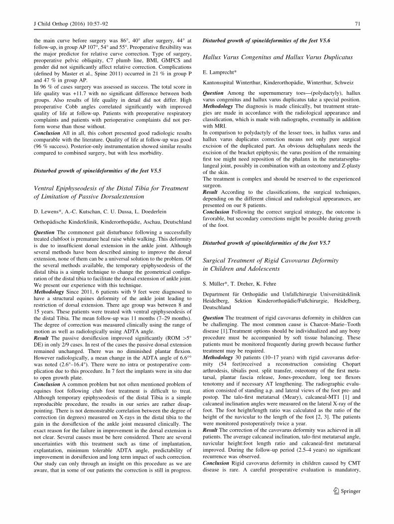
the main curve before surgery was 86�, 40� after surgery, 44� at
follow-up, in group AP 107�, 54� and 55�. Preoperative flexibility was
the major predictor for relative curve correction. Type of surgery,
preoperative pelvic obliquity, C7 plumb line, BMI, GMFCS and
gender did not significantly affect relative correction. Complications
(defined by Master et al., Spine 2011) occurred in 21 % in group P
and 47 % in group AP.
In 96 % of cases surgery was assessed as success. The total score in
life quality was +11.7 with no significant difference between both
groups. Also results of life quality in detail did not differ. High
preoperative Cobb angles correlated significantly with improved
quality of life at follow-up. Patients with preoperative respiratory
complaints and patients with perioperative complaints did not per-
form worse than those without.
Conclusion All in all, this cohort presented good radiologic results
comparable with the literature. Quality of life at follow-up was good
(96 % success). Posterior-only instrumentation showed similar results
compared to combined surgery, but with less morbidity.
Disturbed growth of spine/deformities of the feet V5.5
Ventral Epiphyseodesis of the Distal Tibia for Treatment
of Limitation of Passive Dorsalextension
D. Lewens*, A.-C. Kutschan, C. U. Dussa, L. Doederlein
Orthopadische Kinderklinik, Kinderorthopadie, Aschau, Deutschland
Question The commonest gait disturbance following a successfully
treated clubfoot is premature heal raise while walking. This deformity
is due to insufficient dorsal extension in the ankle joint. Although
several methods have been described aiming to improve the dorsal
extension, none of them can be a universal solution to the problem. Of
the several methods available, the temporary epiphyseodesis of the
distal tibia is a simple technique to change the geometrical configu-
ration of the distal tibia to facilitate the dorsal extension of ankle joint.
We present our experience with this technique.
Methodology Since 2011, 6 patients with 9 feet were diagnosed to
have a structural equines deformity of the ankle joint leading to
restriction of dorsal extension. There age group was between 8 and
15 years. These patients were treated with ventral epiphyseodesis of
the distal Tibia. The mean follow-up was 11 months (7–29 months).
The degree of correction was measured clinically using the range of
motion as well as radiologically using ADTA angle.
Result The passive dorsiflexion improved significantly (ROM [5�DE) in only 2/9 cases. In rest of the cases the passive dorsal extension
remained unchanged. There was no diminished plantar flexion.
However radiologically, a mean change in the ADTA angle of 6.6��was noted (2.6�–16.4�). There were no intra or postoperative com-
plication due to this procedure. In 7 feet the implants were in situ due
to open growth plate.
Conclusion A common problem but not often mentioned problem of
equines foot following club foot treatment is difficult to treat.
Although temporary epiphyseodesis of the distal Tibia is a simple
reproducible procedure, the results in our series are rather disap-
pointing. There is not demonstrable correlation between the degree of
correction (in degrees) measured on X-rays in the distal tibia to the
gain in the dorsiflexion of the ankle joint measured clinically. The
exact reason for the failure in improvement in the dorsal extension is
not clear. Several causes must be here considered. There are several
uncertainties with this treatment such as time of implantation,
explantation, minimum tolerable ADTA angle, predictability of
improvement in dorsiflexion and long term impact of such correction.
Our study can only through an insight on this procedure as we are
aware, that in some of our patients the correction is still in progress.
Disturbed growth of spine/deformities of the feet V5.6
Hallux Varus Congenitus and Hallux Varus Duplicatus
E. Lamprecht*
Kantonsspital Winterthur, Kinderorthopadie, Winterthur, Schweiz
Question Among the supernumerary toes—(polydactyly), hallux
varus congenitus and hallux varus duplicatus take a special position.
Methodology The diagnosis is made clinically, but treatment strate-
gies are made in accordance with the radiological appearance and
classification, which is made with radiographs, eventually in addition
with MRI.
In comparison to polydactyly of the lesser toes, in hallux varus and
hallux varus duplicatus correction means not only pure surgical
excision of the duplicated part. An obvious deltaphalanx needs the
excision of the bracket epiphysis; the varus position of the remaining
first toe might need reposition of the phalanx in the metatarsopha-
langeal joint, possibly in combination with an osteotomy and Z-plasty
of the skin.
The treatment is complex and should be reserved to the experienced
surgeon.
Result According to the classifications, the surgical techniques,
depending on the different clinical and radiological appearances, are
presented on our 8 patients.
Conclusion Following the correct surgical strategy, the outcome is
favorable, but secondary corrections might be possible during growth
of the foot.
Disturbed growth of spine/deformities of the feet V5.7
Surgical Treatment of Rigid Cavovarus Deformity
in Children and Adolescents
S. Muller*, T. Dreher, K. Fehre
Department fur Orthopadie und Unfallchirurgie Universitatsklinik
Heidelberg, Sektion Kinderorthopadie/Fußchirurgie, Heidelberg,
Deutschland
Question The treatment of rigid cavovarus deformity in children can
be challenging. The most common cause is Charcot–Marie–Tooth
disease [1].Treatment options should be individualized and any bony
procedure must be accompanied by soft tissue balancing. These
patients must be monitored frequently during growth because further
treatment may be required.
Methodology 30 patients (10–17 years) with rigid cavovarus defor-
mity (54 feet)received a reconstruction consisting Chopart
arthrodesis, tibialis post. split transfer, osteotomy of the first meta-
tarsal, plantar fascia release, Jones-procedure, long toe flexors
tenotomy and if necessary AT lengthening. The radiographic evalu-
ation consisted of standing a.p. and lateral views of the foot pre- and
postop. The talo-first metatarsal (Meary), calcaneal-MT1 [1] and
calcaneal inclination angles were measured on the lateral X-ray of the
foot. The foot height/length ratio was calculated as the ratio of the
height of the navicular to the length of the foot [2, 3]. The patients
were monitored postoperatively twice a year.
Result The correction of the cavovarus deformity was achieved in all
patients. The average calcaneal inclination, talo-first metatarsal angle,
navicular height:foot length ratio and calcaneal-first metatarsal
improved. During the follow-up period (2.5–4 years) no significant
recurrence was observed.
Conclusion Rigid cavovarus deformity in children caused by CMT
disease is rare. A careful preoperative evaluation is mandatory,
J Child Orthop (2016) 10:57–92 71
123
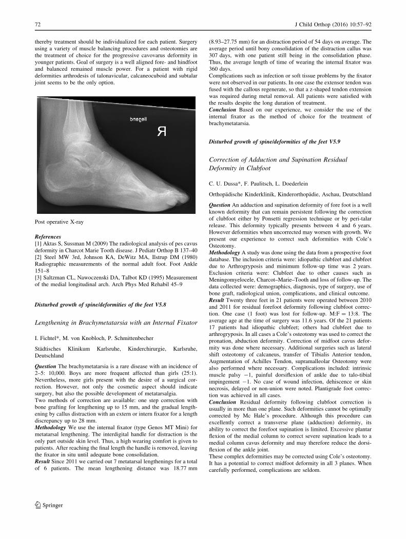
thereby treatment should be individualized for each patient. Surgery
using a variety of muscle balancing procedures and osteotomies are
the treatment of choice for the progressive cavovarus deformity in
younger patients. Goal of surgery is a well aligned fore- and hindfoot
and balanced remained muscle power. For a patient with rigid
deformities arthrodesis of talonavicular, calcaneocuboid and subtalar
joint seems to be the only option.
Post operative X-ray
References[1] Aktas S, Sussman M (2009) The radiological analysis of pes cavus
deformity in Charcot Marie Tooth disease. J Pediatr Orthop B 137–40
[2] Steel MW 3rd, Johnson KA, DeWitz MA, Ilstrup DM (1980)
Radiographic measurements of the normal adult foot. Foot Ankle
151–8
[3] Saltzman CL, Nawoczenski DA, Talbot KD (1995) Measurement
of the medial longitudinal arch. Arch Phys Med Rehabil 45–9
Disturbed growth of spine/deformities of the feet V5.8
Lengthening in Brachymetatarsia with an Internal Fixator
I. Fichtel*, M. von Knobloch, P. Schmittenbecher
Stadtisches Klinikum Karlsruhe, Kinderchirurgie, Karlsruhe,
Deutschland
Question The brachymetatarsia is a rare disease with an incidence of
2–5: 10,000. Boys are more frequent affected than girls (25:1).
Nevertheless, more girls present with the desire of a surgical cor-
rection. However, not only the cosmetic aspect should indicate
surgery, but also the possible development of metatarsalgia.
Two methods of correction are available: one step correction with
bone grafting for lengthening up to 15 mm, and the gradual length-
ening by callus distraction with an extern or intern fixator for a length
discrepancy up to 28 mm.
Methodology We use the internal fixator (type Genos MT Mini) for
metatarsal lengthening. The interdigital handle for distraction is the
only part outside skin level. Thus, a high wearing comfort is given to
patients. After reaching the final length the handle is removed, leaving
the fixator in situ until adequate bone consolidation.
Result Since 2011 we carried out 7 metatarsal lengthenings for a total
of 6 patients. The mean lengthening distance was 18.77 mm
(8.93–27.75 mm) for an distraction period of 54 days on average. The
average period until bony consolidation of the distraction callus was
307 days, with one patient still being in the consolidation phase.
Thus, the average length of time of wearing the internal fixator was
360 days.
Complications such as infection or soft tissue problems by the fixator
were not observed in our patients. In one case the extensor tendon was
fused with the callous regenerate, so that a z-shaped tendon extension
was required during metal removal. All patients were satisfied with
the results despite the long duration of treatment.
Conclusion Based on our experience, we consider the use of the
internal fixator as the method of choice for the treatment of
brachymetatarsia.
Disturbed growth of spine/deformities of the feet V5.9
Correction of Adduction and Supination Residual
Deformity in Clubfoot
C. U. Dussa*, F. Paulitsch, L. Doederlein
Orthopadische Kinderklinik, Kinderorthopadie, Aschau, Deutschland
Question An adduction and supination deformity of fore foot is a well
known deformity that can remain persistent following the correction
of clubfoot either by Ponsetti regression technique or by peri-talar
release. This deformity typically presents between 4 and 6 years.
However deformities when uncorrected may worsen with growth. We
present our experience to correct such deformities with Cole’s
Osteotomy.
Methodology A study was done using the data from a prospective foot
database. The inclusion criteria were: idiopathic clubfeet and clubfeet
due to Arthrogryposis and minimum follow-up time was 2 years.
Exclusion criteria were: Clubfeet due to other causes such as
Meningomyelocele, Charcot–Marie–Tooth and loss of follow-up. The
data collected were: demographics, diagnosis, type of surgery, use of
bone graft, radiological union, complications, and clinical outcome.
Result Twenty three feet in 21 patients were operated between 2010
and 2011 for residual forefoot deformity following clubfoot correc-
tion. One case (1 foot) was lost for follow-up. M:F = 13:8. The
average age at the time of surgery was 11.6 years. Of the 21 patients
17 patients had idiopathic clubfeet; others had clubfeet due to
arthrogryposis. In all cases a Cole’s osteotomy was used to correct the
pronation, abduction deformity. Correction of midfoot cavus defor-
mity was done where necessary. Additional surgeries such as lateral
shift osteotomy of calcaneus, transfer of Tibialis Anterior tendon,
Augmentation of Achilles Tendon, supramalleolar Osteotomy were
also performed where necessary. Complications included: intrinsic
muscle palsy -1, painful dorsiflexion of ankle due to talo-tibial
impingement -1. No case of wound infection, dehiscence or skin
necrosis, delayed or non-union were noted. Plantigrade foot correc-
tion was achieved in all cases.
Conclusion Residual deformity following clubfoot correction is
usually in more than one plane. Such deformities cannot be optimally
corrected by Mc Hale’s procedure. Although this procedure can
excellently correct a transverse plane (adduction) deformity, its
ability to correct the forefoot supination is limited. Excessive plantar
flexion of the medial column to correct severe supination leads to a
medial column cavus deformity and may therefore reduce the dorsi-
flexion of the ankle joint.
These complex deformities may be corrected using Cole’s osteotomy.
It has a potential to correct midfoot deformity in all 3 planes. When
carefully performed, complications are seldom.
72 J Child Orthop (2016) 10:57–92
123
Infantile Tumour Surgery/Varia
Infantile tumour surgery/Varia V6.1
The Delayed Diagnosis in Osteo- und Ewing Sarcoma:
A Facebook-Based Survey
M. Salzmann*1, H. Rechl2, R. von Eisenhart-Rothe2, U. Lenze2, F.
Pohlig2
1Klinikum Schwabing, Kinderorthopadie, Munchen, Deutschland,2Klinikum rechts der Isar, Klinik fur Orthopadie, Munchen,
Deutschland
Question The tumor size and existence of metastases at presentation
are 2 of the most important prognostic factors in Ewing sarcoma and
osteosarcoma. The earliest possible diagnosis is therefore essential to
avoid an unfavorable prognosis. However, the average duration to
diagnosis is still reported often with more than 3 months for both
tumors. Due to the low incidence of these diseases, however, these
data are usually collected over a long period of time and thus have
limited actuality. Social networks like Facebook with their relevant
groups especially for rare diseases such as sarcomas by the avail-
ability of a large number of affected persons represent a
suitable medium for collecting such data. The aim of our study was to
Internet-based survey of the duration of the first symptoms to histo-
logical diagnosis by means of an electronic questionnaire on
Facebook.
Methodology An electronic questionnaire with 10 items was created.
A link to the online questionnaire was posted with the necessary
information in special Facebook groups to osteo- and Ewing-sarco-
mas. After a month, the polls were closed and the data collected were
analyzed statistically.
Result A total of 27 patients were enrolled in our study online with
osteo- and Ewing sarcoma. The average time to diagnosis was
osteosarcoma at 2.92 and Ewing sarcoma at 3.27 months. The most
common tumor location was the lower extremity in both entities. At
the time of diagnosis, 83 % of patients with osteo and 73 % of
patients with Ewing sarcoma were free of metastases. Surgical
resection was performed in the osteosarcoma and Ewing sarcoma
100 % in 91 %. Chemotherapy underwent 92 % of osteosarcoma and
Ewing sarcoma 100 % of the patients. Ewing sarcoma occurred in
55 % of cases additional radiotherapy.
Conclusion Our study confirmed sarcomas long period of onset of
symptoms to diagnosis with osteo- and Ewing sarcoma. Nevertheless,
both entities had only a small proportion of patients at diagnosis on
already detectable metastases. Furthermore, a social network such as
Facebook could be successfully used as a modern medium in com-
bination with an online questionnaire to collect epidemiological data
of rare diseases in our study.
Infantile tumour surgery/Varia V6.2
Primary Malignant Bone and Soft Tissue Sarcoma
in Children and Adolescent: An Analysis of a Nationwide
Sarcoma Center
M. Salzmann*1, F. Pohlig2, H. Rechl2, R. von Eisenhart-Rothe2, N.
C. Berger1, F. Lenze2
1Klinikum Schwabing, Kinderorthopadie, Munchen, Deutschland,2Klinikum rechts der Isar, Klinik fur Orthopadie, Munchen,
Deutschland
Question Primary malignant bone and soft tissue sarcoma constitute
about 10 % of newly diagnosed cancers in children and adolescent.
Pathologic diagnosis in bone and soft tissue tumors can be very dif-
ficult, whereas early diagnosis is crucial. The correct diagnosis and
therapy requires a multidisciplinary team (radiologist, orthopaedic
oncologist, pediatric oncologist and pathologist) to achieve the best
possible results for the patients. Goal of the study was an analysis of
the primary malignant bone and soft tissue sarcoma in children and
adolescent, with special focus on diagnosis delay and outcome.
Methodology This is a retrospective study design. Included were all
children\18 years which were treated for a primary malignant bone
or soft tissue sarcoma between 2000 and 2014. The clinical history,
investigation and treatment were retrospectively analyzed with
respect to localization, biology, age, duration of diagnostic setting,
metastases, kind of operation and outcome.
Result A total of patients 158 were included. 46 % had a diagnosis of
an osteosarcoma and 33 % had a diagnosis of an ewing-sarcoma.
Bone tumors accounted 86 % of the cases. The synovial sarcoma
(33 %) was the most common soft tissue sarcoma. The most common
localization was the femur (42 %), lower leg (21 %) and humerus
(9 %). These preliminary results showed that the duration of diag-
nosis varied from a few weeks to several months. Limb preservation
was the most common therapy for the tumors.
Conclusion In this retrospective study bone tumors clearly outweigh
the soft tissue tumors. The most common tumors were osteosarcoma,
ewingsarcoma and synovial sarcoma. Early diagnosis and absent of
metastasis is crucial for the outcome.
Infantile tumour surgery/Varia V6.3
Influence of Pathological Fractures on the Prognosis
of Primary Malignant Bone Tumors
M. Schlegel*1, M. Zeumer1, S. Burdach1, P.M. Prodinger2, H. Rechl2,
I. von Luettichau1
1Division of Pediatric Hematology/Oncology, Wilhelm Sander Sar-
coma Unit, Roman Herzog Comprehensive Cancer Center, Klinikum
rechts der Isar, Technische Universitat Munchen, Department of
Pediatrics, Munich, Deutschland, 2Department for Orthopedic Sur-
gery, Klinikum rechts der Isar, Technical University of Munich,
Department for Orthopedic Surgery, Munich, Deutschland
Question: The purpose of this study was to investigate whether a
pathological fracture has an effect on the prognosis of patients with
osteosarcoma and Ewing tumor regarding 5-year survival, occurrence
of metastases and local recurrence.
Methodology: We retrospectively analyzed 205 patients with the
histological diagnosis of either osteosarcoma or Ewing tumor. Sur-
vival analysis was performed for all patients and differentiated for the
following subgroups: osteosarcoma, Ewing tumor, adults and
children.
Result: Out of all patients, 127 had an osteosarcoma and 78 had an
Ewing tumor. The age ranged from 3 to 79 years (median age 17). 41
patients (20 %) sustained a pathological fracture. Patients with
pathological fractures showed lower survival rates with 5-year sur-
vival rates of 64 % compared to 83 % (p = 0.023). The difference in
development of metastases between patients with and without a
pathological fracture was not statistically significant (p = 0.286). A
local recurrence occurred in 7 % of the patients without and in 24 %
with a pathological fracture (p = 0.023).
In subgroup analysis, there were no statistically significant differences
between patients with and without a pathological fracture in survival
and local recurrence rates for patients with Ewing tumor and in
children. In contrast, the survival rate decreased from 83 to 59 %
J Child Orthop (2016) 10:57–92 73
123
(p = 0.024) and local recurrence increased from 70 to 87 %
(p = 0.023) in the group with osteosarcoma. For adults, survival rates
decreased from 78 to 51 % (p = 0.004) and local recurrence
increased from 58 to 87 % (p = 0.001). None of the subgroups
showed a statistically significant correlation between pathological
fractures and metastases.
Conclusion: The present study suggests that the occurrence of a
pathological fracture in primary malignant bone tumors has a negative
effect on survival rates and implicates an increased risk of local
recurrence in patients with osteosarcoma and in adults. In patients
with Ewing tumor and in children, no prognostic effect of patholog-
ical fractures could be observed. The influence of pathological
fractures on metastases did not reach statistical significance.
Acknowledgments This work was supported by the Wilhelm Sander
Stiftung.
Infantile tumour surgery/Varia V6.4
Morbus Trevor: Clinical Manifestation and Therapy
of Dysplasia Epiphysealis Hemimelica
C. Wack*, O. Eberhardt, F. Fernandez, T. Wirth
Olgahospital, Orthopadische Klinik, Stuttgart, Deutschland
Question Dysplasia epiphysealis hemimelica or Morbus Trevor is a
realy rare disease with an incidence of 1:1 million. The disease ist
characterised with benign osteocartilaginouse tumors in epiphysis. The
tumor occurs preferred at the lower limb, typically at one side of the
epiphysis. This osteochondrom ist growing from the epiphysis and can
destroy the joint area, so it is differently to the cartilaginous exostosis.
The disease lead to limitations in movement, pain, leg length differ-
ences and deformities. The etiology is until now unknown. Up to date
there is no evidence of a hereditary genesis. The relation of boys to
girls amount to 3:1.
A statement respective frequency of development of malposition and
there consequences is not possible so far.
We report about 12 cases in our department with regard to clinical
symptoms, therapy and progress.
Methodology Between 1999 und 2014 we examined 12 patients with
a dysplasia epiphysealis hemimelica. The classification result from
Azouz et al.
Examined were initial symptoms, classification, affected joint, side,
age, therapeutic intervention, progress and complications.
Result We see 8 boys and 4 girls. The main age was 8 years at
diagnosis, 3 children were diagnosed widely before.
In our 12 patients were 5 patients who had initial multiple symptoms.
Primary symptoms were in over 50 % of the patients swelling, 2
patients indicated pain, a deformity was occurrence in 3 patients. A
limitation of movement was seen in 4 patients.
Mostly pain was indicated at the age of 8–13. In two cases the
diagnoses was an incidental finding after a sprain.
The talus was the most frequently localization, followed by the distal
tibia.
In one case we saw an incidence of both sides at the femurcondyle,
which could treated conservative. In all the other cases a surgical
intervention was necessary, partly with follow interventions.
Conclusion The dysplasia epiphysealis hemimelica (M. Trevor) is
rare and shows an overshoot growing of osteocartilaginouse area in
epiphysis. This run the risk of premature occlusion of epiphysis,
malposition, destruction of the joint area and resultant prearthrosis.
Routine controls of the growth of patients with a M. Trevor are
obligatory require, particularly until the end of growth, but also
beyond that, to prevent deformities and prearthrosis.
Infantile tumour surgery/Varia V6.5
Osteomyelitis of the Patella: Rare Disease and Therefore
Important Not to be Missed
E. Lamprecht*
Kantonsspital Winterthur, Kinderorthopadie, Winterthur, Schweiz
Question Osteomyelitis of the patella is mostly seen in young chil-
dren. Because of its rarity and variety of presentation, the diagnosis
may be missed or delayed.
Methodology Besides peripatellar pain, often with acute exacerbation
with impossibility to walk, the clinical signs are usually swelling,
painful prepatellar bursitis and septic arthritis oft the knee.
Once the diagnosis is clear, treatment follows the strategies of
osteomyelitis with evacuation of pus, debridement of the necrotic
parts of the affected patella and irrigating the knee joint. Systemic and
specific antibiotics are continued according to clinical improvement
and decreasing C-reactive proteine.
Result We present 3 cases of patellar osteomyelitis, the tools to get to
the correct diagnosis, the treatment and the outcome .
Conclusion Our aim is, to point to a rare but easily missed infectious
disease. If correctly treated, the outcome is favorable.
Short survey over the literature.
Poster Presentation
Poster session P.01
3 Entities: 1 Goal: Temporary Hemi-Epiphyseodesis
to Stop Progression of Severe Varus Deformity. Reports
and Review of the Literature for Morbus Blount, Multiple
Epiphyseal Dysplasia and Focal Fibrocartilaginous
Dysplasia
H. Tretow*1, B. Vogt1, G. Gosheger2, B. Schuhknecht2, F. Schiedel1,
R. Rodl1
1Kinderorthopadie, Deformitatenrekonstruktion und Fußchirurgie,
Universitatsklinikum, Munster, Deutschland, 2Klinik fur Allgemeine
Orthopadie und Tumororthopadie, Universitatsklinikum, Munster,
Deutschland
Question In natural course of leg axis development, a slight varus is
physiological in early years of childhood. Besides that, paediatric
orthopaedic surgeons know numerous causes for pathological varus
deformity in the proximal tibia. Among those, we find rare diseases
such as Blount’s disease, multiple epiphyseal dysplasia and the focal
fibrocartilaginous dysplasia. These require surgical treatment beyond
growth guiding by hemiepiphyseodesis.
Methodology We report three different cases of paediatric orthopae-
dic patients with full-blown varus deformity of the proximal tibia
which were symptomatic as well as progressive. The cases are a
matter of Blount’s disease (BD), a fairbank form of multiple epi-
physeal dysplasia (MED), and the focal fibrocartilaginous dysplasia
(FFCD). Lateral hemiepiphyseodesis was performed in each case. The
intention was not to achieve correction of the leg axis deformity by
guided growth but to prevent further progression. More invasive
surgical treatment for axial correction to later points of time were
inevitable part of the concept.
Result By lateral hemiepiphyseodesis, a progression of varus defor-
mity could be stopped in all three cases with even a slight
74 J Child Orthop (2016) 10:57–92
123
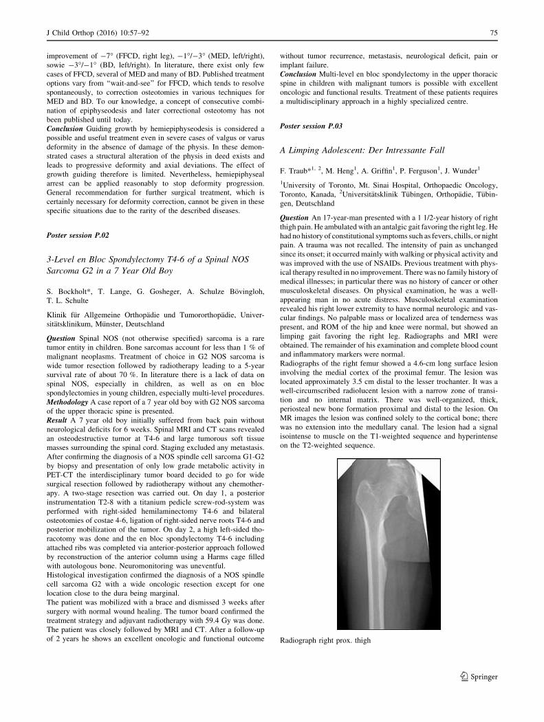
improvement of -7� (FFCD, right leg), -1�/-3� (MED, left/right),
sowie -3�/-1� (BD, left/right). In literature, there exist only few
cases of FFCD, several of MED and many of BD. Published treatment
options vary from ‘‘wait-and-see’’ for FFCD, which tends to resolve
spontaneously, to correction osteotomies in various techniques for
MED and BD. To our knowledge, a concept of consecutive combi-
nation of epiphyseodesis and later correctional osteotomy has not
been published until today.
Conclusion Guiding growth by hemiepiphyseodesis is considered a
possible and useful treatment even in severe cases of valgus or varus
deformity in the absence of damage of the physis. In these demon-
strated cases a structural alteration of the physis in deed exists and
leads to progressive deformity and axial deviations. The effect of
growth guiding therefore is limited. Nevertheless, hemiepiphyseal
arrest can be applied reasonably to stop deformity progression.
General recommendation for further surgical treatment, which is
certainly necessary for deformity correction, cannot be given in these
specific situations due to the rarity of the described diseases.
Poster session P.02
3-Level en Bloc Spondylectomy T4-6 of a Spinal NOS
Sarcoma G2 in a 7 Year Old Boy
S. Bockholt*, T. Lange, G. Gosheger, A. Schulze Bovingloh,
T. L. Schulte
Klinik fur Allgemeine Orthopadie und Tumororthopadie, Univer-
sitatsklinikum, Munster, Deutschland
Question Spinal NOS (not otherwise specified) sarcoma is a rare
tumor entity in children. Bone sarcomas account for less than 1 % of
malignant neoplasms. Treatment of choice in G2 NOS sarcoma is
wide tumor resection followed by radiotherapy leading to a 5-year
survival rate of about 70 %. In literature there is a lack of data on
spinal NOS, especially in children, as well as on en bloc
spondylectomies in young children, especially multi-level procedures.
Methodology A case report of a 7 year old boy with G2 NOS sarcoma
of the upper thoracic spine is presented.
Result A 7 year old boy initially suffered from back pain without
neurological deficits for 6 weeks. Spinal MRI and CT scans revealed
an osteodestructive tumor at T4-6 and large tumorous soft tissue
masses surrounding the spinal cord. Staging excluded any metastasis.
After confirming the diagnosis of a NOS spindle cell sarcoma G1-G2
by biopsy and presentation of only low grade metabolic activity in
PET-CT the interdisciplinary tumor board decided to go for wide
surgical resection followed by radiotherapy without any chemother-
apy. A two-stage resection was carried out. On day 1, a posterior
instrumentation T2-8 with a titanium pedicle screw-rod-system was
performed with right-sided hemilaminectomy T4-6 and bilateral
osteotomies of costae 4-6, ligation of right-sided nerve roots T4-6 and
posterior mobilization of the tumor. On day 2, a high left-sided tho-
racotomy was done and the en bloc spondylectomy T4-6 including
attached ribs was completed via anterior-posterior approach followed
by reconstruction of the anterior column using a Harms cage filled
with autologous bone. Neuromonitoring was uneventful.
Histological investigation confirmed the diagnosis of a NOS spindle
cell sarcoma G2 with a wide oncologic resection except for one
location close to the dura being marginal.
The patient was mobilized with a brace and dismissed 3 weeks after
surgery with normal wound healing. The tumor board confirmed the
treatment strategy and adjuvant radiotherapy with 59.4 Gy was done.
The patient was closely followed by MRI and CT. After a follow-up
of 2 years he shows an excellent oncologic and functional outcome
without tumor recurrence, metastasis, neurological deficit, pain or
implant failure.
Conclusion Multi-level en bloc spondylectomy in the upper thoracic
spine in children with malignant tumors is possible with excellent
oncologic and functional results. Treatment of these patients requires
a multidisciplinary approach in a highly specialized centre.
Poster session P.03
A Limping Adolescent: Der Intressante Fall
F. Traub*1, 2, M. Heng1, A. Griffin1, P. Ferguson1, J. Wunder1
1University of Toronto, Mt. Sinai Hospital, Orthopaedic Oncology,
Toronto, Kanada, 2Universitatsklinik Tubingen, Orthopadie, Tubin-
gen, Deutschland
Question An 17-year-man presented with a 1 1/2-year history of right
thigh pain. He ambulated with an antalgic gait favoring the right leg. He
had no history of constitutional symptoms such as fevers, chills, or night
pain. A trauma was not recalled. The intensity of pain as unchanged
since its onset; it occurred mainly with walking or physical activity and
was improved with the use of NSAIDs. Previous treatment with phys-
ical therapy resulted in no improvement. There was no family history of
medical illnesses; in particular there was no history of cancer or other
musculoskeletal diseases. On physical examination, he was a well-
appearing man in no acute distress. Musculoskeletal examination
revealed his right lower extremity to have normal neurologic and vas-
cular findings. No palpable mass or localized area of tenderness was
present, and ROM of the hip and knee were normal, but showed an
limping gait favoring the right leg. Radiographs and MRI were
obtained. The remainder of his examination and complete blood count
and inflammatory markers were normal.
Radiographs of the right femur showed a 4.6-cm long surface lesion
involving the medial cortex of the proximal femur. The lesion was
located approximately 3.5 cm distal to the lesser trochanter. It was a
well-circumscribed radiolucent lesion with a narrow zone of transi-
tion and no internal matrix. There was well-organized, thick,
periosteal new bone formation proximal and distal to the lesion. On
MR images the lesion was confined solely to the cortical bone; there
was no extension into the medullary canal. The lesion had a signal
isointense to muscle on the T1-weighted sequence and hyperintense
on the T2-weighted sequence.
Radiograph right prox. thigh
J Child Orthop (2016) 10:57–92 75
123
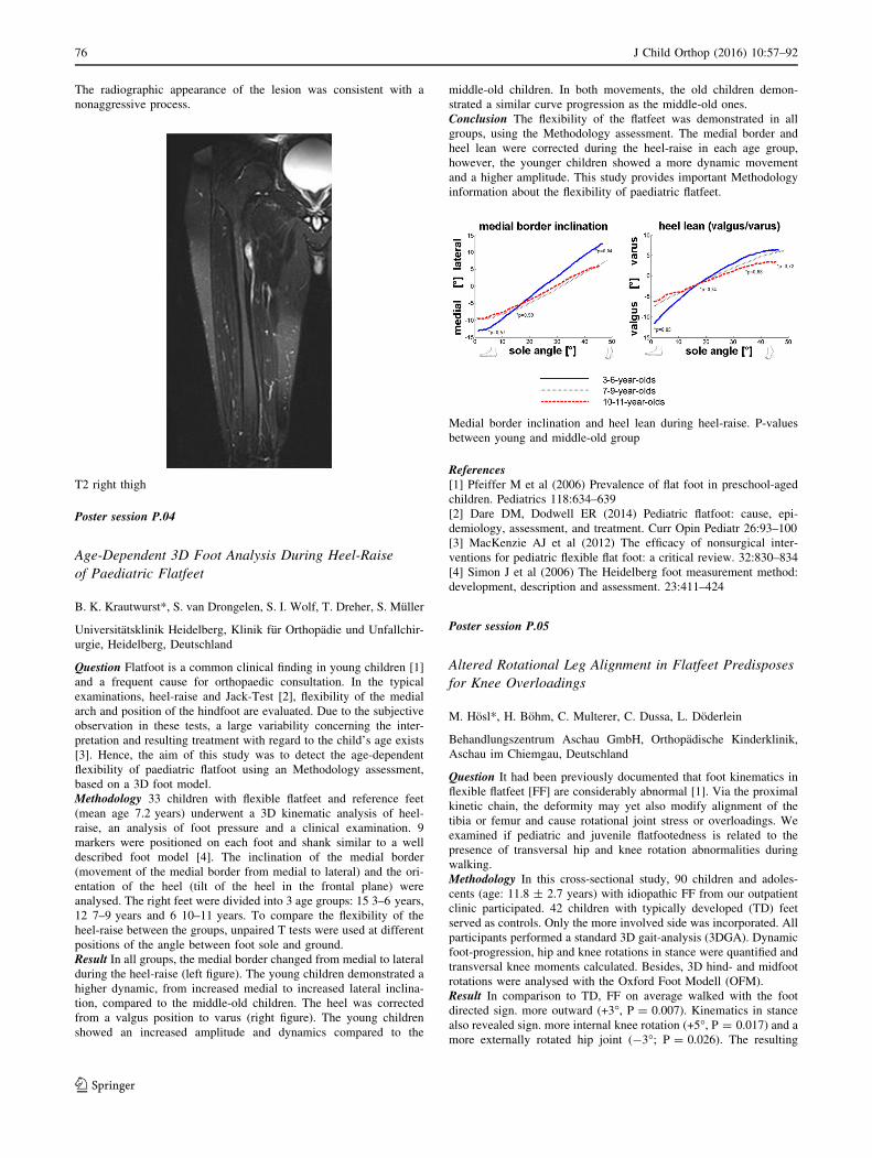
The radiographic appearance of the lesion was consistent with a
nonaggressive process.
T2 right thigh
Poster session P.04
Age-Dependent 3D Foot Analysis During Heel-Raise
of Paediatric Flatfeet
B. K. Krautwurst*, S. van Drongelen, S. I. Wolf, T. Dreher, S. Muller
Universitatsklinik Heidelberg, Klinik fur Orthopadie und Unfallchir-
urgie, Heidelberg, Deutschland
Question Flatfoot is a common clinical finding in young children [1]
and a frequent cause for orthopaedic consultation. In the typical
examinations, heel-raise and Jack-Test [2], flexibility of the medial
arch and position of the hindfoot are evaluated. Due to the subjective
observation in these tests, a large variability concerning the inter-
pretation and resulting treatment with regard to the child’s age exists
[3]. Hence, the aim of this study was to detect the age-dependent
flexibility of paediatric flatfoot using an Methodology assessment,
based on a 3D foot model.
Methodology 33 children with flexible flatfeet and reference feet
(mean age 7.2 years) underwent a 3D kinematic analysis of heel-
raise, an analysis of foot pressure and a clinical examination. 9
markers were positioned on each foot and shank similar to a well
described foot model [4]. The inclination of the medial border
(movement of the medial border from medial to lateral) and the ori-
entation of the heel (tilt of the heel in the frontal plane) were
analysed. The right feet were divided into 3 age groups: 15 3–6 years,
12 7–9 years and 6 10–11 years. To compare the flexibility of the
heel-raise between the groups, unpaired T tests were used at different
positions of the angle between foot sole and ground.
Result In all groups, the medial border changed from medial to lateral
during the heel-raise (left figure). The young children demonstrated a
higher dynamic, from increased medial to increased lateral inclina-
tion, compared to the middle-old children. The heel was corrected
from a valgus position to varus (right figure). The young children
showed an increased amplitude and dynamics compared to the
middle-old children. In both movements, the old children demon-
strated a similar curve progression as the middle-old ones.
Conclusion The flexibility of the flatfeet was demonstrated in all
groups, using the Methodology assessment. The medial border and
heel lean were corrected during the heel-raise in each age group,
however, the younger children showed a more dynamic movement
and a higher amplitude. This study provides important Methodology
information about the flexibility of paediatric flatfeet.
Medial border inclination and heel lean during heel-raise. P-values
between young and middle-old group
References[1] Pfeiffer M et al (2006) Prevalence of flat foot in preschool-aged
children. Pediatrics 118:634–639
[2] Dare DM, Dodwell ER (2014) Pediatric flatfoot: cause, epi-
demiology, assessment, and treatment. Curr Opin Pediatr 26:93–100
[3] MacKenzie AJ et al (2012) The efficacy of nonsurgical inter-
ventions for pediatric flexible flat foot: a critical review. 32:830–834
[4] Simon J et al (2006) The Heidelberg foot measurement method:
development, description and assessment. 23:411–424
Poster session P.05
Altered Rotational Leg Alignment in Flatfeet Predisposes
for Knee Overloadings
M. Hosl*, H. Bohm, C. Multerer, C. Dussa, L. Doderlein
Behandlungszentrum Aschau GmbH, Orthopadische Kinderklinik,
Aschau im Chiemgau, Deutschland
Question It had been previously documented that foot kinematics in
flexible flatfeet [FF] are considerably abnormal [1]. Via the proximal
kinetic chain, the deformity may yet also modify alignment of the
tibia or femur and cause rotational joint stress or overloadings. We
examined if pediatric and juvenile flatfootedness is related to the
presence of transversal hip and knee rotation abnormalities during
walking.
Methodology In this cross-sectional study, 90 children and adoles-
cents (age: 11.8 ± 2.7 years) with idiopathic FF from our outpatient
clinic participated. 42 children with typically developed (TD) feet
served as controls. Only the more involved side was incorporated. All
participants performed a standard 3D gait-analysis (3DGA). Dynamic
foot-progression, hip and knee rotations in stance were quantified and
transversal knee moments calculated. Besides, 3D hind- and midfoot
rotations were analysed with the Oxford Foot Modell (OFM).
Result In comparison to TD, FF on average walked with the foot
directed sign. more outward (+3�, P = 0.007). Kinematics in stance
also revealed sign. more internal knee rotation (+5�, P = 0.017) and a
more externally rotated hip joint (-3�; P = 0.026). The resulting
76 J Child Orthop (2016) 10:57–92
123
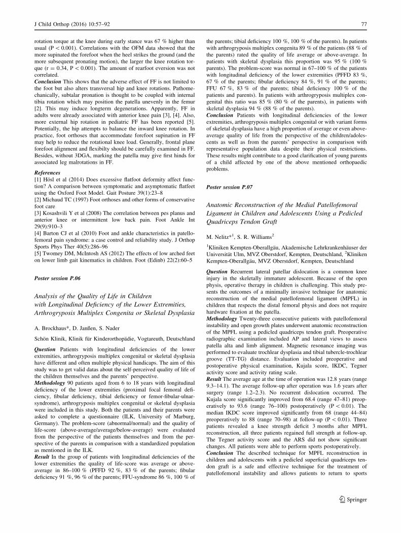
rotation torque at the knee during early stance was 67 % higher than
usual (P\ 0.001). Correlations with the OFM data showed that the
more supinated the forefoot when the heel strikes the ground (and the
more subsequent pronating motion), the larger the knee rotation tor-
que (r = 0.34, P\ 0.001). The amount of rearfoot eversion was not
correlated.
Conclusion This shows that the adverse effect of FF is not limited to
the foot but also alters transversal hip and knee rotations. Pathome-
chanically, subtalar pronation is thought to be coupled with internal
tibia rotation which may position the patella unevenly in the femur
[2]. This may induce longterm degenerations. Apparently, FF in
adults were already associated with anterior knee pain [3], [4]. Also,
more external hip rotation in pediatric FF has been reported [5].
Potentially, the hip attempts to balance the inward knee rotation. In
practice, foot orthoses that accommodate forefoot supination in FF
may help to reduce the rotational knee load. Generally, frontal plane
forefoot alignment and flexibilty should be carefully examined in FF.
Besides, without 3DGA, marking the patella may give first hinds for
associated leg malrotations in FF.
References[1] Hosl et al (2014) Does excessive flatfoot deformity affect func-
tion? A comparison between symptomatic and asymptomatic flatfeet
using the Oxford Foot Model. Gait Posture 39(1):23–8
[2] Michaud TC (1997) Foot orthoses and other forms of conservative
foot care
[3] Kosashvili Y et al (2008) The correlation between pes planus and
anterior knee or intermittent low back pain. Foot Ankle Int
29(9):910–3
[4] Barton CJ et al (2010) Foot and ankle characteristics in patello-
femoral pain syndrome: a case control and reliability study. J Orthop
Sports Phys Ther 40(5):286–96
[5] Twomey DM, McIntosh AS (2012) The effects of low arched feet
on lower limb gait kinematics in children. Foot (Edinb) 22(2):60–5
Poster session P.06
Analysis of the Quality of Life in Children
with Longitudinal Deficiency of the Lower Extremities,
Arthrogryposis Multiplex Congenita or Skeletal Dysplasia
A. Brockhaus*, D. Janßen, S. Nader
Schon Klinik, Klinik fur Kinderorthopadie, Vogtareuth, Deutschland
Question Patients with longitudinal deficiencies of the lower
extremities, arthrogryposis multiplex congenital or skeletal dysplasia
have different and often multiple physical handicaps. The aim of this
study was to get valid datas about the self-perceived quality of life of
the children themselves and the parents’ perspective.
Methodology 90 patients aged from 6 to 18 years with longitudinal
deficiency of the lower extremities (proximal focal femoral defi-
ciency, fibular deficiency, tibial deficiency or femor-fibular-ulnar-
syndrome), arthrogryposis multiplex congenital or skeletal dysplasia
were included in this study. Both the patients and their parents were
asked to complete a questionnaire (ILK, University of Marburg,
Germany). The problem-score (abnormal/normal) and the quality of
life-score (above-average/average/below-average) were evaluated
from the perspective of the patients themselves and from the per-
spective of the parents in comparison with a standardized population
as mentioned in the ILK.
Result In the group of patients with longitudinal deficiencies of the
lower extremities the quality of life-score was average or above-
average in 86–100 % (PFFD 92 %, 83 % of the parents; fibular
deficiency 91 %, 96 % of the parents; FFU-syndrome 86 %, 100 % of
the parents; tibial deficiency 100 %, 100 % of the parents). In patients
with arthrogryposis multiplex congenita 89 % of the patients (88 % of
the parents) rated the quality of life average or above-average. In
patients with skeletal dysplasia this proportion was 95 % (100 %
parents). The problem-score was normal in 67–100 % of the patients
with longitudinal deficiency of the lower extremities (PFFD 83 %,
67 % of the parents; fibular deficiency 84 %, 91 % of the parents;
FFU 67 %, 83 % of the parents; tibial deficiency 100 % of the
patients and parents). In patients with arthrogryposis multiplex con-
genital this ratio was 85 % (80 % of the parents), in patients with
skeletal dysplasia 94 % (88 % of the parents).
Conclusion Patients with longitudinal deficiencies of the lower
extremities, arthrogryposis multiplex congenital or with variant forms
of skeletal dysplasia have a high proportion of average or even above-
average quality of life from the perspective of the children/adoles-
cents as well as from the parents’ perspective in comparison with
representative population data despite their physical restrictions.
These results might contribute to a good clarification of young parents
of a child affected by one of the above mentioned orthopaedic
problems.
Poster session P.07
Anatomic Reconstruction of the Medial Patellofemoral
Ligament in Children and Adolescents Using a Pedicled
Quadriceps Tendon Graft
M. Nelitz*1, S. R. Williams2
1Kliniken Kempten-Oberallgau, Akademische Lehrkrankenhauser der
Universitat Ulm, MVZ Oberstdorf, Kempten, Deutschland, 2Kliniken
Kempten-Oberallgau, MVZ Oberstdorf, Kempten, Deutschland
Question Recurrent lateral patellar dislocation is a common knee
injury in the skeletally immature adolescent. Because of the open
physis, operative therapy in children is challenging. This study pre-
sents the outcomes of a minimally invasive technique for anatomic
reconstruction of the medial patellofemoral ligament (MPFL) in
children that respects the distal femoral physis and does not require
hardware fixation at the patella.
Methodology Twenty-three consecutive patients with patellofemoral
instability and open growth plates underwent anatomic reconstruction
of the MPFL using a pedicled quadriceps tendon graft. Preoperative
radiographic examination included AP and lateral views to assess
patella alta and limb alignment. Magnetic resonance imaging was
performed to evaluate trochlear dysplasia and tibial tubercle-trochlear
groove (TT-TG) distance. Evaluation included preoperative and
postoperative physical examination, Kujala score, IKDC, Tegner
activity score and activity rating scale.
Result The average age at the time of operation was 12.8 years (range
9.3–14.1). The average follow-up after operation was 1.6 years after
surgery (range 1.2–2.3). No recurrent dislocation occurred. The
Kujala score significantly improved from 68.4 (range 47–81) preop-
eratively to 93.6 (range 76–100) postoperatively (P\ 0.01). The
median IKDC score improved significantly from 68 (range 44–84)
preoperatively to 88 (range 70–98) at follow-up (P\ 0.01). Three
patients revealed a knee strength deficit 3 months after MPFL
reconstruction, all three patients regained full strength at follow-up.
The Tegner activity score and the ARS did not show significant
changes. All patients were able to perform sports postoperatively.
Conclusion The described technique for MPFL reconstruction in
children and adolescents with a pedicled superficial quadriceps ten-
don graft is a safe and effective technique for the treatment of
patellofemoral instability and allows patients to return to sports
J Child Orthop (2016) 10:57–92 77
123
without redislocation of the patella. A major advantage of the
quadriceps technique is that no hardware fixation or bony procedure
involving the patella is necessary, making it ideal for use in children.
Poster session P.08
Anterior Knee Pain in a 15 Year Old Girl
with a Hyperplastic Trochlear Groove and Femoral Neck
Rotation Deformity
S. Utzschneider*, C. Ziegler
Ludwig-Maximilians-Universitat, Klinik und Poliklinik fur Orthopa-
die, Physikalische Medizin und Rehabiliation, Munchen, Deutschland
Question A 15 year old girl presented to our ward with a history right
anterior knee pain and a reduced range of motion, both of which had
been persisting for several months. The symptoms were neither pre-
ceded by a traumatic event or dislocation of the patella nor the result
of any known underlying condition. Sports were limited to physical
education classes.
The physical examination showed slight knock knees, moderate
effusion, distinct retropatellar crepitation, hypomobility of the patella
and an extension capability reduced by 20� with free flexion.
MRI studies revealed 2nd to 3rd degree chondral lesions extending
from the proximal apex of the patella over the lateral aspect all the
way into the distal third of the patella. While the medial portion of the
patella appeared to be dysplastic, the trochlear groove seemed to be
deepened. Long leg radiographs showed a mechanical valgus of 5� for
the right and 3� for the left leg. In the additional rotational computer
tomography the femoral antetorsion angle was reduced to 3� on the
right side. The left side was within normal range with 17�. Tibial
torsion was 31� on the right and 33� on the left side, respectively.
A 3 month cycle of physiotherapy yielded an extension improvement
of 5� but no pain relief could be achieved.
Since isolated cartilage repair would surely result in recurrence, we
considered arthroscopic lateral release and a femoral rotation
osteotomy mandatory to correct the reduced femoral neck antetorsion
angle. Optional treatment could consist in coronal axis correction and
trochleoplasty.
How to treat?
Poster session P.09
Arthroscopic Reduction and Primary Osteoplasty
for Severe and Moderate Slipped Capital Femoral
Epiphysis: Surgical Technique
O. Hauschild*, A. El Tayeh, S. Herrmann, P. Helwig, N. Sudkamp, K.
Kuminack
Klinik fur Orthopadie und Unfallchirurgie, Universitatsklinikum,
Freiburg, Deutschland
Question Slipped capital femoral epiphysis (SCFE) is the most
common hip disorder in children and adolescents. In-situ fixation has
traditionally been the treatment of choice even for severely displaced
and unstable slips. As compared to mild slips long term results are,
however, markedly impaired and high rates of avascular necrosis and
early joint degeneration have consistently been reported. Moreover,
more recent studies suggest that the residual CAM type deformity
associated with SCFE may lead to early pathology at the acetabular
rim cartilage and labrum.
The aim of this technical note is to present a novel approach that
combines an arthroscopic reduction technique and screw fixation with
primary osteoplasty in treating Southwick grades 2 and 3 slipped
capital femoral epiphysis (SCFE).
Methodology For this approach an open atraumatic reduction tech-
nique described by Gekeler in 2007 was adapted to an arthroscopic
setting.
The procedure is performed under general anesthesia with the patient
placed in supine position on a radiolucent table.
First a guide wire for a 7.0 mm cannulated screw is placed under
fluoroscopic guidance via lateral stab incision and forwarded to the
metaphyseal area. A 70 degree scope is then inserted into the
peripheral compartment via the proximal anterolateral portal.
Hemarthrosis is evacuated and an anterior working portal is estab-
lished. Now, slight traction is applied to the leg by the assistant
thereby widening the gap between the metaphysis and the acetabular
labrum. A 3.2 mm threaded K wire is inserted into the joint via the
anterior portal and placed through this gap onto the displaced epi-
physis. Upon bony contact the K wire is drilled into the epiphysis.
The epiphysis is now reduced against the metaphysis by manually
pulling on the threaded K wire and simultaneous and coordinated
traction and internal rotation of the leg. As soon as a satisfactory
reduction has been achieved it is secured by advancing the preposi-
tioned wire which in turn is exchanged for the cannulated screw. Now
any residual CAM type deformity is treated by osteoplasty in a
standard fashion.
Result Thus far, ten children have been treated with this technique.
No complications occurred and there was no case of AVN.
Conclusion The present technique is a technically demanding but safe
and feasible treatment option for SCFE. Longer follow-up and larger
series are, however, needed to further evaluate this technique.
Poster session P.10
Biomechanical Evaluation of Two Tension-Band-Principle
Based Tools for Guided Growth: FlexTack vs. Eight-Plate
C. Druschel*1, S. Hoppmann2, R. Mayr3, W. Schmolz3, P. H. Pen-
nekamp2, R. Placzek2
1Charite-Universitatsmedizin Berlin, Centrum fur Muskuloskeletale
Chirurgie, Berlin, Deutschland, 2Universitatsklinikum Bonn, Klinik
und Poliklinik fur Orthopadie und Unfallchirurgie, Bonn, Deutsch-
land, 3Universitatsklinik Innsbruck, Klinik fur Unfallchirurgie,
Innsbruck, Osterreich
Question Stevens et al. introduced in 2007 the eight-Plate to correct
varus and valgus knee deformity and 1 year later also for correction
of fixed knee Flexion deformity [1,2]. Nowadays the eight-Plate
became gold standard for temporary hemiepiphysiodesis instead of
the previously used Blount staples which after explantation may lead
to epiphyseal plate compromise and overcorrection.
The purpose of this biomechanical study was to reveal differences in
mechanical properties (of the tools) such as force absorbtion in
relation to traction-induced displacement and ultimate failure
including possible material failure during simulated epiphyseal
growth.
Methodology Forty fresh frozen porcine femurs were used by opening
the epiphyeal growing zone with a thin chisel respecting the original
anatomy of the epiphyseal line. In four groups with 10 femurs each a
hemiepiphysiodesis was performed:
– SP-1: one hemiepiphysis in the sagittal plane with a 16 mm eight-
Plate using 32 mm screws.
78 J Child Orthop (2016) 10:57–92
123
– SP-2: one hemiepiphysis in the sagittal plane with a 30 mm
FlexTack.
– CP-1: two hemiepiphysis in the coronal plane with two 12 mm
eight-plates using 24 mm screws.
– CP-2: two hemiepiphysis in the coronal plane with one 25 mm
FlexTack.
The specimens were biomechanically tested using a servo-hydraulic
material testing machine (858 Mini Bionix II, MTS, MN, USA) to
determine strength at increasing displacement and failure load (N).
For the statistical evaluation the software SPSS 21.0 (IBM Corpora-
tion, Armonk, NY, USA) was used. The significance level was
determined at p\ 0.05.
Result For hemiepiphysiodesis in sagittal plane there were no sig-
nificant differences found regarding Maximum load force (Fmax) and
displacement at Fmax. for ventral hemiepiphysis a significant dif-
ference in the average maximum force as well as in the displacement
at Fmax was found. The implanted CP-2 group showed a lower
average maximum force and in particular a lower displacement at
Fmax than the CP-1 group.
Conclusion Ventral eight-Plate epiphysiodesis seemed to be superior
in force absorption and fixation failure but a conclusion about a
benefit in clinical use is not possible by this biomechanical data.
Whereas due to the flat design of the FlexTack its implantation may
cause less soft tissue irritation compared to the eight-plate.
References1. Klatt J, Stevens PM. Guided growth for fixed knee flexion defor-
mity. JPO 2008
2. Stevens PM. Guided growth for angular correction: a preliminary
series using a tension band plate. JPO 2007
Poster session P.11
Camptodactyly-Arthropathy-Coxa Vara-Pericarditis
(CACP): Is There a Right Time for Surgical Treatment?
J. Hewera*, R. Pasha
Helios Klinikum Krefeld, Kinderorthopadie, Krefeld, Deutschland
Question We present a 14 years old girl with CACP. That is an
extremely rare rheumatic disease characterized by non-inflammatory
arthropathy with painful contractures of several joints and deformities
of the bone in childhood and adolescence. This genetic disorder is
caused by mutation in the Proteoglycan PRG4 gene on chromosome
1, which is a surface lubricant for joints and tendons. She and her two
siblings suffer from CACP with massive contractures of joints and
deformities in multiple regions. Nevertheless clinical problems like
pain are given just in few joints. In X-rays there is also a pronounced
hallux valgus, a pes excavatus and a lumbar hyperlordosis are seen.
Moreover she presented a progressive and massive contracture of the
hips and a coxa vara with cam impingement on both sides. The
abduction of the hip joint was possible to 5� and very painful. After
diagnostic arthroscopy of the right hip joint with partial synovectomy,
capsulotomy, electrothermal denervation and abrasion of the cam she
had a significantly improved movement of the hip joint without pain.
Same procedure was done on the left side 3 months later with good
result. As the possibilities of surgical treatment are limited with the
risk of recurrences, it is not clear when operative intervention is
recommended.
Does anybody have experience with operative results in patients with
CACP? Is there a ‘‘right’’ time for surgical treatment? Which thera-
peutical steps are recommended?
Poster session P.12
Correction of Gait After Derotation Osteotomies
in Cerebral Palsy: Are The Effects Predictable?
H. Bohm*, M. Hosl, C. Multerer, C. Dussa, L. Doderlein
Orthopadische Kinderklinik, Kinderorthopadie, Aschau, Deutschland
Question Derotation osteotomies of the femur and tibia are estab-
lished procedures to improve transverse plane deformities during
walking with inwardly pointing knees and in- and out toeing gait.
However, effects of femoral derotation osteotomies on gait were
reported to be small and those for the tibia are not known.
Therefore, the aim of the study was to show the relation between the
amount of intraoperative rotation and the changes during gait for
osteotomies at tibia and femur levels, and predict those for the femur
from preoperative clinical and gait data.
Methodology 51 patients with spastic cerebral palsy mean age
13.2 years were included, 46 limbs received rotation only at the
femur, 6 only at the tibia and 11 limbs at both levels. Instrumented
gait analysis and clinical testing was performed pre- and 20.6
(SD = 1.9) months postoperatively. The amount of derotation of
femur and tibia measured intraoperatively, were correlated to the
changes on hip rotation and knee rotation during gait, respectively. In
addition a predictor analysis was performed from preoperative clini-
cal and gait measurements
Result The amount of intraoperative derotation of the femur did not
correlate with the change in hip rotation during walking, whereas the
rotation of the tibia showed an excellent relationship (R = 0.84,
p\ 0.001) with the change in knee rotation. Preoperative hip rotation
during walking, passive hip abduction and foot progression angle
together explained only 38 % of the variability of the postoperative
change in hip rotation during gait. Strength and passive extension of
the hip joint, and hip extension or abduction during walking did not
show any predictive significance.
Prediction from preoperative measures may be only moderate because
growth was shown to affect recurrence of internal hip rotation. This
was confirmed, since adding femoral length change between pre and
postoperative measures to the predictors increased the predictability
from 38 to 49 %.
Conclusion In conclusion changes of knee rotation during gait is
directly predictable from the amount of tibial corrections, contrary the
change in hip rotation was not related to the amount of femoral
derotation, and prediction was only moderate. The predictor analysis
revealed that small changes during walking can be expected when hip
rotation is already close to the natural hip rotation. In addition, growth
during the period until evaluation likely causes a loss of correction in
hip rotation.
Poster session P.13
Correction of Procurvation Deformity near the Knee Joint
in Children by Means of Guiding Growth
L. Schwering*1, V. T. Forster2
1Mathias-Spital, Technische Orthopadie, Rheine, Deutschland,2Universitatsklinikum Freiburg, Department Orthopadie Trauma-
tologie, Freiburg, Deutschland
Question The purpose of this evaluation is to demonstrate the results
of correction of sagittal deformities near the knee joint by means of
guiding growth in children. Guided growth with the 8-plate has
J Child Orthop (2016) 10:57–92 79
123

proved as a valuable tool in the correction of deformities in the frontal
plane near the knee joint like valgus or varus deformity. First P.
Stevens reported about the initial experiences in the correction of
procurvatum. This investigation demonstrates the first finalized
results in the correction of paediatric procurvation of the knee joint by
guiding growth.
Methodology 20 extremities of 13 childrens were operated on for
procurvation deformities near the knee joint because of spina bifida
(7), achondroplasia (4), idiopathic deformity (3), cerebral palsy (2)
infectious disease (1), muscular dystrophy (2) and transepiphyseal bar
(1). The mean age at operation was 12 years and 4 month. The group
consisted from 7 boys and 6 girls.
Therefore 22 8-plates were applied in the distal ventral femur while
14 8-plates were inserted in the proximal ventral tibia. In 11
extremities femur and tibia were treated simultaneously. The opera-
tive technique for the distal ventral femur was described by P.
Stevens, while for the proximal ventral approach to the tibia one
incision over the tuberosity was used to reach the epiphyseal plate
medial and lateral from the tuberosity. Patients’ files and radiographs
were evaluated.
Result Neither during the application nor during the hardware
removal any complications occurred. In six extremities hardware
removal was not necessary because of patients maturity. The 8-plate
remained in situ for a mean duration of 12 months. The mean oper-
ation time/8-plate was 32 min. In 14 of the 20 extremities a full
correction could be achieved while in 5 extremities the residual
growth was too little to create a relevant correction in spite of having
checked the epiphyseal plate by an MRI preoperative. In one case of
posttraumatic deformity the situation remained unchanged.
Conclusion Guiding growth seems to be a valuable tool in procur-
vatum deformities near the knee joint in children. Early intervention
is necessary to avoid failure because of unexpected maturity. Because
of the 3D structure of the growth plates near the knee K-wires instead
of the screws have to be checked accurately by the intensifier.
Potentially this indication will find a wider field in neuroorthopaedic
diseases in the future.
Poster session P.14
Correction of Recurrent Equinus Deformity in Surgical
Treated Clubfeet by Anterior Distal Tibia Epiphyseodesis
N. Floeter*, K. Babin, S. Breyer, M. Rupprecht, K. Schelling, R.
Stuecker
Altonaer Kinderkrankenhaus, Kinderorthopadie, Hamburg,
Deutschland
Question Since the wide spread acceptance of the Ponseti method, the
relapse of clubfeet has become rare, still it is recorded to be 2–5 %. In
contrast the recurrence of surgical treated clubfeet has been docu-
mented to be much higher, between 25 and 45 %. One problem is
recurrent equinus deformity. Several treatment methods are described,
mostly ending up in quite invasive surgical treatments such as
osteotomies or soft tissue releases. As the procurvation of the tibia is
one reason for the limited dorsiflexion in some patients, we want to
evaluate the results of a minimally invasive procedure: the ventral
epiphyseodesis of the distal tibia for treating recurrent equinus
deformity in patients with elevated anterior distal tibia angle (ADTA).
Methodology 20 children (age 8–14, mean 11 years), and 23 feet with
pathologic ADTA were treated with ventral epiphyseodesis of the
distal tibia with 8 plates from 2009 through 2014 in our institution.
We evaluated them radiologically, measuring the ADTA and clini-
cally, measuring the dorsiflexion of the ankle, preoperative and one to
four times until removal of the 8 plate.
Result At the time of abstract admission 11 patients (12 feet) have had
removal of the plate ore closed growth plate. Their mean ADTA
shifted from 86� (SD 2.1) to 74� (SD 6.5).The preoperative mean
dorsiflexion of the ankle was 0.4� (SD 3.8) and improved to 6.3� (SD
4.6). Mean correction time was 19.4 (SD 6.4) months. The shift of the
ADTA and of the dorsiflexion was highly significant (p\ 0.05), but
there was no correlation between the shift in clinical dorsiflexion and
ADTA.
Conclusion The ventral epiphyseodesis of the distal tibia is a valuable
instrument for correcting recurrent equinus deformity in clubfeet with
elevated ADTA. Nevertheless in some patients the dorsiflexion does
not improve by shifting the ADTA. In our opinion there are other
additional clubfoot related deformities like flat top talus or shortening
of the achilles tendon which can be responsible for nonresponding in
those patients.
Poster session P.15
Deformity Correction and Lengthening of the Lower Limb,
45 Surgeries in 24 Patients with Various Skeletal-
Dysplasia
J. K. Correll*, S. Nader
Schon Klinik, Klinik fur Kinderorthopadie, Vogtareuth, Deutschland
Question The skeletal dysplasias are a large, heterogeneous group of
genetic disorders characterised by abnormal development and
remodelling of the bones and cartilage. They typically present with
short stature, disproportion, and various deviations of the lower limb.
Significant advances in the non-surgical treatment of skeletal dys-
plasia have been achieved during the past years. Although extensive
limb lengthening and deformity correction surgery is still an impor-
tant part of the orthopedic treatment.
Methodology We report the results on 45 deformity correction and
limb lengthening surgeries in 24 patients with skeletal dysplasia (29
tibial/16 femoral lengthening). In this study only patients with
chondrodysplasia punctata, pseudoachondroplasia, achondroplasia,
metaphyseal dysplasia, larsen-like syndrome, SEDC and Phosphat-
diabetes were included. The amount of deformity correction, external
fixation time, length gained, mobility of adjacent joints, and incidence
of complications were examined.
In 40 cases correction was done with the Taylor Spatial Frame. In 5
patients the Monolateral Rail System was appropriate. Bifocal
osteotomy was necessary in 6 cases. The average age was 14.2 years,
the mean follow up 2.4 years.
Result The average correction time was 160 days (90–277 days). The
mean duration of external fixation was 240 days (124–519 days). The
mean deformity corrected was 28.9�, the average lengthening was
450 mm.
Major complications requiring surgical treatment were seen in 3
cases. In 1 patient with hypochondroplasia and in 1 patient with
achondroplasia we had to stop tibial lengthening because of poor bone
formation. In 1 patient with SEDC valgus deviation recurred after
1 year, temporary hemiepiphyseodesis was performed. There were 2
cases of subluxation of the knee. Because of technical limitations,
osteotomy couldn’t always cross the CORA, and translation was
accepted. In all examined patients translation regressed with
weightbearing after 1 year.
Conclusion Also in these patients gradual deformity correction and
limb lengthening using the Taylor Spatial Frame is a reliable method
with low complication rate. To determine the right time and amount
of deformity correction finding an accura tediagnosis is important to
predict final height, expected complications and treatment as well as
80 J Child Orthop (2016) 10:57–92
123
extraskeletal changes. In most patients with skeletal dysplasia the
deformity is very complex and caused by intrinsic and extrinsic
factors which should be considered before treatment.
Poster session P.16
Delayed Severe Case of Perthes Disease
G. Salameh*
Waldhofzentrum, Kinderorthopadie, Kronberg, Deutschland
Question Severe case of neglected Perthes disease, boy 9 years old
with pain and severe limping.
Radiological dislocation and deformed flattened femoral head, pelvic
obliquity and static scoliosis
Methodology how to treat??
Result Application of external fixation, distraction of hip joint, second
stage salter osteotomy and reduction of femoral head.
Third stage pelvic support osteotomy.
Alternative treatment options??
Conclusion New method in treatment of neglected perthes disease
with use of external fixation.
Poster session P.17
Different Gait Patterns in Patients with Femoral
Rotational Malalignment: Influence of Gait Analysis
on Treatment Decisions
M. Pallamar*1, A. Kranzl2, D. North2, N. Wasserburger2, R. Ganger1
1Orthopadisches Spital Speising, Kinderorthopadie und Fußchirurgie,
Wien, Osterreich, 2Orthopadisches Spital Speising, Labor fur Gang-
und Bewegungsanalyse, Wien, Osterreich
Question Increased femoral antetorsion and femoral retroversion are
associated with gait rotation malalignment and often require derota-
tion osteotomies to restore anatomy. In symptomatic adolescents with
abnormal rotational alignment additional assessment is done by CT or
MRI and 3D-gait analysis. The purpose of this study was to compare
static and dynamic alignment in these patients.
Methodology 32 patients (21 female, 11 male, mean age 11.3) were
included retrospectively. In these patients rotational malalignment
based on CT (antetorsion angle\10� or[25�) was assessed in overall
55 limbs (in 2 patients only the left side, in 7 Patients only the right
side). Additional 3D-gait analysis was done within 2 months prior or
after CT. Antetorsion and tibia rotation data based on CT scans were
compared to mean rotation values of the pelvis, the femur and tibia
over single stance phase.
Result 19/55 limbs showed increased femoral antetorsion with a
normal tibia torsion (10�–30�) according to CT scans. In all these legs
gait analysis revealed a normal femur rotation based on a dynamic
muscular compensation. In two legs an additional pelvis external
rotation was present.
Based on CT scans 24/55 legs had a rotational malalignment syn-
drome (femoral antetorsion [25� and external tibia torsion [30�).13/24 legs showed a gait pattern with femoral internal rotation and
tibial external resulting in kneeing-in gait. Active muscular com-
pensation in the hole leg and pelvis was assessed in 9/24 legs. In two
legs an external rotated pelvis was combined with a complete
dynamic rotation compensation.
8/55 legs showed increased femoral antetorsion and reduced tibia
external rotation (\10�) on the CT scans, gait analysis revealed in all
8 cases a muscular thigh compensation combined with a reduced or
normal lower leg external rotation.
3/55 legs showed a reduced femoral antetorsion (relative femoral
retroversion) on CT and had external thigh rotation at gait analysis.
1/55 with femoral retroversion on CT showed a complete muscular
compensation with a normal rotation alignment profile in stance phase
while walking.
Conclusion Our results revealed a high number of muscular com-
pensated gait pattern in patients with femoral rotational
malalignment. This fact influences our patient-selection for derotation
osteotomy.
Poster session P.18
Differential Therapy in Infants with Positional Skull
Deformity
H.-J. Kirlum*
Kinderchirurgie und Kinderorthopadie in der Au, Munchen,
Deutschland
Question What are the risk factors for the development of a positional
skull deformity? How effective is the therapy with or without a
moulding helmet?
Since the ‘‘back to sleep’’ campaign more and more children show
remarkable deformities of the skull. Depending on the threshold value
up to 50 % of the infants were affected. The development of the head
shape depends on the growth capability and growth pressure of the
brain. Resent studies showed the possible impairment of the neu-
rodevelopment in deformational plagiocephaly. This emphasises the
importance of an effective therapeutic concept.
The therapeutic concept includes the information of the parents
about the importance and the technique of positioning the baby,
physiotherapy, osteopathy, treatment of cervical spine blockage and
in some cases a moulding helmet. Depending on the age at the first
presentation and the severity of the deformity the different recom-
mendations were given.
Methodology Including the anamnestic parameters, about pregnancy,
birth, positioning, physiotherapy or other therapies, reflexes, mobility
and the visual aspect of the skull deformity were documented. The head
shape was measured with a 3D-imaging system. Anamnestic parame-
ters, therapies, wearing time of the helmet, cephalic-index (CI) and the
cranial vault asymmetry index (CVAI) were selected for further com-
putations. The data of the patients were evaluated by the Department of
Statistics of the Ludwig-Maximilians-University Munich.
Result Risk factors for a skull deformity are caesarean section, vacuum
extractor, multiples, preterm birth and cervical spine blockages. The CI
and the CVAI in all children without a helmet showed a slight
improvement, but the variability of the values was very high. The
improvement of the CI and the CVAI shows a high correlation with the
helmet therapy (p\ 0.0001) and the age at the beginning of the helmet
therapy (p\ 0.05). The wearing time showed also a direct correlation
with the improvement of the CI and the CVAI, but was not significant.
Conclusion The nonsynostotic skull deformities are a challenging
problem for the babies, their parents and the paediatricians. The
structural development of the brain and the possible impairment of the
neurodevelopment require a specific and individual therapeutic con-
cept. The moulding helmet is an important part of this concept. To
what extent the helmet therapy can support the neurodevelopment is
goal of further studies.
J Child Orthop (2016) 10:57–92 81
123
Poster session P.19
Disabling Muscular Weakness and Massive Genua Valga
Due to Rickets Secondary to Coeliac Disease
M. Tedeus*
Kantonsspital Winterthur, Kinderorthopadie, Winterthur, Schweiz
Question Severe rickets are disease only rarely seen in our everyday
work. Presenting our case, we want to point out the aetiology and
symptoms of this disease and sharpen our minds to them.
We are presenting the case of a 17-year-old Syrian girl with Trisomy
21. She suffered from diarrhoea, generalized joint pain, muscular
weakness and severe genua valga. As a result, she was increasingly
unable to walk and became dependent on a wheelchair.
Following extensive diagnostics, in the beginning suspecting a
rheumatoid arthritis, we found severe rickets as the cause of these
symptoms.
Thanks to gluten-restrictive diet and substitution of Vitamin D and
Calcium, the patient soon became painfree, regained muscular
strength and is now able to walk again.
Surgical correction of the genua valga will be performed when the
nutritive status has been completely normalized.
Rickets are a rare cause for joint pain, muscular weakness and axial
deformities.
Our case emphasizes the importance of the ‘‘thinking of’’ in everyday
work.
Poster session P.20
Femoral Torsion in Children with PFFD (Proximal Focal
Femur Deficiency)
M. Klein*, H. Forkl, S. Nader
Schon Klinik, Klinik fur Kinderorthopadie, Vogtareuth, Deutschland
Question PFFD is a congenital rare disorder of the femur often
associated with a length of leg discrepancy, malalignment of the axis
and abnormity of the femoral torsion. A proximal focal femur defi-
ciency often goes with a fibular hemimelia, instability of the knee
joint and deformities of the feet. Due to this deformities affected
children are dependent on orthopedic devices such as orthosis to
compensate leg length inequality or to support the instability of the
knee. There is hardly any literature concerning torsion measurement
in children with PFFD. This study provides information about the
ratios of the torsion of the lower extremities in children with PFFD.
Methodology Between March 2011 and October 2014 eighteen
children with PFFD underwent a torsion measurement with MRI or
CT scan as well as a X-ray of both legs. The torsion of the acetab-
ulum, femur and lower leg, the rotation of the knee and the instability
index (Mc Kibbin Index) were evaluated as well as the length of
femur and tibia. Measurement was done by the method of Schneider
et al. 1997. 9 children were dependent on orthosis, 5 needed ortho-
prosthesis and 4 of them had no orthopedic device.
Result In 15 cases we carried out an MRI, in 3 cases a CT scan. The
mean anteversion of the acetabulum of the affected leg was 12�(opposite side 17�), 7� anteversion of the femoral neck (opposite side
24�), 23� external torsion for the lower leg (opposite side 28�). The
Mc Kibbin Index was 19 (opposite side 41). The femoral length
measured 29.5 cm, the tibial length 27.3 cm (opposite side femoral
35.0 cm and tibial 28.0 cm). The total length of legs discrepancy was
6.3 cm. Three of the eighteen children received a derotation
osteotomy after torsion measuring. 17 (94 %) of our children had an
additional fibulahemimelia on the affected side.
Conclusion Children with PFFD do have a reduced antetorsion of the
femoral neck on the affected leg compared to the other side.
According to our experience a derotation osteotomy simplifies the
fitting of orthetic devices. A very high or low Mc Kibbin Index
(30–40 normal) is associated with more pain and a higher rate of
osteoarthritis. In our children the instability index is strikingly lower
on the affected side compared to the other leg. This is another reason
for us to run a torsion measurement for a correct planning of a
derotation osteotomy. Our goal is to ease the supply with the orthosis
and to improve the quality of life of children with PFFD by reducing
the possibility of pain and osteoarthritis.
Poster session P.21
Gait Changes in Boys with Duchenne Muscular Dystrophy
After Rideau’s Multilevel Soft Tissue Release
J. Funk*1, C. Fritsche1, A. Agres2, S. Lebek1
1Centrum fur Muskuloskeletale Chirurgie-Charite Universitatsmedi-
zin Berlin, Kinder- und Neuroorthopadie, Berlin, Deutschland,2Julius-Wolff-Institut, Charite-Universitatsmedizin Berlin, Berlin,
Deutschland
Question Multilevel soft tissue release according to Rideau can
prolong the ambulatory phase of boys with Duchenne muscular
dystrophy (DMD). Its effect on gait has not been documented with
instrumented gait analysis. The aim of this prospective cohort study is
to analyze the results of Rideau’s soft tissue release with regard to
changes in spatiotemporal gait parameters and ground reaction forces.
Methodology Boys who were eligible for Rideau’s soft tissue release
according to Forst’s criteria were included in this study. Gait analysis
was performed preoperatively, 3 and 12 months postoperatively.
Standard spatiotemporal parameters including velocity, cadence, step
length, walking base, minimal toe clearance, foot progression angle
and double limb support were analyzed. Furthermore, maximal and
total ground reaction forces were assessed. The results were nor-
malized to account for changes related to growth.
Result Between 05/12 and 09/13 5 boys with a mean age of 6.3
(±1.3) years at surgery were included in this study. None of the
spatiotemporal gait parameters showed significant changes between
the pre- and 3 or 12 months postoperative examinations. The same
was found for the maximum anterior-posterior ground reaction forces,
while the maximum vertical and medial forces decreased significantly
after 3 months (1.34 vs. 1.20 and 0.18 vs. 0.10; p = 0.043) and did
not show further changes between 3 and 12 months. Concerning total
forces a significant post-operative increase (1.33 vs. 1.85; p = 0.043)
was followed by a significant decrease (1.85 vs. 1.63; p = 0.043)
during the 3–12 months period for the vertical component while the
other directions showed no changes. Compared to an age and gender
matched group of typically developing children the Duchenne boys
walked with significantly lower toe clearance (1.49 vs. 0.68 cm;
p = 0.036), a tendency towards slower gait (0.44 vs. 0.38; n.s.),
broader walking base (0.16 vs. 0.23; n.s.), lower total vertical force
(1.81 vs. 1.41; n.s.) and higher maximal medial force (0.11 vs. 0.18;
n.s.).
Conclusion The results of this study suggest that boys with Duchenne
muscular dystrophy in this age group walk quite similar to their
typically developing peers. When Rideau’s procedure is done at this
recommended point in time it has no negative effect on gait perfor-
mance. Furthermore, it seems to have a stabilizing effect on gait for at
least 1 year, which might explain its positive influence on the duration
of the ambulatory phase.
82 J Child Orthop (2016) 10:57–92
123
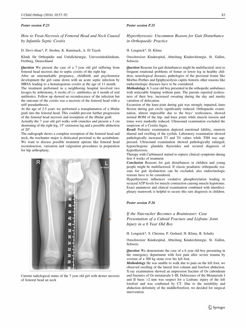
Poster session P.23
How to Treat-Necrosis of Femoral Head and Neck Caused
by Infantile Septic Coxitis
D. Dovi-Akue*, P. Strohm, K. Kuminack, A. El Tayeh
Klinik fur Orthopadie und Unfallchirurgie, Universitatsklinikum,
Freiburg, Deutschland
Question We present the case of a 7 year old girl suffering from
femoral head necrosis due to septic coxitis of the right hip.
After an unremarkable pregnancy, childbirth and psychomotor
development the girl came down with an acute septic infection by
MRSA leading to a hematogenous coxitis at the age of 11 month.
The treatment performed in a neighboring hospital involved two
lavages by arthrotomy, 6 weeks of i.v. antibiotics an 4 month of oral
antibiotics. Follow up showed no recrudescence of the infection but
the outcome of the coxitis was a necrosis of the femoral head with a
stiff pseudarthrosis.
At the age of 2.5 years we performed a transplantation of a fibular
graft into the femoral head. This couldnt prevent further progression
of the femoral head necrosis and resorption of the fibular graft.
Actually the 7 year old girl walks with crunches and present a 3 cm
shortening of the right leg, 15� extension lag and a possible abduction
of 20�.The radiograph shows a complete resorption of the femoral head and
neck, the trochanter major is dislocated proximal to the acetabulum.
We want to discuss possible treatment options like femoral head
reconstruction, varisation and valgisation procedures in preparation
for hip arthroplasty.
Current radiological status of the 7 year old girl with dexter necrosis
of femoral head an neck
Poster session P.25
Hyperthyreosis: Uncommon Reason for Gait Disturbance
in Orthopaedic Practice
H. Lengnick*, H. Klima
Ostschweizer Kinderspital, Abteilung Kinderchirurgie, St. Gallen,
Schweiz
Question Reasons for gait disturbances might be multifaceted: next to
frequent rotational problems of femur or lower leg in healthy chil-
dren, neurological diseases, pathologies of the proximal femur like
Morbus Perthes and Epiphyseolysis capitis femoris other reasons like
endocrinologic diseases have to be considered.
Methodology A 3-year old boy presented in the orthopedic ambulance
with noticeable limping without pain. The parents reported restless-
ness of their boy, increased sweating during the day and mushy
variation of defecation.
Excursion of the knee-joint during gait was strongly impaired, knee
flexion during gait circle significantly reduced. Orthopaedic exami-
nation, almost impossible due to the boys‘ restlessness, showed
normal ROM of the hip- and knee joints while muscle tension and
tonus were markedly reduced. Ultrasound examination excluded the
suspicion of a Coxitis fugax.
Result Pediatric examination depicted emotional lability, enuresis
diurnal and swelling of the eyelids. Laboratory examination showed
pathologically increased T3 and T4 values while TSH was sup-
pressed. Ultrasound examination showed pathologically enlarged,
hypoechogenic glandula thyreoidea and secured diagnosis of
hyperthyreosis.
Therapy with Carbimazol started to repress clinical symptoms during
first 4 weeks of treatment.
Conclusion Reasons for gait disturbances in children and young
people might be multifactored. If classic peadiatric orthopaedic rea-
sons for gait dysfunction can be excluded, also endocrinologic
reasons have to be considered.
Hyperthyreosis influences oxidative phosphorylation leading to
recuced ATP levels for muscle contraction causing muscle hypotonus.
Exact anamnesis and clinical examination combined with interdisci-
plinary teamwork is helpful to secure this rare diagnosis in children.
Poster session P.26
If the Nutcracker Becomes a Brainteaser: Case
Presentation of a Cuboid Fracture and Lisfranc Joint
Injury in a 6 Year Old Boy
H. Lengnick*, S. Christen, P. Gerhard, H. Klima, B. Schultz
Ostschweizer Kinderspital, Abteilung Kinderchirurgie, St. Gallen,
Schweiz
Question We demonstrate the case of a 6 year old boy presenting in
the emergency department with foot pain after severe trauma by
overrun of a 300 kg stone over his left foot.
Methodology He was unable to walk due to pain on the left foot, we
observed swelling of the lateral foot column and forefoot abduction.
X-ray examination showed an impression fracture of Os cuboideum
and fractures of Os metatarsale I–III. Dehiscence of the Metatarsale I
and II basis [2 mm was suspect for a Lisfranc injury of the left
forefoot and was confirmed by CT. Due to the instability and
abduction deformity of the middle/forefoot, we decided for surgical
intervention.
J Child Orthop (2016) 10:57–92 83
123
Result A lateral approach and open reduction of Os cuboideum
impression fracture was performed using the ‘‘Hintermann’’-device to
introduce a Tutobone wedge to hold reposition by K-wire osteosyn-
thesis. Percutaneous fixation of Lisfranc joint was implemented using
one intramedullare K-wire for Os metatarsale II and two K-wires for
parallel Lisfranc joint fixation building the typical triangle. Postop-
erative after-treatment included plaster cast for 6 weeks without
weight bearing, then K-wire displacement and another cast for
6 weeks while full weight bearing was allowed. Intraoperatively, the
calcaneo-cuboid joint was completely reduced, but postoperative
X-ray showed an oblique cuboid position in relation to calcaneus. Six
week postoperative X-ray control showed the improvement of the
oblique calcaneo-cuboid joint position while 12 weeks postopera-
tively the joint position was normal. An orthopaedic shoe inlay
supported foot position of the medial and lateral foot column.
Conclusion Cuboid-impression fractures are, in combination with
Lisfranc joint injury, a very rare foot pathology in children and young
people. The injury mechanism reminds of a nutcracker, when Os
cuboideum is pinched between Calcaneus and the basis of Metatarsale
V. Lisfranc joint injuries may lead to forefoot instability causing pain
and longterm arthritis. Therefore surgical stabilization is necessary to
prevent foot deformity, pain and abrasive wear. Literature describes
only few cases with these kind of fractures in children or young
people, but nutcracker and Lisfranc joint fracture have to be con-
sidered after severe foot trauma.
ReferenceRuffing T et al (2010) Die kindliche Nussknacker-Fraktur, 2010.
Unfallchirurg, 113:495–500
Poster session P.27
Kniest Dysplasia
D. Cerkez*, T. Wirth
Olgahospital, Orthopadische Klinik, Stuttgart, Deutschland
Question Kniest dysplasia is a rare chondrodysplasia with mutation in
the type II collagen gene, COL2A1. It is characterized by dispro-
portionate dwarfism at birth, large head round face, flat midface,
depressed nasal bridge, severe myopia, cataracts, vitreoretinal
degeneration, retinal detachment, Cleft palate, hearing loss, recurrent
otits media, short trunk, short neck, sternal protrusion, kyphoscoliosis,
shortening and bowing of long bone, prominent stiff joints and
clubfoot.
A 5 year old boy presented in October 2014 in our clinic. After birth
the diagnosis of Kniest-dysplasia was made. He had been operated on
his cleft palate. Because of his defective hearing he got hearing aids.
He had also recurrent acute respiratory diseases after postnatal res-
piratory insufficiency. For his mobility he needs orthoses and other
utilities. He could walk short steps free, for longer distance he needs a
walking frame or he uses the therapeutic trainer bike.
Actually he is going into the regular kindergarten with integration
help. He has twice a week physiotherapy with stretching and Galileo
vibration. Now the family is consulting us and asking for better
mobility.
Clinically the patient is mobile on his therapeutic trainer bike with
ankle–foot orthoses. He can demonstrate a free walking but he
manually supports his trunk. We see flexion contractures from both
hips and knees. In supine position there is a flexion/extension 110-30-
0� and an internal/external rotation 20-0-40�. The knee flexion/ex-
tension is 120-30-0. The ankle joint shows good mobility. The feet are
in valgus position with syndactyly of digitus II and III. The spine is
balanced but there is a small right convex scoliosis with hyper-
kyphosis and hyperlordosis. There is a limited spinal flexibility. On
the upper extremity we see on both sides prominent elbow with
extension/flexion 0-10-140�, the supination/pronation is 80-0-70�. On
both sides we see ulna deviation of the wrist and the dorsal exten-
sion/palmar flexion 10-0-90�.On the radiographic pictures there are bilateral coxa vara with broad
metaphysis and missing ossification of both femoral heads and a
dysplastic acetabulum. The feet are flat with ground-glass appearance
of the bone, the talus is also flat. The spine demonstrates a small right
convex scoliosis, hyperkyphosis and hyperlordosis with transforma-
tion of the vertebral bodies.
Treatment for better mobility in particular walking capacity?
Poster session P.28
Large Osteochondromas of the Femoral Neck: Results
of Resection by an Intertrochanteric Osteotomy
C. U. Dussa, C. Eisfeldt, S. Mehler, L. Doederlein*
Orthopadische Kinderklinik, Kinderorthopadie, Aschau, Deutschland
Question Decentring of the hip or severe restriction of ROM in
multiple hereditary osteochondromas (MHO) may cause considerable
functional impairment through expanding dorsocaudal osteochon-
dromas at the medial border of the femoral neck. Resection is
necessary if pain and stiffness as well as radiographic signs of lat-
eralisation of the femoral head occur. As the accessibility of these
major bone masses through an anterior approach is difficult we have
started to excise them through an intertrochanteric osteotomy
approach. We would like to present our results and discuss the indi-
cation for resection.
Methodology Between 2009 and 2014 we have treated 4 patients with
MHO and functional limitations by an intertrochanteric osteotomy
approach and resection of the bone masses. There were 2 girls and
two boys 6,8,11 and 14 years old. All osteochondromas were on the
left side. All osteotomies were internally fixed and the patients were
treated by an early mobilization non weight bearing program for
6–8 weeks.
Result All osteotomies healed uneventfully. The mobility of the hip
joints could be re-established and the hips were concentric at latest
follow up (6 months to 4 years postop.).There was no instance of
AVN.
We indicated surgical resection when there were radiographic signs of
decentration of the hip joint (widening of medial joint space) and
when hip adduction, flexion as well as internal rotation were restricted
to less than 0� of adduction, less than 90� of flexion and less than 0� of
internal rotation, i.e. if an extension, abduction and external rotation
contracture had developed. Pain was also an indicator for removal.
Conclusion Removal of osteochondromas at the inferior border of the
femoral neck seems indicated if progressive extrusion of the hip joint
as well as restriction of hip ROM or pain occur. Removal through an
intertrochanteric osteotomy approach is despite its invasiveness a safe
method to remove all of the pathologically protruding bone by pro-
tecting the vascularity of the femoral head. Immediate mobilization is
possible. Recurrence of the resected osteochondromas had not been
detected until now.
84 J Child Orthop (2016) 10:57–92
123

Poster session P.29
Magnetic Controlled Growing Rods: First Experience
in 12 Patients
T. Lange*, S. Bockholt, G. Gosheger, A. Schulze Bovingloh,
T. L. Schulte
Klinik fur Allgemeine Orthopadie und Tumororthopadie, Univer-
sitatsklinikum, Munster, Deutschland
Question Over the last years magnetic controlled growing rods have
been introduced as an alternative to traditional growing rods [1] to
facilitate distractions without any operative intervention. We report
our experience in 12 patients.
Methodology 12 patients have been operated within 18 months using
magnetic controlled growing rods. The mean followup is 14.5 months
(3–22 months). In these cases Cobb angle, angle of kyphosis (T4-12)
and the distance between the spinous process of T1 and S1 were
measured on X-rays pre-, postoperatively and at followup. Further-
more the number of distraction procedures, the total distraction
distance, the number and type of revision surgeries and complications
were analyzed.
Result 12 patients (8 girls, 4 boys; type of scoliosis: idiopathic
(n = 6), neuromuscular (n = 4), congenital (n = 1), syndromic
(n = 1)) with a mean age of 10.4 y (6.8–15.9 years) have been
operated before skeletal maturity using magnetic controlled growing
rods. All patients received a dual rod construct. The fixation method
varied between pedicle screws (n = 10) and rib and ilium hooks
(n = 2) which were chosen due to dysplastic pedicles. On average 5
distractions (total 54, range 1–8) were carried out on a 3-monthly
basis and 4.3 mm distraction could be reached at each time
(Methodology: 5 mm).
Mean preoperative cobb angle was 72� (52�–109�), after surgery
41� (26�–72�, 43 % correction, p B 0.001) and at followup 41�(28�–93�, 43 % correction, p B 0.001). Mean preoperative kyphosis
angle was 36� (17�–70�), after surgery 25� (2�–63�, 31 % correc-
tion, p B 0.05) and at followup 26� (5�–66�, 28 % correction,
p = 0.07). On average the T1-S1 distance was 291 mm preopera-
tively (211-374 mm), after surgery 325 mm (253–414 mm, 11.7 %
increment, p B 0.001) and at followup 335 mm (267–421 mm,
15 % increment, p B 0.001) resulting in a length growth T1-S1 of
8.3 mm/year.
In 2 patients implant associated complications occurred leading to
revision surgeries (wound infection with subsequent removal of one
rod, implant replacement after rod fracture).
Conclusion It could be shown that the initial surgery using magnetic
controlled growing rods leads to an acceptable Cobb angle correction
which persisted during the following distractions. A kyphosing effect
during distraction could not be detected.
This technique is safe and allows length growth over time of about
8.3 mm/year without any physically demanding consecutive surg-
eries. These results correspond well with the current literature [2].
References[1] Akbarnbia BA (2012) Innovation in growing rod technique: a
study of safety and efficacy of a magnetically controlled growing rod
in a porcine model. Spine
[2] Hickey BA (2014) Early experience of MAGEC magnetic
growing rods in the treatment of early onset scoliosis, Eur Spine J
Poster session P.30
Metamorphosis of Normal Human Lumbar Vertebrae
to Quadruped-Like Shape by VEPTR Induced Growth
Modulation
C. Hasler*1, D. Studer1, P. Buchler2
1Universitatskinderspital beider Basel, Orthopadie, Basel, Schweiz,2Institute for Surgical Technology and Biomechanics Computational
Bioengineering Group, Bern, Schweiz
Question To assess the growth modulating impact of year long dis-
traction-based VEPTR-treatment on vertebral morphology
Methodology We digitally measured lumbar vertebral body heights
(VBH) and upper endplate depths (VBD) at the time of the index
procedure and at follow-up in 9 patients with rib-to-ileum constructs
(group 1) spanning a normal lumbar spine. Nine patients with con-
genital thoracic scoliosis and rib-to-rib constructs but uninstrumented
lumbar spines served as controls (group 2). All had undergone more
than 8 half yearly VEPTR expansions. Wilcoxon signed-rank test was
used for statistical analysis of initial VBH, VBD, height/depth ratio
and at follow-up (significance level 0.05).
Result The average age was 7.1 years (G1) and 5.2 year (G2,
p[ 0.05)) at initial surgery, the average overall follow-up time
5.5 years (p[ 0.05). In both groups VBH increased significantly
without significant intergroup difference. Group 1 did not show sig-
nificant growth in depth, whereas VBD increased significantly in the
control group. As a consequence H/D ratio increased significantly in
group 1 whereas it remained unchanged in group 2. The growth rate in
mm/year was 1.4 (group 1) and 1.1 (group 2, p = 0.45) for height and
-0.3 and 1.1 (p\ 0.05) for depth, respectively.
Conclusion VEPTR growth modulating treatment alters the geometry
of vertebral bodies by increasing the height/depth ratio. We hypoth-
esize that in parallel to the distraction-induced gain of vertebral
height, the implant related deprivation from axial loads (stress-
shielding) impairs anteroposterior growth. The biomechanical con-
sequence of such slender vertebrae exposed to unprotected loads in
case of definitive VEPTR removal at the end of growth is uncertain.
Poster session P.31
Midfoot Joints Compensate the Decreased Dorsal
Extension of Ankle Joint Following Clubfoot Correction
C. U. Dussa*, F. Paulitsch, H. Bohm, M. Hosl, L. Doederlein
Orthopadische Kinderklinik, Kinderorthopadie, Aschau, Deutschland
Question The commonest residual deformity following successful
correction of clubfoot is reduced dorsal extension of the ankle joint.
This may be due to several reasons. We undertook a study to see the
effect of decreased dorsal extension in ankle on the foot.
Methodology The data for the study was acquired from a prospective
Gait laboratory database for clubfeet. All of them underwent foot
analysis using Oxford foot model to assess the kinematics and
kinetics. Inclusion criteria were: clubfoot of idiopathic aetiology, post
correction through surgery, hind foot eversion. Exclusion criteria
J Child Orthop (2016) 10:57–92 85
123
were: neurogenic and syndromal clubfeet, hind foot inversion. In all
58 patients were identified which fulfilled the above criteria. The
observed parameters were dorsal extension of the ankle joint, dorsal
extension of the midfoot, supination of the forefoot, various mea-
surements of the Talus on X-rays. Statistical analysis was done to
determine the significance of individual parameter.
Result 48 Patients with 67 feet were included in the study. Of these,
19 patients had bilateral clubfoot. The compensatory mechanisms
identified in the foot were increased dorsal mobility in the midfoot
especially the increased dorsal extension in the midfoot joint and
increased supination. Statistical analysis showed a strong correlation
between decreased ankle joint dorsal extension and increased dorsi-
flexion of the midfoot joints. However no statistical significance was
found between the decreased ankle joint dorsal extension and maxi-
mal supination of the foot in mid-stance and terminal stance phase.
The measurements of bony landmarks identified the possible cause of
decreased dorsal extension.
Conclusion The effect of decreased ankle joint motion on the foot in
clubfoot has not been mentioned. Dorsal extension of the ankle joint
is necessary for 2nd Rocker motion during gait. Absence of the 2nd
Rocker leads to premature heel raise. This is to some extent com-
pensated by increase in midfoot joint motion predominantly dorsal
extension. This finding has 2 clinical implications: (1) clinically
measured passive dorsal extension of ankle joint is mostly the sum-
mation of dorsal extension in ankle and midfoot joints, (2) it is
important to be aware of this compensation before one considers to
fuse the midfoot joints to correct a midfoot deformity. This may lead
to increased loading (impingement) of ankle joint and pain.
Poster session P.32
Mobility Without Wheelchair Despite Complex
Dysmorphia-Syndrome
S. Kappl*1, M. Reiter2, S. Nader1
1Schon Klinik, Klinik fur Kinderorthopadie, Vogtareuth, Deutsch-
land, 2Sporer Gesundheitsgruppe, Vogtareuth, Deutschland
Question Patients with complex defects of the lower extremities are
obviously massively limited in their mobility. Insufficient operative
treatment or rash amputations frequently result in difficulties of
orthoprothetic care/supply. Therefore sufficient orthoprothetic treat-
ment is facing a permanent wheelchair supply. In our case the positive
effects of psycho-social development are impressivly shown but also
the limits of technical ability by growing demands.
Methodology A 14-year-old female patient with tibial and Patella
aplasia and congenital dysplasia of the hips. In childhood Fixateur
externe and verticalization by wearing ortheses. Attending elementary
school for mentally and physically disabled where not challenged
enough. A sufficient orthoprothetic treatment lead to integration in a
normal school resulting in a normal maturation. The requirements of
taking part in daily working life are provided. Growing demands in
treatment due to free mobility, height, physical education and use of
public transport. Main problems caused by worn out and insufficient
fit of the ortheses due to loss of weight of 7 kg within 6 months.
Showing pain in the rudimentary knees. Modification of ortheses:
Femoral part with dorsal fixation and tube framing, knee flexible,
tibial part with ventral fixation and extension, ring-like framing oft he
foot, soft liner and carbon foot. Reducing weight by using carbonized
GFK and no cosmetic lining.
Result The patient is walking without any aids apart from ortheses,
even stairs in alternating pace, normal height. Problems in stabilizing
the rudimentary knee joints. After calculating the costs the health
insurance is in doubt of necessity of ortheses.
Conclusion Continuously use results in short intervals of maintenance
due to safety aspects. Several soft liners are necessary for changing.
The main focus is put on stabilising the knee joints. The treatment is
team work, the use of new technologies inevitable. Continuous new
developmenst have to be proofed like the Sinterplot-processing.
Tough the manufacture of parts is possible to do out-patient the actual
testing has to be done under close therapeutical supervision i.e. in-
patient. It has to be fought for the preservation of this option despite
the growing costs. The staff might be stressed too much if the syn-
dromes are exceptional. Discussions are helpful. In the end a doctor
should review the result to ensure the quality and to optimize the
process.
Poster session P.33
Multiple Osteochondromata (exostoses) in the Forearm
with Carpal Deviation
J. Willers, E. Dingeldey*, J. Matussek
Orthopadische Klinik Universitat Regensburg, Kinderorthopadie,
Regensburg, Deutschland
Question Seperate lengthening processes of the deformed ulna in a
twisted and deformed forearm yields a better hand function—cases
presentation
Poster session P.34
Musculoskeletal Manifestations in Mucopolysaccharidosis
Type I (Hurler Syndrome) Following Hematopoietic Stem
Cell Transplantation
S. Breyer*1, M. Schmidt2, U. Lobel3, S. Yarar4, R. Stucker1, K.
Ullrich2, I. Muller5, N. Muschol2
1Altonaer Kinderkrankenhaus, Kinderorthopadie, Hamburg,
Deutschland, 2UKE, Padiatrie, Hamburg, Deutschland, 3UKE,
Department of Diagnostic and Interventional Neuroradiology, Ham-
burg, Deutschland, 4UKE, Department of Trauma, Hand and
Reconstructive Surgery, Hamburg, Deutschland, 5UKE, Clinic of
Pediatric Hematology and Oncology, Division for Stem Cell Trans-
plantation, Hamburg, Deutschland
Question Hematopoietic stem cell transplantation (HSCT) is the
treatment of choice in Hurler patients (mucopolysaccharidosis type
I-H, MPS I-H). Following HSCT life expectancy improves signifi-
cantly, whereas musculoskeletal manifestations are assumed to be
progressive. Better understanding of the underlying pathology of joint
and bone disease becoming more relevant. The present study sum-
marizes the progression of musculoskeletal manifestation in 19 Hurler
patients following HSCT and proposes diagnostic algorithms and
treatment recommendations.
Methodology Range of motion (JROM) of shoulders, elbows, hips
and knees in 19 MPS patients, who underwent allogenic HSCT
between 1991 and 2012, were assessed during a mean follow-up
period of 6.4 years (range 0.7–22.5 years). Radiographs of thorax,
spine, pelvis and hands as well as magnetic resonance imaging (MRI)
scans of the craniocervical junction (CCJ) and odontoid process were
analyzed.
Result Nineteen evaluable Hurler patients (10 females, 9 males) were
included in the study. Ten patients achieved long-term engraftment
with chimerism [94 %, seven patients presented with a mixed
86 J Child Orthop (2016) 10:57–92
123
chimerism between 16.7 and 59.3 %, whereas two patients developed
graft failure during follow-up. Ten patients developed acute Graft-
versus-Host Disease (aGvHD). All patients had joint contractures.
The majority of patients showed a steady state or even improvements
in JROM of knees (33 or 56 % resp.), hips (41 or 35 % resp.) and
elbows (56 or 33 % resp.) leading to a full range of motion in
39–61 % of joints. In patients with graft failure progressive restric-
tions in JROM were noted. Assessments of craniocervical stenosis
(CCS) by MRI scan showed stable or improved diameters and cor-
rection of odontoid hypoplasia during follow-up in most cases and
therefore no severe complications were recorded. However thora-
columbar kyphosis, genua valga as well as hip dysplasia were
progressive despite HSCT.
Conclusion HSCT does not only improve long-term survival and
cognitive development, but has a beneficial effect on several mus-
culoskeletal manifestations in Hurler patients. Nevertheless, frequent
monitoring for skeletal complications and supportive therapies are
essential to preserve and improve motor function.
Poster session P.35
Postradiation Acetabular Dysplasia
in Rhabdomyosarcoma
E. Dingeldey, J. Matussek*
Orthopadische Klinik Universitat Regensburg, Kinderorthopadie,
Regensburg, Deutschland
Question Severe pain and leg length discrepancy in a 16 year old boy
with acetabular dysplasia after childhood radiation. PAO and
lengthening processes did not seem to have been sufficient. Muscular
gluteal deficiency seems to produce major treatment difficulties.
Poster session P.36
Proximal Epiphyseal Slip Following Proximal Varus
Derotation Osteotomy of Femur
C. U. Dussa*, L. Doederlein
Orthopadische Kinderklinik, Kinderorthopadie, Aschau, Deutschland
Question A 6-year old boy presented to our clinic with left sided hip
dislocation. I was diagnosed to have Becker-Adams-Syndrome as a
result of chromosomal anomaly.
Clinical examination showed an obese, non cooperative patient
weighing 35 kg. A limb length discrepancy of 1.5 cm shortening on
the left was seen. Passive hip abduction was reduced on the left side
compared to right (R:L = 30�:10�). Otherwise all the other move-
ments of the hip and knee joint are free and full. The ankle joints
showed fixed equines deformity. In addition, the deep tendon reflexes
were exacerbated. Clonus could be elicited. Babinski was positive on
both sides. Walking with support was extremely cumbersome.
X-rays showed a left sided hip dislocation with acetabular dysplasia.
The right hip was normal. A varisation, derotation shortening
osteotomy (DVO) of the proximal femur was performed along with
open reduction of the hip joint. A dega type of acetabuloplasty was
performed. Postoperatively the patient was immobilised in plaster of
Paris cast due to lack of cooperation. Following consolidation of the
osteotomy in 6 weeks, he was referred to a rehabilitation unit. During
mobilisation in a Locomat Trainer, he experienced pain in left hip
which worsened, till the patient could no more be mobilised on
Locomat Trainer. The rehabilitation was discontinued.
Examination on readmission at our hospital, showed elevated tem-
perature 390 �C, increased leucocytes and CRP. X-rays showed
osetolysis of the neck of femur with slip of the proximal femur epi-
physis. An epiphyseal slip due to infection was suspected.
Arthrotomy showed blood stained fluid with no abscess. A val-
gisation osteotomy was done after reduction of the head and
K-Wire pinning. Microbiology showed no bacterial growth. Fol-
low-up after 5 years showed complete necrosis of proximal femur
epiphysis with redislocation of the hip joint and severe hip dys-
plasia. On the right side as well, the hip showed a progressive hip
dislocation.
Discussion Literature shows only one publication with 2 cases
describing similar chain events following surgery. The possible cau-
ses for this complication will be discussed. It is also to be discussed of
the right hip now needs containment. The case has been presented for
its rarity.
Poster session P.38
Reconstruction of a Traumatic Joint Defect in the Forefoot
by Autologous Iliac Apophysis Transplantation in a Child
L. Schwering*1, V. T. Forster2
1Mathias-Spital, Technische Orthopadie, Rheine, Deutschland,2Universitatsklinikum Freiburg, Department Orthopadie Trauma-
tologie, Freiburg, Deutschland
Question So far only a few studies investigated techniques to repair
joint and growth plate defects in the forefoot of children. Trans-
plantation of autologous apophyseal cartilage has shown successful in
reconstruction of joint defects in children in several case reports.
These cases predominantly describe reconstruction of phalangeal joint
defects in hand surgery.
How effective can apophyseal transplantation in forefoot recon-
struction be?
Methodology The case of a 6 year-old girl is subscribed who sus-
tained a wound 2 9 2 cm in size over the medial part of the first
metatarsophalangeal joint in an accident. Besides the extensive
damage to the soft tissue and tendons the radiographs on admission
revealed an osteo-cartilaginous defect of three-fourths of the proximal
phalanx of the first metatarsophalangeal joint. After an initial
debridement and using vacuum bandage for a few days we recon-
structed the joint replacing the osteo-cartilaginous defect with an
autologous cartilage graft of the iliac apophysis and tendon transfer.
Then the residual soft tissue defect was covered with a rotation flap of
the skin by our plastic surgeons. Due to an impaired healing of the
skin flap revision surgery with transplantation of a skin mesh graft
had to be performed 2 weeks later.
Result Complete healing of the wound was achieved 6 weeks after
the injury. Radiographs taken at 4, 6 and 12 months after surgical
correction showed a good integration and remodeling of the grafted
area. Full weight-bearing was allowed after 8 weeks. 6 months fol-
lowing surgery the child presented free from pain with an almost
symmetrical range of movement in the first metatarsophalangeal joint
comparing both feet.
Conclusion We present the medium- term outcome transferring
cartilage of the iliac apophysis to substitute a traumatic joint defect
of the first metatarsophalangeal joint in a pediatric patient. To date
the clinical and radiological result is satisfactorily 24 months after
the injury. Of course the long-term result remains to be seen. In
order to prove the efficacy of using non-vascularize autologous
cartilage grafts in children to repair joint defects in the forefoot
further trials with larger number of cases will be needed in the
future
J Child Orthop (2016) 10:57–92 87
123
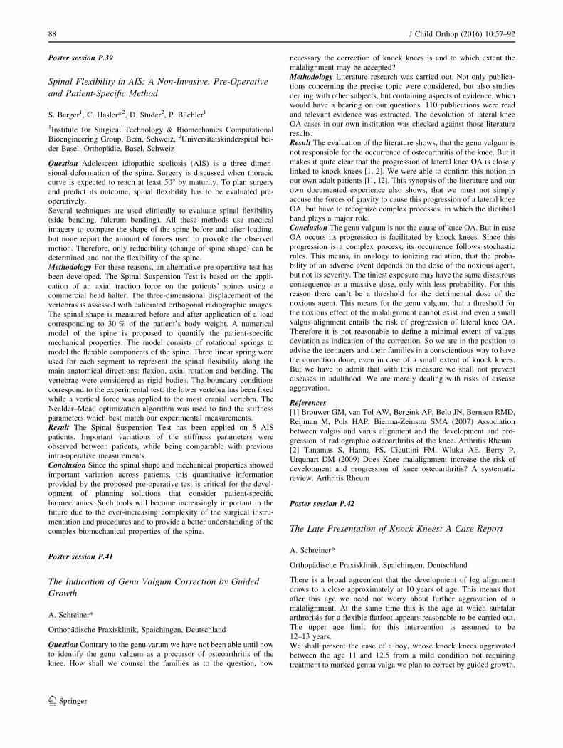
Poster session P.39
Spinal Flexibility in AIS: A Non-Invasive, Pre-Operative
and Patient-Specific Method
S. Berger1, C. Hasler*2, D. Studer2, P. Buchler1
1Institute for Surgical Technology & Biomechanics Computational
Bioengineering Group, Bern, Schweiz, 2Universitatskinderspital bei-
der Basel, Orthopadie, Basel, Schweiz
Question Adolescent idiopathic scoliosis (AIS) is a three dimen-
sional deformation of the spine. Surgery is discussed when thoracic
curve is expected to reach at least 50� by maturity. To plan surgery
and predict its outcome, spinal flexibility has to be evaluated pre-
operatively.
Several techniques are used clinically to evaluate spinal flexibility
(side bending, fulcrum bending). All these methods use medical
imagery to compare the shape of the spine before and after loading,
but none report the amount of forces used to provoke the observed
motion. Therefore, only reducibility (change of spine shape) can be
determined and not the flexibility of the spine.
Methodology For these reasons, an alternative pre-operative test has
been developed. The Spinal Suspension Test is based on the appli-
cation of an axial traction force on the patients’ spines using a
commercial head halter. The three-dimensional displacement of the
vertebras is assessed with calibrated orthogonal radiographic images.
The spinal shape is measured before and after application of a load
corresponding to 30 % of the patient’s body weight. A numerical
model of the spine is proposed to quantify the patient-specific
mechanical properties. The model consists of rotational springs to
model the flexible components of the spine. Three linear spring were
used for each segment to represent the spinal flexibility along the
main anatomical directions: flexion, axial rotation and bending. The
vertebrae were considered as rigid bodies. The boundary conditions
correspond to the experimental test: the lower vertebra has been fixed
while a vertical force was applied to the most cranial vertebra. The
Nealder–Mead optimization algorithm was used to find the stiffness
parameters which best match our experimental measurements.
Result The Spinal Suspension Test has been applied on 5 AIS
patients. Important variations of the stiffness parameters were
observed between patients, while being comparable with previous
intra-operative measurements.
Conclusion Since the spinal shape and mechanical properties showed
important variation across patients, this quantitative information
provided by the proposed pre-operative test is critical for the devel-
opment of planning solutions that consider patient-specific
biomechanics. Such tools will become increasingly important in the
future due to the ever-increasing complexity of the surgical instru-
mentation and procedures and to provide a better understanding of the
complex biomechanical properties of the spine.
Poster session P.41
The Indication of Genu Valgum Correction by Guided
Growth
A. Schreiner*
Orthopadische Praxisklinik, Spaichingen, Deutschland
Question Contrary to the genu varum we have not been able until now
to identify the genu valgum as a precursor of osteoarthritis of the
knee. How shall we counsel the families as to the question, how
necessary the correction of knock knees is and to which extent the
malalignment may be accepted?
Methodology Literature research was carried out. Not only publica-
tions concerning the precise topic were considered, but also studies
dealing with other subjects, but containing aspects of evidence, which
would have a bearing on our questions. 110 publications were read
and relevant evidence was extracted. The devolution of lateral knee
OA cases in our own institution was checked against those literature
results.
Result The evaluation of the literature shows, that the genu valgum is
not responsible for the occurrence of osteoarthritis of the knee. But it
makes it quite clear that the progression of lateral knee OA is closely
linked to knock knees [1, 2]. We were able to confirm this notion in
our own adult patients [I1, I2]. This synopsis of the literature and our
own documented experience also shows, that we must not simply
accuse the forces of gravity to cause this progression of a lateral knee
OA, but have to recognize complex processes, in which the iliotibial
band plays a major role.
Conclusion The genu valgum is not the cause of knee OA. But in case
OA occurs its progression is facilitated by knock knees. Since this
progression is a complex process, its occurrence follows stochastic
rules. This means, in analogy to ionizing radiation, that the proba-
bility of an adverse event depends on the dose of the noxious agent,
but not its severity. The tiniest exposure may have the same disastrous
consequence as a massive dose, only with less probability. For this
reason there can’t be a threshold for the detrimental dose of the
noxious agent. This means for the genu valgum, that a threshold for
the noxious effect of the malalignment cannot exist and even a small
valgus alignment entails the risk of progression of lateral knee OA.
Therefore it is not reasonable to define a minimal extent of valgus
deviation as indication of the correction. So we are in the position to
advise the teenagers and their families in a conscientious way to have
the correction done, even in case of a small extent of knock knees.
But we have to admit that with this measure we shall not prevent
diseases in adulthood. We are merely dealing with risks of disease
aggravation.
References[1] Brouwer GM, van Tol AW, Bergink AP, Belo JN, Bernsen RMD,
Reijman M, Pols HAP, Bierma-Zeinstra SMA (2007) Association
between valgus and varus alignment and the development and pro-
gression of radiographic osteoarthritis of the knee. Arthritis Rheum
[2] Tanamas S, Hanna FS, Cicuttini FM, Wluka AE, Berry P,
Urquhart DM (2009) Does Knee malalignment increase the risk of
development and progression of knee osteoarthritis? A systematic
review. Arthritis Rheum
Poster session P.42
The Late Presentation of Knock Knees: A Case Report
A. Schreiner*
Orthopadische Praxisklinik, Spaichingen, Deutschland
There is a broad agreement that the development of leg alignment
draws to a close approximately at 10 years of age. This means that
after this age we need not worry about further aggravation of a
malalignment. At the same time this is the age at which subtalar
arthrorisis for a flexible flatfoot appears reasonable to be carried out.
The upper age limit for this intervention is assumed to be
12–13 years.
We shall present the case of a boy, whose knock knees aggravated
between the age 11 and 12.5 from a mild condition not requiring
treatment to marked genua valga we plan to correct by guided growth.
88 J Child Orthop (2016) 10:57–92
123
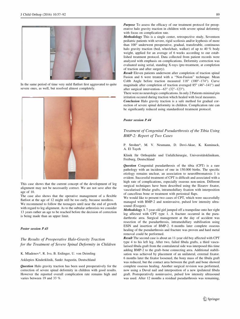
In the same period of time very mild flatfeet first aggravated to quite
severe ones, as well, but resolved almost completely.
This case shows that the current concept of the development of leg
alignment may not be necessarily correct. We are not save after the
age of 10.
The case also shows that the operative management of a flexible
flatfoot at the age of 12 might still be too early, because needless.
We recommend to follow the teenagers until near the end of growth
with regard to leg alignment. As to the subtalar arthrorisis we consider
13 years rather an age to be reached before the decision of correction
is being made than an upper limit.
Poster session P.43
The Results of Preoperative Halo-Gravity Traction
for the Treatment of Severe Spinal Deformity in Children
K. Mladenov*, R. Ivo, B. Erdinger, U. von Deimling
Asklepios Kinderklinik, Sankt Augustin, Deutschland
Question Halo gravity traction has been used preoperatively for the
correction of severe spinal deformity in children with good results.
However the reported overall complication rate remains high and
varies between 19 and 33 %.
Purpose To assess the efficacy of our treatment protocol for preop-
erative halo gravity traction in children with severe spinal deformity
with focus on complication rate.
Methodology This is a single center, retrospective study, Seventeen
pediatric patients with severe, rigid scoliosis and/or kyphosis of more
than 100� underwent preoperative, gradual, transferable, continuous
halo gravity traction (bed, wheelchair, walker) of up to 40 % body
weight, applied for an average of 6 weeks according to our estab-
lished treatment protocol. Data collected from patient records were
analyzed with emphasis on complications. Deformity correction was
evaluated using serial, standing X-rays (pre-treatment, at completion
of traction and after surgery).
Result Eleven patients underwent after completion of traction spinal
Fusion and 6 were treated with a ‘‘Non-Fusion’’ technique. Mean
Cobb Angle before traction measured 118� (100�–174�). Curve
magnitude after completion of traction averaged 85� (46�–141�) and
after surgical intervention—63� (32�–123�).There were no neurologic complications. In only 2 Patients minimal pin
irritation occured during traction which healed with local measures.
Conclusion Halo gravity traction is a safe method for gradual cor-
rection of severe spinal deformity in children. Complication rate can
be significantly reduced using standardized treatment protocol.
Poster session P.44
Treatment of Congenital Pseudarthrosis of the Tibia Using
BMP-2: Report of Two Cases
P. Strohm*, M. V. Neumann, D. Dovi-Akue, K. Kuminack,
A. El Tayeh
Klinik fur Orthopadie und Unfallchirurgie, Universitatsklinikum,
Freiburg, Deutschland
Question Congenital pseudarthrosis of the tibia (CPT) is a rare
pathology with an incidence of one in 150.000 births. The specific
etiology remains unclear, an association to neurofibromatosis 1 is
evident. Successful treatment of CPT is difficult and associated with a
high rate of complications, especially osseous non-union. Different
surgical techniques have been described using the Ilizarov fixator,
vascularised fibular grafts, intramedullary fixation with interposition
of cancellous bone or treatment with periosteal flaps.
We would like to present two cases of CPT, which were successfully
managed with BMP-2 and noninvasive, pulsed low intensity ultra-
sound (Exogen).
Methodology A 7-year-old girl jumped off a trampoline onto her right
leg affected with CPT type 1. A fracture occurred in the pseu-
darthrotic area. Surgical management at the day of accident was
resection of the pseudarthrosis, intramedullary stabilisation using
ESIN and insertion of BMP-2. 6 months later complete osseous
healing of the pseudarthrosis and fracture was proven and hard metal
removal could be performed.
Result The second case is about an 11-year old boy affected with CPT
type 4 to his left leg. After two, failed fibula grafts, a third vascu-
larised fibula graft from the contralateral side was interposed this time
adding BMP-2 to the graft–bone connecting area. Additional stabili-
sation was achieved by placement of an unilateral, external fixator.
6 months later the fixator loosened, the bony mass of the fibula graft
was reduced, but the contact area between the graft and bone showed
complete osseous healing. Another surgical revision was performed,
now using a Duval nail and interposition of a new ipsilateral fibula
graft. Postoperatively noninvasive, pulsed low intensity ultrasound
was used. After 12 months a residual pseudarthrosis was remaining,
J Child Orthop (2016) 10:57–92 89
123
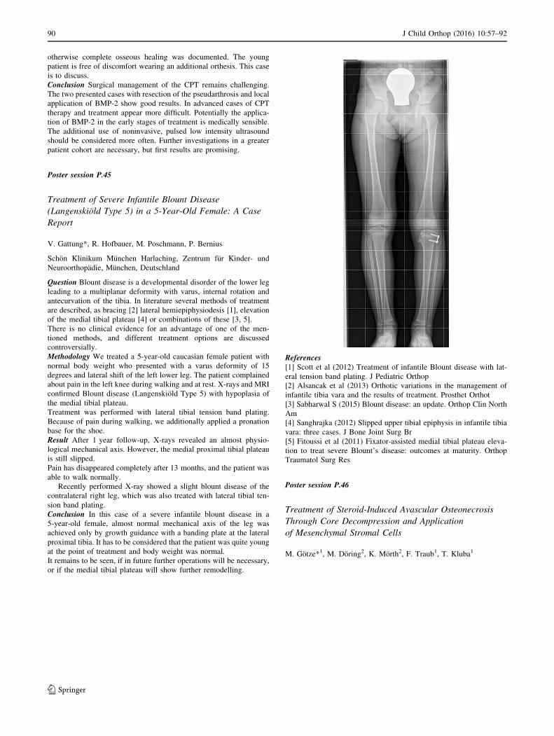
otherwise complete osseous healing was documented. The young
patient is free of discomfort wearing an additional orthesis. This case
is to discuss.
Conclusion Surgical management of the CPT remains challenging.
The two presented cases with resection of the pseudarthrosis and local
application of BMP-2 show good results. In advanced cases of CPT
therapy and treatment appear more difficult. Potentially the applica-
tion of BMP-2 in the early stages of treatment is medically sensible.
The additional use of noninvasive, pulsed low intensity ultrasound
should be considered more often. Further investigations in a greater
patient cohort are necessary, but first results are promising.
Poster session P.45
Treatment of Severe Infantile Blount Disease
(Langenskiold Type 5) in a 5-Year-Old Female: A Case
Report
V. Gattung*, R. Hofbauer, M. Poschmann, P. Bernius
Schon Klinikum Munchen Harlaching, Zentrum fur Kinder- und
Neuroorthopadie, Munchen, Deutschland
Question Blount disease is a developmental disorder of the lower leg
leading to a multiplanar deformity with varus, internal rotation and
antecurvation of the tibia. In literature several methods of treatment
are described, as bracing [2] lateral hemiepiphysiodesis [1], elevation
of the medial tibial plateau [4] or combinations of these [3, 5].
There is no clinical evidence for an advantage of one of the men-
tioned methods, and different treatment options are discussed
controversially.
Methodology We treated a 5-year-old caucasian female patient with
normal body weight who presented with a varus deformity of 15
degrees and lateral shift of the left lower leg. The patient complained
about pain in the left knee during walking and at rest. X-rays and MRI
confirmed Blount disease (Langenskiold Type 5) with hypoplasia of
the medial tibial plateau.
Treatment was performed with lateral tibial tension band plating.
Because of pain during walking, we additionally applied a pronation
base for the shoe.
Result After 1 year follow-up, X-rays revealed an almost physio-
logical mechanical axis. However, the medial proximal tibial plateau
is still slipped.
Pain has disappeared completely after 13 months, and the patient was
able to walk normally.
Recently performed X-ray showed a slight blount disease of the
contralateral right leg, which was also treated with lateral tibial ten-
sion band plating.
Conclusion In this case of a severe infantile blount disease in a
5-year-old female, almost normal mechanical axis of the leg was
achieved only by growth guidance with a banding plate at the lateral
proximal tibia. It has to be considered that the patient was quite young
at the point of treatment and body weight was normal.
It remains to be seen, if in future further operations will be necessary,
or if the medial tibial plateau will show further remodelling.
References[1] Scott et al (2012) Treatment of infantile Blount disease with lat-
eral tension band plating. J Pediatric Orthop
[2] Alsancak et al (2013) Orthotic variations in the management of
infantile tibia vara and the results of treatment. Prosthet Orthot
[3] Sabharwal S (2015) Blount disease: an update. Orthop Clin North
Am
[4] Sanghrajka (2012) Slipped upper tibial epiphysis in infantile tibia
vara: three cases. J Bone Joint Surg Br
[5] Fitoussi et al (2011) Fixator-assisted medial tibial plateau eleva-
tion to treat severe Blount’s disease: outcomes at maturity. Orthop
Traumatol Surg Res
Poster session P.46
Treatment of Steroid-Induced Avascular Osteonecrosis
Through Core Decompression and Application
of Mesenchymal Stromal Cells
M. Gotze*1, M. Doring2, K. Morth2, F. Traub1, T. Kluba1
90 J Child Orthop (2016) 10:57–92
123

1Universitatsklinik Tubingen, Orthopadie, Tubingen, Deutschland,2Universitatsklinik Tubingen, Padiatrie, Tubingen, Deutschland
Question Avascular osteonecrosis (AVN) show a strong association
with corticosteroid treatment in autoimmune and hematological dis-
eases. The most commonly affected sites are the femoral head and the
knee. Steroids seem to play a major role in inducing AVN, although
the particular inducing mechanisms are not known. Decreased per-
fusion, malfunction of endothelial cells as well as coagulopathies are
discussed as possible causes. If the joint surface remains unaffected
by the AVN, core decompression is the treatment of choice. Due to
their differentiation potential, the easy accessibility and their ability to
proliferate quickly, autologous mesenchymal stromal cells (MSC) are
a good option to advance the healing process of AVN when used in
addition to the core decompression.
Methodology Between 2006 und 2013 we treated 12 children and
young adults (sites n = 17) suffering an AVN following therapy with
cytostatics, immunosuppressives and corticosteroids with core
decompression and additional application of autologous MSCs. The
patients were examined clinically and radiologically with a follow-up
of 6 years after surgery (range 1–9 years). Criteria for a successful
treatment were the clinical development regarding pain at rest and on
weight-bearing (visual analog scale), subjective well-being, required
follow-on surgery like joint replacement and the radiologic outcome
compared to the initial images.
Result More than half of the patients showed radiologically regressive
necrotic areas after the treatment, more than 3/4 of the patients
experienced a clinical and subjective improvement of their discom-
fort. Outcome was not dependent on the applied total dose of MSCs
(in millions). 1 Patient had to undergo joint replacement surgery due
to a worsening of the AVN despite the treatment. There were no
negative side effects noted, that could have been traced back to the
MSC application.
Conclusion The application of MSCs in AVN treatment is possible
and primarily safe. The treatment regimen of core decompression and
MSC application can improve the radiologic and clinical outcome of
an AVN. The 12 present trials showed, that the use of MSCs is a
important addition to the classic core decompression treatment of the
AVN.
Poster session P.47
Unilateral Idiopathic Massive Hypertrophy of Intrinsic
Foot Muscles
M. Keim, C. U. Dussa, C. Multerer, L. Doederlein*
Orthopadische Kinderklinik, Kinderorthopadie, Aschau, Deutschland
Question Introduction: Idiopathic hypertrophy of the intrinsic foot
muscles is a rare disorder which can lead to significant stance prob-
lems due to a loss of heel and forefoot contact with the ground. The
patient walks less stable over cushion like soft plantar mass.
Case presentation: We present two boys (3 and 7 years old) each with
a unilateral massive plantar soft tissue swelling that prevents the
affected feet from a plantigrade position. The clinical picture
resembles a rocker-bottom-foot and examination reveals an elastic
subcutaneous painless tumor which covers the medial an plantar
region of the sole. and prevents the heel and forefoot from weight-
bearing.Weightbearing X-Rays in 2 planes of both feet do not reveal
any osseous pathologies or deformities, especially no planovalgus
deformity. MRI-scans show a massive hypertrophy of the intrinsic
plantar muscles especially of the abductor hallucis and the short
hallux flexors. No further abnormalities could be detected.
Question: Beside the problems with shoe fitting and an instability
during barefoot walking no further pathologies could be found.
Nevertheless the finding is of considerable importance because there
is no information concerning further evolution with growth.
We would like to ask if any and if yes which treatment options could
be of value. Muscular bulk resection carries the risk of large scarring
and neurovascular damage, botzulinumtoxion or other denervating
procedures seem to be connected with the risk of recurrence. So there
is still the question what could be done to create two rather symmetric
feet.
Poster session P.48
Valgusdeformity and Malalignement oft the Ankle Caused
by Multiple Enchondroma in a Child Suffering From M.
Ollier
C. Sippel*, B.-C. Vehse
DRK-Kinderklinik Siegen, Kinderorthopadie, Siegen, Deutschland
Question Medical history: *** 9 year old boy presents initially with
massive distension oft he proximal fibula unknown dignity.
*** Caused by the unknown dignity an open histologic veryfication of
an enchondroma and the diagnosis M. Ollier was made.
*** Shortening of the fibula with progressive valgusdeformity oft he
right ankle caused by the enchondroma occured.
*** Hemiepiphyseodesis oft the distal medial tibiaephysis was done
to correct the deformity without complete success.
*** Boy is suffering from pain after sporting activity in the ankle in
spite of wearing a stabilisating orthosis.
X-ray: *** in spite oft he hemiepiphyseodesis valgus-malalignement
of the ankle joint of 20� in the frontal plane
*** Shortening of the Fibula of 1.5 cm with distension of the distal
and proximal fibula caused by the enchondroma.
*** Antecurvation oft he tibia with slightly malalignement of the
distal tibial ephysis in the sagittal plane
Questions In the period of growing the boy is well fitted with a
dynamic ankle foot orthosis
*** would it be the right way to do the correction with closed wedge
osteotomy and lengthening of the fibula after end of growth?
*** would it be better to have an earlier operative treatment because
oft the deformity progress?
Poster session P.49
Vanishing Bone Disease of the Right Lower Leg
T. Lohle*, T. Wirth
Olgahospital, Orthopadische Klinik, Stuttgart, Deutschland
Question Follow-up on a ‘‘How to treat case’’ presented in 2009.
Methodology The first patient contact with the at that time 11 years
old girl with a vanishing bone disease of the right lower leg resulting in
a serious bone instability with valgus deformity was in 2007. In
addition, there was a deformity of the distal femur. The MRI had
shown a massive arterio-venous vessel malformation. The treatment,
which was established at that time, consisted of several alcohol/lipi-
odol intravenous injections under tourniquet with 6 weeks intervals
lasting from October 2007 until February 2009. In total, 13 injections
were done. This treatment resulted in a reduction of the arterio-venous
vessel malformation, however, bony reconstitution did not occur.
J Child Orthop (2016) 10:57–92 91
123
Result After having stabilized the vascular situation, we decided on
stabilizing the bony part by performing a resection of the area of non-
union, intramedullary nailing of both, tibia and fibula plus temporary
external fixation from June to December 2010. A complete bony
healing could be observed. After having removed the external fixation
and cast immobilization for another 10 weeks, the mobilization in
below-knee orthosis could be done.
Conclusion Now the patient can independently walk with a shorter
right leg using the orthosis and a shoe raise. She did so far refuse to
undergo a leg lengthening procedure, which was suggested to her.
Open Access This article is distributed under the terms of the
Creative Commons Attribution 4.0 International License (
http://creativecommons.org/licenses/by/4.0/), which permits unrest-
ricted use, distribution, and reproduction in any medium, provided
you give appropriate credit to the original author(s) and the source,
provide a link to the Creative Commons license, and indicate if
changes were made.
92 J Child Orthop (2016) 10:57–92
123