Embed Size (px)
Citation preview
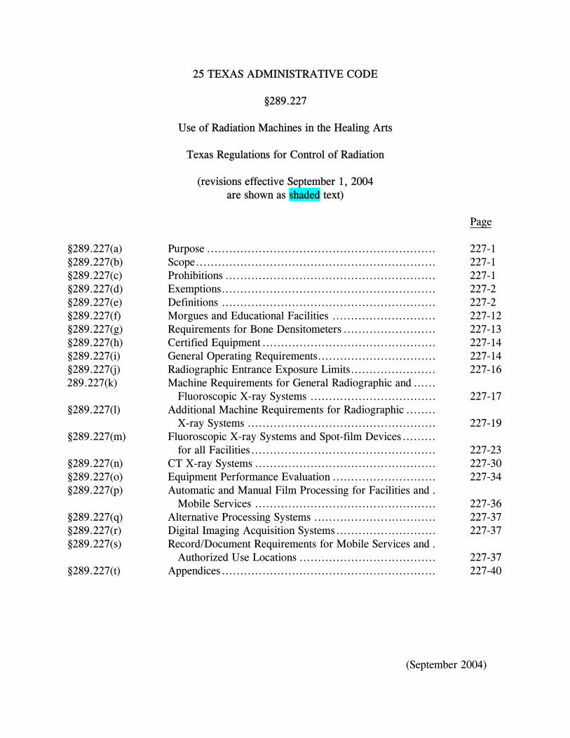
25 TEXAS ADMINISTRATIVE CODE
§289.227
Use of Radiation Machines in the Healing Arts
Texas Regulations for Control of Radiation
(revisions effective September 1, 2004 are shown as shaded text)
Page §289.227(a) Purpose .............................................................. 227-1 §289.227(b) Scope................................................................. 227-1 §289.227(c) Prohibitions ......................................................... 227-1 §289.227(d) Exemptions.......................................................... 227-2 §289.227(e) Definitions .......................................................... 227-2 §289.227(f) Morgues and Educational Facilities ............................ 227-12 §289.227(g) Requirements for Bone Densitometers ......................... 227-13 §289.227(h) Certified Equipment ............................................... 227-14 §289.227(i) General Operating Requirements................................ 227-14 §289.227(j) Radiographic Entrance Exposure Limits....................... 227-16 289.227(k) Machine Requirements for General Radiographic and ......
Fluoroscopic X-ray Systems .................................. 227-17 §289.227(l) Additional Machine Requirements for Radiographic ........ X-ray Systems ................................................... 227-19 §289.227(m) Fluoroscopic X-ray Systems and Spot-film Devices......... for all Facilities.................................................. 227-23 §289.227(n) CT X-ray Systems ................................................. 227-30 §289.227(o) Equipment Performance Evaluation ............................ 227-34 §289.227(p) Automatic and Manual Film Processing for Facilities and . Mobile Services ................................................. 227-36 §289.227(q) Alternative Processing Systems ................................. 227-37 §289.227(r) Digital Imaging Acquisition Systems........................... 227-37 §289.227(s) Record/Document Requirements for Mobile Services and . Authorized Use Locations ..................................... 227-37 §289.227(t) Appendices.......................................................... 227-40
(September 2004)
25 TEXAS ADMINISTRATIVE CODE
§289.227 Use of Radiation Machines in the Healing Arts. (a) Purpose. This section establishes requirements for the use of radiation machines in the healing arts. (b) Scope. (1) The registrant shall be responsible for directing the operation of the radiation machines under the administrative control of the registrant. The registrant shall assure that the requirements of this section are met in the operation of such radiation machines. All usage of such machines under this section shall be made by or under the supervision of a practitioner of the healing arts. (2) In addition to the requirements of this section, all registrants, unless otherwise specified, are subject to the requirements of §289.203 of this title (relating to Notices, Instructions, and Reports to Workers; Inspections), §289.204 of this title (relating to Fees for Certificates of Registration, Radioactive Material Licenses, Emergency Planning and Implementation, and Other Regulatory Services), §289.205 of this title (relating to Hearing and Enforcement Procedures), §289.226 of this title (relating to Registration of Radiation Machine Use and Services), and §289.231 of this title (relating to General Provisions and Standards for Protection Against Machine Produced Radiation). (3) The use of mammography radiation machines is subject to the requirements in §289.230 of this title (relating to Certification of Mammography Systems and Accreditation of Mammography Facilities), with the exceptions listed in §289.230(e)(1) and (2) of this title. The use of dental radiation machines is subject to the requirements in §289.232 of this title (relating to Radiation Control Regulations for Dental Radiation Machines). However, dental radiation machines located in a facility that also has other healing arts radiation machines will be inspected at the intervals specified in §289.231(ll)(1) of this title, and equipment performance evaluations performed at the interval specified for a medical facility in subsection (o)(1) of this section. The use of radiation machines for veterinary medicine is subject to the requirements in §289.233 of this title (relating to Radiation Control Regulations for Radiation Machines for Veterinary Medicine). (c) Prohibitions. (1) The agency may prohibit use of radiation machines that pose significant threat or endanger occupational and public health and safety, in accordance with §289.205 of this title and §289.231 of this title.
227-1 (September 2004)
§289.227(c)(2)
(2) Individuals shall not be exposed to the useful beam except for healing arts purposes and unless such exposure has been authorized by a licensed practitioner of the healing arts. This provision specifically prohibits intentional exposure for the following purposes:
(A) exposure of an individual for training, demonstration, or other non-healing arts purposes; (B) exposure of an individual for the purpose of healing arts screening, except as authorized by §289.226(h)(1) of this title; and (C) exposure of an individual for the purpose of research, except as authorized by §289.226(h)(2) of this title.
(3) Non-image-intensified fluoroscopic equipment shall not be used. (d) Exemptions.
(1) Portable radiation machines designed to be hand-held are exempt from the requirements of subsection (i)(11) of this section. The portable radiation machine shall be held by the tube housing support or handle. (2) Individuals who are sole practitioners and sole operators and the only occupationally exposed individual are exempt from the following requirements: (A) §289.203(b) of this title,“Posting of notices to workers;” (B) §289.203(c) of this title, “Instructions to workers;” and (C) operating and safety procedures in accordance with subsection (i)(2) of this section. (3) Registrants are exempt from the posting of the radiation area requirements in §289.231(x)(1) of this title provided that the operator has continuous surveillance and access control of the radiation area. (e) Definitions. The following words and terms when used in this section shall have the following meaning unless the context clearly indicates otherwise.
(1) Accessible surface - The external surface of the enclosure or housing provided by the manufacturer.
227-2 (September 2004)
§289.227(e)(2)
(2) Aluminum equivalent - The thickness of type 1100 aluminum alloy affording the same attenuation, under specified conditions, as the material in question. The nominal chemical composition of type 1100 aluminum alloy is 99% minimum aluminum, 0.12% copper.
(3) Attenuate - To reduce the exposure rate upon passage of radiation through matter.
(4) Attenuation block - A block or stack, having dimensions 20 centimeters (cm) by 20 cm by 3.8 cm, of type 1100 aluminum alloy or other materials having equivalent attenuation. The nominal chemical composition of type 1100 aluminum alloy is 99% minimum aluminum, 0.12% copper.
(5) Automatic exposure control (AEC) - A device that automatically controls one or more technique factors in order to obtain a required quantity of radiation at preselected locations (See definition for phototimer).
(6) Automatic exposure rate control (AERC) - A device that automatically controls one or more technique factors in order to obtain a required quantity of radiation per unit time at preselected locations.
(7) Barrier (See definition for protective barrier).
(8) Beam axis - A line from the source through the centers of the x-ray fields.
(9) Beam-limiting device - A device that provides a means to restrict the dimensions of the x-ray field.
(10) Beam quality (diagnostic x-ray) - A term that describes the penetrating power of the x-ray beam. This is identified numerically by half-value layer and is influenced by kilovolt peak (kVp) and filtration.
(11) Bone densitometer - A device intended for medical purposes to measure bone density and mineral content by x-ray transmission measurements through the bone and adjacent tissues.
(12) Calibration of instruments - The comparative response or reading of an instrument relative to a series of known radiation values over the range of the instrument. (13) Central axis of the beam - A line passing through the virtual source and the center of the plane figure formed by the edge of the first beam-limiting device.
227-3 (September 2004)
§289.227(e)(14)
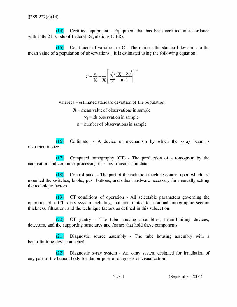
(14) Certified equipment - Equipment that has been certified in accordance with Title 21, Code of Federal Regulations (CFR). (15) Coefficient of variation or C - The ratio of the standard deviation to the mean value of a population of observations. It is estimated using the following equation:
samplein nsobservatio ofnumber =n samplein n observatioith = Xsamplein nsobservatio of mean value = X
population theofdeviation standard estimated = s :where
1-n)X - X(
X1 =
Xs = C
i
i2n
1=i
1/2
∑
(16) Collimator - A device or mechanism by which the x-ray beam is
restricted in size.
(17) Computed tomography (CT) - The production of a tomogram by the acquisition and computer processing of x-ray transmission data.
(18) Control panel - The part of the radiation machine control upon which are mounted the switches, knobs, push buttons, and other hardware necessary for manually setting the technique factors. (19) CT conditions of operation - All selectable parameters governing the operation of a CT x-ray system including, but not limited to, nominal tomographic section thickness, filtration, and the technique factors as defined in this subsection. (20) CT gantry - The tube housing assemblies, beam-limiting devices, detectors, and the supporting structures and frames that hold these components. (21) Diagnostic source assembly - The tube housing assembly with a beam-limiting device attached. (22) Diagnostic x-ray system - An x-ray system designed for irradiation of any part of the human body for the purpose of diagnosis or visualization.
227-4 (September 2004)
§289.227(e)(23)
(23) Entrance exposure - The exposure expressed in roentgens (R), measured in air with the specified technique, calculated or adjusted to represent the exposure at the point where the center of the useful beam enters the patient. (24) Entrance exposure rate - The exposure per unit time at the point where the center of the useful beam enters the patient. (25) Field emission equipment - Equipment that uses an x-ray tube in which electron emission from the cathode is due solely to the action of an electric field. (26) Field size - The dimensions along the major axes of an area in a plane perpendicular to the central axis of the beam at the normal treatment or examination source to image distance and defined by the intersection of the major axes and the 50% isodose line. (27) Filter - Material placed in the useful beam to preferentially absorb selected radiations. (28) Fluoroscopic imaging assembly - A subsystem in which x-ray photons produce a fluoroscopic image. It includes the image receptors such as the image intensifier and spot-film device, electrical interlocks, if any, and structural material providing linkage between the image receptor and diagnostic source assembly. (29) Focal spot - The area projected on the anode of the x-ray tube bombarded by the electrons accelerated from the cathode and from which the useful beam originates. (30) General purpose x-ray system - Any radiographic x-ray system that is not limited by design to radiographic examinations of specific anatomical regions.
(31) Gonadal shield - A protective barrier for the testes or ovaries.
(32) Half-value layer (HVL) - The thickness of a specified material that attenuates the beam of radiation to an extent such that the exposure rate is reduced to one-half of its original value.
(33) Healing arts - Any system, treatment, operation, diagnosis, prescription, or practice for the ascertainment, cure, relief, palliation, adjustment, or correction of any human disease, ailment, deformity, injury, or unhealthy or abnormal physical or mental condition.
227-5 (September 2004)
§289.227(e)(34)
(34) Healing arts screening - The testing of asymptomatic human beings using radiation machines for the detection or evaluation of health indications when such tests are not specifically and individually ordered by a licensed practitioner of the healing arts legally authorized to prescribe such x-ray tests for the purpose of diagnosis or treatment. (35) High level control for fluoroscopy - Any selected mode having an entrance exposure rate above 10 roentgens per minute (R/min). This mode shall meet the high level requirements in subsection (m)(3)(A)(i)(II), (ii)(II), or (iii)(II) of this section. (36) Image intensifier - A device, installed in its housing, that instantaneously converts an x-ray pattern into a corresponding light image of higher energy density. (37) Image receptor - Any device, such as a fluorescent screen or radiographic film, that transforms incident x-ray photons either into a visible image or into another form that can be made into a visible image by further transformations. (38) Irradiation - The exposure of matter to ionizing radiation. (39) kV - Kilovolt. (40) kVp - Kilovolt peak (See definition for peak tube potential). (41) kWs - Kilowatt-second. It is equivalent to 10 E 3 watt-second, where 1 watt-second =1 kV x 1 milliampere (mA) x 1 second. (42) Lead equivalent - The thickness of lead affording the same attenuation, under specified conditions, as the material in question. (43) Leakage radiation - Radiation emanating from the diagnostic source assembly except for the useful beam and radiation produced when the exposure switch or timer is not activated. (44) Leakage technique factors - The technique factors associated with the diagnostic source assembly that are used in measuring leakage radiation. They are defined as follows: (A) for diagnostic source assemblies intended for capacitor energy storage equipment, the maximum-rated peak tube potential and the maximum-rated number of exposures in an hour for operation at the maximum-rated peak tube potential with the quantity of charge per exposure being 10 millicoulombs (10 milliampere-second (mAs)) or the minimum obtainable from the unit, whichever is larger;
227-6 (September 2004)
§289.227(e)(44)(B)
(B) for diagnostic source assemblies intended for field emission equipment rated for pulsed operation, the maximum-rated peak tube potential and the maximum-rated number of x-ray pulses in an hour for operation at the maximum-rated peak tube potential; or (C) for all other diagnostic source assemblies, the maximum-rated peak tube potential and the continuous tube current for the maximum-rated peak tube potential. (45) Licensed medical physicist - An individual holding a current Texas license under the Medical Physics Practice Act, Texas Occupations Code, Chapter 602, with a specialty in diagnostic radiological physics. (46) mA - Milliampere. (47) mAs - Milliampere-second. (48) Medical research - The investigation of various health risks and diseases. (49) Mobile service operation - The provision of radiation machines and personnel at temporary sites for limited time periods. The radiation machines may be fixed inside a motorized vehicle or may be a portable radiation machine that may be removed from the vehicle and taken into a facility for use. (50) Multiple slice tomogram system - A computed tomography x-ray system that obtains x-ray transmission data simultaneously during a single scan to produce more than one tomogram. (51) Nominal tomographic section thickness - The full-width at half-maximum of the sensitivity profile taken at the center of the cross sectional volume over which x-ray transmission data are collected.
(52) Non-certified radiographic equipment - Equipment manufactured and assembled prior to certification requirements of Title 21, CFR, effective as specified in Title 21, CFR, Part 1020.30(a). (53) Patient - An individual subjected to healing arts examination, diagnosis, or treatment. (54) Peak tube potential - The maximum value of the potential difference in kilovolts across the x-ray tube during an exposure.
(55) Phantom - A volume of material behaving in a manner that can be related to tissue with respect to the attenuation and scattering of radiation.
227-7 (September 2004)
§289.227(e)(56)
(56) Phototimer - A method for controlling exposures to image receptors by the amount of radiation that reaches a radiation monitoring device. The radiation monitoring device is part of an electronic circuit that controls the duration of time the tube is activated (See definition for automatic exposure control). (57) Portable x-ray equipment (See definition for x-ray equipment). (58) Practitioner of the healing arts (practitioner) - A person licensed to practice healing arts by either the Texas State Board of Medical Examiners as a physician, the Texas Board of Chiropractic Examiners, or the Texas State Board of Podiatry Examiners. (59) Primary protective barrier - (See definition for protective barrier). (60) Protective apron - An apron made of radiation attenuating materials used to reduce radiation exposure. (61) Protective barrier - A barrier of radiation absorbing materials used to reduce radiation exposure. The types of protective barriers are as follows: (A) primary protective barrier - A barrier sufficient to attenuate the useful beam to the required degree. (B) secondary protective barrier - A barrier sufficient to attenuate the stray radiation to the required degree. (62) Protective glove - A glove made of radiation attenuating materials used to reduce radiation exposure. (63) Radiograph - An image receptor on which the image is created directly or indirectly by an x-ray exposure and results in a permanent record.
(64) Reference plane - A plane that is displaced from and parallel to the tomographic plane. (65) Scan - The complete process of collecting x-ray transmission data for the production of a tomogram. Data can be collected simultaneously during a single scan for the production of one or more tomograms. (66) Scan increment - The amount of relative displacement of the patient with respect to the CT x-ray system between successive scans measured along the direction of such displacement.
227-8 (September 2004)
§289.227(e)(67)
(67) Scan sequence - A preselected set of two or more scans performed consecutively under preselected CT conditions of operation.
(68) Scan time - The period of time between the beginning and end of x-ray transmission data accumulation for a single scan.
(69) Scattered radiation - Radiation that has been deviated in direction during passage through matter. (70) Secondary protective barrier (See definition for protective barrier). (71) Shutter - A device attached to the tube housing assembly that can totally intercept the useful beam and that has a lead equivalency not less than that of the tube housing assembly. (72) Single tomogram system - CT x-ray system that obtains x-ray transmission data during a scan to produce a single tomogram. (73) Source - The focal spot of the x-ray tube.
(74) Source-to-image receptor distance (SID) - The distance from the source to the center of the input surface of the image receptor. (75) Source-to-skin distance (SSD) - The distance from the source to the skin of the patient. (76) Special purpose x-ray system - Any radiographic x-ray system that is limited by design to radiographic examinations of specific anatomical regions. Special purpose x-ray systems include, but are not limited to, dedicated chest units, cystography units, and head and skull units. (77) Special procedures - The application of special x-ray equipment and specialized techniques to obtain required diagnostic information. Special procedures include, but are not limited to, angiography, cardiac catheterization, myelography, and surgery. (78) Spot film - A radiograph that is made during a fluoroscopic examination to permanently record conditions that exist during that fluoroscopic procedure. (79) Spot film device - A device intended to transport and/or position a radiographic image receptor between the x-ray source and fluoroscopic image receptor. It includes a device intended to hold a cassette over the input end of an image intensifier for the purpose of making a radiograph.
(80) Stationary x-ray equipment - (See definition for x-ray equipment).
227-9 (September 2004)
§289.227(e)(81)
(81) Stray radiation - The sum of leakage and scattered radiation. (82) Supervision - The delegating, by the practitioner, of the task of applying radiation to persons who perform tasks under the practitioner's control and who are certified under the Medical Radiologic Technologist Act, Texas Occupations Code, Chapter 601. The practitioner assumes full responsibility for these tasks and shall assure that the tasks will be administered correctly. (83) Target - The part of a radiation machine head that by design intercepts a beam of accelerated particles with subsequent emission of other radiation. (84) Technique chart - A chart that provides all necessary generator control settings and geometry needed to make clinical radiographs when the radiography system is in manual mode. (85) Technique factors - The conditions of operation that are specified as follows:
(A) for capacitor energy storage equipment, peak tube potential in kV and quantity of charge in mAs; (B) for field emission equipment rated for pulsed operation, peak tube potential in kV and number of x-ray pulses; (C) for CT equipment designed for pulsed operations, peak tube potential in kV, scan time in seconds, and either tube current in mA, x-ray pulse width in seconds, and the number of x-ray pulses per scan or the product of tube current, x-ray pulse width, and the number of x-ray pulses in mAs; (D) for CT equipment not designed for pulsed operation, peak tube potential in kV, and either tube current in mA and scan time in seconds or the product of tube current and exposure time in mAs when the scan time and exposure time are equivalent; and (E) for all other equipment, peak tube potential in kV and either tube current in mA and exposure time in seconds or the product of tube current and exposure time in mAs. (86) Tomogram - The depiction of the x-ray attenuation properties of a section through the body. (87) Tomographic plane - The geometric plane that is identified as corresponding to the output.
227-10 (September 2004)
§289.227(e)(88)
(88) Tomographic section - The volume of an object whose x-ray attenuation properties are imaged in a tomogram. (89) Traceable to a national standard - This indicates that a quantity or a measurement has been compared to a national standard, for example, the National Institute of Standards and Technology, directly or indirectly through one or more intermediate steps and that all comparisons have been documented. (90) Tube - An x-ray tube, unless otherwise specified. (91) Tube housing assembly - The tube housing with tube installed. It includes high-voltage and/or filament transformers and other appropriate elements when such are contained within the tube housing. (92) Useful beam - Radiation that passes through the window, aperture, cone, or other collimating device of the source housing. Also referred to as the primary beam. (93) X-ray control - A device that controls input power to the x-ray high-voltage generator and/or the x-ray tube. It includes equipment such as timers, phototimers, automatic brightness stabilizers, and similar devices that control the technique factors of an x-ray exposure. (94) X-ray equipment - An x-ray system, subsystem, or component thereof. For the purposes of this rule, types of x-ray equipment are as follows: (A) portable x-ray equipment - x-ray equipment mounted on a permanent base with wheels and/or casters for moving while completely assembled. Portable x-ray equipment may also include equipment designed to be hand-carried; or (B) stationary x-ray equipment - x-ray equipment that is installed in a fixed location. (95) X-ray field - That area of the intersection of the useful beam and any one of the set of planes parallel to and including the plane of the image receptor, whose perimeter is the locus of points at which the exposure rate is one-fourth of the maximum in the intersection. (96) X-ray high-voltage generator - A device that transforms electrical energy from the potential supplied by the x-ray control to the tube operating potential. The device may also include means for transforming alternating current to direct current, filament transformers for the x-ray tubes, high-voltage switches, electrical protective devices, and other appropriate elements.
227-11 (September 2004)
§289.227(e)(97)
(97) X-ray system - An assemblage of components for the controlled production of x rays. It includes minimally an x-ray high-voltage generator, an x-ray control, a tube housing assembly, a beam-limiting device, and the necessary supporting structures. Additional components that function with the system are considered integral parts of the system. (98) X-ray subsystem - Any combination of two or more components of an x-ray system. (99) X-ray tube - Any electron tube that is designed to be used primarily for the production of x rays.
(f) Morgues and educational facilities. (1) Morgues shall comply with the following requirements: (A) subsection (b)(1) and (2) of this section concerning scope; (B) subsection (c) of this section concerning prohibitions; (C) subsection (e) of this section concerning definitions, as applicable; (D) subsection (i)(2) of this section concerning operating and safety procedures; (E) subsection (i)(4) of this section concerning protective devices; (F) subsection (i)(11) of this section concerning holding of tube; (G) subsection (k)(1) of this section concerning warning labels; (H) subsection (m)(1)(A) of this section concerning fluoroscopy; and (I) subsection (s)(1)(A)-(I), and (R) of this section concerning records. (2) Facilities conducting training using non-humans shall comply with all the requirements of this section except for the following:
(A) subsection (i)(5) of this section concerning operator credentialing;
227-12 (September 2004)
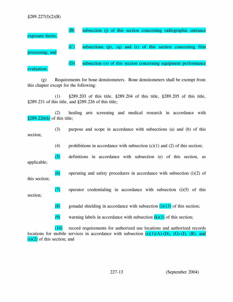
§289.227(f)(2)(B)
(B) subsection (j) of this section concerning radiographic entrance exposure limits;
(C) subsections (p), (q) and (r) of this section concerning film processing; and
(D) subsection (o) of this section concerning equipment performance evaluation. (g) Requirements for bone densitometers. Bone densitometers shall be exempt from this chapter except for the following:
(1) §289.203 of this title, §289.204 of this title, §289.205 of this title, §289.231 of this title, and §289.226 of this title; (2) healing arts screening and medical research in accordance with §289.226(h) of this title;
(3) purpose and scope in accordance with subsections (a) and (b) of this section;
(4) prohibitions in accordance with subsection (c)(1) and (2) of this section; (5) definitions in accordance with subsection (e) of this section, as applicable; (6) operating and safety procedures in accordance with subsection (i)(2) of this section; (7) operator credentialing in accordance with subsection (i)(5) of this section; (8) gonadal shielding in accordance with subsection (i)(13) of this section; (9) warning labels in accordance with subsection (k)(1) of this section; (10) record requirements for authorized use locations and authorized records locations for mobile services in accordance with subsection (s)(1)(A)-(D), (G)-(J), (R), and (s)(2) of this section; and
227-13 (September 2004)
§289.227(g)(11)
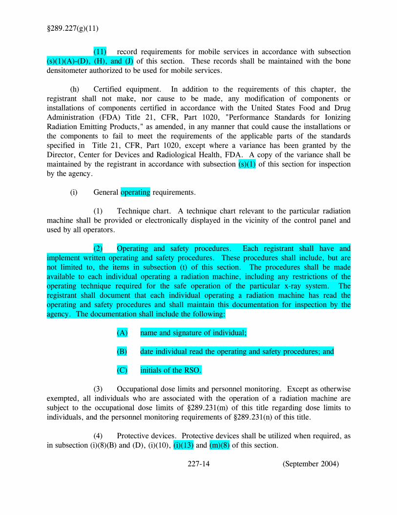
(11) record requirements for mobile services in accordance with subsection (s)(1)(A)-(D), (H), and (J) of this section. These records shall be maintained with the bone densitometer authorized to be used for mobile services. (h) Certified equipment. In addition to the requirements of this chapter, the registrant shall not make, nor cause to be made, any modification of components or installations of components certified in accordance with the United States Food and Drug Administration (FDA) Title 21, CFR, Part 1020, "Performance Standards for Ionizing Radiation Emitting Products," as amended, in any manner that could cause the installations or the components to fail to meet the requirements of the applicable parts of the standards specified in Title 21, CFR, Part 1020, except where a variance has been granted by the Director, Center for Devices and Radiological Health, FDA. A copy of the variance shall be maintained by the registrant in accordance with subsection (s)(1) of this section for inspection by the agency. (i) General operating requirements.
(1) Technique chart. A technique chart relevant to the particular radiation machine shall be provided or electronically displayed in the vicinity of the control panel and used by all operators. (2) Operating and safety procedures. Each registrant shall have and implement written operating and safety procedures. These procedures shall include, but are not limited to, the items in subsection (t) of this section. The procedures shall be made available to each individual operating a radiation machine, including any restrictions of the operating technique required for the safe operation of the particular x-ray system. The registrant shall document that each individual operating a radiation machine has read the operating and safety procedures and shall maintain this documentation for inspection by the agency. The documentation shall include the following:
(A) name and signature of individual;
(B) date individual read the operating and safety procedures; and (C) initials of the RSO.
(3) Occupational dose limits and personnel monitoring. Except as otherwise exempted, all individuals who are associated with the operation of a radiation machine are subject to the occupational dose limits of §289.231(m) of this title regarding dose limits to individuals, and the personnel monitoring requirements of §289.231(n) of this title.
(4) Protective devices. Protective devices shall be utilized when required, as in subsection (i)(8)(B) and (D), (i)(10), (i)(13) and (m)(8) of this section.
227-14 (September 2004)
§289.227(i)(4)(A)
(A) Protective devices shall be of no less than 0.25 millimeter (mm) lead equivalent material except as specified in subsections (i)(13) and (m)(8)(B)(i) of this section. (B) Protective devices, including aprons, gloves, and shields shall be checked annually for defects such as holes, cracks, and tears. These checks may be performed by the registrant by visual or tactile means, or x-ray imaging. If a defect is found, protective devices shall be replaced or removed from service until repaired. A record of this test shall be made and maintained by the registrant in accordance with subsection (s)(1) of this section for inspection by the agency.
(5) Operator credentialing. Individuals who operate radiation machines for human use shall meet the appropriate credentialing requirements of rules issued in accordance with the Medical Radiologic Technologist Certification Act, Texas Occupations Code, Chapter 601. Copies of the credentialing document shall be maintained at the locations(s) where the individual is working.
(6) Practice of medical physics. Surveys, tests, or evaluations required by this section may constitute the practice of medical physics and, therefore, require a license from the Texas Board of Licensure for Professional Medical Physicists in accordance with the Medical Physics Practice Act, Texas Occupations Code, Chapter 602.
(7) Exposure of individuals other than the patient. No individual other than a patient, operator, and ancillary personnel shall be in the x-ray room or area while exposures are being made unless such individual's assistance is required.
(8) Holding of patient or image receptor.
(A) When a patient or image receptor must be held in position during radiography, mechanical supporting or restraining devices shall be used when the exam permits.
(B) If a patient or image receptor must be held by an individual during an exposure, that individual shall be protected with appropriate shielding devices described in paragraph (4) of this subsection.
(C) The registrant's written operating and safety procedures required by paragraph (2) of this subsection shall include the following:
(i) a list of circumstances in which mechanical holding devices cannot be routinely utilized; and
227-15 (September 2004)
§289.227(i)(8)(C)(ii)
(ii) a procedure used for selecting an individual to hold or support the patient or image receptor.
(D) In those cases where the patient must hold the image receptor, any portion of the body other than the area of clinical interest struck by the useful beam shall be protected by not less than 0.25 mm lead equivalent material. (9) Viewing system and contact with patient.
(A) Windows, mirrors, closed circuit television, or another method shall be provided to permit the operator to continuously observe the patient during irradiation.
(B) The operator shall be able to maintain verbal, visual, and aural contact with the patient.
(10) Operator position. The operator position during the exposure shall be such that the operator's exposure is as low as reasonably achievable (ALARA) and the operator is a minimum of six feet from the source of radiation or protected by an apron, gloves, or other shielding having a minimum of 0.25 lead equivalent material.
(11) Holding of tube. In no case shall an individual hold the tube or tube housing assembly supports during any radiographic exposure. (12) Patient protection. Notwithstanding the provisions of subsection (i)(7) of this section, other patients who are in line with the primary beam and who cannot be removed from the room shall be protected by whole body protective barriers of a minimum of 0.25 mm lead equivalent material or so positioned that the nearest portion of their body is at least six feet from both the tube head and the nearest edge of the image receptor. (13) Gonadal shielding. Gonadal shielding shall be used on patients when the gonads are in or within 5 cm of the useful beam. This requirement does not apply if the shielding will interfere with the diagnostic procedure. Gonadal shielding shall be of at least 0.5 mm lead equivalent material. (j) Radiographic entrance exposure limits. The in-air exposure determined for the technique used by the registrant for the specified average human adult patient thickness for routine medical radiography shall not exceed the entrance exposure limits in the following Table I.
227-16 (September 2004)

§289.227(j)
TABLE I - RADIOGRAPHIC ENTRANCE EXPOSURE LIMITS Technique Patient Thickness (cm) Exposure Limit (mR) Chest (PA)
(Non-Grid) 23 20
(Grid) 23 30
Abdomen (KUB) 23 450
Lumbo-Sacral Spine (AP) 23 550
Cervical Spine (AP) 13 120
Thoracic Spine (AP) 23 325
Full Spine 23 300
Skull (lateral) 15 150
Foot (DP) 8 50
(k) Machine requirements for general radiographic and fluoroscopic x-ray systems.
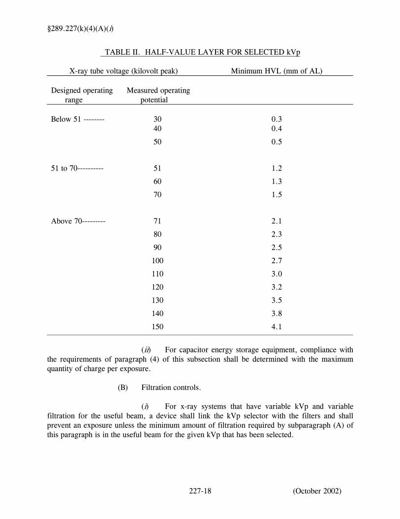
(1) Warning label. The warning label will meet the requirements of §289.231(z) of this title. (2) Mechanical support of tube head. The tube housing assembly shall be adjusted to remain stable during an exposure unless tube housing movement is a designed function of the x-ray system. (3) Battery charge indicator. On battery-powered x-ray generators, visual means shall be provided on the control panel to indicate whether the battery is in a state of charge adequate for proper operation. (4) Beam quality. The following requirements apply to beam quality. (A) Half-value layer. (i) The half-value layer of the useful beam for a given x-ray tube potential shall not be less than the values shown in the following Table II. If it is necessary to determine such half-value layer at an x-ray tube potential that is not listed in Table II, linear interpolation may be made.
227-17 (September 2004)
§289.227(k)(4)(A)(i)
TABLE II. HALF-VALUE LAYER FOR SELECTED kVp
X-ray tube voltage (kilovolt peak) Minimum HVL (mm of AL) Designed operating Measured operating range potential
Below 51 -------- 30 0.3 40 0.4
50 0.5
51 to 70---------- 51 1.2
60 1.3
70 1.5
Above 70--------- 71 2.1
80 2.3
90 2.5
100 2.7
110 3.0
120 3.2
130 3.5
140 3.8
150 4.1
(ii) For capacitor energy storage equipment, compliance with
the requirements of paragraph (4) of this subsection shall be determined with the maximum quantity of charge per exposure. (B) Filtration controls. (i) For x-ray systems that have variable kVp and variable filtration for the useful beam, a device shall link the kVp selector with the filters and shall prevent an exposure unless the minimum amount of filtration required by subparagraph (A) of this paragraph is in the useful beam for the given kVp that has been selected.
227-18 (October 2002)
§289.227(k)(4)(B)(ii)
(ii) Any other system having removable filters shall be required to have the minimum amount of filtration as required by subparagraph (A)(i) of this paragraph permanently located in the useful beam during each exposure. (5) Multiple tubes. Where two or more radiographic tubes are controlled by one exposure switch, the tube or tubes that have been selected shall be clearly indicated prior to initiation of the exposure. This indication shall be both on the x-ray control panel and at or near the tube housing assembly that has been selected. (6) Technique and exposure indicators. (A) The technique factors to be used during an exposure shall be indicated before the exposure begins except when automatic exposure controls are used, in which case the technique factors that are set prior to the exposure shall be indicated. (B) On equipment having fixed technique factors, the requirement of subparagraph (A) of this paragraph may be met by permanent markings. (C) The x-ray control shall provide visual indication of the production of x rays. (D) The indicated technique factors shall be accurate to meet manufacturer's specifications. If these specifications are not available from the manufacturer, the factors shall be accurate to within plus or minus 10% of the indicated setting. (7) X-ray control. An x-ray control shall be incorporated into each x-ray system such that an exposure can be terminated by the operator at any time except for an exposure of 0.5 seconds or less or during serial radiography when means shall be provided to permit completion of any single exposure of the series in process. (l) Additional machine requirements for radiographic x-ray systems. This subsection does not apply to fluoroscopic or CT x-ray systems. (1) Beam limitation. Beam limitation shall be as follows. (A) Stationary general purpose x-ray systems. (i) Beam-limiting devices shall restrict the useful beam to the area of clinical interest as follows: (I) the misalignment of the x-ray field for a manual rectangular collimator shall be within 2.0% of the SID for the length or width of the image receptor;
227-19 (September 2004)
§289.227(l)(1)(A)(i)(II)
(II) the x-ray field for a circular or polygon collimator shall not exceed the diagonal of the image receptor by more than 2.0% of the SID; or (III) the misalignment of the x-ray field for an automatic or semi-automatic collimator shall be within 3.0% of the SID for the length and width of the image receptor and shall be within 4.0% of the SID, without regard to the sign, of the sum of the difference of the length and width of the image receptor. (ii) A method shall be provided for visually defining the perimeter of the x-ray field. The total misalignment of the edges of the visually defined field with the respective edges, either the length or width, of the x-ray field shall not exceed 2.0% of the SID. (iii) A numerical SID indicator shall be present and shall be accurate to within 2.0% of the SID. (iv) The system shall indicate when the axis of the x-ray field is perpendicular to the plane of the image receptor. (v) The center of the x-ray field, when perpendicular to the image receptor, shall be accurate to within 2.0% of the SID with respect to the center of the image receptor. (vi) The beam-limiting device shall numerically indicate the field size in the plane of the image receptor. (vii) Indication of field size dimensions and SIDs shall be specified in inches and/or centimeters. (viii) The field size indicated on the beam-limiting device shall be within 2.0% of the SID along the width and length, separately, of the actual x-ray field size. (B) Portable x-ray equipment. Portable x-ray equipment shall comply with the requirements in subparagraph (A) of this paragraph, as applicable, based on manufacturer's design. (C) Radiographic systems designed for one image receptor size. Radiographic equipment designed for only one image receptor size at a fixed SID shall provide a means to do the following: (i) limit the x-ray field to no greater than the dimensions of the image receptor at the SID, and to align the center of the x-ray field with the center of the image receptor to within 2.0% of the SID center; or
227-20 (September 2004)
§289.227(l)(1)(C)(ii)
(ii) align the x-ray field such that the x-ray field does not extend beyond any edge of the image receptor at the SID. (D) Special purpose x-ray systems. (i) When the x-ray beam is perpendicular to the plane of the image receptor, a means shall be provided to do the following: (I) limit the x-ray field such that the x-ray field does not exceed each dimension of the image receptor by more than 2.0% of the SID; and (II) align the center of the x-ray field with the center of the image receptor to within 2.0% of the SID. (ii) The requirements of clause (i) of this subparagraph may be met with a system that meets the requirements for a general purpose x-ray system as specified in subparagraph (A)(i)-(iv) of this paragraph or, when alignment means are also provided, may be met with either of the following: (I) an assortment of removable, fixed-aperture, beam-limiting devices sufficient to meet the requirement for each combination of image receptor size and SID for which the unit is designed with each such device having clear and permanent markings to indicate the image receptor size and SID for which it is designed; or (II) a beam-limiting device having multiple fixed apertures sufficient to meet the requirement for each combination of image receptor size and SID for which the radiation machine is designed. Permanent, clearly legible markings shall indicate the image receptor size and SID for which each aperture is designed and shall indicate which aperture is in position for use. (2) Radiation exposure control devices. Radiation exposure control devices shall include the following: (A) Timers. Means shall be provided to terminate the exposure at a preset time interval, a preset product of current and time, a preset number of pulses, or a preset radiation exposure to the image receptor. In addition, it shall not be possible to make an exposure when the timer is set to a "zero" or "off" position if either position is provided. (B) AEC. When AEC is provided, the following shall occur. (i) Indication shall be made on the control panel when this mode of operation is selected.
227-21 (September 2004)
§289.227(l)(2)(B)(ii)
(ii) If the x-ray tube potential is equal to or greater than 50 kVp, the minimum exposure time for field emission equipment rated for pulsed operation shall be equal to or less than a time interval equivalent to two pulses. (iii) The minimum exposure time for all equipment other than that specified in clause (ii) of this subparagraph shall be equal to or less than 0.0167 second or a time interval required to deliver 5 mAs, whichever is greater. (iv) Either the product of peak x-ray tube potential, current, and exposure time shall be limited to not more than 60 kWs per exposure, or the product of x-ray tube current and exposure time shall be limited to not more than 600 mAs per exposure except that, when the x-ray tube potential is less than 50 kVp, the product of x-ray tube current and exposure time shall be limited to not more than 2,000 mAs per exposure. (v) A visible and/or audible signal shall indicate when an exposure has been terminated at the limits required by clause (iv) of this subparagraph, and manual resetting shall be required before further automatically timed exposures can be made. (C) Exposure interval reproducibility. When all technique factors are held constant, including control panel selections associated with AEC systems, the coefficient of variation of exposure interval for both manual and AEC systems shall not exceed 0.05. This requirement applies to clinically used techniques. (3) SSD. All mobile or portable radiographic systems shall be provided with means to limit the SSD to equal to or greater than 30 cm. (4) Exposure reproducibility. When all technique factors are held constant, including control panel selections associated with AEC systems, the coefficient of variation of exposure for both manual and AEC systems shall not exceed 0.05. This requirement applies to clinically used techniques. (5) Linearity. The average ratios of exposure mR to the indicated mAs product obtained at any two consecutive mA or mAs settings shall not differ by more than 0.10 times their sum, where X1 and X2 are the average mR/mAs values obtained at each of two consecutive tube current settings:
( )2121 10.0 XXXX +≤−
(6) Radiation from capacitor energy storage equipment in standby status.
Radiation emitted from the x-ray tube when the exposure switch or timer is not activated shall not exceed a rate of 2 milliroentgens per hour (mR/hr) at 5 cm from any accessible surface of the diagnostic source assembly, with the beam-limiting device fully open.
227-22 (September 2004)
§289.227(m)
(m) Fluoroscopic x-ray systems and spot-film devices for all facilities. (1) Limitation of the useful beam. Limitation of the useful beam shall be as follows. (A) Primary barrier. (i) The fluoroscopic imaging assembly shall be provided with a primary protective barrier that intercepts the entire cross section of the useful beam at any SID. (ii) The x-ray tube used for fluoroscopy shall not produce x rays unless the barrier is in position to intercept the useful beam and the imaging device is in place and operable. (iii) The exposure rate due to transmission through the barrier with the attenuation block in the useful beam, combined with radiation through the image intensifier if provided, shall not exceed 3.34 x 10⁻³% of the entrance exposure rate at a distance of 10 cm from any accessible surface of the fluoroscopic imaging assembly beyond the plane of the image receptor. (B) Measuring compliance of barrier transmission. (i) The exposure rate due to transmission through the primary protective barrier combined with radiation through the image intensifier shall be determined by measurements averaged over an area of 100 cm2 with no linear dimension greater than 20 cm. (ii) If the source is below the tabletop, the measurement shall be made with the input surface of the fluoroscopic imaging assembly positioned 30 cm above the tabletop. (iii) If the source is above the tabletop and the SID is variable, the measurement shall be made with the end of the beam-limiting device or spacer as close to the tabletop as it can be placed, provided that it shall not be closer than 30 cm. (iv) Movable grids and compression devices shall be removed from the useful beam during the measurement. (v) The attenuation block shall be positioned in the useful beam 10 cm from the point of measurement of entrance exposure rate and between this point and the input surface of the fluoroscopic imaging assembly. (vi) The collimator shall be fully open when the measurement is made.
227-23 (September 2004)
§289.227(m)(1)(C)
(C) X-ray field. (i) Compliance with clauses (ii)-(vii) of this subparagraph shall be determined with the beam axis perpendicular to the plane of the image receptor. (ii) Equipment with a fixed SID and the capability of a visible area of no greater than 300 cm2 shall be provided with either stepless adjustment of the x-ray field or a means to further limit the x-ray field at the image receptor to 125 cm2 or less. If the equipment is provided with stepless adjustment, the minimum x-ray field size at the maximum SID shall be less than or equal to 5 cm by 5 cm at the image receptor. (iii) Equipment with a variable SID or a fixed SID with the capability of a visible area of greater than 300 cm2 shall be provided with stepless adjustment of the field size. The minimum x-ray field size at the maximum SID shall be less than or equal to 5 cm by 5 cm at the image receptor. (iv) Neither the length nor the width of the x-ray field in the plane of the image receptor shall exceed that of the visible area of the image receptor by more than 3.0% of the SID. The sum of the excess length and the excess width shall be no greater than 4.0% of the SID. (v) For rectangular x-ray fields used with circular image receptors, the error in alignment shall be determined along the length and width dimensions of the x-ray field that pass through the center of the visible area of the image receptor. (vi) For fluoroscopic equipment with only a manual mode of collimation, the x-ray field produced shall be limited to the area of the spot-film cassette at 16 inches above tabletop. Additionally, during fluoroscopy, the beam shall be restricted to the area of the input phosphor. (vii) Spot-film devices shall meet the following additional requirements. (I) Means shall be provided between the source and the patient for adjustment of the x-ray field size in the plane of the film to the size of that portion of the film that has been selected on the spot-film selector. (-a-) Such adjustment shall be automatically accomplished except when the x-ray field size in the plane of the film is smaller than that of the selected portion of the film.
227-24 (October 2002)
§289.227(m)(1)(C)(vii)(I)(-b-)
(-b-) The total misalignment of the edges of the x-ray field with the respective edges of the selected portion of the image receptor along the length or width dimensions of the x-ray field in the plane of the image receptor shall not exceed 3.0% of the SID when adjusted for full coverage of the selected portion of the image receptor. (-c-) The sum, without regard to sign of the misalignment along any two orthogonal dimensions, shall not exceed 4.0% of the SID. (II) The center of the x-ray field in the plane of the film shall be aligned with the center of the selected portion of the film to within 2.0% of the SID. (2) Activation of the fluoroscopic tube. X-ray production in the fluoroscopic mode shall be controlled by a device that requires continuous pressure by the fluoroscopist for the entire time of the exposure (continuous pressure type switch). When recording serial fluoroscopic images, the fluoroscopist shall be able to terminate the x-ray exposures at any time, but means may be provided to permit completion of any single exposure of the series in process. (3) Entrance exposure rate allowable limits. (A) The following requirements apply to fluoroscopic equipment manufactured prior to May 19, 1995. (i) Equipment with AERC. Fluoroscopic equipment that is provided with AERC shall not be operable at any combination of tube potential and current that will result in an exposure rate in excess of 2.58 x 10⁻³ coulomb per kilogram per minute (C/kg/min) (10 roentgens per minute (10 R/min)) at the point where the center of the useful beam enters the patient, except: (I) during recording of fluoroscopic images, excluding last image hold; or (II) when an optional high-level control is provided. When so provided, the equipment shall not be operable at any combination of tube potential and current that will result in an exposure rate in excess of 1.29 x 10⁻³ C/kg/min (5 R/min) at the point where the center of the useful beam enters the patient, unless the high-level control is activated. Special means of activation of high-level controls shall be required. The high-level control shall be operable only when continuous manual activation is provided by the operator. A continuous signal audible to the fluoroscopist shall indicate that the high-level control is being employed.
227-25 (October 2002)
§289.227(m)(3)(A)(ii)
(ii) Equipment without AERC (manual mode). Fluoroscopic equipment that is not provided with AERC shall not be operable at any combination of tube potential and current that will result in an exposure rate in excess of 1.29 x 10⁻³ C/kg/min (5 R/min) at the point where the center of the useful beam enters the patient, except: (I) during recording of fluoroscopic images, excluding last image hold; or (II) when an optional high-level control is activated. Special means of activation of high-level controls shall be required. The high-level control shall be operable only when continuous manual activation is provided by the operator. A continuous signal audible to the fluoroscopist shall indicate that the high-level control is being employed. (iii) Equipment with both an AERC mode and a manual mode. Fluoroscopic equipment that is provided with both an AERC mode and a manual mode shall not be operable at any combination of tube potential and current that will result in an exposure rate in excess of 2.58 x 10⁻³ C/kg/min (10 R/min) in either mode at the point where the center of the useful beam enters the patient except: (I) during recording of fluoroscopic images, excluding last image hold; or (II) when the mode or modes have an optional high-level control, in which case that mode or modes shall not be operable at any combination of tube potential and current that will result in an exposure rate in excess of 1.29 x 10⁻³ C/kg/min (5 R/min) at the point where the center of the useful beam enters the patient, unless the high level control is activated. Special means of activation of high-level control shall be required. The high level control shall be operable only when continuous manual activation is provided by the operator. A continuous signal audible to the fluoroscopist shall indicate that the high-level is being employed. (iv) Measuring compliance. Compliance with subparagraph (A) of this paragraph shall be determined as follows. (I) If the source is below the x-ray table, the exposure rate shall be measured at 1 cm above the tabletop or cradle. (II) If the source is above the x-ray table, the exposure rate shall be measured at 30 cm above the tabletop with the end of the beam-limiting device or spacer positioned as closely as possible to the point of measurement.
227-26 (October 2002)
§289.227(m)(3)(A)(iv)(III)
(III) In a C-arm type of fluoroscope, the exposure rate shall be measured at 30 cm from the input surface of the fluoroscopic imaging assembly provided that the end of the beam-limiting device or spacer is no closer than 30 cm from the input surface of the fluoroscopic imaging assembly. The applicable limit shall not be exceeded at any available SID. (IV) In a lateral (horizontal) type of fluoroscope, the exposure rate shall be measured at a point 15 cm from the centerline of the x-ray table and in the direction of the x-ray source with the end of the beam-limiting device or spacer positioned as closely as possible to the point of measurement. If the table top is movable, it shall be positioned as closely as possible to the lateral x-ray source, with the end of the beam-limiting device or spacer no closer than 15 cm to the centerline of the x-ray table. (B) The following requirements apply to fluoroscopic equipment manufactured on and after May 19, 1995. (i) Fluoroscopic equipment operable at any combination of tube potential and current that will result in an exposure rate greater than 1.29 x 10⁻³ C/kg/min (5 R/min) at the point where the center of the useful beam enters the patient shall be equipped with AERC. Provision for manual selection of technique factors may be provided. (ii) Fluoroscopic equipment shall not be operable at any combination of tube potential and current that will result in an exposure rate in excess of 2.58 x 10⁻³ C/kg/min (10 R/min) at the point where the center of the useful beam enters the patient except: (I) During the recording of images from an x-ray image-intensifier tube using photographic film or a video camera when the x-ray source is operated in a pulsed mode. (II) When the high-level control is activated, the equipment shall not be operable at any combination of tube potential and current that will result in an exposure rate in excess of 5.16 x 10⁻³ C/kg/min (20 R/min) at the point where the center of the useful beam enters the patient. Special means of activation of high-level controls shall be required. The high-level control shall only be operable when continuous manual activation is provided by the operator. A continuous signal audible to the fluoroscopist shall indicate that the high-level control is being employed. (iii) Measuring compliance. Compliance with subparagraph (B) of this paragraph shall be determined as follows. (I) If the source is below the x-ray table, the exposure rate shall be measured at 1 cm above the tabletop or cradle.
227-27 (October 2002)
§289.227(m)(3)(B)(iii)(II)
(II) If the source is above the x-ray table, the exposure rate shall be measured at 30 cm above the tabletop with the end of the beam-limiting device or spacer positioned as closely as possible to the point of measurement. (III) In a C-arm type of fluoroscope, the exposure rate shall be measured at 30 cm from the input surface of the fluoroscopic imaging assembly provided that the end of the beam-limiting device or spacer is no closer than 30 cm from the input surface of the fluoroscopic imaging assembly. The applicable limit shall not be exceeded at any available SID. (IV) In a lateral (horizontal) type of fluoroscope, the exposure rate shall be measured at a point 15 cm from the centerline of the x-ray table and in the direction of the x-ray source with the end of the beam-limiting device or spacer positioned as closely as possible to the point of measurement. If the table top is movable, it shall be positioned as closely as possible to the lateral x-ray source, with the end of the beam-limiting device or spacer no closer than 15 cm to the centerline of the x-ray table. (C) For hand-held fluoroscopes, the exposure rate shall be measured at the point closest to the source. (D) Periodic measurement of entrance exposure rate shall be performed as follows by a licensed medical physicist. (i) Such measurements shall be made within 30 days of installation, annually, and within 30 days after any maintenance of the system that might affect the exposure rate. (ii) Results of these measurements shall be posted where any fluoroscopist may have ready access to such results while using the fluoroscope and maintained in accordance with subsection (s)(1) of this section for inspection by the agency. The measurement results shall be stated in R/min and include the technique factors used in determining such results. The name of the person performing the measurements and the date the measurements were performed shall be included in the results. (iii) Conditions of periodic measurement of entrance exposure rate are as follows. (I) The measurement shall be made in accordance with subparagraph (A)(iv) or (B)(iii) of this paragraph, as applicable. (II) X-ray systems that do not incorporate an AERC shall utilize a milliamperage and kVp typical of the clinical use of the x-ray system. Materials should be placed in the useful beam between the detection and imaging systems when conducting these periodic measurements to protect the imaging system.
227-28 (September 2004)
§289.227(m)(3)(D)(iii)(III)
(III) X-ray systems that do incorporate an AERC shall have sufficient material placed in the useful beam to produce a milliamperage and kVp typical of the clinical use of the x-ray system. (4) Measurements of the output rate. Measurements of the output rate of the fluoroscopic unit shall be performed with a calibrated dosimetry system. The dosimetry system shall have been calibrated within the preceding 24 months and the calibration shall be traceable to a national standard. During the calendar year in which the dosimetry system is not calibrated, an intercomparison to a system calibrated within the previous 12 months shall be performed. (5) Indication of potential and current. During fluoroscopy and cinefluorography, the kV and the mA shall be continuously indicated at the control panel and/or the fluoroscopist's position. (6) Source-to-skin distance (SSD).
(A) Means shall be provided to limit the SSD to the following:
(i) not less than 38 centimeters on stationary fluoroscopes; and
(ii) not less than 30 centimeters on mobile and portable fluoroscopes. (B) For image-intensified fluoroscopes intended for specific surgical application that would be prohibited at the SSDs specified in subparagraph (A) of this paragraph, provisions may be made for operation at shorter SSDs, but in no case less than 20 cm. The registrant's written operating and safety procedures shall provide precautionary measures to be adhered to during the use of the shorter source to skin distance, in accordance with manufacturer's precautions, if provided. (C) The SSD shall not be less than the FDA approved variance for a specific manufacturer of a hand-held fluoroscope. (7) Fluoroscopic timer. (A) Means shall be provided to preset the cumulative on-time of the fluoroscopic x-ray tube. The maximum cumulative time of the timing device shall not exceed five minutes without resetting.
227-29 (September 2004)
§289.227(m)(7)(B)
(B) A signal audible to the fluoroscopist shall indicate the completion of any preset cumulative on-time. Such signal shall continue to sound while x rays are produced until the timing device is reset. In lieu of such signal, the timer shall terminate the beam after the preset cumulative on-time is completed. (8) Control of scattered radiation. (A) Fluoroscopic configuration, including fluoroscopic table designs, shall not permit any portion of any individual's body, except the head, neck, and extremities, to be exposed to scattered radiation emanating from above or below the tabletop unless the radiation has passed through not less than a total of 0.25 mm lead equivalent material. The material may be, but is not limited to, drapes, self-supporting curtains, or viewing shields, in addition to any lead equivalency provided by a protective apron. (B) Where sterile fields or special procedures prohibit the use of normal protective barriers or drapes, all of the following conditions shall be met. (i) All persons, except the patient, in the room where fluoroscopy is performed shall wear protective aprons that provide a shielding equivalent of 0.5 mm of lead. (ii) The fluoroscopic field size shall be reduced to the absolute minimum required for the procedure being performed (area of clinical interest). (iii) Operating and safety procedures shall reflect the above conditions, and fluoroscopy personnel shall exhibit awareness of situations requiring the use and/or nonuse of the protective drapes. (C) For image-intensified fluoroscopic equipment with only a manual mode of collimation, the x-ray field produced shall be limited to the area of the spot-film cassette at 16 inches above tabletop. Additionally, during fluoroscopy, the beam shall be restricted to the area of the input phosphor. (n) CT x-ray systems. (1) Equipment requirements shall include the following. (A) Warning label. The warning label will meet the requirements of §289.231(z) of this title.
(B) The x-ray control shall provide visual indication of the production of x rays.
227-30 (September 2004)
§289.227(n)(1)(C)
(C) The indicated technique factors shall be accurate to meet manufacturer's specifications. If these specifications are not available from the manufacturer, the factors shall be accurate to within plus or minus 10% of the indicated setting. (D) Tomographic plane indication and alignment. (i) For any single tomogram system, means shall be provided to permit visual determination of the tomographic plane or a reference plane offset from the tomographic plane. (ii) For any multiple slice tomogram system, means shall be provided to permit visual determination of the location of a reference plane. The reference plane can be offset from the location of the tomographic planes. (iii) If a device using a light source is used to satisfy the requirements of clause (i) or (ii) of this subparagraph, the light source shall provide illumination levels sufficient to permit visual determination of the location of the tomographic plane or reference plane under ambient light conditions of up to 500 lux. (E) Indication of CT conditions of operation. The CT x-ray system shall be designed such that the CT conditions of operation to be used during a scan or a scan sequence are indicated prior to the initiation of a scan or a scan sequence. On equipment having all or some of these conditions of operation at fixed values, this requirement may be met by permanent markings. Indication of CT conditions of operation shall be visible from any position from which scan initiation is possible. (F) Initiation of operation. (i) The x-ray control and gantry shall provide visual indication whenever x rays are produced and, if applicable, whether the shutter is open or closed. (ii) Means shall be provided to require operator initiation of each individual scan or series of scans. (iii) All emergency buttons/switches shall be clearly labeled as to their functions. (G) Termination of exposure.
227-31 (September 2004)
§289.227(n)(1)(G)(i)
(i) Means shall be provided to terminate the x-ray exposure automatically by either de-energizing the x-ray source or shuttering the x-ray beam in the event of equipment failure affecting data collection. Such termination shall occur within an interval that limits the total scan time to no more than 110% of its preset value through the use of either a backup timer or devices that monitor equipment function. (ii) A signal visible to the operator shall indicate when the x-ray exposure has been terminated through the means required by clause (i) of this subparagraph. (iii) The operator shall be able to terminate the x-ray exposure at any time during a scan or series of scans under CT x-ray system control, of greater than 0.5 seconds duration. Termination of the x-ray exposure shall necessitate resetting of the CT conditions of operation prior to initiation of another scan. (H) Additional requirements applicable to CT x-ray systems. Additional requirements applicable to CT x-ray systems containing a gantry manufactured after September 3, 1985, are as follows. (i) The total error in the indicated location of the tomographic plane or reference plane shall not exceed 5 mm. (ii) If the x-ray production period is less than 0.5 seconds, the indication of x-ray production shall be actuated for at least 0.5 seconds. Indicators at or near the gantry shall be discernible from any point external to the patient opening where insertion of any part of the human body into the primary beam is possible. (iii) The deviation of indicated scan increment versus actual increment shall not exceed plus or minus 1 mm with any mass from 0 to 100 kilograms (kg) resting on the support device. The patient support device shall be incremented from a typical starting position to the maximum incremented distance or 30 cm, whichever is less, and then returned to the starting position. Measurement of actual versus indicated scan increment can be taken anywhere along this travel. (2) Facility design requirements shall include the following. (A) Provision shall be made for two-way aural communication between the patient and the operator at the control panel. (B) Windows, mirrors, closed-circuit television, or an equivalent shall be provided to permit continuous observation of the patient during irradiation and shall be so located that the operator can observe the patient from the control panel.
227-32 (October 2002)
§289.227(n)(2)(B)(i)
(i) Should the viewing system described in subparagraph (B) of this paragraph fail or be inoperative, treatment shall not be performed with the unit until the system is restored. (ii) In a facility that has a primary viewing system by electronic means and an alternate viewing system, should the viewing system described in subparagraph (B) of this paragraph fail or be inoperative, treatment shall not be performed with the unit until one of the systems is restored. (3) Measurements of the radiation output of the CT x-ray system, using the computed tomography dose index (CTDI) as recommended by the American Association of Physicists in Medicine (AAPM) and the International Council on Radiation Protection (ICRP), shall be performed by a licensed medical physicist. (A) Performance of the measurements shall be: (i) at intervals not to exceed 12 months; (ii) when major maintenance, except x-ray tube replacement, that could affect radiation output is performed; and (iii) when a major change in equipment operation is accomplished, for example, introduction of a new software package. (B) Measurements of the radiation output of a CT x-ray system shall be performed with a calibrated dosimetry system. The dosimetry system shall have been calibrated within the preceding 24 months and the calibration shall be traceable to a national standard. During the calendar year in which the dosimetry system is not calibrated, an intercomparison to a system calibrated within the previous 12 months shall be performed. (C) Records of dose measurements in this paragraph shall be maintained by the registrant in accordance with subsection (s)(1) of this section for inspection by the agency. (4) A maintenance schedule shall be developed and followed. This schedule shall be included in the registrant’s operating and safety procedures and shall include but may not be limited to the following: (A) dose measurements required by paragraph (3)(A) of this subsection; (B) acquisition of images by a licensed medical physicist obtained with phantoms and using the same processing mode and CT conditions of operation as are used to perform dose measurements required by paragraph (3)(A) of this subsection; and
227-33 (September 2004)
§289.227(n)(4)(C)
(C) acquisition of images by the registrant for quality control purposes obtained with phantoms and using protocol and intervals recommended by the manufacturer or the licensed medical physicist.
(5) The registrant shall maintain the images specified in paragraph (4)(B) and (C) of this subsection in accordance with subsection (s)(1) of this section for inspection by the agency. The images may be maintained by either of the following methods: (A) photographic copies of the images obtained from the image display device; or (B) images stored in digital form. (o) Equipment performance evaluation. (1) For radiographic, fluoroscopic, and CT x-ray systems, the tests listed in paragraphs (5)-(7) of this subsection shall be performed by or under the supervision of a licensed medical physicist, at the frequency listed in the following table.
Type of Machine
Frequency
CT
1 year
Fluoroscopy
1 year
Radiographic Podiatric use only
4 years
All other Radiographic
2 years
(2) Records of the test results, including any numerical readings shall be maintained by the registrant in accordance with subsection (s)(1) of this section for inspection by the agency. (3) Any items not meeting the specifications of the tests shall be corrected or repaired. The correction or repair shall begin within 30 days following the check and shall be performed according to a plan designated by the registrant. Correction or repair shall be completed no longer than 90 days from discovery unless authorized by the agency. Records of corrections or repairs shall be maintained by the registrant in accordance with subsection (s)(1) of this section for inspection by the agency.
227-34 (September 2004)
§289.227(o)(4)
(4) The registrant shall ensure that measurements of the radiation output of an x-ray system are performed with a calibrated dosimetry system. The dosimetry system shall have been calibrated within the preceding 24 months and the calibration shall be traceable to a national standard. During the calendar year in which the dosimetry system is not calibrated, an intercomparison to a system calibrated within the previous 12 months shall be performed. The registrant shall make and maintain a record of the dosimetry system calibration. If the registrant has caused the tests to be performed by a licensed medical physicist, registered in accordance with §289.226(b)(10) of this title, that licensed medical physicist shall make the record. (5) Radiographic x-ray equipment performance evaluation. (A) Timer. The accuracy of the timer shall meet the manufacturer's specifications. If the manufacturer's specifications are not obtainable, the timer accuracy shall be plus or minus 10% of the indicated time with testing performed at 0.5 second. (B) Exposure reproducibility. Exposure reproducibility shall meet the requirements of subsection (l)(4) of this section. (C) Linearity. mR/mAs stations shall meet the requirements of subsection (l)(5) of this section. (D) kVp. If the registrant possesses documentation of the appropriate manufacturer's kVp specifications, the radiation machine shall meet those specifications. If the registrant does not possess documentation of the appropriate manufacturer's kVp specifications, the indicated kVp shall be accurate to within plus or minus 10% of the indicated setting at no less than three points over the usual operating range of the machine. (E) Tube stability. The x-ray tube shall remain physically stable during exposures. In cases where tubes are designed to move during exposure, the registrant shall assure proper and free movement of the unit. (F) Collimation. The following items shall meet the requirements of subsection (l)(1) of this section: (i) numerical indicators of x-ray field size; (ii) light field versus x-ray field congruence; (iii) automatic and semi-automatic collimators unless disabled; and (iv) center of x-ray field alignment with center of image receptor.
227-35 (September 2004)
§289.227(o)(5)(G)
(G) Entrance exposure limits. Entrance exposure limits shall meet the requirements specified in subsection (j) of this section. (6) Fluoroscopic x-ray systems and spot film devices equipment performance evaluation. Fluoroscopic equipment shall meet the requirements of subsection (m)(1)(C) and (m)(3) and (4) of this section. (7) CT x-ray systems equipment performance evaluation. CT x-ray systems shall meet the requirements of subsection (n)(1)(H) and (n)(3) of this section. (p) Automatic and manual film processing for facilities and mobile services. (1) Films shall be developed in accordance with the time-temperature relationships recommended by the film manufacturer. The specified developer temperature for automatic processing and the time-temperature chart for manual processing shall be posted in the darkroom. If the registrant determines an alternate time-temperature relationship is more appropriate for a specific facility, that time-temperature relationship shall be documented and posted. (2) Chemicals shall be replaced according to the chemical manufacturer's or supplier's recommendations or at an interval not to exceed three months. (3) Darkroom light leak tests shall be performed and any light leaks corrected at intervals not to exceed six months. (4) Lighting in the film processing/loading area shall be maintained with the filter, bulb wattage, and distances recommended by the film manufacturer for that film emulsion or with products that provide an equivalent level of protection against fogging. (5) Corrections or repairs of the light leaks or other deficiencies in paragraphs (2)-(4) of this subsection shall be initiated within 72 hours of discovery and completed no longer than 15 days from detection of the deficiency unless a longer time is authorized by the agency. Records of the correction or repairs shall include the date and initials of the individual performing these functions and shall be maintained in accordance with subsection (s)(1) of this section for inspection by the agency. (6) Documentation of the items in paragraphs (2), (3), and (5) of this subsection shall be maintained at the site where performed and shall include the date and initials of the individual completing these items. These records shall be maintained in accordance with subsection (s)(1) of this section for inspection by the agency.
227-36 (September 2004)
§289.227(q)
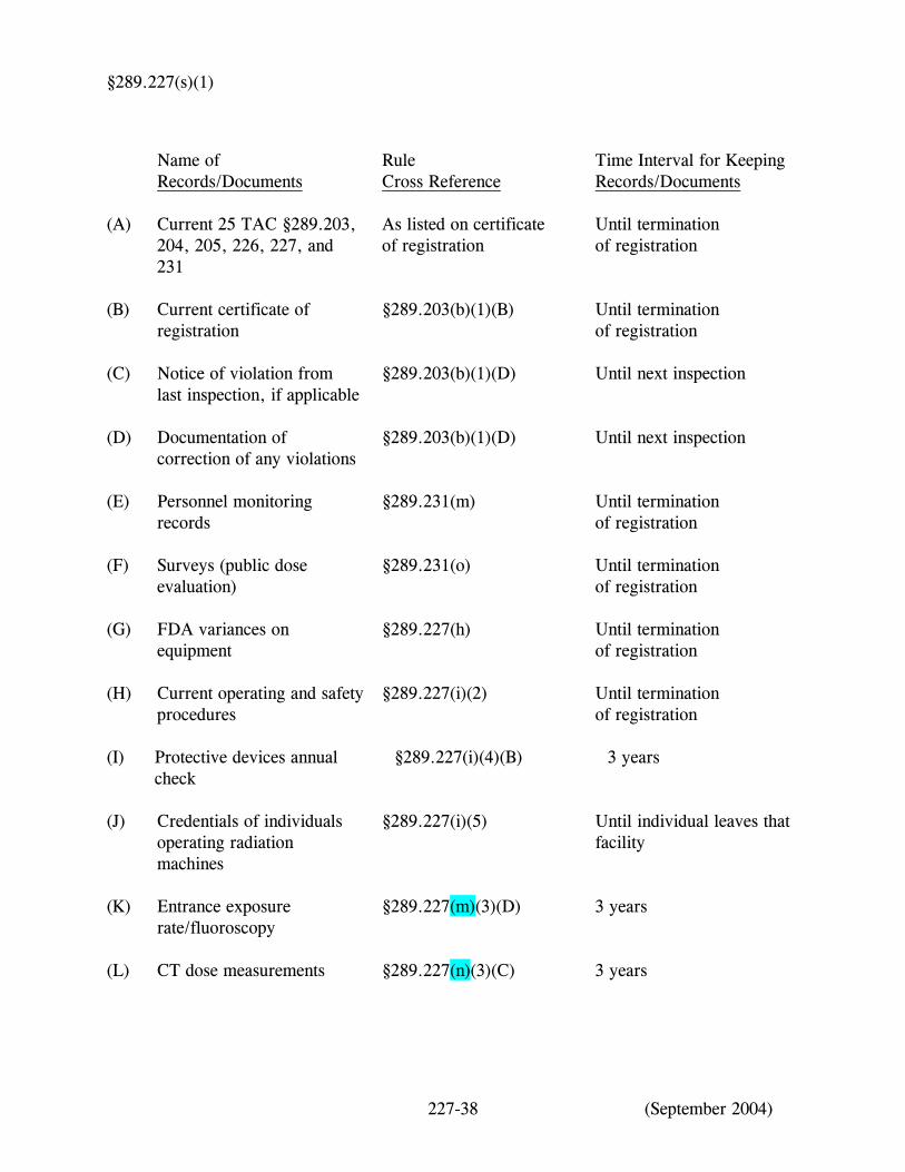
(q) Alternative processing systems. Users of daylight processing systems, laser processors, self-processing film units, or other alternative processing systems shall follow manufacturer's recommendations for image processing. Documentation that the registrant is following manufacturer’s recommendations shall include the date and initials of the individual completing the document and shall be maintained at the site where performed in accordance with subsection (s)(1) of this section for inspection by the agency. (r) Digital imaging acquisition systems. Users of digital imaging acquisition systems shall follow quality assurance/quality control protocol for image processing established by the manufacturer or, if no manufacturer's protocol is available, by the registrant. The registrant shall include the protocols, whether established by the registrant or the manufacturer, in its operating and safety procedures. The registrant shall document the frequency at which the quality assurance/quality control protocol is performed. Documentation shall include the date and initials of the individual completing the document and shall be maintained at the site where performed in accordance with subsection (s)(1) of this section for inspection by the agency. (s) Record/document requirements for mobile services and authorized use locations. (1) Each registrant shall maintain the following records/documents at each site, including authorized records sites for mobile services at the time intervals specified, for inspection by the agency. The records may be maintained in electronic format.
THIS PORTION OF THE PAGE LEFT INTENTIONALLY BLANK
227-37 (September 2004)
§289.227(s)(1)
Name of Records/Documents
Rule Cross Reference
Time Interval for Keeping Records/Documents
(A)
Current 25 TAC §289.203, 204, 205, 226, 227, and 231
As listed on certificate of registration
Until termination of registration
(B)
Current certificate of registration
§289.203(b)(1)(B)
Until termination of registration
(C)
Notice of violation from last inspection, if applicable
§289.203(b)(1)(D)
Until next inspection
(D)
Documentation of correction of any violations
§289.203(b)(1)(D)
Until next inspection
(E)
Personnel monitoring records
§289.231(m)
Until termination of registration
(F)
Surveys (public dose evaluation)
§289.231(o)
Until termination of registration
(G)
FDA variances on equipment
§289.227(h)
Until termination of registration
(H)
Current operating and safety procedures
§289.227(i)(2)
Until termination of registration
(I)
Protective devices annual check
§289.227(i)(4)(B)
3 years
(J)
Credentials of individuals operating radiation machines
§289.227(i)(5)
Until individual leaves that facility
(K)
Entrance exposure rate/fluoroscopy
§289.227(m)(3)(D)
3 years
(L)
CT dose measurements
§289.227(n)(3)(C)
3 years
227-38 (September 2004)
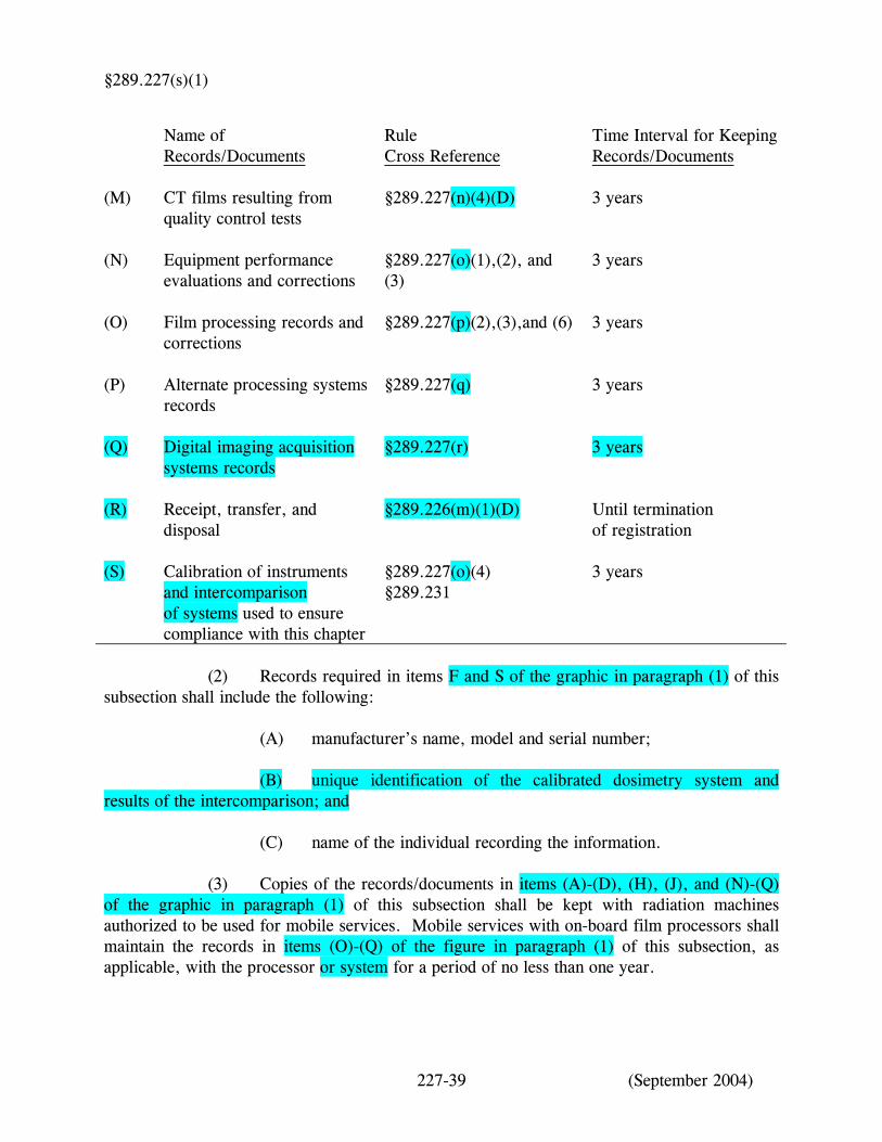
§289.227(s)(1)
Name of Records/Documents
Rule Cross Reference
Time Interval for Keeping Records/Documents
(M)
CT films resulting from quality control tests
§289.227(n)(4)(D)
3 years
(N)
Equipment performance evaluations and corrections
§289.227(o)(1),(2), and (3)
3 years
(O)
Film processing records and corrections
§289.227(p)(2),(3),and (6)
3 years
(P)
Alternate processing systems records
§289.227(q)
3 years
(Q)
Digital imaging acquisition systems records
§289.227(r)
3 years
(R)
Receipt, transfer, and disposal
§289.226(m)(1)(D)
Until termination of registration
(S)
Calibration of instruments and intercomparison of systems used to ensure compliance with this chapter
§289.227(o)(4) §289.231
3 years
(2) Records required in items F and S of the graphic in paragraph (1) of this
subsection shall include the following: (A) manufacturer’s name, model and serial number; (B) unique identification of the calibrated dosimetry system and results of the intercomparison; and (C) name of the individual recording the information.
(3) Copies of the records/documents in items (A)-(D), (H), (J), and (N)-(Q) of the graphic in paragraph (1) of this subsection shall be kept with radiation machines authorized to be used for mobile services. Mobile services with on-board film processors shall maintain the records in items (O)-(Q) of the figure in paragraph (1) of this subsection, as applicable, with the processor or system for a period of no less than one year.
227-39 (September 2004)
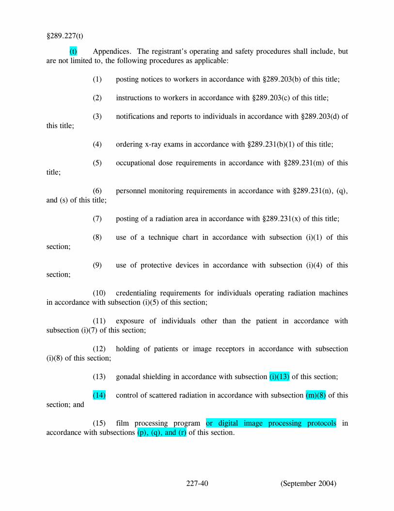
§289.227(t)
(t) Appendices. The registrant's operating and safety procedures shall include, but are not limited to, the following procedures as applicable: (1) posting notices to workers in accordance with §289.203(b) of this title; (2) instructions to workers in accordance with §289.203(c) of this title; (3) notifications and reports to individuals in accordance with §289.203(d) of this title; (4) ordering x-ray exams in accordance with §289.231(b)(1) of this title; (5) occupational dose requirements in accordance with §289.231(m) of this title; (6) personnel monitoring requirements in accordance with §289.231(n), (q), and (s) of this title; (7) posting of a radiation area in accordance with §289.231(x) of this title; (8) use of a technique chart in accordance with subsection (i)(1) of this section; (9) use of protective devices in accordance with subsection (i)(4) of this section; (10) credentialing requirements for individuals operating radiation machines in accordance with subsection (i)(5) of this section; (11) exposure of individuals other than the patient in accordance with subsection (i)(7) of this section; (12) holding of patients or image receptors in accordance with subsection (i)(8) of this section; (13) gonadal shielding in accordance with subsection (i)(13) of this section; (14) control of scattered radiation in accordance with subsection (m)(8) of this section; and (15) film processing program or digital image processing protocols in accordance with subsections (p), (q), and (r) of this section.
227-40 (September 2004)