Embed Size (px)
Citation preview

Cite as: S. Banerjee et al., Science 10.1126/science.aad7974 (2016).
REPORTS
First release: 28 January 2016 www.sciencemag.org (Page numbers not final at time of first release) 1
The superfamily of AAA+ (ATPases Associated with diverse cellular Activities) proteins are molecular chaperones that serve major roles in cellular protein quality control, in DNA replication, transcription, recombination and repair, in membrane fusion, in movement of intracellular cargo, and in cell cycle regulation (1, 2). These oligomeric ring-like en-zymes, which typically form hexamers, contain common functional features including one or more conserved AAA+ ATPase cassettes in each protomer that bind and hydrolyze ATP at the interface between adjacent subunits. The ac-quired energy from binding and catalysis of nucleotides in-duces a series of conformational changes to enable these enzymes to modulate their substrates. p97 is a well-characterized member of this protein family, that binds multiple proteins including deubiquitinating enzymes as well as ubiquitin-binding adaptors and ligases (3–7). The central role that p97 plays in protein quality control makes it an attractive target for cancer chemotherapy, as disrup-tion of both of these processes hinder cancer cell survival (8). Upregulation of p97 expression in cancer cells supports this premise (9), as do reports of numerous structurally di-verse inhibitor types (10–14), and notably, an example of a potent inhibitor that entered a Phase I clinical trial in 2015 (15).
Structurally, each p97 protomer has a predicted molecu-
lar weight of ~90 kDa and is comprised of an N-terminal domain, two tandem ATPase domains designated as D1 and D2 that pack as hexameric rings, and a short C-terminal domain. As a multi-domain, dynamic molecular machine, structural studies of full-length p97 by X-ray crystallography have been limited so far to medium resolution (3.5 Å to 4.7 Å) (16), although higher resolution structures of sub-complexes that contain D1 and the N-domain or the D2 do-main have revealed further architectural insights in these regions of the protein (16–20). Determination of the atomic resolution structure of native, full-length p97 in different conformational states and in complex with inhibitors is therefore of considerable biological and clinical interest.
Using cryo-EM, we first performed structural analysis of p97 without addition of exogenous nucleotides. This struc-ture, determined at an overall resolution of 2.4 Å, shows the expected architecture of the hexameric complex (fig. S1, A and B). ADP occupies both the D1 and D2 domain nucleo-tide binding pockets (fig. S1, C and D). Analysis of the con-formational variants that emerge during 3D classification suggests that subtle conformational heterogeneity in the ADP-bound D2 hexamer (fig. S2) is a likely reason for the lower resolutions achieved in X-ray crystallographic anal-yses of full-length p97. Overall, the cryo-EM derived struc-ture of the D2 domain in the full-length hexamer is
2.3 Å resolution cryo-EM structure of human p97 and mechanism of allosteric inhibition Soojay Banerjee,1* Alberto Bartesaghi,1* Alan Merk,1 Prashant Rao,1 Stacie L. Bulfer,2 Yongzhao Yan,3 Neal Green,4 Barbara Mroczkowski,5 R. Jeffrey Neitz,2 Peter Wipf,3 Veronica Falconieri,1 Raymond J. Deshaies,6 Jacqueline L. S. Milne,1 Donna Huryn,3 Michelle Arkin,2 Sriram Subramaniam1†
1Laboratory of Cell Biology, National Cancer Institute, Bethesda, MD 20892, USA. 2Small Molecule Discovery Center, Pharmaceutical Chemistry, School of Pharmacy, University of
California, San Francisco, CA 94143, USA. 3University of Pittsburgh Chemical Diversity Center, University of Pittsburgh, Pittsburgh, PA 15260, USA. 4Leidos Biomedical Research Inc.,
Frederick, MD 21702, USA. 5Division of Cancer Treatment and Diagnosis, National Cancer Institute, Bethesda, MD 20892, USA. 6Division of Biology and Biological Engineering and
Howard Hughes Medical Institute, California Institute of Technology, Pasadena, CA 91107, USA.
*These authors contributed equally to this work. †Corresponding author. E-mail: [email protected]
p97 is a hexameric AAA ATPase that is an attractive target for cancer drug development. Here, we report cryo-EM structures for ADP-bound, full-length, hexameric wild-type p97 in the presence and absence of an allosteric inhibitor at resolutions of 2.3 Å and 2.4 Å, respectively. We also report cryo-EM structures at ~3.3 Å, 3.2 Å and 3.3 Å resolutions respectively, for three distinct, co-existing functional states of p97 with occupancies of 0, 1 or 2 molecules of ATPγS per protomer. A large corkscrew-like change in molecular architecture coupled with upward displacement of the N-domain is observed only when ATPγS is bound to both D1 and D2 domains. These cryo-EM structures establish the sequence of nucleotide-driven structural changes in p97 at atomic resolution. They also enable elucidation of the binding mode of an allosteric small molecule inhibitor to p97 and illustrate how inhibitor binding at the interface between D1 and D2 domains prevents propagation of the conformational changes necessary for p97 function.
on August 9, 2019
http://science.sciencem
ag.org/D
ownloaded from

First release: 28 January 2016 www.sciencemag.org (Page numbers not final at time of first release) 2
comparable to that determined for full-length p97 at 3.5 Å by X-ray crystallography, except for some differences in the relative disposition of the N-domain (16) and better defini-tion of the C-terminal α-helical subdomain that spans ami-no acid residues 645-763.
Several classes of inhibitors, including those that act in an allosteric or competitive manner, have been identified that impair p97 ATPase activity (10–14, 21–27). Determina-tion of the cryo-EM structure at 2.3 Å resolution of p97 in complex with UPCDC30245, a phenyl indole derivative (fig. S3) that is structurally related to a recently described series of allosteric inhibitors (14) shows that this compound (IC50 for inhibition ~27 nM) binds at the junction between the D1 and D2 domains (Fig. 1, A and B). A detailed view of the structure of a single p97 protomer shows the location of the inhibitor and bound ADP (Fig. 1, C to E), including features such as the interaction of the ADP β-phosphate with a water molecule in the D1 domain (Fig. 1D) and hydrogen-bonding of a water molecule to the α-phosphate moiety and to the hydroxyl group on the ribose ring proximal to the α-phosphate group (Fig. 1E). Furthermore, the distal hydroxyl group of the ribose ring interacts with a water molecule hy-drogen-bonded to side-chain oxygen atom of Asn 660, the main-chain carbonyl atom of residue Asp 478 and another water molecule. Local map resolution measured by RESMAP (28) indicates that the density is weakest in the N-domain, consistent with plots of B-factor profile of the refined struc-ture, which show that average B-factor is highest in the N-domain and in the D1-D2 connector region, and lowest in the core of the D1 and D2 domains (Fig. 1, F and G, and fig. S4). The most ordered regions are at the p97 core, with a clear gradient toward higher B-factors at the periphery (movie S1).
Selected examples of the quality of the cryo-EM map in the D2 domain (residues 481-763) are presented in Fig. 2, A to C. It is conceivable that the two discrete conformations of the Arg 599 side chain (Fig. 2C) are relevant to the ability of p97 to locally adapt to structural variations in bound sub-strates since this residue has been implicated in substrate interactions in the pore region surrounded by the D2 hex-amer. UPCDC30245 binding does not result in significant changes in conformation of the polypeptide backbone, in-cluding at the nucleotide binding sites (fig. S5). Visualiza-tion of density for the bound inhibitor in the map enables determination of its conformation in the bound state and the local interactions involved in its binding at the D1-D2 interface. The inhibitor conformation is most ordered in the half that includes the indole ring that nestles into a binding pocket (Fig. 2, D to F, and fig. S6A). This inhibitor-binding site is sandwiched by the C-terminal regions of two α-helical segments that range from residues 483-498 and 523-534. The binding tunnel is capped at the end where the fluori-
nated indole ring is docked between two loop elements (amino acid residues 506-512 and 610-618) that lead into two parallel β-strands. The other end of the binding cleft is open and solvent exposed (Fig. 2, D and E), with the pipera-zine ring of the inhibitor protruding outwards into the bulk solvent. Residues that interact with ADP in the D1 and D2 sites are summarized in fig. S6, B and C, respectively.
The key interactions that anchor the molecule arise from main chain carbonyl oxygen of Val 493 hydrogen-bonded to the N-H of the indole ring (2.9 Å), weak interaction of the fluorine atom present on the indole ring with the main-chain carbonyl oxygen atom of Ser 511, hydrogen-bonding of the thiol group of Cys 535 to the nitrogen atom in the piper-idine ring (3.4 Å), and the side-chain of Glu 498 hydrogen-bonded to the nitrogen atom in the linker between the piperadine and piperazine groups (3.2 Å). The rest of the binding environment is largely hydrophobic in nature, stemming from interactions with residues Phe 618, Pro 496, 510 and 571, Ala 537 and Val 497 (Fig. 2E). Interactions at the indole ring end are central to the binding of UPCDC30245 to p97, with a potential edge-to-face, weak pi-pi interaction between Phe 618 and the indole ring. Con-sistent with these findings, recent functional studies show that substitutions at the 5-position of the indole ring (where the fluorine atom is attached) have profound effects on ac-tivity, with both steric and electronic factors being im-portant (14). The interactions at the piperazine ring end are minimal, suggesting high conformational flexibility in this region, although the highly negatively charged outer surface of the binding tunnel (fig. S6A) may enable better anchoring to the surface of p97. Mutagenesis studies have shown that the D1-D2 interface is very important for p97 function (11, 12), and provide support for the structural studies we report here.
To unravel the intra- and inter-protomer conformational changes that occur with ATP binding, we performed struc-tural analysis of p97 in the presence of ATPγS at physiologi-cally relevant concentrations. Earlier crystallographic studies of p97 with ATP analogs reveal only modest struc-tural changes (16), raising the concern that crystal packing constraints may have prevented capturing functionally rele-vant protein conformational changes. Cryo-EM methods have been applied previously to study p97 conformational changes; however, these structures have been at very low resolutions (~ 20 – 30 Å; (29–31)), and did not result in de-finitive identification of the structural changes relative to the ADP-bound conformation. Here, we report separation of three well-defined, co-existing conformational states at near-atomic resolution (~3.2 Å/3.3 Å) that are simultaneous-ly populated when ATPγS is added to p97 (Fig. 3 and figs. S7 and S8). In conformation I, ADP is bound in both D1 and D2 nucleotide binding domains; in conformation II, ADP is
on August 9, 2019
http://science.sciencem
ag.org/D
ownloaded from

First release: 28 January 2016 www.sciencemag.org (Page numbers not final at time of first release) 3
bound in the D1 domain while ATPγS is bound in the D2 domain, and in conformation III, ATPγS is bound in the nu-cleotide binding regions of both D1 and D2.
The differences in structure between the three states re-veal an unambiguous and step-wise evolution of the con-formational change with binding of ATPγS (Fig. 3, A to C). Conformation II differs from I only at the D2 domain, while the N and D1 domains remain essentially unchanged (Fig. 3, D to F). The changes in the D2 domain occur largely proxi-mal to the nucleotide-binding region, and result from the exchange of the bound ADP with ATPγS, as verified by di-rect visualization of density for the additional phosphate moiety in the binding site (fig. S8). Within each protomer, there is a ~10-15° rotational twist of the D2 domain with respect to the D1 domain, with the D1-D2 linker region serv-ing as a hinge. This relative rotational displacement of the D2 ring with respect to the D1 ring is consistent with atomic force microscopic studies that have reported ATP-dependent rotation in p97 (32).
The difference between conformation II and III is pri-marily in the N and D1 domains, with minimal changes in the D2 domain (Fig. 3, G to I). Here again, the change in conformation is directly associated with the exchange of ADP for ATPγS, with the density map demonstrating density for the additional phosphate moiety present in the D1 nu-cleotide binding site (fig. S8). The most striking feature of this transition is the large-scale motion of the N-domain away from the plane of the D1 hexamer. The structure of the N and D1 domains in conformation III is essentially the same as that reported from X-ray crystallographic analysis of an isolated N-D1 fragment derived from a mutant p97 (33). The changes include rearrangements at the peripheral helix in D1 and its the interface with the D2 domain, and movement of the N-domain from its original isoplanar, or “down” position to an “up” position, where it is rotated by ~75° and displaced by ~14 Å. This out-of-plane confor-mation of the N-D1 domain and the double occupancy of both nucleotide-binding regions with ATPγS have not been reported in any of the full-length p97 structures solved by X-ray crystallography (16). Additionally, the presence of ATPγS in conformations II and III results in the stabilization of the C-terminal helix of the D2 domain, and extension of the traceable density from residue 763 to residue 768 (fig. S8, G to I).
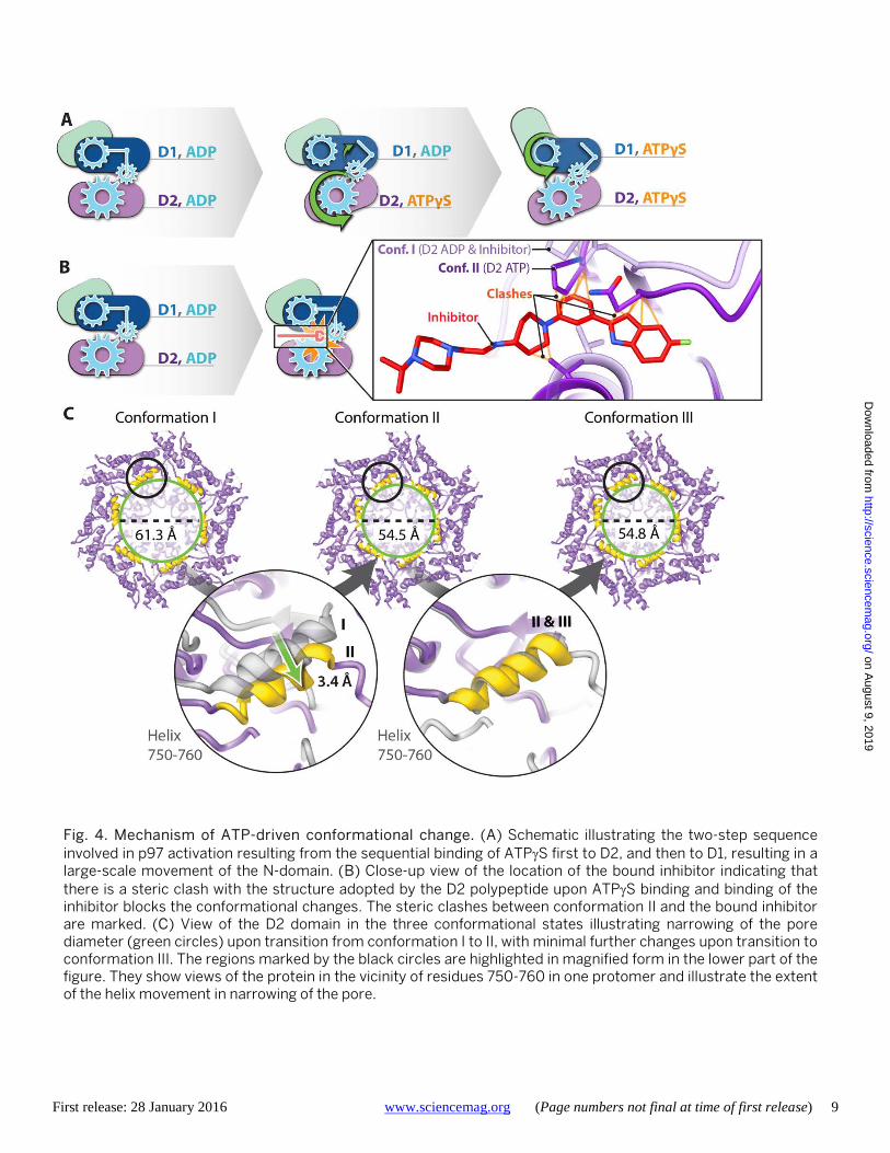
Our structural analyses thus show that the effect of ATPγS binding is to cause a two-step, sequential conforma-tional change in the D2 and D1 hexameric layers, respective-ly (Fig. 4A). Analysis of the images without imposition of 6-fold symmetry shows that the majority of the molecules are in one of the three conformations obtained with the use of symmetry, indicating that the structural changes between
conformations I, II and III are cooperative in nature (fig. S9). The location of the inhibitor binding site at the inter-face of domains D1 and D2 suggests that inhibitor binding prevents the nucleotide-driven conformational changes at the interface that are pivotal for p97 activity. To explore this further, we superimposed the structure of the p97-UPCDC30245 complex onto that of the ATPγS bound p97 conformation II. The superposed models (Fig. 4B and fig. S8J) suggest that there would be steric clashes between the indole ring of the inhibitor and residues in the D2 loop (amino acid residues 611-616). Even if there is some flexibil-ity in the D2 domain that enables some level of binding, key interactions that enable the binding of UPCDC30245 in the p97/ADP complex, such as that between the terminal fluo-rine atom and the backbone carbonyl Ser 511 (Fig. 2D) are not conserved in the ATPγS bound state. These observations indicate that UPCDC30245 is a conformation-selective in-hibitor that preferentially binds the ADP state to act literally as a “wrench in the works”.
The pivot-like movement of the D2 hexamer with ATPγS binding also affects pore dimensions (Fig. 4C). Thus, with ATPγS binding, the diameter of the cytoplasmic face of the pore in the center of the D2 hexamer (measured by the α-carbon of residue 755 in the 750-760 helix) contracts from ~61Å to ~54Å (movie S2), while the average outer (~ 28 Å) and inner (~ 6 Å) diameters of the D1 are similar in the three conformations. Many of the residues that line the cen-tral pore are negatively charged, and could be important in the context of the binding of ubiquitylated substrates. The cavity at the center is large enough to accommodate sub-strates destined for degradation, but given that the changes in size with nucleotide binding are relatively small, our re-sults are consistent with the suggestion (16) that substrates degraded by p97 may not be threaded through the central axis across the length of the barrel.
The ability to determine atomic resolution structures for multiple conformational states that are present simultane-ously in a dynamic molecular machine such as p97 is likely to be an increasingly common signature of the application of cryo-EM methods in structural biology. p97 mediates its function in the cell by interacting with a large array of effec-tor proteins, most of which bind at the N-domain, which undergoes a large movement with ATP binding as our pre-sent work and previous structural studies demonstrate (5, 18, 34–36). Cryo-EM studies could therefore be very useful for a more detailed understanding of the structural basis of these interactions in normal cells and in diseases such as cancer. Further, the delineation of the inhibitor-bound p97 structure provides another example (besides β-galactosidase (37)) of a regulatory metabolic enzyme where cryo-EM at ~ 2 Å resolution enables visualization of structural detail such as hydrogen-bonded water molecules and side chain hetero-
on August 9, 2019
http://science.sciencem
ag.org/D
ownloaded from
First release: 28 January 2016 www.sciencemag.org (Page numbers not final at time of first release) 4
geneity. The discovery of the specific mode of binding of a compound that blocks p97 activity provides new insights into the interactions and residues that are critical for func-tion, and will hopefully enable the design of clinically useful inhibitors.
REFERENCES AND NOTES 1. P. I. Hanson, S. W. Whiteheart, AAA+ proteins: Have engine, will work. Nat. Rev.
Mol. Cell Biol. 6, 519–529 (2005). Medline doi:10.1038/nrm1684 2. T. Ogura, A. J. Wilkinson, AAA+ superfamily ATPases: Common structure—diverse
function. Genes Cells 6, 575–597 (2001). Medline doi:10.1046/j.1365-2443.2001.00447.x
3. D. Barthelme, R. T. Sauer, Origin and functional evolution of the Cdc48/p97/VCP AAA+ protein unfolding and remodeling machine. J. Mol. Biol. (2015). Medline doi:10.1016/j.jmb.2015.11.015
4. M. Otter-Nilsson, R. Hendriks, E. I. Pecheur-Huet, D. Hoekstra, T. Nilsson, Cytosolic ATPases, p97 and NSF, are sufficient to mediate rapid membrane fusion. EMBO J. 18, 2074–2083 (1999). Medline doi:10.1093/emboj/18.8.2074
5. H. Meyer, C. C. Weihl, The VCP/p97 system at a glance: Connecting cellular function to disease pathogenesis. J. Cell Sci. 127, 3877–3883 (2014). Medline doi:10.1242/jcs.093831
6. D. Avci, M. K. Lemberg, Clipping or extracting: Two ways to membrane protein degradation. Trends Cell Biol. 25, 611–622 (2015). Medline doi:10.1016/j.tcb.2015.07.003
7. L. Fang, C. Hemion, A. C. Pinho Ferreira Bento, C. C. Bippes, J. Flammer, A. Neutzner, Mitochondrial function in neuronal cells depends on p97/VCP/Cdc48-mediated quality control. Front. Cell. Neurosci. 9, 16 (2015). Medline doi:10.3389/fncel.2015.00016
8. R. J. Deshaies, Proteotoxic crisis, the ubiquitin-proteasome system, and cancer therapy. BMC Biol. 12, 94 (2014). Medline
9. C. W. Valle, T. Min, M. Bodas, S. Mazur, S. Begum, D. Tang, N. Vij, Critical role of VCP/p97 in the pathogenesis and progression of non-small cell lung carcinoma. PLOS ONE 6, e29073 (2011). Medline doi:10.1371/journal.pone.0029073
10. T. F. Chou, S. J. Brown, D. Minond, B. E. Nordin, K. Li, A. C. Jones, P. Chase, P. R. Porubsky, B. M. Stoltz, F. J. Schoenen, M. P. Patricelli, P. Hodder, H. Rosen, R. J. Deshaies, Reversible inhibitor of p97, DBeQ, impairs both ubiquitin-dependent and autophagic protein clearance pathways. Proc. Natl. Acad. Sci. U.S.A. 108, 4834–4839 (2011). Medline doi:10.1073/pnas.1015312108
11. T. F. Chou, K. Li, K. J. Frankowski, F. J. Schoenen, R. J. Deshaies, Structure-activity relationship study reveals ML240 and ML241 as potent and selective inhibitors of p97 ATPase. ChemMedChem 8, 297–312 (2013). Medline doi:10.1002/cmdc.201200520
12. P. Magnaghi, R. D’Alessio, B. Valsasina, N. Avanzi, S. Rizzi, D. Asa, F. Gasparri, L. Cozzi, U. Cucchi, C. Orrenius, P. Polucci, D. Ballinari, C. Perrera, A. Leone, G. Cervi, E. Casale, Y. Xiao, C. Wong, D. J. Anderson, A. Galvani, D. Donati, T. O’Brien, P. K. Jackson, A. Isacchi, Covalent and allosteric inhibitors of the ATPase VCP/p97 induce cancer cell death. Nat. Chem. Biol. 9, 548–556 (2013). Medline doi:10.1038/nchembio.1313
13. P. Polucci, P. Magnaghi, M. Angiolini, D. Asa, N. Avanzi, A. Badari, J. Bertrand, E. Casale, S. Cauteruccio, A. Cirla, L. Cozzi, A. Galvani, P. K. Jackson, Y. Liu, S. Magnuson, B. Malgesini, S. Nuvoloni, C. Orrenius, F. R. Sirtori, L. Riceputi, S. Rizzi, B. Trucchi, T. O’Brien, A. Isacchi, D. Donati, R. D’Alessio, Alkylsulfanyl-1,2,4-triazoles, a new class of allosteric valosine containing protein inhibitors. Synthesis and structure-activity relationships. J. Med. Chem. 56, 437–450 (2013). Medline doi:10.1021/jm3013213
14. C. Alverez, M. R. Arkin, S. L. Bulfer, R. Colombo, M. Kovaliov, M. G. LaPorte, C. Lim, M. Liang, W. J. Moore, R. J. Neitz, Y. Yan, Z. Yue, D. M. Huryn, P. Wipf, Structure-activity study of bioisosteric trifluoromethyl and pentafluorosulfanyl indole inhibitors of the AAA ATPase p97. ACS Med. Chem. Lett. 6, 1225–1230 (2015). doi:10.1021/acsmedchemlett.5b00364
15. D. J. Anderson et al., “Inhibition of the AAA-ATPase p97 with the first in class inhibitor CB-5083 as a novel approach to treat cancer.” Paper presented at the American Association for Cancer Research annual meeting, Philadelphia, 18 to
22 April 2015. 16. J. M. Davies, A. T. Brünger, W. I. Weis, Improved structures of full-length p97, an
AAA ATPase: Implications for mechanisms of nucleotide-dependent conformational change. Structure 16, 715–726 (2008). Medline doi:10.1016/j.str.2008.02.010
17. S. J. Kim, J. Cho, E. J. Song, S. J. Kim, H. M. Kim, K. E. Lee, S. W. Suh, E. E. Kim, Structural basis for ovarian tumor domain-containing protein 1 (OTU1) binding to p97/valosin-containing protein (VCP). J. Biol. Chem. 289, 12264–12274 (2014). Medline doi:10.1074/jbc.M113.523936
18. P. Hänzelmann, A. Buchberger, H. Schindelin, Hierarchical binding of cofactors to the AAA ATPase p97. Structure 19, 833–843 (2011). Medline doi:10.1016/j.str.2011.03.018
19. I. Dreveny, H. Kondo, K. Uchiyama, A. Shaw, X. Zhang, P. S. Freemont, Structural basis of the interaction between the AAA ATPase p97/VCP and its adaptor protein p47. EMBO J. 23, 1030–1039 (2004). Medline doi:10.1038/sj.emboj.7600139
20. W. K. Tang, D. Xia, Altered intersubunit communication is the molecular basis for functional defects of pathogenic p97 mutants. J. Biol. Chem. 288, 36624–36635 (2013). Medline doi:10.1074/jbc.M113.488924
21. E. Chapman, N. Maksim, F. de la Cruz, J. J. La Clair, Inhibitors of the AAA+ chaperone p97. Molecules 20, 3027–3049 (2015). Medline doi:10.3390/molecules20023027
22. C. J. Fang, L. Gui, X. Zhang, D. R. Moen, K. Li, K. J. Frankowski, H. J. Lin, F. J. Schoenen, T. F. Chou, Evaluating p97 inhibitor analogues for their domain selectivity and potency against the p97-p47 complex. ChemMedChem 10, 52–56 (2015). Medline doi:10.1002/cmdc.201402420
23. T. F. Chou, R. J. Deshaies, Development of p97 AAA ATPase inhibitors. Autophagy 7, 1091–1092 (2011). Medline doi:10.4161/auto.7.9.16489
24. S. Tao, J. Tillotson, E. M. Wijeratne, Y. M. Xu, M. Kang, T. Wu, E. C. Lau, C. Mesa, D. J. Mason, R. V. Brown, J. J. La Clair, A. A. Gunatilaka, D. D. Zhang, E. Chapman, Withaferin A analogs that target the AAA+ chaperone p97. ACS Chem. Biol. 10, 1916–1924 (2015). Medline
25. P. Yi, A. Higa, S. Taouji, M. G. Bexiga, E. Marza, D. Arma, C. Castain, B. Le Bail, J. C. Simpson, J. Rosenbaum, C. Balabaud, P. Bioulac-Sage, J. F. Blanc, E. Chevet, Sorafenib-mediated targeting of the AAA⁺ ATPase p97/VCP leads to disruption of the secretory pathway, endoplasmic reticulum stress, and hepatocellular cancer cell death. Mol. Cancer Ther. 11, 2610–2620 (2012). Medline doi:10.1158/1535-7163.MCT-12-0516
26. S. J. Brown et al., in Probe Reports from the NIH Molecular Libraries Program (National Center for Biotechnology Information, Bethesda, MD, 2010).
27. M. S. Chimenti, S. L. Bulfer, R. J. Neitz, A. R. Renslo, M. P. Jacobson, T. L. James, M. R. Arkin, M. J. Kelly, A fragment-based ligand screen against part of a large protein machine: The ND1 domains of the AAA+ ATPase p97/VCP. J. Biomol. Screen. 20, 788–800 (2015). Medline doi:10.1177/1087057115570550
28. A. Kucukelbir, F. J. Sigworth, H. D. Tagare, Quantifying the local resolution of cryo-EM density maps. Nat. Methods 11, 63–65 (2014). Medline doi:10.1038/nmeth.2727
29. X. Zhang, A. Shaw, P. A. Bates, R. H. Newman, B. Gowen, E. Orlova, M. A. Gorman, H. Kondo, P. Dokurno, J. Lally, G. Leonard, H. Meyer, M. van Heel, P. S. Freemont, Structure of the AAA ATPase p97. Mol. Cell 6, 1473–1484 (2000). Medline
30. H. Niwa, C. A. Ewens, C. Tsang, H. O. Yeung, X. Zhang, P. S. Freemont, The role of the N-domain in the ATPase activity of the mammalian AAA ATPase p97/VCP. J. Biol. Chem. 287, 8561–8570 (2012). Medline doi:10.1074/jbc.M111.302778
31. H. O. Yeung, A. Förster, C. Bebeacua, H. Niwa, C. Ewens, C. McKeown, X. Zhang, P. S. Freemont, Inter-ring rotations of AAA ATPase p97 revealed by electron cryomicroscopy. Open Biol 4, 130142 (2014). Medline doi:10.1098/rsob.130142
32. K. Noi, D. Yamamoto, S. Nishikori, K. Arita-Morioka, T. Kato, T. Ando, T. Ogura, High-speed atomic force microscopic observation of ATP-dependent rotation of the AAA+ chaperone p97. Structure 21, 1992–2002 (2013). Medline doi:10.1016/j.str.2013.08.017
33. W. K. Tang, D. Li, C. C. Li, L. Esser, R. Dai, L. Guo, D. Xia, A novel ATP-dependent conformation in p97 N-D1 fragment revealed by crystal structures of disease-related mutants. EMBO J. 29, 2217–2229 (2010). Medline doi:10.1038/emboj.2010.104
34. W. S. Chia, D. X. Chia, F. Rao, S. Bar Nun, S. Geifman Shochat, ATP binding to
on August 9, 2019
http://science.sciencem
ag.org/D
ownloaded from
First release: 28 January 2016 www.sciencemag.org (Page numbers not final at time of first release) 5
p97/VCP D1 domain regulates selective recruitment of adaptors to its proximal N-domain. PLOS ONE 7, e50490 (2012). Medline doi:10.1371/journal.pone.0050490
35. F. Beuron, I. Dreveny, X. Yuan, V. E. Pye, C. McKeown, L. C. Briggs, M. J. Cliff, Y. Kaneko, R. Wallis, R. L. Isaacson, J. E. Ladbury, S. J. Matthews, H. Kondo, X. Zhang, P. S. Freemont, Conformational changes in the AAA ATPase p97-p47 adaptor complex. EMBO J. 25, 1967–1976 (2006). Medline
36. B. Almeida, I. A. Abreu, C. A. Matos, J. S. Fraga, S. Fernandes, M. G. Macedo, R. Gutiérrez-Gallego, P. J. Pereira, A. L. Carvalho, S. Macedo-Ribeiro, SUMOylation of the brain-predominant Ataxin-3 isoform modulates its interaction with p97. Biochim. Biophys. Acta 1852, 1950–1959 (2015). Medline doi:10.1016/j.bbadis.2015.06.010
37. A. Bartesaghi, A. Merk, S. Banerjee, D. Matthies, X. Wu, J. L. Milne, S. Subramaniam, 2.2 Å resolution cryo-EM structure of β-galactosidase in complex with a cell-permeant inhibitor. Science 348, 1147–1151 (2015). Medline
ACKNOWLEDGMENTS
This research was supported by funds from the Center for Cancer Research, National Cancer Institute, NIH, Bethesda, MD, and with federal funds from the National Cancer Institute, National Institutes of Health, under Chemical Biology Consortium Contract No. HHSN261200800001E Agreement No. 29XS127TO15. RJD is an investigator of the HHMI. This work utilized the computational resources of the NIH HPC Biowulf cluster (http://hpc.nih.gov). We thank K. Moynihan, R. Mueller, and J. Cometa for technical assistance with electron microscopy, F. Ulmer, P. Mooney and C. Booth for advice and assistance with optimizing K2 detector performance, and M.G. Laporte for contributions to the medicinal chemistry program. The University of Pittsburgh has filed a provisional patent application that covers the inhibitor compound described in this manuscript. RD is on the Scientific Advisory Board of Cleave Biosciences. The density maps and refined atomic models have been deposited with the Electron Microscopy Data Bank with accession numbers of EMD-3295, 3296, 3297, 3298 and 3299 and the Protein Data Bank with matching accession numbers of PDB-5FTJ, 5FTK, 5FTL, 5FTM and 5FTN, respectively for native p97 in the presence and absence of bound inhibitor, and for p97 conformational states I, II and III observed in the presence of ATPγS.
SUPPLEMENTARY MATERIALS www.sciencemag.org/cgi/content/full/science.aad7974/DC1 Materials and Methods Figs. S1 to S9 Movies S1 and S2 3 November 2015; accepted 14 January 2016 Published online 28 January 2016 10.1126/science.aad7974
on August 9, 2019
http://science.sciencem
ag.org/D
ownloaded from

First release: 28 January 2016 www.sciencemag.org (Page numbers not final at time of first release) 6
Fig. 1. Atomic resolution model derived from cryo-EM structure of p97 in the presence of bound inhibitor. (A and B) Top and side views, respectively, of the cryo-EM structure of the p97 hexamer presented as a ribbon diagram, showing the N (green), D1 (blue) and D2 (purple) domains. The ADP molecule is colored in cyan. The inhibitor (red) is bound at the junction between the D1 and D2 domains. The relative position of each domain in the primary sequence is indicated. (C) Ribbon diagram of a p97 protomer highlighting the location of the bound inhibitor (red) relative to the two bound nucleotides (cyan) in D1 and D2 domains. (D and E) Density maps for bound nucleotides establishing that ADP is bound to both D1 and D2 domains, and visualization of densities for tightly bound water molecules (colored red - highlighted in yellow) at the nucleotide binding sites. (F and G) Top, and side-view representations, respectively, of the structure (uncorrected density map), color-coded to represent variation in resolution across the protein as determined using the program RESMAP (28).
on August 9, 2019
http://science.sciencem
ag.org/D
ownloaded from

First release: 28 January 2016 www.sciencemag.org (Page numbers not final at time of first release) 7
Fig. 2. Depiction of cryo-EM map quality and inhibitor interactions. (A) Illustration of hydrophobic packing between the aromatic side-chains of Trp 551 and Phe 552, and the presence of a hole in the aromatic ring of Phe 576. (B) Density for water molecules hydrogen-bonded to the Arg 599 backbone and evidence for alternate conformations of the terminal guanidinium group, shown in orthogonal orientations. The main chain oxygen atom of Phe 552 shown in (A) is hydrogen-bonded to the Arg 599 guanidinium moiety. (C) Density for Arg 638, part of the “arginine finger” motif at the outer surface of the D2 domain. (D) LIGPLOT representation showing residues in p97 that are in close proximity to the inhibitor, highlighting key H-bond interactions between the nitrogen on the indole ring and the backbone carbonyl oxygen atom of Val 493 and side-chain of Glu 498 with the nitrogen atom in the linker of the inhibitor. The interaction between the fluorine atom present on the indole ring of the inhibitor and the main-chain carbonyl atom of Ser 511 is also indicated. (E and F) Close-up view of the conformation of UPCDC30245 bound to p97. There is strong density for the bound inhibitor for the segment encompassing the indole group to the piperidine ring and virtually no density at the other extremity, where the molecule is expected to be highly flexible because of minimal interactions with the protein.
on August 9, 2019
http://science.sciencem
ag.org/D
ownloaded from

First release: 28 January 2016 www.sciencemag.org (Page numbers not final at time of first release) 8
Fig. 3. Cryo-EM structures at ~ 3.3 Å, ~ 3.2 Å and ~ 3.3 Å resolution respectively of three distinct p97 conformational states populated upon addition of ATPγS. (A to C) Side views of molecular surface models of the three states, color coded to show the N-terminal, D1 and D2 domains in green, blue and purple respectively. The green arrows indicate the motion of the D2 domain in the transition from conformation I to II (B), and the motion of the N domain in the transition from conformation II to III (C). (D to F) Superposition of the polypeptide backbones of conformations I and II in ribbon representation to illustrate that the N and D1 domains display similar conformations, but that there are significant differences in the D2 domain, as indicated by the green arrows. The color scheme for conformation II is as in (A) to (C), with conformation I shown in orange. (G to I) Superposition of the polypeptide backbones of conformations II and III in ribbon representation to illustrate that the D2 domain is similar, but that there are significant differences in conformation of the N and D1 domains, as indicated by the green arrows. The color scheme for conformation III is as in (A) to (C), with conformation II now shown in orange. The insets in (D) and (G) show top views of the D1 domain, while the insets in (E) and (H) show top views of the D2 domain, providing context for the superpositions shown in the main panels.
on August 9, 2019
http://science.sciencem
ag.org/D
ownloaded from
First release: 28 January 2016 www.sciencemag.org (Page numbers not final at time of first release) 9
Fig. 4. Mechanism of ATP-driven conformational change. (A) Schematic illustrating the two-step sequence involved in p97 activation resulting from the sequential binding of ATPγS first to D2, and then to D1, resulting in a large-scale movement of the N-domain. (B) Close-up view of the location of the bound inhibitor indicating that there is a steric clash with the structure adopted by the D2 polypeptide upon ATPγS binding and binding of the inhibitor blocks the conformational changes. The steric clashes between conformation II and the bound inhibitor are marked. (C) View of the D2 domain in the three conformational states illustrating narrowing of the pore diameter (green circles) upon transition from conformation I to II, with minimal further changes upon transition to conformation III. The regions marked by the black circles are highlighted in magnified form in the lower part of the figure. They show views of the protein in the vicinity of residues 750-760 in one protomer and illustrate the extent of the helix movement in narrowing of the pore.
on August 9, 2019
http://science.sciencem
ag.org/D
ownloaded from

2.3 Å resolution cryo-EM structure of human p97 and mechanism of allosteric inhibition
Sriram SubramaniamR. Jeffrey Neitz, Peter Wipf, Veronica Falconieri, Raymond J. Deshaies, Jacqueline L. S. Milne, Donna Huryn, Michelle Arkin and Soojay Banerjee, Alberto Bartesaghi, Alan Merk, Prashant Rao, Stacie L. Bulfer, Yongzhao Yan, Neal Green, Barbara Mroczkowski,
published online January 28, 2016
ARTICLE TOOLS http://science.sciencemag.org/content/early/2016/01/27/science.aad7974
MATERIALSSUPPLEMENTARY http://science.sciencemag.org/content/suppl/2016/01/27/science.aad7974.DC1
CONTENTRELATED http://stke.sciencemag.org/content/sigtrans/9/420/re3.full
REFERENCES
http://science.sciencemag.org/content/early/2016/01/27/science.aad7974#BIBLThis article cites 34 articles, 9 of which you can access for free
PERMISSIONS http://www.sciencemag.org/help/reprints-and-permissions
Terms of ServiceUse of this article is subject to the
registered trademark of AAAS. is aScienceAmerican Association for the Advancement of Science. No claim to original U.S. Government Works. The title
Science, 1200 New York Avenue NW, Washington, DC 20005. 2017 © The Authors, some rights reserved; exclusive licensee (print ISSN 0036-8075; online ISSN 1095-9203) is published by the American Association for the Advancement ofScience
on August 9, 2019
http://science.sciencem
ag.org/D
ownloaded from


![[2005] 현대자동차 노동강도 평가와 대안 마련을 위한 연구 (p97~146)](https://img.dokumen.tips/doc/110x75/55b94f9cbb61eb47308b456e/2005-p97146.jpg)