Embed Size (px)
Citation preview

MOLECULAR PAINLaedermann et al. Molecular Pain 2014, 10:19http://www.molecularpain.com/content/10/1/19
RESEARCH Open Access
Voltage-gated sodium channel expression inmouse DRG after SNI leads to re-evaluation ofprojections of injured fibersCédric J Laedermann1*†, Marie Pertin1†, Marc R Suter1 and Isabelle Decosterd1,2
Abstract
Background: Dysregulation of voltage-gated sodium channels (Navs) is believed to play a major role in nerve fiberhyperexcitability associated with neuropathic pain. A complete transcriptional characterization of the different isoformsof Navs under normal and pathological conditions had never been performed on mice, despite their widespread use inpain research. Navs mRNA levels in mouse dorsal root ganglia (DRG) were studied in the spared nerve injury (SNI)and spinal nerve ligation (SNL) models of neuropathic pain. In the SNI model, injured and non-injured neuronswere intermingled in lumbar DRG, which were pooled to increase the tissue available for experiments.
Results: A strong downregulation was observed for every Navs isoform expressed except for Nav1.2; even Nav1.3,known to be upregulated in rat neuropathic pain models, was lower in the SNI mouse model. This suggestsdifferences between these two species. In the SNL model, where the cell bodies of injured and non-injured fibersare anatomically separated between different DRG, most Navs were observed to be downregulated in the L5 DRGreceiving axotomized fibers. Transcription was then investigated independently in the L3, L4 and L5 DRG in theSNI model, and an important downregulation of many Navs isoforms was observed in the L3 DRG, suggesting thepresence of numerous injured neurons there after SNI. Consequently, the proportion of axotomized neurons in theL3, L4 and L5 DRG after SNI was characterized by studying the expression of activating transcription factor 3(ATF3). Using this marker of nerve injury confirmed that most injured fibers find their cell bodies in the L3 and L4DRG after SNI in C57BL/6 J mice; this contrasts with their L4 and L5 DRG localization in rats. The spared sural nerve,through which pain hypersensitivity is measured in behavioral studies, mostly projects into the L4 and L5 DRG.
Conclusions: The complex regulation of Navs, together with the anatomical rostral shift of the DRG harboringinjured fibers in C57BL/6 J mice, emphasize that caution is necessary and preliminary anatomical experimentsshould be carried out for gene and protein expression studies after SNI in mouse strains.
Keywords: Activating transcription factor 3 (ATF3), Dorsal root ganglia (DRG), Nerve injury, Neuropathic pain,Quantitative real time polymerase chain reaction (qRT-PCR), Sciatic nerve, Spared nerve injury (SNI), Spinal nerveligation (SNL), Voltage-gated sodium channels (Navs)
BackgroundIncreased electrical activity is a major mechanism in thedevelopment of neuropathic pain following peripheralnerve injury. Spontaneous electrical discharges can ori-ginate from both injured and non-injured nerve fibersor from dorsal root ganglia (DRG) [1-7]. Voltage-gated
* Correspondence: [email protected]†Equal contributors1Pain Center, Department of Anesthesiology, University Hospital Center andUniversity of Lausanne, Lausanne 1011, SwitzerlandFull list of author information is available at the end of the article
© 2014 Laedermann et al.; licensee BioMed CeCreative Commons Attribution License (http:/distribution, and reproduction in any mediumDomain Dedication waiver (http://creativecomarticle, unless otherwise stated.
sodium channels (Navs) are key transmitters in cellularexcitability [8], and are essential for pain transmission[9]. Navs are heteromeric proteins composed of a large,pore-forming α-subunits and small β-auxiliary subunits[10,11]. Of the nine distinct channel isoforms described(Nav1.1 to Nav1.9), Nav1.5, Nav1.8 and Nav1.9 are resist-ant to tetrodotoxin (TTX). All isoforms, except Nav1.4and Nav1.5, are expressed in DRG. Nav1.7 is the mosthighly expressed, TTX-sensitive isoform in rats [12-17].It has been proposed that nerve-injury-mediated hyper-excitability results from the altered expression of Navs
ntral Ltd. This is an Open Access article distributed under the terms of the/creativecommons.org/licenses/by/2.0), which permits unrestricted use,, provided the original work is properly credited. The Creative Commons Publicmons.org/publicdomain/zero/1.0/) applies to the data made available in this

Figure 1 Constitutive mRNA expression of Navs isoforms insham mouse DRG. Constitutive levels of mRNA were determinedusing qPCR and normalized using GAPDH as a reference gene. Dataare expressed as mean ± SEM, n = 4 samples (2 animals per sample).
Laedermann et al. Molecular Pain 2014, 10:19 Page 2 of 11http://www.molecularpain.com/content/10/1/19
[18-20]. In rats, these changes in Navs expression occurin both injured and non-injured neurons [19,21-23]. Indifferent experimental models of neuropathic pain in rats,the mRNAs of most Navs were downregulated in the DRG[16,24-27] except for an increase of Nav1.3 transcript[16,27,28]. Navs changes in mouse models of neuropathicpain have, however, not been investigated.The various animal models of neuropathic pain, involving
transection and/or ligation of nerves from the hind paw,exhibit different relations between injured and non-injuredfibers. The first behavioral model of nerve injury wasthe complete sciatic nerve transection [29]. This modeldoes not adequately reflect the partial nerve injuriesobserved in most patients with neuropathic pain, whichalso involves signals coming from intact sensory neurons[30]. Since then, models of partial injuries have beendescribed which also allowed evoked behavioral testing ofthe hind paw. The L5 spinal nerve ligation (SNL) is an ex-perimental neuropathic pain model which displays a clearseparation between injured and non-injured cell bodies[31]. This model does not allow cross-talk between injuredand non-injured cell bodies in L5 and L4 DRG, respect-ively. The spared nerve injury model (SNI) [32] involvesthe lesion of two terminal branches of the sciatic nerve,the common peroneal and tibial nerves, sparing the suralnerve, and inducing mechanical and thermal hypersensitiv-ity in the latter territory. In this model, injured and intactnerves intermingle in the same DRG, which may allowcross-excitation between cell bodies [33,34] in addition toephaptic cross-talk along nerve fibers [35]. Originally a ratmodel, the SNI model was later transposed and validated inmice [36]. To our knowledge, a careful characterization ofinjured and non-injured nerve fibers projecting into DRGhas not been carried out in mice after SNI, and the as-sumption of neuroanatomical similarities between the twospecies—rats and mice—may not be correct [37].In this study, we investigated the changes in Navs tran-
scription in mouse DRG following SNI and SNL surgery.To correlate Navs expression to injury we also studiedthe projection of injured and intact fibers into the L3, L4and L5 DRG after SNI.
Results and discussionExpression of Navs in mouse L4 and L5 DRGFirst, the level of expression of Navs in the DRG of sham-operated mice was assessed using qRT-PCR (Figure 1).Constitutively, Nav1.2 is the most expressed TTX-sensitiveisoform in mouse L4 and L5 DRG; this differs from rats,where this isoform is only faintly expressed [12,16,17]. It isnoteworthy that significant variability in the expression forthis isoform was observed (see Figure 2). Nav1.7, whichis the main TTX-sensitive isoform expressed in rat DRG,was also well represented in mice, as it was the secondmost expressed TTX-sensitive isoform. Consistent with
observations in rats, the two TTX-resistant isoforms—Nav1.8 and Nav1.9—were also highly expressed in mouseL4 and L5 DRG. The qRT-PCR products of all the Navsisoforms were subcloned and sequenced, and, surprisingly,despite the careful design of specific in silico primers,some of the amplicons were indeed seen to be the cross-amplification products of other isoforms. In order to avoidartefactual results, all final primers used in this study(Table 1) were validated by sequencing the amplicons(see Methods). These results suggest that amplicons shouldalways be sequenced when studying a highly conservedfamily of proteins such as Navs.
Downregulation of Navs expression after SNI injuryNext, Navs mRNA regulation after SNI in mice wasanalyzed. In order to reduce the number of animals ne-cessary for experiments, and because DRG only containscarce amounts of tissue, DRG are commonly pooled to-gether. In procedures corresponding to those previouslycarried out in rats, mouse L4 and L5 DRG were pooled,as it was assumed that these would contain a mixtureof the cell bodies from injured and non-injured fibers.In comparison to the sham-operated mice, SNI induceda significant downregulation of every isoform tested(Figure 2A and Table 2) except for Nav1.2. This downregula-tion may prove to be sustained as Nav1.7 (−33%, p= 0.019)and Nav1.8 (−38%, p = 0.007) were still significantlydownregulated 6 weeks after SNI (data now shown).At the same time point, Nav1.3 downregulation hadnot reached significance (−24%, p = 0.43). The otherisoforms were not tested.

A
C
B
Figure 2 SNI and SNL modulate Navs mRNA expression in mouse DRG. Transcription profile: (A) one week after SNI in pooled L4 and L5DRG. Nav1.1, Nav1.3, Nav1.6, Nav1.7, Nav1.8 and Nav1.9 were downregulated after SNI. Only Nav1.2 remained unchanged. *p < 0.05, ***p < 0.001,Student’s t test. The bar graph represents the SNI/sham ratios.% changes in transcripts and p-values are found in Table 2. (B) One week after SNLin L4 and L5 DRG. In injured L5 DRG (black bars), Nav1.1, Nav1.6, Nav1.7, Nav1.8 and Nav1.9 were significantly lower. Nav1.2 and Nav1.3 wereunchanged. In non-injured L4 DRG (white bars), only Nav1.1 was lower but Nav1.2, Nav1.3, Nav1.6, Nav1.7, Nav1.8 and Nav1.9 remained unchanged.*p < 0.05, **p < 0.01, ***p < 0.001, two-way ANOVA and post hoc Bonferroni tests. The bar graph represents the SNL/sham ratios for L4 and L5DRG.% change and p-values are found in Table 3. (C) One week after SNI in separated L3, L4 and L5 DRG. Nav1.1 was significantly downregulatedin L3 and L4. Nav1.2, Nav1.3 and Nav1.6 remained statistically unchanged in every DRG tested. Nav1.7 was only significantly downregulated in L3.Nav1.8 and Nav1.9 were downregulated in all three DRG. *p < 0.05, **p < 0.01, ***p < 0.001, two-way ANOVA and post hoc Bonferroni tests. The bargraph represents the SNI/sham ratios for L3, L4 and L5 DRG.% change and p-values are found in Table 4.
Laedermann et al. Molecular Pain 2014, 10:19 Page 3 of 11http://www.molecularpain.com/content/10/1/19
These results highlighted an important difference be-tween mice and rats: whereas Nav1.3 was upregulatedin rats [16,28,38,39] after SNI, a downregulation of thisisoform was observed in mice after SNI. Despite contro-versies about the role of sodium channels in neuropathicpain, the upregulation of Nav1.3 is commonly acceptedas an important mechanism beyond neuropathic pain-associated hyperexcitability in rats [27,39]. This was re-cently confirmed by the gene’s knockdown in a rat modelof nerve injury, which led to an attenuation of the nerveinjury-induced neuropathic pain symptoms [40]. However,this study’s results indicated that Nav1.3 might be involveddifferently in mice, and this was corroborated by thenormal development of neuropathic pain symptoms inNav1.3 null mutant mice [41].How a decrease in Navs mRNA in the DRG could
contribute to hyperexcitability remains subject to debate.A redistribution of the mRNA from the cell bodies tothe sciatic nerve has been shown for Nav1.8, where this
isoform can be translated and regain function [42]. In aprevious paper, this study’s authors reported the inter-esting fact that following SNI in mice, Nav1.8 proteinexpression in the sciatic nerve increased [43]. More-over, the level of mRNA does not necessarily correlateto amounts of protein, and further investigation will benecessary to unravel the physiological meaning of a de-crease in Navs transcripts in the DRG.
Downregulation of Navs expression after SNL injuryBecause the L4 and L5 DRG contained adjacent injuredand non-injured neurons after SNI, and in order tosolely investigate the role of axotomy on Navs expression,the following procedure to be performed was SNL. TheL4 (non-injured) and L5 (injured) DRG were compared totheir respective DRG in sham-operated mice.A highly significant decrease in the mRNA expression
of most of the Navs isoforms was observed in the injuredL5 DRG. Only the Nav1.2 and Nav1.3 isoforms remained

Table 1 List of primers sequences
Genename
Primer sequence 5′-3′ Primerconcentration
GAPDH (Fw) TCCATGACAACTTTGGCATTG 200 nM
(Rev) CAGTCTTCTGGGTGGCAGTGA
ATF3 (Fw) AGCTGAGATTCGCCATCCAGAA 200 nM
(Rev) CTCGCCGCCTCCTTTTCCT
Nav1.1 (Fw) AACAAGCTTGATTCACATACAATAAG 200 nM
(Rev) AGGAGGGCGGACAAGCTG
Nav1.2 (Fw) GGGAACGCCCATCAAAGAAG 100 nM
(Rev) ACGCTATCGTAGGAAGGTGG
Nav1.3 (Fw) AGGCATGAGGGTGGTTGTGAACG 300 nM
(Rev) CAGAAGATGAGGCACACCAGTAGC
Nav1.6 (Fw) AGTAACCCTCCAGAATGGTCCAA 200 nM
(Rev) GTCTAACCAGTTCCACGGGTCT
Nav1.7 (Fw) TCCTTTATTCATAATCCCAGCCTCAC 200 nM
(Rev) GATCGGTTCCGTCTCTCTTTGC
Nav1.8 (Fw) ACCGACAATCAGAGCGAGGAG 200 nM
(Rev) ACAGACTAGAAATGGACAGAATCACC
Nav1.9 (Fw) TGAGGCAACACTACTTCACCAATG 300 nM
(Rev) AGCCAGAAACCAAGGTACTAATGATG
Table 3 Changes in transcriptional level of Navs in injured(L5) and non-injured (L4) DRG after SNL
% of modification(SNL/sham)
p-values oftreatment (SNL)for each DRG
Overall p-value oftreatment (SNL)
DRGs L5 L4 L5 L4
Nav1.1 −61% −33% ** * p < 0.001
Nav1.2 −19% −36% ns ns p = 0.013
Nav1.3 +14% −23% ns ns p = 0.923
Nav1.6 −63% −18% ** ns p = 0.004
Nav1.7 −53% −15% * ns p = 0.015
Nav1.8 −74% +8% *** ns p = 0.003
Nav1.9 −78% −17% *** ns p = 0.002
% of change of Navs transcript in SNL as compared to sham in injured L5 andnon-injured L4 DRG. 2–way ANOVA with independent measures to test foroverall treatment effect and post hoc Bonferroni to test for statistical significanceof treatment in each DRG (*p < 0.05, **p < 0.01 and ***p < 0.001).
Laedermann et al. Molecular Pain 2014, 10:19 Page 4 of 11http://www.molecularpain.com/content/10/1/19
unchanged in the injured L5 DRG (Figure 2B and Table 3).In contrast, most of Navs isoform expressions in thenon-injured L4 DRG remained unchanged in comparisonto sham-operated mice, with the exception of a decreaseof Nav1.1 mRNA.The results for Nav1.6, Nav1.7, Nav1.8 and Nav1.9 seemed
to indicate that their downregulation occurred exclusivelyin injured DRG. This was consistent with a previous studyperformed using a rat SNL model [44] where the authorsreported a similar dichotomy for Nav1.8 and Nav1.9. How-ever, this observation contrasts with the authors’ previousstudy carried in the rat after SNL [16], where small butsignificant increases of Nav1.6, Nav1.7, Nav1.8 and Nav1.9were observed in the non-injured L4 DRG.
Table 2 Changes in transcriptional level of Navs in pooledL4/L5 DRG after SNI
% of modification (SNI/sham) p-values of treatment (SNI)
DRGs L4/L5
Nav1.1 −45% p < 0.001
Nav1.2 −46% p = 0.155
Nav1.3 −44% p = 0.011
Nav1.6 −34% p < 0.001
Nav1.7 −31% p < 0.0001
Nav1.8 −38% p < 0.0001
Nav1.9 −40% p < 0.0001
% of change of Navs transcript in SNI as compared to sham in pooled L4/L5DRG. Student’s t test to compare sham to SNI for every Navs isoform.
Comparing Navs expression modifications after SNI and SNLComparing observations of SNI and SNL results on theregulation of Nav1.6, Nav1.7, Nav1.8 and Nav1.9 furthersupported the fact that axotomy is responsible for theirdownregulation. A mixture of injured and non-injuredDRG neurons revealed a ~40% decrease (L4/L5 SNI), and aDRG of enriched injured fibers revealed a decrease of ~65%(L5 SNL), in contrast to the lack of modification in DRGenriched in non-injured fibers (L4 SNL).Nav1.1 was the only isoform to be downregulated in
both non-injured L4 and injured L5 DRG, and this prob-ably explains why it was the isoform which was most con-sistently lower in the SNI model (−65%). Even though cellbodies of injured and non-injured nerves are anatomicallyseparated in different DRG, the decrease of Nav1.1 in theL4 DRG suggested possible cross-talk between injured andnon-injured distal fibers in the SNL model.Nav1.3 remains unchanged in the L4 and L5 DRG. This
result seemed to contrast with the significant downregula-tion of Nav1.3 in the SNI experiment, and suggested thatdifferent types of lesion (either more distal or proximal)may have differing effects depending on the isoform. Fur-thermore, this lack of modification also contrasted with theauthors’ previous study on the rat SNL model [16], furthersupporting differences between mice and rats.
Regulation of Navs in distinct DRG after SNI leads toreassessment of the innervationTo refine our analysis of SNI effects on Navs regulation,we collected L4 and L5 DRG separately after surgery,instead of combining them. We also collected L3 DRG,because, as can be seen in the dissection procedure(Figure 3), and with regard to the differential anatomicalrelationships in mouse strains described by Rigaud et al.[37], these DRG were likely to provide fibers to the sciaticnerve. Nav1.1 mRNA was significantly lower in L3 and

Figure 3 Representative postero-lateral view of mouse DRGdissection. In the photograph, the L3, L4 and L5 spinal nerves(black arrows), linked to L3, L4 and L5 DRG, respectively, are themain contributors of the sciatic nerve. L6 does not contribute to thesciatic nerve. Sites of SNI, SNIv(cp,t) and SNL lesions are shown. SNI,Spared Nerve Injury; SNIv(cp,t), Spared Nerve Injury variant, sparingcommon peroneal (cp) and tibial (t) nerves; SNL, Spinal Nerve Ligation.
Laedermann et al. Molecular Pain 2014, 10:19 Page 5 of 11http://www.molecularpain.com/content/10/1/19
L4 DRG, and remained unchanged in L5 (Figure 2Cand Table 4). Nav1.2, Nav1.3 and Nav1.6 mRNA expressionremained unchanged across the three DRG as a whole, des-pite an observed trend to being lower in L3. Nav1.7 mRNAwas significantly lower in L3 DRG, but remained statisti-cally unchanged in L4 and L5. Nav1.8 mRNA was stronglydownregulated in L3 and L4 DRG, and was also downregu-lated in L5 DRG, but to a minor extent. Nav1.9 mRNA wasdownregulated to a similar level in all three DRG.It was previously observed that Nav1.7, Nav1.8 and
Nav1.9 were principally downregulated in injured fibers(Figure 2B) in the SNL model. The greater decrease ofthese three isoforms in the L3 DRG than in L5, furthersupports the possibility that L3 also harbors injured fibersfollowing SNI surgery.
Identification of L3, L4 and L5 DRG in C57BL/6 J miceSegmentation of the lumbar vertebral column varies signifi-cantly between different strains of mice [45]. Rigaud et al.recently demonstrated that the vast majority of theDBA/2 J strain (97%) possessed five lumbar bony segments,whereas most of the C57BL/6 J strain (86%) possessed six[37]. Furthermore, these two strains also showed intra-species variability, and presented five or six segments, oreven an asymmetrical fusion of the sixth lumbar vertebra.Because of this variability between strains, this study de-scribed the precise dissection procedure for harvestingthe L3, L4 and L5 DRG in C57BL/6 J mice.
Table 4 Changes in transcriptional levels of Navs in L3, L4and L5 DRG after SNI
% of modification(SNI/sham)
p-values oftreatment (SNI)for each DRG
Overall p-value oftreatment (SNI)
DRG L3 L4 L5 L3 L4 L5
Nav1.1 −43% −52% −9% * * ns p = 0.002
Nav1.2 −30% −34% −34% ns ns ns p = 0.112
Nav1.3 −27% −9% −38% ns ns ns p = 0.113
Nav1.6 −32% −42% −11% ns ns ns p = 0.008
Nav1.7 −35% −16% −16% ** ns ns p < 0.001
Nav1.8 −47% −49% −19% *** *** ** p < 0.001
Nav1.9 −27% −37% −29% * * * p < 0.001
% of change of Navs transcript in SNI as compared to sham in L3, L4 and L5DRG independently. 2–way ANOVA with independent measures to test foroverall treatment effect and post hoc Bonferroni to test for statisticalsignificance of treatment in each DRG (*p < 0.05, **p < 0.01 and ***p < 0.001).

Laedermann et al. Molecular Pain 2014, 10:19 Page 6 of 11http://www.molecularpain.com/content/10/1/19
Figure 3 shows a representative photograph of thesciatic nerve, the L2 to L6 spinal nerves with their DRG,and spinal cord of a C57BL/6 J mouse after dissection.The sites of SNI, a SNI variant (sparing the commonperoneal (cp) and tibial (t) nerves and noted as SNIv(cp,t))[36], and SNL injuries are illustrated on the picture andon the drawn extensions of the sciatic nerve trifurcationinto sural, common peroneal and tibial nerves. Followingthe sciatic nerve in the rostral direction leads to the firstbifurcation heading to the L5 spinal nerve and to thebranches leading to L4 and L3 DRG. Fibers from the sciaticnerve can be seen continuing towards the L3 DRG. Basedon the dissection, it is likely that the L3 DRG also receiveafferents from the femoral/saphenous nerve. Unlike in rats,none of the fibers in the sciatic nerve in mice originate fromthe L6 DRG; this seems to confirm that to find mouse DRGhomologous to the rat, we must make a rostral shift [37].
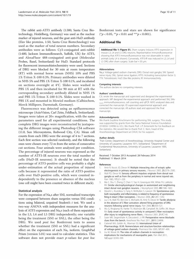
Injured fibers in the SNI model in mice project into L3 and L4In rats, 98% of sciatic nerve fibers originate from the L4and L5 DRG, whereas the somas of the saphenous nerve(part of the femoral nerve) fibers are located in the L3DRG [46]. Therefore, the L4 and L5 DRG are those ofinterest in the SNI rat model. However, Rigaud et al.demonstrated that the functional and anatomical homo-logues of the rat L4 and L5 DRG were rather in the L3and L4 DRG in mice [37]. This, together with the presentstudy’s observation that L3 DRG showed a stronger down-regulation of the Navs transcript than L5, suggested that itmay be necessary to reconsider which ganglia are likely toharbor the somas in the SNI mouse model. We conse-quently investigated the amount of injured fiber receivedby each ganglion after SNI. The expression of ATF3 wasstudied; it is a member of ATF/CREB family and a markerof axotomized neurons [47,48]. In sham-operated animals,ATF3-immunoreactivity (IR) was barely observable andreached a maximum of 8% for L3 (Additional file 1: FigureS1 and Figure 4B). Because naïve animals showed no IRfor ATF3 (Additional file 1: Figure S1), it is likely that sur-gery itself activates ATF3 expression, as has already beenproposed [49]. Seven days after SNI surgery, the percent-age of ATF3-positive cells in L3 (37%) and L4 DRG (34%)was significantly higher than in sham-operation conditions(Figure 4A, B), yet in L5 the low percentage of ATF3-positive cells observed (3%) remained at sham levels. Thisresult contrasts with the strong increase of ATF3 expres-sion observed in L5 in rats after SNI [50], and clearly con-firms that in mice, the cell bodies of most of the commonperoneal and tibial injured fibers are located in L3 and L4DRG rather than in L5.The mRNA expression of ATF3 in the L3 to L5 DRG
was also studied using qRT-PCR. This approach supportedfindings of a very significant increase of ATF3 in L3 and L4DRG, but no change in expression in L5 (Figure 4C).
So what is the relevance of the L5 DRG in the SNImouse model? The above approach was used on the SNIvariant, transecting only the sural nerve (SNIv(cp,t)) inorder to investigate which ganglia the fibers from this nervewould project into. Sham surgery revealed 8%, 7% and4% of ATF3-IR cells for L3, L4 and L5 DRG, respectively(Figure 5B), which was not different from sham-conditionpercentages of the traditional SNI. After SNIv(cp,t), therewas a significant increase of ATF3-IR cells in L4 (17%)and L5 (15%) DRG compared to sham conditions (Figure 5Aand B). Conversely, the number of injured cells had notincreased in L3 (7%), suggesting that the sural nerveoriginates in the L4 and L5 DRG.In summary, after SNI, the L3 DRG was comprised of
a substantial proportion (~ 40%) of neurons from the in-jured common peroneal and tibial nerves, but none fromthe sural nerve. L4 DRG was constituted of neurons fromthe injured common peroneal and tibial nerves (~ 35%)and neurons from the sural nerve (~ 15%). Finally, theL5 DRG was comprised of no injured neurons from theinjured common peroneal and tibial nerves after SNI,but did contain 15% of sural nerve neurons (Figure 6).This demonstrated that when using the SNI mouse
model, DRG should be pooled with caution becausethe L3, L4 and L5 DRG provided very different profilesof injured and non-injured neurons. It should be kept inmind that the mixture of neurons from all three individu-ally taken DRG might affect or dilute the overall analysisand results. Furthermore, when performing behavioral paintests in mice, and as Rigaud et al. [37] already proposed, L4injury is more suitable for studying neuropathic pain-likehyperalgesia in the SNL model. Because this study aimedto investigate the modification of Navs expression in DRGenriched in injured fibers, we did not re-perform SNLsurgery on the L4 DRG.
ConclusionWe showed that the expression of most Navs mRNAswas lower in the L3 and L4 DRG after SNI in mice. Nav1.3showed either a slight downregulation, or an absence ofregulation, after SNI and SNL, which contrasted with therobust upregulation observed in rats. This inter-speciesdifference should be further investigated in nerve-injurymouse models. Investigating Navs expression in the L3,L4 and L5 DRG independently, lead to a re-evaluationof where injured neurons are projected after SNI. The in-jured common peroneal and tibial nerves projected intothe L3 and L4 DRG, and the non-injured sural nerve pro-jected into the L4 and L5 DRG in C57BL/6 J mice. This isof great importance when investigating nerve-injury medi-ated modifications in DRG after SNI in mice. We suggestthat the L3 or L4 DRG should be harvested to target andenrichment in somas of injured fibers and L5 to enrich forthe soma of non-injured fibers.

Figure 4 ATF3 expression increases in mouse L3 and L4 DRG neurons after SNI, but not L5. (A) Representative immunofluorescenceshowing that ATF3 (marker of injured neurons, red) was mostly up-regulated in L3 and L4 DRG neurons (HuD positive cells, green) after SNI. Scalebar = 50 μm. (B) Quantification of ATF3-immunoreactivity (IR) in L3, L4 and L5 DRG neurons one week after SNI or sham surgery. ATF3-IR washigher in L3 and L4 after SNI, but remained the same in L5 DRG. Data are expressed as mean ± SEM, n = 4 animals in each group. ***p < 0.001,two-way ANOVA and post hoc Bonferroni tests. (C) mRNA levels of ATF3 one week after SNI compared to sham surgery in L3, L4 and L5 DRG.ATF3 mRNA was higher in L3 and L4 after SNI, but not in L5. Levels of transcripts were first normalized to GAPDH as a reference gene, and thento sham for each DRG. Data are expressed as mean ± SEM, n = 3–4 animals in each group. ***p < 0.001, two-way ANOVA and post hoc Bonferronitests. SNI, Spared Nerve Injury.
Laedermann et al. Molecular Pain 2014, 10:19 Page 7 of 11http://www.molecularpain.com/content/10/1/19
MethodsSurgeryAll procedures were approved by the Canton of Vaud’sCommittee on Animal Experimentation (Switzerland),in accordance with Swiss Federal Law on Animal Welfareand International Association for the Study of Painguidelines [51].The spared nerve injury (SNI) model of neuropathic
pain was previously described in rats [32,52] and mice [36].Briefly, adult C57BL/6 J mice (Charles River, L’Arbresle,France) were anesthetized with 1.5% isoflurane and afterexposure of the sciatic nerve, the common peroneal and
tibial nerves were ligated together with a 6.0 silk suture(Ethicon, Johnson and Johnson AG, Zug, Switzerland)and transected. In the SNI variant (SNIv(cp,t)) [36] theligation and transection were performed on the suralnerve, leaving the common peroneal and tibial nervesintact. The incision was closed in distinct layers (muscleand skin). Sham surgery was performed similarly exceptfor the nerve ligation and transection.Spinal nerve ligation (SNL) surgery was adapted from
the procedure described by Kim and Chung [31], andtransposed to mice. Briefly, after skin and muscle inci-sion the L5 transverse process of vertebra was exposed

Figure 5 ATF3 expression increases in mouse L4 and L5 DRG neurons after SNIv(cp,t), but not L3. (A) Representative immunofluorescenceshowing that ATF3 (marker of injured neurons, red) was mostly upregulated in L4 and L5 DRG neurons (HuD positive cells, green) after SNIv(cp,t).Scale bar = 50 μm. (B) Quantification of ATF3-immunoreactivity (IR) in L3, L4 and L5 DRG neurons one week after SNIv(cp,t) or sham surgery. ATF3was higher in L4 and L5 DRG when the sural nerve was injured, but not in L3. Data are expressed as mean ± SEM, n = 4 animals in eachgroup. ***p < 0.001, two-way ANOVA and post hoc Bonferroni tests. SNIv(cp,t), Spared Nerve Injury variant, sparing common peroneal (cp)and tibial (t) nerves.
Laedermann et al. Molecular Pain 2014, 10:19 Page 8 of 11http://www.molecularpain.com/content/10/1/19
and carefully removed. The L4 and L5 spinal nerves wereexposed and the L5 spinal nerve was tightly ligated. Theincision was closed in distinct layers (muscle and skin).
DissectionBriefly, mice were terminally anesthetized with sodiumpentobarbital (Esconarkon; Streuli Pharma AG, Uznach,Switzerland) and the biceps femoris muscle of the leftthigh was incised. The genus descendes artery was usedas a reference for the muscle incision, which lead to theexposure of the sciatic nerve and the trifurcation of theperipheral branches: common peroneal, tibial and suralnerves. The sciatic nerve was followed in the rostral
direction, removing muscle tissue until reaching the verte-bral column. Vertebral lamina, pedicles and spinous pro-cesses were trimmed away to expose the spinal cord andDRG. For the nomenclature of DRG, refer to Figure 3.
Quantitative real-time reverse transcription PCR (qRT-PCR)Ipsilateral DRG were rapidly dissected and collected inRNAlater solution (Qiagen, Basel, Switzerland). For SNI,2 series of mice were used, one where the L4 and L5were pooled together, as usually done (4 DRG pooledfrom 2 mice per sample), and one series where L3, L4 andL5 were dissected separately (8 DRG pooled from 8 miceper sample). For SNL, L4 and L5 DRG were consistently

Figure 6 Schematic view of sciatic nerve branches withprojections of injured fibers into DRG. The schematic view showsthat common peroneal and tibial nerves predominantly originate inthe L3 and L4 DRG (red fibers), while the sural nerve mainlyoriginates from the L4 and L5 (blue fibers). SNI, Spared Nerve Injury;SNIv(cp,t), Spared Nerve Injury variant, sparing common peroneal (cp)and tibial (t) nerves.
Laedermann et al. Molecular Pain 2014, 10:19 Page 9 of 11http://www.molecularpain.com/content/10/1/19
separated (2 DRG pooled from 2 mice per sample) as theyrepresented non-injured and injured neurons, respectively.For all conditions tested, n = 3–4 / sample were used.mRNA was extracted and purified using a RNeasy Plus MiniKit (Qiagen) and quantified using a RNA 6000 Nano Assay(Agilent Technologies AG, Basel, Switzerland). A total of600 ng of RNA was reverse-transcribed for each sampleusing Omniscript Reverse Transcriptase Kit (Qiagen).Primer sequences and working concentrations for Navsα-subunits, ATF3 and glyceraldehyde-3-phosphate de-hydrogenase (GAPDH) can be found in Table 1.We used GAPDH as a reference gene to normalize Navs
mRNA expression since it is stable between sham andSNI conditions (M-value = 0.30), taking into accountthe efficiencies of qPCR reaction. Gene-specific mRNAanalyses were performed using the iQ SYBR-greenSupermix (BioRad, Reinach, Switzerland) and the iQ5real-time PCR detection system (BioRad). Only reactionswith appropriate amplification and melting curves wereanalyzed. All samples were run in triplicate. Normalizedtranscripts were then expressed as a ratio of the level inSNI and SNL models to that in sham-operated mice.The bar graphs in Figures 2A, B and C represent theseratios for each isoform. Each qPCR product was se-quenced to confirm the specificity of amplification.Briefly, qPCR products were first loaded on a low-meltagarose gel to confirm the size of the amplicon. Ampli-cons were then subcloned in a pGEM-T Vector System(Promega, Madison, WI, USA), and sent for sequencingusing T7 promoter (Fasteris, Geneva, Switzerland). AllqPCR products were validated as specific for each ofthe Navs tested using the primers in Table 1.
ImmunofluorescenceOne week after sham SNI or SNIv(cp,t) surgery, animalswere terminally anesthetized with sodium pentobarbital(Esconarkon), and then transcardially perfused with salinesolution, directly followed by paraformaldehyde 4% dilutedin phosphate buffered saline (PBS). The L3 to L5 DRG weredissected, post-fixed at 4°C for 90 min and then transferredin sucrose solution (20% sucrose in PBS) overnight. Thefollowing day, tissues were mounted in cryoembeddingfluid (Tissue-Tek; Sakura Finetek, Zoeterwoude, Holland),frozen, cryosectioned in 12 μm-thick sections and thaw-mounted onto slides.
Laedermann et al. Molecular Pain 2014, 10:19 Page 10 of 11http://www.molecularpain.com/content/10/1/19
The rabbit anti-ATF3 antibody (1:300, Santa Cruz Bio-technology, Heidelberg, Germany) was used as the nuclearmarker of injured neurons, and the goat anti-HuD antibody(Elav like proteins, 1:50, Santa Cruz Biotechnology) wasused as the marker of total neuron numbers. Secondaryantibodies were as follows: Cy3-conjugated anti-rabbit(1:400, Jackson ImmunoResearch, Suffolk, UK) for ATF3,and AlexaFluor 488-conjugated anti-goat (MolecularProbes, Basel, Switzerland) for HuD. Standard protocolsfor fluorescent immunohistochemistry were used. Sectionsof DRG were blocked for 30 min at room temperature(RT) with normal horse serum (NHS) 10% and PBS1X-Triton X-100 0.3%. Primary antibodies were dilutedin NHS 5% and PBS 1X-Triton X-100 0.1%, and incubatedon sections overnight at 4°C. Slides were washed inPBS 1X and then incubated for 90 min at RT with thecorresponding secondary antibody diluted in NHS 1%and PBS 1X-Triton X-100 0.1%. Slides were washed inPBS 1X and mounted in Mowiol medium (Calbiochem,Merck Millipore, Darmstadt, Germany).Fluorescence was detected using an epifluorescence
microscope (AxioVision, Carl Zeiss, Feldbach, Switzerland).Images were taken at 20× magnification, with the sameparameters used for all experimental conditions. Thecomplete DRG images were reconstructed by juxtapos-ing the different images using Photoshop CS4 software(11.0, Sun Microsystems, Redwood City, CA). Mean cellcounts from each DRG were the average of 4 to 7 sections.Each first section was selected randomly, and the followingones were chosen every 72 m from the series of consecutivecut sections. Four animals were analyzed per condition.The percentage of injured neurons was expressed as thenumber of ATF3-IR neurons over the total number ofcells (HuD-IR neurons). It should be noted that thepercentage of ATF3-positive cells was probably a slightunder-estimation of the actual proportion of injuredcells because it represented the ratio of ATF3-positivecells over HuD-positive cells, which were counted in-dependently to the presence or absence of the nucleus(one cell might have been counted twice in different stack).
Statistical analysisFor the expression of Navs after SNI, normalized transcriptswere compared between sham surgeries versus SNI condi-tions using bilateral, unpaired Student’s t test. We used atwo-way ANOVA with independent measures for the ana-lysis of ATF3 expression and Navs (each of them separately)in the L3, L4 and L5 DRG independently; one variablebeing the treatment (SNI or SNL), the other being theDRG. We used post hoc Bonferroni tests to assesswhether the treatment (SNI or SNL) had a significanteffect on the expression of each Nav isoform. GraphPadPrism (version 5.01) was used to calculate statistics. Thissoftware does not provide exact p-values for post hoc
Bonferroni tests and stars are shown for significance(*p < 0.05, **p < 0.01 and ***p < 0.001).
Additional file
Additional file 1: Figure S1. Sham surgery induces ATF3 expression inmouse L3, L4 and L5 DRG neurons. Representative immunofluorescenceshowing that ATF3 immunofluorescence was not observable in naïveanimals (only L4 is shown). Conversely, ATF3-IR was induced in L3, L4 andL5 DRG after sham surgery. Scale bar = 50 μm.
AbbreviationsNavs: Voltage-gated sodium channels; DRG: Dorsal root ganglia; SNI: Sparednerve injury; SNL: Spinal nerve ligation; ATF3: Activating transcription factor 3;TTX: Tetrodotoxin; HuD: Elav-like proteins; IR: Immunoreactivity.
Competing interestsThe authors declare no competing interests.
Authors’ contributionsCJL wrote the manuscript and supervised and designed the experimentalapproach. MP designed the experimental approach and performed SNI, SNIv(cp,t), immunofluorescence, cell counting and qPCR. M.R.S analyzed data andcorrected the manuscript. ID supervised experimental approach andcorrected the manuscript. All authors read and approved the finalmanuscript.
AcknowledgmentsWe thank Guylène Kirschmann for performing SNL surgery. This studywas supported by grants from the Swiss National Science Foundation(31003A-124996 to I.D.). We thank Prof. J-P Antonietti for his feedback onthe statistics. We would like to thank Prof. C. Kern, head of theAnesthesiology Department at CHUV, for his support.
Author details1Pain Center, Department of Anesthesiology, University Hospital Center andUniversity of Lausanne, Lausanne 1011, Switzerland. 2Department ofFundamental Neurosciences, University of Lausanne, Lausanne 1005,Switzerland.
Received: 7 October 2013 Accepted: 28 February 2014Published: 11 March 2014
References1. Amir R, Kocsis JD, Devor M: Multiple interacting sites of ectopic spike
electrogenesis in primary sensory neurons. J Neurosci 2005, 25:2576–2585.2. Wall PD, Devor M: Sensory afferent impulses originate from dorsal root
ganglia as well as from the periphery in normal and nerve injured rats.Pain 1983, 17:321–339.
3. Ma C, Shu Y, Zheng Z, Chen Y, Yao H, Greenquist KW, White FA, LaMotteRH: Similar electrophysiological changes in axotomized and neighboringintact dorsal root ganglion neurons. J Neurophysiol 2003, 89:1588–1602.
4. Djouhri L, Koutsikou S, Fang X, McMullan S, Lawson SN: Spontaneous pain,both neuropathic and inflammatory, is related to frequency of spontaneousfiring in intact C-fiber nociceptors. J Neurosci 2006, 26:1281–1292.
5. Liu C-N, Wall PD, Ben-Dor E, Michaelis M, Amir R, Devor M: Tactile allodyniain the absence of C-fiber activation: altered firing properties of DRGneurons following spinal nerve injury. Pain 2000, 85:503–521.
6. Wu G, Ringkamp M, Hartke TV, Murinson BB, Campbell JN, Griffin JW, MeyerRA: Early onset of spontaneous activity in uninjured C-fiber nociceptorsafter injury to neighboring nerve fibers. J Neurosci 2001, 21:RC140.
7. Suter MR, Siegenthaler A, Decosterd I, Ji RR: Perioperative nerve blockade:clues from the bench. Anesthesiol Res Pract 2011, 2011:124898.
8. Catterall WA, Goldin AL, Waxman SG: International Union ofPharmacology. XLVII. Nomenclature and structure-function relationshipsof voltage-gated sodium channels. Pharmacol Rev 2005, 57:397–409.
9. Liu M, Wood JN: The roles of sodium channels in nociception:implications for mechanisms of neuropathic pain. Pain Med 2011,12(Suppl 3):S93–S99.

Laedermann et al. Molecular Pain 2014, 10:19 Page 11 of 11http://www.molecularpain.com/content/10/1/19
10. Catterall WA: From ionic currents to molecular mechanisms: the structureand function of voltage-gated sodium channels. Neuron 2000, 26:13–25.
11. Brackenbury WJ, Isom LL: Na channel beta subunits: overachievers of theIon channel family. Front Pharmacol 2011, 2:53.
12. Ho C, O’Leary ME: Single-cell analysis of sodium channel expression indorsal root ganglion neurons. Mol Cell Neurosci 2011, 46:159–166.
13. Fukuoka T, Noguchi K: Comparative study of voltage-gated sodiumchannel alpha-subunits in non-overlapping four neuronal populations inthe rat dorsal root ganglion. Neurosci Res 2011, 70:164–171.
14. Black JA, Dib-Hajj S, McNabola K, Jeste S, Rizzo MA, Kocsis JD, Waxman SG:Spinal sensory neurons express multiple sodium channel alpha-subunitmRNAs. Brain Res Mol Brain Res 1996, 43:117–131.
15. Rush AM, Cummins TR, Waxman SG: Multiple sodium channels and theirroles in electrogenesis within dorsal root ganglion neurons. J Physiol2007, 579:1–14.
16. Berta T, Poirot O, Pertin M, Ji RR, Kellenberger S, Decosterd I: Transcriptionaland functional profiles of voltage-gated Na(+) channels in injured andnon-injured DRG neurons in the SNI model of neuropathic pain. Mol CellNeurosci 2008, 37:196–208.
17. Fukuoka T, Kobayashi K, Yamanaka H, Obata K, Dai Y, Noguchi K: Comparativestudy of the distribution of the alpha-subunits of voltage-gated sodiumchannels in normal and axotomized rat dorsal root ganglion neurons.J Comp Neurol 2008, 510:188–206.
18. Chahine M, Ziane R, Vijayaragavan K, Okamura Y: Regulation of Na vchannels in sensory neurons. Trends Pharmacol Sci 2005, 26:496–502.
19. Gold MS, Weinreich D, Kim CS, Wang R, Treanor J, Porreca F, Lai J:Redistribution of NaV1.8 In uninjured axons enables neuropathic pain.J Neurosci 2003, 23:158–166.
20. Study RE, Kral MG: Spontaneous action potential activity in isolated dorsalroot ganglion neurons from rats with a painful neuropathy. Pain 2005,65:235–242.
21. Pertin M, Ji RR, Berta T, Powell AJ, Karchewski L, Tate SN, Isom LL, Woolf CJ,Gilliard N, Spahn DR, Decosterd I: Upregulation of the voltage-gatedsodium channel beta2 subunit in neuropathic pain models:characterization of expression in injured and non-injured primarysensory neurons. J Neurosci 2005, 25:10970–10980.
22. Decosterd I, Ji RR, Abdi S, Tate S, Woolf CJ: The pattern of expression ofthe voltage-gated sodium channels Na(v)1.8 and Na(v)1.9 does notchange in uninjured primary sensory neurons in experimentalneuropathic pain models. Pain 2002, 96:269–277.
23. Fukuoka T, Noguchi K: Contribution of the spared primary afferent neuronsto the pathomechanisms of neuropathic pain. Mol Neurobiol 2002, 26:57–67.
24. Sleeper AA, Cummins TR, Dib-Hajj SD, Hormuzdiar W, Tyrrell L, Waxman SG,Black JA: Changes in expression of two tetrodotoxin-resistant sodiumchannels and their currents in dorsal root ganglion neurons after sciaticnerve injury but not rhizotomy. J Neurosci 2000, 20:7279–7289.
25. Dib-Hajj S, Black JA, Felts P, Waxman SG: Down-regulation of transcriptsfor Na channel alpha-SNS in spinal sensory neurons following axotomy.Proc Natl Acad Sci U S A 1996, 93:14950–14954.
26. Dib-Hajj SD, Tyrrell L, Black JA, Waxman SG: NaN, a novel voltage-gated Nachannel, is expressed preferentially in peripheral sensory neurons anddown-regulated after axotomy. Proc Natl Acad Sci U S A 1998, 95:8963–8968.
27. Cummins TR, Waxman SG: Down-regulation of tetrodotoxin-resistant sodiumcurrents and upregulation of a rapidly repriming tetrodotoxin-sensitivesodium current in small spinal sensory neurons after nerve injury.J Neurosci 1997, 17:3503–3514.
28. Waxman SG, Kocsis JD, Black JA: Type III sodium channel mRNA isexpressed in embryonic but not adult spinal sensory neurons, and isreexpressed following axotomy. J Neurophysiol 1994, 72:466–470.
29. Wall PD, Devor M, Inbal R, Scadding JW, Schonfeld D, Seltzer Z, TomkiewiczMM: Autotomy following peripheral nerve lesions: experimentalanaesthesia dolorosa. Pain 1979, 7:103–111.
30. Campbell JN, Meyer RA: Mechanisms of neuropathic pain. Neuron 2006,52:77–92.
31. Kim SH, Chung JM: An experimental model for peripheral neuropathyproduced by segmental spinal nerve ligation in the rat. Pain 1992,50:355–363.
32. Decosterd I, Woolf CJ: Spared nerve injury: an animal model of persistentperipheral neuropathic pain. Pain 2000, 87:149–158.
33. Devor M, Wall PD: Cross-excitation in dorsal root ganglia of nerve-injuredand intact rats. J Neurophysiol 1990, 64:1733–1746.
34. Amir R, Devor M: Chemically mediated cross-excitation in rat dorsal rootganglia. J Neurosci 1996, 16:4733–4741.
35. Lisney SJW, Pover CM: Coupling between fibers involved in sensory nerveneuromata in cats. J Neurol Sci 1983, 59:255–264.
36. Bourquin A-F, Süveges M, Pertin M, Gilliard N, Sardy S, Davison AC, SpahnDR, Decosterd I: Assessment and analysis of mechanical allodynia-likebehavior induced by spared nerve injury (SNI) in the mouse. Pain 2006,122:14.e11–14.e14.
37. Rigaud M, Gemes G, Barabas M-E, Chernoff DI, Abram SE, Stucky CL, HoganQH: Species and strain differences in rodent sciatic nerve anatomy:implications for studies of neuropathic pain. Pain 2008, 136:188–201.
38. Lindia JA, Köhler MG, Martin WJ, Abbadie C: Relationship between sodiumchannel NaV1.3 expression and neuropathic pain behavior in rats.Pain 2005, 117:145–153.
39. Black JA, Cummins TR, Plumpton C, Chen YH, Hormuzdiar W, Clare JJ,Waxman SG: Upregulation of a silent sodium channel after peripheral,but not central, nerve injury in DRG neurons. J Neurophysiol 1999,82:2776–2785.
40. Samad OA, Tan AM, Cheng X, Foster E, Dib-Hajj SD, Waxman SG: Virus-mediatedshRNA knockdown of Nav1.3 In Rat dorsal root ganglion attenuates nerveinjury-induced neuropathic pain. Mol Ther 2013, 21:49–56.
41. Nassar MA, Baker MD, Levato A, Ingram R, Mallucci G, McMahon SB, WoodJN: Nerve injury induces robust allodynia and ectopic discharges inNav1.3 null mutant mice. Mol Pain 2006, 2:33.
42. Thakor DK, Lin A, Matsuka Y, Meyer EM, Ruangsri S, Nishimura I, Spigelman I:Increased peripheral nerve excitability and local NaV1.8 mRNA up-regulationin painful neuropathy. Mol Pain 2009, 5:14.
43. Laedermann CJ, Cachemaille M, Kirschmann G, Pertin M, Gosselin RD, Chang I,Albesa M, Towne C, Schneider BL, Kellenberger S, Abriel H, Decosterd I:Dysregulation of voltage-gated sodium channels by ubiquitin ligaseNEDD4-2 in neuropathic pain. J Clin Invest 2013, 123:3002–3013.
44. Fukuoka T, Yamanaka H, Kobayashi K, Okubo M, Miyoshi K, Dai Y, Noguchi K:Re-evaluation of the phenotypic changes in L4 dorsal root ganglionneurons after L5 spinal nerve ligation. Pain 2012, 153:68–79.
45. Green EL: Genetic and non-genetic factors which influence the type ofthe skeleton in an inbred strain of mice. Genetics 1941, 26:192–222.
46. Swett JE, Torigoe Y, Elie VR, Bourassa CM, Miller PG: Sensory neurons of therat sciatic nerve. Exp Neurol 1991, 114:82–103.
47. Tsujino H, Kondo E, Fukuoka T, Dai Y, Tokunaga A, Miki K, Yonenobu K, OchiT, Noguchi K: Activating transcription factor 3 (ATF3) induction byaxotomy in sensory and motoneurons: a novel neuronal marker of nerveinjury. Mol Cell Neurosci 2000, 15:170–182.
48. Tsuzuki K, Kondo E, Fukuoka T, Yi D, Tsujino H, Sakagami M, Noguchi K:Differential regulation of P2X3 mRNA expression by peripheral nerveinjury in intact and injured neurons in the rat sensory ganglia. Pain 2001,91:351–360.
49. Shortland PJ, Baytug B, Krzyzanowska A, McMahon SB, Priestley JV, Averill S:ATF3 expression in L4 dorsal root ganglion neurons after L5 spinal nervetransection. Eur J Neurosci 2006, 23:365–373.
50. Takahashi N, Kikuchi S, Dai Y, Kobayashi K, Fukuoka T, Noguchi K:Expression of auxiliary beta subunits of sodium channels in primaryafferent neurons and the effect of nerve injury. Neuroscience 2003,121:441–450.
51. Zimmermann M: Ethical guidelines for investigations of experimentalpain in conscious animals. Pain 1983, 16:109–110.
52. Pertin M, Gosselin R-D, Decosterd I: The Spared Nerve Injury Model ofNeuropathic Pain. In Pain Research, Volume Volume 851. Edited by Luo ZD.233 Spring Street, New York, NY 10013-1578: Humana Press Inc; 2012:205–212.[Methods in Molecular Biology].
doi:10.1186/1744-8069-10-19Cite this article as: Laedermann et al.: Voltage-gated sodium channelexpression in mouse DRG after SNI leads to re-evaluation of projectionsof injured fibers. Molecular Pain 2014 10:19.