Embed Size (px)
Citation preview
124I-huA33 Antibody Uptake Is Driven by A33 AntigenConcentration in Tissues from Colorectal Cancer PatientsImaged by Immuno-PET
Joseph A. O’Donoghue1, Peter M. Smith-Jones2, John L. Humm1, Shutian Ruan2, Daniel A. Pryma2, Achim A. Jungbluth3,Chaitanya R. Divgi2, Jorge A. Carrasquillo2, Neeta Pandit-Taskar2, Yuman Fong4, Vivian E. Strong4, Nancy E. Kemeny5,Lloyd J. Old3, and Steven M. Larson2
1Department of Medical Physics, Memorial Sloan-Kettering Cancer Center, New York, New York; 2Department of Radiology,Memorial Sloan-Kettering Cancer Center, New York, New York; 3Ludwig Institute for Cancer Research (New York Branch), MemorialSloan-Kettering Cancer Center, New York, New York; 4Department of Surgery, Memorial Sloan-Kettering Cancer Center, New York,New York; and 5Department of Medicine, Memorial Sloan-Kettering Cancer Center, New York, New York
The primary aim of this analysis was to examine the quantitativefeatures of antibody–antigen interactions in tumors and normaltissue after parenteral administration of antitumor antibodies tohuman patients. Methods: Humanized anti-A33 antibody (10mg) labeled with the positron-emitting radionuclide 124I (124I-huA33) was injected intravenously in 15 patients with colorectalcancer. Clinical PET/CT was performed approximately 1 wklater, followed by a detailed assay of surgically removed tissuespecimens including radioactivity counting, autoradiography,immunohistochemistry, and antigen density determination.Results: PET/CT showed high levels of antibody targeting intumors and normal bowel. In tissue specimens, the spatial dis-tribution of 124I-huA33 conformed to that of A33 antigen, andthere was a linear relationship between the amount of boundantibody and antigen concentration. Antibody uptake was highin 1- to 2-mm regions of antigen-positive tumor cells (mean,;0.05 percentage injected dose per gram) and in antigen-posi-tive normal colonic mucosa (mean, ;0.03 percentage injecteddose per gram). The estimated binding site occupancy for tumorand normal colon was 20%–50%. Conclusion: The in vivo bio-distribution of 124I-huA33 in human patients 1 wk after antibodyadministration was determined by A33 antigen expression. Ourdata imply that the optimal strategy for A33-based radioimmu-notherapy of colon cancer will consist of a multistep treatmentusing a radionuclide with short-range (a- or b-particle) emissions.
Key Words: huA33; colorectal cancer; 124I; immuno-PET;radioimmunotherapy
J Nucl Med 2011; 52:1–8DOI: 10.2967/jnumed.111.095596
A33 is a cell surface glycoprotein abundantly expressedin 95% of colon carcinomas and by epithelial cells of the
normal intestine but not by other normal tissues (1–3). TheA33 antigen is a member of the immunoglobulin superfamily,with homology to cell adhesion and tight junction–associatedproteins (4,5). The antigen has been purified from human coloncancer cells, the protein sequence determined, the complemen-tary DNA cloned, and the mouse homolog identified (4,6).Because of its restricted tissue localization and high level ofexpression, the A33 antigen is appealing as a therapeutic target,and a variety of anti-A33–based approaches are under preclin-ical investigation as potential therapies for colon cancer (7–11).
Early clinical studies (12,13) were performed with murineanti-A33 monoclonal antibody radiolabeled with 131I or 125Ito assess its potential for radioimmunotherapy. Among thefindings of these studies was that antibody localization atprimary and metastatic tumor sites was extremely persistent,with significant retention beyond 6 wk after administration,whereas the initially high uptake in normal intestine gradu-ally diminished with time. The applicability of murine A33was restricted by the development of an antimouse IgG im-mune response and, in an attempt to overcome this limitation,a fully humanized complementarity determining region-grafted A33 IgG1 (huA33) was developed (14). However,subsequent studies showed that repetitive administrations ofhuA33 could elicit a human antihuman antibody response(15). Humanized antibody A33 (huA33) has been shown tobe equivalent to the mouse antibody in competitive bindingassays and localization studies in animal models (14) and canbe radioiodinated with retention of immunoreactivity.
The relatively long half-life (4.2 d) of 124I allows PET tobe performed for up to a week after the administration of124I-labeled antibody (16), with radiation doses to normaltissues comparable with those of the 131I-labeled antibody.It has been shown that quantitative noninvasive imaging of124I is possible using a dedicated PET system (17,18).
We performed a clinical study of 124I-huA33 PET inpatients with colorectal cancer. In 15 patients, for whomsurgery was prescheduled as a standard of care, a PET/CTscan was acquired approximately 1 wk after antibody admin-
Received Jul. 11, 2011; revision accepted Sep. 9, 2011.For correspondence or reprints contact: Joseph A. O’Donoghue, Department
of Medical Physics, Memorial Sloan-Kettering Cancer Center, 1275 York Ave.,New York, NY 10065.E-mail: [email protected] online nnnnnnnnnnnn.COPYRIGHT ª 2011 by the Society of Nuclear Medicine, Inc.
ANTIBODY–ANTIGEN RELATIONSHIPS FOR HUA33 • O’Donoghue et al. 1
jnm095596-pm n 11/8/11
Journal of Nuclear Medicine, published on November 8, 2011 as doi:10.2967/jnumed.111.095596
Copyright 2011 by Society of Nuclear Medicine.
by on January 30, 2018. For personal use only. jnm.snmjournals.org Downloaded from
istration, immediately before the surgical removal of tumorand elements of neighboring normal tissues. Surgicallyexcised tissues were subsequently processed for ex vivoquantification of antibody uptake, antigen density determi-nation, digital autoradiography (DAR), and histologic orimmunohistochemical staining. In this paper, we describethe findings of this study, focusing on the ex vivo measure-ments and their clinical implications. The clinical PET/CT isdescribed only to the extent that it has an impact on these. Amore comprehensive description of the clinical imagingstudy of 124I-huA33 is provided elsewhere (19).
MATERIALS AND METHODS
Clinical Study with 124I-huA33Under the auspices of a protocol approved by the Institutional
Review Board and an Investigational New Drug applicationapproved by the Food and Drug Administration, 15 patients(mean age, 66 y; range, 52–77; 11 men, 4 women) with colorectalcancer were intravenously administered 10 mg of huA33 labeledwith 124I after providing informed consent (Trial registration ID,NCT00199862). On average, the administered activity was 200MBq (5.4 mCi), and the range was 44–400 MBq (1.2–10.7mCi). There were no adverse events related to huA33 administra-tion for any patient in the study. Patients were imaged by PET/CTapproximately 7 d later (range, 5–9 d) and underwent presched-uled surgery thereafter. Tumor and normal tissue samples wereobtained at the time of surgery, as part of the standard of care,and portions of these were used in the analysis.
Antibody RadiolabelingFor clinical PET/CT, huA33 antibody was radiolabeled with 124I, in
accordance with USP ,797. (20), using the IODO-GEN (Pierce)method (21). 124I was either prepared by an in-house cyclotron(TR14/4; Ebco) or purchased commercially (IBA Molecular). Detailsof the labeling procedure are provided in the supplemental data (avail-able online only at http://jnm.snmjournals.org). The average radio-chemical yield was 92% 6 6% and, after ion exchange purification,the mean radiochemical purity was 98%6 1%. Endotoxin levels wereconsistently below 0.4 EU/mL, all solutions were sterile, and the aver-age immunoreactivity of the 124I-huA33 was 90% 6 5%.
For ex vivo saturation binding studies, huA33 antibody wasradiolabeled with 131I purchased commercially (Nordion) usingthe IODO-GEN method (21) to a specific activity of 220–260MBq/mg (6–7 mCi/mg). Details of the labeling procedure areprovided in supplemental data. The radiochemical yield wasgreater than 90%, and 131I-huA33 was more than 99% pure.
Tissue Sample ProcessingTissue samples recovered from surgery were divided into
portions for g-well counting and subsequent antigen density deter-mination as well as DAR and tissue staining analysis.
Antigen Density Determination by Saturation Binding Analysis.Tissue samples were weighed, snap-frozen in liquid nitrogen, andimmediately counted in a g-well counter, together with appropri-ate 124I standards. These were then stored at 280�C for 2 mo toallow decay of 124I. Thereafter, tissue was thawed on ice, diced,put into 20 mM N-(2-hydroxyethyl)piperazine-N9-(2-ethanesul-fonic acid)-KOH buffer (pH 7.4), and homogenized on ice with4 · 10 s 24,000-rpm bursts with a disperser (T18 Basic ULTRA-TURRAX; IKA). Membranes were isolated by centrifugation at
5,000g for 20 min at 4�C and resuspended in phosphate-bufferedsaline using a Potter-Elvehjem tissue grinder. The protein content ofmembrane preparations was assayed using bicinchoninic acid reagent(Pierce) and measuring the absorption of the Cu(I) complex at 540 nm.
Saturation studies used 50–100 mg of membranes in phosphate-buffered saline incubated with increasing amounts of 131I-huA33.Mixtures were incubated at room temperature for 60 min beforerapid filtration through a glass fiber filter (GF/C; Whatman) pre-soaked in 1% bovine serum albumin/10 mM Tris-HCl (0.85%NaCl, pH 7.4). Filters were washed with 4 · 1 mL of ice-cold10 mM Tris-HCl (0.85% NaCl, pH 7.4) and counted with a NaI(Tl) well scintillation counter. Each assay was performed in trip-licate. Nonspecific binding was defined as that observed in thepresence of 100 nM unlabeled huA33. The saturation binding datawere evaluated using the least-squares fitting routine of the com-puter program Origin (version 7.5; Microcal Software) and byScatchard transformation of the data. The maximum number ofbinding sites (Bmax) and dissociation constant (Kd) values wereobtained for all tissue samples, with the consistency of the Kd
value acting as an experimental control.DAR and Tissue Staining Analysis. Tissue samples for DAR and
tissue staining analysis were individually wrapped in a layer ofheavy-duty clear plastic wrap film and immersed into prechilledmethylbutane (Fisher Scientific) at 280�C for 10 min. The frozentissues were then embedded in optimal-cutting-temperature (OCT)compound (VWR Scientific) on dry ice and transferred to a 280�C freezer for 30 min. Sets of (typically 10) contiguous frozen 8-mm-thick sections were cut from each tissue sample using aHM500 cryostat microtome (Microm) and collected on glassmicroscope slides.
Adjacent tissue sections from the same contiguous set were usedfor DAR and for histologic and immunohistochemical staining.
A minimum of 3 frozen tissue sections from each specimen wereplaced in a film cassette against a Fujifilm BAS-MS2325 imagingplate (Fuji Photo Film Co.). Latent images were read out aftervariable exposure times (mean, 61 h; range, 18–165 h) using aFujifilm BAS-1800II Bio-Imaging Analyzer (Fuji Photo Film Co.)at a 50-mm-pixel resolution. DAR image intensity was characterizedby the machine readout parameter of photostimulable luminescenceper square millimeter (PSL/mm2). DAR calibration studies wereperformed to relate PSL/mm2 to cumulated activity concentration(MBq h/mL). 124I-huA33 uptake was derived from region-of-interest(ROI) analysis of the DAR images using Multi Gauge software(version 2.2; Fujifilm). ROIs were drawn to encompass entire tissuesections to facilitate comparison with the well counter measurementsof tissue blocks. In addition, ROIs were drawn around identifiablezones in the DAR images, which were subsequently related to thehistologic features of the tissue sections.
For conventional morphologic assessment, tissue sections werefixed in 10% phosphate-buffered formalin solution (15 min), washedwith phosphate-buffered solution, and stained with hematoxylin andeosin (Sigma-Aldrich Inc.).
For immunohistochemical detection of A33 antigen, tissuesections were fixed in 10% neutral-buffered formalin (5 min),endogenous peroxidase was suppressed with H2O2, and a proteinblock for the reduction of nonspecific staining was applied. Thereaftermurine monoclonal antibody A33 was applied at a concentration of0.5 mg/mL overnight at 4�C. Primary antibody was detected usingbiotinylated horse antimouse-secondary reagent (1:200; Vector Labs),followed by avidin-biotin-complex system (ABC-Elite; VectorLabs) with 39-39-diaminobenzidine (DAB; Biogenex) as a chrom-
2 THE JOURNAL OF NUCLEAR MEDICINE • Vol. 52 • No. 12 • December 2011
jnm095596-pm n 11/8/11
by on January 30, 2018. For personal use only. jnm.snmjournals.org Downloaded from
ogen. Sections were counterstained with hematoxylin. Negativecontrol slides, omitting the primary antibody, were included in allassays. Images of stained sections were obtained with a BX600digital microscope (Olympus).
DAR System CalibrationThe DAR system response was calibrated using 124I activity
standards. Standards were constructed by mixing known activitiesof 124I in known volumes of OCT compound. Uniform mixing wasfacilitated by including red food dye in the diluent and heating theOCT compound to 80�C. Subsequently, the activity-containingOCT compound was poured into moulds and frozen on dry ice.Sections of 8-mm thickness were cut and collected on glass micro-scope slides in a manner identical to that for the clinical speci-mens. At least 3 sets of serially diluted activity-containing sectionswere then exposed to imaging plates for times ranging from 5 to116 h before being read out. ROIs were drawn on the images ofthe activity standards, interior to the visible edges, and over abackground location. The signal from the activity standards wasquantified in terms of mean background-corrected PSL/mm2.
The cumulated activity concentration in a standard, as, wascalculated by:
~as 5
Z Texp
0
as0expð 2 ltÞdt 5 as0�1 2 exp
�2 lTexp
���l;
where as0 was the activity concentration in the standard at thebeginning of the exposure, of duration Texp, and l was the physicaldecay constant of 124I (0.00692 h21).
This procedure enabled the DAR system to be calibrated interms of photostimulable luminescence per unit area per unitcumulated activity concentration (PSL/mm2 per MBq h/mL).
DAR-Derived Fractional Tissue Uptake perUnit Volume
For clinical tissue samples, values of cumulated activity perunit volume were determined from ROI analysis of the DARimages using the system calibration factor. The implied activityconcentrations at the beginning of the exposure were thencompared with the total activity administered, corrected for decayto the beginning of the exposure, to derive fractional uptake perunit volume—that is:
FROI 5 aon ROI=Aon;
where FROI was the fractional uptake per unit volume in the ROI,aon ROI was the activity concentration in the ROI at the beginningof the DAR exposure, and Aon was the total administered activitycorrected for decay to the beginning of the DAR exposure.
In terms of known or directly measured parameters:
FROI 5 l~aROI
A0expð 2 lTonÞ�1 2 exp
�1 2 lTexp
��;
where A0 was the total administered activity, and Ton was the timebetween the administration of activity and the beginning of theDAR exposure of duration Texp.
Statistical AnalysisBivariate relationships were assessed by linear regression
analysis. Differences between regression coefficients (m1, m2)were compared using the Student t test on the test statistic
t 5 ðm12m2Þ=sm12m2, where sm1 2 m2
is the SE of the differencebetween regression coefficients according to Chapter 18 of Zar(22). SD and SE are used to denote standard deviation and stand-ard error, respectively.
RESULTS
Clinical PET/CT Images of 124I-huA33
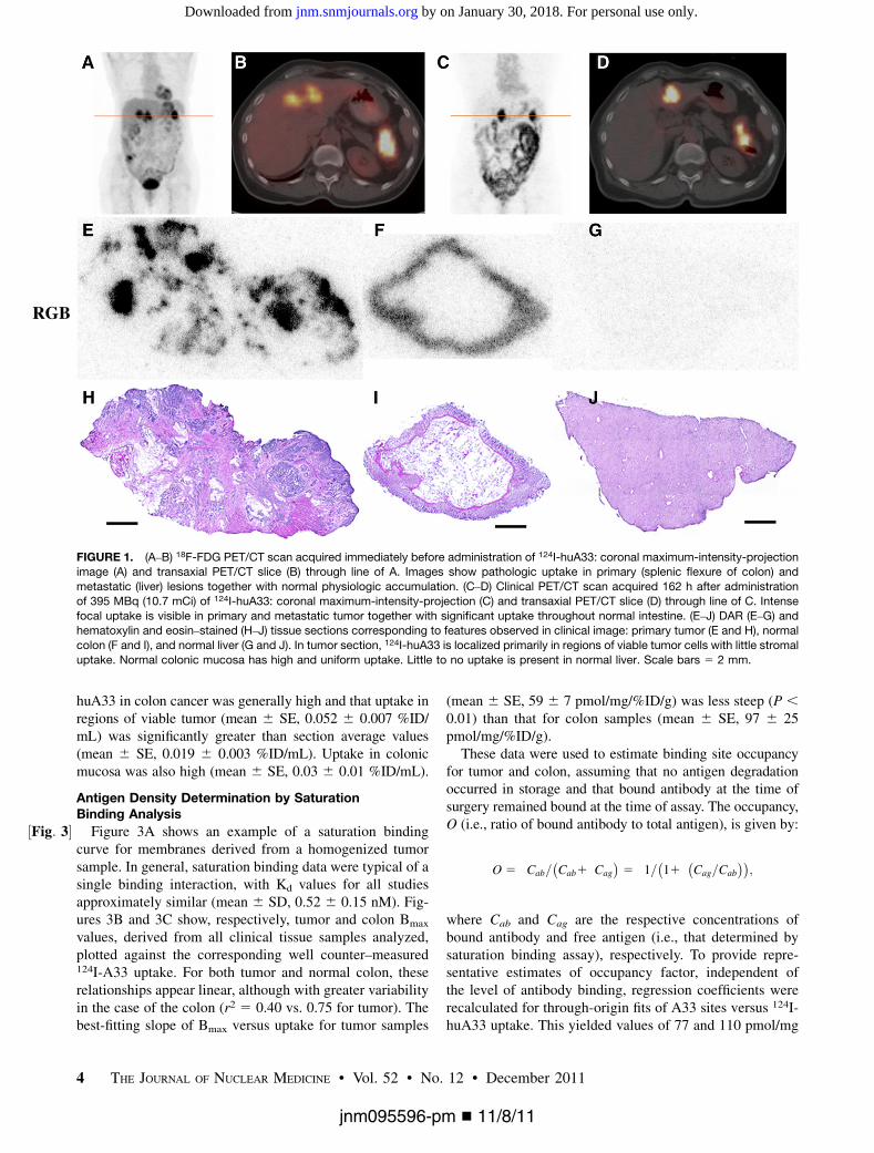
PET/CT images of 124I-huA33 were generally of suffi-ciently high quality for diagnostic interpretation at approx-imately 1 wk after administration (19). Examples of suchimages are provided in ½Fig: 1�Figures 1 and 2. The 124I-huA33maximum-intensity-projection ½Fig: 2�images (Figs. 1C and 2C)indicate the presence of activity primarily in A33-express-ing tissue but also in the circulation (cardiac blood pool andblood vessels). Although the entire intestinal tract wasclearly visualized, little activity was present in any otherparenchymal or connective tissue. The axial PET/CTimages (Figs. 1D and 2D) show focal 124I-huA33 uptakein primary and metastatic tumor and also normal colon.
Patterns of 124I-huA33 Uptake in ClinicalTissue Sections
Twenty-nine tissue samples from 14 patients were evalu-able by DAR, including both tumor (16) and nontumor (13)material. Figures 1E–1J show a set of DAR and hematoxylinand eosin–stained sections of tissues surgically removed afteracquisition of the 124I-huA33 clinical images shown in Fig-ures 1C and 1D. Focal uptake was apparent in regions ofviable tumor cells with little uptake in tumor-infiltratingstroma. The DAR images also show high uptake in normalcolonic mucosa, consistent with the known expression pat-tern of A33 antigen (1–3), with relatively low uptake in liver.ROIs drawn on the DAR around the whole tumor tissuesection and around several of the focal high-activity zonesindicated that the whole-section average uptake of 0.025percentage injected dose per milliliter (%ID/mL) was con-siderably less than that in the focal regions (;0.10 %ID/mL).
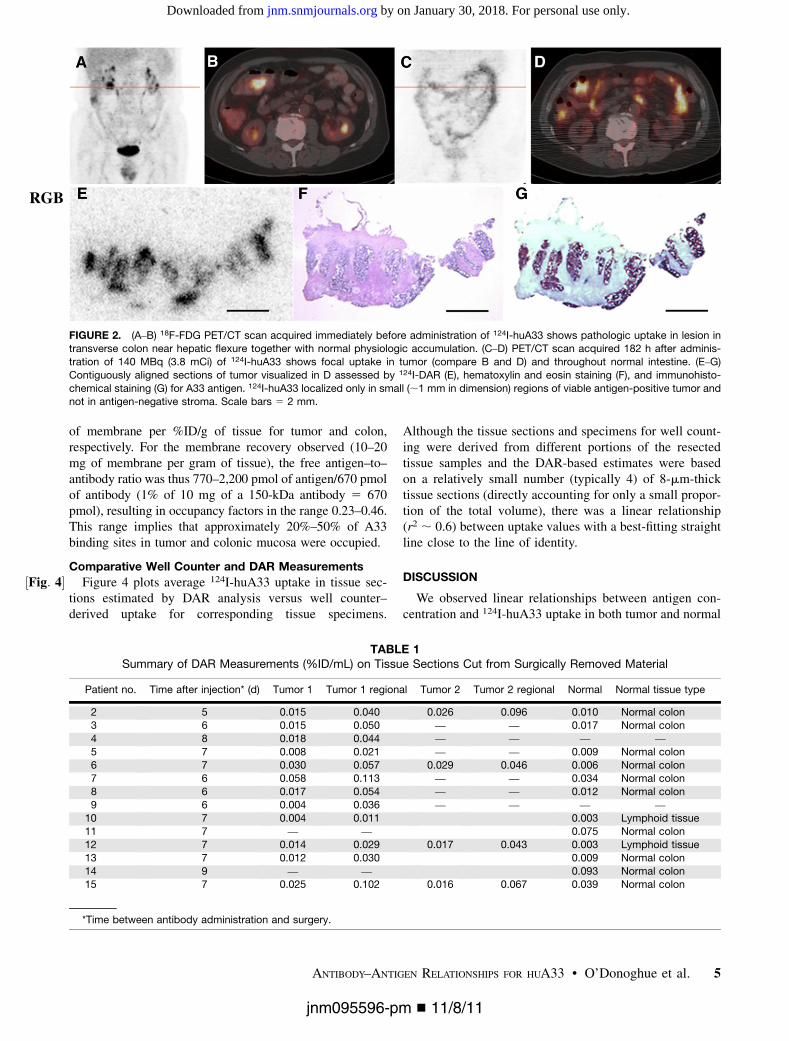
Figures 2E–2G show another tumor tissue section imageset (contiguously aligned sections assessed by DAR, hem-atoxylin and eosin staining, and immunohistochemistrystaining for A33 antigen). Comparison of these imagesindicates that the distribution of 124I-huA33 conformedclosely to viable antigen-positive tumor. ROI analysis indi-cated that the average section uptake (0.018 %ID/mL) wasconsiderably less than that in viable antigen-positive tumorregions (;0.044 %ID/mL).
½Table 1�Table 1 summarizes the DAR-based measurements for allpatients studied in terms of 124I-huA33 uptake expressed in%ID/mL. Section average and regional (i.e., antigen-posi-tive) uptake from up to 2 separate tumor locations wereexamined together with normal colonic mucosa if available.Noncolon normal tissues generally had low uptake as exem-plified by the lymphoid tissue samples for patients 10 and 12(0.003 %ID/mL) and by the normal liver sample shown inFigures 1G and 1J) (0.002 %ID/mL) for patient 15 (notincluded in table). Table 1 indicates that the uptake of
ANTIBODY–ANTIGEN RELATIONSHIPS FOR HUA33 • O’Donoghue et al. 3
jnm095596-pm n 11/8/11
by on January 30, 2018. For personal use only. jnm.snmjournals.org Downloaded from
huA33 in colon cancer was generally high and that uptake inregions of viable tumor (mean 6 SE, 0.052 6 0.007 %ID/mL) was significantly greater than section average values(mean 6 SE, 0.019 6 0.003 %ID/mL). Uptake in colonicmucosa was also high (mean 6 SE, 0.03 6 0.01 %ID/mL).
Antigen Density Determination by SaturationBinding Analysis
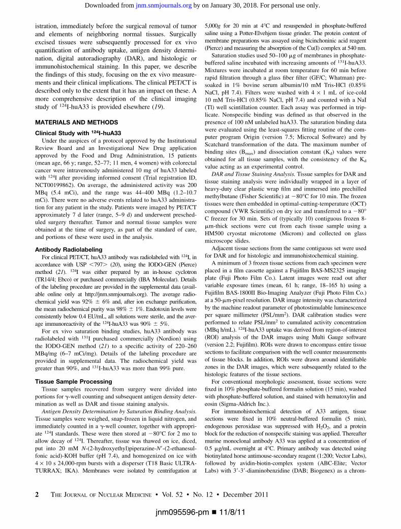
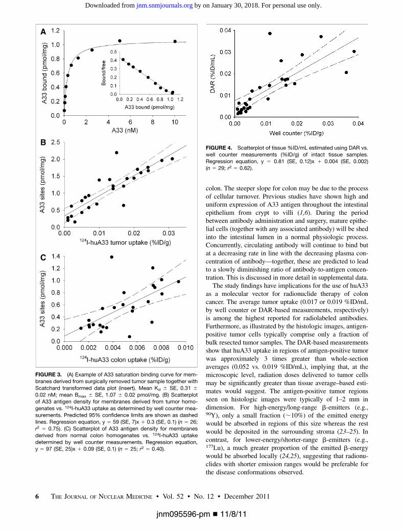
½Fig: 3� Figure 3A shows an example of a saturation bindingcurve for membranes derived from a homogenized tumorsample. In general, saturation binding data were typical of asingle binding interaction, with Kd values for all studiesapproximately similar (mean 6 SD, 0.52 6 0.15 nM). Fig-ures 3B and 3C show, respectively, tumor and colon Bmax
values, derived from all clinical tissue samples analyzed,plotted against the corresponding well counter–measured124I-A33 uptake. For both tumor and normal colon, theserelationships appear linear, although with greater variabilityin the case of the colon (r2 5 0.40 vs. 0.75 for tumor). Thebest-fitting slope of Bmax versus uptake for tumor samples
(mean 6 SE, 59 6 7 pmol/mg/%ID/g) was less steep (P ,0.01) than that for colon samples (mean 6 SE, 97 6 25pmol/mg/%ID/g).
These data were used to estimate binding site occupancyfor tumor and colon, assuming that no antigen degradationoccurred in storage and that bound antibody at the time ofsurgery remained bound at the time of assay. The occupancy,O (i.e., ratio of bound antibody to total antigen), is given by:
O 5 Cab=�Cab1 Cag
�5 1=
�11
�Cag=Cab
��;
where Cab and Cag are the respective concentrations ofbound antibody and free antigen (i.e., that determined bysaturation binding assay), respectively. To provide repre-sentative estimates of occupancy factor, independent ofthe level of antibody binding, regression coefficients wererecalculated for through-origin fits of A33 sites versus 124I-huA33 uptake. This yielded values of 77 and 110 pmol/mg
FIGURE 1. (A–B) 18F-FDG PET/CT scan acquired immediately before administration of 124I-huA33: coronal maximum-intensity-projectionimage (A) and transaxial PET/CT slice (B) through line of A. Images show pathologic uptake in primary (splenic flexure of colon) and
metastatic (liver) lesions together with normal physiologic accumulation. (C–D) Clinical PET/CT scan acquired 162 h after administration
of 395 MBq (10.7 mCi) of 124I-huA33: coronal maximum-intensity-projection (C) and transaxial PET/CT slice (D) through line of C. Intense
focal uptake is visible in primary and metastatic tumor together with significant uptake throughout normal intestine. (E–J) DAR (E–G) andhematoxylin and eosin–stained (H–J) tissue sections corresponding to features observed in clinical image: primary tumor (E and H), normal
colon (F and I), and normal liver (G and J). In tumor section, 124I-huA33 is localized primarily in regions of viable tumor cells with little stromal
uptake. Normal colonic mucosa has high and uniform uptake. Little to no uptake is present in normal liver. Scale bars 5 2 mm.
RGB
4 THE JOURNAL OF NUCLEAR MEDICINE • Vol. 52 • No. 12 • December 2011
jnm095596-pm n 11/8/11
by on January 30, 2018. For personal use only. jnm.snmjournals.org Downloaded from
of membrane per %ID/g of tissue for tumor and colon,respectively. For the membrane recovery observed (10–20mg of membrane per gram of tissue), the free antigen–to–antibody ratio was thus 770–2,200 pmol of antigen/670 pmolof antibody (1% of 10 mg of a 150-kDa antibody 5 670pmol), resulting in occupancy factors in the range 0.23–0.46.This range implies that approximately 20%–50% of A33binding sites in tumor and colonic mucosa were occupied.
Comparative Well Counter and DAR Measurements
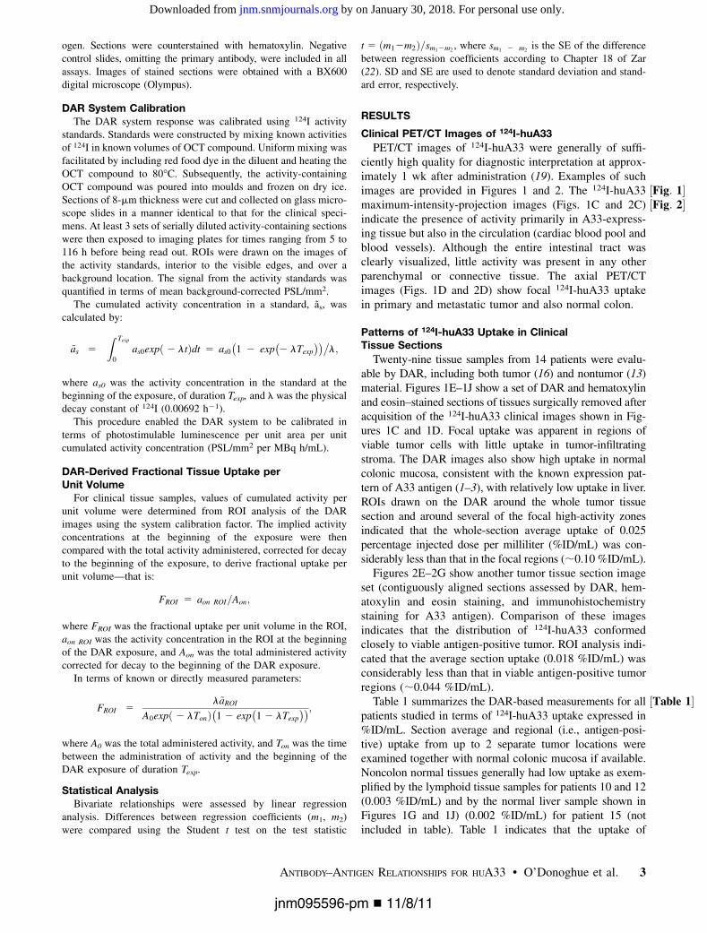
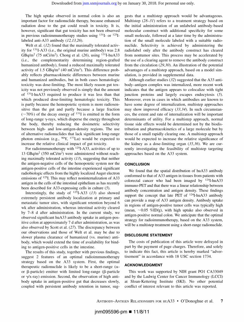
½Fig: 4� Figure 4 plots average 124I-huA33 uptake in tissue sec-tions estimated by DAR analysis versus well counter–derived uptake for corresponding tissue specimens.
Although the tissue sections and specimens for well count-ing were derived from different portions of the resectedtissue samples and the DAR-based estimates were basedon a relatively small number (typically 4) of 8-mm-thicktissue sections (directly accounting for only a small propor-tion of the total volume), there was a linear relationship(r2 ; 0.6) between uptake values with a best-fitting straightline close to the line of identity.
DISCUSSION
We observed linear relationships between antigen con-centration and 124I-huA33 uptake in both tumor and normal
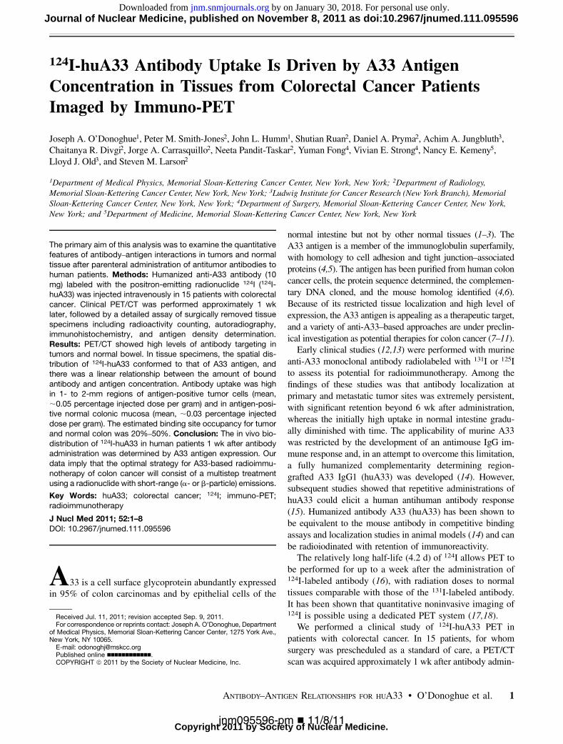
FIGURE 2. (A–B) 18F-FDG PET/CT scan acquired immediately before administration of 124I-huA33 shows pathologic uptake in lesion intransverse colon near hepatic flexure together with normal physiologic accumulation. (C–D) PET/CT scan acquired 182 h after adminis-
tration of 140 MBq (3.8 mCi) of 124I-huA33 shows focal uptake in tumor (compare B and D) and throughout normal intestine. (E–G)
Contiguously aligned sections of tumor visualized in D assessed by 124I-DAR (E), hematoxylin and eosin staining (F), and immunohisto-
chemical staining (G) for A33 antigen. 124I-huA33 localized only in small (;1 mm in dimension) regions of viable antigen-positive tumor andnot in antigen-negative stroma. Scale bars 5 2 mm.
RGB
TABLE 1Summary of DAR Measurements (%ID/mL) on Tissue Sections Cut from Surgically Removed Material
Patient no. Time after injection* (d) Tumor 1 Tumor 1 regional Tumor 2 Tumor 2 regional Normal Normal tissue type
2 5 0.015 0.040 0.026 0.096 0.010 Normal colon3 6 0.015 0.050 — — 0.017 Normal colon
4 8 0.018 0.044 — — — —
5 7 0.008 0.021 — — 0.009 Normal colon
6 7 0.030 0.057 0.029 0.046 0.006 Normal colon7 6 0.058 0.113 — — 0.034 Normal colon
8 6 0.017 0.054 — — 0.012 Normal colon
9 6 0.004 0.036 — — — —
10 7 0.004 0.011 0.003 Lymphoid tissue11 7 — — 0.075 Normal colon
12 7 0.014 0.029 0.017 0.043 0.003 Lymphoid tissue
13 7 0.012 0.030 0.009 Normal colon
14 9 — — 0.093 Normal colon15 7 0.025 0.102 0.016 0.067 0.039 Normal colon
*Time between antibody administration and surgery.
ANTIBODY–ANTIGEN RELATIONSHIPS FOR HUA33 • O’Donoghue et al. 5
jnm095596-pm n 11/8/11
by on January 30, 2018. For personal use only. jnm.snmjournals.org Downloaded from
colon. The steeper slope for colon may be due to the processof cellular turnover. Previous studies have shown high anduniform expression of A33 antigen throughout the intestinalepithelium from crypt to villi (1,6). During the periodbetween antibody administration and surgery, mature epithe-lial cells (together with any associated antibody) will be shedinto the intestinal lumen in a normal physiologic process.Concurrently, circulating antibody will continue to bind butat a decreasing rate in line with the decreasing plasma con-centration of antibody—together, these are predicted to leadto a slowly diminishing ratio of antibody-to-antigen concen-tration. This is discussed in more detail in supplemental data.
The study findings have implications for the use of huA33as a molecular vector for radionuclide therapy of coloncancer. The average tumor uptake (0.017 or 0.019 %ID/mLby well counter or DAR-based measurements, respectively)is among the highest reported for radiolabeled antibodies.Furthermore, as illustrated by the histologic images, antigen-positive tumor cells typically comprise only a fraction ofbulk resected tumor samples. The DAR-based measurementsshow that huA33 uptake in regions of antigen-positive tumorwas approximately 3 times greater than whole-sectionaverages (0.052 vs. 0.019 %ID/mL), implying that, at themicroscopic level, radiation doses delivered to tumor cellsmay be significantly greater than tissue average–based esti-mates would suggest. The antigen-positive tumor regionsseen on histologic images were typically of 1–2 mm indimension. For high-energy/long-range b-emitters (e.g.,90Y), only a small fraction (;10%) of the emitted energywould be absorbed in regions of this size whereas the restwould be deposited in the surrounding stroma (23–25). Incontrast, for lower-energy/shorter-range b-emitters (e.g.,177Lu), a much greater proportion of the emitted b-energywould be absorbed locally (24,25), suggesting that radionu-clides with shorter emission ranges would be preferable forthe disease conformations observed.
FIGURE 3. (A) Example of A33 saturation binding curve for mem-
branes derived from surgically removed tumor sample together with
Scatchard transformed data plot (insert). Mean Kd 6 SE, 0.31 60.02 nM; mean Bmax 6 SE, 1.07 6 0.02 pmol/mg. (B) Scatterplotof A33 antigen density for membranes derived from tumor homo-
genates vs. 124I-huA33 uptake as determined by well counter mea-
surements. Predicted 95% confidence limits are shown as dashed
lines. Regression equation, y 5 59 (SE, 7)x 1 0.3 (SE, 0.1) (n 5 26;r2 5 0.75). (C) Scatterplot of A33 antigen density for membranes
derived from normal colon homogenates vs. 124I-huA33 uptake
determined by well counter measurements. Regression equation,y 5 97 (SE, 25)x 1 0.09 (SE, 0.1) (n 5 25; r2 5 0.40).
FIGURE 4. Scatterplot of tissue %ID/mL estimated using DAR vs.
well counter measurements (%ID/g) of intact tissue samples.
Regression equation, y 5 0.81 (SE, 0.12)x 1 0.004 (SE, 0.002)(n 5 29; r2 5 0.62).
6 THE JOURNAL OF NUCLEAR MEDICINE • Vol. 52 • No. 12 • December 2011
jnm095596-pm n 11/8/11
by on January 30, 2018. For personal use only. jnm.snmjournals.org Downloaded from
The high uptake observed in normal colon is also animportant factor for radionuclide therapy, because enhancedradiation dose to the gut could result in toxicity. It is,however, significant that gut toxicity has not been observedin previous radioimmunotherapy studies using 131I- or 125I-labeled anti-A33 antibody (12,13,26).Welt et al. (12) found that the maximally tolerated activ-
ity for 131I-A33 (i.e., the original murine antibody) was 2.8GBq/m2 (75 mCi/m2). Chong et al. (26), using 131I-huA33(i.e., the complementarity determining region-graftedhumanized antibody), found a reduced maximally toleratedactivity of 1.5 GBq/m2 (40 mCi/m2). This difference prob-ably reflects pharmacokinetic differences between murineand humanized antibodies, but in both cases hematologictoxicity was dose-limiting. The most likely reason gut tox-icity was not previously observed is simply that the amountof 131I-huA33 required to produce it was less than thatwhich produced dose-limiting hematologic toxicity. Thisis partly because the hemopoietic system is more radiosen-sitive than the gut and partly because a large fraction(;70%) of the decay energy of 131I is emitted in the formof long-range g-rays, which disperse the energy throughoutthe body, thereby reducing the dosimetric selectivitybetween high- and low-antigen-density regions. The useof alternative radionuclides that lack significant long-rangephoton emission (e.g., 90Y, 177Lu) would be expected toincrease the relative clinical impact of gut toxicity.For radioimmunotherapy with 125I-A33, activities of up to
13 GBq/m2 (350 mCi/m2) were administered without reach-ing maximally tolerated activity (13), suggesting that neitherthe antigen-negative cells of the hemopoietic system nor theantigen-positive cells of the intestine experienced significantradiobiologic effects from the highly localized Auger electronemissions of 125I. This may reflect noninternalization of A33antigen in the cells of the intestinal epithelium as has recentlybeen described for A33-expressing cells in culture (5).Interestingly, the study of 125I-A33 (13) also showed
extremely persistent antibody localization at primary andmetastatic tumor sites, with significant retention beyond 6wk after administration, whereas intestinal activity clearedby 7–8 d after administration. In the current study, weobserved significant huA33 antibody uptake in antigen-pos-itive colon at approximately 7 d after administration, as wasalso observed by Scott et al. (27). The discrepancy betweenour observations and those of Welt et al. may be due toslower plasma clearance of humanized (vs. murine) anti-body, which would extend the time of availability for bind-ing to antigen-positive cells in the intestine.The results of this study, together with previous findings,
suggest 2 features of an optimal radioimmunotherapystrategy based on the A33 system. First, the optimaltherapeutic radionuclide is likely to be a short-range (a-or b-particle) emitter with limited long-range (b-particleor g/x-ray) emission. Second, the observation of high anti-body uptake in antigen-positive gut that decreases slowly,coupled with persistent antibody retention in tumor, sug-
gests that a multistep approach would be advantageous.Multistep (28–31) refers to a treatment strategy based onthe initial administration of an unlabeled antibody-basedmolecular construct with additional specificity for somesmall molecule, followed at a later time by the administra-tion of the small molecule labeled with a suitable radio-nuclide. Selectivity is achieved by administering theradiolabel only after the antibody construct has clearedfrom nontumor sites. This process may be accelerated bythe use of a clearing agent to remove the antibody constructfrom the circulation (29,30). An illustration of the potentialadvantages of a multistep approach, based on a model sim-ulation, is provided in supplemental data.
Although earlier studies (32) suggested that the A33 anti-body–antigen complex was internalized, more recent workindicates that the antigen appears to colocalize with tightjunction proteins and largely escapes endocytosis (5).Moreover, even in cases in which antibodies are known tohave some degree of internalization, multistep approachesmay show improved efficacy (33,34). In such circumstan-ces, the extent and rate of internalization will be importantdeterminants of utility. For a multistep approach, normaltissue side effects would be determined not by the biodis-tribution and pharmacokinetics of a large molecule but bythose of a small rapidly clearing one. A multistep approachwould be expected to increase the relative significance ofthe kidney as a dose-limiting organ (35,36). We are cur-rently investigating the feasibility of multistep targetingapproaches based on the A33 system.
CONCLUSION
We found that the spatial distribution of huA33 antibodyconformed to that of A33 antigen in tissues from patients withcolorectal cancer who had been imaged by 124I-huA33immuno-PET and that there was a linear relationship betweenantibody concentration and antigen density. These findingssupport the concept that late PET of 124I-huA33 antibodycan provide a map of A33 antigen density. Antibody uptakein regions of antigen-positive tumor cells was typically high(mean, ;0.05 %ID/g), with high uptake also observed inantigen-positive normal colon. We anticipate that the optimalstrategy for radioimmunotherapy, based on the A33 system,will be a multistep treatment using a short-range radionuclide.
DISCLOSURE STATEMENT
The costs of publication of this article were defrayed inpart by the payment of page charges. Therefore, and solelyto indicate this fact, this article is hereby marked “adver-tisement” in accordance with 18 USC section 1734.
ACKNOWLEDGMENT
This work was supported by NIH grant PO1 CA33049and by the Ludwig Center for Cancer Immunology (LCCI)at Sloan-Kettering Institute (SKI). No other potentialconflict of interest relevant to this article was reported.
ANTIBODY–ANTIGEN RELATIONSHIPS FOR HUA33 • O’Donoghue et al. 7
jnm095596-pm n 11/8/11
by on January 30, 2018. For personal use only. jnm.snmjournals.org Downloaded from
REFERENCES
1. Garinchesa P, Sakamoto J, Welt S, Real FX, Rettig WJ, Old LJ. Organ-specific
expression of the colon cancer antigen A33, a cell surface target for antibody-
based therapy. Int J Oncol. 1996;9:465–471.
2. Johnstone CN, White SJ, Tebbutt NC, et al. Analysis of the regulation of the A33
antigen gene reveals intestine-specific mechanisms of gene expression. J Biol
Chem. 2002;277:34531–34539.
3. Sakamoto J, Kojima H, Kato J, Hamashima H, Suzuki H. Organ-specific expres-
sion of the intestinal epithelium-related antigen A33, a cell surface target for
antibody-based imaging and treatment in gastrointestinal cancer. Cancer Chemo-
ther Pharmacol. 2000;46:S27–S32.
4. Heath JK, White SJ, Johnstone CN, et al. The human A33 antigen is a trans-
membrane glycoprotein and a novel member of the immunoglobulin superfam-
ily. Proc Natl Acad Sci USA. 1997;94:469–474.
5. Ackerman ME, Chalouni C, Schmidt MM, et al. A33 antigen displays persistent
surface expression. Cancer Immunol Immunother. 2008;57:1017–1027.
6. Johnstone CN, Tebbutt NC, Abud HE, et al. Characterization of mouse A33
antigen, a definitive marker for basolateral surfaces of intestinal epithelial cells.
Am J Physiol Gastrointest Liver Physiol. 2000;279:G500–G510.
7. Almqvist Y, Orlova A, Sjostrom A, et al. In vitro characterization of At-211-
labeled antibody A33: a potential therapeutic agent against metastatic colorectal
carcinoma. Cancer Biother Radiopharm. 2005;20:514–523.
8. Almqvist Y, Steffen AC, Tolmachev V, Divgi CR, Sundin A. In vitro and in vivo
characterization of Lu-177-huA33: a radioimmunoconjugate against colorectal
cancer. Nucl Med Biol. 2006;33:991–998.
9. Deckert PM, Bornmann WG, Ritter G, et al. Specific tumour localisation of a
huA33 antibody: carboxypeptidase A conjugate and activation of methotrexate-
phenylalanine. Int J Oncol. 2004;24:1289–1295.
10. Coelho V, Dernedde J, Petrausch U, et al. Design, construction, and in vitro
analysis of A33scFv: CDy, a recombinant fusion protein for antibody-directed
enzyme prodrug therapy in colon cancer. Int J Oncol. 2007;31:951–957.
11. Cortez C, Tomaskovic-Crook E, Johnston APR, et al. Influence of size, surface,
cell line, and kinetic properties on the specific binding of A33 antigen-targeted
multilayered particles and capsules to colorectal cancer cells. ACS Nano. 2007;
1:93–102.
12. Welt S, Divgi CR, Kemeny N, et al. Phase I/II study of iodine 131-labeled
monoclonal-antibody A33 in patients with advanced colon-cancer. J Clin Oncol.
1994;12:1561–1571.
13. Welt S, Scott A, Divgi C, et al. Phase I/II study of iodine 125-labeled monoclonal
antibody A33 in patients with advanced colon cancer. J Clin Oncol. 1996;14:
1787–1797.
14. King DJ, Antoniw P, Owens RJ, et al. Preparation and preclinical evaluation of
humanized A33 immunoconjugates for radioimmunotherapy. Br J Cancer.
1995;72:1364–1372.
15. Ritter G, Cohen LS, Williams C, Richards EC, Old LJ, Welt S. Serological
analysis of human anti-human antibody responses in colon cancer patients trea-
ted with repeated doses of humanized monoclonal antibody A33. Cancer Res.
2001;61:6851–6859.
16. Divgi CR, Pandit-Taskar N, Jungbluth AA, et al. Preoperative characterisation of
clear-cell renal carcinoma using iodine-124-labelled antibody chimeric G250 (I-
124-cG250) and PET in patients with renal masses: a phase I trial. Lancet Oncol.
2007;8:304–310.
17. Pentlow KS, Graham MC, Lambrecht RM, Cheung NKV, Larson SM. Quanti-
tative imaging of I-124 using positron emitting tomography with applications to
radioimmunodiagnosis and radioimmunotherapy. Med Phys. 1991;18:357–366.
18. Pentlow KS, Graham MC, Lambrecht RM, et al. Quantitative imaging of iodine-
124 with PET. J Nucl Med. 1996;37:1557–1562.
19. Carrasquillo JA, Pandit-Taskar N, O’Donoghue JA, et al. 124I-huA33 antibody
PET of colorectal cancer. J Nucl Med. 2011;52:1173–1180.
20. United States Pharmacopeial Convention, Inc. Pharmaceutical compounding–
sterile preparations. USP ,797.. First Supplement to The United Stated Phar-
macopeia, 27th rev.; and The National Formulary, 22nd ed. Rockville, MD:
United States Pharmacopeial Convention, Inc.; 2004.
21. Fraker PJ, Speck JC. Protein and cell-membrane iodinations with a sparingly
soluble chloramide, 1,3,4,6-tetrachloro-3A,6A-diphenylglycoluril. Biochem Bio-
phys Res Commun. 1978;80:849–857.
22. Zar JH. Biostatistical Analysis. Fifth ed. Upper Saddle River, NJ: Prentice Hall; 2010.
23. Humm JL. Dosimetric aspects of radiolabeled antibodies for tumor therapy. J
Nucl Med. 1986;27:1490–1497.
24. Bardies M, Chatal JF. Absorbed doses for internal radiotherapy from 22 beta-
emitting radionuclides - beta-dosimetry of small spheres. Phys Med Biol.
1994;39:961–981.
25. O’Donoghue JA, Bardies M, Wheldon TE. Relationships between tumor size and
curability for uniformly targeted therapy with beta-emitting radionuclides. J
Nucl Med. 1995;36:1902–1909.
26. Chong G, Lee F, Hopkins W, et al. Phase I trial of I-131-huA33 in patients with
advanced colorectal carcinoma. Clin Cancer Res. 2005;11:4818–4826.
27. Scott AM, Lee FT, Jones R, et al. A phase I trial of humanized monoclonal
antibody A33 in patients with colorectal carcinoma: Biodistribution, pharmaco-
kinetics, and quantitative tumor uptake. Clin Cancer Res. 2005;11:4810–4817.
28. Boerman OC, van Schaijk FG, Oyen WJG, Corstens FHM. Pretargeted radio-
immunotherapy of cancer: Progress step by step. J Nucl Med. 2003;44:400–411.
29. Forero A, Weiden PL, Vose JM, et al. Phase 1 trial of a novel anti-CD20 fusion
protein in pretargeted radioimmunotherapy for B-cell non-Hodgkin lymphoma.
Blood. 2004;104:227–236.
30. Green DJ, Pagel JM, Pantelias A, et al. Pretargeted radioimmunotherapy for B-
Cell lymphomas. Clin Cancer Res. 2007;13:5598s–5603s.
31. Gold DV, Goldenberg DM, Karacay H, et al. A novel bispecific, trivalent antibody
construct for targeting pancreatic carcinoma. Cancer Res. 2008;68:4819–4826.
32. Daghighian F, Barendswaard E, Welt S, et al. Enhancement of radiation dose to
the nucleus by vesicular internalization of iodine-125-labeled A33 monoclonal
antibody. J Nucl Med. 1996;37:1052–1057.
33. Zhang M, Zhang Z, Garmestani K, et al. Pretarget radiotherapy with an anti-
CD25 antibody streptavidin fusion protein was effective in therapy of leukemia/
lymphoma xenografts. Proc Natl Acad Sci USA. 2003;100:1891–1895.
34. Sato N, Hassan R, Axworthy DB, et al. Pretargeted radioimmunotherapy of
mesothelin-expressing cancer using a tetravalent single-chain Fv-streptavidin
fusion protein. J Nucl Med. 2005;46:1201–1209.
35. Forster GJ, Santos EB, Smith-Jones PM, Zanzonico P, Larson SM. Pretargeted
radioimmunotherapy with a single-chain antibody/streptavidin construct and ra-
diolabeled DOTA-biotin: strategies for reduction of the renal dose. J Nucl Med.
2006;47:140–149.
36. O’Donoghue J. Relevance of external beam dose-response relationships to kid-
ney toxicity associated with radionuclide therapy. Cancer Biother Radiopharm.
2004;19:378–387.
8 THE JOURNAL OF NUCLEAR MEDICINE • Vol. 52 • No. 12 • December 2011
jnm095596-pm n 11/8/11
by on January 30, 2018. For personal use only. jnm.snmjournals.org Downloaded from
Doi: 10.2967/jnumed.111.095596Published online: November 8, 2011.J Nucl Med. Lloyd J. Old and Steven M. LarsonChaitanya R. Divgi, Jorge A. Carrasquillo, Neeta Pandit-Taskar, Yuman Fong, Vivian E. Strong, Nancy E. Kemeny, Joseph A. O'Donoghue, Peter M. Smith-Jones, John L. Humm, Shutian Ruan, Daniel A. Pryma, Achim A. Jungbluth, from Colorectal Cancer Patients Imaged by Immuno-PET
I-huA33 Antibody Uptake Is Driven by A33 Antigen Concentration in Tissues124
http://jnm.snmjournals.org/content/early/2011/11/08/jnumed.111.095596This article and updated information are available at:
http://jnm.snmjournals.org/site/subscriptions/online.xhtml
Information about subscriptions to JNM can be found at:
http://jnm.snmjournals.org/site/misc/permission.xhtmlInformation about reproducing figures, tables, or other portions of this article can be found online at:
the manuscript and the final, published version.typesetting, proofreading, and author review. This process may lead to differences between the accepted version of
ahead of print area, they will be prepared for print and online publication, which includes copyediting,JNMthe copyedited, nor have they appeared in a print or online issue of the journal. Once the accepted manuscripts appear in
. They have not beenJNM ahead of print articles have been peer reviewed and accepted for publication in JNM
(Print ISSN: 0161-5505, Online ISSN: 2159-662X)1850 Samuel Morse Drive, Reston, VA 20190.SNMMI | Society of Nuclear Medicine and Molecular Imaging
is published monthly.The Journal of Nuclear Medicine
© Copyright 2011 SNMMI; all rights reserved.
by on January 30, 2018. For personal use only. jnm.snmjournals.org Downloaded from