Embed Size (px)
Citation preview

11.2.Muscles and movement

• State the roles of bones, ligaments, muscles, tendons and nerves in human movement.
• Label a diagram of the human elbow joint, including cartilage, synovial fluid, joint capsule, named bones and antagonistic muscles (biceps and triceps).
• Outline the functions of the structures in the human elbow joint named above.
• Compare the movements of the hip joint and the knee joint.
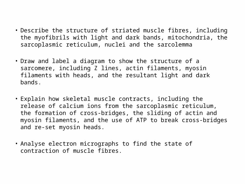
• Describe the structure of striated muscle fibres, including the myofibrils with light and dark bands, mitochondria, the sarcoplasmic reticulum, nuclei and the sarcolemma
• Draw and label a diagram to show the structure of a sarcomere, including Z lines, actin filaments, myosin filaments with heads, and the resultant light and dark bands.
• Explain how skeletal muscle contracts, including the release of calcium ions from the sarcoplasmic reticulum, the formation of cross-bridges, the sliding of actin and myosin filaments, and the use of ATP to break cross-bridges and re-set myosin heads.
• Analyse electron micrographs to find the state of contraction of muscle fibres.
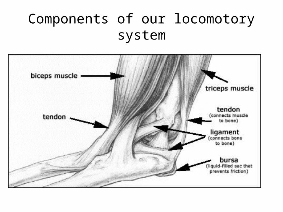
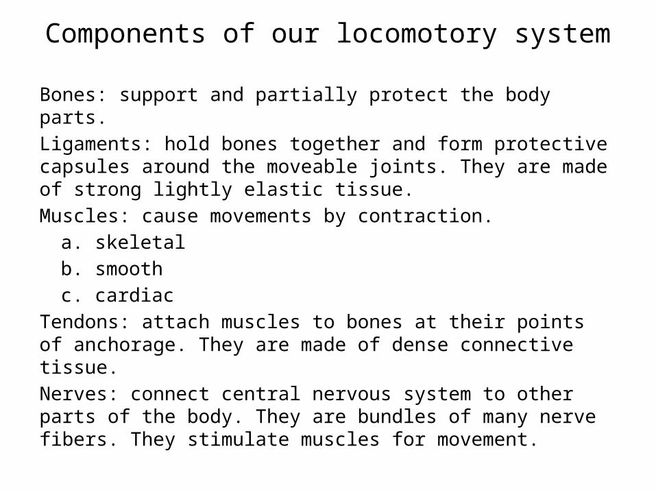
Components of our locomotory system
Components of our locomotory system
Bones: support and partially protect the body parts.Ligaments: hold bones together and form protective capsules around the moveable joints. They are made of strong lightly elastic tissue.Muscles: cause movements by contraction.
a. skeletalb. smoothc. cardiac
Tendons: attach muscles to bones at their points of anchorage. They are made of dense connective tissue.Nerves: connect central nervous system to other parts of the body. They are bundles of many nerve fibers. They stimulate muscles for movement.

The human elbow joint

The components of human elbow jointHumerus, radius, ulna:Biceps muscle:Triceps muscles:Ligaments:Capsule:Synovial membrane:Synovial fluid:Cartilage:

Types of joints:
• Movable joints: provide controlled movement also known as synovial joints.
a. Ball and socket joints: are found in hip joints that permit movements in all three planes (circular movement: circumduction)
b. Hinge joint: restricts the movement to one plane (flexion and extension. Example: Knee joint

The human knee joint

MUSCLE CONTRACTION
AND MOVEMENT
© 2012 Parson Education, Inc.

• Muscles and bones interact to produce movement.
• Muscles– are connected to bones by tendons and– can only contract, requiring an antagonistic
muscle to• reverse the action and• relengthen muscles.
The skeleton and muscles interact in movement
© 2012 Parson Education, Inc.

Figure 30.7A
Biceps contracted,triceps relaxed(extended)
Biceps
Triceps
TendonsTriceps
Biceps
Tricepscontracted,bicepsrelaxed
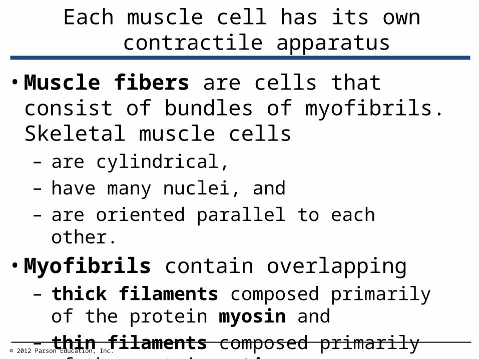
• Muscle fibers are cells that consist of bundles of myofibrils. Skeletal muscle cells– are cylindrical,– have many nuclei, and– are oriented parallel to each other.
• Myofibrils contain overlapping– thick filaments composed primarily of the
protein myosin and– thin filaments composed primarily of the
protein actin.
Each muscle cell has its own contractile apparatus
© 2012 Parson Education, Inc.

• Sarcomeres are– repeating groups of overlapping thick and thin
filaments and– the contractile unit—the fundamental unit of
muscle action.
Each muscle cell has its own contractile apparatus
© 2012 Parson Education, Inc.

Figure 30.8_1
Muscle
Several muscle fibers
Single muscle fiber(cell)
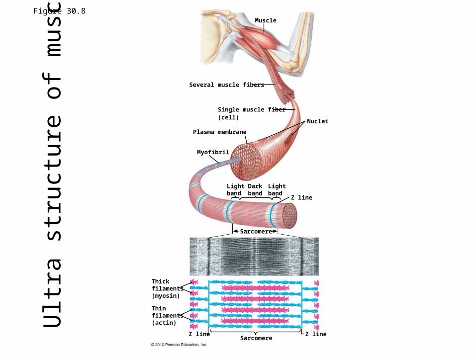
Figure 30.8Muscle
Several muscle fibers
Single muscle fiber(cell)
Plasma membrane
Nuclei
Myofibril
Lightband
Darkband
Lightband
Z line
Sarcomere
SarcomereZ lineZ line
Thickfilaments(myosin)
Thinfilaments(actin)U
ltra
stru
ctur
e of
mus
cle
fiber
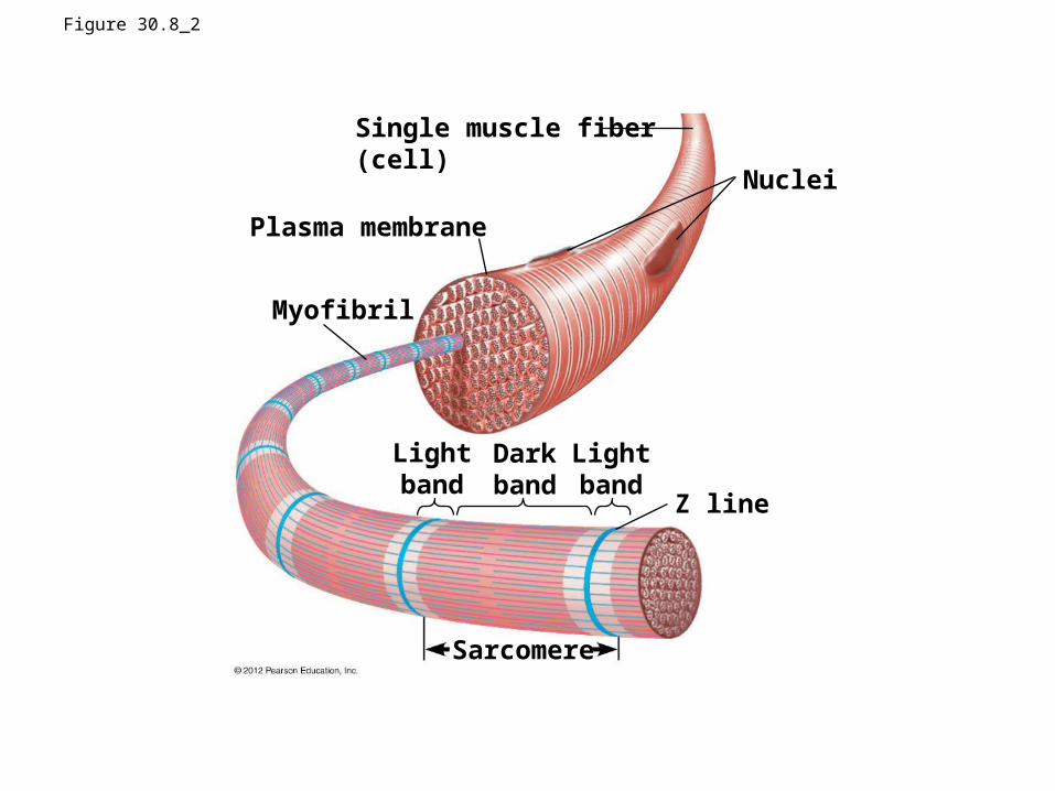
Figure 30.8_2
Plasma membrane
Nuclei
Myofibril
Lightband
Darkband
Lightband
Z line
Sarcomere
Single muscle fiber(cell)
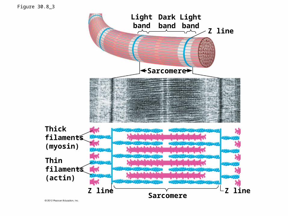
Figure 30.8_3
SarcomereZ lineZ line
Thickfilaments(myosin)
Thinfilaments(actin)
Lightband
Darkband
Lightband
Z line
Sarcomere

Figure 30.8_4
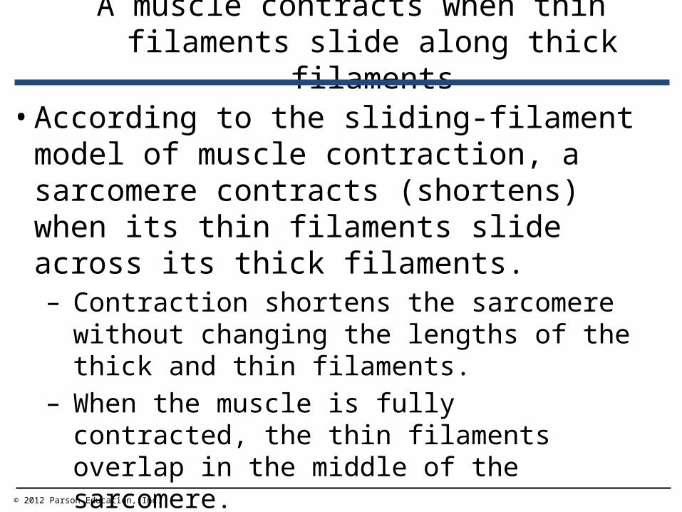
• According to the sliding-filament model of muscle contraction, a sarcomere contracts (shortens) when its thin filaments slide across its thick filaments.– Contraction shortens the sarcomere without
changing the lengths of the thick and thin filaments.
– When the muscle is fully contracted, the thin filaments overlap in the middle of the sarcomere.
A muscle contracts when thin filaments slide along thick filaments
© 2012 Parson Education, Inc.

Figure 30.9A
Relaxed muscle
Contractingmuscle
Fully contractedmuscle
Dark band
Sarcomere
Contracted sarcomere
Z Z

• Myosin heads of the thick filaments– bind ATP and– extend to high-energy states.
• Myosin heads then – attach to binding sites on the actin molecules
and– pull the thin filaments toward the center of the
sarcomere.
A muscle contracts when thin filaments slide along thick filaments
© 2012 Parson Education, Inc.

Figure 30.9BThinfilaments
Thinfilament
Thickfilament
Thick filament
Z lineActin
Myosin head (low-energy configuration)
Myosin head (high-energy configuration)
Cross-bridge
Newpositionof Z lineThin filament moves
toward center of sarcomere.
1
2
3
ATP
ADPP
ADPP
4
ADP P
5
Myosin head (pivoting tolow-energy configuration)
ATP Myosin head (low-energy configuration)

Figure 30.9B_s1
Thinfilaments
Thick filament
Z line

Figure 30.9B_s2
Thinfilaments
Thick filament
Z line
Myosin head (low-energy configuration)
Actin
Thinfilament
Thickfilament
1
ATP

Figure 30.9B_s3
Thinfilaments
Thick filament
Z line
Myosin head (low-energy configuration)
Myosin head (high-energy configuration)
Actin
Thinfilament
Thickfilament
1
2
ATP
ADPP

Figure 30.9B_s4
Cross-bridge
3
ADPP
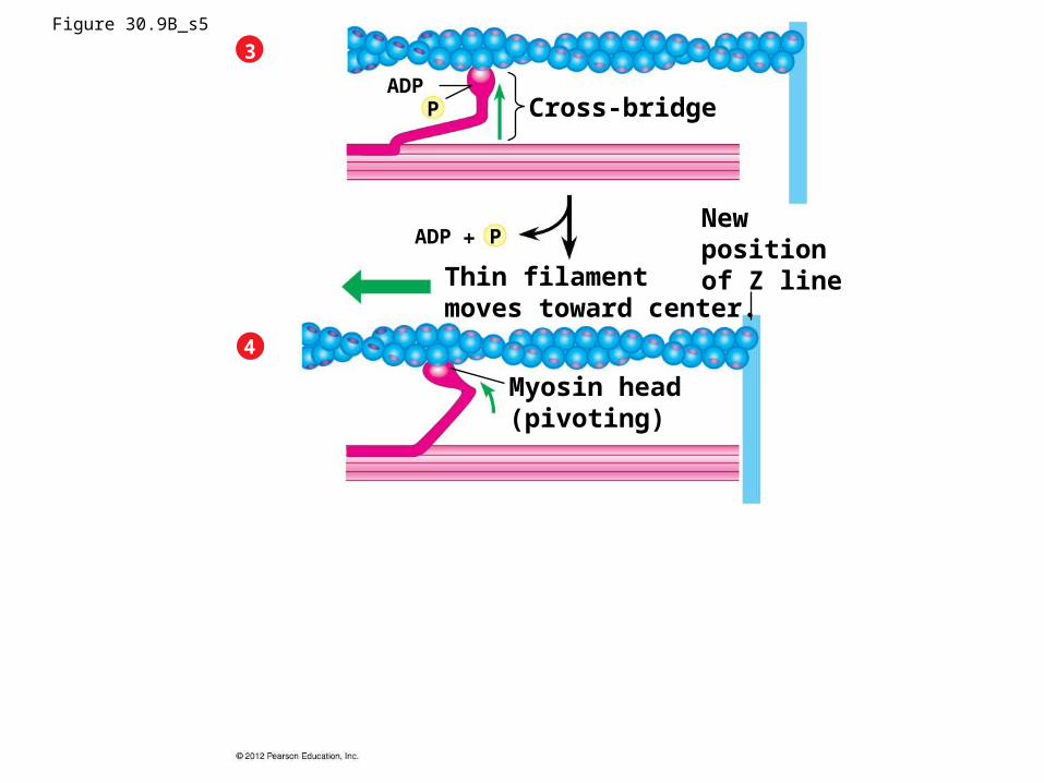
Figure 30.9B_s5
Myosin head(pivoting)
4
Newpositionof Z lineThin filament
moves toward center.
ADP P
Cross-bridge
3
ADPP

Figure 30.9B_s6
Myosin head(low-energy)
Myosin head(pivoting)
ATP
5
4
Newpositionof Z lineThin filament
moves toward center.
ADP P
Cross-bridge
3
ADPP
• A motor neuron– carries an action potential to a muscle cell,– releases the neurotransmitter acetylcholine
from its synaptic terminal, and– initiates a muscle contraction.
Motor neurons stimulate muscle contraction
© 2012 Parson Education, Inc.

Figure 30.10A
Motor neuronaxon
Synapticterminal
T tubule
Action potentialMitochondrion
Endoplasmicreticulum (ER)
Myofibril
Plasma membraneSarcomere
Ca2
releasedfrom ER
• An action potential in a muscle cell– passes along T tubules and– into the center of the muscle fiber.
• Calcium ions– are released from the endoplasmic reticulum
and– initiate muscle contraction by moving the
regulatory protein tropomyosin away from the myosin-binding sites on actin.
Motor neurons stimulate muscle contraction
© 2012 Parson Education, Inc.

Figure 30.10B
Myosin-binding sites blocked
Myosin-binding sites exposed
Myosin-binding site
Ca2 floods thecytoplasmicfluid
ActinTropomyosin Ca2-binding sites
Troponin complex

• A motor unit consists of– a neuron and– the set of muscle fibers it controls.
• More forceful muscle contractions result when additional motor units are activated.
Motor neurons stimulate muscle contraction
© 2012 Parson Education, Inc.

Figure 30.10C Spinal cord
Motor neuroncell body
Nerve
Motor neuronaxon
Synapticterminals
Muscle
Tendon
Muscle fibers(cells)
Nuclei
Bone
Motorunit 1
Motorunit 2