Embed Size (px)
Citation preview
1
Enzymes II
Department of Biochemistry (J.D.) 2013
Mechanism of enzyme reaction, metalloenzymes,
kinetics, activity, enzymes in medicine
2
Active site of enzyme
• small part of molecule, deep cleft or pocket on the surface
• three-dimensional environment shields the substrate from water
• in oligomeric enzymes - interface between subunits
• made by side chains of AA which are distant in primary structure
• the site is really active - protein flexibility facilitates the conformation
changes fitting to substrate
• substrate is bound relatively weakly
3
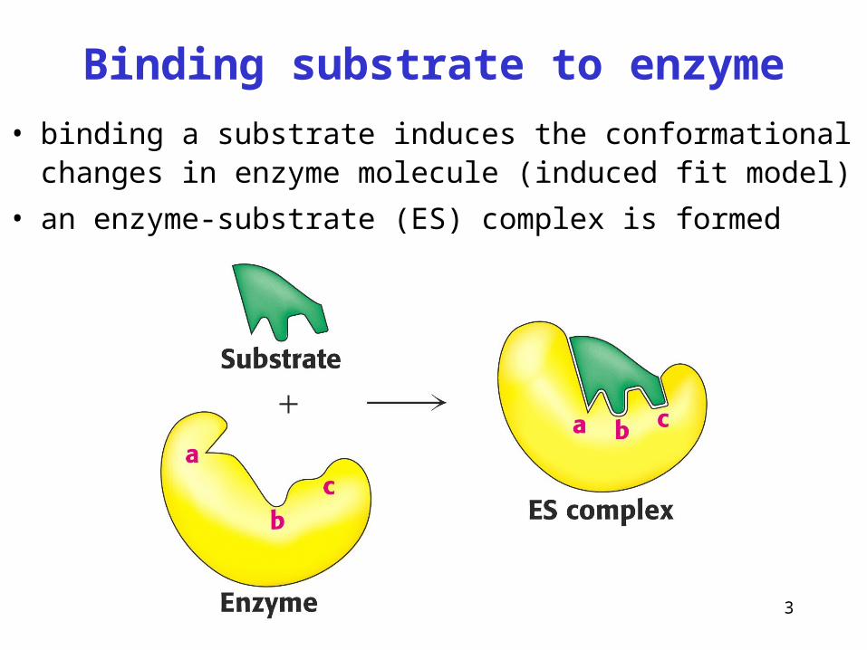
Binding substrate to enzyme
• binding a substrate induces the conformational changes in enzyme molecule (induced fit model)
• an enzyme-substrate (ES) complex is formed
4
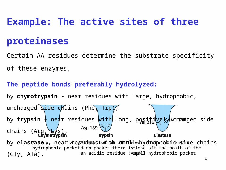
A deep, relativelyhydrophobic pocket
At the bottom of thedeep pocket there isan acidic residue (Asp)
Two residues of valineclose off the mouth of thesmall hydrophobic pocket
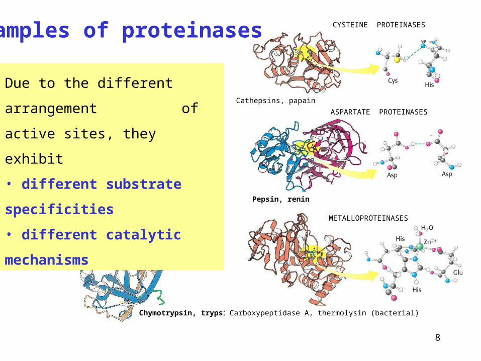
Example: The active sites of three proteinases
Certain AA residues determine the substrate specificity of these enzymes.
The peptide bonds preferably hydrolyzed:
by chymotrypsin - near residues with large, hydrophobic, uncharged side chains (Phe, Trp),
by trypsin – near residues with long, positively charged side chains (Arg, Lys),
by elastase - near residues with small hydrophobic side chains (Gly, Ala).
5
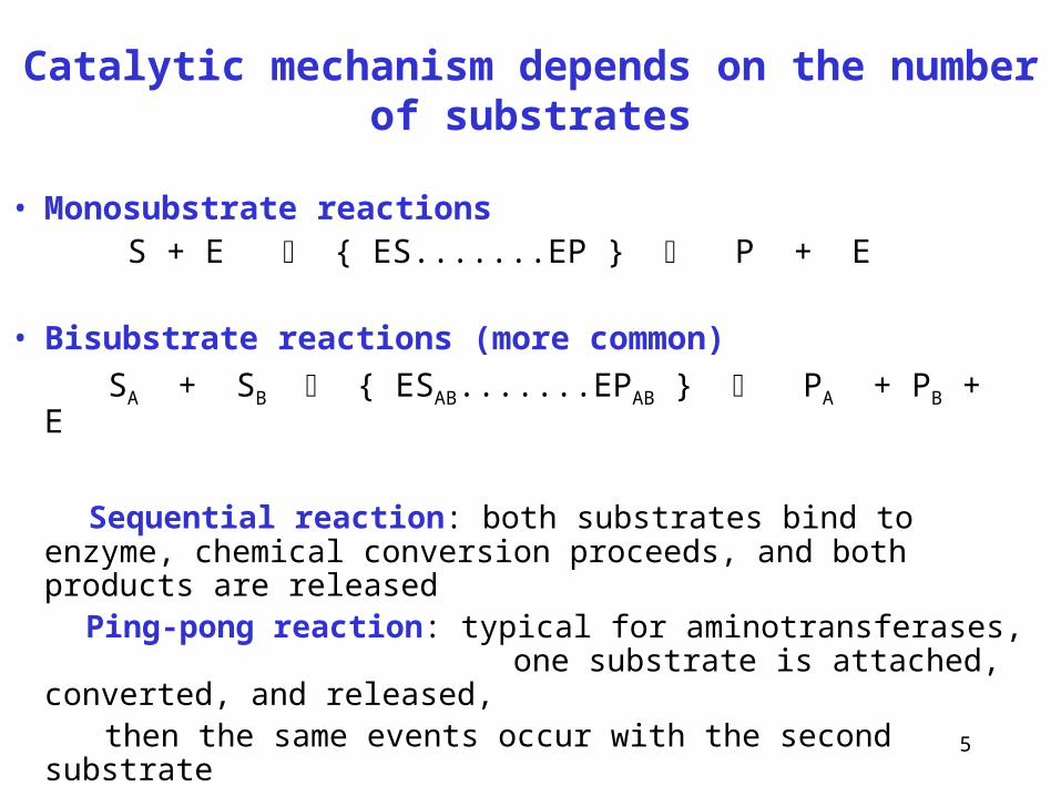
Catalytic mechanism depends on the number of substrates
• Monosubstrate reactions S + E { ES.......EP } P + E
• Bisubstrate reactions (more common)
SA + SB { ESAB.......EPAB } PA + PB + E
Sequential reaction: both substrates bind to enzyme, chemical conversion proceeds, and both products are released
Ping-pong reaction: typical for aminotransferases, one substrate is attached, converted, and released,
then the same events occur with the second substrate
6
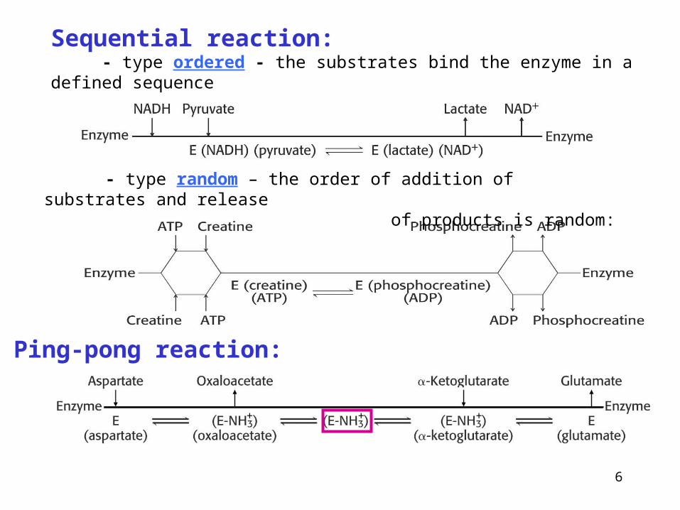
Sequential reaction: - type ordered - the substrates bind the enzyme in a defined sequence
- type random – the order of addition of substrates and release of products is random:
Ping-pong reaction:
+ + +
7
Catalytic groups • located in active site
• involved the chemical conversion of substrate
• nucleophilic (cysteine -SH, serine -OH)
• acidic (Asp, Glu), basic (His, Arg, Lys)
Examples of different types of catalytic mechanisms:
• Catalysis through proximity and orientation effects (strained reactants)
• Covalent catalysis – formation of transitory covalent bonds between enzyme and substrate
• Acid-base catalysis – protonization of substrates or catalytic groups of enzyme
• Metal ion catalysis mediating redox reactions or shielding negative el. charges
• Electrostatic catalysis (after excluding water from the active site by binding a substrate)
8
SERINE PROTEASES
Serine 195
Chymotrypsin, trypsin
CYSTEINE PROTEINASES
Carboxypeptidase A, thermolysin (bacterial)
METALLOPROTEINASES
Cathepsins, papain
ASPARTATE PROTEINASES
Pepsin, renin
Examples of proteinases
Due to the different arrangement
of active sites, they exhibit
• different substrate specificities
• different catalytic mechanisms
9
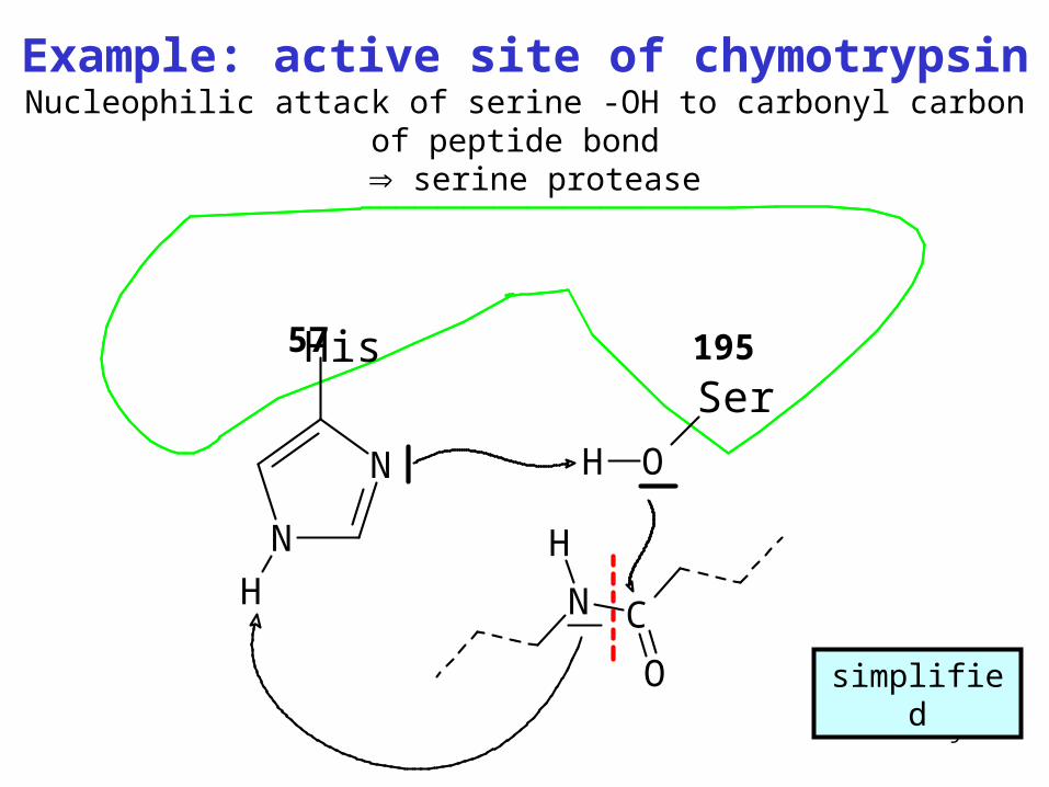
Example: active site of chymotrypsinNucleophilic attack of serine -OH to carbonyl carbon of peptide bond
serine protease
N C
O
H
His
N
N
H
Ser
OH
195
57
simplified
10
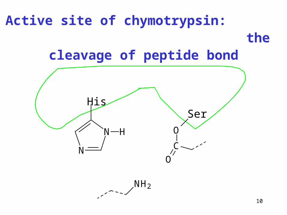
Active site of chymotrypsin: the cleavage of peptide bond
His
N
N H
Ser
O
C
O
NH2
11
Metalloenzymes
• contain functioning metal cations (prosthetic groups) directly
involved in catalyzed reaction, metal ions attached tightly
(Enz-M)
• some enzymes need metal ions just for activation,
they are associated relatively weekly (Enz …M),
e.g. Ca2+ (coagulation factors), Mg2+ (kinases)
12
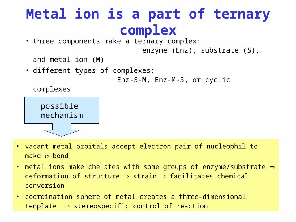
Metal ion is a part of ternary complex
• three components make a ternary complex: enzyme (Enz), substrate (S), and metal ion (M)
• different types of complexes: Enz-S-M, Enz-M-S, or cyclic complexes
• vacant metal orbitals accept electron pair of nucleophil to make -bond
• metal ions make chelates with some groups of enzyme/substrate deformation
of structure strain facilitates chemical conversion
• coordination sphere of metal creates a three-dimensional template
stereospecific control of reaction
possible mechanism
13
Molybdenum (Mo)
• Some oxidoreductases
• cofactor – molybdopterin
• Xanthine oxidase (xanthine uric acid)
• Sulfite oxidase (sulfite HSO3- sulfate SO4
2-)
Mo in food: legumes, wholemeal cereals
14
Zinc (Zn)• Many enzymes
• Alcohol dehydrogenase (ethanol acetaldehyde)
• Carbonic anhydrase (H2O + CO2 H2CO3)
• Carboxypeptidases (cleavage of polypeptides from C-terminal)
• Cu, Zn-superoxide dismutase (cytosolic isoform)
(2 •O2- + 2 H+ O2 + H2O2)
Zn in food: red meat, lobsters, legumes, seeds, wholemeal cereals
15
Copper (Cu)• many oxidoreductases
• Ceruloplasmin (ferroxidase) (Fe2+ Fe3+)
• Cytochrome-c-oxidase (R.Ch., electron transfer to O2)
• Monoamine oxidases (MAO, inactivation of biogenic
amines, side product H2O2, see Med. Chem. II, p. 60)
• Dopamine hydroxylase (dopamine noradrenaline)
• Lysyl oxidase (collagen maturation, Lys alLys)
Cu in food: liver, meat, cocoa, legumes, nuts
16
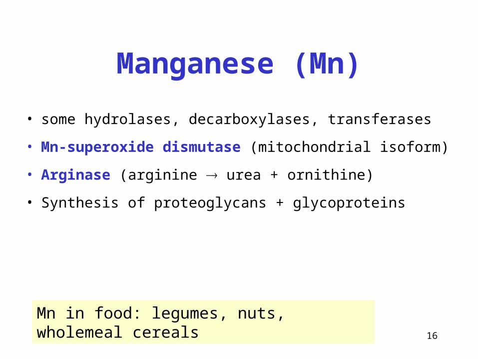
Manganese (Mn)
• some hydrolases, decarboxylases, transferases
• Mn-superoxide dismutase (mitochondrial isoform)
• Arginase (arginine urea + ornithine)
• Synthesis of proteoglycans + glycoproteins
Mn in food: legumes, nuts, wholemeal cereals
17
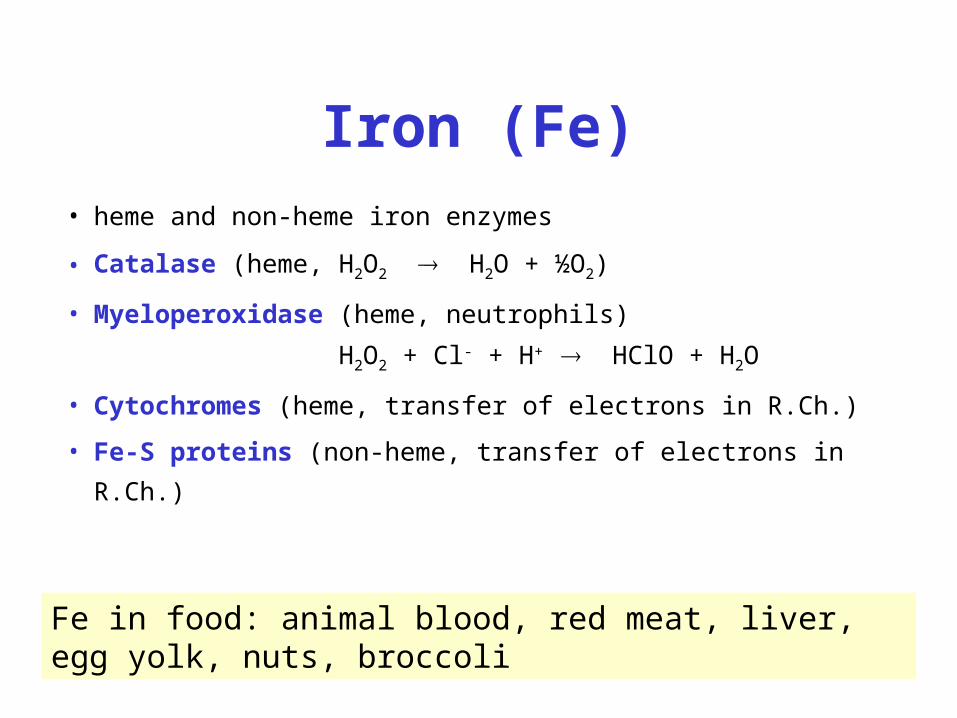
Iron (Fe)
• heme and non-heme iron enzymes
• Catalase (heme, H2O2 H2O + ½O2)
• Myeloperoxidase (heme, neutrophils)
H2O2 + Cl- + H+ HClO + H2O
• Cytochromes (heme, transfer of electrons in R.Ch.)
• Fe-S proteins (non-heme, transfer of electrons in R.Ch.)
Fe in food: animal blood, red meat, liver, egg yolk, nuts, broccoli
18
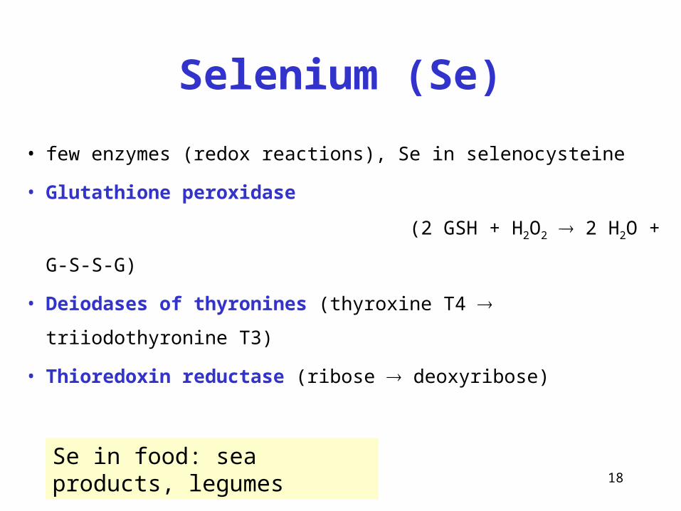
Selenium (Se)
• few enzymes (redox reactions), Se in selenocysteine
• Glutathione peroxidase
(2 GSH + H2O2 2 H2O + G-S-S-G)
• Deiodases of thyronines (thyroxine T4 triiodothyronine T3)
• Thioredoxin reductase (ribose deoxyribose)
Se in food: sea products, legumes
19
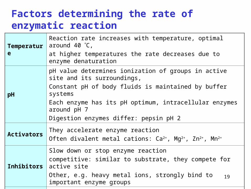
Factors determining the rate of enzymatic reaction
TemperatureReaction rate increases with temperature, optimal around 40 C,
at higher temperatures the rate decreases due to enzyme denaturation
pH
pH value determines ionization of groups in active site and its surroundings,
Constant pH of body fluids is maintained by buffer systems
Each enzyme has its pH optimum, intracellular enzymes around pH 7
Digestion enzymes differ: pepsin pH 2
ActivatorsThey accelerate enzyme reaction
Often divalent metal cations: Ca2+, Mg2+, Zn2+, Mn2+
Inhibitors
Slow down or stop enzyme reaction
competitive: similar to substrate, they compete for active site
Other, e.g. heavy metal ions, strongly bind to important enzyme groups
Substrate
concentration At high substrate concentration, enzyme is saturated, so the reactions proceeds with maximal velocity, graphically expressed as saturation curve
20
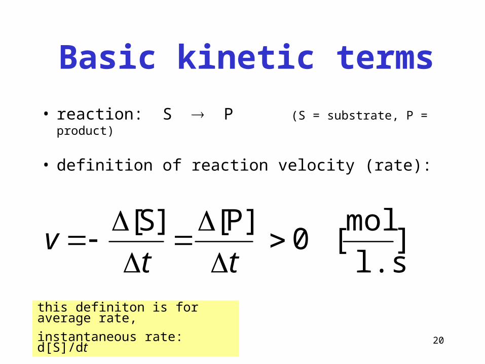
Basic kinetic terms
• reaction: S P (S = substrate, P = product)
• definition of reaction velocity (rate):
]l.s
mol[0
]P[]S[
ttv
this definiton is for average rate,
instantaneous rate: d[S]/dt
21
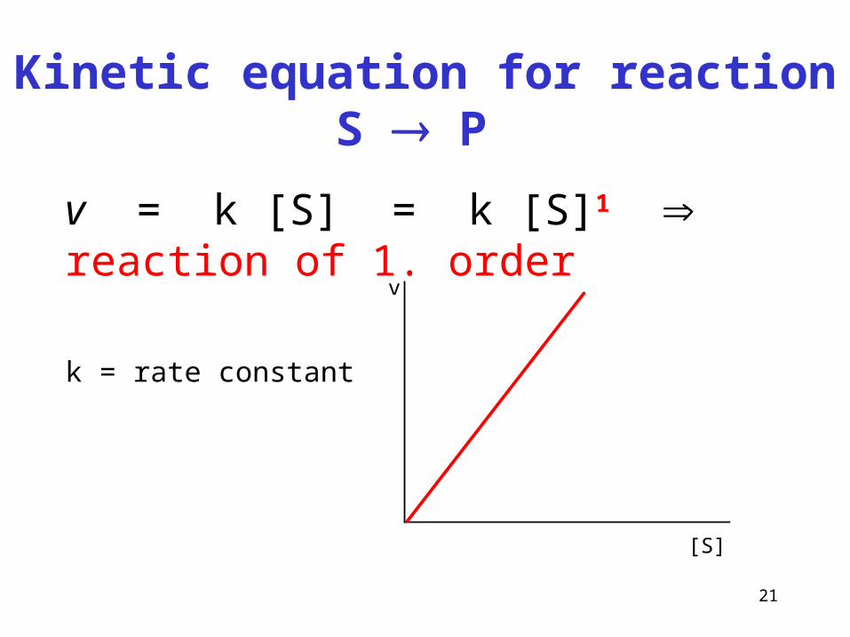
Kinetic equation for reaction S P
v = k [S] = k [S]1 reaction of 1. order
k = rate constantv
[S]
22
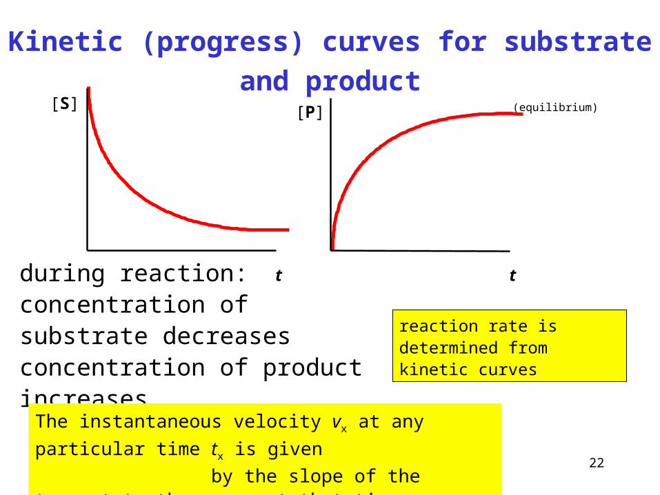
Kinetic (progress) curves for substrate and product
[P]
t
[S]
t
(equilibrium)
during reaction:concentration of substrate decreasesconcentration of product increases
reaction rate is determined from kinetic curves
The instantaneous velocity vx at any particular time tx is given
by the slope of the tangent to the curve at that time.
23
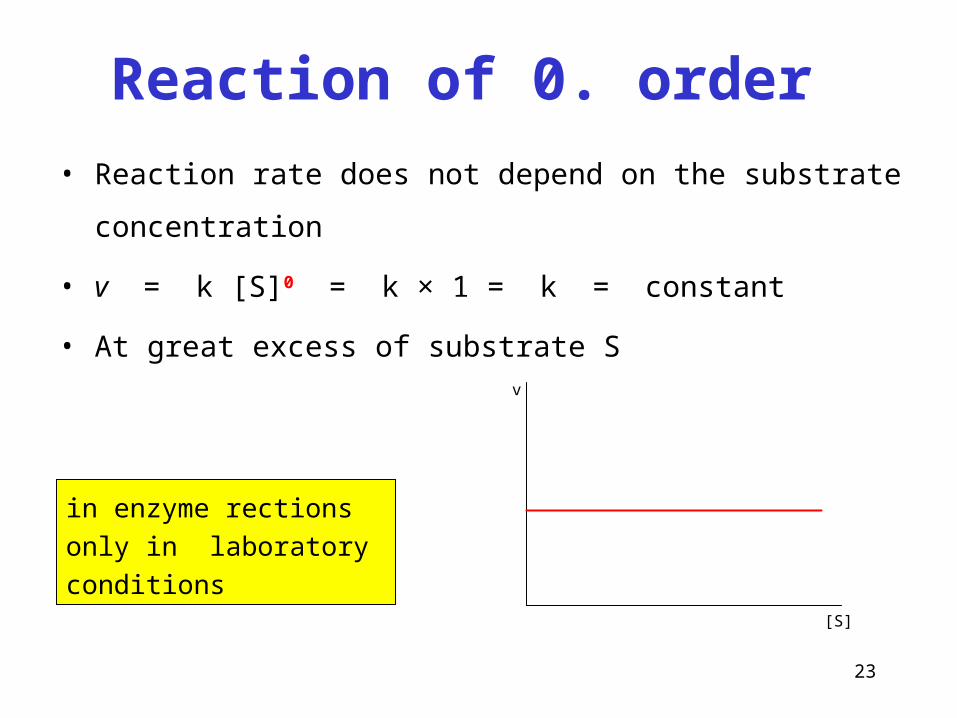
Reaction of 0. order
• Reaction rate does not depend on the substrate concentration
• v = k [S]0 = k × 1 = k = constant
• At great excess of substrate S
v
[S]
in enzyme rections only in laboratory conditions
24
Initial velocity vo
• The highest value of velocity
• It is not influenced by the decrease of substrate
nor the reverse change of product
• Determined from kinetic curves at the time t = 0
25
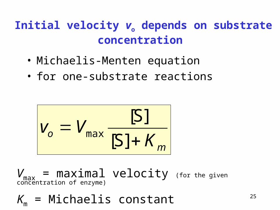
Initial velocity vo depends on substrate concentration
• Michaelis-Menten equation• for one-substrate reactions
mo K
Vv
]S[
]S[max
Vmax = maximal velocity (for the given concentration of enzyme)
Km = Michaelis constant

26
The graph of previous equation is saturation curve
Km mol/l[S]
vo
Vmax
enzym nasycensubstrátem
Vmax
2
enzyme saturated by substrate
27
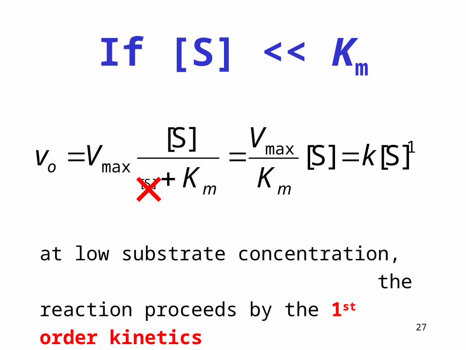
If [S] << Km
1maxmax ]S[]S[
]S[
]S[
kK
V
KVv
mmo
at low substrate concentration,
the reaction proceeds by the 1st order kinetics
×
28
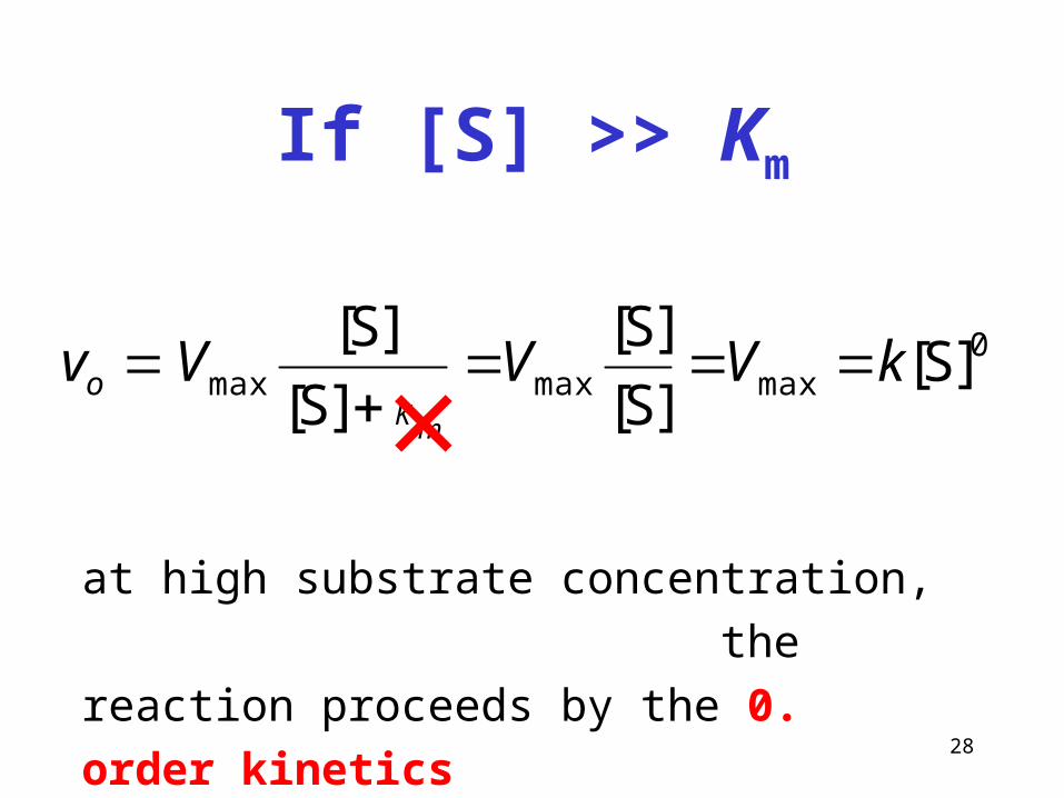
If [S] >> Km
0maxmaxmax ]S[
]S[
]S[
]S[
]S[kVVVv
mKo
at high substrate concentration,
the reaction proceeds by the 0. order kinetics
×
29
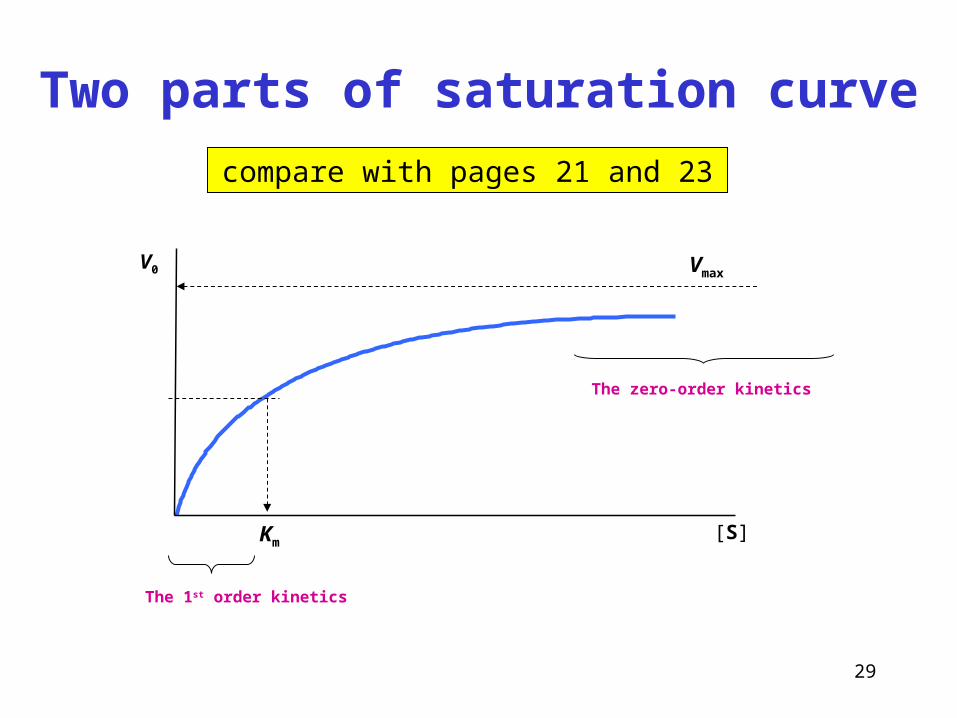
Two parts of saturation curve
compare with pages 21 and 23
[S]
V0 Vmax
Km
The zero-order kinetics
The 1st order kinetics
30
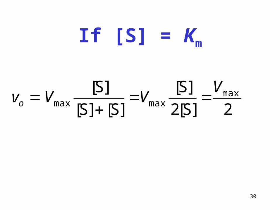
If [S] = Km
2]S[2
]S[
]S[]S[
]S[ maxmaxmax
VVVvo

31
Significance of Km and Vmax
• the Michaelis constant Km is the concentration of substrate S which gives half
the maximal velocity Vmax (50 % saturation of enzyme)
• the Km has the dimension of concentration (mol/l)
• Km is inversely related to the affinity of enzyme for its substrate.
If more substrates with similar structure exist, then the best natural substrate is
one with the least value of Km
• if there is a need to measure the activity of enzyme, the substrate concentration
has to be at least several times higher than the Km value.
32
[P]
t
[S1]
[S2]
[S3]
[S4]
v0 1v0 2
v0 3v0 4
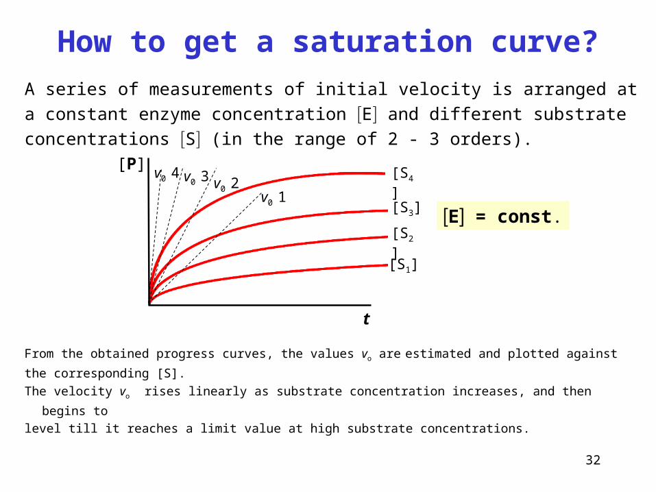
From the obtained progress curves, the values vo are estimated and plotted against
the corresponding [S].
The velocity vo rises linearly as substrate concentration increases, and then begins to
level till it reaches a limit value at high substrate concentrations.
How to get a saturation curve?
E = const.
A series of measurements of initial velocity is arranged at a constant enzyme
concentration E and different substrate concentrations S (in the range of 2 - 3 orders).
33
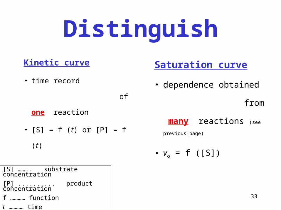
Distinguish
Kinetic curve
• time record
of one reaction
• [S] = f (t) or [P] = f (t)
Saturation curve
• dependence obtained
from many
reactions (see previous page)
• vo = f ([S])[S] …….. substrate concentration
[P] .......... product concentration
f ………… function
t ………… time
vo ……….. initial velocity
34
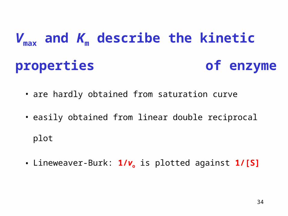
Vmax and Km describe the kinetic properties
of enzyme
• are hardly obtained from saturation curve
• easily obtained from linear double reciprocal plot
• Lineweaver-Burk: 1/vo is plotted against 1/[S]
35
mo K
Vv
]S[
]S[max
]S[]S[
]S[1
]S[
]S[11
maxmax
mm
o
K
V
K
Vv
]S[
111
maxmax
V
K
Vvm
o
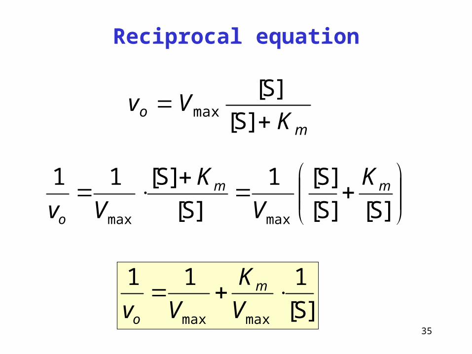
Reciprocal equation
36
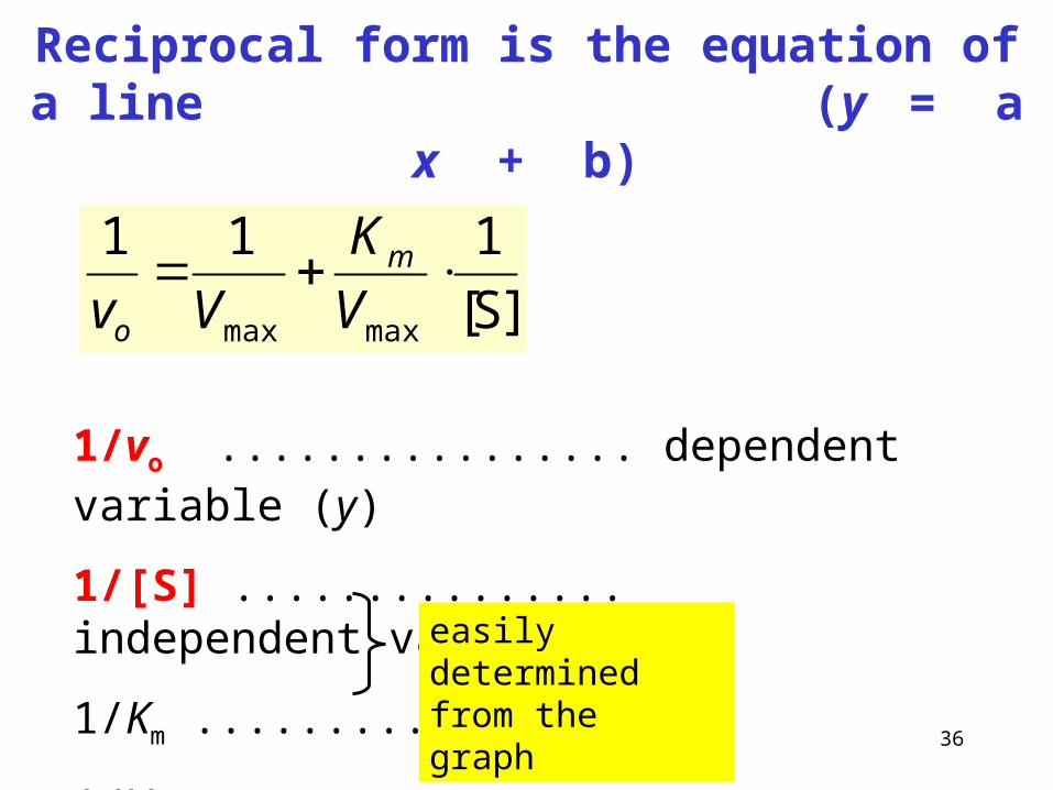
Reciprocal form is the equation of a line (y = a x + b)
]S[
111
maxmax
V
K
Vvm
o
1/vo ................ dependent variable (y)
1/[S] ............... independent variable (x)
1/Km ...............
1/Vmax .............
easily determined from the graph
37
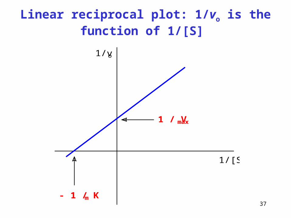
Linear reciprocal plot: 1/vo is the function of 1/[S]
1/vo
1/[S]
1 / Vmax
- 1 / Km
38
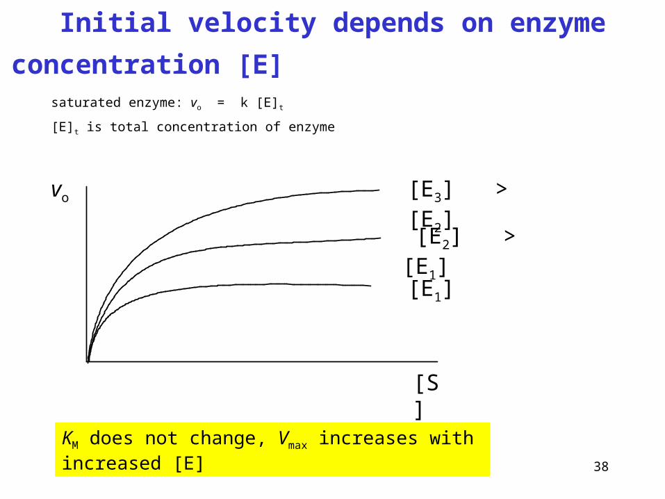
vo
[S]
[E1]
[E2] > [E1]
[E3] > [E2]
KM does not change, Vmax increases with increased [E]
Initial velocity depends on enzyme concentration [E]
saturated enzyme: vo = k [E]t
[E]t is total concentration of enzyme
39
How to quantify enzymes
in biological material?
• very low concentrations of enzymes
• in very complex mixtures
• in the presence of many other proteins
• simple chemical reactions cannot be used, they are not
specific enough to distinguish individual enzymes
40
Enzyme quantity in a biological sample
can be determined by two ways
• catalytic concentration
• μkat/l
• the product of enzyme
reaction is determined
• mass concentration
• μg/l
• enzyme itself is determined
as antigen
(immunochemical assay)
41
Catalytic activity of enzyme
• unit katal, 1 kat = mol/s
• such amount of enzyme that catalyzes the conversion
of one mole of substrate per one second IU (international unit), 1 IU = μmol/min
1 μkat = 60 IU, 1 IU = 16.6 nkat
Catalytic concentration of enzyme
• activity related to the volume of body fluid (blood serum)
• typical units: mkat/l, kat/l
42
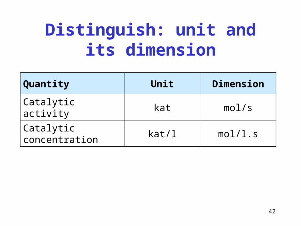
Distinguish: unit and its dimension
Quantity Unit Dimension
Catalytic activity kat mol/s
Catalytic concentration kat/l mol/l.s
43
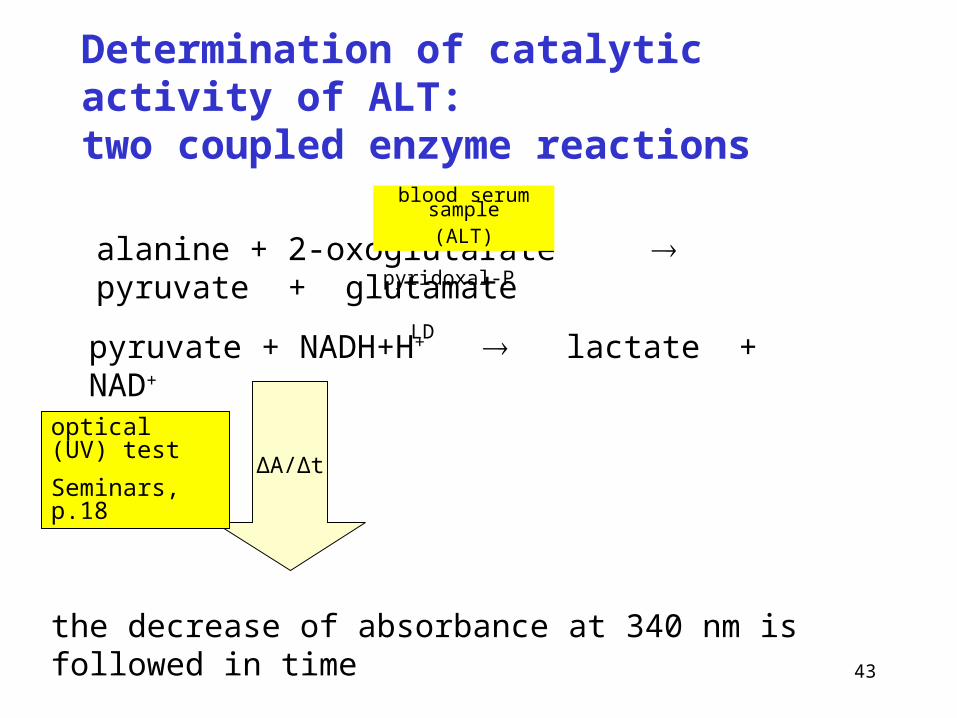
Determination of catalytic activity of ALT: two coupled enzyme reactions
alanine + 2-oxoglutarate pyruvate + glutamate
blood serum sample(ALT)
pyridoxal-P
pyruvate + NADH+H+ lactate + NAD+
ΔA/Δt
optical (UV) test
Seminars, p.18
the decrease of absorbance at 340 nm is followed in time
LD
44
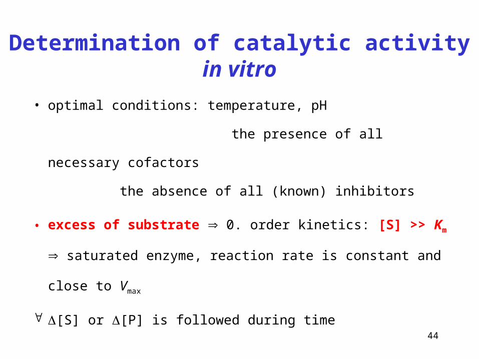
Determination of catalytic activity in vitro
• optimal conditions: temperature, pH
the presence of all necessary cofactors
the absence of all (known) inhibitors
• excess of substrate 0. order kinetics: [S] >> Km saturated
enzyme, reaction rate is constant and close to Vmax
[S] or [P] is followed during time
45
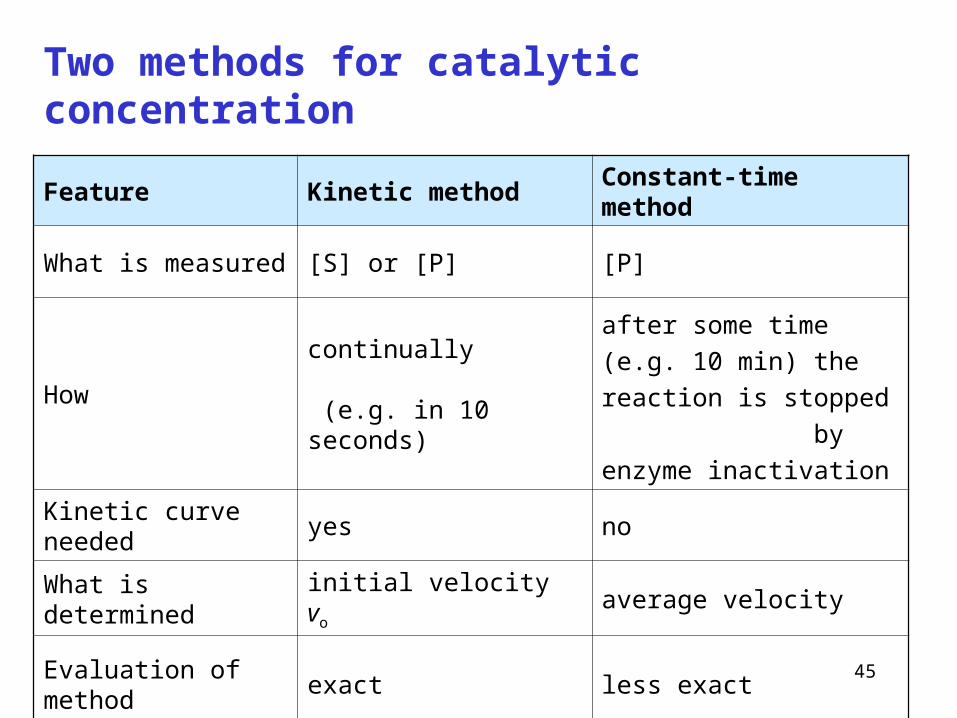
Two methods for catalytic concentration
Feature Kinetic method Constant-time method
What is measured [S] or [P] [P]
Howcontinually (e.g. in 10 seconds)
after some time (e.g. 10 min)
the reaction is stopped
by enzyme inactivation
Kinetic curve needed yes no
What is determined initial velocity vo average velocity
Evaluation of method exact less exact
46
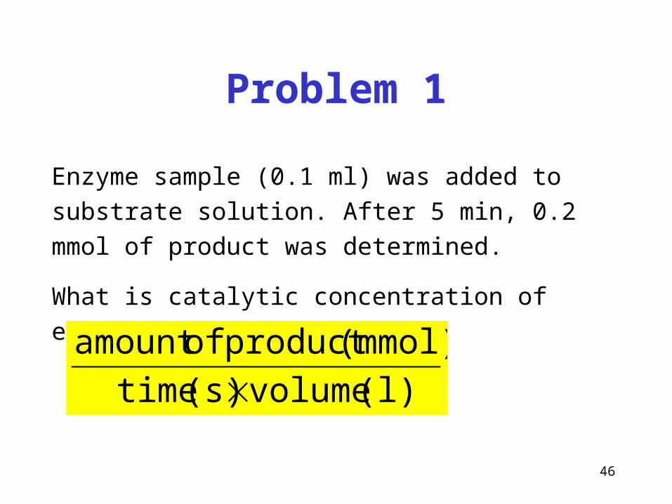
Problem 1
Enzyme sample (0.1 ml) was added to substrate solution.
After 5 min, 0.2 mmol of product was determined.
What is catalytic concentration of enzyme?
(l) volume (s) time
(mmol)product ofamount
47
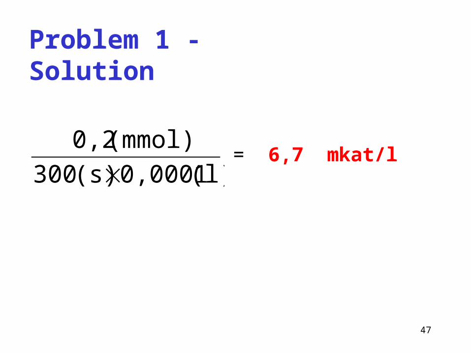
Problem 1 - Solution
(l) 0,0001 (s) 300
(mmol) 0,2
= 6,7 mkat/l
48
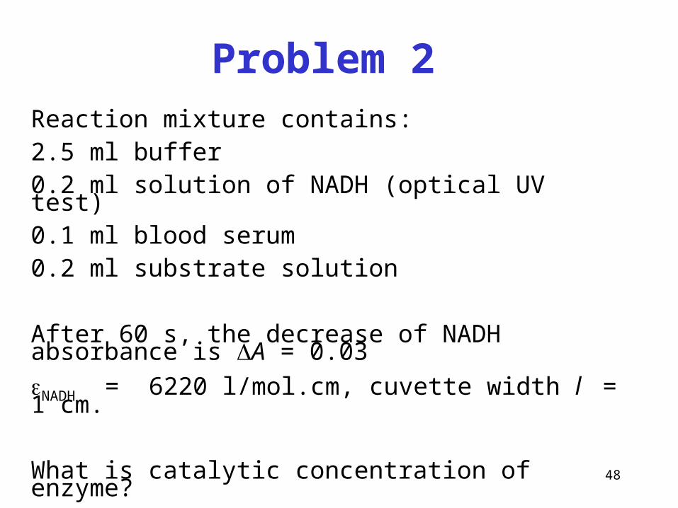
Problem 2Reaction mixture contains: 2.5 ml buffer 0.2 ml solution of NADH (optical UV test)0.1 ml blood serum 0.2 ml substrate solution
After 60 s, the decrease of NADH absorbance is A = 0.03
NADH = 6220 l/mol.cm, cuvette width l = 1 cm. What is catalytic concentration of enzyme?
49
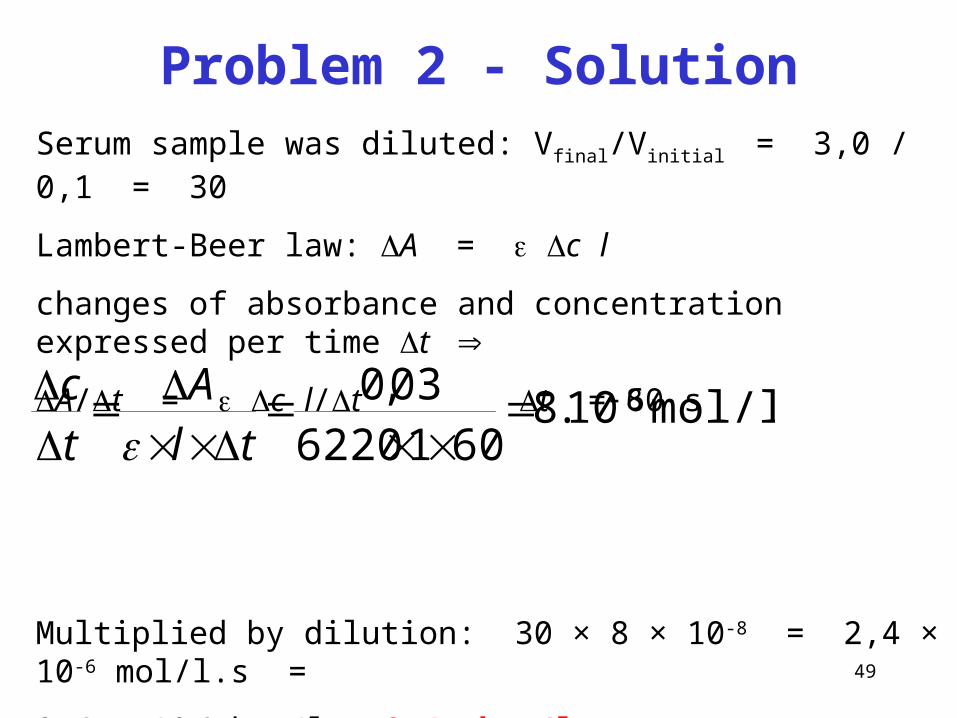
Problem 2 - SolutionSerum sample was diluted: Vfinal/Vinitial = 3,0 / 0,1 = 30
Lambert-Beer law: A = c l
changes of absorbance and concentration expressed per time t
A/t = c l/t t = 60 s
Multiplied by dilution: 30 × 8 × 10-8 = 2,4 × 10⁻⁶ mol/l.s =
2,4 × 10⁻⁶ kat/l = 2,4 kat/l
mol/l.s10.86016220
03,0 8
tlA
tc
50
Consider that
catalytic concentration = rate of chemical reaction
kat/l = mol/l.s
51
Irreversible inhibitors
Irreversible inhibitors are usually compounds not of biological origin,
bind onto enzyme mostly covalently and make substrate binding impossible.
Heavy metal ions, organophosphates, cyanides
bind and inhibit irreversibly enzymes during isolation.
Inhibitors reduce enzyme activity
Reversible inhibitors
In contrast with irreversible inhibitors, reversible inhibitors bind to the enzyme
loosely and can rapidly dissociate from the enzyme-inhibitor complex.
These inhibitors are classified as competitive, non-competitive.
52
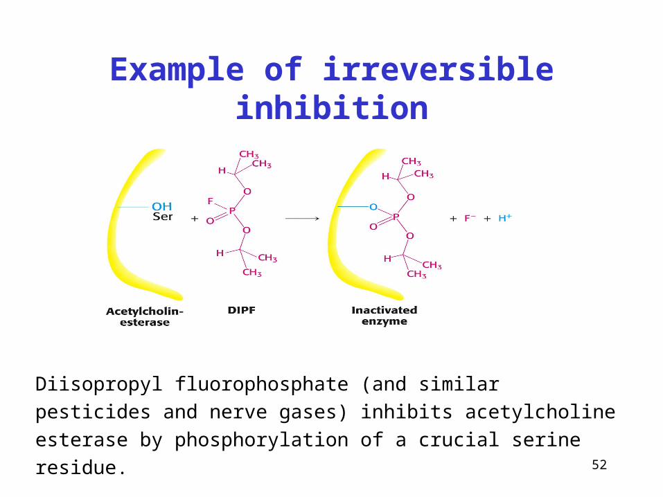
Example of irreversible inhibition
Diisopropyl fluorophosphate (and similar pesticides and nerve gases)
inhibits acetylcholine esterase by phosphorylation of a crucial serine
residue.
53
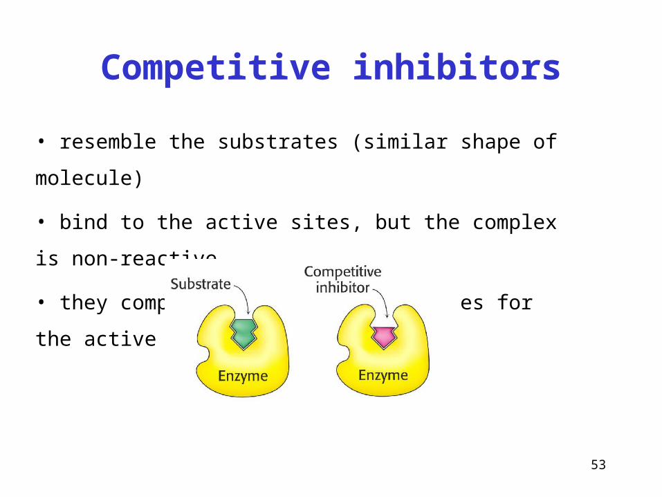
• resemble the substrates (similar shape of molecule)
• bind to the active sites, but the complex is non-reactive
• they compete with normal substrates for the active sites
Competitive inhibitors
54
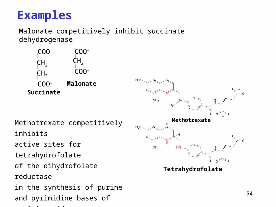
Malonate competitively inhibit succinate dehydrogenase
Examples
Methotrexate
Tetrahydrofolate
Methotrexate competitively inhibits
active sites for tetrahydrofolate
of the dihydrofolate reductase
in the synthesis of purine and
pyrimidine bases of nucleic acids.
It is used to treat cancer.
Malonate
Succinate
COO–
CH2
CH2
COO–
COO–
CH2
COO–
55
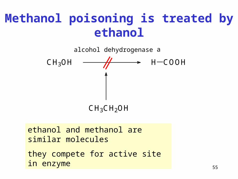
Methanol poisoning is treated by ethanol
CH3OH H COOH
alkoholdehydrogenasa
CH3CH2OH
ethanol and methanol are similar molecules
they compete for active site in enzyme
alcohol dehydrogenase
56
Km [S]
v0
Vmax
Km
1 / v0
1 / [S]
1 / Vmax
- 1 / Km
No inhibitor
Competitive inhibitor
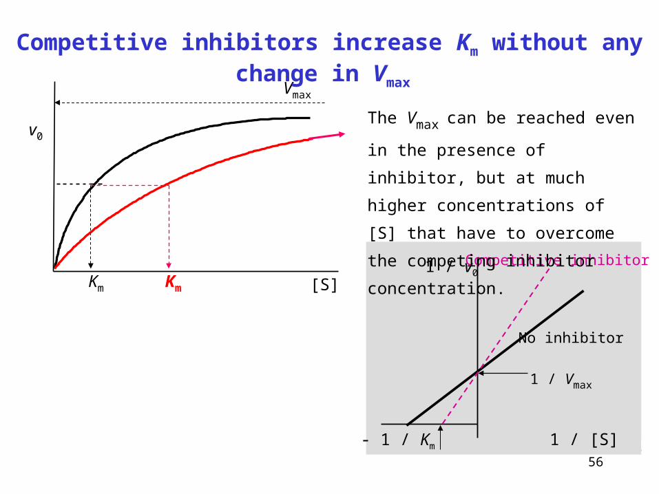
Competitive inhibitors increase Km without any change in Vmax
The Vmax can be reached even in the
presence of inhibitor, but at much higher
concentrations of [S] that have to
overcome the competing inhibitor
concentration.
57
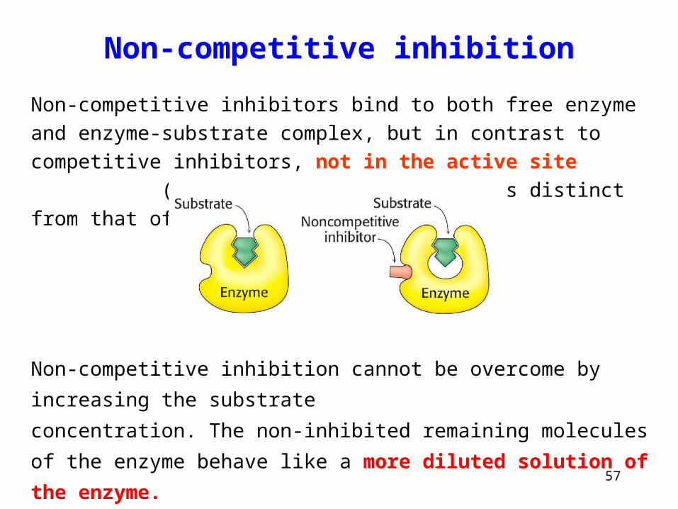
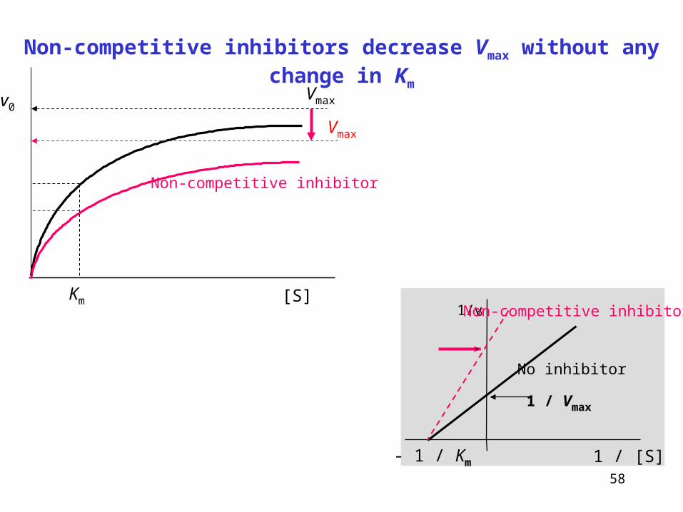
Non-competitive inhibition
Non-competitive inhibitors bind to both free enzyme and enzyme-substrate
complex, but in contrast to competitive inhibitors, not in the active site
(the structure of inhibitor is distinct from that of substrate).
Non-competitive inhibition cannot be overcome by increasing the substrate
concentration. The non-inhibited remaining molecules of the enzyme behave
like a more diluted solution of the enzyme.
58
1/vo
1 / [S]
1 / Vmax
- 1 / Km
Non-competitive inhibitor
No inhibitor
Km [S]
v0Vmax
Vmax
Non-competitive inhibitor
Non-competitive inhibitors decrease Vmax without any change in Km
59
Many drugs are inhibitors of enzymes
• Acetylsalicylic acid, ibuprofen (cyclooxygenase) - see Seminars, p. 67
• Statins (HMG-CoA reductase) inhibit cholesterol synthesis (e.g. lovastatin)
• ACE Inhibitors (angiotensin-converting enzyme) – treatment of hypertension
• Reversible acetylcholinesterase inhibitors (e.g. neostigmine) – myastenia gravis,
post-surgical atonia,
• Brain acetylcholinesterase inhibitors (rivastigmine, galantamin) – Alzheimer d.
• Many antibiotics inhibit bacterial enzymes:
Penicillins – inhibit bacterial transpeptidases (formation of bacterial cell-wall)
Tetracyclins, macrolides, chloramphenicol – inhibit bacterial proteosynthesis
Fluoroquinolones (ciprofloxacin) – inhibit bacterial topoisomerase
60
1. Regulation of enzyme quantity
2. Regulation of enzyme activity
3. Availability and concentration of substrate and/or cofactor
(in vivo less
important, in vitro critical condition)
Regulation principles in enzyme reactions (three general aspects)
61
Regulation of enzyme quantity
• Controlled enzyme proteosynthesis
constitutive and induced gene expresion,
regulation of transcription rate,
posttranscriptional RNA processing,
regulation of translation rate and posttranslational
modifications
• Controlled enzyme degradation
specific intracellular proteinases determine
biological half-life of enzymes
62
Regulation of enzyme activity
• Activation by partial and irreversible proteolysis
• Reversible covalent modification
• Allosteric regulation
63
Enzyme activation by partial proteolysis
• Active enzyme is formed by irreversible cleavage of
certain sequence from proenzyme (zymogen) molecule
• Proteinases in digestion (pepsinogen pepsin)
• Factors of blood coagulation
• Proteinases caspases in apoptosis
64
Reversible covalent enzyme modification
• phosphorylation, catalyzed by kinases
• transfer of phosphoryl -PO32- from ATP to -OH group of enzyme
(Ser, Thr, Tyr)
• reversible process, dephosphorylation catalyzed by
phosphatase, hydrolysis of phosphoester
• Other modifications: carboxylation, acetylation, prenylation ...
65
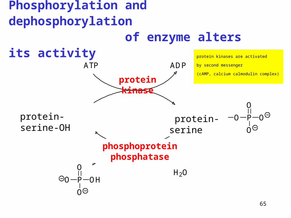
Phosphorylation and dephosphorylation of enzyme alters its activity
Enzym Serin OH Enzym Serin O P
O
O
O
ATP ADP
H2OP
O
O
OHO
protein kinase
phosphoprotein phosphatase
protein-serine-OH protein-serine
protein kinases are activated
by second messenger
(cAMP, calcium calmodulin complex)
66
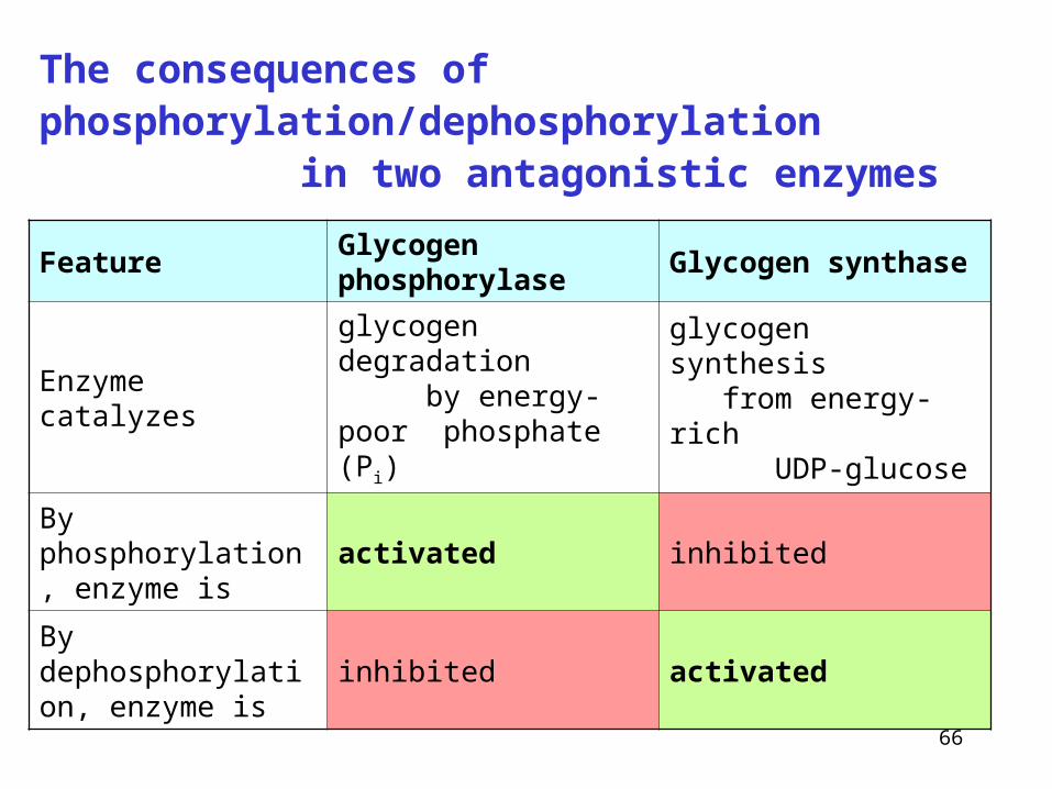
The consequences of phosphorylation/dephosphorylation in two antagonistic enzymes
Feature Glycogen phosphorylase Glycogen synthase
Enzyme catalyzesglycogen degradation by energy-poor phosphate (Pi)
glycogen synthesis from energy-rich UDP-glucose
By phosphorylation, enzyme is
activated inhibited
By dephosphorylation, enzyme is
inhibited activated
67
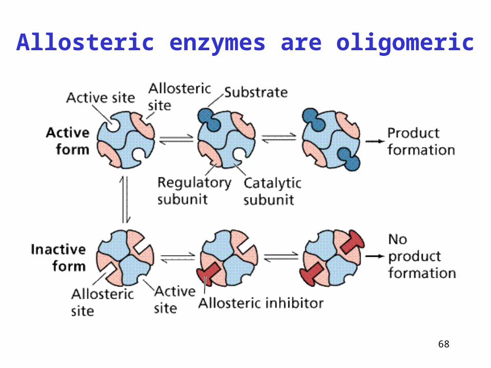
Allosteric enzymes are oligomeric
• more subunits, often regulatory and catalytic
• effector, structurally different from substrate - often product,
binds to enzyme to allosteric site (other than active site)
• binding effector triggers the changes in conformation and
activity allosteric activation / inhibition
68
Allosteric enzymes are oligomeric
69
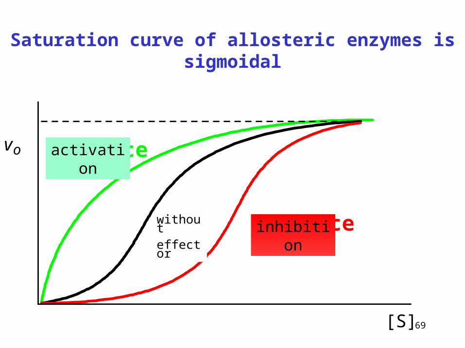
Saturation curve of allosteric enzymes is sigmoidal
[S]
vo aktivace
inhibicebez
efektoru
activation
without effector
inhibition
70
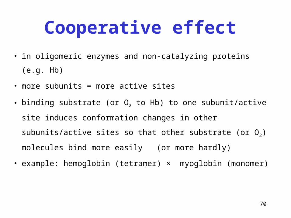
Cooperative effect
• in oligomeric enzymes and non-catalyzing proteins (e.g. Hb)
• more subunits = more active sites
• binding substrate (or O2 to Hb) to one subunit/active site
induces conformation changes in other subunits/active sites so
that other substrate (or O2) molecules bind more easily (or
more hardly)
• example: hemoglobin (tetramer) × myoglobin (monomer)
71
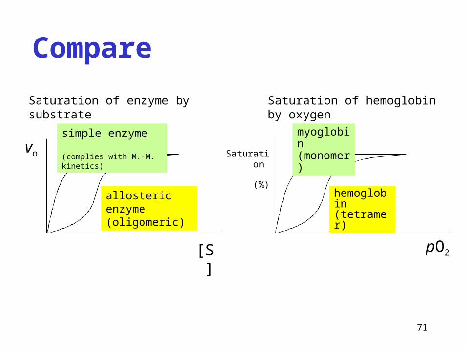
Compare
Saturation of enzyme by substrate Saturation of hemoglobin by oxygen
vo
[S] pO2
Saturation
(%)
myoglobin (monomer)
hemoglobin (tetramer)
allosteric enzyme (oligomeric)
simple enzyme (complies with M.-M. kinetics)
72
Three utilizations of enzymes in medicine
1. enzymes as indicators of pathological condition
2. enzymes as analytic reagents in clinical chemistry
3. enzymes as drugs
73
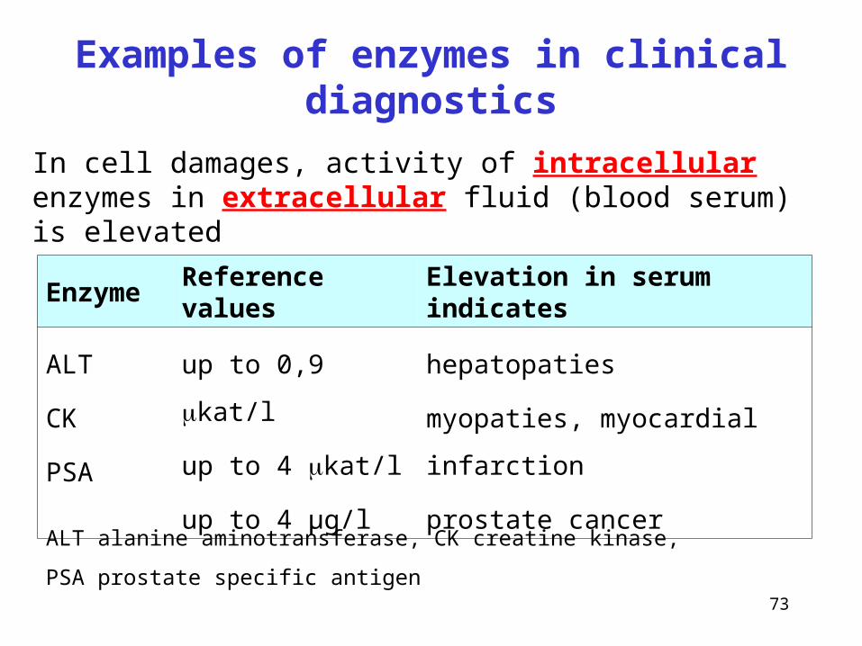
Examples of enzymes in clinical diagnostics
Enzyme Reference values Elevation in serum indicates
ALT
CK
PSA
up to 0,9 kat/l
up to 4 kat/l
up to 4 μg/l
hepatopaties
myopaties, myocardial infarction
prostate cancer
ALT alanine aminotransferase, CK creatine kinase,
PSA prostate specific antigen
In cell damages, activity of intracellular enzymes in extracellular fluid (blood serum) is elevated
74
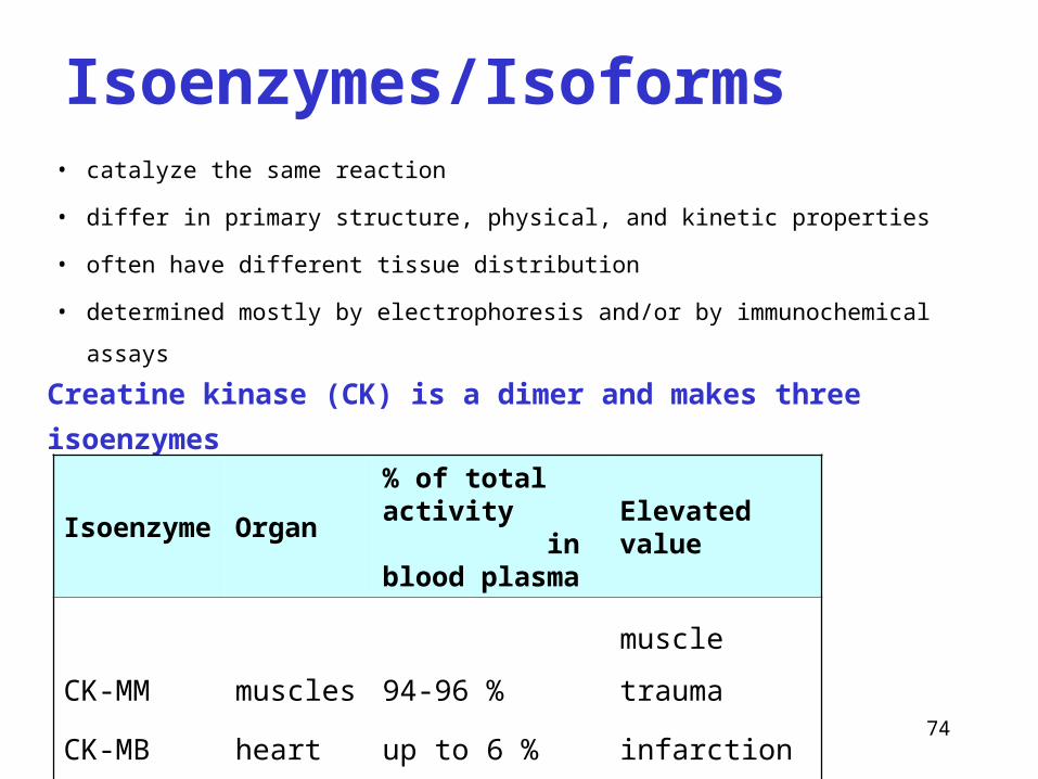
Creatine kinase (CK) is a dimer and makes three isoenzymes
Isoenzyme Organ% of total activity in blood plasma
Elevated value
CK-MM
CK-MB
CK-BB
muscles
heart
brain
94-96 %
up to 6 %
traces
muscle trauma
infarction
brain damage
• catalyze the same reaction
• differ in primary structure, physical, and kinetic properties
• often have different tissue distribution
• determined mostly by electrophoresis and/or by immunochemical assays
Isoenzymes/Isoforms
75
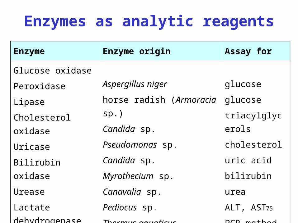
Enzymes as analytic reagents
Enzyme Enzyme origin Assay for
Glucose oxidase
Peroxidase
Lipase
Cholesterol oxidase
Uricase
Bilirubin oxidase
Urease
Lactate dehydrogenase
Taq polymerase
Aspergillus niger
horse radish (Armoracia sp.)
Candida sp.
Pseudomonas sp.
Candida sp.
Myrothecium sp.
Canavalia sp.
Pediocus sp.
Thermus aquaticus
glucose
glucose
triacylglycerols
cholesterol
uric acid
bilirubin
urea
ALT, AST
PCR method
76
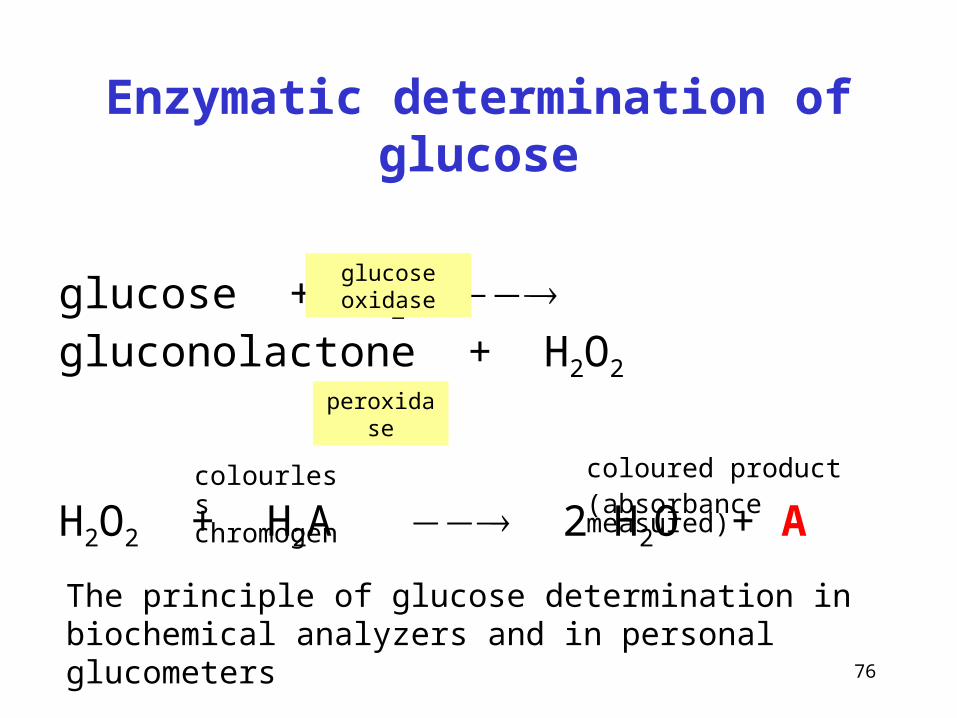
Enzymatic determination of glucose
glucose + O2 gluconolactone + H2O2
H2O2 + H2A 2 H2O + A
glucose oxidase
peroxidase
colourless chromogen
coloured product(absorbance measured)
The principle of glucose determination in biochemical analyzers and in personal glucometers
77
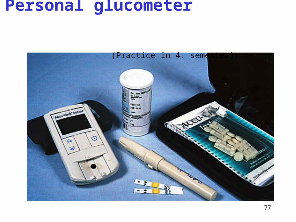
Personal glucometer
(Practice in 4. semester)
78
Pancreatic enzymes in therapy
• enzyme mixtures (lipase, amylase, proteinases)
of animal origin
• indication: insufficient secretion of pancreas,
cystic fibrosis
• 3 × times daily a capsule during meal
gastro-resistant capsules, they survive the passage through stomach, soluble and active in duodenum
79
Asparaginase in leukemia treatment
• Catalyzes the hydrolysis of asparagine amide group (deamidation)
• Asn + H2O Asp + NH3
• L-asparagine is necessary for the proteosynthesis of some cancer cells
• Hydrolysis of Asp reduces the cell proliferation (see also Seminars, p. 19)
Enzyme fibrinolytics
• thrombolytic drugs, dissolve blood clots in veins
• urokinase (human enzyme, serine protease)
• converts plasminogen to plasmin which degrades fibrin
thrombolysis
• venous thrombosis, pulmonary embolism, acute myocardial infarction
80
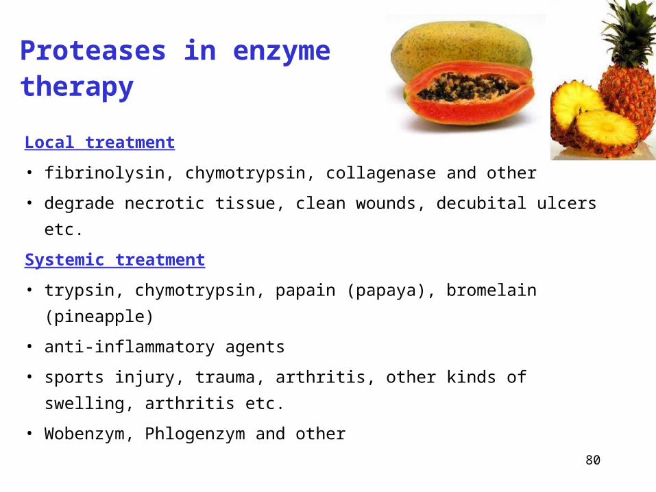
Proteases in enzyme therapy
Local treatment
• fibrinolysin, chymotrypsin, collagenase and other
• degrade necrotic tissue, clean wounds, decubital ulcers etc.
Systemic treatment
• trypsin, chymotrypsin, papain (papaya), bromelain (pineapple)
• anti-inflammatory agents
• sports injury, trauma, arthritis, other kinds of swelling, arthritis etc.
• Wobenzym, Phlogenzym and other