Embed Size (px)
Citation preview
1
ADDITION BY ME
CHAPTER 20 (GENES X)
PROMOTERS AND ENHANCERS
2
Eukaryotic gene control: purposes and general principles
• Unlike bacterial cells and most single cell eukaryotes, cells in multicellular organisms have relatively few genes that are reversibly regulated by environmental conditions
• However, gene control in multicellular organisms is important for development and differentiation, and is generally not reversible
3
Regulatory elements in eukaryotic DNA often are many kilobases from start sites
• The basic principles that control transcription in bacteria also
apply to eukaryotic organisms: transcription is initiated at a
specific base pair and is controlled by the binding of trans-
acting proteins (transcription factors) to cis-acting regulatory
DNA sequences
• However, eukaryotic cis-acting elements are often much
further from the promoter they regulate, and transcription
from a single promoter may be regulated by binding of
multiple transcription factors to alternative control elements.
4
Promoter sequence components can beidentified by:
Transcription control sequences can be identified by analysis of a 5’-deletion series.
Finding precise consensus sequence that is bound by a particular factor in multiple promoters.
Introduction of point mutations at particular base pairs and testing the promoter function.
Finding the proteins which bound to promoters by footprinting and mutation.
5
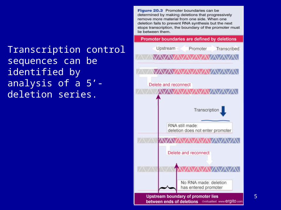
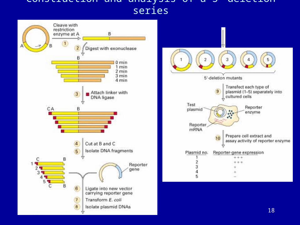
Transcription control sequences can be identified by analysis of a 5’-deletion series.
6
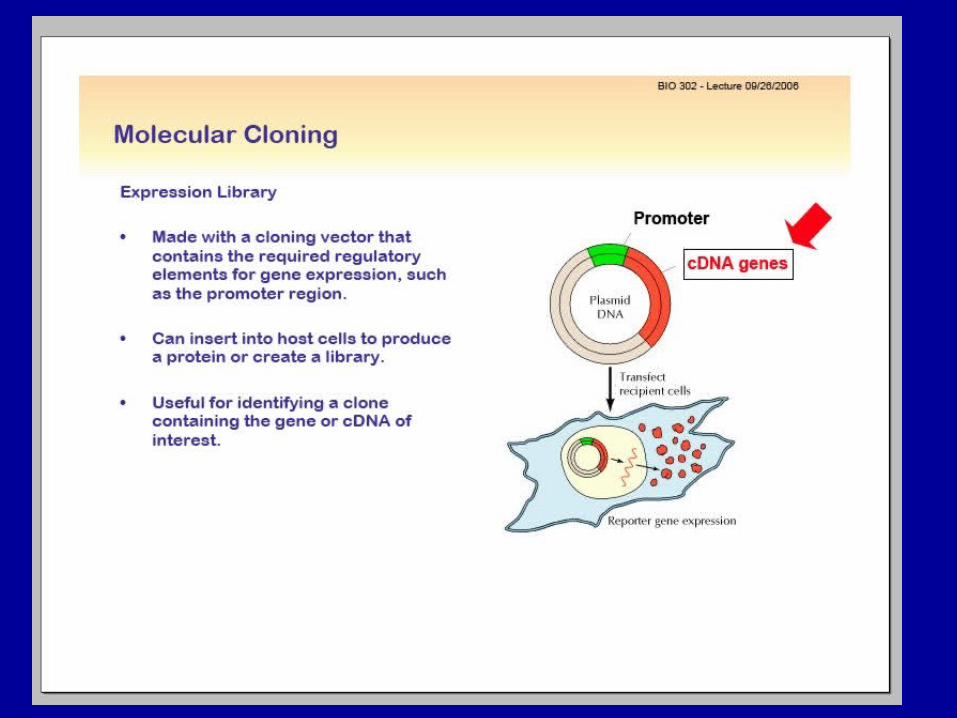
REPORTER GENES
The primary objective is to identify gene regulatory sequences by testing a large number of deletion and point mutants in an in vivo cell transfection assays.
Promoter mapping studies can be done by creating artificial “reporter gene” by fusing regulatory region of the test gene to a heterologous gene coding sequences that direct the synthesis of a readily detectable protein product.
The steady state level of the reporter protein in the transfected cell is directly correlated to the steady state level of reporter mRNA, then it is possible to use protein based reporter assays for promoter mapping experiments.
7
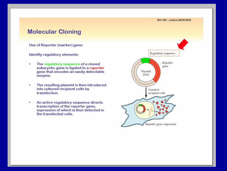
Repoter gene constructs
When trying to determine what are the cis-acting regulatory regions
that control the expression (transcription) levels of a gene, is usually
routine to clone a large piece of DNA containing these sequences and
hook it to a downstream sequence that encodes an easy-to-measure
protein called a reporter.
This is a lot more convenient and accurate than trying to estimate the
absolute levels of mRNA.
In vivo transgene reporter expression driven by the promoter of
interest will reveal the temporal/spatial pattern of expression of the
gene, measured by an easier method than the alternative in situ
hybridization.
8
Reporter genes:
1. must encode a protein activity that is
similar to one already present in the cell.
2. The protein assay should be sensitive
enough, reproducible and easy to perform.
3. The reporter protein function should not
interfere with host cellular processes in a way
that will alter intracellular signaling pathways
or metabolic rates.
9
Repoter gene constructs:
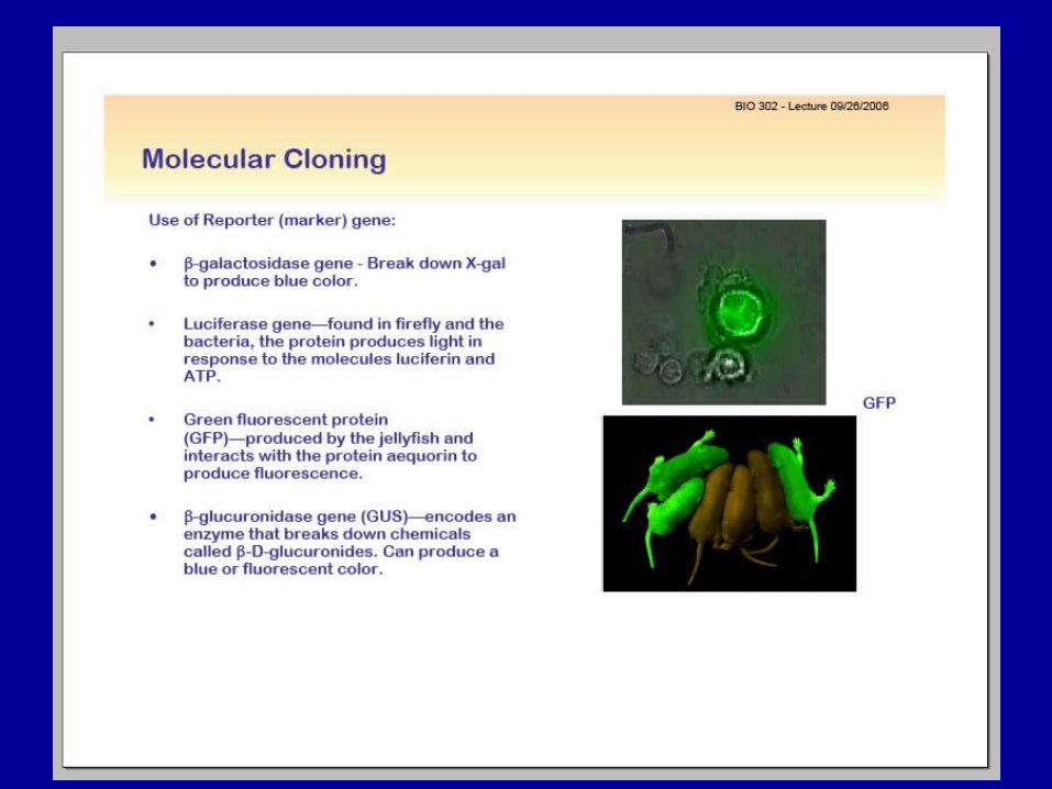
The most used reporters are:bacterial -galactosidase,chloramfenicol acetyl transferase (CAT),fire-fly luciferaseand most recently, the green fluorescent protein (GFP) which
allows measurements in living cells or organisms.
Part of the whole of a protein rather than a promoter can also be fused in frame to this reporters to find out where it localizes. Reporter gene must not being expressed endogenously in the cell types that you are working on, so their background should be null.
10
11
12
13
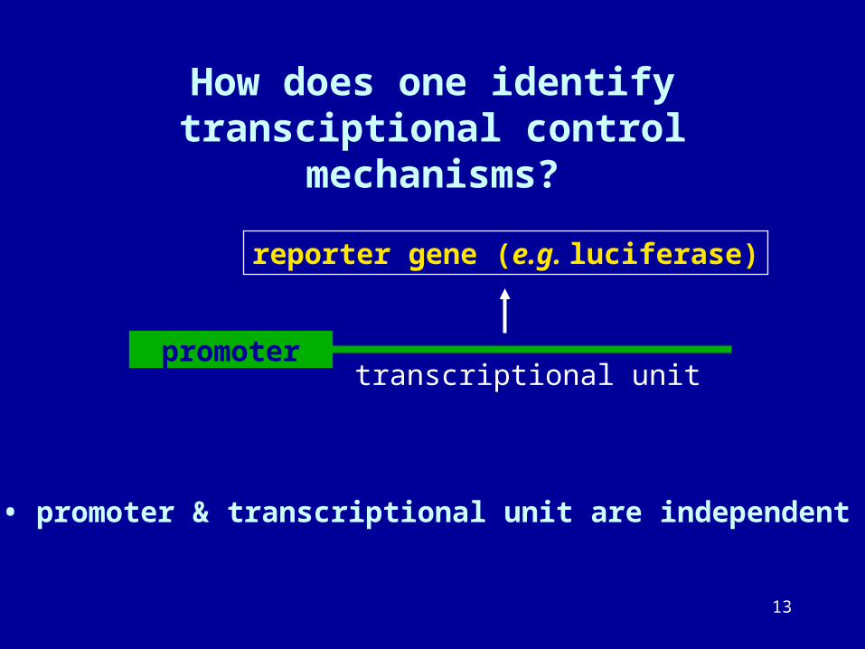
How does one identify transciptional control mechanisms?
promotertranscriptional unit
• promoter & transcriptional unit are independent
reporter gene (e.g. luciferase)
14
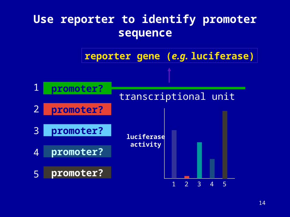
Use reporter to identify promoter sequence
promoter?transcriptional unit
reporter gene (e.g. luciferase)
promoter?
promoter?
promoter?
promoter?
1
2
3
4
5
luciferaseactivity
1 2 3 4 5
15
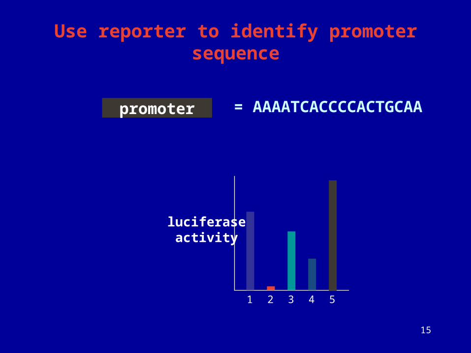
Use reporter to identify promoter sequence
promoter
luciferaseactivity
1 2 3 4 5
= AAAATCACCCCACTGCAA
16
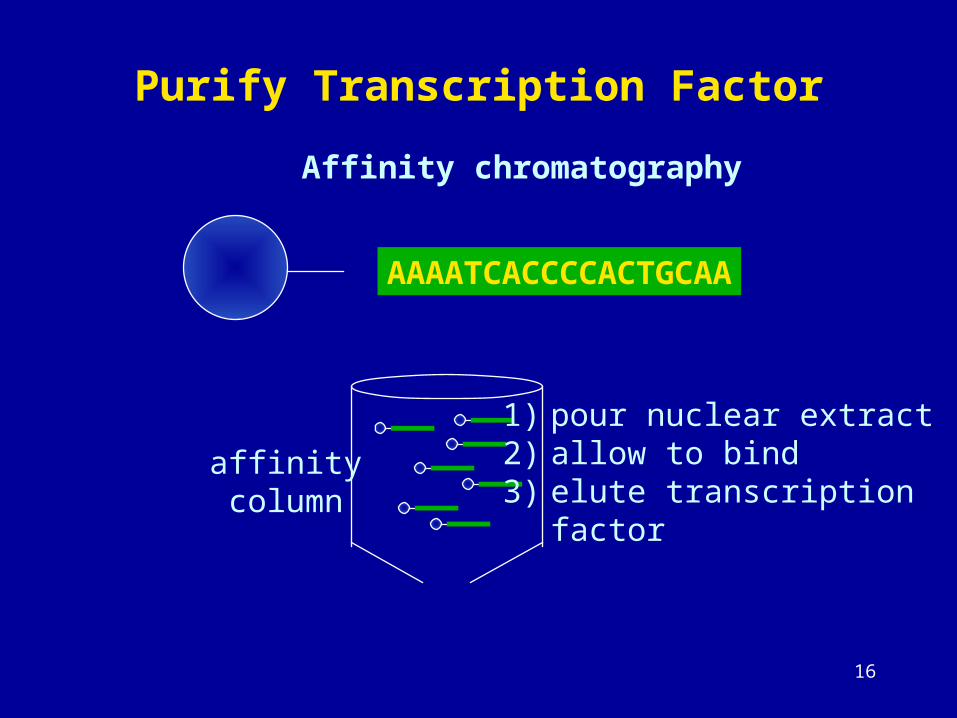
Purify Transcription Factor
AAAATCACCCCACTGCAA
Affinity chromatography
affinitycolumn
1) pour nuclear extract2) allow to bind3) elute transcription
factor
17
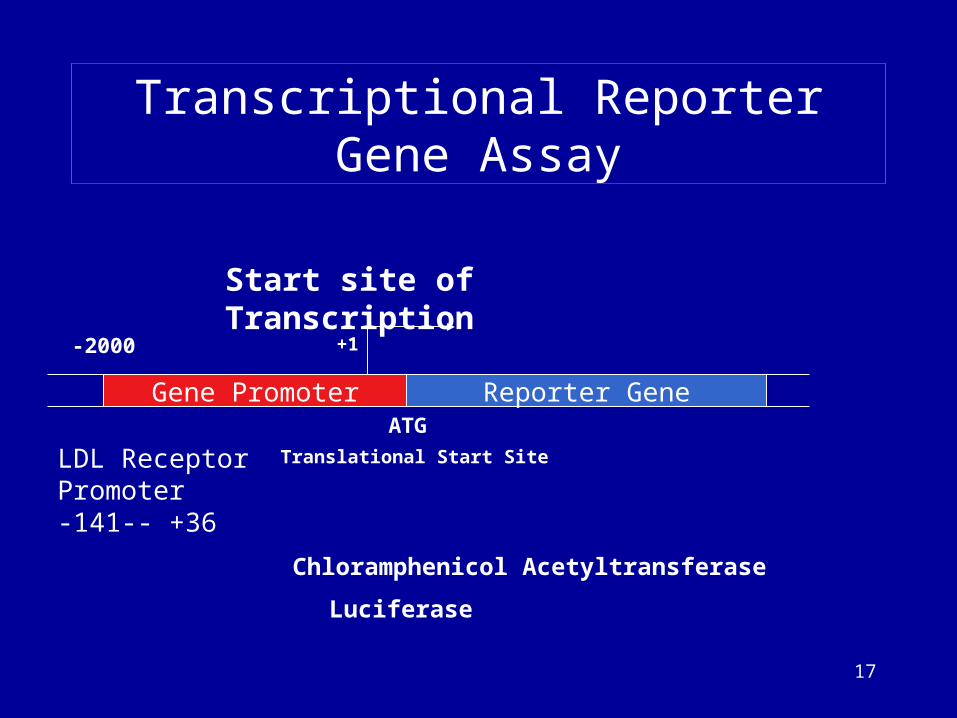
Gene Promoter Reporter Gene
Start site of Transcription
Transcriptional Reporter Gene Assay
+1
Translational Start Site
ATG
-2000
Chloramphenicol Acetyltransferase
Luciferase
LDL Receptor Promoter-141-- +36
18
Construction and analysis of a 5-deletion series
19
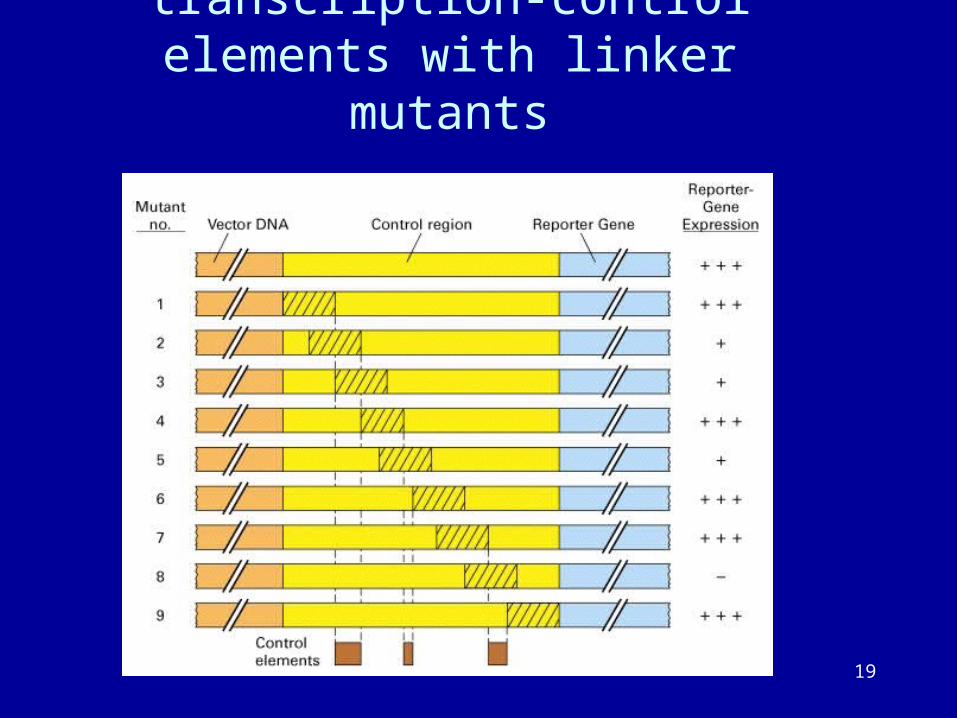
Identification of transcription-control elements with linker mutants
20
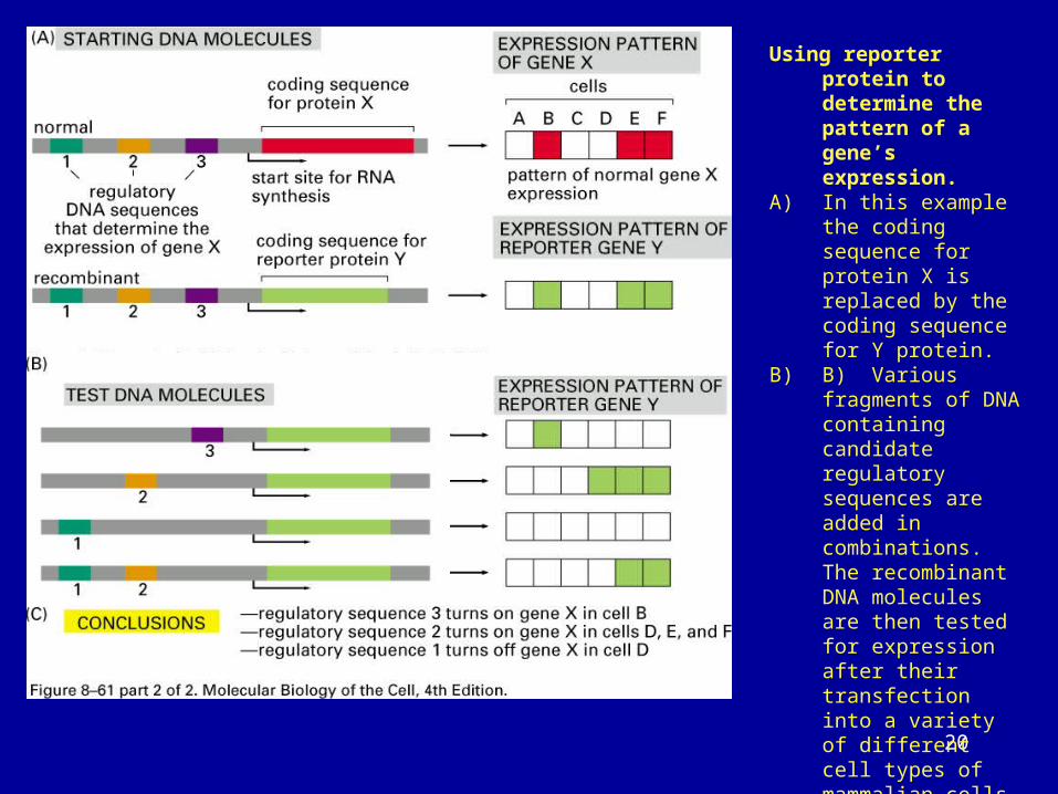
asUsing reporter protein to
determine the pattern of a gene’s expression.
A) In this example the coding sequence for protein X is replaced by the coding sequence for Y protein.
B) B) Various fragments of DNA containing candidate regulatory sequences are added in combinations. The recombinant DNA molecules are then tested for expression after their transfection into a variety of different cell types of mammalian cells, and the result is summarized in C.
For experiments in eukaryotic cells, two commonly used reporter proteins are enzymes b-
galactosidase (b-gal) and green fluorescent protein or GFP.
21
METHODS FOR STUDYING DNA-PROTEIN
INTERACTIONS
A. DNA footprinting
B. Electrophoretic mobility shift assay
C. Chromatin immunoprecipitation assay
22
DNA FOOTPRINTING
identify protein-DNA interactions
23
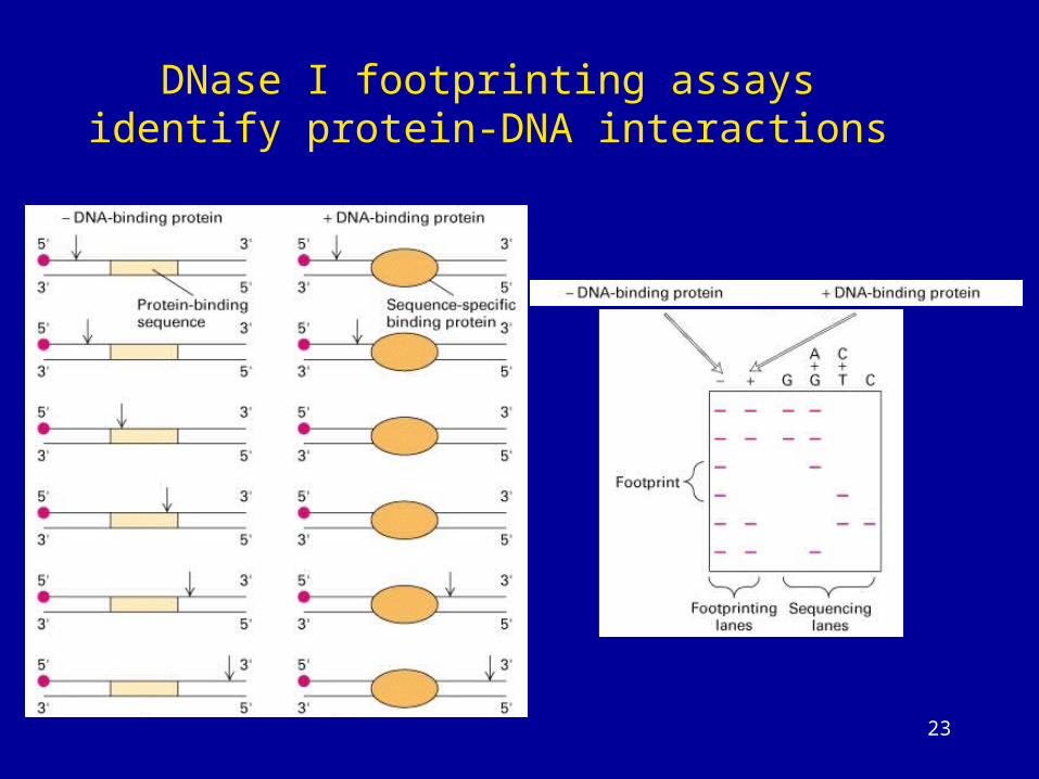
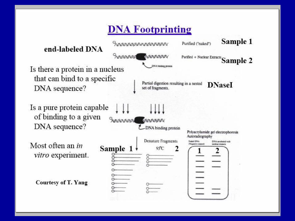
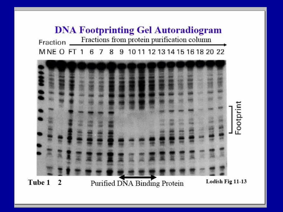
DNase I footprinting assays identify protein-DNA interactions
24
25
26
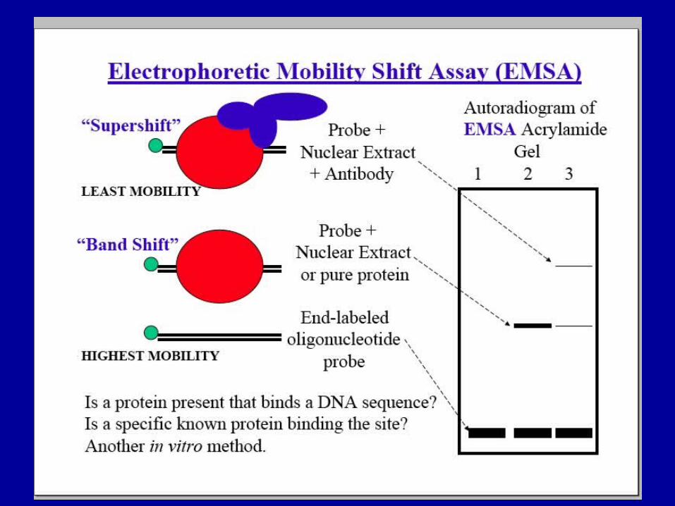
Electrophoretic mobility shift assay
identify protein-DNA interactions
27
28
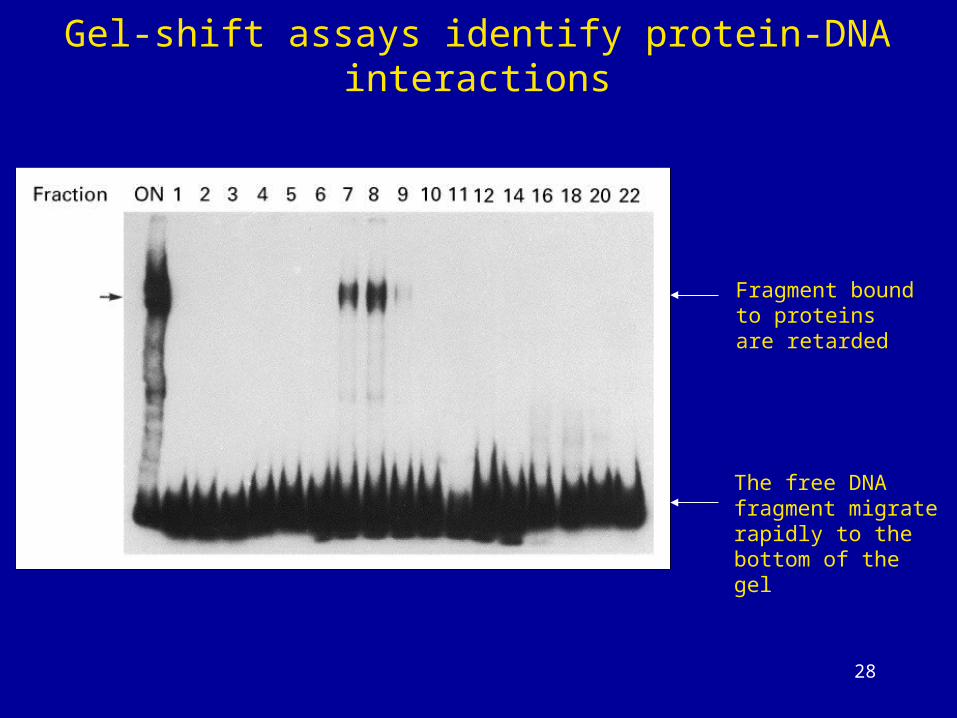
Gel-shift assays identify protein-DNA interactions
Fragment bound to proteins are retarded
The free DNA fragment migrate rapidly to the bottom of the gel
29
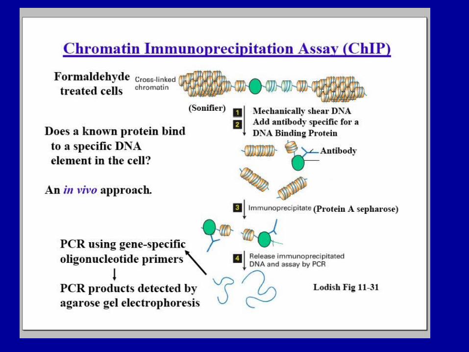
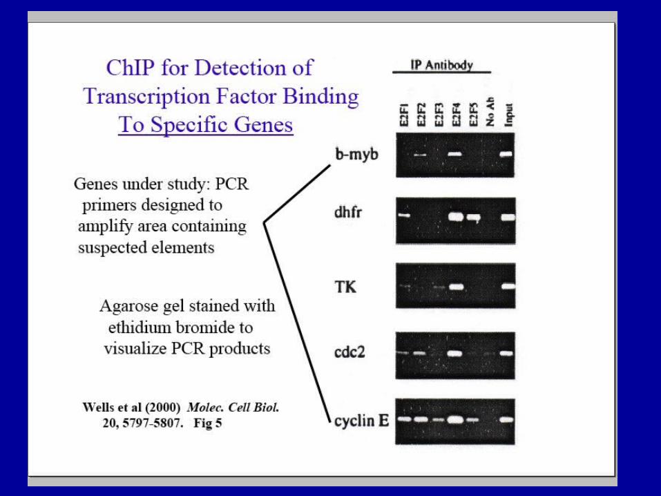
ChIP (Chromatin ImmunoPrecipitation)
Chromatin immunoprecipitation, or ChIP, refers to
a procedure used to determine whether a given
protein binds to a specific DNA sequence in vivo
30
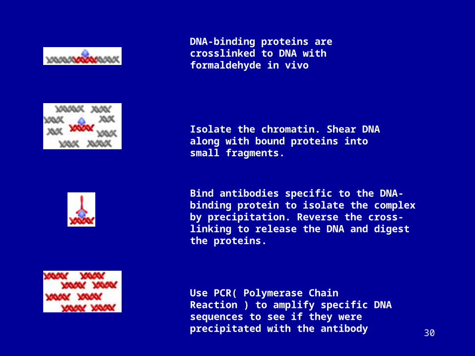
DNA-binding proteins are crosslinked to DNA with formaldehyde in vivo
Isolate the chromatin. Shear DNA along with bound proteins into small fragments.
Bind antibodies specific to the DNA-binding protein to isolate the complex by precipitation. Reverse the cross-linking to release the DNA and digest the proteins.
Use PCR( Polymerase Chain Reaction ) to amplify specific DNA sequences to see if they were precipitated with the antibody
31
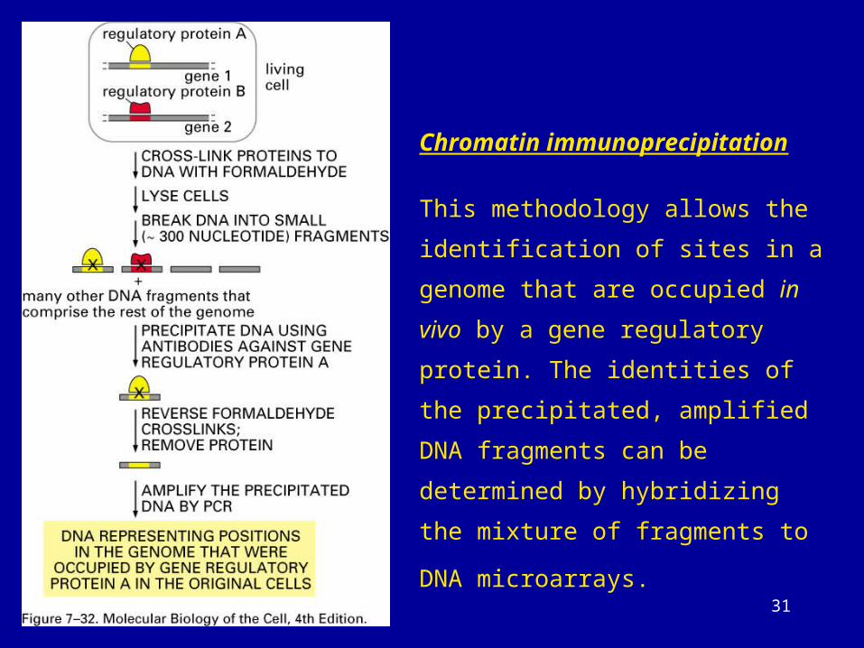
Chromatin immunoprecipitation
This methodology allows the identification
of sites in a genome that are occupied in
vivo by a gene regulatory protein. The
identities of the precipitated, amplified
DNA fragments can be determined by
hybridizing the mixture of fragments to
DNA microarrays.
32
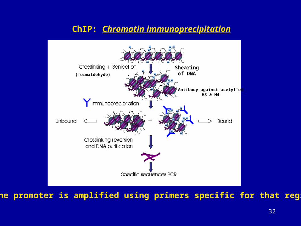
ChIP: Chromatin immunoprecipitation
(formaldehyde)
Shearing of DNA
Antibody against acetyl’edH3 & H4
Gene promoter is amplified using primers specific for that region
33
34
35
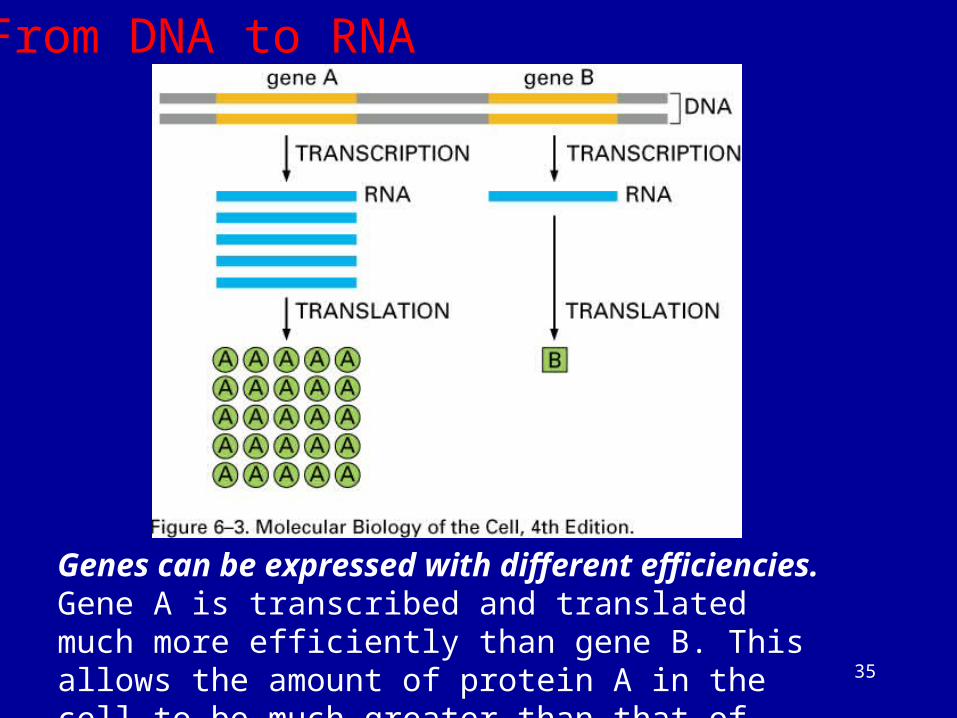
Genes can be expressed with different efficiencies.Gene A is transcribed and translated much more efficiently than gene B. This allows the amount of protein A in the cell to be much greater than that of protein B.
From DNA to RNA
36
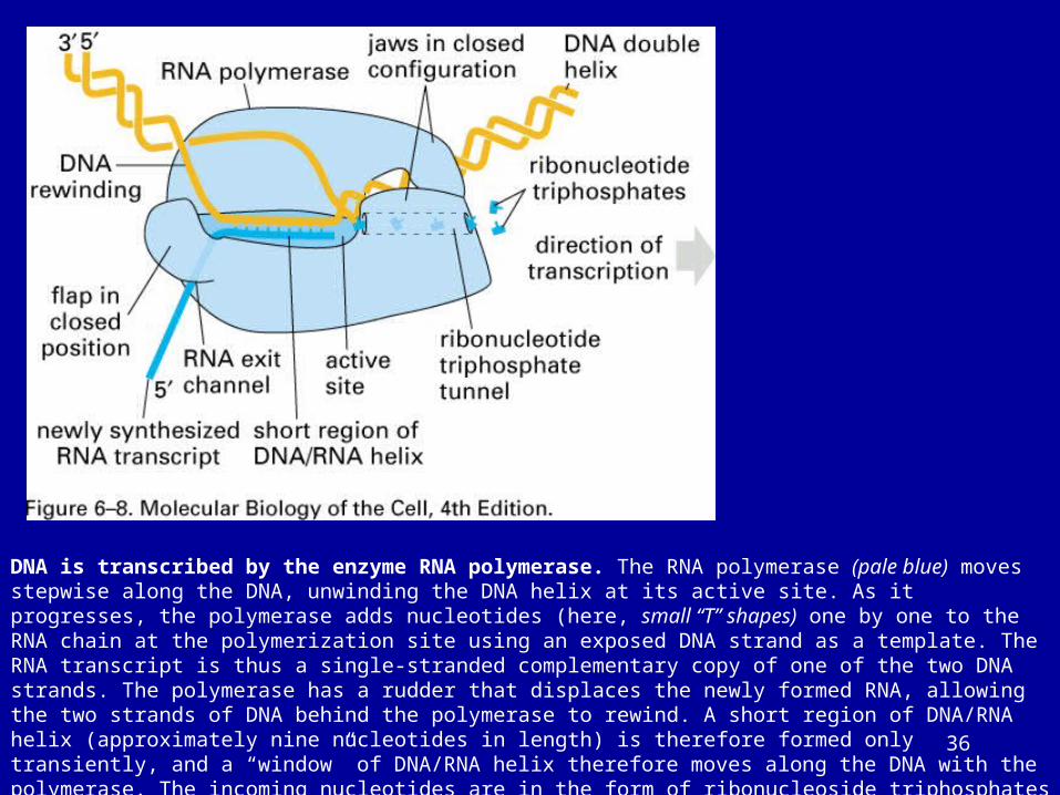
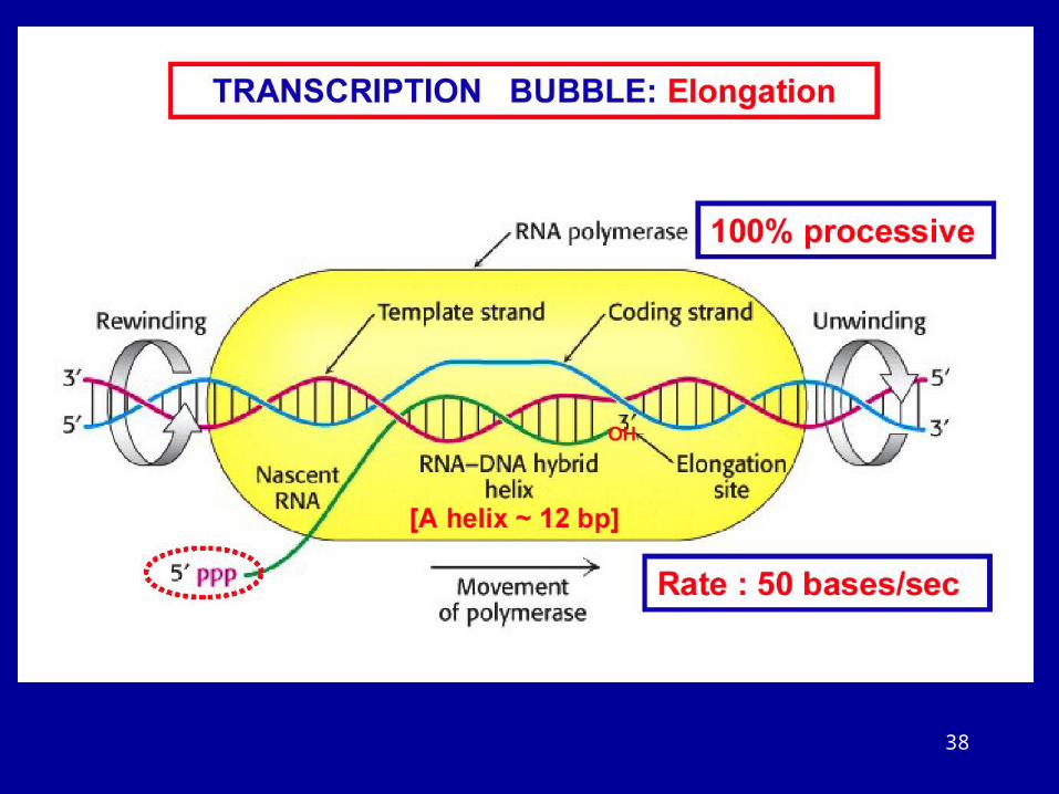
DNA is transcribed by the enzyme RNA polymerase. The RNA polymerase (pale blue) moves stepwise along the DNA, unwinding the DNA helix at its active site. As it progresses, the polymerase adds nucleotides (here, small “T” shapes) one by one to the RNA chain at the polymerization site using an exposed DNA strand as a template. The RNA transcript is thus a single-stranded complementary copy of one of the two DNA strands. The polymerase has a rudder that displaces the newly formed RNA, allowing the two strands of DNA behind the polymerase to rewind. A short region of DNA/RNA helix (approximately nine nucleotides in length) is therefore formed only transiently, and a “window” of DNA/RNA helix therefore moves along the DNA with the polymerase. The incoming nucleotides are in the form of ribonucleoside triphosphates (ATP, UTP, CTP, and GTP), and the energy stored in their phosphate–phosphate bonds provides the driving force for the polymerization reaction (see Figure 5–4).
37
38
39
40
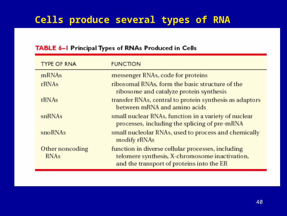
Cells produce several types of RNA
41
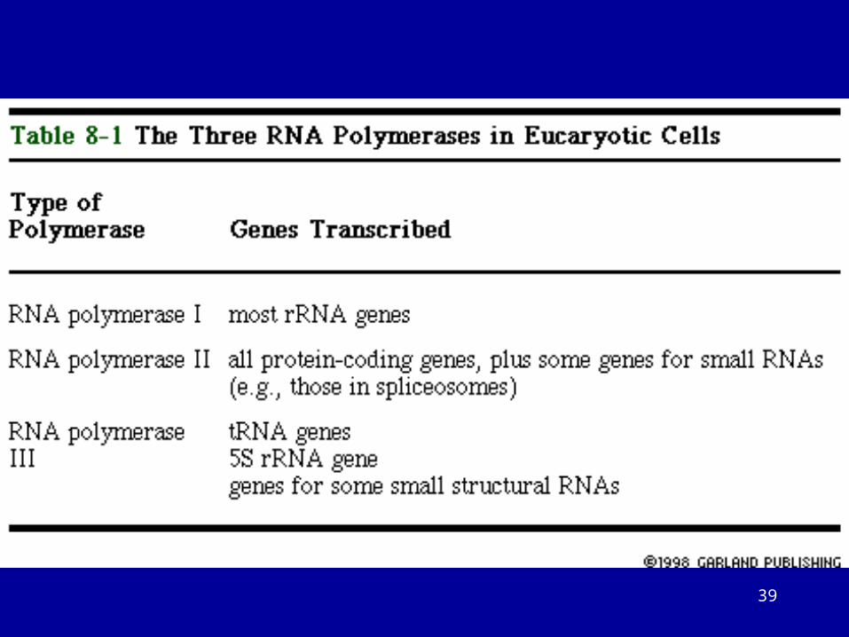
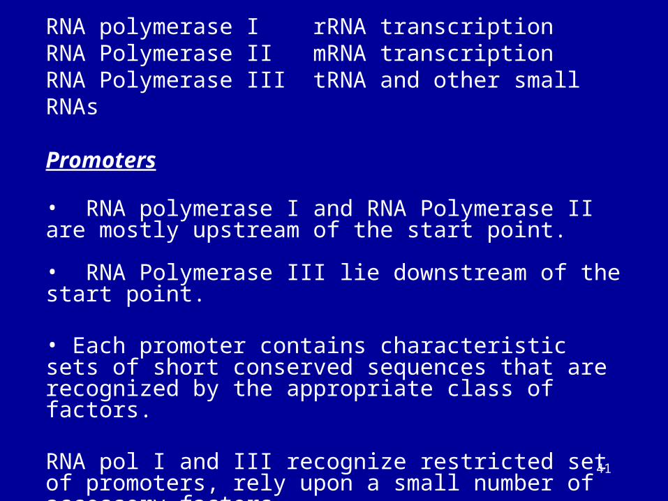
RNA polymerase I rRNA transcriptionRNA Polymerase II mRNA transcriptionRNA Polymerase III tRNA and other small RNAs
Promoters
• RNA polymerase I and RNA Polymerase II are mostly upstream of the start point.
• RNA Polymerase III lie downstream of the start point.
• Each promoter contains characteristic sets of short conserved sequences that are recognized by the appropriate class of factors.
RNA pol I and III recognize restricted set of promoters, rely upon a small number of accessory factors.
RNA pol II show more variation in sequence, cis acting elements are spread out over a region of >200bp.
42
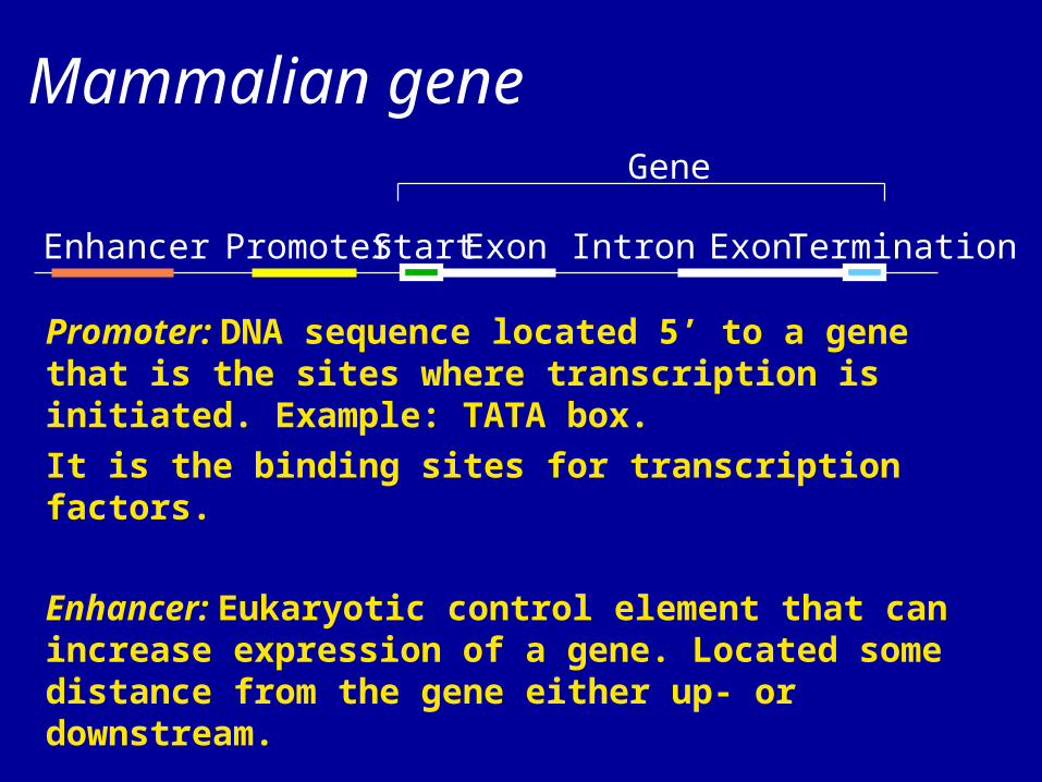
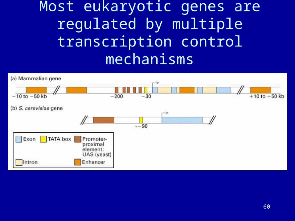
Start TerminationExon ExonIntronPromoterEnhancer
Mammalian gene
Promoter: DNA sequence located 5’ to a gene that is the sites where transcription is initiated. Example: TATA box.
It is the binding sites for transcription factors.
Enhancer: Eukaryotic control element that can increase expression of a gene. Located some distance from the gene either up- or downstream.
Human gene number estimate: 28,000–34,000 genes.
Gene
43
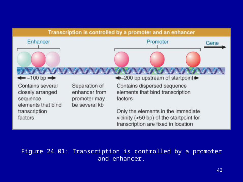
Figure 24.01: Transcription is controlled by a promoter and enhancer.
44
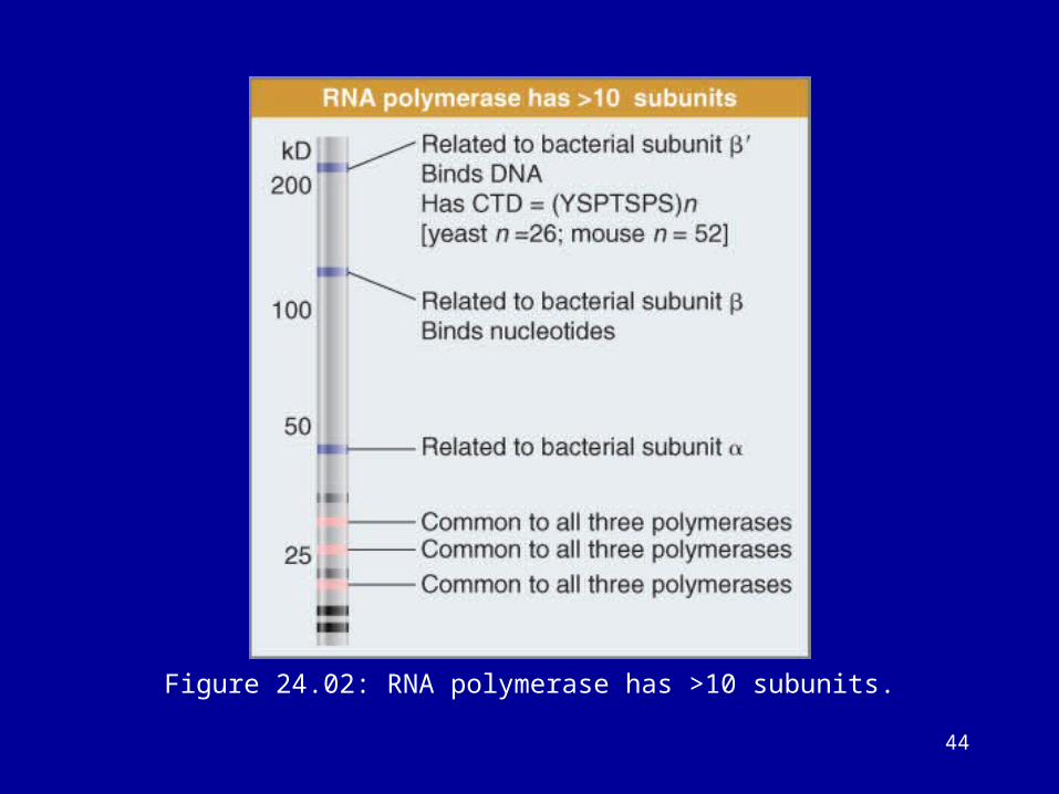
Figure 24.02: RNA polymerase has >10 subunits.
45
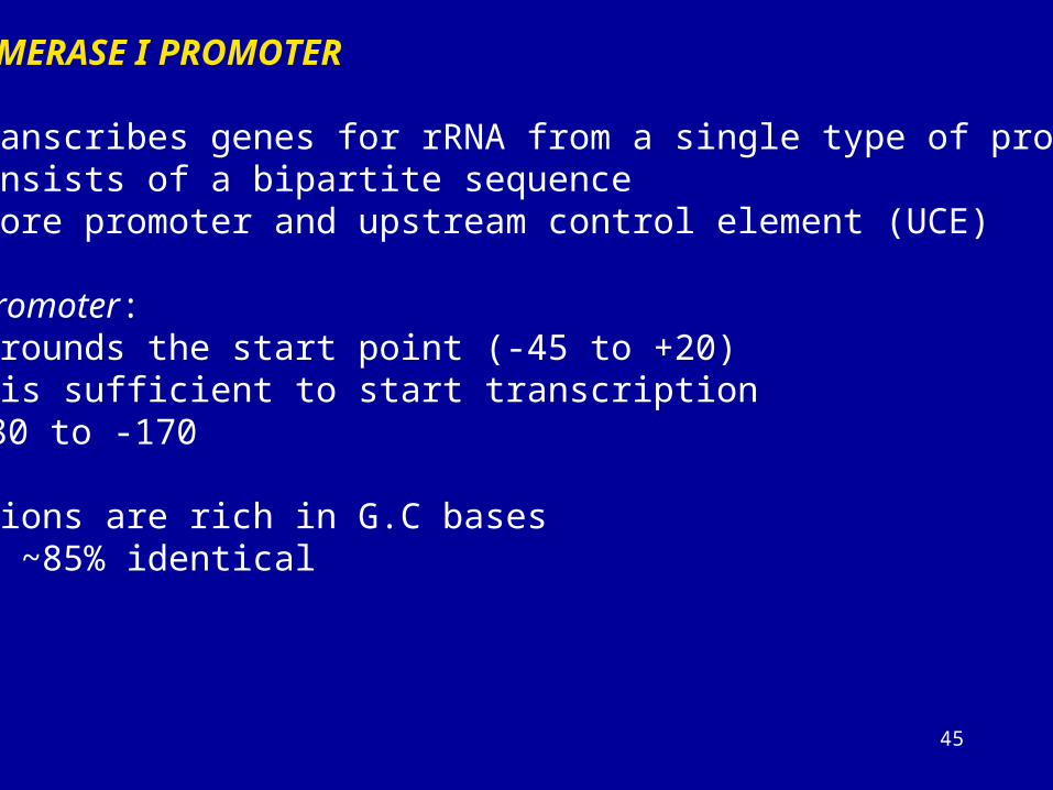
RNA POLYMERASE I PROMOTER
• It transcribes genes for rRNA from a single type of promoter• It consists of a bipartite sequence• The core promoter and upstream control element (UCE)
The core promoter:Surrounds the start point (-45 to +20)It is sufficient to start transcription
UCE: -180 to -170
Both regions are rich in G.C basesThey are ~85% identical
46
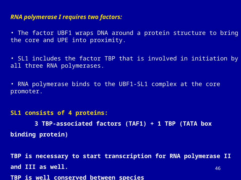
RNA polymerase I requires two factors:
• The factor UBF1 wraps DNA around a protein structure to bring the core and UPE into proximity.
• SL1 includes the factor TBP that is involved in initiation by all three RNA polymerases.
• RNA polymerase binds to the UBF1-SL1 complex at the core promoter.
SL1 consists of 4 proteins:
3 TBP-associated factors (TAF1) + 1 TBP (TATA box binding protein)
TBP is necessary to start transcription for RNA polymerase II and III as well.
TBP is well conserved between species
TBP does not directly bind to G.C rich DNA. Other factors are also necessary.
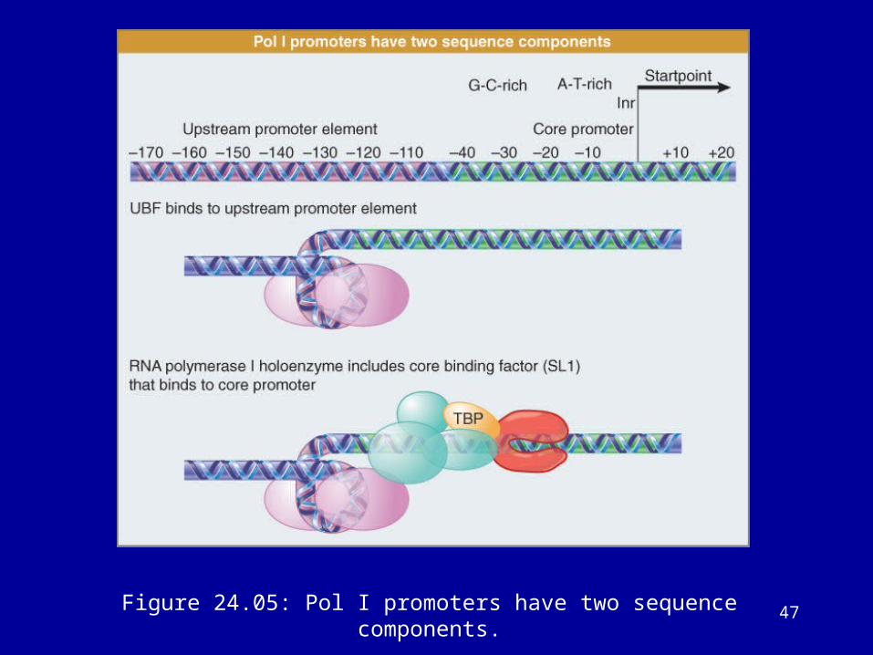
47Figure 24.05: Pol I promoters have two sequence components.
48
RNA Polymerase III promoter:
The promoters have two general classes
1) Promoters for 5S and tRNA genes are internal. They lie
downstream of the start point between positions +55
and +80 within the gene.
2) The promoters for snRNA genes lie upstream of the
start point.
3) There are two types of internal promoters: Type I and
Type II
Type I Box A and Box C
Type IIBox A and Box B
49
There are three accessory factors:
TFIIIAZinc finger protein
TFIIIB TBP and two proteins (B” and BRF proteins)
TFIIIC contains 5 subunit, >500 kD
TFIIIA and TFIIIC do not fully affect the initiation
reaction (assembly factors), their role is to bind TFIIIB
at the right location.
TFIIIB is crucial. It allows RNA pol III to bind at the
start site point (initiation factor or positioning factor).
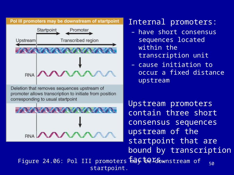
50Figure 24.06: Pol III promoters may be downstream of startpoint.
• Internal promoters:– have short consensus sequences
located within the transcription unit
– cause initiation to occur a fixed distance upstream
• Upstream promoters contain three short consensus sequences upstream of the startpoint that are bound by transcription factors.
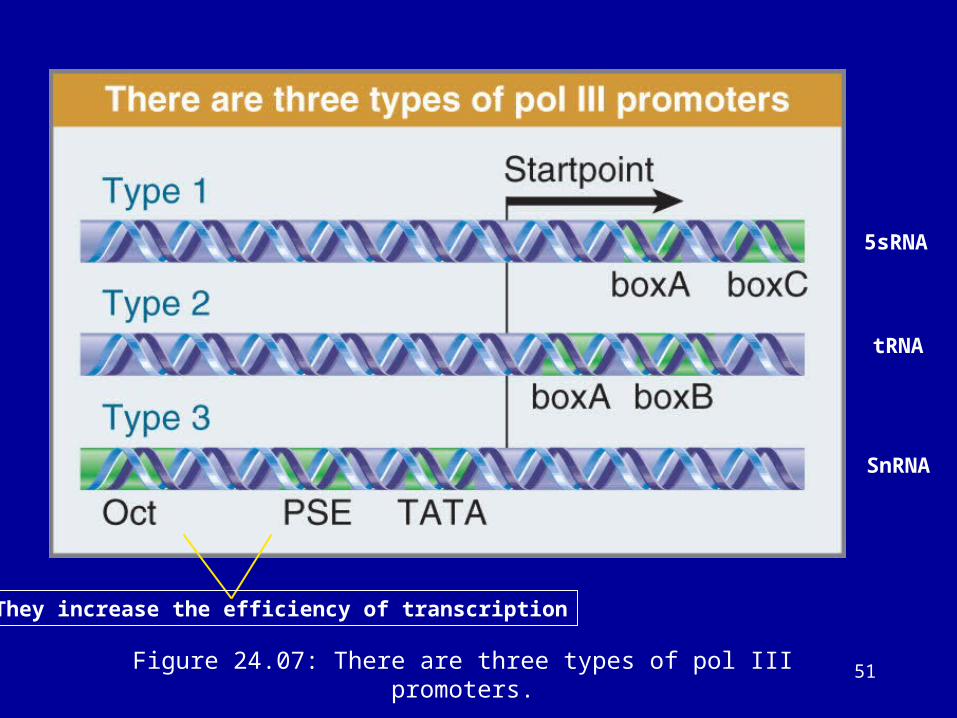
51Figure 24.07: There are three types of pol III promoters.
5sRNA
tRNA
SnRNA
They increase the efficiency of transcription

52Figure 24.08: Type 2 internal promoters use TFIIIC.
tRNA
• TFIIIA and TFIIIC bind to the consensus sequences and enable TFIIIB to bind at the startpoint.
• TFIIIB has TBP as one subunit and enables RNA polymerase to bind.
TFIIIB Is the Commitment Factor for Pol III Promoters
53
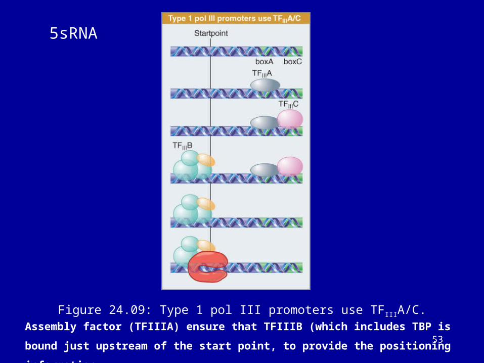
Figure 24.09: Type 1 pol III promoters use TFIIIA/C.
5sRNA
Assembly factor (TFIIIA) ensure that TFIIIB (which includes TBP is bound just upstream of
the start point, to provide the positioning information.
54
The Startpoint for RNA Polymerase II
• RNA polymerase II requires general transcription factors (called TFIIX) to initiate transcription.
• RNA polymerase II promoters have a short conserved sequence Py2CAPy5 (the initiator InR) at the startpoint.
55
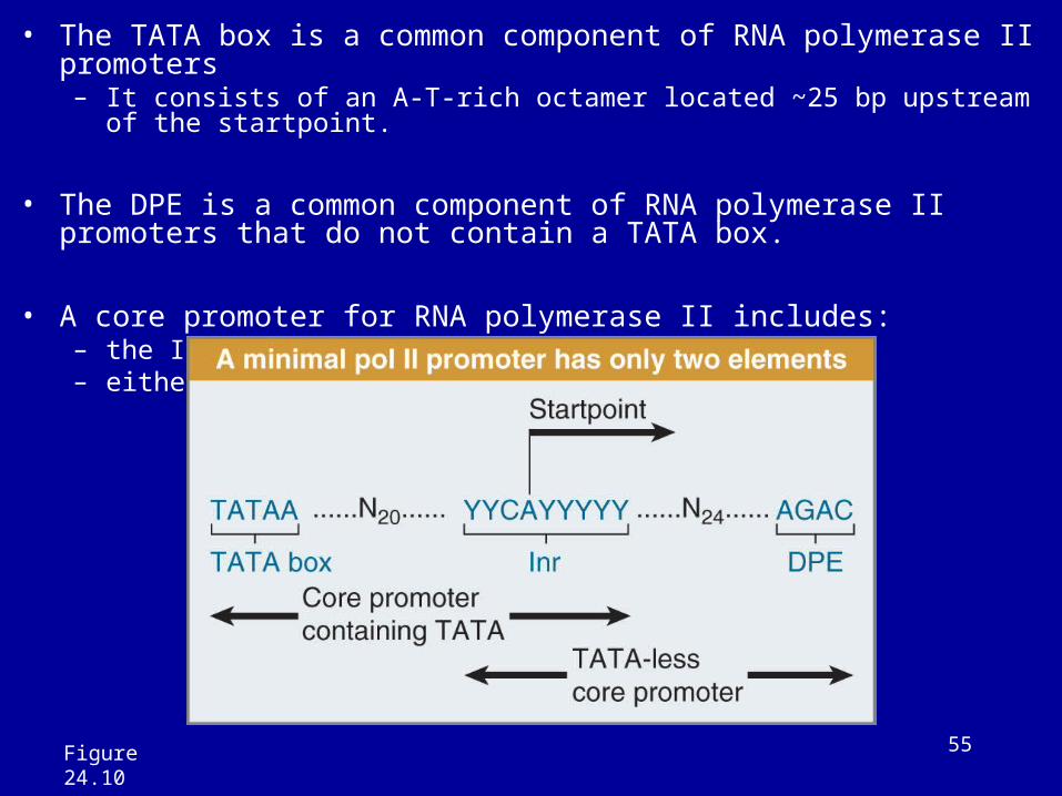
• The TATA box is a common component of RNA polymerase II promoters – It consists of an A-T-rich octamer located ~25 bp upstream of the startpoint.
• The DPE is a common component of RNA polymerase II promoters that do not contain a TATA box.
• A core promoter for RNA polymerase II includes:– the InR– either a TATA box or a DPE
Figure 24.10
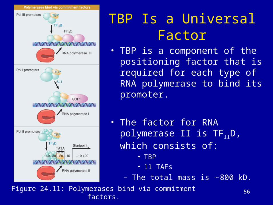
56Figure 24.11: Polymerases bind via commitment factors.
TBP Is a Universal Factor
• TBP is a component of the positioning factor that is required for each type of RNA polymerase to bind its promoter.
• The factor for RNA polymerase II is TFIID, which consists of:
• TBP
• 11 TAFs
– The total mass is ∼800 kD.
57
TBP Binds DNA in an Unusual Way
• TBP binds to the TATA box in the minor groove of DNA.
• It forms a saddle around the DNA and bends it by ∼80°.
• Some of the TAFs resemble histones and may form a structure resembling a histone octamer.
58
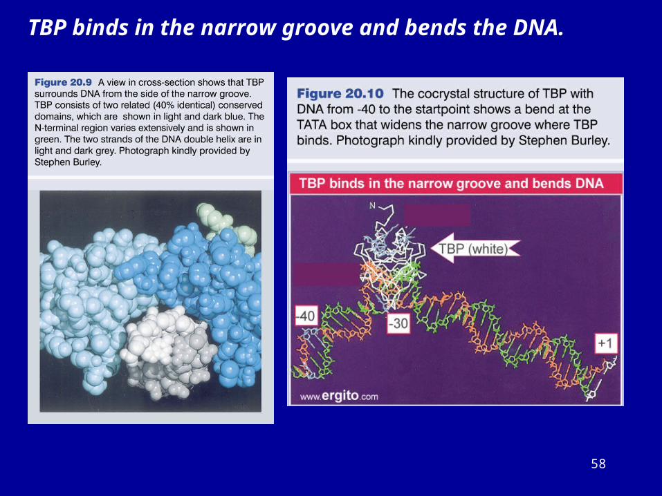
TBP binds in the narrow groove and bends the DNA.
59
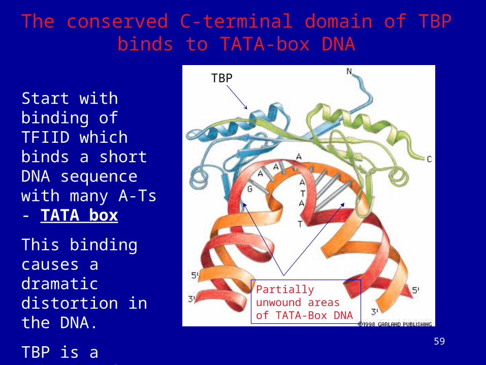
Start with binding of TFIID which binds a short DNA sequence with many A-Ts - TATA box
This binding causes a dramatic distortion in the DNA.
TBP is a subunit of TFIID which binds the TATA box.
Partially unwound areas of TATA-Box DNA
The conserved C-terminal domain of TBP binds to TATA-box DNA
TBP
60
Most eukaryotic genes are regulated by multiple transcription control
mechanisms
61
The basal apparatus assembles at the promoter
Binding of TFIID to the TATA box is the first step in initiation.
Other transcription factors bind to the complex in a defined order, extending the length of the protected region on DNA.
When RNA polymerase II binds to the complex, it initiates transcription.

62
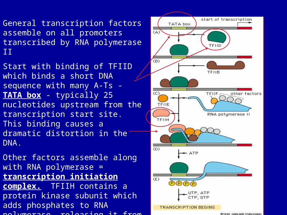
RNA polymerase II requires general transcription factors
Initiation of transcription of a eucaryotic gene by RNA polymerase II.
To begin transcription, RNA
polymerase requires a number of
general transcription factors (called
TFIIA, TFIIB, and so on). (A) The
promoter contains a DNA sequence
called the TATA box, which is
located 25 nucleotides away from the
site at which transcription is initiated.
(B) The TATA box is recognized and
bound by transcription factor TFIID,
which then enables the adjacent
binding of TFIIB (C).
63
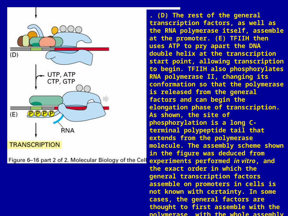
. (D) The rest of the general transcription factors, as well as the RNA polymerase itself, assemble at the promoter. (E) TFIIH then uses ATP to pry apart the DNA double helix at the transcription start point, allowing transcription to begin. TFIIH also phosphorylates RNA polymerase II, changing its conformation so that the polymerase is released from the general factors and can begin the elongation phase of transcription. As shown, the site of phosphorylation is a long C-terminal polypeptide tail that extends from the polymerase molecule. The assembly scheme shown in the figure was deduced from experiments performed in vitro, and the exact order in which the general transcription factors assemble on promoters in cells is not known with certainty. In some cases, the general factors are thought to first assemble with the polymerase, with the whole assembly subsequently binding to the DNA in a single step. The general transcription factors have been highly conserved in evolution; some of those from human cells can be replaced in biochemical experiments by the corresponding factors from simple yeasts.
64
General transcription factors assemble on all promoters transcribed by RNA polymerase II
Start with binding of TFIID which binds a short DNA sequence with many A-Ts - TATA box - typically 25 nucleotides upstream from the transcription start site. This binding causes a dramatic distortion in the DNA.
Other factors assemble along with RNA polymerase = transcription initiation complex. TFIIH contains a protein kinase subunit which adds phosphates to RNA polymerase, releasing it from the complex to start transcription
65
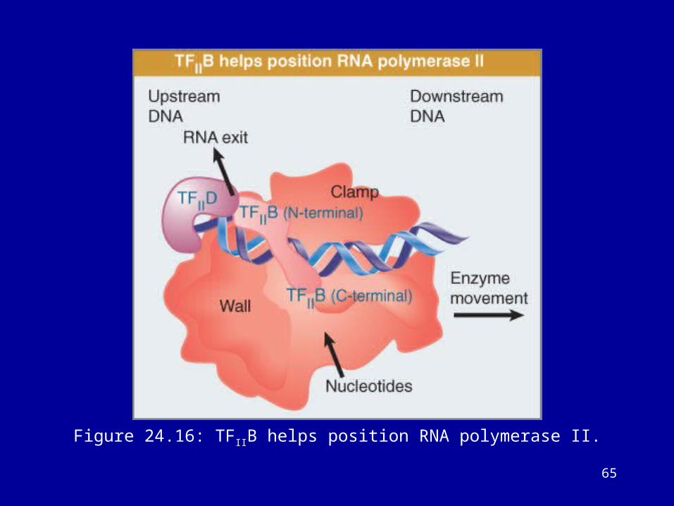
Figure 24.16: TFIIB helps position RNA polymerase II.
66
The basal / general transcription factors (core promoter factors):
TFIID: It has many subunits. TBP and TAFs
TBP is the only general transcription factor makes sequence-specific contacts with
DNA.
Within TFIID as a free protein complex, the factor TAFII230 binds to TBP and
controls the ability of TBP to bind to DNA.
The N-terminal domain of TAFII230 must be displaced from the DNA-binding
surface of TBP in order for TFIID to bind to DNA.
TFIIA: binds to TBP and stablilizes the TBP-DNA interaction. It also antogonizes transcription repressors, it physically displaces or blocks several negative transcriptional regulators from TFIID complex.
TFIIB: it is involved in the selection of transcription start sites, possibly setting distances between promoters and transcription start sites. It interacts with diverse activators that may serve to recruit TFIIB to the promoters.
67
TFIIF:
It binds tightly to RNA pol.II, suppresses nonspecific DNA
binding, and stabilizes the preinitiation complex.
It stimulates polymerase elongation rates by suppressing
transient pause during transcription.
Its subunit RAP74 makes contacts with DNA to position the
template during initiation. RAP74 has an ATP-dependent DNA
helicase activity. It affects the DNA topology, facilitates
promoter melting.
68
TFIIE: Interacts with TFIIH; involved in recruitment, stimulation of TFIIH and
promoter opening.
TFIIH: Its subunits have at least three enzymatic activities: DNA-dependent
ATPase, ATP-dependent helicase, and CTD kinase.
Two of its subunits have helicase activity (5’ to 3’ DNA helicase activity),
essential promoter opening and promoter escape.
TFIIH kinase/cyclin subcomplex: Cdks phosphorylate serines in the RNA
polymerase II CTD.
TFIIH kinase also functions as a Cdk activating kinase (CAK) and regulates
cell cycle transitions.
Some of its subunits required for nucleotide excision repair (transcriptional
coupled repair function).
69
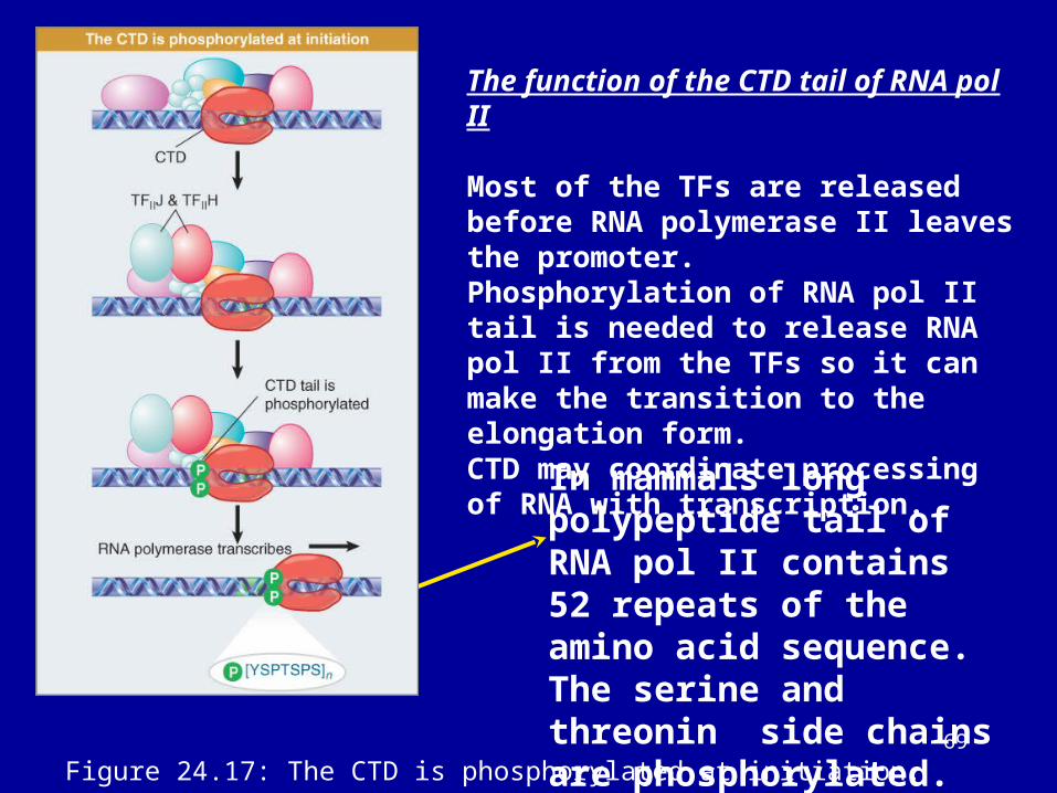
The function of the CTD tail of RNA pol II
Most of the TFs are released before RNA polymerase II leaves the promoter.Phosphorylation of RNA pol II tail is needed to release RNA pol II from the TFs so it can make the transition to the elongation form.CTD may coordinate processing of RNA with transcription.
In mammals long polypeptide tail of RNA pol II contains 52 repeats of the amino acid sequence. The serine and threonin side chains are phosphorylated.
Figure 24.17: The CTD is phosphorylated at initiation.
70
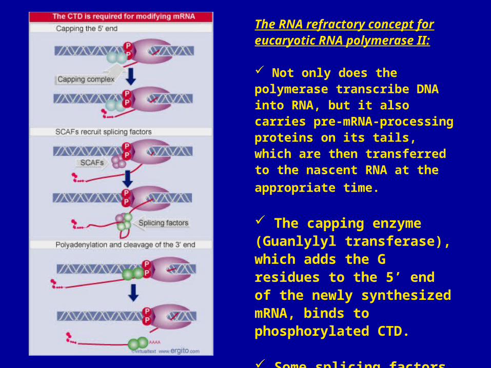
The RNA refractory concept for eucaryotic RNA polymerase II:
Not only does the polymerase transcribe DNA into RNA, but it also carries pre-mRNA-processing proteins on its tails, which are then transferred to the nascent RNA at the
appropriate time.
The capping enzyme (Guanlylyl transferase), which adds the G residues to the 5’ end of the newly synthesized mRNA, binds to phosphorylated CTD.
Some splicing factors bind to CTD and also some components of the cleavage/polyadenylation apparatus.
71
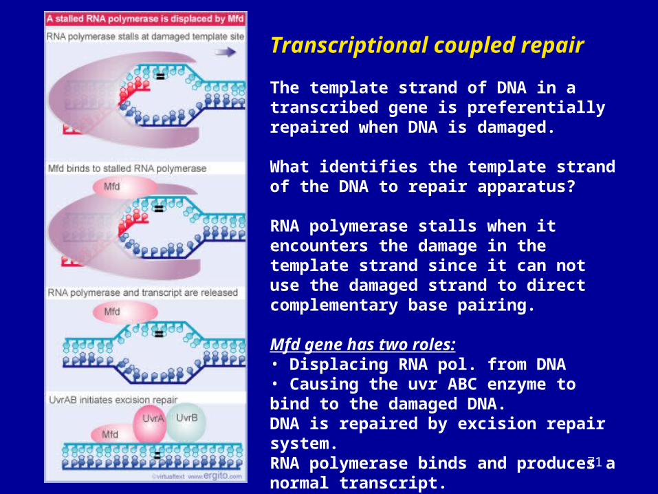
Transcriptional coupled repair
The template strand of DNA in a transcribed gene is preferentially repaired when DNA is damaged.
What identifies the template strand of the DNA to repair apparatus?
RNA polymerase stalls when it encounters the damage in the template strand since it can not use the damaged strand to direct complementary base pairing.
Mfd gene has two roles:• Displacing RNA pol. from DNA• Causing the uvr ABC enzyme to bind to the damaged DNA.DNA is repaired by excision repair system.RNA polymerase binds and produces a normal transcript.
72
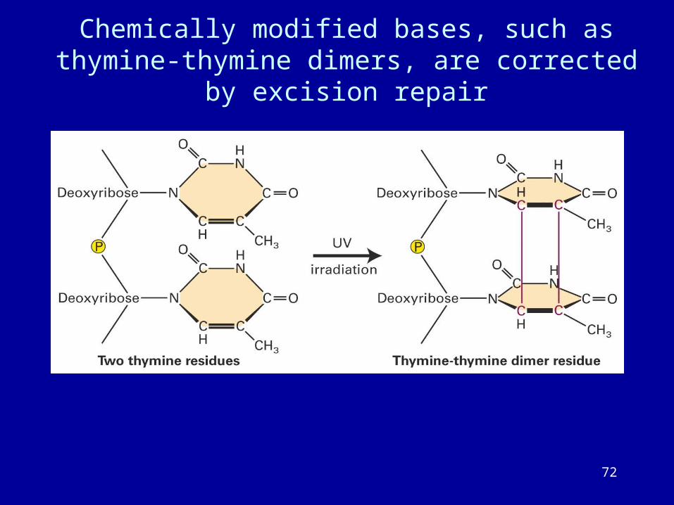
Chemically modified bases, such as thymine-thymine dimers, are corrected by excision repair
73
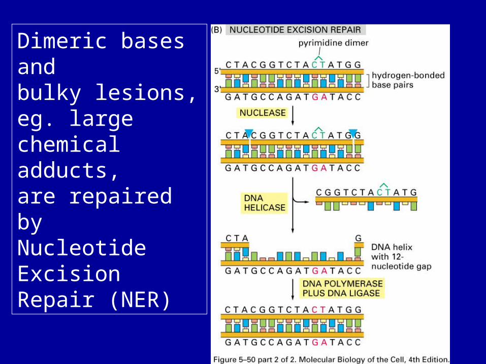
Dimeric bases andbulky lesions,eg. large chemical adducts,are repaired by Nucleotide Excision Repair (NER)
74
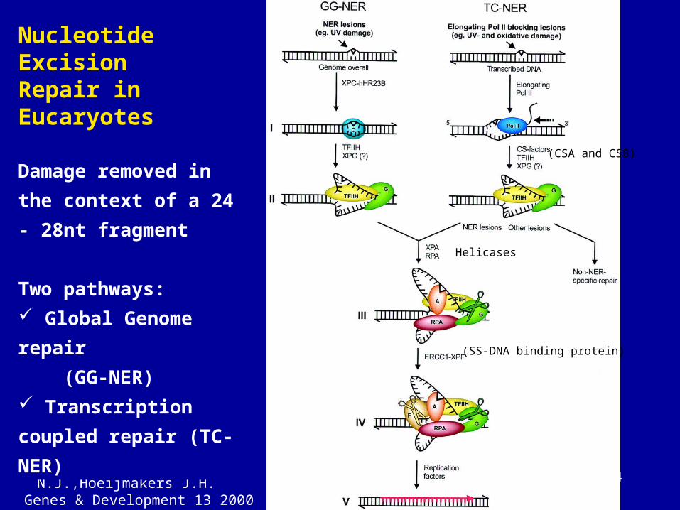
Rereference: de Laat W.L., Jaspers N.J.,Hoeijmakers J.H.
Genes & Development 13 2000
Nucleotide ExcisionRepair in Eucaryotes
Damage removed in the
context of a 24 - 28nt
fragment
Two pathways:
Global Genome repair
(GG-NER)
Transcription coupled
repair (TC-NER)
(Helicases)
(SS-DNA binding protein)
(CSA and CSB)
75
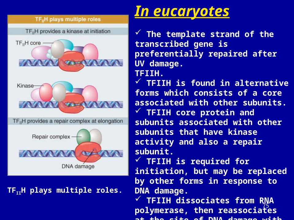
In eucaryotes The template strand of the transcribed gene is preferentially repaired after UV damage.TFIIH. TFIIH is found in alternative forms which consists of a core associated with other subunits. TFIIH core protein and subunits associated with other subunits that have kinase activity and also a repair subunit. TFIIH is required for initiation, but may be replaced by other forms in response to DNA damage. TFIIH dissociates from RNA polymerase, then reassociates at the site of DNA damage with additional coupling components (i.e. Rad26). The repair function may require modification or degradation of RNA pol.
TFIIH plays multiple roles.
76
Short sequence elements bind activators
Short conserved sequence elements are
dispersed in the region preceding the startpoint.
The upstream elements increase the
frequency of initiation.
The factors that bind to them to stimulate
transcription are called activators.
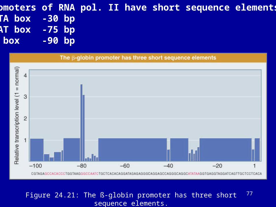
77Figure 24.21: The ß-globin promoter has three short sequence elements.
Promoters of RNA pol. II have short sequence elements.TATA box -30 bpCAAT box -75 bpGC box -90 bp
78
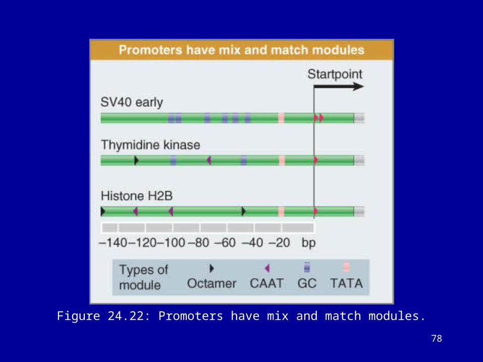
Figure 24.22: Promoters have mix and match modules.
79
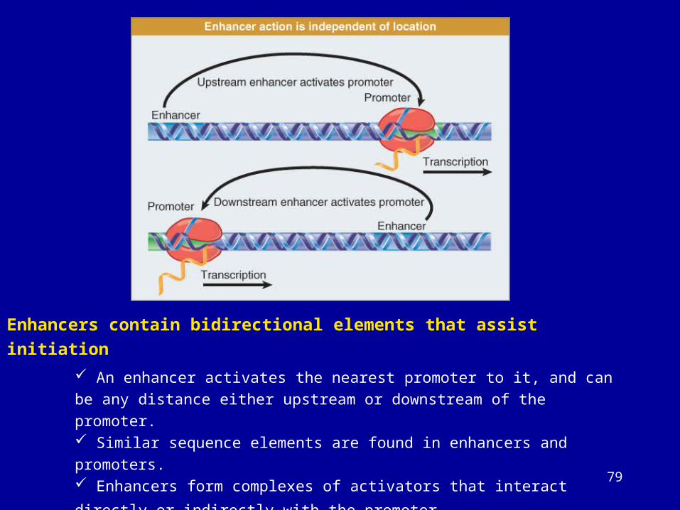
Enhancers contain bidirectional elements that assist initiation
An enhancer activates the nearest promoter to it, and can be any distance either
upstream or downstream of the promoter. Similar sequence elements are found in enhancers and promoters. Enhancers form complexes of activators that interact directly or indirectly with
the promoter.
80
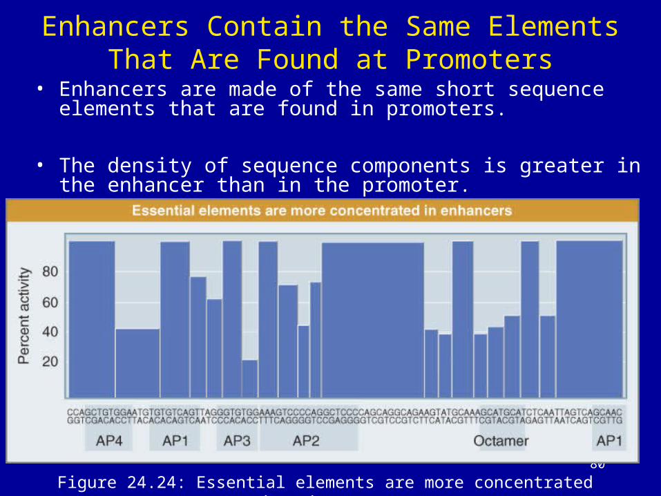
Figure 24.24: Essential elements are more concentrated in enhancers.
Enhancers Contain the Same Elements That Are Found at Promoters
• Enhancers are made of the same short sequence elements that are found in promoters.
• The density of sequence components is greater in the enhancer than in the promoter.
81
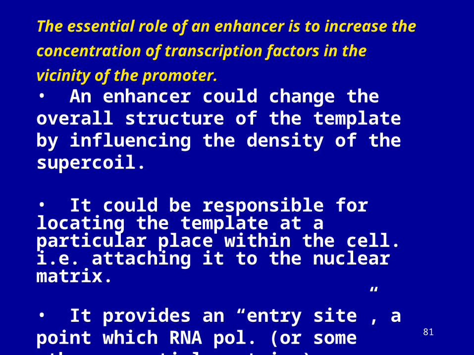
The essential role of an enhancer is to increase the
concentration of transcription factors in the vicinity of the
promoter.• An enhancer could change the overall structure of the template by influencing the density of the supercoil.
• It could be responsible for locating the template at a particular place within the cell. i.e. attaching it to the nuclear matrix.
• It provides an “entry site”, a point which RNA pol. (or some other essential proteins) associate with chromatin.
82
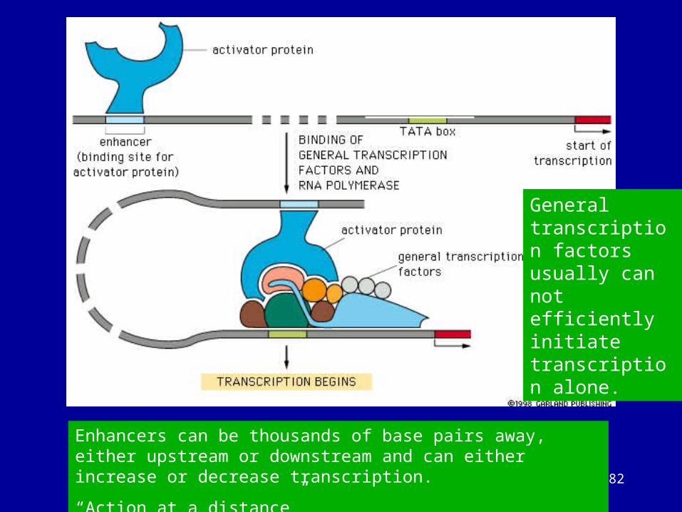
A model for gene activation from an enhancer
General transcription factors usually can not efficiently initiate transcription alone.
Enhancers can be thousands of base pairs away, either upstream or downstream and can either increase or decrease transcription.
“Action at a distance”
83
Enhancers work by increasing the concentration of activators near the promoter
Enhancers usually work only in cis configuration with a target promoter.
Enhancers can be made to work in trans configuration by linking the DNA that contains the target promoter to the DNA that contains the enhancer via a protein bridge or by catenating the two molecules.
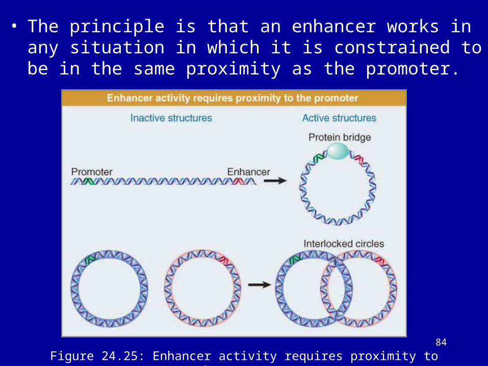
The principle is that an enhancer works in any situation in which it is constrained to be in the same proximity as the promoter.
84
Figure 24.25: Enhancer activity requires proximity to the promoter.
• The principle is that an enhancer works in any situation in which it is constrained to be in the same proximity as the promoter.
85
Gene Expression Is Associated with Demethylation
• Demethylation at the 5 end of the gene is necessary for transcription.
86
CpG Islands Are Regulatory Targets
• CpG islands surround the promoters of constitutively expressed genes where they are unmethylated.
• CpG islands also are found at the promoters of some tissue-regulated genes.
87
Gene expression is associated with demethylation
•The state of methylation of DNA is controlled by • methylases, which add methyl groups to the 5 position of cytosine• demethylases which remove the methyl group.
•There are two types of DNA methylase:
• De novo methylase• modifies the DNA at a new position
recognizes DNA by the presence of a specific sequence
Maintenance methylaseacts on hemimethylated sites to convert to fully methylated
sites. It is a ubiquitious enzyme.
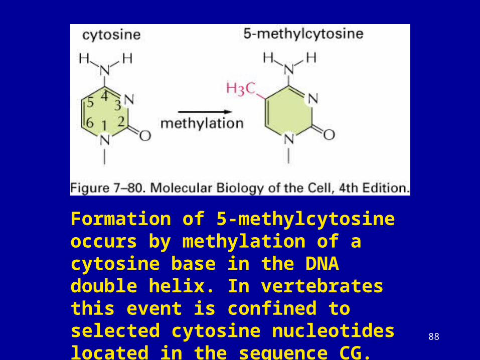
88
Formation of 5-methylcytosine occurs by methylation of a cytosine base in the DNA double helix. In vertebrates this event is confined to selected cytosine nucleotides located in the sequence CG.
89
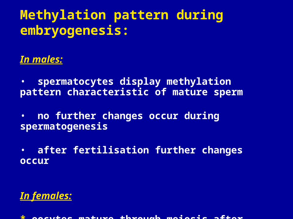
Methylation pattern during embryogenesis:
In males:
• spermatocytes display methylation pattern characteristic of mature sperm
• no further changes occur during spermatogenesis
• after fertilisation further changes occur
In females:
* oocytes mature through meiosis after birth and methylation pattern is established.
90
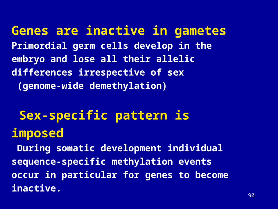
Genes are inactive in gametesPrimordial germ cells develop in the embryo and lose all
their allelic differences irrespective of sex
(genome-wide demethylation)
Sex-specific pattern is imposed During somatic development individual sequence-specific
methylation events occur in particular for genes to
become inactive.
91
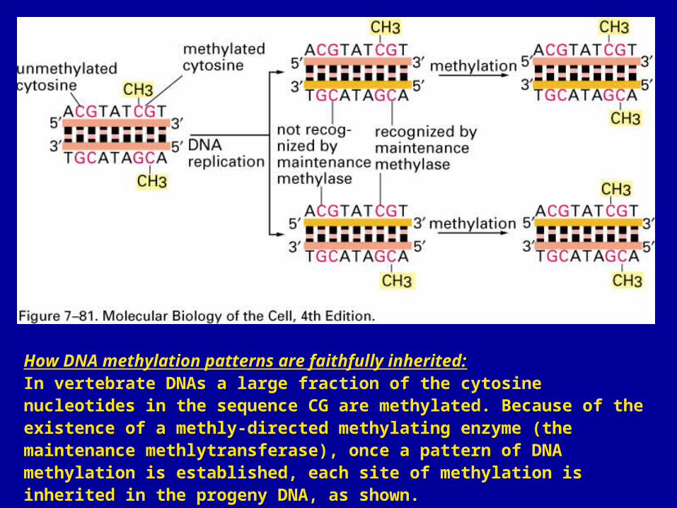
How DNA methylation patterns are faithfully inherited:In vertebrate DNAs a large fraction of the cytosine nucleotides in the sequence CG are methylated. Because of the existence of a methly-directed methylating enzyme (the maintenance methlytransferase), once a pattern of DNA methylation is established, each site of methylation is inherited in the progeny DNA, as shown.
92
Gene expression is associated with demethylation
93
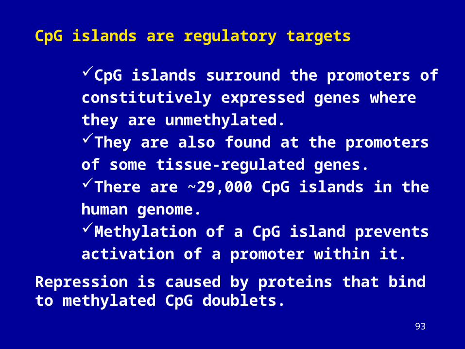
CpG islands are regulatory targets
CpG islands surround the promoters of
constitutively expressed genes where they are
unmethylated. They are also found at the promoters of some
tissue-regulated genes. There are ~29,000 CpG islands in the human
genome. Methylation of a CpG island prevents activation
of a promoter within it.
Repression is caused by proteins that bind to methylated CpG doublets.
94
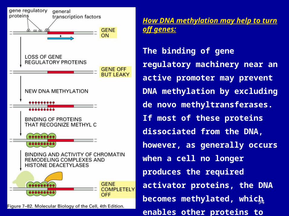
How DNA methylation may help to turn off genes:
The binding of gene regulatory
machinery near an active promoter may
prevent DNA methylation by excluding
de novo methyltransferases. If most of
these proteins dissociated from the
DNA, however, as generally occurs
when a cell no longer produces the
required activator proteins, the DNA
becomes methylated, which enables
other proteins to bind, and these shut
down the gene completely by further
altering chromatin structure.
95
96
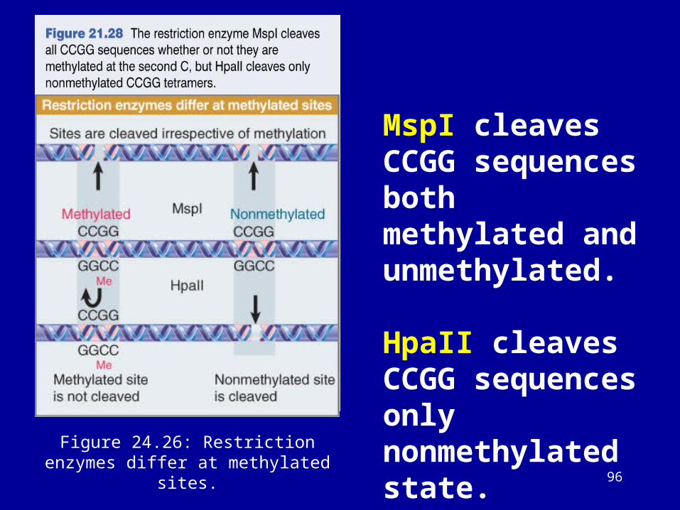
Figure 24.26: Restriction enzymes differ at methylated sites.
MspI cleaves CCGG sequences both methylated and unmethylated.
HpaII cleaves CCGG sequences only nonmethylated state.
97
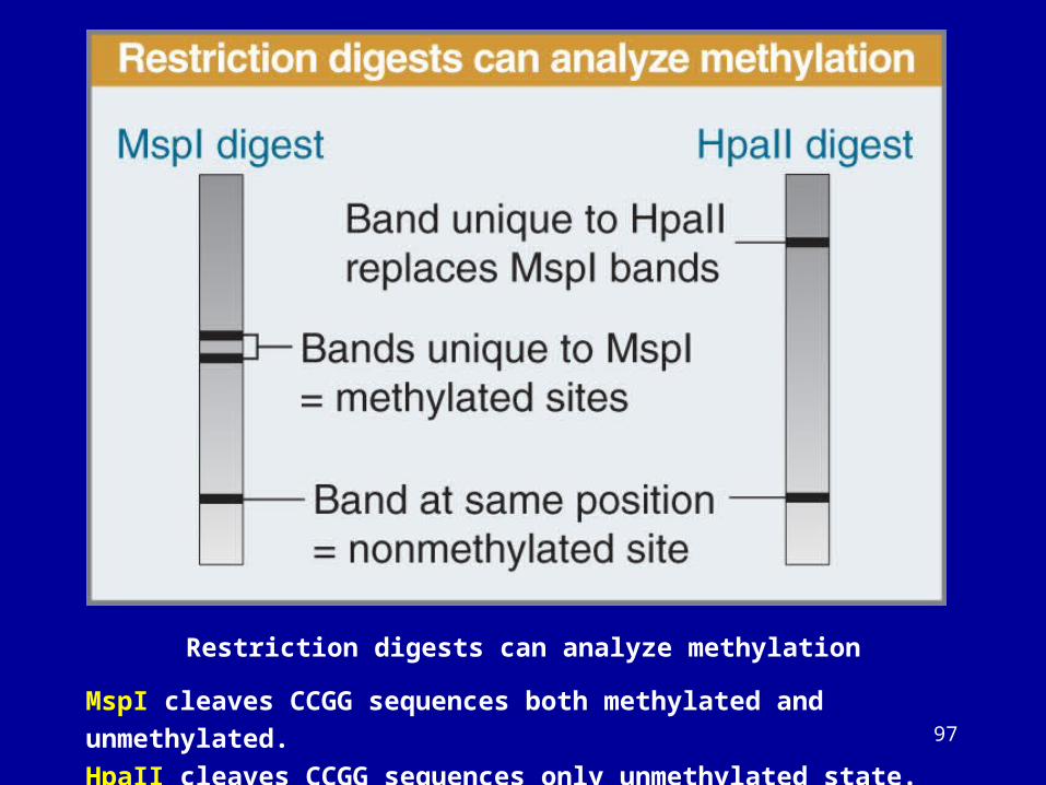
Restriction digests can analyze methylation
MspI cleaves CCGG sequences both methylated and unmethylated.
HpaII cleaves CCGG sequences only unmethylated state.
98
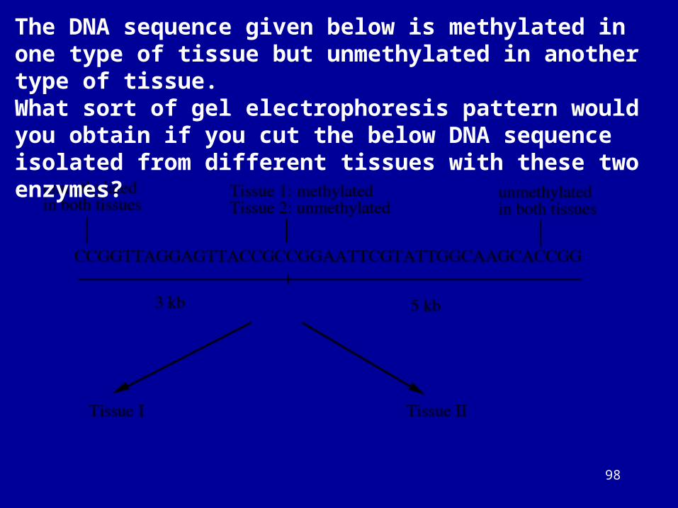
The DNA sequence given below is methylated in one type of tissue but unmethylated in another type of tissue.What sort of gel electrophoresis pattern would you obtain if you cut the below DNA sequence isolated from different tissues with these two enzymes?
99
Methylation status was checked by “methylation specific PCR (MSP)”
• All unmethylated cytosines are deaminated and sulfonated, converting them to
uracils, while 5-methylcytosines remain unaltered.
(Sodium bisulfite converts unmethylated cytosine to uracil).
• Thus, the sequence of the treated DNA will differ depending on whether the DNA
is originally methylated or unmethylated.
• Also, the initially complementary DNA strands will no longer be complementary
after cytosine conversion.
• Primers for use in MSP can be designed to specifically amplify either bisulfite-
sensitive, unmethylated strand or a bisulfite-resisant, methylated strand, based
upon these chemically-induced differences.
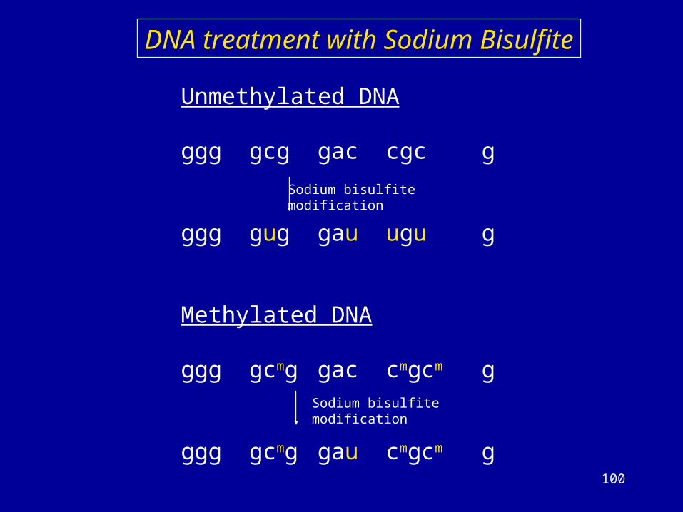
100
Unmethylated DNA
ggg gcg gac cgc g
ggg gug gau ugu g
Methylated DNA
ggg gcmg gac cmgcm g
ggg gcmg gau cmgcm g
Sodium bisulfitemodification
Sodium bisulfitemodification
DNA treatment with Sodium Bisulfite
101
Methylation specific PCR
The PCR primers are designed to specifically amplify the promoter regions of the gene of interest.
If the DNA samples was originally unmethylated, an MSP reaction product will be detectable when using primer set (labeled as “U”) designed to be complementary to the unmethylated DNA sequence.
No product will be generated using primer set (labeled as “M”) designed to be complementary to the derivative methylated DNA sequence.
Conversely, an MSP product will be generated only using the M primer set if the sample was originally methylated, and the U primers will not amplify such a template.
102
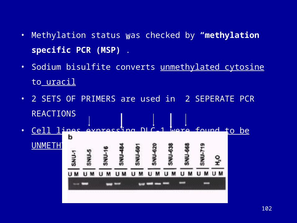
• Methylation status was checked by “methylation specific PCR (MSP)”.
• Sodium bisulfite converts unmethylated cytosine to uracil
• 2 SETS OF PRIMERS are used in 2 SEPERATE PCR REACTIONS
• Cell lines expressing DLC-1 were found to be UNMETHYLATED.


![SLC15A2 genomic variation is associated with the ... · and regulatory regions such as promoters and enhancers, [11, 12] allowing a systemic approach to biomarker age of 56 years](https://img.dokumen.tips/doc/110x75/5e53b8f60bdf6b1f7a334b35/slc15a2-genomic-variation-is-associated-with-the-and-regulatory-regions-such.jpg)