Embed Size (px)
Citation preview
1
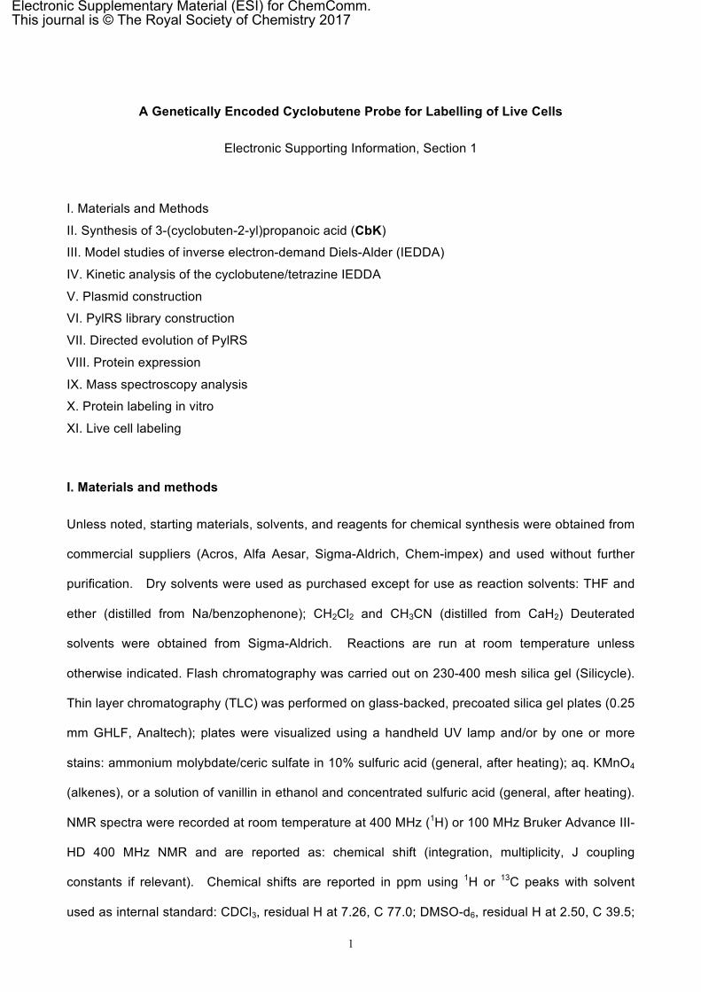
A Genetically Encoded Cyclobutene Probe for Labelling of Live Cells
Electronic Supporting Information, Section 1
I. Materials and Methods
II. Synthesis of 3-(cyclobuten-2-yl)propanoic acid (CbK)
III. Model studies of inverse electron-demand Diels-Alder (IEDDA)
IV. Kinetic analysis of the cyclobutene/tetrazine IEDDA
V. Plasmid construction
VI. PylRS library construction
VII. Directed evolution of PylRS
VIII. Protein expression
IX. Mass spectroscopy analysis
X. Protein labeling in vitro
XI. Live cell labeling
I. Materials and methods
Unless noted, starting materials, solvents, and reagents for chemical synthesis were obtained from
commercial suppliers (Acros, Alfa Aesar, Sigma-Aldrich, Chem-impex) and used without further
purification. Dry solvents were used as purchased except for use as reaction solvents: THF and
ether (distilled from Na/benzophenone); CH2Cl2 and CH3CN (distilled from CaH2) Deuterated
solvents were obtained from Sigma-Aldrich. Reactions are run at room temperature unless
otherwise indicated. Flash chromatography was carried out on 230-400 mesh silica gel (Silicycle).
Thin layer chromatography (TLC) was performed on glass-backed, precoated silica gel plates (0.25
mm GHLF, Analtech); plates were visualized using a handheld UV lamp and/or by one or more
stains: ammonium molybdate/ceric sulfate in 10% sulfuric acid (general, after heating); aq. KMnO4
(alkenes), or a solution of vanillin in ethanol and concentrated sulfuric acid (general, after heating).
NMR spectra were recorded at room temperature at 400 MHz (1H) or 100 MHz Bruker Advance III-
HD 400 MHz NMR and are reported as: chemical shift (integration, multiplicity, J coupling
constants if relevant). Chemical shifts are reported in ppm using 1H or 13C peaks with solvent
used as internal standard: CDCl3, residual H at 7.26, C 77.0; DMSO-d6, residual H at 2.50, C 39.5;
Electronic Supplementary Material (ESI) for ChemComm.This journal is © The Royal Society of Chemistry 2017
2
D2O, HOD at 4.79. Multiplicity was reported as follows: s = singlet, d = doublet, t = triplet, q =
quartet, m = multiplet, b = broad. IR spectra were acquired on neat films (diamond, ATR mode)
with selected absorbances reported in wavenumbers (cm-1). UV absorbance measurements for
kinetic studies were conducted on a Shimadzu UV2401-PC. Absorbance spectrum and intensity
were measured on Shimadzu UV2401-PC and Thermo Scientific GENESYS 10S UV/Vis
Spectrophotometer. Fluorescence spectra and intensities were recorded on Horiba FluoroMax 4
spectrometer and BioTek Synergy H1 Hybrid plate reader. Sodium dodecyl sulfate-
polyacrylamide gel electrophoresis (SDS-PAGE) was performed on Bio-Rad mini-PROTEAN
electrophoresis system. Bio-Rad Prestained Protein Ladder was applied to at least one lane of
each gel for the estimation of apparent molecular weights. Protein gels were stained by
Coomassie Brilliant Blue staining and visualized using Bio-Rad Molecular Imager ChemiDoc XRS+
System. For in-gel fluorescence imaging, either Bio-Rad Molecular Imager ChemiDoc XRS+
System or GE Typhoon FLA9500 was used. Live cells were imaged on an Olympus FV500
inverted (Olympus IX-81) confocal microscope. Abbreviations: tetrahydrofuran (THF), hexanes
(Hex), ethyl acetate (EA), diethyl ether (ether), PBS (phosphate-buffered saline); rt (room
temperature), rbf (round bottom flask).
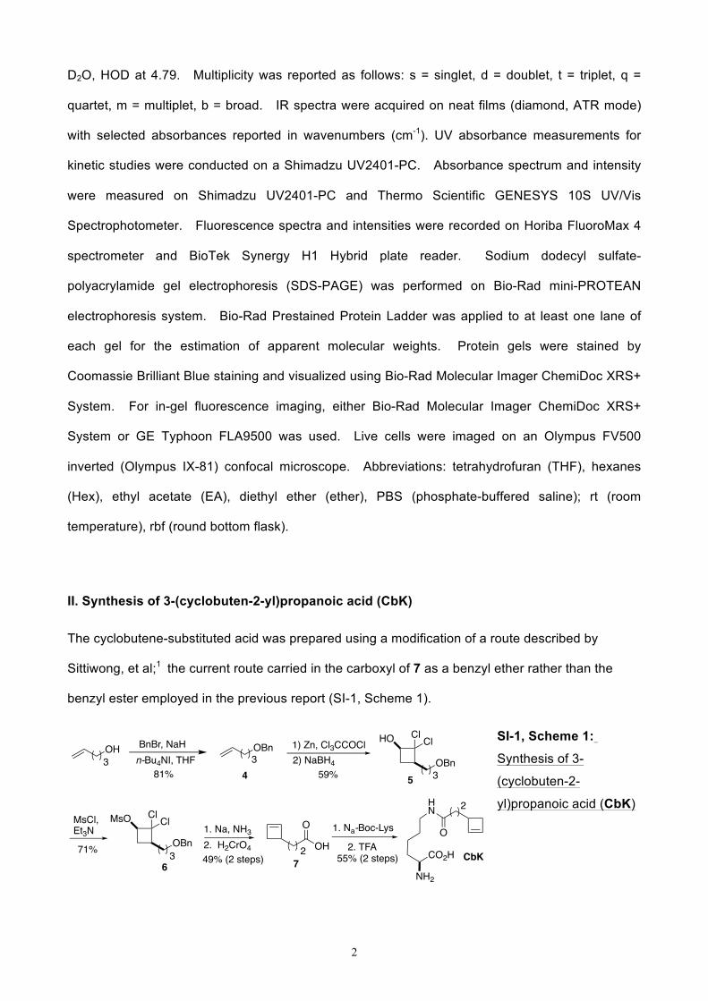
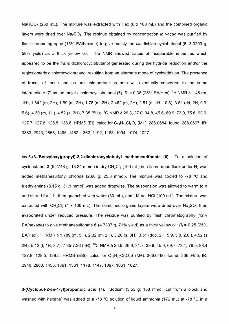
II. Synthesis of 3-(cyclobuten-2-yl)propanoic acid (CbK)
The cyclobutene-substituted acid was prepared using a modification of a route described by
Sittiwong, et al;1 the current route carried in the carboxyl of 7 as a benzyl ether rather than the
benzyl ester employed in the previous report (SI-1, Scheme 1).
SI-1, Scheme 1:
Synthesis of 3-
(cyclobuten-2-
yl)propanoic acid (CbK)
OH3
BnBr, NaHn-Bu4NI, THF
1) Zn, Cl3CCOCl2) NaBH4
HO
3OBn
MsCl,Et3N
MsO
3OBn
2
4 5
76
OBn3
Cl Cl
Cl Cl
OH
O
81% 59%
71%49% (2 steps)
1. Na, NH32. H2CrO4
1. Na-Boc-Lys
2. TFA
2
O
NH2
CO2H
HN
CbK55% (2 steps)
3
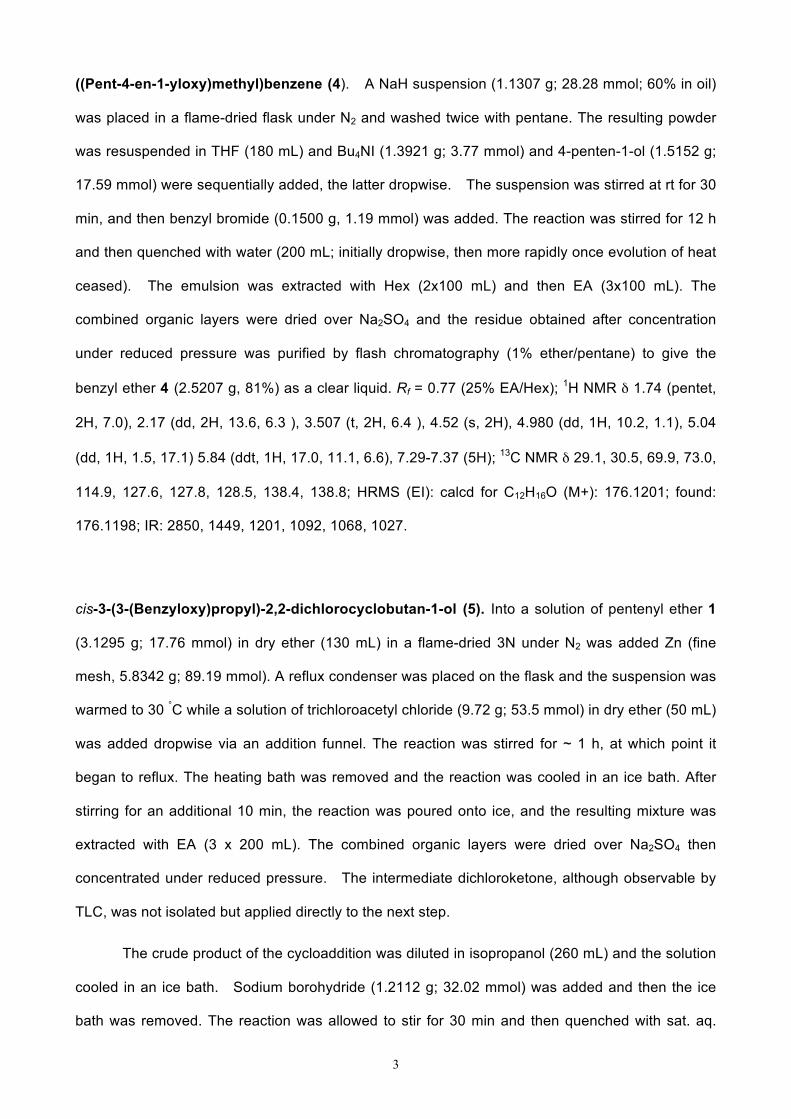
((Pent-4-en-1-yloxy)methyl)benzene (4). A NaH suspension (1.1307 g; 28.28 mmol; 60% in oil)
was placed in a flame-dried flask under N2 and washed twice with pentane. The resulting powder
was resuspended in THF (180 mL) and Bu4NI (1.3921 g; 3.77 mmol) and 4-penten-1-ol (1.5152 g;
17.59 mmol) were sequentially added, the latter dropwise. The suspension was stirred at rt for 30
min, and then benzyl bromide (0.1500 g, 1.19 mmol) was added. The reaction was stirred for 12 h
and then quenched with water (200 mL; initially dropwise, then more rapidly once evolution of heat
ceased). The emulsion was extracted with Hex (2x100 mL) and then EA (3x100 mL). The
combined organic layers were dried over Na2SO4 and the residue obtained after concentration
under reduced pressure was purified by flash chromatography (1% ether/pentane) to give the
benzyl ether 4 (2.5207 g, 81%) as a clear liquid. Rf = 0.77 (25% EA/Hex); 1H NMR δ 1.74 (pentet,
2H, 7.0), 2.17 (dd, 2H, 13.6, 6.3 ), 3.507 (t, 2H, 6.4 ), 4.52 (s, 2H), 4.980 (dd, 1H, 10.2, 1.1), 5.04
(dd, 1H, 1.5, 17.1) 5.84 (ddt, 1H, 17.0, 11.1, 6.6), 7.29-7.37 (5H); 13C NMR δ 29.1, 30.5, 69.9, 73.0,
114.9, 127.6, 127.8, 128.5, 138.4, 138.8; HRMS (EI): calcd for C12H16O (M+): 176.1201; found:
176.1198; IR: 2850, 1449, 1201, 1092, 1068, 1027.
cis-3-(3-(Benzyloxy)propyl)-2,2-dichlorocyclobutan-1-ol (5). Into a solution of pentenyl ether 1
(3.1295 g; 17.76 mmol) in dry ether (130 mL) in a flame-dried 3N under N2 was added Zn (fine
mesh, 5.8342 g; 89.19 mmol). A reflux condenser was placed on the flask and the suspension was
warmed to 30 °C while a solution of trichloroacetyl chloride (9.72 g; 53.5 mmol) in dry ether (50 mL)
was added dropwise via an addition funnel. The reaction was stirred for ~ 1 h, at which point it
began to reflux. The heating bath was removed and the reaction was cooled in an ice bath. After
stirring for an additional 10 min, the reaction was poured onto ice, and the resulting mixture was
extracted with EA (3 x 200 mL). The combined organic layers were dried over Na2SO4 then
concentrated under reduced pressure. The intermediate dichloroketone, although observable by
TLC, was not isolated but applied directly to the next step.
The crude product of the cycloaddition was diluted in isopropanol (260 mL) and the solution
cooled in an ice bath. Sodium borohydride (1.2112 g; 32.02 mmol) was added and then the ice
bath was removed. The reaction was allowed to stir for 30 min and then quenched with sat. aq.
4
NaHCO3 (250 mL). The mixture was extracted with Hex (6 x 100 mL) and the combined organic
layers were dried over Na2SO4. The residue obtained by concentration in vacuo was purified by
flash chromatography (15% EA/hexane) to give mainly the cis-dichlorocyclobutanol (5, 3.0203 g,
59% yield) as a thick yellow oil. The NMR showed traces of inseparable impurities which
appeared to be the trans dichlorocyclobutanol generated during the hydride reduction and/or the
regiosiomeric dichlorocyclobutanol resulting from an alternate mode of cycloaddition. The presence
of traces of these species are unimportant as both will eventually converted to the same
intermediate (7) as the major dichlorocyclobutanol (5). Rf = 0.38 (25% EA/Hex); 1H NMR δ 1.48 (m,
1H), 1.642 (m, 2H), 1.69 (m, 2H), 1.78 (m, 2H), 2.462 (m, 2H), 2.51 (d, 1H, 10.8), 3.51 (dd, 2H, 9.9,
5.6), 4.30 (m, 1H), 4.52 (s, 2H), 7.35 (5H); 13C NMR δ 26.8, 27.0, 34.8, 45.6, 69.9, 73.0, 75.6, 93.0,
127.7, 127.8, 128.5, 138.6; HRMS (EI): calcd for C14H18Cl2O2 (M+): 288.0684; found: 288.0697; IR:
3383, 2943, 2856, 1495, 1452, 1362, 1192, 1163, 1094, 1074, 1027.
cis-3-(3-(Benzyloxy)propyl)-2,2-dichlorocyclobutyl methanesulfonate (6). To a solution of
cyclobutanol 2 (5.2748 g; 18.24 mmol) in dry CH2Cl2 (100 mL) in a flame-dried flask under N2 was
added methanesulfonyl chloride (2.96 g; 25.8 mmol). The mixture was cooled to -78 °C and
triethylamine (3.15 g; 31.1 mmol) was added dropwise. The suspension was allowed to warm to rt
and stirred for 1 h, then quenched with water (30 mL), and 1M aq. HCl (100 mL). The mixture was
extracted with CH2Cl2 (4 x 100 mL). The combined organic layers were dried over Na2SO4 then
evaporated under reduced pressure. The residue was purified by flash chromatography (12%
EA/hexane) to give methanesulfonate 6 (4.7337 g, 71% yield) as a thick yellow oil: Rf = 0.25 (25%
EA/Hex); 1H NMR δ 1.789 (m, 5H), 2.32 (m, 2H), 3.20 (s, 3H), 3.51 (ddd, 2H, 5.9, 3.0, 2.6 ), 4.52 (s,
2H), 5.12 (t, 1H, 8.7), 7.35-7.36 (5H); 13C NMR δ 26.9, 26.9, 31.7, 39.6, 45.9, 69.7, 73.1, 78.5, 88.4,
127.8, 128.5, 138.5; HRMS (ESI): calcd for C15H20Cl2O4S (M+): 366.0460; found: 366.0455; IR:
2940, 2860, 1453, 1361, 1361, 1178, 1147, 1097, 1061, 1027.
3-(Cyclobut-2-en-1-yl)propanoic acid (7). Sodium (3.53 g; 153 mmol; cut from a block and
washed with hexane) was added to a -78 °C solution of liquid ammonia (172 mL) at -78 °C in a
5
flame-dried flask under N2. To the resulting blue solution was added a solution of
methanesulfonate 3 (3.1667 g; 8.62 mmol) in dry THF (2 mL). The suspension was stirred at -
78 °C for 1 h, after which additional sodium (2.06 g; 89.6 g) was added. The reaction was stirred
for an additional 30 min and then slowly quenched with sat. aq. NH4Cl. (100 mL). The mixture was
allowed to warm to rt over 5 h, during which time the ammonia was allowed to evaporate. The
resulting suspension was diluted with aq. 1M HCl (100 mL), and the mixture extracted with ether (4
x 50 mL). The combined organic layers were dried over Na2SO4 and the majority of the solvent
was carefully removed by concentration at 80 mm. The majority of the crude alcohol was subjected
to oxidation (next step) without purification. A small portion of the volatile alcohol was purified by
flash chromatography (20% ether/pentane) and/or microscale bulb-to-bulb distillation (75 °C at 30
mm) for NMR analysis: Rf = 0.31 (25% EA/Hex); 1H NMR δ 1.57 (m, 4H), 2.07 (d, 1H, 13.4), 2.67
(dd, 1H, 13.7, 4.2), 2.82 (ddd, 1H, 6.1, 4.2), 3.66 (t, 2H, 6.3), 6.06 (d, 1H, 2.6), 6.11 (d, 1H, 2.4);
13C NMR δ 30.8, 31.3, 36.9, 43.9, 63.2, 135.5, 140.8; IR: 3314, 3037, 2914, 2846, 1451, 1325,
1288, 1052, 1008, 918, 902, 694.
The partially concentrated solution of alcohol obtained in the previous step was dissolved in
acetone (105 mL). Into the 0 °C solution was dropwise added chromic acid in aqueous acetone
(Jones' reagent, ~2M, 21 mL). The suspension was stirred for 10 min and then extracted with
pentane (8 x 75 mL). The combined extracts were washed with 1M NaOH (4x50 mL). The
combined alkaline washes were cooled in an ice bath and acidified (6 M HCl) to a pH of ~ 1. The
solution was extracted with ether (5 x 150 mL) and the combined organic layers were dried over
Na2SO4 and then concentrated under reduced pressure. The residue was purified by flash
chromatography on acidified silica (230-400 mesh silica gel rinsed with 0.1M sulfuric acid, and then
dried overnight at 120 °C) with 50% ether/pentane to provide 3-(cyclobut-2-en-1-yl)propanoic acid
7 (0.5343 g, 49% over two steps) as a light yellow liquid: Rf = 0.22 (25% EA/Hex); 1H NMR δ 1.83
(q, 2H, 7.2), 2.10 (d, 1H, 13.9), 2.39 (t, 2H, 7.2 ), 2.68 (dd, 1H, 13.9, 4.5), 2.86 (ddd, 1H, 7.2, 4.6,
4.6), 6.084 (m, 2H), 11.51 (s, 1H); 13C NMR δ 29.4, 32.5, 36.4, 43.2, 136.0, 139.9, 180.6; HRMS
(EI): calcd for C7H10O2 (M+): 126.0681; found: 126.0683; IR: 2915, 1702, 1410, 1274, 1216.
6
N6-(3-(Cyclobut-2-en-1-yl)propionyl)-L-lysine (CbK). To an ice-bath cooled solution of
cyclobutene derived acid (252 mg, 2 mmol, 1 eq.) in anhydrous CH2Cl2 (10 mL) was slowly added
oxalyl chloride (171 uL, 5 mmol, 2.5 eq.), followed by 1 drop of N,N-dimethylformamide. The
solution was allowed to warm to rt and stirred for 2 h. After careful removal of solvent and volatile
byproducts under reduced pressure (~ 50 mm), the residue was dissolved in 1 mL THF and added
to a 0 °C solution of Nα-Boc-L-lysine (592 mg, 2 mmol, 1 eq.) in a mixture of 1 N NaOH (5 mL) and
THF (5 mL). The reaction was stirred overnight at rt and then recooled to 0°C and washed with
cold ether (3x5 mL). The aqueous layer was then acidified to between pH 1 and 2 and extracted
with EA (3x20 mL). The combined organic layers were dried over Na2SO4 and concentrated under
reduced pressure. The residue was dissolved in a mixture of CH2Cl2 (5 mL) and trifluoroacetic
acid (5 mL) and the reaction stirred for 1 h. The residue obtained after concentration was
dissolved in minimal amount of methanol and the product precipitated by addition of ether. The
collected precipitate was dried under vacuum to furnish (279 mg, 55% yield): 1H NMR (D2O/NaOH)
δ 6.03-6.01 (m, 2H), 3.07-3.05 (m, 3H), 2.71-2.68 (m, 1H), 2.58-2.54 (m, 1H), 2.18-2.15 (m, 2H),
2.01-1.97(m, 1H), 1.70-1.62(m, 2H), 1.53-1.43(m, 4H), 1.30-1.25 (m, 2H); 13C NMR (D2O/NaOH) δ
183.6, 176.9, 140.6, 136.3, 56.1, 43.0, 39.4, 36.1, 34.5, 30.4, 28.4, 22.6; HRMS (ESI) calc. for
[C13H22N2O3+H]: 255.1703; found: 255.1702.
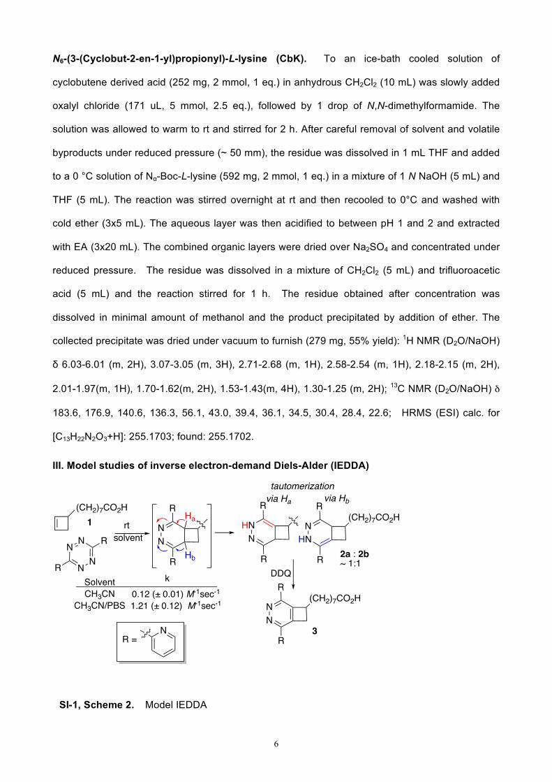
III. Model studies of inverse electron-demand Diels-Alder (IEDDA)
SI-1, Scheme 2. Model IEDDA
N N
NN
R
R
(CH2)7CO2H
HNN
R
R
SolventCH3CN
CH3CN/PBS
rtsolvent
0.12 (± 0.01) M-1sec-1
1.21 (± 0.12) M-1sec-1 NN
R
R(CH2)7CO2H
1
3R =
N
NHN
R
R(CH2)7CO2H
DDQ
NN
R
R
Hb
Ha
k
2a : 2b ~ 1:1
tautomerizationvia Ha via Hb
7
3-(Cyclobut-2-en-1-yl)octanoic acid (1) was prepared using a published procedure.1
8-(2,5-Di(pyridin-2-yl)-3,4-diazabicyclo[4.2.0]octa-1,4-dien-7-yl)octanoic acid & 8-(2,5-
di(pyridin-2-yl)-3,4-diazabicyclo[4.2.0]octa-2,5-dien-7-yl)octanoic acid (2a, 2b). Commercially
available 3,6-di(pyridin-2-yl)-1,2,4,5-tetrazine (0.1299 g, 0.55 mmol, 1.1 equiv, Sigma-Aldrich) was
dissolved in 5 mL of a 1:1 mixture of THF and CH3CN (both distilled) in a round bottom flask. A
magnetic stir bar was added and the flask was capped with a septum. Dissolution of the tetrazine
resulted in a clear and bright pink solution. Cyclobutene 1 (0.5mmol, 0.0981g 1.0 eq.)1 was added
and the reaction was allowed to stir overnight, gradually darkening to a red-orange solution
containing a white precipitate. The solid (0.0460 g, 23%) was collected by vacuum filtration and
determined by NMR to be an inseparable mixture of dihydrodiazine tautomers (2a, 2b): 1H (MeOD,
400MHz) δ 1.23 (s, 8H), 1.35 (t, 3H, 7.4), 1.47 (t, 2H, 6.4), 1.57 (q, 1H, 7.52), 1.76 (m, 2H), 2.09 (t,
2H 7.4), 2.17 (t, 2H, 7.4), 2.57 (t, 1H, 10.8), 3.13 (m, 2H), 3.60 (m, 1H), 4.26 (d, 1H, 9.4), 6.96 (m,
1H), 7.01 (d, 1H, 8.04), 7.08 (m, 1H), 7.33 (m 2H), 7.56 (m, 1H), 7.72 (m, 2H), 7.82 (dt, 1H, 7.7,
1.5), 7.94 (d, 1H, 8), 8.07 (d, 1H, 7.9), 8.10 (d, 1H, 4.1), 8.31 dd, 2H, 19.1, 4.3 ), 8.43 (d, 1H, 4.4),
8.64 (s, 1H) δ 12.01 (s, 2H).
8-(2,5-Di(pyridin-2-yl)-3,4-diazabicyclo[4.2.0]octa-1,3,5-trien-7-yl)octanoic acid (3). 3,6-
Di(pyridin-2-yl)-1,2,4,5-tetrazine (0.0520 g, 0.22 mmol, 1.1 eq.) was placed in a flame-dried, N2-
backfilled 10 ml RBF equipped with a magnetic stir bar and a capped with a rubber septum. A
solution of 8-(cyclobut-2-en-1-yl)octanoic acid (1, 0.0393g, 0.2 mmol, 1.0 eq.) in 3 mL of distilled
tetrahydrofuran was added and the stirred mixture was heated to 65 °C (oil bath) for 2.5 h.
Dissolution of the tetrazine resulted in a bright pink solution which gradually took on an orange-red
color as reaction proceeded. 2,3-Dichloro-5,6-dicyano-1,4-benzoquinone (DDQ, 0.0499 g, 0.22
mmol, 1.1eq) was added, and the reaction solution immediately became a deep red color. TLC (10%
CH3OH/ CH2Cl2) revealed a UV active spot with an Rf just lower than the tetrazine starting material.
The reaction was concentrated in vacuo and the residue submitted directly to flash column
chromatography (~30 cm Si in a column of 2.5 cm diameter) using a gradient of 0 % (200mL) to 2%
8
(400 mL) to 6% (400 mL) CH3OH/CH2Cl2 which incorporated 2 drops of glacial acetic acid per 100
mL volume. The residue obtained after concentration was redissolved in 0.5mL of methanol and
placed in a -20 °C freezer. The dipyridyl diazine 3 crystallized out of this solution after 18 h and
was collected by filtration (0.0471g 58%): Rf = 0.15 (10% CH3OH/CH2Cl2); 1H (CDCl3 400MHz) δ
1.32 (m 8H), δ 1.62 (m 2H), δ 2.33 (t, 3H, 6.8), δ 3.42 (dd, 1H, 16.0, 2.2), 3.85 (dd 15.8, 4.84), 4.08
(m 1H) δ 7.35 (m, 2H), 7.87 (dq, 2H, 11.74, 1.68), 8.64 (t, 2H, 9.12), 8.73 (t, 2H, 4.9); 13C (CDCl3,
100MHz, 10 sec delay, 0 Hz line broadening) δ 24.7, 26.9, 28.7, 28.8, 29.1, 31.6, 34.1, 38.8, 47.1,
122.0, 122.1, 124.3, 124.4, 136.9, 137.1, 146.9, 149.6, 149.7, 151.7, 153.1, 153.3, 154.2, 154.4,
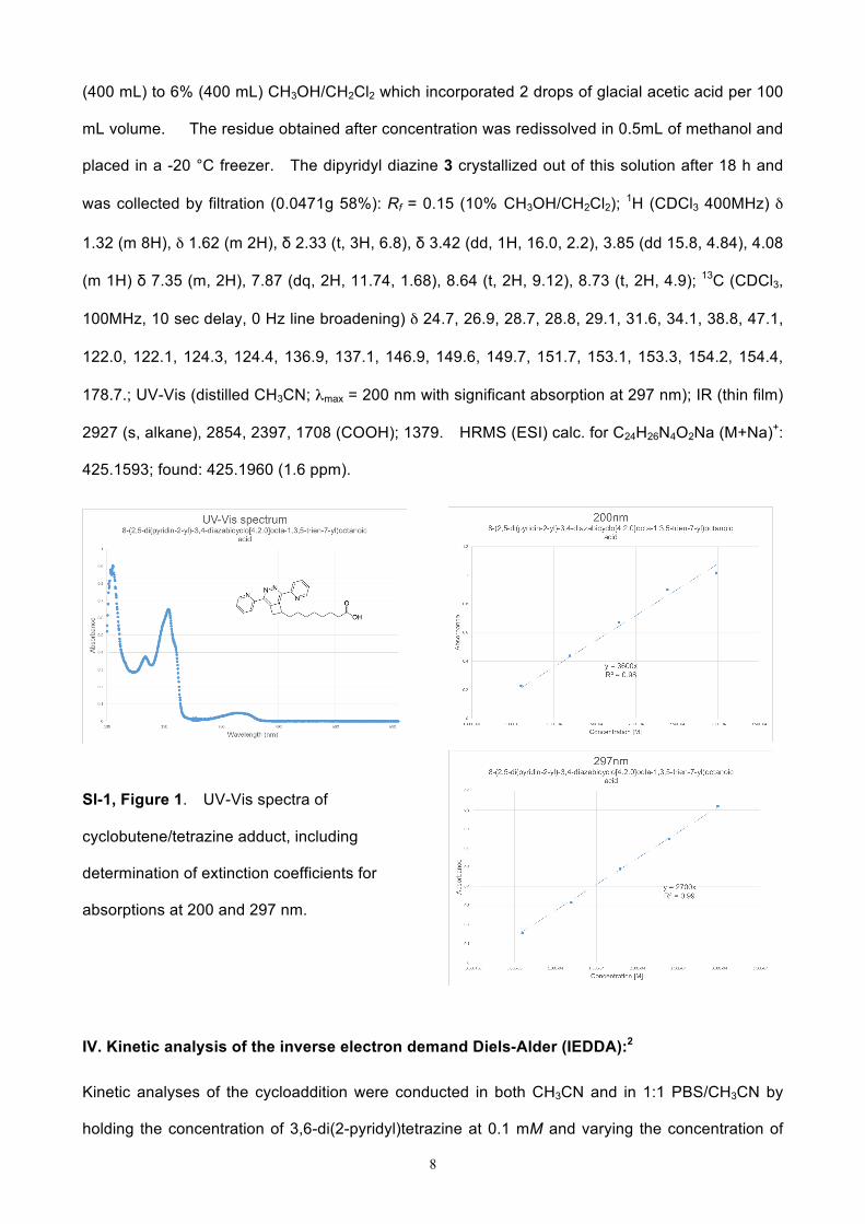
178.7.; UV-Vis (distilled CH3CN; λmax = 200 nm with significant absorption at 297 nm); IR (thin film)
2927 (s, alkane), 2854, 2397, 1708 (COOH); 1379. HRMS (ESI) calc. for C24H26N4O2Na (M+Na)+:
425.1593; found: 425.1960 (1.6 ppm).
SI-1, Figure 1. UV-Vis spectra of
cyclobutene/tetrazine adduct, including
determination of extinction coefficients for
absorptions at 200 and 297 nm.
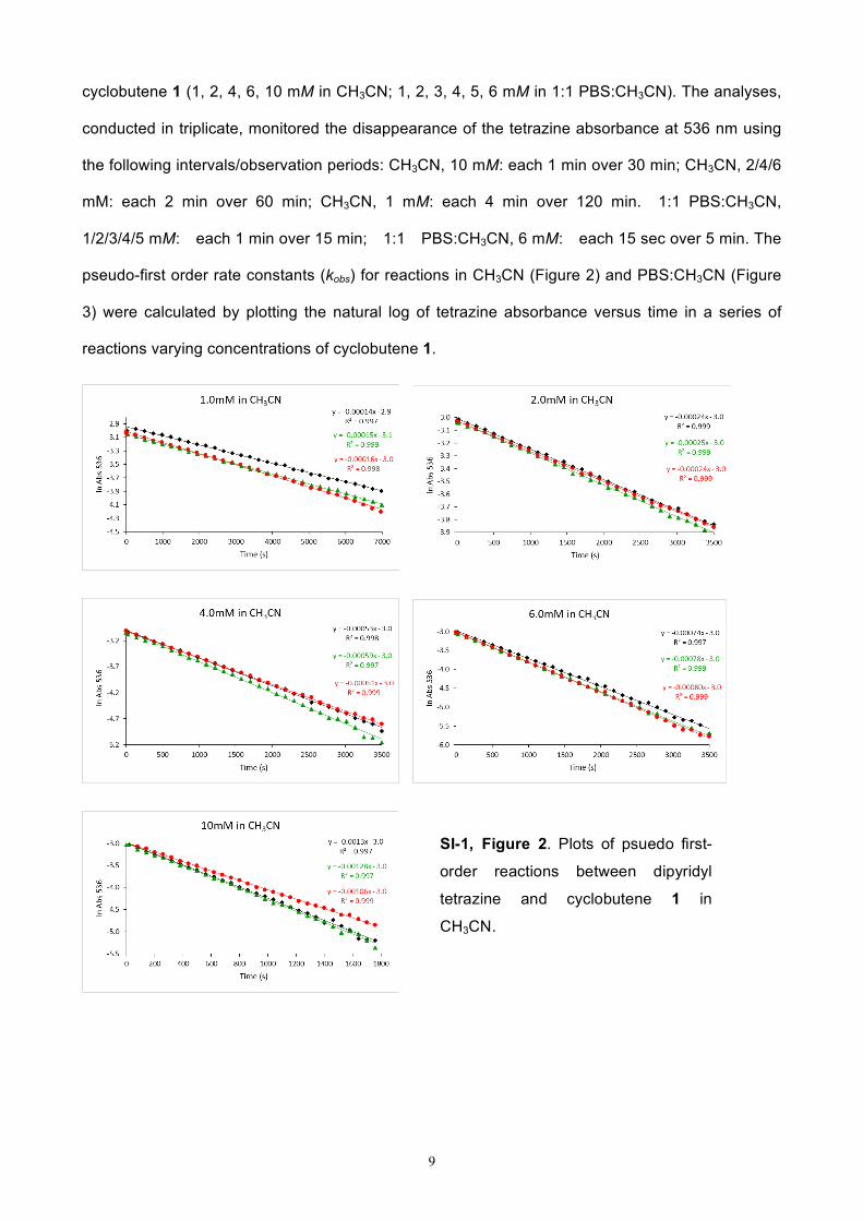
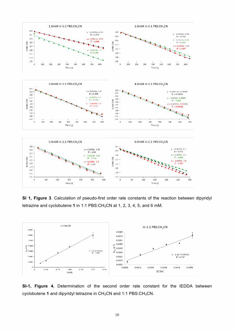
IV. Kinetic analysis of the inverse electron demand Diels-Alder (IEDDA):2
Kinetic analyses of the cycloaddition were conducted in both CH3CN and in 1:1 PBS/CH3CN by
holding the concentration of 3,6-di(2-pyridyl)tetrazine at 0.1 mM and varying the concentration of
9
cyclobutene 1 (1, 2, 4, 6, 10 mM in CH3CN; 1, 2, 3, 4, 5, 6 mM in 1:1 PBS:CH3CN). The analyses,
conducted in triplicate, monitored the disappearance of the tetrazine absorbance at 536 nm using
the following intervals/observation periods: CH3CN, 10 mM: each 1 min over 30 min; CH3CN, 2/4/6
mM: each 2 min over 60 min; CH3CN, 1 mM: each 4 min over 120 min. 1:1 PBS:CH3CN,
1/2/3/4/5 mM: each 1 min over 15 min; 1:1 PBS:CH3CN, 6 mM: each 15 sec over 5 min. The
pseudo-first order rate constants (kobs) for reactions in CH3CN (Figure 2) and PBS:CH3CN (Figure
3) were calculated by plotting the natural log of tetrazine absorbance versus time in a series of
reactions varying concentrations of cyclobutene 1.
SI-1, Figure 2. Plots of psuedo first-
order reactions between dipyridyl
tetrazine and cyclobutene 1 in
CH3CN.
10
SI 1, Figure 3. Calculation of pseudo-first order rate constants of the reaction between dipyridyl
tetrazine and cyclobutene 1 in 1:1 PBS:CH3CN at 1, 2, 3, 4, 5, and 6 mM.
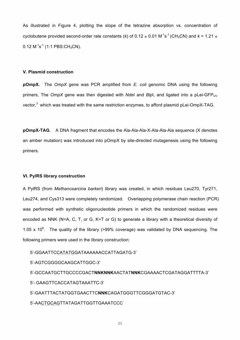
SI-1, Figure 4. Determination of the second order rate constant for the IEDDA between
cyclobutene 1 and dipyridyl tetrazine in CH3CN and 1:1 PBS:CH3CN.
11
As illustrated in Figure 4, plotting the slope of the tetrazine absorption vs. concentration of
cyclobutene provided second-order rate constants (k) of 0.12 ± 0.01 M-1s-1 (CH3CN) and k = 1.21 ±
0.12 M-1s-1 (1:1 PBS:CH3CN).
V. Plasmid construction
pOmpX. The OmpX gene was PCR amplified from E. coli genomic DNA using the following
primers, The OmpX gene was then digested with NdeI and BlpI, and ligated into a pLei-GFPUV
vector,3 which was treated with the same restriction enzymes, to afford plasmid pLei-OmpX-TAG.
pOmpX-TAG. A DNA fragment that encodes the Ala-Ala-Ala-X-Ala-Ala-Ala sequence (X denotes
an amber mutation) was introduced into pOmpX by site-directed mutagenesis using the following
primers.
VI. PylRS library construction
A PylRS (from Methanosarcina barkeri) library was created, in which residues Leu270, Tyr271,
Leu274, and Cys313 were completely randomized. Overlapping polymerase chain reaction (PCR)
was performed with synthetic oligonucleotide primers in which the randomized residues were
encoded as NNK (N=A, C, T, or G, K=T or G) to generate a library with a theoretical diversity of
1.05 x 10⁶. The quality of the library (>99% coverage) was validated by DNA sequencing. The
following primers were used in the library construction:
5’-GGAATTCCATATGGATAAAAAACCATTAGATG-3’
5’-AGTCGGGGCAAGCATTGGC-3’
5’-GCCAATGCTTGCCCCGACTNNKNNKAACTATNNKCGAAAACTCGATAGGATTTTA-3’
5’- GAAGTTCACCATAGTAAATTC-3’
5’-GAATTTACTATGGTGAACTTCNNKCAGATGGGTTCGGGATGTAC-3’
5’-AACTGCAGTTATAGATTGGTTGAAATCCC
12
VII. Directed evolution of PylRS
The resulting PylRS library was subjected to a negative selection to remove PylRS variants that
could charge tRNAPyl with natural amino acid as previosly described, followed by a positive
selection to identify functional PylRS variants. Briefly, the negative selection uses the toxic
barnase gene with amber mutations at permissive sites (Gln2TAG and Asp44TAG) and was
carried out in the absence of CbK. The positive selection is based on resistance to
chloramphenicol (Cm), which is conferred by the suppression of an amber mutation at a permissive
site (Asp112) in the chloramphenicol acetyltransferase-encoding gene in the presence of tRNAPyl,
CbK, and functional PylRS mutants. The surviving PylRS variants were subsequently screened
for chloramphenicol resistance level in the presence and absence of CbK. A few clones that
survived on 75 µg/mL chloramphenicol in the presence of CbK and did not grow on 35 µg/mL
chloramphenicol in the absence of CbK were identified. Among these clones, CbKRS
(Leu274Met and Cys313Ala), displayed the fastest growth rate in the presence of chloramphenicol.
VIII. Protein expression
E. coli GeneHogs cells harboring pBK-CbKRS and pLei-sfGFP-N149TAG4 were cultured in 50 mL
of LB medium containing 50 µg/mL kanamycin and 34 µg/mL chloramphenicol at 37 oC with
shaking. When the OD600 of the culture reached 0.6, the protein expression was induced by the
addition of IPTG (0.5 mM) and CbK (1.0 mM). After 24 h of cultivation at room temperature, cells
were collected by centrifugation (5,000g, 10 min), resuspended in lysis buffer (20 mM potassium
phosphate, pH 7.4, 150 mM NaCl, and 20 mM imidazole), and disrupted by sonication. Cellular
debris was removed by centrifugation (21,000g, 30 min, 4 oC). The cell-free lysate was applied to
Ni Sepharose 6 Fast Flow resin (GE Healthcare). Protein purification followed manufacturer’s
instructions. Purified protein was desalted using the Amicon Econo-Pac® 10DG column (BioRad
Laboratories, Inc) using PBS buffer.
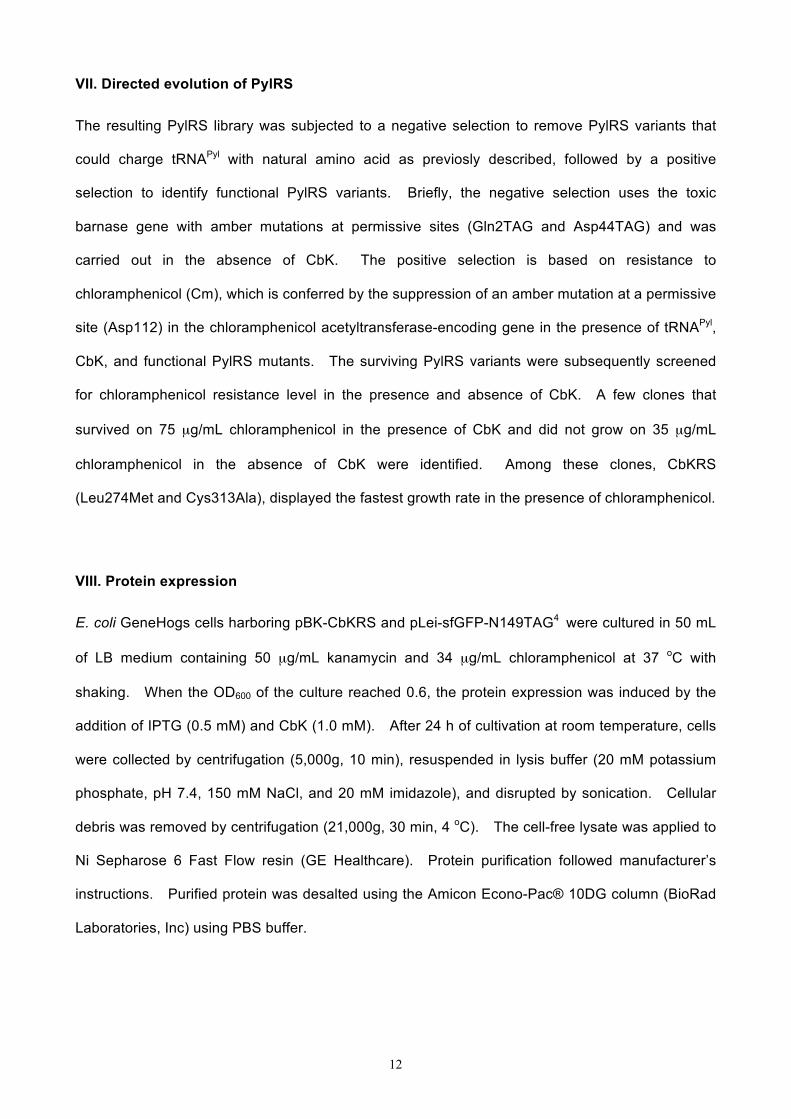
13
SI-1, Figure 5. SDS-PAGE analysis of protein purification.
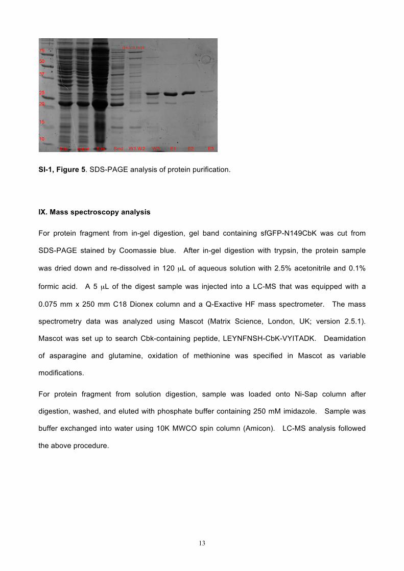
IX. Mass spectroscopy analysis
For protein fragment from in-gel digestion, gel band containing sfGFP-N149CbK was cut from
SDS-PAGE stained by Coomassie blue. After in-gel digestion with trypsin, the protein sample
was dried down and re-dissolved in 120 µL of aqueous solution with 2.5% acetonitrile and 0.1%
formic acid. A 5 µL of the digest sample was injected into a LC-MS that was equipped with a
0.075 mm x 250 mm C18 Dionex column and a Q-Exactive HF mass spectrometer. The mass
spectrometry data was analyzed using Mascot (Matrix Science, London, UK; version 2.5.1).
Mascot was set up to search Cbk-containing peptide, LEYNFNSH-CbK-VYITADK. Deamidation
of asparagine and glutamine, oxidation of methionine was specified in Mascot as variable
modifications.
For protein fragment from solution digestion, sample was loaded onto Ni-Sap column after
digestion, washed, and eluted with phosphate buffer containing 250 mM imidazole. Sample was
buffer exchanged into water using 10K MWCO spin column (Amicon). LC-MS analysis followed
the above procedure.
14
SI-1, Figure 6. LC-MS of in-gel digested sfGFP-N149CbK. Expected, 684.40 Da, z = 3; 1026.10
Da, z = 2. Observed 684.35 Da, z = 3; 1026.02 Da, z = 2.
X. Protein labeling in vitro
Purified sfGFP-N149CbK (10 µL, 0.24 mg/mL) in PBS buffer was treated with 1 µL of Fl-Tet (in
DMSO) at indicated concentrations. The reaction mixture was incubated at room temperature
with agitation for indicated period of time. Solution of 5-norbonene-2-methanol (1 µL, 100 mM) in
DMSO was added to quench the reaction. The quenched reaction was then mixed with SDS-PAGE
sample loading buffer (2x), heated at 95 oC for 15 min, and loaded onto wells of SDS-PAGE.
Fluorescence detection was performed before staining by Coomassie blue. Protein gels were
imaged using Bio-Rad Molecular Imager ChemDoc XRS+ or GE Typhoon FLA9500. Coomassie
blue-stained gels were imaged using Bio-Rad Molecular Imager. As a control, wild-type sfGFP
(10 µL, 0.24 mg/mL) was subjected to labeling and analysis under the same conditions.
15
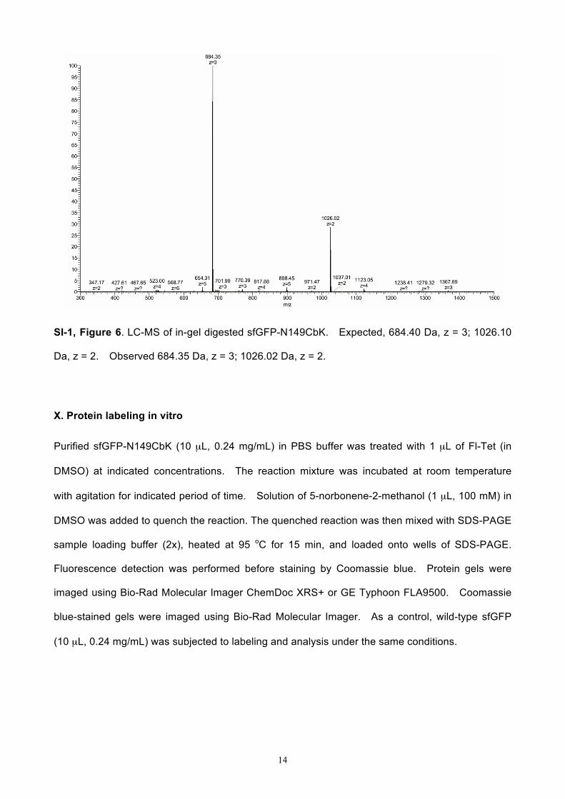
SI-1, Figure 7. In vitro protein labeling of sfGFP variants with cyclobutene-tetrazine reaction.
Fluorescence intensities of protein bands (Figure 3 of the maintext) were estimated using ImageJ
software. (A) Reaction progress of sfGFP-N149CbK protein labeling with 50 µM of Fl-Tet; (B)
Labeling of sfGFP-N149CbK protein with varied concentrations of Fl-Tet for 80 minutes.
16
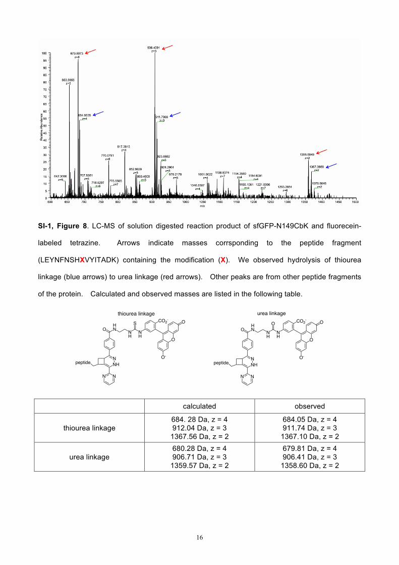
SI-1, Figure 8. LC-MS of solution digested reaction product of sfGFP-N149CbK and fluorecein-
labeled tetrazine. Arrows indicate masses corrsponding to the peptide fragment
(LEYNFNSHXVYITADK) containing the modification (X). We observed hydrolysis of thiourea
linkage (blue arrows) to urea linkage (red arrows). Other peaks are from other peptide fragments
of the protein. Calculated and observed masses are listed in the following table.
calculated observed
thiourea linkage 684. 28 Da, z = 4 912.04 Da, z = 3
1367.56 Da, z = 2
684.05 Da, z = 4 911.74 Da, z = 3
1367.10 Da, z = 2
urea linkage 680.28 Da, z = 4 906.71 Da, z = 3
1359.57 Da, z = 2
679.81 Da, z = 4 906.41 Da, z = 3
1358.60 Da, z = 2
OHN N
HNH
O CO2-
O
O-
O
peptide NHN
N N
OHN N
HNH
S CO2-
O
O-
O
peptide NHN
N N
thiourea linkage urea linkage
17
XI. Live cell labeling
E. coli cells expressing either wild-type OmpX or OmpX-CbK mutant proteins were harvested by
centrifugation (21,000 g, 5 min, 4 °C). Collected cells were washed three times with PBS buffer.
Washed cells were resuspended in PBS buffer. Fl-Tet stock solution was added into cell
suspension to a final concentration of 100 µM. After incubation at room temperature with
agitation for 60 min, cells were collected by centrifugation (21,000 g, 5 min, 4 °C) and resuspended
in PBS buffer. Cell suspensions were placed on the surface of a glass slide and covered with a
glass cover slip for imaging. Cells were imaged on an Olympus FV500 inverted (Olympus IX-81)
confocal microscope. Excitation wavelength (495 nm) and emission filter (510 nm) were used in
the imaging experiments.
References:
1 (a) W. Sittiwong, D. K. Zinniel, R. J. Fenton, D. D. Marshall, C. B. Story, B. Kim, J. Lee, R.
Powers, R. G. Barletta, and P. H. Dussault, “Development of Cyclobutene- and Cyclobutane-
Functionalized Fatty Acids with Inhibitory Activity against Mycobacterium tuberculosis.”
ChemMedChem, 2014, 9, 1838; (b) W. Sittiwong. Synthesis and Application of Four-Membered
Carbocycle Amphiphiles. Ph.D. Thesis, University of Nebraska-Lincoln, Lincoln, NE, 2014.
2 (a) F. Sicilio, and M.D. Peterson, “Ratio errors in pseudo first order reactions.” J. Chem. Ed.
1961, 38, 576; (b) J.F. Corbett, “Pseudo first-order kinetics.” J. Chem. Ed. 1972, 49, 663.
3 N. Wang, T. Ju, W. Niu and J. Guo, “Fine-tuning Interaction between Aminoacyl-tRNA
Synthetase and tRNA for Efficient Synthesis of Proteins Containing Unnatural Amino Acids” ACS
Synth. Biol., 2014, 4, 207-212.
4 X. Shang, X. Song, C. Faller, R. Lai, H. Li, R. Cerny, W. Niu and J. Guo, “Fluorogenic protein
labeling using a genetically encoded unstrained alkene” Chem. Sci., 2017, 8, 1141-1145.