Embed Size (px)
Citation preview

Materials and methods
1.1 Materials
With the purity of over 98%, 7-Ethyl-10-hydroxylcamptothecin (SN38) and
irinotecan hydrochloride (CPT-11) were purchased from Meilun Biotechnology Co.,
LTD (Dalian, China). All other commercially obtained chemical reagents were analytical
grade or better.
1.2 Preparation of Nanocrystals-SN38 (NCsS)
10 mg of SN38 and 1 mL ammonium hydroxide were added in 3 mL DI water. The
reaction was kept stirring at room temperature and away from light, until turning to
transparent solution, which means that all the lactone forms of SN38 transformed to
the carboxylate acid (CA-SN38). Next, the transparent solution was diluted to 0.5 ~ 1
mg/mL with DI water and freeze-dried to induce the carboxylate acid of SN38
converting into active lactone form. During the freeze drying, the nanocrystals were
reconstructed and took the form of fluffy powders. The freeze-drying powders could
be dispersible in 5% glucose or water for injection.
1.3 1H NMR Spectra
The 1H NMR spectra, recorded by Varian INOVA spectrometer at 400 MHz in DMSO
d6, was applied to confirm that the NCs were completely permuted with lactone form
of SN38. Chemical shifts were displayed as δ value (ppm) against the internal standard
compound – Tetramethylsilane (TMS). Also, the coupling constants (J) were marked
as hertz (HZ). Multiplicities were recorded as singlet (s), doublet (d), triplet (t) and
multiplet (m).
1.4 High Performance Liquid Chromatography (HPLC)
The qualitative and quantitative detection of SN38 and CPT-11 were realized by an
Agilent HPLC system (Agilent 1260 Infinity, Agilent technology, USA), including an
Agilent quat pump, an Agilent variable wavelength detector and a C18 reversed-phase
analytical column (250mm × 4.5 mm, 5 μm, Scienhome, China)1. 36 mM sodium
dihydrogen phosphate, 4 mM sodium 1-heptanesulfonate (pH = 3.0, adjusted with
phosphoric acid solution) and acetonitrile (65 : 35) were applied as the mobile phase,
Electronic Supplementary Material (ESI) for ChemComm.This journal is © The Royal Society of Chemistry 2019

with a flow rate of 1 mL/min and a column temperature of 35 C. The UV detector was
set at 380 nm.
1.5 pH-dependent dynamic equilibrium
1 mg/mL SN38 or CPT-11 stock solution dissolved with DMSO was diluted to 5
μg/mL with 10 mM buffer solution at varying pH values, including 5.0, 6.0, 7.0, 8.0 and
9.0. Samples were placed at 37 C for 2 h to achieve the equilibrum between lactone
and carboxylate acid form,2 and analyzed by HPLC to characterize the ratio of the
closed-lactone at different pH values.
1.6 HPLC tandem triple-quad mass spectrometry (LC–MS/MS)
The quantitative detection of SN38 and CPT-11 were realized by a LC-MS/MS
(Agilent 6410B, Agilent technology, USA)3, including an Agilent binary pump and an
ODS analytical column (50 × 4.6 mm, 1.8 μm, Diamonsil). 10mM ammonium acetate
(0.1% formic acid, pH = 3) and acetonitrile (65:35) were applied as the mobile phase,
with a flow rate of 0.4 mL/min and an injection volume of 1 μL. The retention time of
CPT-11 and SN38 were 0.84 and 1.31 min respectively. Multiple reaction monitoring
(MRM) positive mode was set to analyze CPT-11 and SN38. The parameters of mass
spectrometer were applied as following settings: CPT-11 transition (m/z),
587.3→167.1, collision energy, 44 eV; SN38 transition (m/z), 393.3→349.1, collision
energy, 24 eV; gas temperature, 350 C; nebulizer pressure, 35 psi and spray voltage,
4000 V.
1.7 Characterization of NCsS
Size and Zeta Potentials. The particle size, size distribution and Zeta potentials of
NCs were measured by Dynamic Light Scattering (DLS, Zetasizer Nano ZS90, Malvern,
UK) at 25 C.
Morphology. By using field emission scanning electron microscope (JSM – 7500F,
JEOL, Japan), the Scanning Electron Microscope (SEM) image of NCs was gained at 15
keV working voltage. The Transmission Electron Microscope (TEM) imaging of NCsS
was performed on an H-600 TEM (Hitachi, Japan) with negative staining by 2%
phosphotungstic acid.
Crystalline Form. Powder X-ray Diffraction (PXRD) analyses were applied to reveal
the formation mechanism of NCs from 10° to 70° with a minimum step size 2θ of

0.001° at a detecting speed of 0.045° 2θ/min. To analyze crystalline states, SN38
powder and freeze-dried NCsS powder were included.
1.8 Cell lines
The 4T1 cells4 were cultured in RPMI-1640 supplemented with 10% fetal bovine
serum and 1% penicillin streptomycin combination, in a humidified atmosphere of 5%
CO2 at 37 °C.
1.9 Cytotoxicity assays
The cytotoxicity of free CPT-11, free SN38 and NCsS were determined by 3- (4, 5-
Dimethylthiazol-2-yl) - 2, 5- diphenyltetrazolium bromide assay (MTT assay) in 4T1
cells. Briefly, cells were seeded in 96 well plates at a density of 5000 cells per well and
incubated for 12 h. The medium was replaced by 100 μL medium with serial dilutions
of CPT-11, SN38 or NCsS (CPT-11 from 10 μg/mL to 100 μg/mL and SN38 from 0.1
μg/mL to 10 μg/mL) and were incubated for 24 h. Next, cells were treated with 100 μL
MTT solution (0.5 μg/mL in PBS) for 4 h at 37 °C. Then, the medium was discarded and
100 μL DMSO was added to dissolve the formazan crystal formed by viable cells. The
absorbance at 470 nm was recorded by a microplate reader (Tecan spark 10M, China).
Cells treated with fresh RPMI-1640 medium were set as control. Cell viability (%) was
calculated in accordance with the following equation:
Cell viability (%) =
Atest - Ablank
Acontrol - Ablank
× 100%.
1.10 Cellular uptake study
The 4T1 cells were seeded in 6 well plates at a density of 5 × 10⁵ cells per well and
incubated for 12 h. Next, 4T1 cells were incubated with free CPT-11, SN38 or NCsS at
an isomolar concentration of 13 nM for 1, 2 or 4 h, in a humidified atmosphere of 5%
CO2 at 37 °C. Then, the cells were washed with cold PBS for 3 times and obtained by
centrifuging at 2000 rpm for 3 min. And 0.3 mL DI water was added to resuspend
centrifuged cells. Finally, the cells suspension was lysed in the freezing and thawing
cycle for 5 times to release the intracellular uptake drugs. The lysed cell suspension
(100 μL) and organic solvent mixture (200 μL, methanol/acetonitrile, 1:1 and 0.5%
ethylic acid) were mixed. After vortex mixing for 5 min, the supernatant was obtained

by twice centrifuging at 13, 500 rpm for 15 min and analyzed by LC-MS/MS.
Meanwhile, 20 μL lysed cell suspension was applied to detect the total protein content
by BCA assay reagent kit (Thermo Fisher Scientific, USA). The results were represented
by the amount of CPT-11, SN38 or NCsS (nmol) in per milligram protein.
1.11 Confocal laser scanning microscopy (CLSM)
The intracellular distribution of free SN38 and NCsS were studied in 4T1 cells by the
CLSM assay. Cells were seeded in confocal dishes at a density of 1 × 10⁵ cells per well.
After 12 h, the original medium was removed and medium containing 13 nM of free
SN38 or NCsS was added. After incubation for 2 h, culture medium was removed, and
cells were rinsed with PBS for 3 times. The lysosomes were stained with Lyso-Tracker
Red (KGMP006, KeyGEN BioTECH, China) for 1 h. After being fixed with 4% (w/v)
paraformaldehyde operation solution for 20 min, the cell nuclei were stained with
DAPI for 15 min. Then cells were washed with PBS for 3 times and were observed with
CLSM (LSM-800, ZEISS, Germany), with emission wavelength of 454 nm for DAPI, 530
nm for SN38, and 590 nm for Lyso-Tracker Red.
1.12 Animals and BALB/c mice bearing 4T1 tumors
Healthy male Sprague-Dawley rats and female BALB/c mice (6 weeks old) were
purchased from Institute of Laboratory Animals of Sichuan Academy of Medical
Sciences & Sichuan Provincial People’s Hospital (Chengdu, China). All the animal
experiments presented were approved of the Institutional Animal Care and Ethic
Committee of Sichuan University in accordance with the Chinese guidelines for the
care and use of laboratory animals.
4T1 cells were orthotopically injected into the right underarm mammary fat pad at
a density of 1 × 10⁵ cells per BALB/c mouse in cold PBS4, 5. The volume of tumor was
measured by a digital caliper every other day, and was calculated by the following
equation: V = 0.5 × length × width2.
1.13 Pharmacokinetics
The pharmacokinetics of CPT-11 and NCsS were evaluated on male SD rats (n = 5).
Irinotecan hydrochloride (CPT-11), a prodrug of SN38, is widely applied in treating
many kinds of carcinoma. According to previous reports3, 6, it was dissolved in 5%

dextrose at pH 6.0 (1 mg/mL) for injection. The free CPT-11 solution or NCsS was
administered intravenously at an equivalent dosage of 5.6 mg/kg SN38. At planned
time points after tail vein injection (0.17, 0.33, 0.50, 1, 2, 4, 8 and 12h), 100 μL of blood
was collected from infraorbital venous plexus. Organic solvent mixture (200 μL,
methanol/acetonitrile, 1:1 and 0.5% ethylic acid) was added and eddied for 5 min. The
supernatant was acquired by twice centrifugation at 13, 500 rpm for 15 min and
analyzed by LC-MS/MS.
1.14 Biodistribution
The tumor-bearing mice were randomly divided into two groups (n = 20) with
intravenously injection of free CPT-11 solution or NCsS at an equivalent dosage of 10
mg/kg SN38, when tumor volume reached approximately 300 mm3. Mice were
sacrificed at planned time points after injection (1, 4, 8 and 24 h, n = 5). Major organs
(heart, liver, spleen, lung and kidney) and tumor were collected, washed with normal
saline solution, dried with filter paper, weighed and homogenized with double volume
of PBS (mL/g). Samples were disposed in accordance with the pharmacokinetics. The
concentrations of drug in different organs were detected by LC-MS/MS.
The penetration of free SN38 or NCsS were evaluated by immunofluorescence
staining. SN38 was dissolved in a mixed solvent, which was composed of dimethyl
sulfoxide, polyethylene glycol 400, ethanol and 5% glucose (3:3:2:2, in volume) for
injection. The tumor-bearing mice were tail intravenously injected with free SN38
solution or NCsS at an equivalent dosage of 10 mg/kg SN38. Mice were sacrificed 8 h
after injection and tumors were collected. The tumor sections were prepared and
stained with CD31 (EPR3094, Alexa Fluor® 647, Thermo Fisher Scientific, USA) to mark
the tumor vessel by the reported method.7 The nuclei were stained with DAPI and
sections were observed with the CLSM on the uniform conditions, with an emission
wavelength of 454 nm for DAPI, 530 nm for SN38, and 668 nm for CD31.
1.15 In vivo antitumor efficacy
The tumor-bearing mice were randomly divided into three groups (n = 5) and were
tail intravenously injected with normal saline solution (NS), free CPT-11 solution or
NCsS at an equivalent dosage of 10 mg/kg SN38, when tumor volume reached 50 ~
100 mm3. The dosage regimen was 5 injections with each in every 3 days. The weight
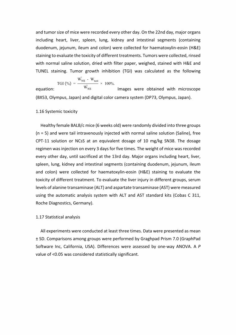
and tumor size of mice were recorded every other day. On the 22nd day, major organs
including heart, liver, spleen, lung, kidney and intestinal segments (containing
duodenum, jejunum, ileum and colon) were collected for haematoxylin-eosin (H&E)
staining to evaluate the toxicity of different treatments. Tumors were collected, rinsed
with normal saline solution, dried with filter paper, weighed, stained with H&E and
TUNEL staining. Tumor growth inhibition (TGI) was calculated as the following
equation: Images were obtained with microscope TGI (%) =
WNS - Wtest
WNS × 100%.
(BX53, Olympus, Japan) and digital color camera system (DP73, Olympus, Japan).
1.16 Systemic toxicity
Healthy female BALB/c mice (6 weeks old) were randomly divided into three groups
(n = 5) and were tail intravenously injected with normal saline solution (Saline), free
CPT-11 solution or NCsS at an equivalent dosage of 10 mg/kg SN38. The dosage
regimen was injection on every 3 days for five times. The weight of mice was recorded
every other day, until sacrificed at the 13rd day. Major organs including heart, liver,
spleen, lung, kidney and intestinal segments (containing duodenum, jejunum, ileum
and colon) were collected for haematoxylin-eosin (H&E) staining to evaluate the
toxicity of different treatment. To evaluate the liver injury in different groups, serum
levels of alanine transaminase (ALT) and aspartate transaminase (AST) were measured
using the automatic analysis system with ALT and AST standard kits (Cobas C 311,
Roche Diagnostics, Germany).
1.17 Statistical analysis
All experiments were conducted at least three times. Data were presented as mean
± SD. Comparisons among groups were performed by Graghpad Prism 7.0 (GraphPad
Software Inc, California, USA). Differences were assessed by one-way ANOVA. A P
value of <0.05 was considered statistically significant.

Fig. S1 Camptothecin analogues.

Fig. S2 UV-spectra of NCsS, SN38 (A) and CPT-11 (B). SN38 or CPT-11 stock solutions
and NCsS solution were diluted by deionized water to about 5 μg/mL and recorded by
a UV-Visible Spectrometer (Cary 100 Conc, Varian, USA).

Fig. S3 pH-dependent dynamic equilibrium of SN38 and CPT-11. A: Ratios of lactone
form between the chemical equilibrium of SN38 or CPT-11 on varying pH (n = 3). The
dynamic equilibrimu between lactone form and carboxylate acid form of SN38 (B) and
CPT-11 (C) monitored by HPLC.

Fig. S4 Characterization of free SN38, reaction product (CA-SN38) and
pharmaceutical product (NCsS-SN38) with HPLC (6.97, 3.42 and 6.97 min,
respectively).

Fig. S5 5 mg NCsS powder (left) and SN38 (right) with the same weight.

Fig. S6 1H NMR spectra of SN38 (A) and NCsS (B) (DMSO-d6, 400MHz). A: δ 10.30(s,
1H), 8.02(d, J = 8.4Hz, 1H), 7.42-7.39(m, 2H), 7.25(s, 1H), 6.49(s, 1H), 5.42(s, 2H),
5.27(s, 2H), 3.08(d, J = 7.2Hz, 2H), 1.92-1.81(m, 2H), 1.30(t, J = 7.2Hz, 3H), 0.88(t, J =
7.2Hz, 3H). B: δ 10.29 (brs, 1H), 8.02 (d, J = 6.0Hz, 1H), 7.41-7.39 (m, 2H), 7.24 (s, 1H),
6.50 (s, 1H), 5.42(s, 2H), 5.25 (s, 2H), 3.09-3.07 (m, 2H), 1.88-1.85 (m, 2H), 1.28 (t, J =
7.2Hz, 3H), 0.88 (t, J = 7.2Hz, 3H).

Fig. S7 Fluorescence excitation (A) and emission (B) spectra of SN38 (yellow) and
NCsS (carmine) recorded by a fluorescence spectrophotometer (RF-6000, Shimadzu,
Japan), with an emission and excitation wavelength of 550 nm and 380 nm
respectively .

Fig. S8 CLSM images of 4T1 cells incubated with free SN38 and NCsS for 2 h. The
nuclei were stained with DAPI (blue), SN38 fluorescence was shown in green and
lysosomes were stained with Lyso – tracker in red. The scale bars represent 5 μm.

Fig. S9 The concentration of CPT-11 and time curves of injected CPT-11 in SD rats.
Data represent mean ± SD, n = 5.

Fig. S10 Biodistribution of NCsS or SN38 converted from injected free CPT-11 (CPT-
SN38) in major organs, including heart (A), liver (B), spleen (C), lung (D), kidney (E); (F):
The concentration of CPT-11 in major organs 1, 4, 8 and 24 h after injection of free
CPT-11. Data represent mean ± SD, n = 5. **, p < 0.01, compared with the free CPT-11
group.

Fig. S11 The histological analysis on major organs of tumor-bearing mice from
differently treated groups to evaluate the antitumor efficacy and toxicity. The scale
bars represent 50 μm.

Fig. S12 The histological analysis on major intestinal segments of tumor-bearing mice
administrated with different treatments to evaluate the toxicity. The scale bars
represent 50 μm.

Fig. S13 Systemic toxicity evaluation. (A) Body weight shift of health BALB/c mice
treated with free CPT-11, NCsS or saline. ALT (B) and AST (C) levels in the serum of the
mice determined at 24 h after the fifth administration. (D) The histological analysis on
major organs of mice from differently treated groups. (E) The histological analysis on
major intestinal segments of mice administrated with different treatment.

References
1. M. T. Baylatry, A. C. Joly, J. P. Pelage, L. Bengrine-Lefevre, J. L. Prugnaud, A. Laurent and C. Fernandez, J Chromatogr B, 2010, 878, 738-742.
2. U. Y. Lau, L. T. Benoit, N. S. Stevens, K. K. Emmerton, M. Zaval, J. H. Cochran and P. D. Senter, Mol Pharmaceut, 2018, 15, 4063-4072.
3. H. Liu, H. Lu, L. F. Liao, X. M. Zhang, T. Gong and Z. R. Zhang, Drug Deliv, 2015, 22, 701-709.
4. Y. Lv, C. Xu, X. Zhao, C. Lin, X. Yang, X. Xin, L. Zhang, C. Qin, X. Han, L. Yang, W. He and L. Yin, ACS Nano, 2018, 12, 1519-1536.
5. H. Zhou, Z. Y. Fan, J. J. Deng, P. K. Lemons, D. C. Arhontoulis, W. B. Bowne and H. Cheng, Nano Lett, 2016, 16, 3268-3277.
6. S. Q. Hu, E. Lee, C. Wang, J. Q. Wang, Z. X. Zhou, Y. X. Li, X. Y. Li, J. B. Tang, D. H. Lee, X. R. Liu and Y. Q. Shen, J Control Release, 2015, 220, 175-179.
7. R. Tsumura, S. Manabe, H. Takashima, Y. Koga, M. Yasunaga and Y. Matsumura, J Control Release, 2018, 284, 49-56.