Embed Size (px)
Citation preview
Dr. Kaan Yücel http://yeditepeanatomy1.org Yeditepe Anatomy
THIGH & POPLITEAL FOSSA23. February.2012 Thursday
THIGHThe femoral region (thigh) is the region of the free lower limb that lies between the gluteal, abdominal,
and perineal regions proximally and the knee region distally: anteriorly, it is separated from the abdominal wall by the inguinal ligament; posteriorly, it is separated from the gluteal region by the gluteal fold superficially, and by the inferior margins of the gluteus maximus and quadratus femoris on deeper planes.
Structures enter and leave the top of the thigh by three routes: posteriorly , the thigh is continuous with the gluteal region and the major structure passing between the two regions is the sciatic nerve; anteriorly , the thigh communicates with the abdominal cavity through the aperture between the inguinal ligament and pelvic bone, and major structures passing through this aperture are the iliopsoas and pectineus muscles, the femoral nerve, artery and vein, and lymphatic vessels; medially , structures (including the obturator nerve and associated vessels) pass between the thigh and pelvic cavity through the obturator canal. Vessels and nerves passing between the thigh and leg pass through the popliteal fossa posterior to the knee joint.
Superficial Fascia of the ThighThe membranous layer of the superficial fascia of the anterior abdominal wall extends into the thigh and
is attached to the deep fascia (fascia lata) about a fingerbreadth below the inguinal ligament.Deep Fascia of the Thigh (Fascia Lata)
The deep fascia encloses the thigh like a trouser leg and at its upper end is attached to the pelvis and the inguinal ligament. On its lateral aspect, it is thickened to form the iliotibial tract. The saphenous opening is a gap in the deep fascia in the front of the thigh just below the inguinal ligament. Great saphenous vein penetrates this fascia and passes through the hiatus saphenus to drain into the femoral vein. The saphenous opening is filled with loose connective tissue called the cribriform fascia.
Fascial Compartments of the ThighThe thigh is divided into three compartments by intermuscular septa between the posterior aspect of the
femur and the fascia lata, each having muscles, nerves, and arteries.: 1) anterior compartment of thigh contains muscles that mainly extend the leg at the knee joint;2) medial compartment of thigh consists of muscles that mainly adduct the thigh at the hip joint;3) posterior compartment of thigh contains muscles that mainly extend the thigh at the hip joint and flex the leg at the knee joint.
The sciatic nerve innervates muscles in the posterior compartment of thigh, the femoral nerve innervates muscles in the anterior compartment of thigh, and the obturator nerve innervates most muscles in the medial compartment of thigh (Kaan’s note: F.O.S. anterior-medial-posterior compartments of the thigh).
The major artery, vein, and lymphatic channels enter the thigh anterior to the pelvic bone and pass through the femoral triangle inferior to the inguinal ligament. Inguinal ligament extends between the anterior superior iliac spine and the pubic tubercle. It is the most inferior part of the aponeourosis of the external oblique muscle (one of the muscles of the anterolateral abdominal wall).
BonesThe skeletal support for the thigh is the femur. Most of the large muscles in the thigh insert into the
proximal ends of the two bones of the leg (tibia and fibula) and flex and extend the leg at the knee joint. The distal end of the femur provides origin for the gastrocnemius muscles, which are predominantly in the posterior compartment of the leg and plantarflex the foot.
Muscleshttp://www.youtube.com/yeditepeanatomy 1
Muscles of the thigh are arranged in three compartments separated by intermuscular septa. The anterior compartment of thigh contains the sartorius and the four large quadriceps femoris
muscles (rectus femoris, vastus lateralis, vastus medialis, and vastus intermedius). All are innervated by the femoral nerve. In addition, the terminal ends of the psoas major and iliacus muscles pass into the upper part of the anterior compartment from sites of origin on the posterior abdominal wall.
The medial compartment of thigh contains six muscles (gracilis, pectineus, adductor longus, adductor brevis, adductor magnus, and obturator externus). All except pectineus, which is innervated by the femoral nerve, and part of the adductor magnus, which is innervated by the sciatic nerve, are innervated by the obturator nerve.
The posterior compartment of thigh contains three large muscles termed the "hamstrings." All are innervated by the sciatic nerve.
Anterior compartment The large anterior compartment of the thigh contains the anterior thigh muscles, the flexors of the hip
and extensors of the knee. psoas major and iliacus act on the hip joint sartorius and rectus femoris act on both the hip and knee joints vastus muscles act on the knee joint.
Iliopsoas-psoas major and iliacusAlthough the iliacus and psoas major originate as separate muscles in the abdomen, both insert by a
common tendon onto the lesser trochanter of the femur and together are usually referred to as the iliopsoas muscle. The lateral part of the iliopsoas, the iliacus, and its long medial part, the psoas major, arise from the iliac fossa and lumbar vertebrae, respectively. Thus it is the only muscle attached to the vertebral column, pelvis, and femur. It is in a unique position not only to produce movement but to stabilize (fixate).
The iliopsoas is the chief flexor of the thigh, the most powerful of the hip flexors with the longest range. Iliopsoas can also contribute to lateral rotation of the thigh. Although it is one of the body's most powerful muscles, it is relatively hidden, with most of its mass located in the posterior wall of the abdomen and greater pelvis. The iliopsoas is also a postural muscle, active during standing in maintaining normal lumbar lordosis and resisting hyperextension of the hip joint.
Quadriceps femorisThe quadriceps femoris (L., four-headed femoral muscle) forms the main bulk of the anterior thigh
muscles and collectively constitutes the largest and one of the most powerful muscles in the body. It covers almost all the anterior aspect and sides of the femur. The large quadriceps femoris muscle consists of three vastus muscles (vastus medialis, vastus intermedius, and vastus lateralis) and rectus femoris muscle (See Table 1 on page 4 for origins, insertions, functions and innervations of these muscles). Collectively, the quadriceps is a two-joint muscle capable of producing action at both the hip and knee.
The quadriceps is the great extensor of the leg. The quadriceps femoris muscle mainly extends the leg at the knee joint, but the rectus femoris component also assists flexion of the thigh at the hip joint. Because the vastus muscles insert into the margins of the patella as well as into the quadriceps femoris tendon, they stabilize the position of the patella during knee joint movement.
Testing the quadriceps is performed with the person in the supine position with the knee partly flexed. The person extends the knee against resistance. During the test, contraction of the rectus femoris should be observable and palpable if the muscle is acting normally, indicating that its nerve supply is intact.
The quadriceps femoris is innervated by the femoral nerve. A tap with a tendon hammer on the patellar ligament therefore tests reflex activity mainly at spinal cord levels L3 and L4.
Vastus musclesThe vastus muscles originate from the femur, whereas the rectus femoris muscle originates from the
pelvic bone. All attach first to the patella by the quadriceps femoris tendon and then to the tibia by the patellar ligament. The names of the three large vastus muscles (vasti) indicate their position around the femoral shaft.Vastus lateralis, the largest component of the quadriceps, lies on the lateral side of the thigh.Vastus medialis covers the medial side of the thigh.Vastus intermedius lies deep to the rectus femoris, between the vastus medialis and vastus. lateralis.It is difficult to isolate the function of the three vastus muscles.
http://www.youtube.com/yeditepeanatomy 2

Dr. Kaan Yücel http://yeditepeanatomy1.org Yeditepe Anatomy
A tiny muscle (articularis genus) originates from the femur just inferior to the origin of the vastus intermedius and inserts into the suprapatellar bursa associated with the knee joint. This articular muscle, which is often part of the vastus intermedius muscle, pulls the synovial membrane superiorly during extension of the leg, thereby preventing folds of the membrane from being compressed between the femur and the patella within the knee joint.
Rectus femorisThe rectus femoris received its name because it runs straight down the thigh (L. rectus, straight).
Unlike the vastus muscles, which cross only the knee joint, the rectus femoris muscle crosses both the hip and the knee joints; hence it is capable of flexing the thigh at the hip joint and extending the leg at the knee joint.The rectus femoris has two tendinous heads of origin from the pelvic bone: one from the anterior inferior iliac spine (straight head); the other from a roughened area of the ilium immediately superior to the acetabulum (reflected head).
The rectus femoris is the only part of the quadriceps that crosses the hip joint, and as a hip flexor it acts with and like the iliopsoas during the preswing and initial swing phases of walking. The rectus femoris is susceptible to injury and avulsion from the anterior inferior iliac spine during kicking, hence the name “kicking muscle.”
Patellar ligamentThe patellar ligament is functionally the continuation of the quadriceps femoris tendon below the
patella and is attached above to the apex and margins of the patella and below to the tibial tuberosity. Sartorius
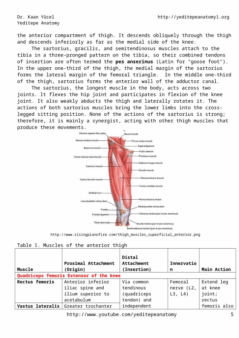
The sartorius, the “tailor's muscle” (L. sartus, patched or repaired), is long and ribbon-like. It is the most superficial muscle in the anterior compartment of thigh. It descends obliquely through the thigh and descends inferiorly as far as the medial side of the knee.
The sartorius, gracilis, and semitendinosus muscles attach to the tibia in a three-pronged pattern on the tibia, so their combined tendons of insertion are often termed the pes anserinus (Latin for "goose foot"). In the upper one-third of the thigh, the medial margin of the sartorius forms the lateral margin of the femoral triangle. In the middle one-third of the thigh, sartorius forms the anterior wall of the adductor canal.
The sartorius, the longest muscle in the body, acts across two joints. It flexes the hip joint and participates in flexion of the knee joint. It also weakly abducts the thigh and laterally rotates it. The actions of both sartorius muscles bring the lower limbs into the cross-legged sitting position. None of the actions of the sartorius is strong; therefore, it is mainly a synergist, acting with other thigh muscles that produce these movements.
http://www.risingpianofire.com/thigh_muscles_superficial_anterior.png
http://www.youtube.com/yeditepeanatomy 3
Table 1. Muscles of the anterior thigh
MuscleProximal Attachment (Origin)
Distal Attachment (Insertion) Innervation Main Action
Quadriceps femoris Extensor of the kneeRectus femoris Anterior inferior iliac spine
and ilium superior to acetabulum
Via common tendinous (quadriceps tendon) and independent attachments to base of patella; indirectly via patellar ligament to tibial tuberosity; medial and lateral vasti also attach to tibia and patella via aponeuroses (medial and lateral patellar retinacula)
Femoral nerve (L2, L3, L4)
Extend leg at knee joint; rectus femoris also steadies hip joint and helps iliopsoas flex thigh
Vastus lateralis Greater trochanter and lateral lip of linea aspera of femur
Vastus medialis Intertrochanteric line and medial lip of linea aspera of femur
Vastus intermedius Anterior and lateral surfaces of shaft of femur
Sartorius Anterior superior iliac spine and superior part of notch inferior to it
Superior part of medial surface of tibia
Femoral nerve (L2, L3)
Flexes, abducts, and laterally rotates thigh at hip joint; flexes leg at knee joint (medially rotating leg when knee is flexed)
Quadriceps Femoris as a Knee-Joint StabilizerThe quadriceps femoris is an important extensor muscle for the knee joint. Its tone greatly strengthens
the joint; therefore, this muscle mass must be carefully examined when disease of the knee joint is suspected. Both thighs should be examined, and the size, consistency, and strength of the quadriceps muscles should be tested. Reduction in size caused by muscle atrophy can be tested by measuring the circumference of each thigh a fixed distance above the superior border of the patella. The vastus medialis muscle extends farther distally than the vastus lateralis. The vastus medialis is the first part of the quadriceps muscle to atrophy in knee-joint disease and the last to recover.
Rupture of the Rectus FemorisThe rectus femoris muscle can rupture in sudden violent extension movements of the knee joint. The muscle belly retracts proximally, leaving a gap that may be palpable on the anterior surface of the thigh. In complete rupture of the muscle, surgical repair is indicated.
Rupture of the Ligamentum PatellaeThis can occur when a sudden flexing force is applied to the knee joint when the quadriceps femoris muscle is actively contracting.
Medial compartmentThere are six muscles in the medial compartment of the thigh: gracilis, pectineus, adductor longus,
adductor brevis, adductor magnus, and obturator externus (See Table 2 on page 6 for origins, insertions, functions and innervations of these muscles). Collectively, all these muscles except the obturator externus mainly adduct the thigh at the hip joint; the adductor longus and magnus may also medially rotate the thigh. Obturator externus is a lateral rotator of the thigh at the hip joint. All adductor muscles, except the “hamstring part” of the adductor magnus and part of the pectineus are supplied by the obturator nerve (L2-L4). The hamstring part of the adductor magnus is supplied by the tibial part of the sciatic nerve (L4).
GracilisThe gracilis (L., slender) is a long, strap-like muscle and is the most medial muscle of the thigh and the
most superficial of the muscles in the medial compartment of thigh. It is the weakest member of the adductor group. It is the only one of the group to cross the knee joint as well as the hip joint. It descends almost vertically
http://www.youtube.com/yeditepeanatomy 4
Dr. Kaan Yücel http://yeditepeanatomy1.org Yeditepe Anatomy
down the medial side of the thigh. The gracilis is a synergist in adducting the thigh, flexing the knee, and rotating the leg medially when the knee is flexed. It acts with the other two “pes anserinus” muscles to add stability to the medial aspect of the extended knee, much as the gluteus maximus and tensor fasciae latae do via the iliotibial tract on the lateral side.
PectineusThe pectineus is a flat quadrangular muscle located in the anterior part of the superomedial aspect of the
thigh. It often appears to be composed of two layers, superficial and deep, and these are generally innervated by two different nerves. Because of the dual nerve supply and the muscle's actions (the pectineus adducts and flexes the thigh and assists in medial rotation of the thigh), it is actually a transitional muscle between the anterior and medial compartments.
Adductor longusThe adductor longus is a large, fan-shaped muscle and is the most anteriorly placed of the adductor
group. The adductor longus contributes to the floor of the femoral triangle, and its medial margin forms the medial border of the femoral triangle. The muscle also forms the proximal posterior wall of the adductor canal.
Adductor brevisThe adductor brevis, the short adductor, lies deep to the pectineus and adductor longus. As the
obturator nerve emerges from the obturator canal to enter the medial compartment of the thigh, it splits into an anterior and a posterior division. The two divisions pass anterior and posterior to the adductor brevis.
Adductor magnusThe adductor magnus is the largest and deepest of the muscles in the medial compartment of thigh. It
is also the most powerful and most posterior muscle in the adductor group. The muscle forms the distal posterior wall of the adductor canal. The lateral part of the muscle is often termed the "adductor part" of the adductor magnus. The medial part of the adductor magnus, often called the "hamstring part”. The two parts differ in their attachments, nerve supply, and main actions (See Table 2).
Adductor HiatusThe adductor hiatus is an opening or gap between the aponeurotic distal attachment of the adductor
part of the adductor magnus and the tendinous distal attachment of the hamstring part. The adductor hiatus transmits the femoral artery and vein from the adductor canal in the thigh to the popliteal fossa posterior to the knee. The opening is located just lateral and superior to the adductor tubercle of the femur.
Obturator externusThe obturator externus is a flat fan-shaped muscle that is deeply placed in the superomedial part of the
thigh. Obturator externus externally rotates the thigh at the hip joint and is innervated by the posterior branch of the obturator nerve.
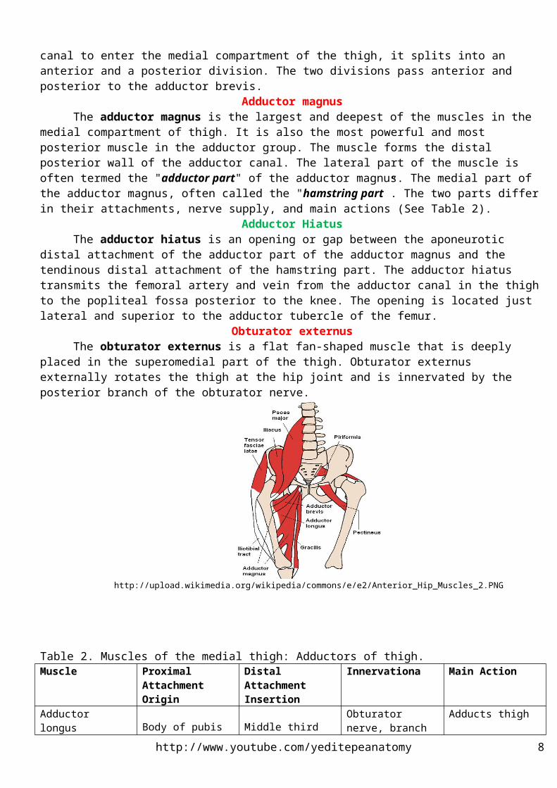
http://upload.wikimedia.org/wikipedia/commons/e/e2/Anterior_Hip_Muscles_2.PNG
http://www.youtube.com/yeditepeanatomy 5
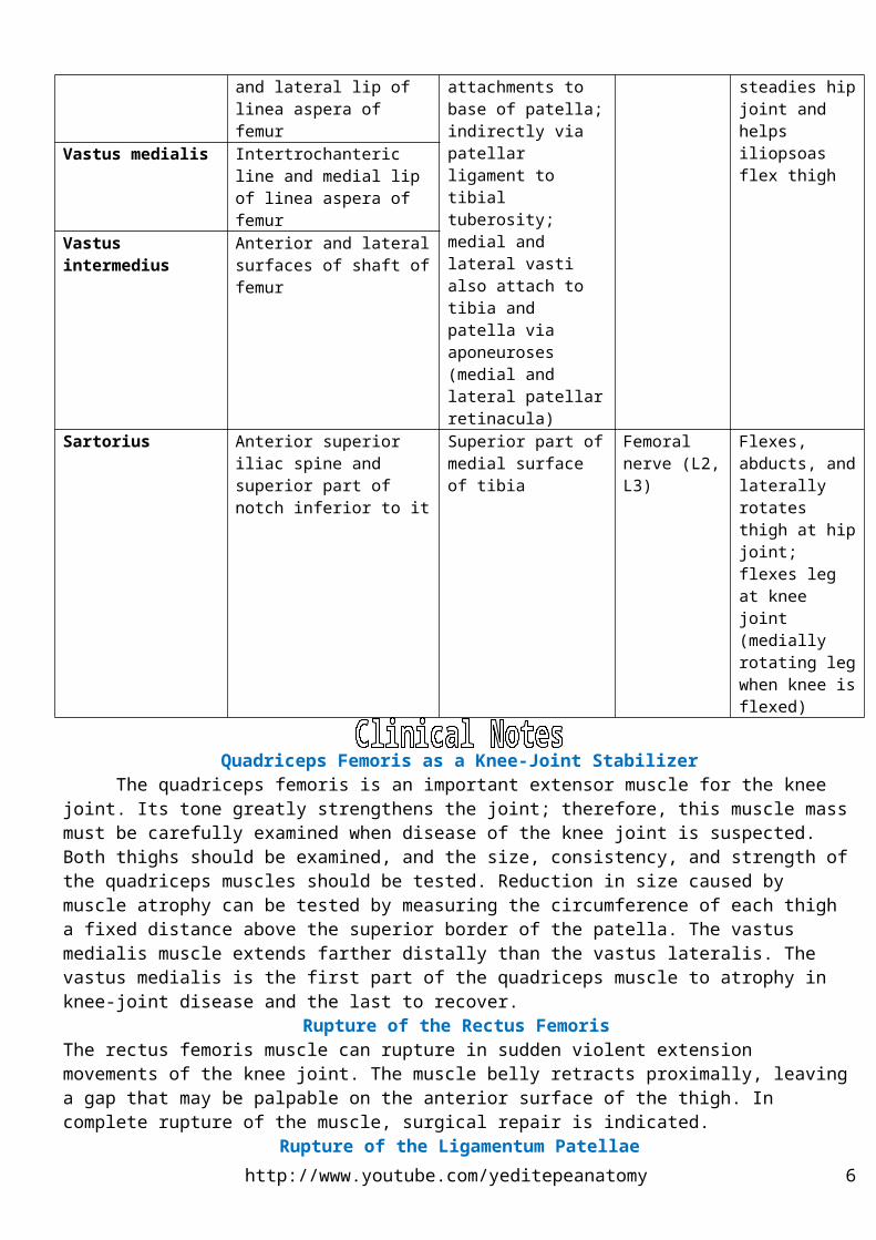
Table 2. Muscles of the medial thigh: Adductors of thigh.Muscle Proximal Attachment
OriginDistal Attachment Insertion
Innervationa Main Action
Adductor longusBody of pubis inferior to pubic crest
Middle third of linea aspera of femur
Obturator nerve, branch of, anterior division (L2, L3, L4)
Adducts thigh
Adductor brevis Body and inferior ramus of pubis
Pectineal line and proximal part of linea aspera of femur
Adducts thigh; to some extent flexes it
Adductor magnus Adductor part: inferior ramus of pubis, ramus of ischium
Hamstrings part: ischial tuberosity
Adductor part: gluteal tuberosity, linea aspera, medial supracondylar line
Hamstring part: adductor tubercle of femur
Adductor part: obturator nerve (L2, L3, L4), branches of posterior division Hamstring part: tibial part of sciatic nerve (L4)
Adducts thigh Adductor part: flexes thigh Hamstrings part: extends thigh
Gracilis Body and inferior ramus of pubis
Superior part of medial surface of tibia.
Obturator nerve (L2, L3)
Adducts thigh; flexes leg; helps rotate leg medially
Pectineus Superior ramus of pubis
Pectineal line of femur, just inferior to lesser trochanter
Femoral nerve (L2, L3); may receive a branch from obturator nerve
Adducts and flexes thigh; assists with medial rotation of thigh
Obturator externus Margins of obturator foramen and obturator membrane
Trochanteric fossa of femur
Obturator nerve (L2, L3)
Laterally rotates thigh; steadies head of femur in acetabulum
a Collectively, the five muscles listed are the adductors of the thigh, but their actions are more complex (e.g., they act as flexors of the hip joint during flexion of the knee joint and are active during walking).b The spinal cord segmental innervation is indicated (e.g., “L2, L3, L4” means that the nerves supplying the adductor longus are derived from the second to fourth lumbar segments of the spinal cord). Numbers in boldface (L3) indicate the main segmental innervation. Damage to one or more of the listed spinal cord segments or to the motor nerve roots arising from them results in paralysis of the muscles concerned.
Posterior compartmentThree of the four muscles in the posterior aspect of the thigh are hamstrings. The hamstring muscles are:
(1) semitendinosus, (2) semimembranosus, and (3) biceps femoris (long head). The hamstring muscles (“hamstrings” for short) share common features: Proximal attachment to the ischial tuberosity deep to the gluteus maximus. Distal attachment to the bones of the leg. Thus they span and act on two joints, producing extension at the hip joint and flexion at the knee joint. Innervation by the tibial division of the sciatic nerve.The long head of the biceps femoris meets all these conditions, but the short head of the biceps, the fourth muscle of the posterior compartment, fails to meet any of them. As a group, the hamstrings flex the leg at the knee joint and extend the thigh at the hip joint. They are also rotators at both joints (See Table 3 on page 8 for origins, insertions, functions and innervations of these muscles). .
The hamstrings received their name because it is common to tie hams (pork thighs) up for curing and/or smoking with a hook around these muscle tendons. To test the hamstrings, the person flexes their leg against resistance. Normally, these muscles—especially their tendons on each side of the popliteal fossa—should be prominent as they bend the knee.
Biceps femoris
http://www.youtube.com/yeditepeanatomy 6

Dr. Kaan Yücel http://yeditepeanatomy1.org Yeditepe Anatomy
The fusiform biceps femoris, as its name indicates, has two heads: a long head and a short head and is lateral in the posterior compartment of thigh. Together, fibers from the two heads form a tendon, which is palpable on the lateral side of the distal thigh.
SemitendinosusThe semitendinosus, as its name indicates, is half tendinous. The semitendinosus muscle is medial to
the biceps femoris muscle in the posterior compartment of thigh. The semitendinosus flexes the leg at the knee joint and extends the thigh at the hip joint. Working with the semimembranosus, it also medially rotates the thigh at the hip joint and medially rotates the leg at the knee joint.
SemimembranosusThe semimembranosus is a broad muscle that is also aptly named because of the flattened membranous
form of its proximal attachment to the ischial tuberosity. The semimembranosus muscle lies deep to the semitendinosus muscle in the posterior compartment of thigh. The semimembranosus flexes the leg at the knee joint and extends the thigh at the hip joint. Working with the semitendinosus muscle, it medially rotates the thigh at the hip joint and the leg at the knee joint.
Table 3. Muscles of the posterior thigh: Extensors of hip and flexors of knee
MuscleaProximal Attachment Distal Attachment Innervationb Main Action
Semitendinosus
Ischial tuberosity
Medial surface of superior part of tibia
Tibial division of sciatic nerve part of tibia (L5, S1, S2)
Extend thigh; flex leg and rotate it medially when knee is flexed; when thigh and leg are flexed, these muscles can extend trunk
SemimembranosusPosterior part of medial condyle of tibia; reflected attachment forms oblique popliteal ligament (to lateral femoral condyle)
Biceps femoris Long head: ischial tuberosity Short head: linea aspera and lateral supracondylar line of femur
Lateral side of head of fibula; tendon is split at this site by fibular collateral ligament of knee
Long head: tibial division of sciatic nerve (L5, S1, S2) Short head: common fibular division of sciatic nerve (L5, S1, S2)
Flexes leg and rotates it laterally when knee is flexed; extends thigh (e.g., accelerating mass during first step of gait).
http://www.deeptissue.com/learn/knee/hamstrings.htm
http://www.youtube.com/yeditepeanatomy 7
ArteriesThree arteries enter the thigh: the femoral artery, obturator artery, and inferior gluteal artery. Of
these, the femoral artery is the largest and supplies most of the lower limb. The three arteries contribute to an anastomotic network of vessels around the hip joint.
Femoral arteryThe femoral artery, distal continuation of the external iliac artery, is the primary artery of the lower
limb. It begins as the external iliac artery passes under the inguinal ligament. It traverses the femoral triangle and the adductor canal of Hunter. It enters the femoral triangle deep to the midpoint of the inguinal ligament (midway between the ASIS and the pubic tubercle), lateral to the femoral vein on the anterior aspect of the upper thigh. The femoral artery passes vertically through the femoral triangle and then continues down the thigh in the adductor canal. It leaves the canal by passing through the adductor hiatus in the adductor magnus muscle. It ends at the opening in the adductor magnus muscle, passing through the adductor canal and entering the popliteal space to become the popliteal artery behind the knee.
The femoral artery is palpable in the femoral triangle just inferior to the inguinal ligament midway between the anterior superior iliac spine and the pubic symphysis.
Branches of the femoral artery1) Superficial circumflex iliac artery 2) Superficial epigastric artery crosses the inguinal ligament and runs to the region of the umbilicus.3) Superficial external pudendal artery 4) Deep external pudendal artery5) Profunda femoris artery is a large and important branch that arises from the lateral side of the femoral artery
below the inguinal ligament. 6) Descending genicular artery is a small branch that arises from the femoral artery near its termination
Deep artery of thighThe largest branch of the femoral artery and the chief artery to the thigh is the deep artery of thigh
(profunda femoris artery), which originates from the lateral side of the femoral artery in the femoral triangle. and ends as the fourth perforating artery. The deep artery of thigh has lateral & medial circumflex femoral branches and three perforating branches.
Obturator arteryThe obturator artery originates as a branch of the internal iliac artery in the pelvic cavity and enters
the medial compartment of thigh through the obturator canal. It accompanies the obturator nerve through the obturator canal (i.e., the upper part of the obturator foramen).
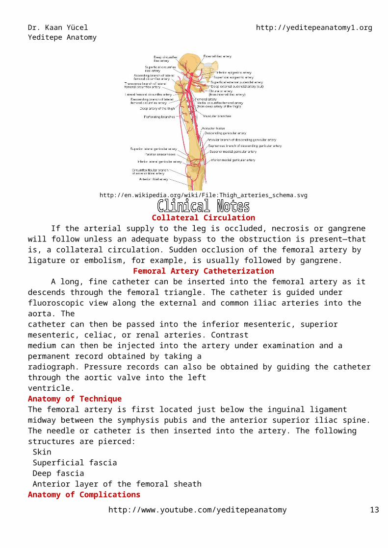
http://en.wikipedia.org/wiki/File:Thigh_arteries_schema.svg
Collateral Circulation
http://www.youtube.com/yeditepeanatomy 8

Dr. Kaan Yücel http://yeditepeanatomy1.org Yeditepe Anatomy
If the arterial supply to the leg is occluded, necrosis or gangrene will follow unless an adequate bypass to the obstruction is present—that is, a collateral circulation. Sudden occlusion of the femoral artery by ligature or embolism, for example, is usually followed by gangrene.
Femoral Artery CatheterizationA long, fine catheter can be inserted into the femoral artery as it descends through the femoral triangle.
The catheter is guided under fluoroscopic view along the external and common iliac arteries into the aorta. Thecatheter can then be passed into the inferior mesenteric, superior mesenteric, celiac, or renal arteries. Contrastmedium can then be injected into the artery under examination and a permanent record obtained by taking aradiograph. Pressure records can also be obtained by guiding the catheter through the aortic valve into the leftventricle.Anatomy of TechniqueThe femoral artery is first located just below the inguinal ligament midway between the symphysis pubis and the anterior superior iliac spine. The needle or catheter is then inserted into the artery. The following structures are pierced: Skin Superficial fascia Deep fascia Anterior layer of the femoral sheathAnatomy of ComplicationsThe femoral vein lies immediately medial to the artery and may be entered in error. The nonpulsatile nature of the vein on palpation should exclude this possibility. Since the hip joint lies posterior to the femoral artery, the erroneous passage of the needle through the posterior arterial wall may cause it to pierce the psoas muscle and enter the joint cavity. Some difficulty may be experienced in passing the catheter up the femoral artery if the artery is tortuous or if there is extensive atherosclerosis of the arterial wall.
Obtaining blood sample from the femoral arteryThe femoral artery at the groin is readily punctured by a hypodermic needle and is the most convenient site to obtain arterial blood samples. Arteriography of the peripheral leg vessels is also easily performed via femoral artery.
Traumatic Injury to Arteries of the Lower LimbInjury to the large femoral artery can cause rapid exsanguination of the patient. Unlike in the upper
extremity, arterial injuries of the lower limb do not have a good prognosis. The collateral circulations around the hip and knee joints, although present, are not as adequate as that around the shoulder and elbow. Damage to a neighboring large vein can further complicate the situation and causes further impairment of the circulation to the distal part of the limb. The femoral artery is superficial where it lies in the femoral triangle and in consequence easily injured.
Aneurysms of the Lower ExtremityThese occur much less frequently than abdominal aortic aneurysms and are usually caused by
atherosclerosis. Most patients are over 50 years of age, and the common sites are the femoral and popliteal arteries. The diagnosis is usually made by finding an expansile swelling along the course of the artery. Patients may present in the emergency department with complications, which include sudden embolic obstruction to arteries distal to the aneurysm or sudden thrombotic occlusion of the aneurysm. Pressure on neighboring nerves may give rise to symptoms; for example, an enlarging popliteal aneurysm may press on the tibial nerve, causing pain in the foot. Rupture of femoral or popliteal aneurysms is rare.
VeinsVeins in the thigh consist of superficial and deep veins. Deep veins generally follow the arteries and
have similar names. Superficial veins are in the superficial fascia, interconnect with deep veins, and do not generally accompany arteries. The largest of the superficial veins in the thigh is the great saphenous vein. Many small veins curve around the medial and lateral aspects of the thigh and ultimately drain into the great saphenous vein. Superficial veins from the lower part of the back of the thigh join the small saphenous vein in the popliteal fossa.
Great saphenous vein
http://www.youtube.com/yeditepeanatomy 9

The great saphenous vein originates from a venous arch on the dorsal aspect of the foot and ascends along the medial side of the lower limb to the proximal thigh. Here it passes through the saphenous ring in deep fascia covering the anterior thigh to connect with the femoral vein in the femoral triangle. The superficial circumflex iliac vein, the superficial epigastric vein, and the external pudendal veins drain into the great saphenous vein.
Femoral veinThe femoral vein is the continuation of the popliteal vein proximal to the adductor hiatus. It ascends
through the thigh, lying at first on the lateral side of the artery, then posterior to it, and finally on its medial side. The femoral vein enters the femoral sheath lateral to the femoral canal and ends posterior to the inguinal ligament, where it becomes the external iliac vein. In the inferior part of the femoral triangle, the femoral vein receives the deep vein of the thigh, the great saphenous vein, and other tributaries.
Profunda Femoris VeinThe profunda femoris vein receives tributaries that correspond to the branches of the artery. It drains
into the femoral vein.Obturator Vein
The obturator vein receives tributaries that correspond to the branches of the artery. It drains into the internal iliac vein.
The Great Saphenous Vein in Coronary Bypass SurgeryIn patients with occlusive coronary disease caused by atherosclerosis,the diseased arterial segment can
be bypassed by inserting a graft consisting of a portion of the great saphenous vein. The venous segment is reversed so that its valves do not obstruct the arterial flow. Following removal of the great saphenous vein at the donor site, the superficial venous blood ascends the lower limb by passing through perforating veins and entering the deep veins.The great saphenous vein can also be used to bypass obstructions of the brachial or femoral arteries.
Deep Vein Thrombosis and Long-Distance Air TravelPassengers who sit immobile for hours on long-distance flights are very prone to deep vein thrombosis
in the legs. Preventative measures include stretching of the legs every hour to improve the venous circulation.Femoral Vein Catheterization
Femoral vein catheterization is used when rapid access to a large vein is needed. The femoral vein has a constant relationship to the medial side of the femoral artery just below the inguinal ligament and is easily cannulated. Anatomy of the Procedure1. The skin of the thigh below the inguinal ligament is supplied by the genitofemoral nerve; this nerve isblocked with a local anesthetic.2. The femoral pulse is palpated midway between the anterior superior iliac spine and the symphysis pubis, andthe femoral vein lies immediately medial to it.3. At a site about two fingerbreadths below the inguinal ligament, the needle is inserted into the femoral vein
NervesThere are three major nerves in the thigh, each associated with one of the three compartments. The
femoral nerve is associated with the anterior compartment of thigh, the obturator nerve is associated with the medial compartment of thigh, and the sciatic nerve is associated with the posterior compartment of thigh.
Femoral nerveThe femoral nerve (L2-L4) is the largest branch of the lumbar plexus. The femoral nerve originates
from the lumbar plexus (spinal cord segments L2-L4) in the abdomen within the psoas major and descends posterolaterally through the pelvis to approximately the midpoint of the inguinal ligament. It lies behind the fascia iliaca and enters the thigh lateral to the femoral artery and the femoral sheath, behind the inguinal ligament. In the femoral triangle the femoral nerve lies on the lateral side of the femoral artery and is outside the femoral sheath, which surrounds the vessels. The femoral nerve supplies all the muscles of the anterior compartment of the thigh. Note that the femoral nerve does not enter the thigh within the femoral sheath.
Before entering the thigh, the femoral nerve supplies branches to the iliacus and pectineus muscles.
http://www.youtube.com/yeditepeanatomy 10

Dr. Kaan Yücel http://yeditepeanatomy1.org Yeditepe Anatomy
Immediately after passing under the inguinal ligament, the femoral nerve divides into anterior and posterior branches, which supply muscles of the anterior compartment of thigh and skin on the anterior and medial aspects of the thigh and on the medial sides of the leg and foot. Branches of the femoral nerve include: o anterior division gives off two cutaneous and two muscular branches. The cutaneous branches are the medial cutaneous nerve of the thigh and the intermediate cutaneous nerves that supply the skin of the medial and anterior surfaces of the thigh, respectively. The muscular branches supply the sartorius and the pectineus.o posterior division gives off one cutaneous branch, the saphenous nerve, and muscular branches to the
quadriceps muscle. o numerous motor nerves, which supply the quadriceps femoris muscles (rectus femoris, vastus lateralis,
vastus intermedius, and vastus medialis muscles) and the sartorius muscle; andThe terminal cutaneous branch of the femoral nerve, the saphenous nerve, descends through the
femoral triangle, lateral to the femoral sheath containing the femoral vessels. The saphenous nerve accompanies the femoral artery through the adductor canal, but does not pass through the adductor hiatus with the femoral artery. It terminates in the region of the ball of the big toe. and supplies skin and fascia on the anteromedial aspects of the knee, leg, and foot.
Obturator nerveThe obturator nerve enters the medial compartment of thigh by passing through the obturator canal. It
supplies most of the adductor muscles and skin on the medial aspect of the thigh. As the obturator nerve enters the thigh, it divides into two branches which are separated by the adductor brevis muscle: o posterior branch supplies the obturator externus and adductor brevis muscles and the part of adductor magnus that attaches to the linea aspera;o anterior branch and is behind the pectineus and adductor longus muscles-it supplies branches to the adductor longus, gracilis, and adductor brevis muscles, and often contributes to the supply of the pectineus muscle, and cutaneous branches innervate the skin on the medial side of the thigh.
Sciatic nerveThe sciatic nerve leaves the gluteal region as it descends in the midline of the thigh. It is overlapped
posteriorly by the adjacent margins of the biceps femoris and semimembranosus muscles. It lies on the posterior aspect of the adductor magnus muscle. In the lower third of the thigh it ends by dividing into the tibial and common peroneal nerves. These nerves travel vertically down the thigh and enter the popliteal fossa posterior to the knee. Here, they meet the popliteal artery and vein. The sciatic nerve innervates all muscles in the posterior compartment of thigh and then its branches continue into the leg and foot.
http://www.youtube.com/yeditepeanatomy 11

Lateral cutaneous nerve of the thighThe lateral cutaneous nerve of the thigh, a branch of the lumbar plexus (L2 and 3), enters the thigh
behind the lateral end of the inguinal ligament. Having divided into anterior and posterior branches, it supplies the skin of the lateral aspect of the thigh and knee. It also supplies the skin of the lower lateral quadrant of the buttock.
Medial cutaneous nerve of the thighThe medial cutaneous nerve of the thigh, a branch of the femoral nerve, supplies the medial aspect of
the thigh and joins the patellar plexus.Intermediate cutaneous nerve of the thigh
The intermediate cutaneous nerve of the thigh, a branch of the femoral nerve, divides into two branches that supply the anterior aspect of the thigh and joins the patellar plexus.
Branches from the anterior division of the obturator nerve supply a variable area of skin on the medial aspect of the thigh.
The patellar plexus lies in front of the knee and is formed from the terminal branches of the lateral, intermediate, and medial cutaneous nerves of the thigh and the infrapatellar branch of the saphenous nerve.
Posterior cutaneous nerve of the thighThe posterior cutaneous nerve of the thigh, a branch of the sacral plexus descends on the back of the
thigh, and in the popliteal fossa it pierces the deep fascia and supplies the skin. It gives off numerous branches to the skin on the back of the thigh and the upper part of the leg.
Femoral Nerve InjuryThe femoral nerve (L2, 3, and 4) enters the thigh from behind the inguinal ligament, at a point midway
between the anterior superior iliac spine and the pubic tubercle; it lies about a fingerbreadth lateral to the femoral pulse. About 2 in. (5 cm) below the inguinal ligament, the nerve splits into its terminal branches.
The femoral nerve can be injured in stab or gunshot wounds, but a complete division of the nerve is rare. The following clinical features are present when the nerve is completely divided:oMotor: The quadriceps femoris muscle is paralyzed, and the knee cannot be extended. In walking, this is compensated for to some extent by use of the adductor muscles.
http://www.youtube.com/yeditepeanatomy 12
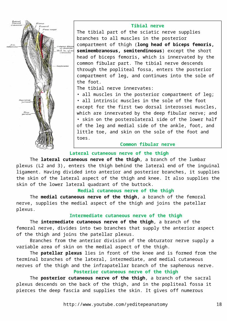
Tibial nerveThe tibial part of the sciatic nerve supplies branches to all muscles in the posterior compartment of thigh (long head of biceps femoris, semimembranosus, semitendinosus) except the short head of biceps femoris, which is innervated by the common fibular part. The tibial nerve descends through the popliteal fossa, enters the posterior compartment of leg, and continues into the sole of the foot. The tibial nerve innervates: • all muscles in the posterior compartment of leg;• all intrinsic muscles in the sole of the foot except for the first two dorsal interossei muscles, which are innervated by the deep fibular nerve; and• skin on the posterolateral side of the lower half of the leg and medial side of the ankle, foot, and little toe, and skin on the sole of the foot and toes.
Common fibular nerveThe common fibular part of the sciatic nerve innervates the short head of biceps femoris in the posterior compartment of thigh and then continues into the lateral and anterior compartments of leg and onto the foot. The common fibular nerve innervates: • all muscles in the anterior and lateral compartments of leg;• one muscle (extensor digitorum brevis) on the dorsal aspect of the foot;• the first two dorsal interossei muscles in the sole of the foot; and• skin over the lateral aspect of the leg, and ankle, and over the dorsal aspect of the foot and toes.
Dr. Kaan Yücel http://yeditepeanatomy1.org Yeditepe Anatomy
oSensory: Skin sensation is lost over the anterior and medial sides of the thigh, over the medial side of the lower part of the leg, and along the medial border of the foot as far as the ball of the big toe; this area is normally supplied by the saphenous nerve.
Obturator Nerve InjuryThe obturator nerve (L2, 3, and 4) enters the thigh as anterior and posterior divisions through the upper
part of the obturator foramen. The anterior division descends in front of the obturator externus and the adductor brevis, deep to the floor of the femoral triangle. The posterior division descends behind the adductor brevis and in front of the adductor magnus.
It is rarely injured in penetrating wounds, in anterior dislocations of the hip joint, or in abdominal herniae through the obturator foramen. It may be pressed on by the fetal head during parturition. The following clinical features occur: oMotor: All the adductor muscles are paralyzed except the hamstring part of the adductor magnus, which issupplied by the sciatic nerve.oSensory: The cutaneous sensory loss is minimal on the medial aspect of the thigh.
Referred Pain from the Hip JointThe femoral nerve not only supplies the hip joint but, via the intermediate and medial cutaneous nerves
of the thigh, also supplies the skin of the front and medial side of the thigh. It is not surprising, therefore, for pain originating in the hip joint to be referred to the front and medial side of the thigh. The posterior division of the obturator nerve supplies both the hip and knee joints. This would explain why hip joint disease sometimes gives rise to pain in the knee joint.
Lymph Nodes of the ThighThe deep inguinal lymph nodes lie along the medial side of the terminal part of the femoral vein, and
the most superior is usually located in the femoral canal. They receive all the lymph from the superficial inguinal nodes via lymph vessels that pass through the cribriform fascia of the saphenous opening. They also receive lymph from the deep structures of the lower limb that have ascended in lymph vessels alongside the arteries, some having passed through the popliteal nodes. The efferent lymph vessels from the deep inguinal nodes ascend into the abdominal cavity through the femoral canal and drain into the external iliac nodes.
Lymph from the skin and superficial fascia on the back of the thigh drains upward and forward into the vertical group of superficial inguinal lymph nodes.Neurovascular Structures and Relationships in Anteromedial Thigh
FEMORAL TRIANGLEThe femoral triangle, a subfascial formation, lies at the uper part of the anterior thigh. The femoral
triangle is bounded:o Superiorly by the inguinal ligament o Medially by the medial border of the adductor longus.o Laterally by the sartorius; the apex of the femoral triangle is where the medial border of the sartorius crosses the lateral border of the adductor longus.The muscular floor of the femoral triangle is formed by the iliopsoas laterally and the pectineus medially. The roof of the femoral triangle is formed by the fascia lata and cribriform fascia, subcutaneous tissue, and skin.The contents of the femoral triangle, from lateral to medial, are the:1) Femoral nerve and its (terminal) branches2) Femoral sheath and its contents3) Femoral artery and several of its branches4) Femoral vein and its proximal tributaries (e.g., the great saphenous and deep femoral veins).5) Deep inguinal lymph nodes and associated lymphatic vessels
The femoral triangle is bisected by the femoral artery and vein, which pass to and from the adductor canal inferiorly at the triangle's apex. The adductor canal is an intermuscular passageway deep to the sartorius by which the major neurovascular bundle of the thigh traverses the middle third of the thigh.
FEMORAL SHEATHThe femoral sheath is a funnel-shaped fascial tube that passes deep to the inguinal ligament. It
terminates inferiorly by blending with the adventitia of the femoral vessels. The sheath encloses proximal parts http://www.youtube.com/yeditepeanatomy 13

of the femoral vessels and creates the femoral canal medial to them. Its anterior wall is continuous above with the fascia transversalis, and its posterior wall with the fascia iliaca.
The femoral sheath is formed by an inferior prolongation of transversalis and iliopsoas fascia from the abdomen.The femoral sheath allows the femoral artery and vein to glide deep to the inguinal ligament during movements of the hip joint.
The femoral sheath lining the vascular compartment is subdivided internally into three smaller compartments by vertical septa of extraperitoneal connective tissue that extend from the abdomen along the femoral vessels. The compartments of the femoral sheath are the:1) Lateral compartment for the femoral artery.2) Intermediate compartment for the femoral vein.3) Medial compartment, which constitutes the femoral canal.
Femoral CanalThe femoral canal is the smallest of the three compartments of the femoral sheath. It is conical and short
(approximately 1.25 cm) and lies between the medial edge of the femoral sheath and the femoral vein. Extends distally to the level of the proximal edge of the saphenous opening. Allows the femoral vein to expand when venous return from the lower limb is increased, or when increased intraabdominal pressure causes a temporary stasis in the vein (as during a Valsalva maneuver, i.e., taking a breath and holding it, often while bearing down).
Contains loose connective tissue, fat, a few lymphatic vessels, and sometimes a deep inguinal lymph node (lacunar node).
The base (upper opening) of the femoral canal is the oval femoral ring formed by the small (approximately 1 cm wide) proximal opening at its abdominal end. The femoral septum, which is a condensation of extraperitoneal tissue, closes the ring. The femoral canal contains fatty connective tissue, all the efferent lymph vessels from the deep inguinal lymph nodes, and one of the deep inguinal lymph nodes.
The boundaries of the femoral ring are: Laterally, the vertical septum between the femoral canal and femoral vein. Posteriorly, the superior ramus of the pubis covered by the pectineus muscle and its fascia. Medially, the lacunar ligament (Gimbernant’s ligament) Anteriorly, the medial part of the inguinal ligament.The femoral canal has two functions: first, as a dead space for expansion of the distended femoral vein, and, second, as a lymphatic pathway from the lower limb to the external iliac nodes.
The femoral sheath is adherent to the walls of the blood vessels and inferiorly blends with the tunica adventitia of these vessels. The part of the femoral sheath that forms the medially located femoral canal is not adherent to the walls of the small lymph vessels; it is this site that forms a potentially weak area in the abdomen. A protrusion of peritoneum could be forced down the femoral canal, pushing the femoral septum before it. Such a condition is known as a femoral hernia.
Adductor CanalThe adductor canal (Subsartorial canal; Hunter canal) is a long, narrow intermuscular cleft in the
middle third of the thigh beneath the sartorius muscle. It extends from the apex of the femoral triangle, where the sartorius crosses over the adductor longus, to the adductor hiatus in the tendon of the adductor magnus.
The adductor canal contains the terminal part of the femoral artery, femoral vein (lies behind the artery), deep lymph vessels, saphenous nerve, nerve to the vastus medialis, and terminal part of the obturator nerve.The adductor canal is bounded: Anteriorly and laterally by the vastus medialis. Posteriorly by the adductors longus and magnus. Medially by the sartorius, which forms roof of the canal.
The adductor hiatus is different than the adductor canal and is located at a more inferior level, just proximal to the medial supracondylar ridge. This hiatus is a gap between the aponeurotic adductor and the tendinous hamstrings attachments of the adductor magnus.
Femoral Hernia
http://www.youtube.com/yeditepeanatomy 14

Dr. Kaan Yücel http://yeditepeanatomy1.org Yeditepe Anatomy
The great importance of the femoral canal,of course, that is a potential point of weakness in the abdominal wall through which a femoral hernia may develop. Unlike the inguinal hernia, that is never due to a congenital sac,and, although cases do occur rarely in children, it is never found in the newborn.
The hernial sac descends through the femoral canal within the femoral sheath, creating a femoral hernia. The femoral sheath is a protrusion of the fascial envelope lining the abdominal walls and surrounds the femoral vessels and lymphatics for about 1 in. (2.5 cm) below the inguinal ligament. The femoral artery, as it entersthe thigh below the inguinal ligament, occupies the lateral compartment of the sheath. The femoral vein, which lies on its medial side and is separated from it by a fibrous septum, occupies the intermediate compartment. Thelymph vessels, which are separated from the vein by a fibrous septum, occupy the most medial compartment.The femoral canal, the compartment for the lymphatics, occupies the medial part of the sheath.
Its upper opening is referred to as the femoral ring. The femoral septum, which is a condensation of extraperitoneal tissue, plugs the opening of the femoral ring. A femoral hernia is more common in women than in men (possibly because of a wider pelvis and femoral canal).
http://www.laparoscopyhospital.com/femoral-hernia.html
POPLITEAL FOSSAThe popliteal fossa is an important area of transition between the thigh and leg and is the major route by
which structures pass from one region to the other. The popliteal fossa is a mostly fat-filled compartment of the lower limb. The fossa is most prominent when the knee joint is flexed. Superficially, when the knee is flexed, the popliteal fossa is evident as a diamond-shaped depression posterior to the knee joint.
The popliteal fossa is formed between muscles in the posterior compartments of thigh and leg. Superficially, the popliteal fossa is bounded: Superolaterally by the biceps femoris (superolateral border). Superomedially by the semimembranosus, lateral to which is the semitendinosus (superomedial border). Inferolaterally and inferomedially by the lateral and medial heads of the gastrocnemius, respectively (inferolateral and inferomedial borders). Posteriorly by skin and popliteal fascia (roof).
Contents1) Termination of the small saphenous vein2) Popliteal arteries and veins and their branches and tributaries3) Tibial and common fibular nerves4) Posterior cutaneous nerve of thigh 5) Popliteal lymph nodes and lymphatic vessels
Fascia of popliteal fossaThe popliteal fascia is a strong sheet of deep fascia, continuous superiorly with the fascia lata and
inferiorly with the deep fascia of the leg. The popliteal fascia forms a protective covering for neurovascular structures passing from the thigh through the popliteal fossa to the leg, and a relatively loose but functional retaining “retinaculum” (retaining band) for the hamstring tendons. Often the fascia is pierced by the small saphenous vein.
Neurovascular structures & relationships in popliteal fossaAll important neurovascular structures that pass from the thigh to the leg do so by traversing the
popliteal fossa. Progressing from superficial to deep (posterior to anterior) within the fossa, as in dissection, the
http://www.youtube.com/yeditepeanatomy 15
nerves are encountered first, then the veins. The arteries lie deepest, directly on the surface of the femur, joint capsule, and investing fascia of the popliteus forming the floor of the fossa.
Nerves in Popliteal FossaThe sciatic nerve usually ends at the superior angle of the popliteal fossa by dividing into the tibial and common fibular nerves.
Tibial and common fibular nervesThe tibial and common fibular nerves originate proximal to the popliteal fossa as the two major
branches of the sciatic nerve. They are the most superficial of the neurovascular structures in the popliteal fossa and enter the region directly from above under the margin of the biceps femoris muscle: tibial nerve ,medial and larger terminal branch of the sciatic nerve, descends vertically through the popliteal fossa and exits deep to the margin of plantaris muscle to enter the posterior compartment of leg; common fibular nerve , lateral, smaller terminal branch of the sciatic nerve, exits by following the medial border of the biceps femoris tendon over the lower lateral margin of the popliteal fossa, and continues to the lateral side of the leg where it swings around the neck of the fibula and enters the lateral compartment of leg.
Tibial nerveThe tibial nerve arises in the lower third of the thigh. It runs downward through the popliteal fossa, lying
first on the lateral side of the popliteal artery, then posterior to it, and finally medial to it. The popliteal vein lies between the nerve and the artery throughout its course. The nerve enters the posterior compartment of the leg by passing beneath the soleus muscle.
The tibial nerve is the most superficial of the three main central components of the popliteal fossa (i.e., nerve, vein, and artery); however, it is still in a deep and protected position. The tibial nerve bisects the fossa as it passes from its superior to its inferior angle.
Branches of the tibial nerveCutaneous: The sural nerve descends between the two heads of the gastrocnemius muscle and is usually
joined by the sural communicating branch of the common peroneal nerve. The sural nerve supplies the skin of the lateral and posterior part of the lower one-third of the leg. The sural nerve accompanies the small saphenous vein behind the lateral malleolus and is distributed to the skin along the lateral border of the foot and the lateral side of the little toe.
Muscular branches supply both heads of the gastrocnemius and the plantaris, soleus, and popliteus. Common Peroneal Nerve
The common peroneal nerve arises in the lower third of the thigh. It leaves the fossa by crossing superficially the lateral head of the gastrocnemius muscle. It then passes behind the head of the fibula, winds laterally around the neck of the bone, pierces the peroneus longus muscle, and divides into two terminal branches: the superficial peroneal nerve and the deep peroneal nerve. As the nerve lies on the lateral aspect of the neck of the fibula, it is subcutaneous and can easily be rolled against the bone.Branches Cutaneous: The sural communicating branch runs downward and joins the sural nerve. The lateral cutaneous nerve of the calf supplies the skin on the lateral side of the back of the leg.
Muscular branch to the short head of the biceps femoris muscle, which arises high up in the popliteal fossa.Posterior Cutaneous Nerve of the Thigh
The posterior cutaneous nerve of the thigh terminates by supplying the skin over the popliteal fossa. The nerve traverses most of the length of the posterior compartment of the thigh deep to the fascia lata; only its terminal branches enter the subcutaneous tissue as cutaneous nerves.
Obturator NerveThe obturator nerve leaves the subsartorial canal with the femoral artery by passing through the opening in
the adductor magnus and terminates by supplying the knee joint.Blood Vessels in Popliteal Fossa
Popliteal artery and veinThe popliteal artery is the continuation of the femoral artery in the anterior compartment of thigh, and
begins as the femoral artery passes posteriorly through the adductor hiatus in the adductor magnus muscle. The popliteal artery descends obliquely through the fossa with the tibial nerve and enters the posterior compartment of leg where it ends just lateral to the midline of the leg by dividing into the anterior and posterior tibial arteries at the inferior border of the popliteus.
http://www.youtube.com/yeditepeanatomy 16
Dr. Kaan Yücel http://yeditepeanatomy1.org Yeditepe Anatomy
The popliteal artery is the deepest of the neurovascular structures in the popliteal fossa and is therefore difficult to palpate; however, a pulse can usually be detected by deep palpation medial to the midline. In the popliteal fossa, the popliteal artery gives rise to branches, which contribute to vascular anastomoses around the knee. The genicular arteries are the superior lateral, superior medial, middle, inferior lateral, and inferior medial genicular arteries. They participate in the formation of the periarticular genicular anastomosis, a network of vessels surrounding the knee that provides collateral circulation capable of maintaining blood supply to the leg during full knee flexion, which may kink the popliteal artery.
The popliteal vein is superficial to and travels with the popliteal artery. The popliteal vein ascends through the popliteal fossa to the aperture in the adductor magnus, where it becomes the femoral vein. The popliteal vein is formed by formation of anterior and posterior tibial veins at the lower border of popliteus. Throughout its course, the vein lies close to the popliteal artery, lying superficial to it and in the same fibrous sheath. More superiorly, the popliteal vein lies posterior to the artery, between this vessel and the overlying tibial nerve. The small saphenous vein pierces the deep popliteal fascia and enters the popliteal vein.
Arterial PalpationEvery health professional should know the precise position of the main arteries within the lower limb,
for he or she may be called on to arrest a severe hemorrhage or palpatedifferent parts of the arterial tree in patients with arterial occlusion.
The femoral artery enters the thigh behind the inguinal ligament at a point midway between the anterosuperior iliac spine and the symphysis pubis. The artery is easily palpated here because it can be pressed backward against the pectineus and the superior ramus of the pubis.
The popliteal artery can be felt by gentle palpation in the depths of the popliteal space provided that the deep fasciais fully relaxed by passively flexing the knee joint.
Lymph Nodes in Popliteal FossaThe superficial popliteal lymph nodes are usually small and lie in the subcutaneous tissue.. The deep
popliteal lymph nodes surround the vessels and receive lymph from the joint capsule of the knee and the lymphatic vessels that accompany the deep veins of the leg. The lymphatic vessels from the popliteal lymph nodes follow the femoral vessels to the deep inguinal lymph nodes.
Roof of popliteal fossaThe roof of the popliteal fossa is covered by superficial fascia and skin. The most important structure in
the superficial fascia is the small saphenous vein. This vessel joins with the popliteal vein. One other structure that passes through the roof of the fossa is the posterior cutaneous nerve of thigh, which descends through the thigh superficial to the hamstring muscles, passes through the roof of the popliteal fossa, and then continues inferiorly with the small saphenous vein to innervate skin on the upper half of the back of the leg.
http://www.cambridgeorthopaedics.com/cambridgeanaesthetics/advancednerveblocks/images/popliteal%20block/popliteal%20fossa.gif
http://www.youtube.com/yeditepeanatomy 17
http://www.youtube.com/yeditepeanatomy 18