Embed Size (px)
Citation preview
SPECT dopamine transporters. document.docx
QIBA Profile: Quantifying Dopamine Transporters with 123Iodine Labeled Ioflupane in Neurodegenerative Disease (Short Title: SPECT dopamine transporters)Stage: A. Initial Draft
Notation in this TemplateTemplate Element Appears as InstructionsBoilerplate text Plain black text Don't change.
Should appear in all profiles.Example text Plain grey text Provides an example of content and wording
appropriate to that location.Rewrite it to your needs and change the text color back to Automatic (which will make it black).
Placeholder <text in angle brackets> Replace text and <> with your text.Use Find/Replace for ones that appear frequently.
Guidance Comment with "GUIDANCE" at the top.
Delete it when you've followed it and don't need it anymore.
Version 0.1 of February 16, 2016
5
10
SPECT dopamine transporters. document.docx
Version 0.1 of February 16, 2016
15
SPECT dopamine transporters. document.docx
Table of ContentsChange Log:................................................................................................................................................. 4Open Issues:.................................................................................................................................................5Closed Issues:...............................................................................................................................................51. Executive Summary..................................................................................................................................62. Clinical Context and Claims......................................................................................................................73. Profile Activities....................................................................................................................................... 9
3.1. Pre-delivery.....................................................................................................................................103.1.1 Discussion..................................................................................................................................103.1.2 Specification..............................................................................................................................10
3.2. Installation...................................................................................................................................... 103.2.1 Discussion..................................................................................................................................103.2.2 Specification..............................................................................................................................10
3.3. Periodic QA..................................................................................................................................... 103.3.1 Discussion..................................................................................................................................103.3.2 Specification..............................................................................................................................11
3.4. Subject Selection.............................................................................................................................113.4.1 Discussion..................................................................................................................................113.4.2 Specification..............................................................................................................................11
3.5. Subject Handling............................................................................................................................. 113.4.1 Discussion..................................................................................................................................113.4.2 Specification..............................................................................................................................11
3.6. Image Data Acquisition................................................................................................................... 123.6.1 Discussion..................................................................................................................................123.6.2 Specification..............................................................................................................................12
3.7. Image Data Reconstruction.............................................................................................................123.7.1 Discussion..................................................................................................................................123.7.2 Specification..............................................................................................................................12
3.8. Image QA.........................................................................................................................................123.8.1 Discussion..................................................................................................................................123.8.2 Specification..............................................................................................................................13
3.9. Image Distribution...........................................................................................................................133.9.1 Discussion..................................................................................................................................133.9.2 Specification..............................................................................................................................13
3.10. Image Analysis...............................................................................................................................133.10.1 Discussion................................................................................................................................143.10.2 Specification............................................................................................................................14
3.11. Image Interpretation.....................................................................................................................143.11.1 Discussion................................................................................................................................143.11.2 Specification............................................................................................................................14
4. Assessment Procedures......................................................................................................................... 154.1. Assessment Procedure: Voxel Noise...............................................................................................154.2. Assessment Procedure: <Parameter Y>..........................................................................................154.3. Assessment Procedure: PET Calibration Factor...............................................................................16
Version 0.1 of February 16, 2016
20
25
30
35
40
45
50
55
60
SPECT dopamine transporters. document.docx
References................................................................................................................................................. 17Appendices................................................................................................................................................ 18
Appendix A: Acknowledgements and Attributions.................................................................................18Appendix B: Background Information....................................................................................................18Appendix C: Conventions and Definitions..............................................................................................18Appendix D: Model-specific Instructions and Parameters.....................................................................19
Version 0.1 of February 16, 2016
65
70
SPECT dopamine transporters. document.docx
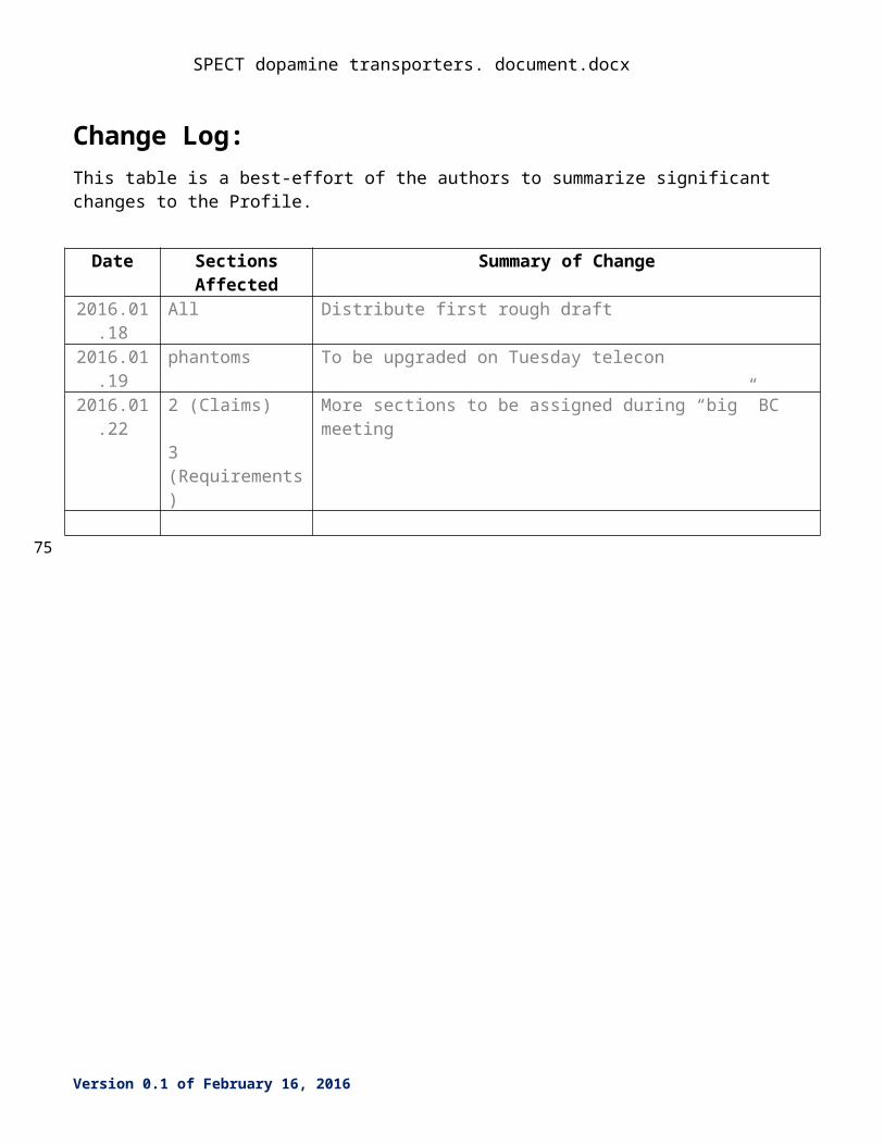
Change Log:This table is a best-effort of the authors to summarize significant changes to the Profile.
Date Sections Affected Summary of Change2016.01.18 All Distribute first rough draft2016.01.19 phantoms To be upgraded on Tuesday telecon2016.01.22 2 (Claims)
3 (Requirements)
More sections to be assigned during “big” BC meeting
Version 0.1 of February 16, 2016
75
SPECT dopamine transporters. document.docx
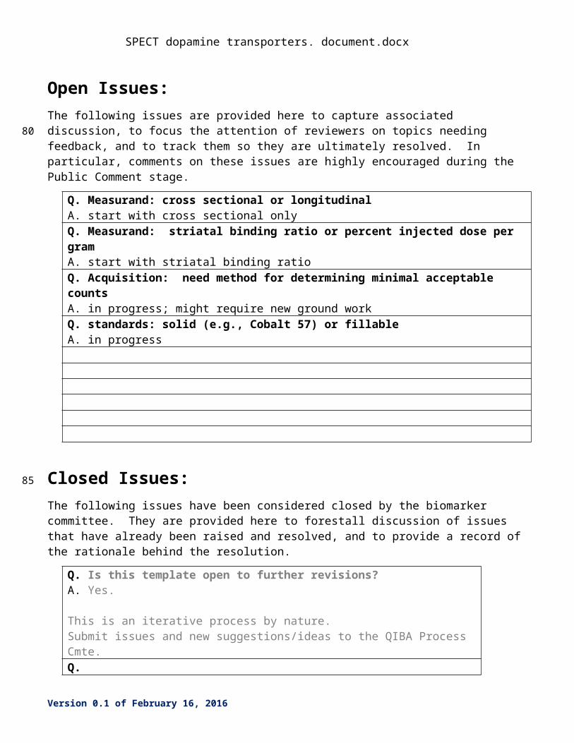
Open Issues:The following issues are provided here to capture associated discussion, to focus the attention of reviewers on topics needing feedback, and to track them so they are ultimately resolved. In particular, comments on these issues are highly encouraged during the Public Comment stage.
Q. Measurand: cross sectional or longitudinalA. start with cross sectional onlyQ. Measurand: striatal binding ratio or percent injected dose per gramA. start with striatal binding ratioQ. Acquisition: need method for determining minimal acceptable countsA. in progress; might require new ground workQ. standards: solid (e.g., Cobalt 57) or fillableA. in progress
Closed Issues:The following issues have been considered closed by the biomarker committee. They are provided here to forestall discussion of issues that have already been raised and resolved, and to provide a record of the rationale behind the resolution.
Q. Is this template open to further revisions?A. Yes.
This is an iterative process by nature.Submit issues and new suggestions/ideas to the QIBA Process Cmte.Q.A.
Version 0.1 of February 16, 2016
80
85
SPECT dopamine transporters. document.docx
1. Executive SummaryParkinsonism is a major health problem. Distinguishing Parkinson’s disease (PD) from other movement disorders that can mimic it has important implications for management. The goal of this QIBA Profile is to optimize the performance of Iodine-123 (123I) ioflupane single photon emission computed tomography (SPECT) for quantifying the concentration of regional cerebral dopamine transporters (DaT) in patients who are being evaluated for neurodegenerative disorders.
The Claim (Section 2): This profile claims that compliance with its specifications will produce measurements of DaT that can distinguish patients with PD from matched controls. The claim is based on an observation that idiopathic PD is associated with dopminergic degeneration in the subtantia nigra, which in turn is manifested by a loss of DaT activity in the basal ganglia. The loss is first observed in the most posterior aspect of the putamen, and then seems to march anteriorly. As a result, quantifying DaT in the posterior putamen can distinguish patients with PD from matched controls. The Activities (Section 3) describe what needs to be done to make measurements that reliably distinguish patients from controls with confidence. Requirements are placed on the Actors that participate in those activities as necessary to achieve the Claim. Assessment Procedures (Section 4) for evaluating specific requirements are defined as needed.
This QIBA Profile, Quantifying Dopamine Transporters with 123Iodine Labeled Ioflupane in Neurodegenerative Disease, addresses quantitative SPECT imaging, which is often used as a diagnostic, as well as a longitudinal biomarker of disease progression or response to treatment. It places requirements on Acquisition Devices, Technologists, Radiologists, Reconstruction Software and Image Analysis Tools involved in Subject Handling, Image Data Acquisition, Image Data Reconstruction, Image QA and Image Analysis.
The requirements are focused on achieving sufficient accuracy and avoiding unnecessary variability of the DaT measurements to distinguish patients with PD from matched controls.
The clinical performance target is to achieve a 95% confidence interval for the striatal binding ratio with both a reproducibility and a repeatability of +/- 15%.
This document is intended to help clinicians basing decisions on this biomarker, imaging staff generating this biomarker, vendor staff developing related products, purchasers of such products and investigators designing trials with imaging endpoints.
Note that this document only states requirements to achieve the claim, not “requirements on standard of care.” Conformance to this Profile is secondary to properly caring for the patient.
QIBA Profiles addressing other imaging biomarkers using CT, MRI, PET and Ultrasound can be found at qibawiki.rsna.org.
Version 0.1 of February 16, 2016
90
95
100
110
115
120
SPECT dopamine transporters. document.docx
2. Clinical Context and ClaimsClinical Context
Parkinson’s disease (PD) is a major health problem. The prevalence is increasing as the population ages. Onset can be insidious, which can make the diagnosis challenging on clinical grounds alone. A number of radiopharmaceuticals that can quantify several different components of the pre-synaptic dopamine system have been shown to help distinguish between idiopathic PD and movement disorders that mimic it. This profile focuses on a marketed radiopharmaceutical for this use, Iodine-123 (123I) labeled ioflupane (methyl (1R,2S,3S,5S)- 3-(4-iodophenyl)- 8-(3-fluoropropyl)- 8-azabicyclo[3.2.1]octane- 2-carboxylate).
Conformance to this Profile by all relevant staff and equipment supports the following claim(s):
Claim 1: Cross sectional: A measured striatal binding ratio (SBR) is within +/- 15% of the true SBR. During the initial presentation of newly symptomatic patients, a diagnosis of Parkinson’s disease (PD) is consistent with a finding of a SBR in the posterior putamen that is 50% or less than the value in properly matched controls.
Claim 2: Longitudinal: For a measured change in SBR of X, a 95% confidence interval for the true change is [X-15%, X+15%].
These claims hold when: Anatomical imaging, such as magnetic resonance imaging (MRI), has already ruled out other
causes of parkinsonism, such as stroke; The patient has not been taking drugs or nutritional supplements that can transiently influence
the measurements; The patient does not have a deformity or condition that prevents proper positioning in the
scanner; The patient can tolerate the imaging procedures well enough to prevent motion from
confounding the acquisition; The administration of the radiopharmaceutical is not confounded by infiltration of the dose; Et cetera
DiscussionThe primary measurand, or outcome measure, is the specific binding ratio (SBR) obtained in the striatum, and usually divided into separate values for the caudate, anterior putamen, and posterior putamen. While research studies sometimes include the SBR for other structures, such as the substantia nigra pars compacta, the thalamus, amygdala, and hippocampus, these regions are beyond the scope of this profile.
The SBR is defined as the count density in a striatal region of interest (ROI) divided by a reference region count density minus 1, and is roughly equivalent to the non-displaceable binding potential (BPnd)
Version 0.1 of February 16, 2016
125
130
135
140
145
150
155
160
SPECT dopamine transporters. document.docx
The reference region is ideally the cerebellum, as it contains no known dopaminergic proteins or messenger RNA for these proteins. Acceptable alternatives include the occipital cortex, particularly when the axial field of view is limited.
An alternative outcome measure is the fraction of the injected dose per unit volume in a ROI expressed in units of kBq/mL.
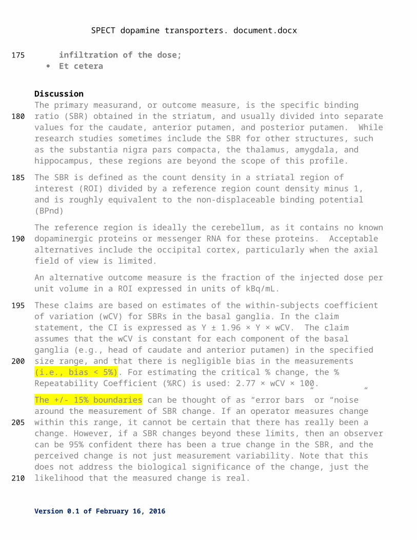
These claims are based on estimates of the within-subjects coefficient of variation (wCV) for SBRs in the basal ganglia. In the claim statement, the CI is expressed as Y ± 1.96 × Y × wCV. The claim assumes that the wCV is constant for each component of the basal ganglia (e.g., head of caudate and anterior putamen) in the specified size range, and that there is negligible bias in the measurements (i.e., bias < 5%). For estimating the critical % change, the % Repeatability Coefficient (%RC) is used: 2.77 × wCV × 100.
The +/- 15% boundaries can be thought of as “error bars” or “noise” around the measurement of SBR change. If an operator measures change within this range, it cannot be certain that there has really been a change. However, if a SBR changes beyond these limits, then an observer can be 95% confident there has been a true change in the SBR, and the perceived change is not just measurement variability. Note that this does not address the biological significance of the change, just the likelihood that the measured change is real.
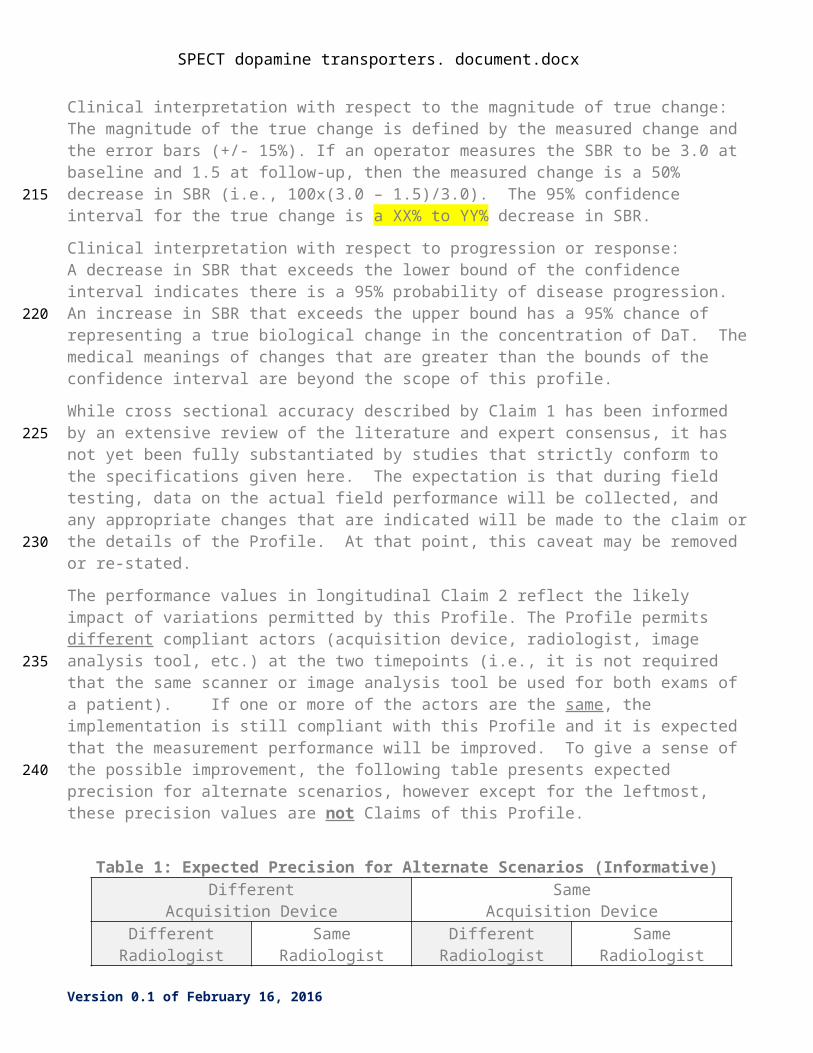
Clinical interpretation with respect to the magnitude of true change: The magnitude of the true change is defined by the measured change and the error bars (+/- 15%). If an operator measures the SBR to be 3.0 at baseline and 1.5 at follow-up, then the measured change is a 50% decrease in SBR (i.e., 100x(3.0 – 1.5)/3.0). The 95% confidence interval for the true change is a XX% to YY% decrease in SBR.
Clinical interpretation with respect to progression or response:A decrease in SBR that exceeds the lower bound of the confidence interval indicates there is a 95% probability of disease progression. An increase in SBR that exceeds the upper bound has a 95% chance of representing a true biological change in the concentration of DaT. The medical meanings of changes that are greater than the bounds of the confidence interval are beyond the scope of this profile.
While cross sectional accuracy described by Claim 1 has been informed by an extensive review of the literature and expert consensus, it has not yet been fully substantiated by studies that strictly conform to the specifications given here. The expectation is that during field testing, data on the actual field performance will be collected, and any appropriate changes that are indicated will be made to the claim or the details of the Profile. At that point, this caveat may be removed or re-stated.
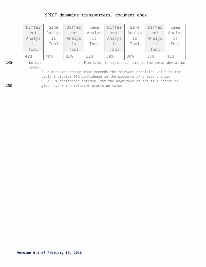
The performance values in longitudinal Claim 2 reflect the likely impact of variations permitted by this Profile. The Profile permits different compliant actors (acquisition device, radiologist, image analysis tool, etc.) at the two timepoints (i.e., it is not required that the same scanner or image analysis tool be used for both exams of a patient). If one or more of the actors are the same, the implementation is still compliant with this Profile and it is expected that the measurement performance will be improved. To give a sense of the possible improvement, the following table presents expected precision for alternate scenarios, however except for the leftmost, these precision values are not Claims of this Profile.
Version 0.1 of February 16, 2016
165
170
175
180
185
190
195
200

SPECT dopamine transporters. document.docx
Table 1: Expected Precision for Alternate Scenarios (Informative)Different
Acquisition DeviceSame
Acquisition DeviceDifferent
RadiologistSame
RadiologistDifferent
RadiologistSame
RadiologistDifferent Analysis
Tool
Same Analysis
Tool
Different Analysis
Tool
Same Analysis
Tool
Different Analysis
Tool
Same Analysis
Tool
Different Analysis
Tool
Same Analysis
Tool47% 46% 33% 32% 38% 36% 13% 11%
Notes: 1. Precision is expressed here as the total deviation index.2. A measured change that exceeds the relevant precision value in the table indicates 95% confidence in the presence of a true change. 3. A 95% confidence interval for the magnitude of the true change is given by: ± the relevant precision value.
Version 0.1 of February 16, 2016
205
210
SPECT dopamine transporters. document.docx
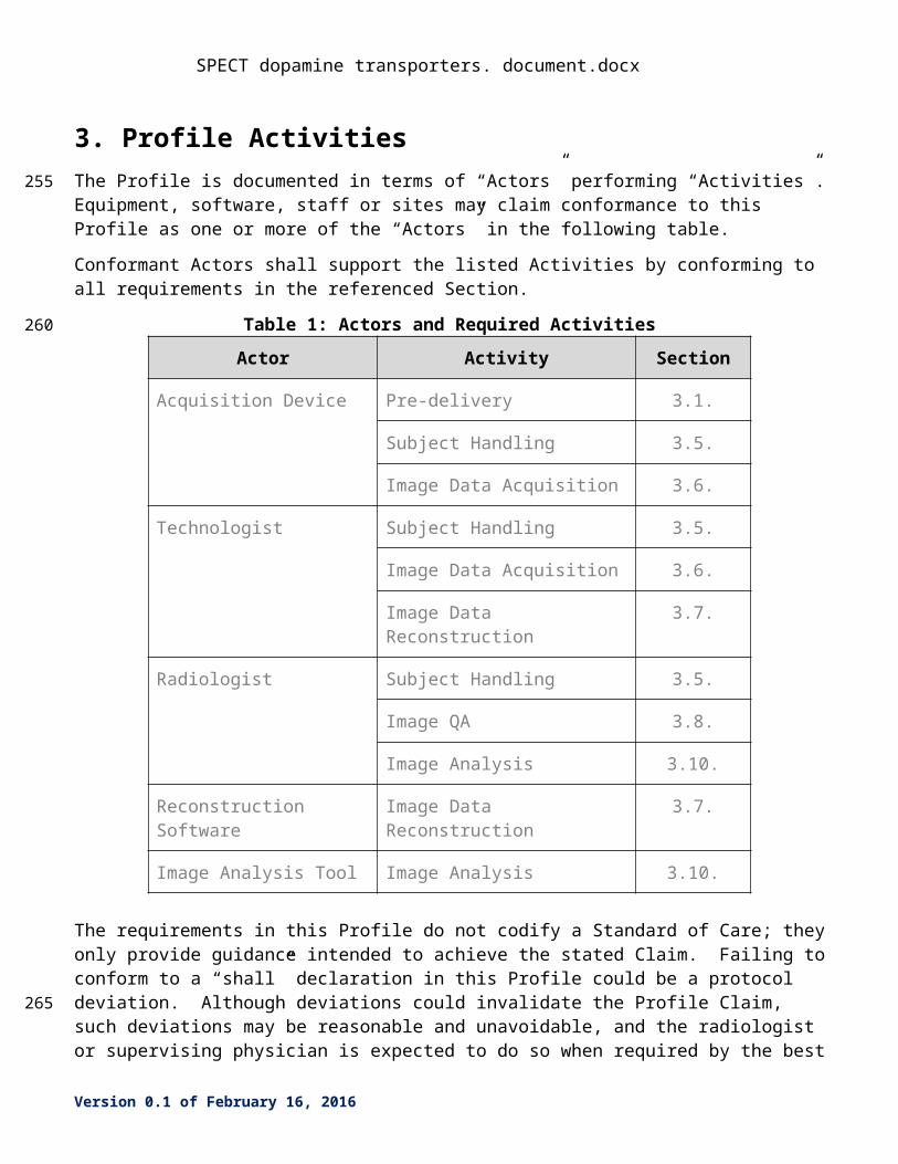
3. Profile ActivitiesThe Profile is documented in terms of “Actors” performing “Activities”. Equipment, software, staff or sites may claim conformance to this Profile as one or more of the “Actors” in the following table.
Conformant Actors shall support the listed Activities by conforming to all requirements in the referenced Section.
Table 1: Actors and Required Activities
Actor Activity Section
Acquisition Device Pre-delivery 3.1.
Subject Handling 3.5.
Image Data Acquisition 3.6.
Technologist Subject Handling 3.5.
Image Data Acquisition 3.6.
Image Data Reconstruction 3.7.
Radiologist Subject Handling 3.5.
Image QA 3.8.
Image Analysis 3.10.
Reconstruction Software Image Data Reconstruction 3.7.
Image Analysis Tool Image Analysis 3.10.
The requirements in this Profile do not codify a Standard of Care; they only provide guidance intended to achieve the stated Claim. Failing to conform to a “shall” declaration in this Profile could be a protocol deviation. Although deviations could invalidate the Profile Claim, such deviations may be reasonable and unavoidable, and the radiologist or supervising physician is expected to do so when required by the best interest of the patient or research subject. How study sponsors and others decide to handle deviations for their own purposes is entirely up to them.
The sequencing of the Activities specified in this Profile are shown in Figure 1:
<activity sequence diagram>Figure 1: <Title of the Profile> - Activity Sequence
Version 0.1 of February 16, 2016
215
220
225
SPECT dopamine transporters. document.docx
Version 0.1 of February 16, 2016
SPECT dopamine transporters. document.docx
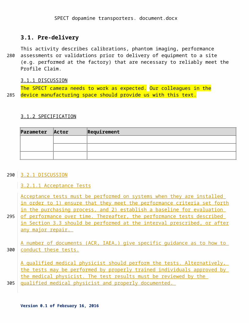
3.1. Pre-delivery
This activity describes calibrations, phantom imaging, performance assessments or validations prior to delivery of equipment to a site (e.g. performed at the factory) that are necessary to reliably meet the Profile Claim.
3.1.1 DISCUSSION The SPECT camera needs to work as expected. Our colleagues in the device manufacturing space should provide us with this text.
3.1.2 SPECIFICATION
Parameter Actor Requirement
3.2.1 DISCUSSION
3.2.1.1 Acceptance Tests
Acceptance tests must be performed on systems when they are installed in order to 1) ensure that they meet the performance criteria set forth in the purchasing process, and 2) establish a baseline for evaluation of performance over time. Thereafter, the performance tests described in Section 3.3 should be performed at the interval prescribed, or after any major repair.
A number of documents (ACR, IAEA,) give specific guidance as to how to conduct these tests.
A qualified medical physicist should perform the tests. Alternatively, the tests may be performed by properly trained individuals approved by the medical physicist. The test results must be reviewed by the qualified medical physicist and properly documented.
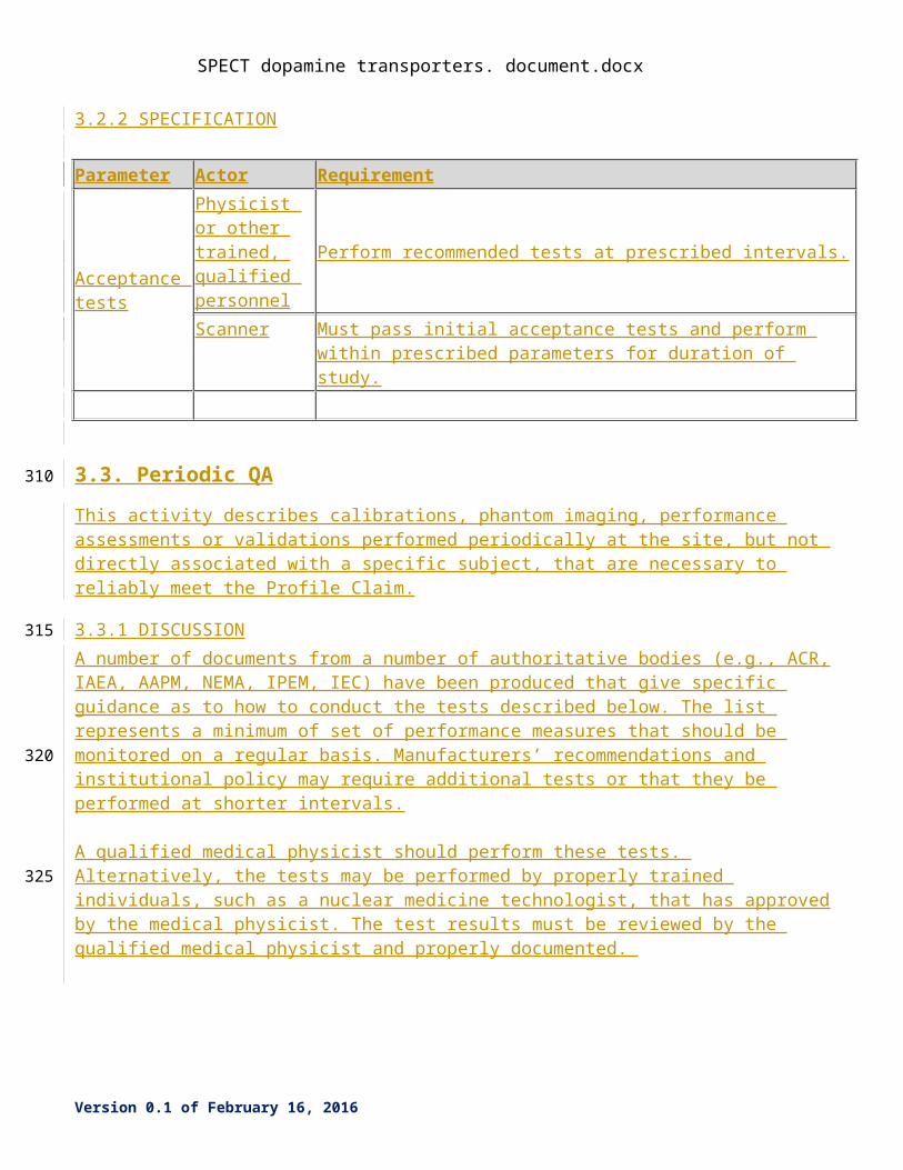
3.2.2 SPECIFICATION
Parameter Actor RequirementAcceptance tests
Physicist or other trained, qualified personnel
Perform recommended tests at prescribed intervals.
Scanner Must pass initial acceptance tests and perform within prescribed
Version 0.1 of February 16, 2016
230
235
240
245
250
255
SPECT dopamine transporters. document.docx
Parameter Actor Requirementparameters for duration of study.
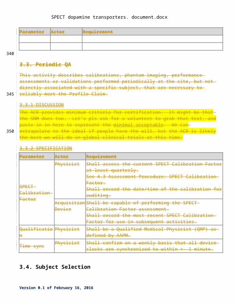
3.3. Periodic QA
This activity describes calibrations, phantom imaging, performance assessments or validations performed periodically at the site, but not directly associated with a specific subject, that are necessary to reliably meet the Profile Claim.
3.3.1 DISCUSSIONA number of documents from a number of authoritative bodies (e.g., ACR, IAEA, AAPM, NEMA, IPEM, IEC) have been produced that give specific guidance as to how to conduct the tests described below. The list represents a minimum of set of performance measures that should be monitored on a regular basis. Manufacturers’ recommendations and institutional policy may require additional tests or that they be performed at shorter intervals.
A qualified medical physicist should perform these tests. Alternatively, the tests may be performed by properly trained individuals, such as a nuclear medicine technologist, that has approved by the medical physicist. The test results must be reviewed by the qualified medical physicist and properly documented.
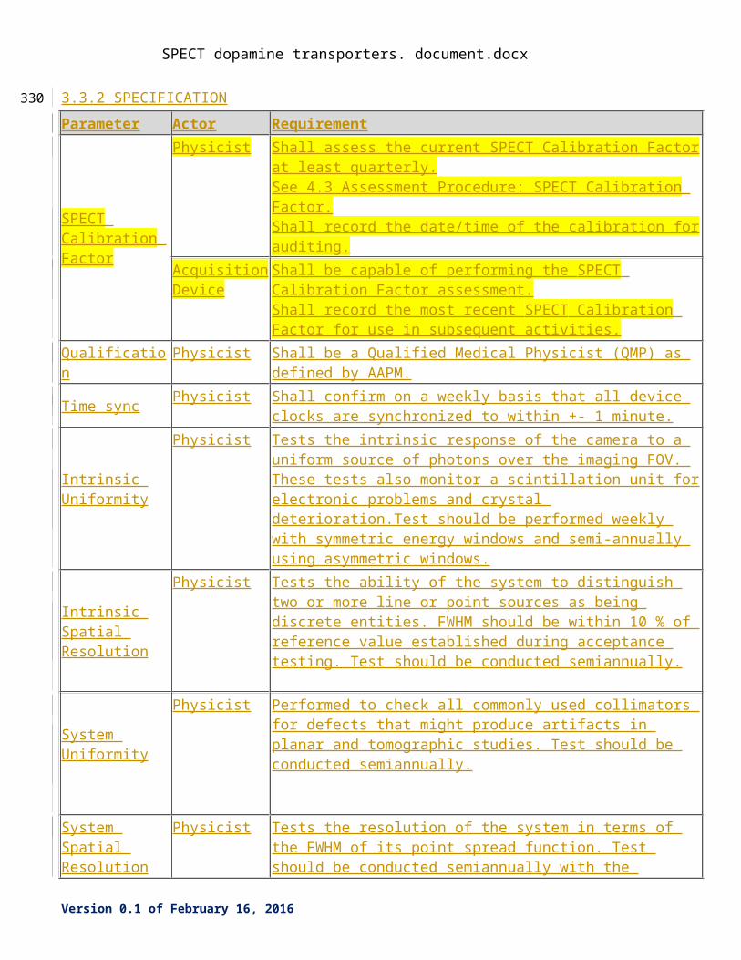
3.3.2 SPECIFICATION
Parameter Actor Requirement
SPECT Calibration Factor
Physicist Shall assess the current SPECT Calibration Factor at least quarterly.See 4.3 Assessment Procedure: SPECT Calibration Factor.Shall record the date/time of the calibration for auditing.
Acquisition Device
Shall be capable of performing the SPECT Calibration Factor assessment.Shall record the most recent SPECT Calibration Factor for use in subsequent activities.
Qualification Physicist Shall be a Qualified Medical Physicist (QMP) as defined by AAPM.
Time sync Physicist Shall confirm on a weekly basis that all device clocks are synchronized to within +- 1 minute.
Intrinsic Uniformity
Physicist Tests the intrinsic response of the camera to a uniform source of photons over the imaging FOV. These tests also monitor a scintillation unit for electronic problems and crystal deterioration.Test should be performed weekly with symmetric energy windows and semi-annually using asymmetric windows.
Intrinsic Spatial Resolution
Physicist Tests the ability of the system to distinguish two or more line or point sources as being discrete entities. FWHM should be within 10 % of reference value established during acceptance testing. Test should be conducted semiannually.
Version 0.1 of February 16, 2016
260
265
270
SPECT dopamine transporters. document.docx
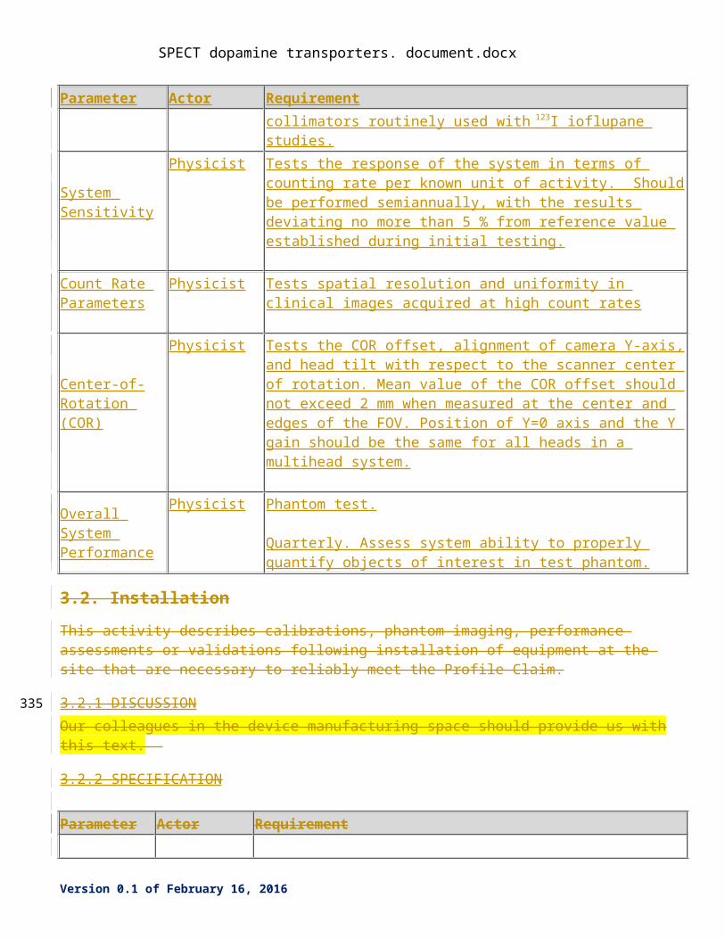
Parameter Actor Requirement
System Uniformity
Physicist Performed to check all commonly used collimators for defects that might produce artifacts in planar and tomographic studies. Test should be conducted semiannually.
System Spatial Resolution
Physicist Tests the resolution of the system in terms of the FWHM of its point spread function. Test should be conducted semiannually with the collimators routinely used with 123 I ioflupane studies.
System Sensitivity
Physicist Tests the response of the system in terms of counting rate per known unit of activity. Should be performed semiannually, with the results deviating no more than 5 % from reference value established during initial testing.
Count Rate Parameters
Physicist Tests spatial resolution and uniformity in clinical images acquired at high count rates
Center-of-Rotation (COR)
Physicist Tests the COR offset, alignment of camera Y-axis, and head tilt with respect to the scanner center of rotation. Mean value of the COR offset should not exceed 2 mm when measured at the center and edges of the FOV. Position of Y=0 axis and the Y gain should be the same for all heads in a multihead system.
Overall System Performance
Physicist Phantom test.
Quarterly. Assess system ability to properly quantify objects of interest in test phantom.
3.2. Installation
This activity describes calibrations, phantom imaging, performance assessments or validations following installation of equipment at the site that are necessary to reliably meet the Profile Claim.
3.2.1 DISCUSSION Our colleagues in the device manufacturing space should provide us with this text.
3.2.2 SPECIFICATION
Parameter Actor Requirement
Version 0.1 of February 16, 2016
275
SPECT dopamine transporters. document.docx
Parameter Actor Requirement
3.3. Periodic QA
This activity describes calibrations, phantom imaging, performance assessments or validations performed periodically at the site, but not directly associated with a specific subject, that are necessary to reliably meet the Profile Claim.
3.3.1 DISCUSSION The ACR provides minimum criteria for certification. It might be that the SNM does too. Let’s pls ask for a volunteer to grab that text, and paste in in here to represent the minimal acceptable. We can extrapolate to the ideal if people have the will, but the ACR is likely the best we will do in global clinical trials at this time.
3.3.2 SPECIFICATION
Parameter Actor Requirement
SPECT Calibration Factor
Physicist Shall assess the current SPECT Calibration Factor at least quarterly.See 4.3 Assessment Procedure: SPECT Calibration Factor.Shall record the date/time of the calibration for auditing.
Acquisition Device
Shall be capable of performing the SPECT Calibration Factor assessment.Shall record the most recent SPECT Calibration Factor for use in subsequent activities.
Qualification Physicist Shall be a Qualified Medical Physicist (QMP) as defined by AAPM.
Time sync Physicist Shall confirm on a weekly basis that all device clocks are synchronized to within +- 1 minute.
3.4. Subject Selection
This activity describes criteria and procedures related to the selection of appropriate imaging subjects that are necessary to reliably meet the Profile Claim.
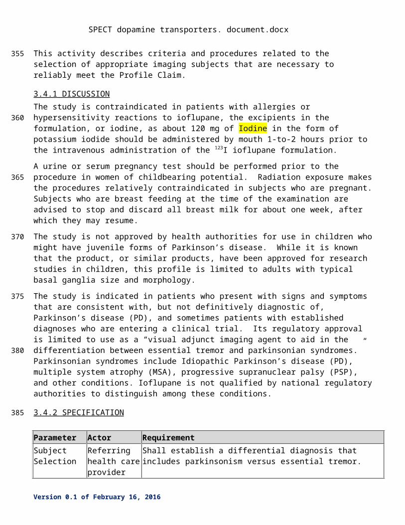
3.4.1 DISCUSSION The study is contraindicated in patients with allergies or hypersensitivity reactions to ioflupane, the excipients in the formulation, or iodine, as about 120 mg of Iodine in the form of potassium iodide should be administered by mouth 1-to-2 hours prior to the intravenous administration of the 123I ioflupane formulation.
A urine or serum pregnancy test should be performed prior to the procedure in women of childbearing potential. Radiation exposure makes the procedures relatively contraindicated in subjects who are pregnant. Subjects who are breast feeding at the time of the examination are advised to stop and discard all breast milk for about one week, after which they may resume.
Version 0.1 of February 16, 2016
280
285
290
295
300
SPECT dopamine transporters. document.docx
The study is not approved by health authorities for use in children who might have juvenile forms of Parkinson’s disease. While it is known that the product, or similar products, have been approved for research studies in children, this profile is limited to adults with typical basal ganglia size and morphology.
The study is indicated in patients who present with signs and symptoms that are consistent with, but not definitively diagnostic of, Parkinson’s disease (PD), and sometimes patients with established diagnoses who are entering a clinical trial. Its regulatory approval is limited to use as a “visual adjunct imaging agent to aid in the differentiation between essential tremor and parkinsonian syndromes.” Parkinsonian syndromes include Idiopathic Parkinson’s disease (PD), multiple system atrophy (MSA), progressive supranuclear palsy (PSP), and other conditions. Ioflupane is not qualified by national regulatory authorities to distinguish among these conditions.
3.4.2 SPECIFICATION
Parameter Actor Requirement
Subject Selection
Referring health care provider
Shall establish a differential diagnosis that includes parkinsonism versus essential tremor.
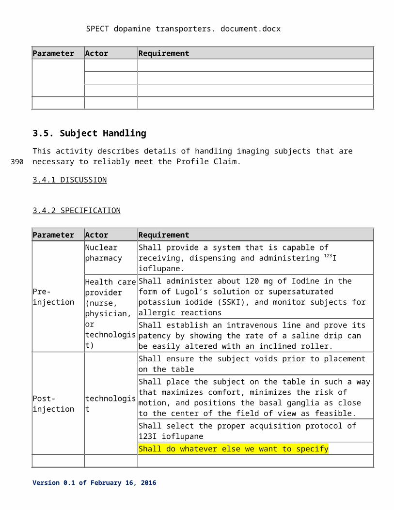
3.5. Subject Handling
This activity describes details of handling imaging subjects that are necessary to reliably meet the Profile Claim.
3.4.1 DISCUSSION
3.4.2 SPECIFICATION
Parameter Actor Requirement
Pre-injection
Nuclear pharmacy
Shall provide a system that is capable of receiving, dispensing and administering 123I ioflupane.
Health care provider (nurse, physician, or technologist)
Shall administer about 120 mg of Iodine in the form of Lugol’s solution or supersaturated potassium iodide (SSKI), and monitor subjects for allergic reactionsShall establish an intravenous line and prove its patency by showing the rate of a saline drip can be easily altered with an inclined roller.
Post-injection technologist Shall ensure the subject voids prior to placement on the table
Version 0.1 of February 16, 2016
305
310
315
320
SPECT dopamine transporters. document.docx
Parameter Actor RequirementShall place the subject on the table in such a way that maximizes comfort, minimizes the risk of motion, and positions the basal ganglia as close to the center of the field of view as feasible.Shall select the proper acquisition protocol of 123I ioflupaneShall do whatever else we want to specify
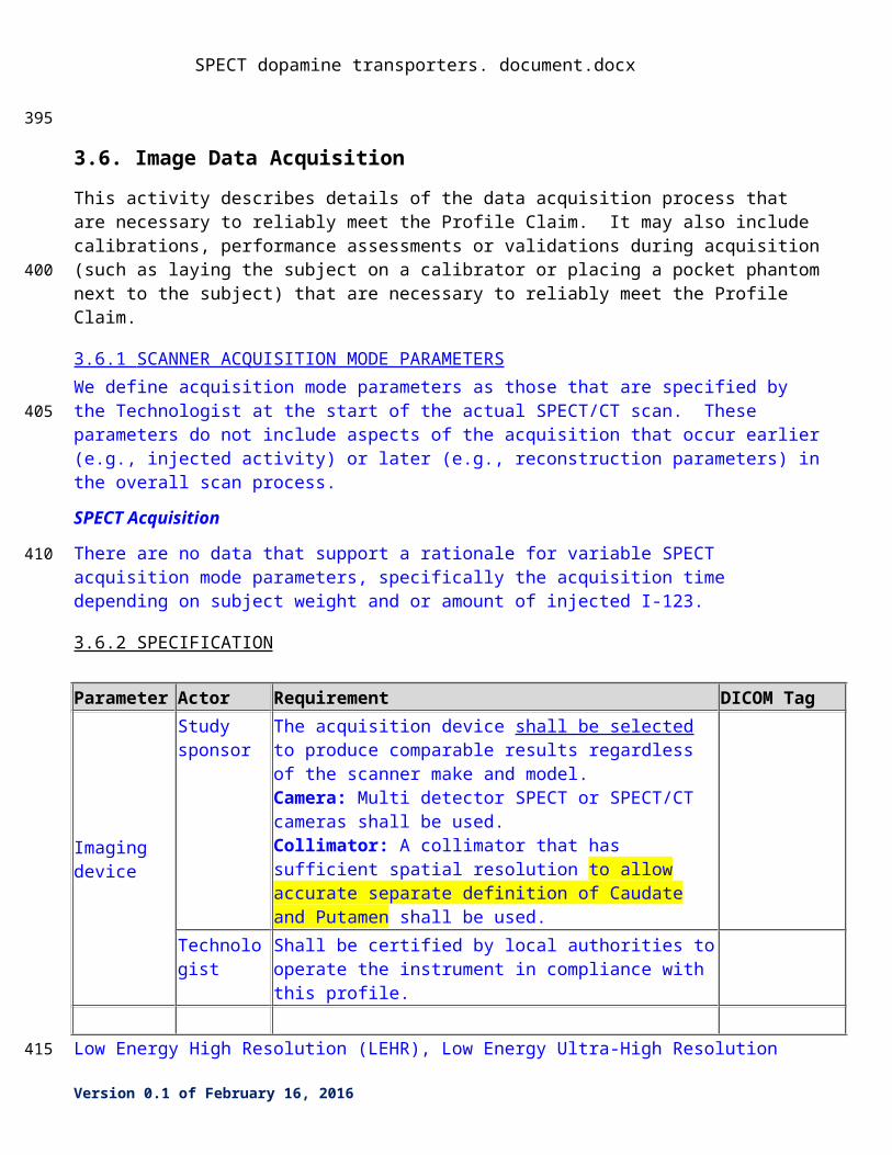
3.6. Image Data Acquisition
This activity describes details of the data acquisition process that are necessary to reliably meet the Profile Claim. It may also include calibrations, performance assessments or validations during acquisition (such as laying the subject on a calibrator or placing a pocket phantom next to the subject) that are necessary to reliably meet the Profile Claim.
3.6.1 SCANNER ACQUISITION MODE PARAMETERS We define acquisition mode parameters as those that are specified by the Technologist at the start of the actual SPECT/CT scan. These parameters do not include aspects of the acquisition that occur earlier (e.g., injected activity) or later (e.g., reconstruction parameters) in the overall scan process.
SPECT Acquisition
There are no data that support a rationale for variable SPECT acquisition mode parameters, specifically the acquisition time depending on subject weight and or amount of injected I-123.
3.6.2 SPECIFICATION
Parameter Actor Requirement DICOM Tag
Imaging device
Study sponsor
The acquisition device shall be selected to produce comparable results regardless of the scanner make and model.Camera: Multi detector SPECT or SPECT/CT cameras shall be used.Collimator: A collimator that has sufficient spatial resolution to allow accurate separate definition of Caudate and Putamen shall be used.
Technologist Shall be certified by local authorities to operate the instrument in compliance with this profile.
Low Energy High Resolution (LEHR), Low Energy Ultra-High Resolution (LEUHR) and fan beam collimators with manufacturer specified (or measured according to NEMA standards) planar system resolution of < 8 mm FWHM (in ‘air’ at 10 cm distance) typically meets the above requirement. ME collimators, which reduce septal penetration may be insufficient in terms of the resolution requirement. If available,
Version 0.1 of February 16, 2016
325
330
335
340
SPECT dopamine transporters. document.docx
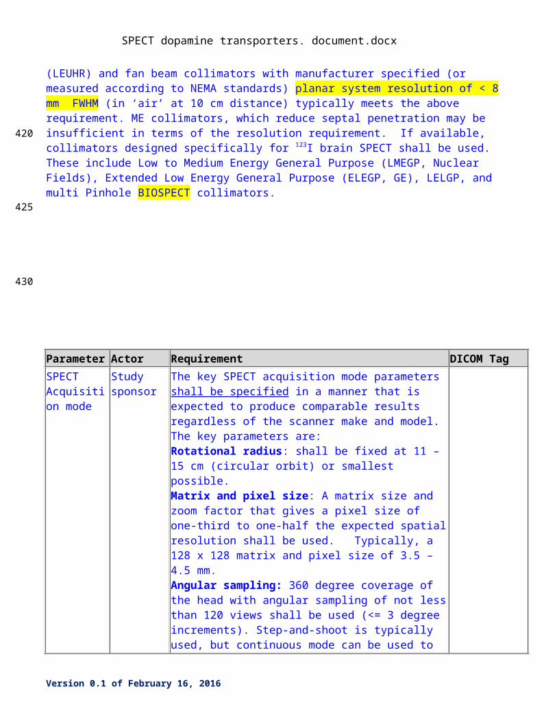
collimators designed specifically for 123I brain SPECT shall be used. These include Low to Medium Energy General Purpose (LMEGP, Nuclear Fields), Extended Low Energy General Purpose (ELEGP, GE), LELGP, and multi Pinhole BIOSPECT collimators.
Parameter Actor Requirement DICOM Tag
SPECT Acquisition mode
Study sponsor
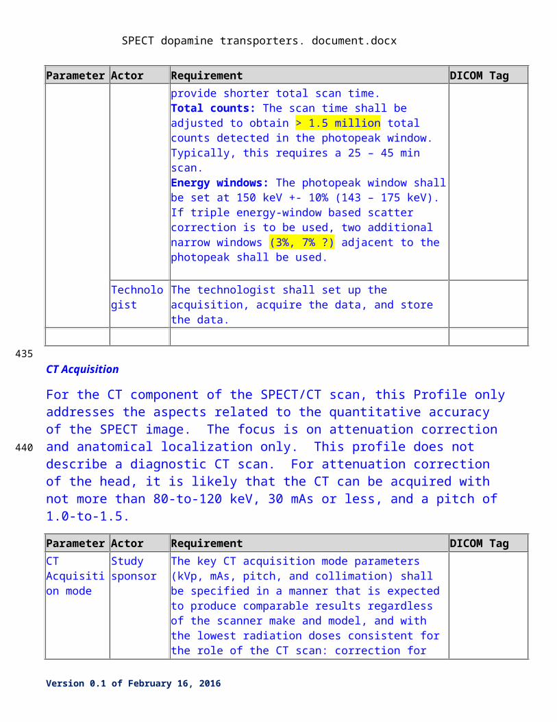
The key SPECT acquisition mode parameters shall be specified in a manner that is expected to produce comparable results regardless of the scanner make and model. The key parameters are:Rotational radius: shall be fixed at 11 – 15 cm (circular orbit) or smallest possible.Matrix and pixel size: A matrix size and zoom factor that gives a pixel size of one-third to one-half the expected spatial resolution shall be used. Typically, a 128 x 128 matrix and pixel size of 3.5 – 4.5 mm.Angular sampling: 360 degree coverage of the head with angular sampling of not less than 120 views shall be used (<= 3 degree increments). Step-and-shoot is typically used, but continuous mode can be used to provide shorter total scan time.Total counts: The scan time shall be adjusted to obtain > 1.5 million total counts detected in the photopeak window. Typically, this requires a 25 – 45 min scan.Energy windows: The photopeak window shall be set at 150 keV +- 10% (143 – 175 keV). If triple energy-window based scatter correction is to be used, two additional narrow windows (3%, 7% ?) adjacent to the photopeak shall be used.
Technologist The technologist shall set up the acquisition, acquire the data, and store the data.
CT Acquisition
Version 0.1 of February 16, 2016
345
350
355
SPECT dopamine transporters. document.docx
For the CT component of the SPECT/CT scan, this Profile only addresses the aspects related to the quantitative accuracy of the SPECT image. The focus is on attenuation correction and anatomical localization only. This profile does not describe a diagnostic CT scan. For attenuation correction of the head, it is likely that the CT can be acquired with not more than 80-to-120 keV, 30 mAs or less, and a pitch of 1.0-to-1.5.
Parameter Actor Requirement DICOM Tag
CT Acquisition mode
Study sponsor
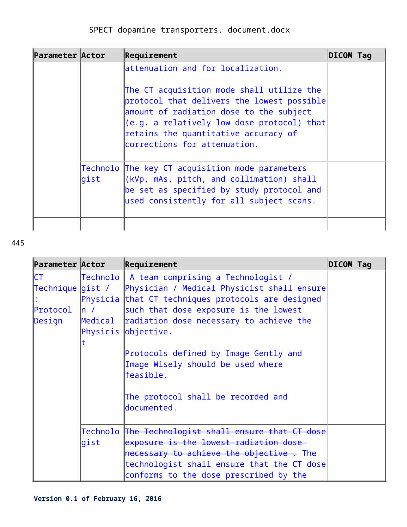
The key CT acquisition mode parameters (kVp, mAs, pitch, and collimation) shall be specified in a manner that is expected to produce comparable results regardless of the scanner make and model, and with the lowest radiation doses consistent for the role of the CT scan: correction for attenuation and for localization.
The CT acquisition mode shall utilize the protocol that delivers the lowest possible amount of radiation dose to the subject (e.g. a relatively low dose protocol) that retains the quantitative accuracy of corrections for attenuation.
Technologist The key CT acquisition mode parameters (kVp, mAs, pitch, and collimation) shall be set as specified by study protocol and used consistently for all subject scans.
Parameter Actor Requirement DICOM Tag
CT Technique: Protocol Design
Technologist / Physician / Medical Physicist
A team comprising a Technologist / Physician / Medical Physicist shall ensure that CT techniques protocols are designed such that dose exposure is the lowest radiation dose necessary to achieve the objective.
Protocols defined by Image Gently and Image Wisely should be used where feasible.
The protocol shall be recorded and documented.
Technologist The Technologist shall ensure that CT dose exposure is the lowest radiation dose necessary to achieve the objective . The technologist shall ensure that the CT dose conforms to the dose prescribed by the supervising physician or protocol.
Version 0.1 of February 16, 2016
360
SPECT dopamine transporters. document.docx
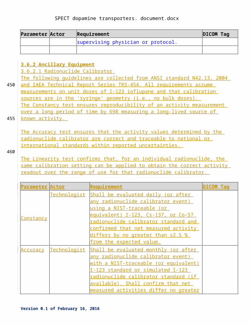
3.6.2 Ancillary Equipment3.6.2.1 Radionuclide Calibrator The following guidelines are collected from ANSI standard N42.13, 2004 and IAEA Technical Report Series TRS-454. All requirements assume measurements on unit doses of I-123 ioflupane and that calibration sources are in the 'syringe' geometry (i.e., no bulk doses). The Constancy test ensures reproducibility of an activity measurement over a long period of time by 698 measuring a long-lived source of known activity.
The Accuracy test ensures that the activity values determined by the radionuclide calibrator are correct and traceable to national or international standards within reported uncertainties.
The Linearity test confirms that, for an individual radionuclide, the same calibration setting can be applied to obtain the correct activity readout over the range of use for that radionuclide calibrator.
Parameter Actor Requirement DICOM Tag
Constancy
Technologist Shall be evaluated daily (or after any radionuclide calibrator event) using a NIST-traceable (or equivalent) I-123, Cs-137, or Co-57 radionuclide calibrator standard and confirmed that net measured activity differs by no greater than ±2.5 % from the expected value.
Accuracy
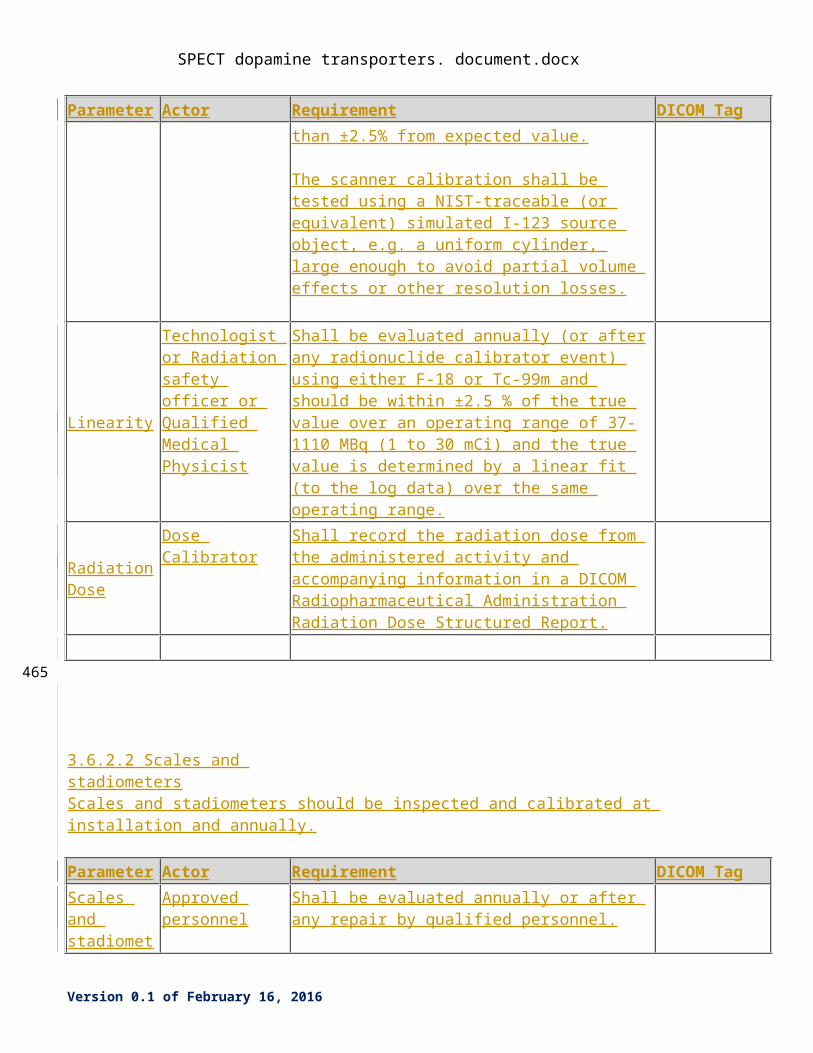
Technologist Shall be evaluated monthly (or after any radionuclide calibrator event) with a NIST-traceable (or equivalent) I-123 standard or simulated I-123 radionuclide calibrator standard (if available). Shall confirm that net measured activities differ no greater than ±2.5% from expected value.
The scanner calibration shall be tested using a NIST-traceable (or equivalent) simulated I-123 source object, e.g. a uniform cylinder, large enough to avoid partial volume effects or other resolution losses.
Linearity
Technologist or Radiation safety officer or Qualified Medical Physicist
Shall be evaluated annually (or after any radionuclide calibrator event) using either F-18 or Tc-99m and should be within ±2.5 % of the true value over an operating range of 37-1110 MBq (1 to 30 mCi) and the true value is determined by a linear fit (to the log data) over the same operating range.
Radiation Dose
Dose Calibrator Shall record the radiation dose from the administered activity and accompanying information in a DICOM Radiopharmaceutical Administration Radiation Dose Structured Report.
Version 0.1 of February 16, 2016
365
370
375
SPECT dopamine transporters. document.docx
Parameter Actor Requirement DICOM Tag
3.6.2.2 Scales and stadiometers
Scales and stadiometers should be inspected and calibrated at installation and annually.
Parameter Actor Requirement DICOM Tag
Scales and stadiometers
Approved personnel
Shall be evaluated annually or after any repair by qualified personnel.
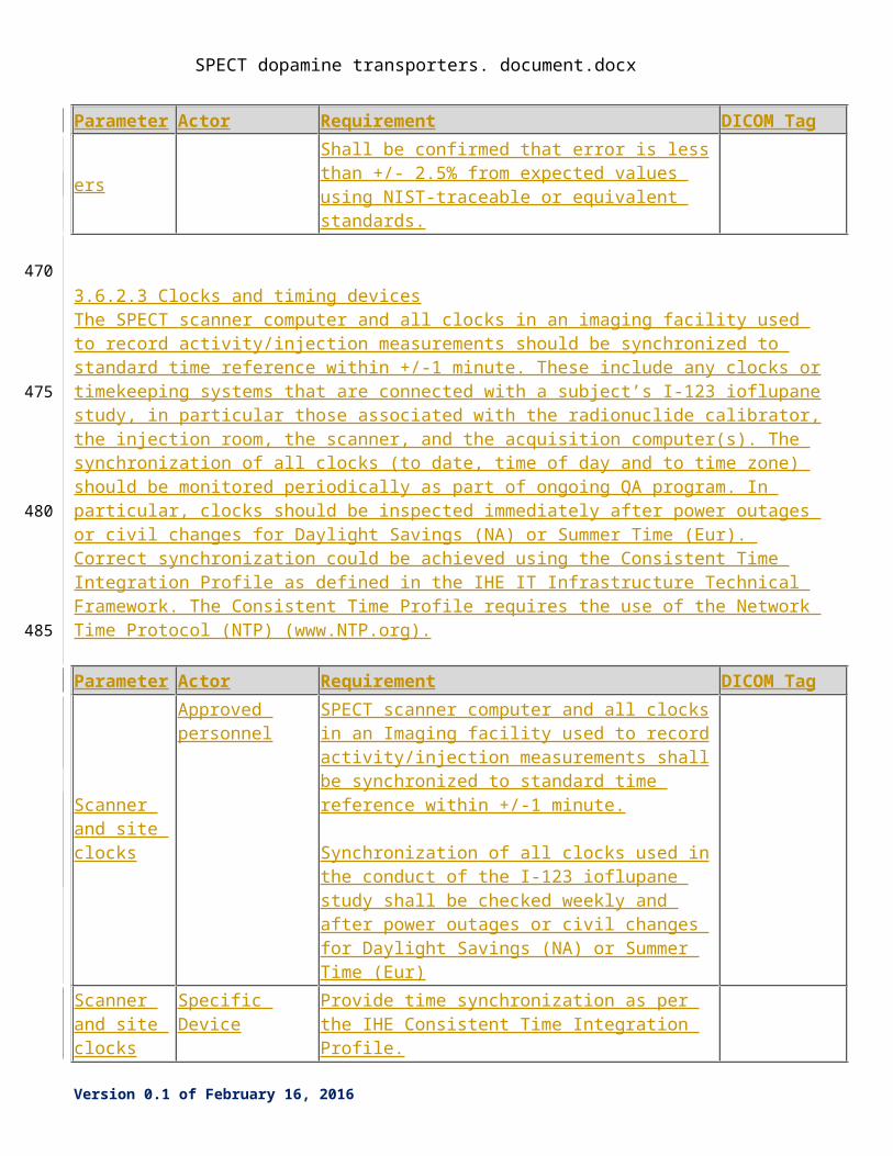
Shall be confirmed that error is less than +/- 2.5% from expected values using NIST-traceable or equivalent standards.
3.6.2.3 Clocks and timing devicesThe SPECT scanner computer and all clocks in an imaging facility used to record activity/injection measurements should be synchronized to standard time reference within +/-1 minute. These include any clocks or timekeeping systems that are connected with a subject’s I-123 ioflupane study, in particular those associated with the radionuclide calibrator, the injection room, the scanner, and the acquisition computer(s). The synchronization of all clocks (to date, time of day and to time zone) should be monitored periodically as part of ongoing QA program. In particular, clocks should be inspected immediately after power outages or civil changes for Daylight Savings (NA) or Summer Time (Eur). Correct synchronization could be achieved using the Consistent Time Integration Profile as defined in the IHE IT Infrastructure Technical Framework. The Consistent Time Profile requires the use of the Network Time Protocol (NTP) (www.NTP.org).
Parameter Actor Requirement DICOM Tag
Scanner and site clocks
Approved personnel
SPECT scanner computer and all clocks in an Imaging facility used to record activity/injection measurements shall be synchronized to standard time reference within +/-1 minute.
Synchronization of all clocks used in the conduct of the I-123 ioflupane study shall be checked weekly and after power outages or civil changes for Daylight Savings (NA) or Summer Time (Eur)
Scanner and site clocks
Specific Device Provide time synchronization as per the IHE Consistent Time Integration Profile.
Version 0.1 of February 16, 2016
380
385
390
395
SPECT dopamine transporters. document.docx
Parameter Actor Requirement DICOM Tag
Dose calibrator clock
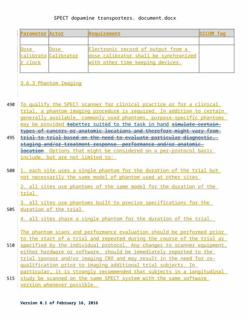
Dose Calibrator Electronic record of output from a dose calibrator shall be synchronized with other time keeping devices.
3.6.3 Phantom Imaging
To qualify the SPECT scanner for clinical practice or for a clinical trial, a phantom imaging procedure is required. In addition to certain generally available, commonly used phantoms, purpose-specific phantoms may be provided tobetter suited to the task in hand simulate certain types of cancers or anatomic locations and therefore might vary from trial to trial based on the need to evaluate particular diagnostic, staging and/or treatment response performance and/or anatomic location. Options that might be considered on a per-protocol basis include, but are not limited to:
1. each site uses a single phantom for the duration of the trial but not necessarily the same model of phantom used at other sites
2. all sites use phantoms of the same model for the duration of the trial
3. all sites use phantoms built to precise specifications for the duration of the trial
4. all sites share a single phantom for the duration of the trial.
The phantom scans and performance evaluation should be performed prior to the start of a trial and repeated during the course of the trial as specified by the individual protocol. Any changes to scanner equipment, either hardware or software, should be immediately reported to the trial sponsor and/or imaging CRO and may result in the need for re-qualification prior to imaging additional trial subjects. In particular, it is strongly recommended that subjects in a longitudinal study be scanned on the same SPECT system with the same software version whenever possible.
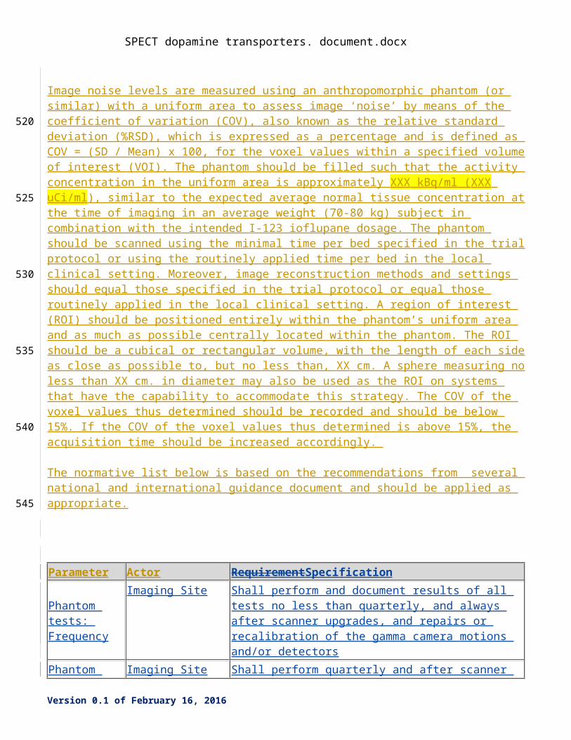
Image noise levels are measured using an anthropomorphic phantom (or similar) with a uniform area to assess image ‘noise’ by means of the coefficient of variation (COV), also known as the relative standard deviation (%RSD), which is expressed as a percentage and is defined as COV = (SD / Mean) x 100, for the voxel values within a specified volume of interest (VOI). The phantom should be filled such that the activity concentration in the uniform area is approximately XXX kBq/ml (XXX uCi/ml), similar to the expected average normal tissue concentration at the time of imaging in an average weight (70-80 kg) subject in combination with the intended I-123 ioflupane dosage. The phantom should be scanned using the minimal time per bed specified in the trial protocol or using the routinely applied time per bed in the local clinical setting. Moreover, image reconstruction methods and settings should equal those specified in the trial protocol or equal those routinely applied in the local clinical setting. A region of interest (ROI) should be positioned entirely within the phantom’s uniform area and as much as possible centrally located within the phantom. The ROI should be a cubical or rectangular volume, with the length of each
Version 0.1 of February 16, 2016
400
405
410
415
420
425
430
SPECT dopamine transporters. document.docx
side as close as possible to, but no less than, XX cm. A sphere measuring no less than XX cm. in diameter may also be used as the ROI on systems that have the capability to accommodate this strategy. The COV of the voxel values thus determined should be recorded and should be below 15%. If the COV of the voxel values thus determined is above 15%, the acquisition time should be increased accordingly.
The normative list below is based on the recommendations from several national and international guidance document and should be applied as appropriate.
Parameter Actor RequirementSpecification
Phantom tests: Frequency
Imaging Site Shall perform and document results of all tests no less than quarterly, and always after scanner upgrades, and repairs or recalibration of the gamma camera motions and/or detectors
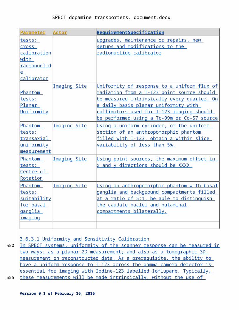
Phantom tests: cross calibration with radionuclide calibrator
Imaging Site Shall perform quarterly and after scanner upgrades, maintenance or repairs, new setups and modifications to the radionuclide calibrator
Phantom tests:Planar Uniformity
Imaging Site Uniformity of response to a uniform flux of radiation from a I-123 point source should be measured intrinsically every quarter. On a daily basis planar uniformity with collimators used for I-123 imaging should be performed using a Tc-99m or Co-57 source
Phantom tests:transaxial uniformity measurement
Imaging Site Using a uniform cylinder, or the uniform section of an anthropomorphic phantom filled with I-123, obtain a within slice variability of less than 5%.
Phantom tests: Centre of Rotation
Imaging Site Using point sources, the maximum offset in x and y directions should be XXXX.
Phantom tests:suitability for basal ganglia imaging
Imaging Site Using an anthropomorphic phantom with basal ganglia and background compartments filled at a ratio of 5:1, be able to distinguish the caudate nuclei and putaminal compartments bilaterally.
3.6.3.1 Uniformity and Sensitivity CalibrationIn SPECT systems, uniformity of the scanner response can be measured in two ways: as a planar 2D measurement; and also as a tomographic 3D measurement on reconstructed data. As a prerequisite, the ability to have a uniform response to I-123 across the gamma camera detector is essential for imaging with Iodine-123 labelled Ioflupane. Typically, these measurements will be made intrinsically, without the
Version 0.1 of February 16, 2016
435
440
445
SPECT dopamine transporters. document.docx
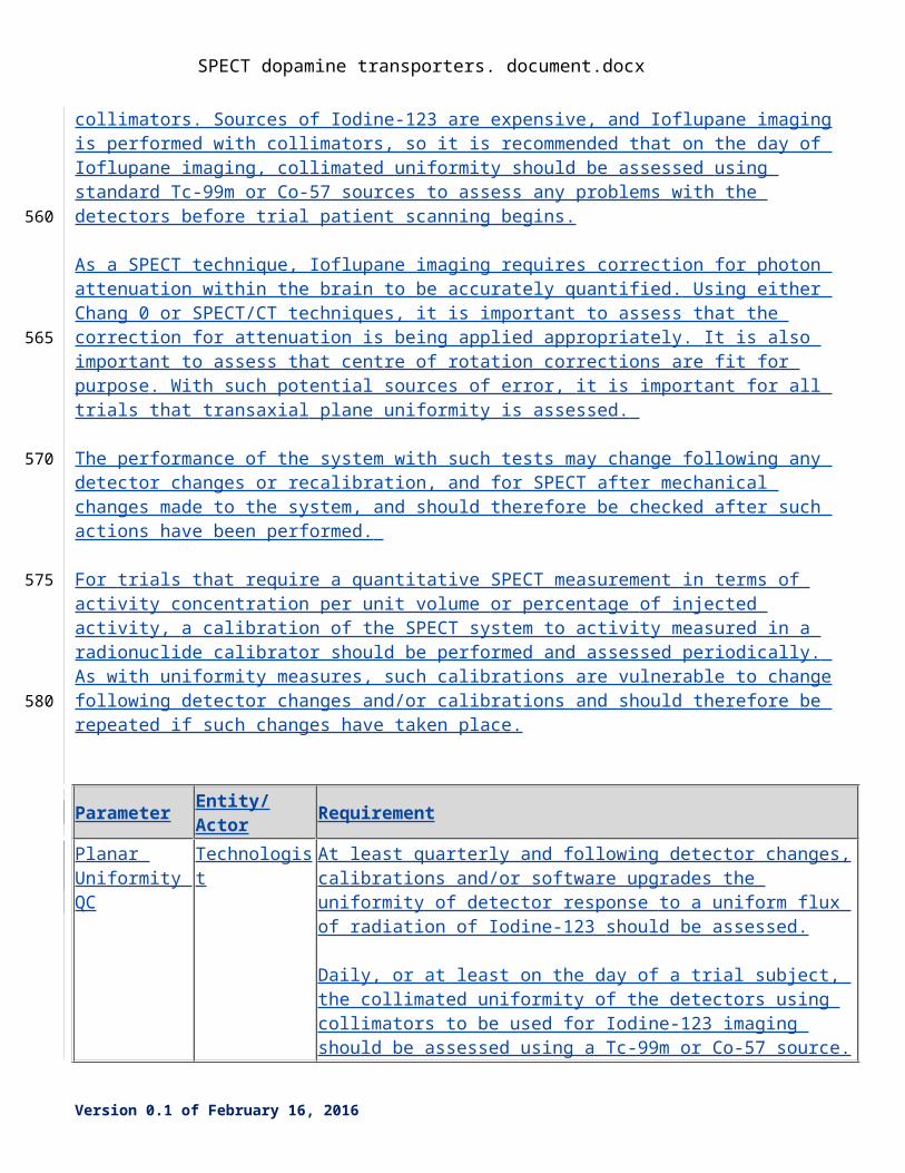
use of collimators. Sources of Iodine-123 are expensive, and Ioflupane imaging is performed with collimators, so it is recommended that on the day of Ioflupane imaging, collimated uniformity should be assessed using standard Tc-99m or Co-57 sources to assess any problems with the detectors before trial patient scanning begins.
As a SPECT technique, Ioflupane imaging requires correction for photon attenuation within the brain to be accurately quantified. Using either Chang 0 or SPECT/CT techniques, it is important to assess that the correction for attenuation is being applied appropriately. It is also important to assess that centre of rotation corrections are fit for purpose. With such potential sources of error, it is important for all trials that transaxial plane uniformity is assessed.
The performance of the system with such tests may change following any detector changes or recalibration, and for SPECT after mechanical changes made to the system, and should therefore be checked after such actions have been performed.
For trials that require a quantitative SPECT measurement in terms of activity concentration per unit volume or percentage of injected activity, a calibration of the SPECT system to activity measured in a radionuclide calibrator should be performed and assessed periodically. As with uniformity measures, such calibrations are vulnerable to change following detector changes and/or calibrations and should therefore be repeated if such changes have taken place.
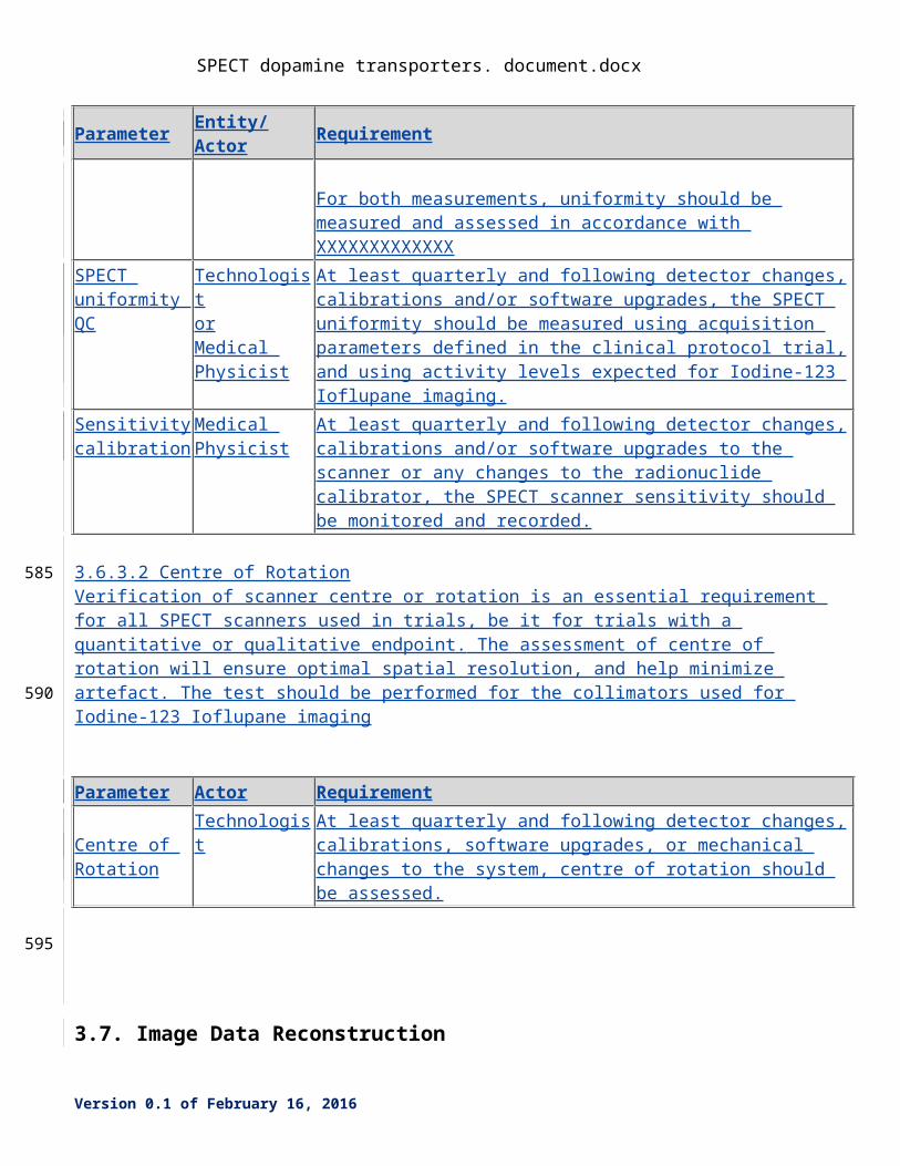
Parameter Entity/Actor RequirementPlanar Uniformity QC
Technologist At least quarterly and following detector changes, calibrations and/or software upgrades the uniformity of detector response to a uniform flux of radiation of Iodine-123 should be assessed.
Daily, or at least on the day of a trial subject, the collimated uniformity of the detectors using collimators to be used for Iodine-123 imaging should be assessed using a Tc-99m or Co-57 source.
For both measurements, uniformity should be measured and assessed in accordance with XXXXXXXXXXXXX
SPECT uniformity QC
TechnologistorMedical Physicist
At least quarterly and following detector changes, calibrations and/or software upgrades, the SPECT uniformity should be measured using acquisition parameters defined in the clinical protocol trial, and using activity levels expected for Iodine-123 Ioflupane imaging.
Sensitivity calibration
Medical Physicist
At least quarterly and following detector changes, calibrations and/or software upgrades to the scanner or any changes to the radionuclide calibrator, the SPECT scanner sensitivity should be monitored and recorded.
3.6.3.2 Centre of RotationVerification of scanner centre or rotation is an essential requirement for all SPECT scanners used in trials,
Version 0.1 of February 16, 2016
450
455
460
465
470
SPECT dopamine transporters. document.docx
be it for trials with a quantitative or qualitative endpoint. The assessment of centre of rotation will ensure optimal spatial resolution, and help minimize artefact. The test should be performed for the collimators used for Iodine-123 Ioflupane imaging
Parameter Actor Requirement
Centre of Rotation
Technologist At least quarterly and following detector changes, calibrations, software upgrades, or mechanical changes to the system, centre of rotation should be assessed.
3.7. Image Data Reconstruction
This activity describes criteria and procedures related to producing images from the acquired data that are necessary to reliably meet the Profile Claim.
3.7.1 DISCUSSION
3.7.2 SPECIFICATION
Parameter Actor Requirement
3.8. Image QA
This activity describes criteria and evaluations of the images that are necessary to reliably meet the Profile Claim.
3.8.1 DISCUSSION Tumor Size can affect the accuracy of measurements. Both theoretical considerations and the groundwork projects done by QIBA indicate that for tumors that are small, errors in measurement represent a greater percentage of the measured size. For tumors that are smaller than the limits defined in this profile, please see the profile produced by the QIBA Small Nodule group for more information on imaging recommendations and performance claims. For tumors that are extremely large, the limitations on measurement are based less on imaging physics and more on anatomy. Such tumors are likely to cross anatomical boundaries and abut structures that make consistent segmentation difficult.
Version 0.1 of February 16, 2016
475
480
485
490
495
SPECT dopamine transporters. document.docx
Tumor Margin Conspicuity refers to the clarity with which the boundary of the tumor can be discerned from the surroundings. Conspicuity can directly impact the ability to segment the tumor to properly determine its volume. Conspicuity problems can derive from poor contrast enhancement, from the inherent texture, homogeneity or structure of the tumor, or from attachment of the tumor to other structures.
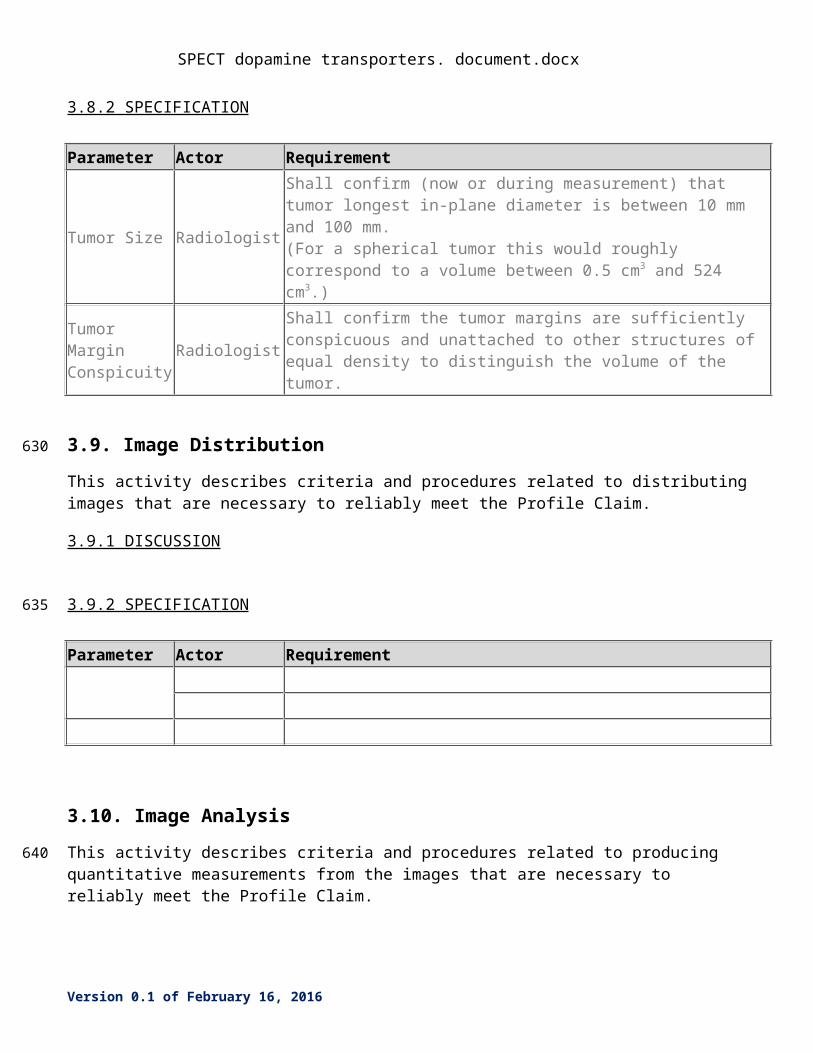
3.8.2 SPECIFICATION
Parameter Actor Requirement
Tumor Size Radiologist
Shall confirm (now or during measurement) that tumor longest in-plane diameter is between 10 mm and 100 mm. (For a spherical tumor this would roughly correspond to a volume between 0.5 cm3 and 524 cm3.)
Tumor Margin Conspicuity Radiologist
Shall confirm the tumor margins are sufficiently conspicuous and unattached to other structures of equal density to distinguish the volume of the tumor.
3.9. Image Distribution
This activity describes criteria and procedures related to distributing images that are necessary to reliably meet the Profile Claim.
3.9.1 DISCUSSION
3.9.2 SPECIFICATION
Parameter Actor Requirement
3.10. Image Analysis
This activity describes criteria and procedures related to producing quantitative measurements from the images that are necessary to reliably meet the Profile Claim.
3.10.1 DISCUSSION
The Image Analyst using computer workstation analysis tools shall perform the specified measurements. The main quantitative data analysis task is to determine the Specific Binding Ratios (SBR) of Ioflupane DaTscan for the right and left caudate and putamen. The derived results are then compared to an age
Version 0.1 of February 16, 2016
500
505
510
515
520
SPECT dopamine transporters. document.docx
normalized database to provide a reference for the SBR versus age matched normals. The profile describes the data analysis methodology.
Quantitative Specific Binding Ratio (SBR) of Ioflupane DaTscan will be based upon patient SBR and compared to an age normalized database (or striatal phantom or digital reference object as the case may be). Qualified systems will be able to achieve a SBR within a certain range (i.e., ±5% of reference value) for quantitative imaging of I-123 Ioflupane for the DaTscan phantom (described in this profile). The profile does not seek to make disease determination but to provide the methodology for data analysis and also for qualification of systems and processing for I-123 Ioflupane DaTscan data analysis.
Input Data:
The output images from Image Reconstruction are considered the input for Image Analysis. Once stored on the analysis workstation the image data will be processed for region of interest image analysis as described below. The original input data will be maintained as a separate file and will be stored along with the processed data for image analysis.
Methods to be Used:
Uptake in the striatum (i.e., caudate and putamen) and background region (e.g., cerebellum or occipital region) is characterized by defining a region-of-interest (ROI). The measurand is the specific binding ratio and is determined from the following equation:
(eq 1)
where the backgrndROI
counts are normalized to the same ROI volume as the striatal ROI (i.e., caudate
or putamen).
Regions of interests will be drawn on preprocessed images as described below.
On spatial normalized SPECT image volumes the transaxial slice with the highest striatal uptake is identified and the 8 hottest striatal slices around it are averaged to generate a single slice image.
Regions of interest (ROI) are then placed on the left and right caudate, the left and right putamen, and the occipital cortex (reference tissue). It should be clear which values belong to which striatal structures. This can be done by capturing DICOM coordinates along with ROI values or secondary screen capture of the ROI for identification.
Count densities for each region are extracted and used to calculate specific binding ratios (SBRs) for each of the striatal regions. SBR is calculated as (target region/reference region)-1, as described above in eq 1.
Required characteristics of resulting data:
The specific trial protocol shall prospectively define the SBR parameter that is required for the striatum and the caudate and putamen, specifically. Some studies may also compare different metrics (e.g., right to left asymmetry or caudate to putamen ratio) and will require recording multiple parameters. SBR measures (and the analysis tools used to obtain them, including software version) shall be used
Version 0.1 of February 16, 2016
525
530
535
540
545
550
555
560
SPECT dopamine transporters. document.docx
consistently across all subjects and across all sequential SBR measurements.
SBR’s are intended as a measure of relative uptake and in that sense, can be regarded as dimensionless (unitless)
It should be clear which values belong to which structures (e.g., the whole striatum, left – right caudate, left – right putamen). This can be done by capturing DICOM coordinates along with the SBR or secondary screen captures of the ROI for identification. It should be reported what background region was used for normalization (e.g., occipital cortex or cerebellum).
The analysis software should generate a report
3.10.2 SPECIFICATION
Parameter Actor Requirement
Specific Binding Ratio
Image Analyst Analysis WorkstationShall have a suitable monitor of appropriate size and pixel density for diagnostic viewing of medical images. Shall be placed in a room with in room lighting appropriate for image data analysis and interpretation (i.e., a radiology reading room). Shall have appropriate computation power and memory to carryout ROI or VOI data analysis.Post processed image for data analysisImage for data analysis shall be reconstructed in accordance with parameters as described in Section 3.7. If needed image is spatially normalized. The transaxial slice with the highest striatal uptake is identified and the 8 hottest striatal slices around it are averaged to generate a single slice imageROI software analysis toolsUsing analysis workstation tools, regions of interest are placed on the left and right caudate, the left and right putamen, and the occipital cortex (reference tissue). Count densities for each region are extracted to calculate SBRs for each of the striatal regions and for the striatum as a whole. Need to decide if ROIs are drawn by hand or automatically. Also need to decide if image based partial volume correction will be used. Finally, do we make a statement about if MRI is available it can be used for striatum (i.e., caudate and putamen) definition.Age matched normal database?
3.11. Image Interpretation
This activity describes criteria and procedures related to clinically interpreting the measurements and images that are necessary to reliably meet the Profile Claim.
Version 0.1 of February 16, 2016
565
570
SPECT dopamine transporters. document.docx
3.11.1 DISCUSSION
3.11.2 SPECIFICATION
Parameter Actor Requirement
Version 0.1 of February 16, 2016
575
580
SPECT dopamine transporters. document.docx
4. Assessment ProceduresTo conform to this Profile, participating staff and equipment (“Actors”) shall support each activity assigned to them in Table 1.
To support an activity, the actor shall conform to the requirements (indicated by “shall language”) listed in the specifications table of the activity subsection in Section 3.
Although most of the requirements described in Section 3 can be assessed for conformance by direct observation, some of the performance-oriented requirements cannot, in which case the requirement will reference an assessment procedure in a subsection here in Section 4.
Formal claims of conformance by the organization responsible for an Actor shall be in the form of a published QIBA Conformance Statement. Vendors publishing a QIBA Conformance Statement shall provide a set of “Model-specific Parameters” (as shown in Appendix D) describing how their product was configured to achieve conformance. Vendors shall also provide access or describe the characteristics of the test set used for conformance testing.
4.1. Assessment Procedure: Voxel Noise
This procedure can be used by a vendor or an imaging site to assess the voxel noise of reconstructed images. Voxel noise is assessed in terms of the standard deviation of pixel values when imaging a material with uniform density.
The assessor shall first warm up the scanner’s x-ray tube and perform calibration scans (often called air-calibration scans) according to scanner manufacturer recommendations. The assessor shall then scan a phantom of uniform density, such as the ACR CT Accreditation Program (CTAP) Phantom’s module 3, which is a 20 cm diameter cylinder of water equivalent material. The phantom shall be placed at the isocenter of the scanner. The acquisition protocol and reconstruction parameters shall conform to this Profile (See Section 3.6.2 and 3.7.2). The same protocol and parameters shall be used when performing the assessments in 4.1 and 4.2.
When the scan is performed, the assessor shall select a single representative slice from the uniformity portion of the phantom. An approximately circular region of interest (ROI) of at least 400 mm2 shall be placed near the center of the phantom.
The assessor shall record the values reported for the ROI mean and standard deviation.
The procedure described above is provided as a reference method. Sites or vendors may submit to QIBA a proposed alternative method (such as using the water phantom portion of a manufacturer’s QA phantom) and evidence that the results produced by the proposed method are equivalent to this reference method. Upon review and approval by QIBA, the alternative method will also become an accepted assessment procedure in this Profile.
The test procedure described here is based on the use of conventional filtered backprojection reconstruction methods; extreme care must be taken when iterative reconstruction methods are used as their use may invalidate some of the assumptions inherent in this method.
Version 0.1 of February 16, 2016
585
590
595
600
605
610
615
SPECT dopamine transporters. document.docx
4.2. Assessment Procedure: <Parameter Y>
4.3. Assessment Procedure: SPECT Calibration Factor
This procedure can be used by a vendor, physicist or an imaging site to assess the SPECT Calibration Factor of an acquisition device. SPECT Calibration Factor is assessed in terms of compensating value that needs to be applied to get the image voxel values produced by the acquisition device to match the known activity in kBq/mL of scanned phantom. The units of the SPECT Calibration factor are kBq/mL divided by the arbitrary units used by the acquisition device to record image voxel values.
The assessor shall scan a phantom of uniform …We’ve got text describing recipes for scanning bottles of various sizes filled with purportedly known concentrations of radioactivity. The question is whether we will replace with a solid standard.
Version 0.1 of February 16, 2016
620
625
630
SPECT dopamine transporters. document.docx
References
Version 0.1 of February 16, 2016
635
SPECT dopamine transporters. document.docx
AppendicesAppendix A: Acknowledgements and Attributions
Appendix B: Background Information
Appendix C: Conventions and Definitions
Version 0.1 of February 16, 2016
640
645
SPECT dopamine transporters. document.docx
Appendix D: Model-specific Instructions and Parameters
For acquisition modalities, reconstruction software and software analysis tools, profile conformance requires meeting the activity specifications above in Sections 2, 3 and 4.
This Appendix provides, as an informative tool, some specific acquisition parameters, reconstruction parameters and analysis software parameters that are expected to be compatible with meeting the profile requirements. Just using these parameters without meeting the requirements specified in the profile is not sufficient to achieve conformance. Conversely, it is possible to use different compatible parameters and still achieve conformance.
Sites using models listed here are encouraged to consider using these parameters for both simplicity and consistency. Sites using models not listed here may be able to devise their own settings that result in data meeting the requirements.
IMPORTANT: The presence of a product model/version in these tables does not imply it has demonstrated conformance with the QIBA Profile. Refer to the QIBA Conformance Statement for the product.
Table D.1 Model-specific Parameters for Acquisition DevicesAcquisition Device Settings Compatible with Conformance
Acme MedicalCT LightsV3.14
Submitted by: Gotham University Hospital
kVp 120Number of Data Channels (N) 64Width of Each Data Channel (T, in mm) 0.625Gantry Rotation Time in seconds 1.0mA 120Pitch 0.984Scan FoV Large Body (500mm)
Table D.2 Model-specific Parameters for Reconstruction SoftwareReconstruction Software Settings Compatible with Conformance
Acme MedicalCT WSV3.14
Reconstructed Slice Width, mm 1.25Reconstruction Interval 1.0mmDisplay FOV, mm 350Recon kernel STD
Version 0.1 of February 16, 2016
650
655
660
665
SPECT dopamine transporters. document.docx
Version 0.1 of February 16, 2016