Embed Size (px)
Citation preview
24-11-2017
1
? Medical mystery ?
A case report
N. Boeckx, MD, PhD
03-10-2017
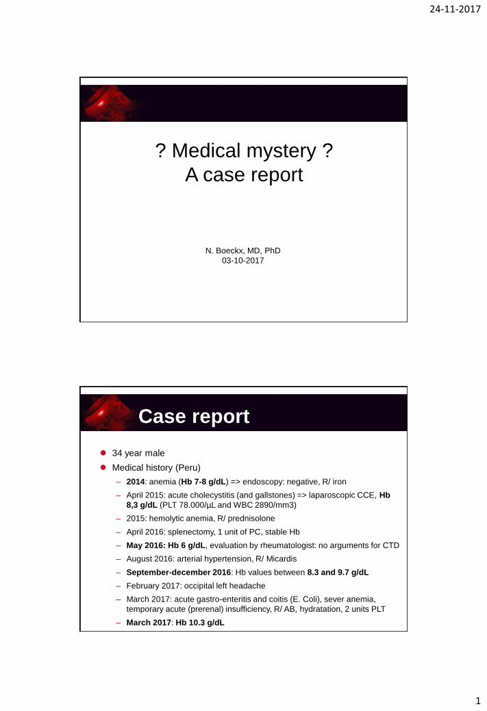
Case report
34 year male
Medical history (Peru)
– 2014: anemia (Hb 7-8 g/dL) => endoscopy: negative, R/ iron
– April 2015: acute cholecystitis (and gallstones) => laparoscopic CCE, Hb
8,3 g/dL (PLT 78.000/µL and WBC 2890/mm3)
– 2015: hemolytic anemia, R/ prednisolone
– April 2016: splenectomy, 1 unit of PC, stable Hb
– May 2016: Hb 6 g/dL, evaluation by rheumatologist: no arguments for CTD
– August 2016: arterial hypertension, R/ Micardis
– September-december 2016: Hb values between 8.3 and 9.7 g/dL
– February 2017: occipital left headache
– March 2017: acute gastro-enteritis and coitis (E. Coli), sever anemia,
temporary acute (prerenal) insufficiency, R/ AB, hydratation, 2 units PLT
– March 2017: Hb 10.3 g/dL
24-11-2017
2
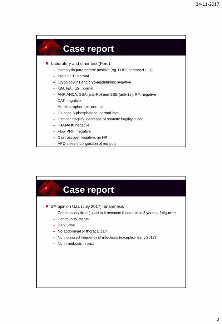
Case report
Laboratory and other test (Peru):
– Hemolysis parameters: positive (eg. LHD: increased +++)
– Protein EF: normal
– Cryoglobulins and cryo-agglutinins: negative
– IgM, IgA, IgG: normal
– ANF, ANCA, SSA (anti-Ro) and SSB (anti-1a), RF: negative
– DAT: negative
– Hb-electrophoresis: normal
– Glucose-6-phosphatase: normal level
– Osmotic fragility: decrease of osmotic fragility curve
– HAM-test: negative
– Flow PNH: negative
– Gastroscopy: negative, no HP
– APO spleen: congestion of red pulp
Case report
2nd opinion UZL (July 2017): anamnesis
– Continuously tired (‘used to it because it lasts since 3 years’), fatigue ++
– Continuous icterus
– Dark urine
– No abdominal or thoracal pain
– No increased frequency of infections (exception early 2017)
– No thrombosis in past

24-11-2017
3
Case report
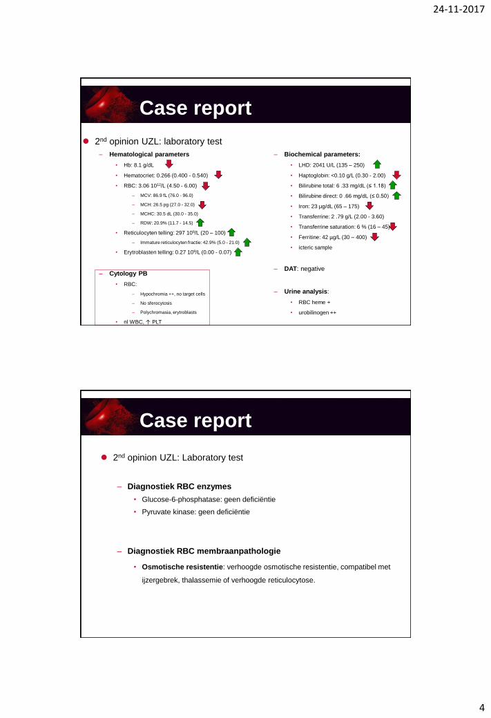
2nd opinion UZL: laboratory test
– Hematological parameters
• Hb: 8.1 g/dL
• Hematocriet: 0.266 (0.400 - 0.540)
• RBC: 3.06 1012/L (4.50 - 6.00)
– MCV: 86.9 fL (76.0 - 96.0)
– MCH: 26.5 pg (27.0 - 32.0)
– MCHC: 30.5 dL (30.0 - 35.0)
– RDW: 20.9% (11.7 - 14.5)
• Reticulocyten telling: 297 109/L (20 – 100)
– Immature reticulocyten fractie: 42.9% (5.0 - 21.0)
• Erytroblasten telling: 0.27 109/L (0.00 - 0.07)
– Cytology PB
• RBC:
– Hypochromia ++, no target cells
– No sferocytosis
– Polychromasia, erytroblasts
• nl WBC, ↑ PLT
2nd opinion UZL: Laboratory test
– Biochemical parameters:
• LHD: 2041 U/L (135 – 250)
• Haptoglobin: <0.10 g/L (0.30 - 2.00)
• Bilirubine total: 6 .33 mg/dL (≤ 1.18)
• Bilirubine direct: 0 .66 mg/dL (≤ 0.50)
• Iron: 23 µg/dL (65 – 175)
• Transferrine: 2 .79 g/L (2.00 - 3.60)
• Transferrine saturation: 6 % (16 – 45)
• Ferritine: 42 µg/L (30 – 400)
• icteric sample
– DAT: negative
– Urine analysis:
• RBC heme +
• urobilinogen ++
Hemolytic anemia
Inherited
Hemolytic
Anemias
Acquired
Hemolytic
Anemias
Sickle Cell Anemia
24-11-2017
4
Case report
2nd opinion UZL: laboratory test
– Hematological parameters
• Hb: 8.1 g/dL
• Hematocriet: 0.266 (0.400 - 0.540)
• RBC: 3.06 1012/L (4.50 - 6.00)
– MCV: 86.9 fL (76.0 - 96.0)
– MCH: 26.5 pg (27.0 - 32.0)
– MCHC: 30.5 dL (30.0 - 35.0)
– RDW: 20.9% (11.7 - 14.5)
• Reticulocyten telling: 297 109/L (20 – 100)
– Immature reticulocyten fractie: 42.9% (5.0 - 21.0)
• Erytroblasten telling: 0.27 109/L (0.00 - 0.07)
– Cytology PB
• RBC:
– Hypochromia ++, no target cells
– No sferocytosis
– Polychromasia, erytroblasts
• nl WBC, ↑ PLT
2nd opinion UZL: Laboratory test
– Biochemical parameters:
• LHD: 2041 U/L (135 – 250)
• Haptoglobin: <0.10 g/L (0.30 - 2.00)
• Bilirubine total: 6 .33 mg/dL (≤ 1.18)
• Bilirubine direct: 0 .66 mg/dL (≤ 0.50)
• Iron: 23 µg/dL (65 – 175)
• Transferrine: 2 .79 g/L (2.00 - 3.60)
• Transferrine saturation: 6 % (16 – 45)
• Ferritine: 42 µg/L (30 – 400)
• icteric sample
– DAT: negative
– Urine analysis:
• RBC heme +
• urobilinogen ++
Case report
2nd opinion UZL: Laboratory test
– Diagnostiek RBC enzymes
• Glucose-6-phosphatase: geen deficiëntie
• Pyruvate kinase: geen deficiëntie
– Diagnostiek RBC membraanpathologie
• Osmotische resistentie: verhoogde osmotische resistentie, compatibel met
ijzergebrek, thalassemie of verhoogde reticulocytose.
24-11-2017
5
Case report
2nd opinion UZL: Laboratory test
– Diagnostiek RBC membraanpathologie (Uitgevoerd door LHUB-ULB Site Anderlecht (Hopital Erasme))
• Cryohemolyse niet uitgevoerd
• EMA 2.0% (refw: < 19.0%)
• Ektacytometrie Het profiel is abnormaal maar niet typisch voor erfelijke
sferocytose.
• Elektroforese niet uitgevoerd
BESLUIT: Screeningstests hebben de aanwezigheid van erfelijke
sferocytose NIET aangetoond.
Case report
2nd opinion UZL: Laboratory test
– Flow:
• PNH clone WBC
– 99% of monocytes
– 99% of granulocytes
• PNH clone RBC
– 53% (type II and III) RBC
24-11-2017
6
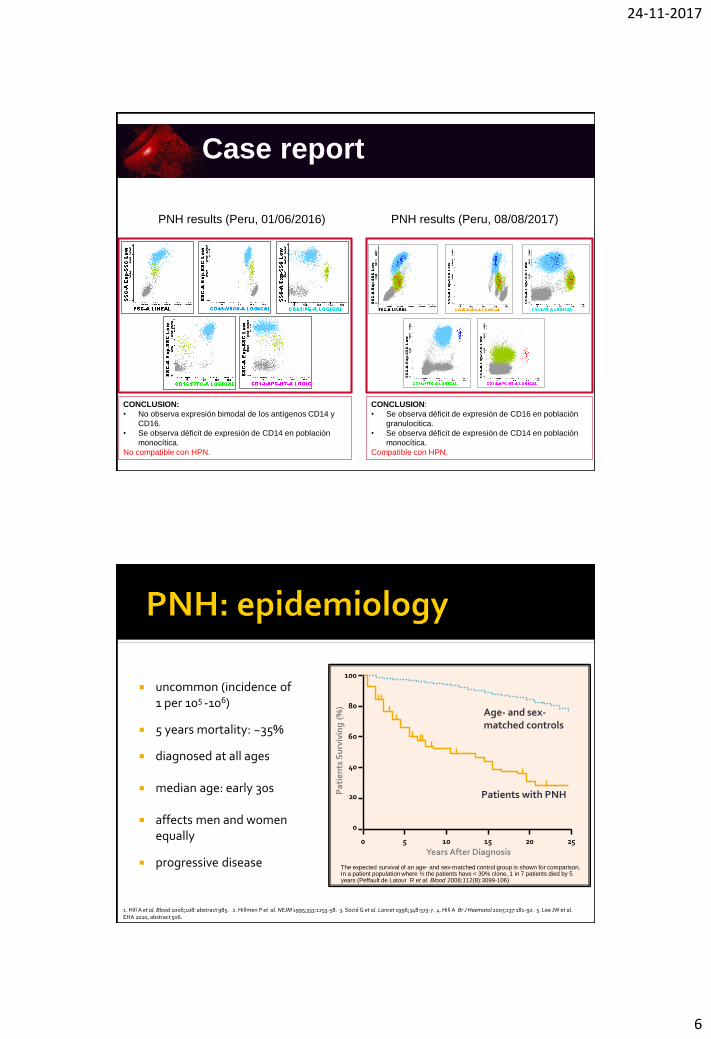
PNH results (Peru, 08/08/2017)PNH results (Peru, 01/06/2016)
CONCLUSION:
• No observa expresión bimodal de los antígenos CD14 y
CD16.
• Se observa déficit de expresión de CD14 en población
monocítica.
No compatible con HPN.
CONCLUSION:
• Se observa déficit de expresión de CD16 en población
granulocitica.
• Se observa déficit de expresión de CD14 en población
monocítica.
Compatible con HPN.
Case report
uncommon (incidence of 1 per 105 -106)
5 years mortality: ~35%
diagnosed at all ages
median age: early 30s
affects men and women equally
progressive disease
100
80
60
40
20
0
0 5 10 15 20 25
Years After Diagnosis
Pa
tie
nts
Su
rviv
ing
(%
)
The expected survival of an age- and sex-matched control group is shown for comparison. In a patient population where ½ the patients have < 30% clone, 1 in 7 patients died by 5 years (Peffault de Latour R et al. Blood 2008;112(8):3099-106)
Age- and sex-matched controls
Patients with PNH
1. Hill A et al. Blood 2006;108: abstract 985. 2. Hillmen P et al. NEJM 1995;333:1253-58. 3. Socié G et al. Lancet 1996;348:573-7. 4. Hill A Br J Haematol 2007;137:181-92. 5. Lee JW et al. EHA 2010, abstract 506.
24-11-2017
7
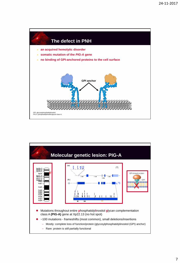
The defect in PNH
an acquired hemolytic disorder
somatic mutation of the PIG-A gene
no binding of GPI-anchored proteins to the cell surface
GPI anchor
GPI, glycosylphosphatidylinositol;
PIG-A, phosphatidylinositol glycan class A;
Mutations throughout entire phosphatidylinositol glycan complementation
class A (PIG-A) gene at Xp22.13 (no hot spot)
~100 mutations - frameshifts (most common), small deletions/insertions
– Mostly: complete loss of function/protein (glycosylphosphatidylinositol (GPI) anchor)
– Rare: protein is still partially functional
Molecular genetic lesion: PIG-A

24-11-2017
8
Hematopoietic
Stem Cell
CD59, CD109, CD90
RBC
CD5555
CD58
CD59
PrPc
AChE
JMH Ag
Dombroch
HG Ag
CD55
CD58
CD59
CD109
PrPc
GP500
Gova/b
Platelets
CD55 CD58
CD59 CD14
CD16 CD24
CD48 CD66b
CD66c CD87
CD109 CD157
LAPNB1 PrPc
p50-80 GPI-80
ADP-RT NA1/NA2
PMN
CD14 CD55
CD58 CD59
CD48 CD52
CD87 CD109
CD157 Group-8
PrPc GPI-80
CD16
Monocytes
B Cells
CD24 CD55
CD58 CD59
CD48 PrPc
CD73 CD108
T Cells
CD55 CD58
CD59 CD48
CD52 CD87
CD108 PrPc
ADP-RT CD73
CD90 CD109
CD16
NK CellsCD55
CD58
CD59
CD48
CD52
PrPc
CD16
CD55
The defect in PNH
CD59GPI anchor
GPI, glycosylphosphatidylinositol;
PIG-A, phosphatidylinositol glycan class A;
RBC, red blood cell
CD55
Prevents formation and augments
instability of the C3 convertases,
attenuating the complement cascade
CD59
Forms a defensive shield for RBCs
from complement-mediated lysis
Inhibits the assembly of the
membrane attack complex
24-11-2017
9
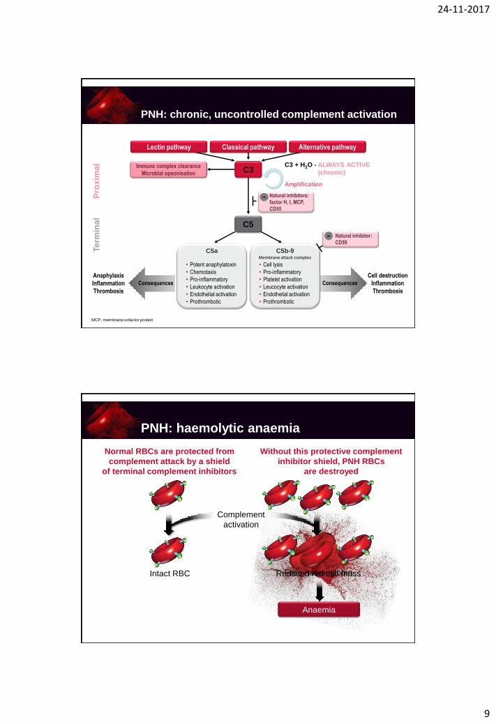
PNH: chronic, uncontrolled complement activation
Lectin pathway Classical pathway
Immune complex clearance
Microbial opsonisation C3
C5
Pro
xim
al
Te
rmin
al
Natural inhibitors:
factor H, I, MCP,
CD55
-
C3 + H2O - ALWAYS ACTIVE
(chronic)
Amplification
C5a
• Potent anaphylatoxin
• Chemotaxis
• Pro-inflammatory
• Leukocyte activation
• Endothelial activation
• Prothrombotic
C5b-9Membrane attack complex
• Cell lysis
• Pro-inflammatory
• Platelet activation
• Leucocyte activation
• Endothelial activation
• Prothrombotic
Consequences Consequences
Alternative pathway
Natural inhibitor:
CD59-
MCP, membrane cofactor protein
Anaphylaxis
Inflammation
Thrombosis
Cell destruction
Inflammation
Thrombosis
Anaemia
Reduced red cell massFree haemoglobin
Normal RBCs are protected from
complement attack by a shield
of terminal complement inhibitors
Without this protective complement
inhibitor shield, PNH RBCs
are destroyed
Intact RBC
Complement
activation
PNH: haemolytic anaemia
24-11-2017
10
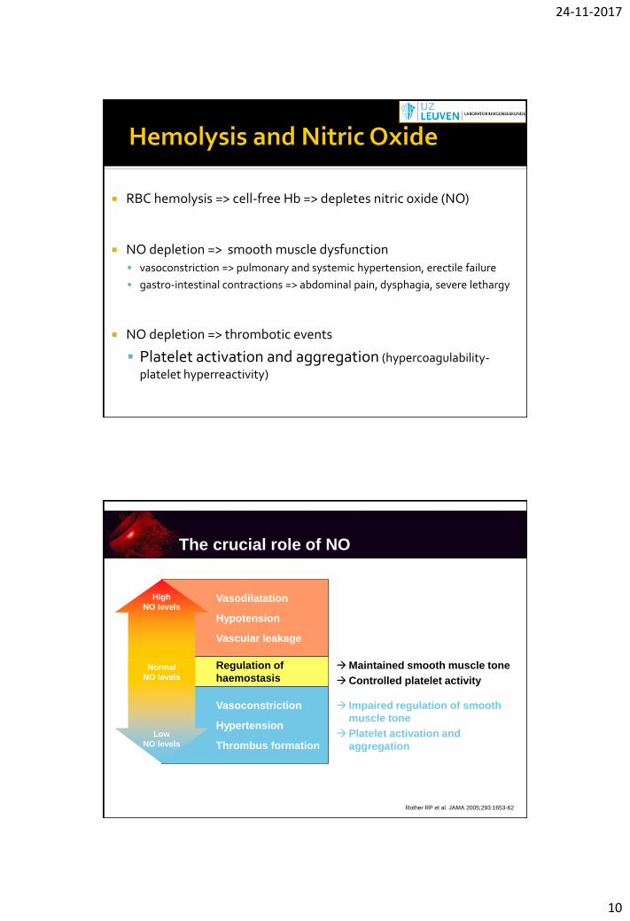
RBC hemolysis => cell-free Hb => depletes nitric oxide (NO)
NO depletion => smooth muscle dysfunction vasoconstriction => pulmonary and systemic hypertension, erectile failure
gastro-intestinal contractions => abdominal pain, dysphagia, severe lethargy
NO depletion => thrombotic events
Platelet activation and aggregation (hypercoagulability-
platelet hyperreactivity)
The crucial role of NO
Normal
NO levels
High
NO levelsVasodilatation
Hypotension
Vascular leakage
Low
NO levels
Vasoconstriction
Hypertension
Thrombus formation
Regulation of
haemostasis
Rother RP et al. JAMA 2005;293:1653-62
Maintained smooth muscle tone
Controlled platelet activity
Impaired regulation of smooth
muscle tone
Platelet activation and
aggregation
24-11-2017
11
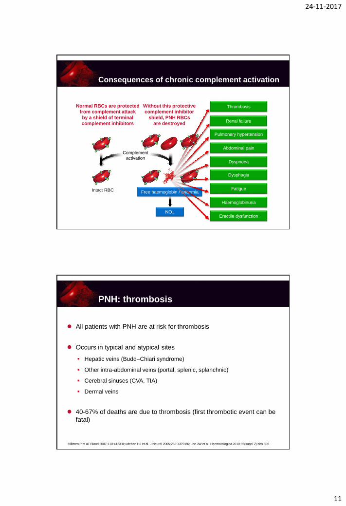
Consequences of chronic complement activation
Normal RBCs are protected
from complement attack
by a shield of terminal
complement inhibitors
Without this protective
complement inhibitor
shield, PNH RBCs
are destroyed
Intact RBC
Complement
activation
Free haemoglobin / anaemia
NO↓
Thrombosis
Fatigue
Abdominal pain
Dyspnoea
Dysphagia
Haemoglobinuria
Erectile dysfunction
Pulmonary hypertension
Renal failure
PNH: thrombosis
All patients with PNH are at risk for thrombosis
Occurs in typical and atypical sites
Hepatic veins (Budd–Chiari syndrome)
Other intra-abdominal veins (portal, splenic, splanchnic)
Cerebral sinuses (CVA, TIA)
Dermal veins
40-67% of deaths are due to thrombosis (first thrombotic event can be
fatal)
Hillmen P et al. Blood 2007;110:4123-8; udebert HJ et al. J Neurol 2005;252:1379-86; Lee JW et al. Haematologica 2010;95(suppl 2):abs 506
24-11-2017
12
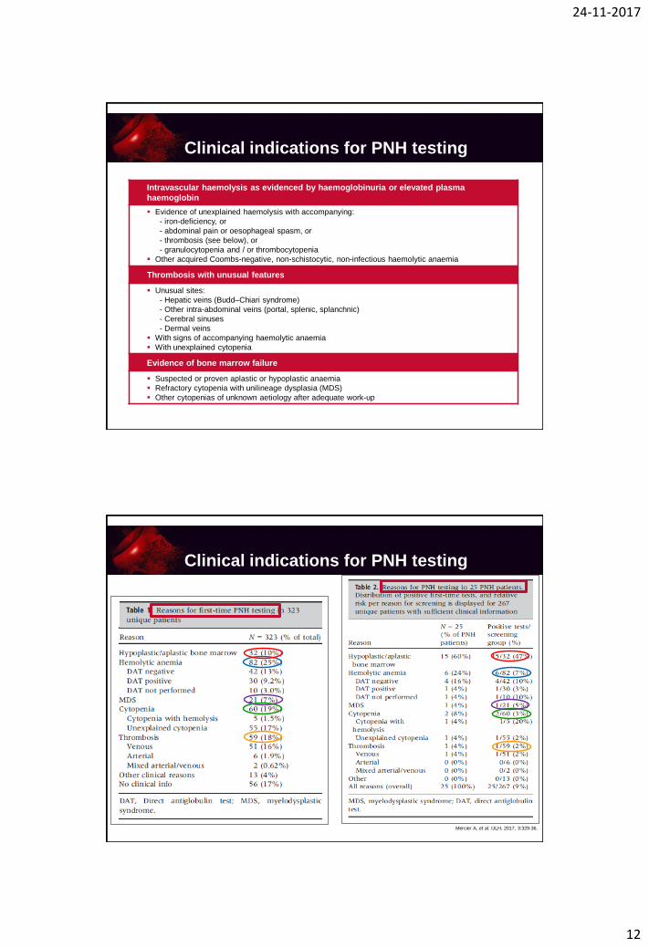
Intravascular haemolysis as evidenced by haemoglobinuria or elevated plasma
haemoglobin
Evidence of unexplained haemolysis with accompanying:
- iron-deficiency, or
- abdominal pain or oesophageal spasm, or
- thrombosis (see below), or
- granulocytopenia and / or thrombocytopenia
Other acquired Coombs-negative, non-schistocytic, non-infectious haemolytic anaemia
Thrombosis with unusual features
Unusual sites:
- Hepatic veins (Budd–Chiari syndrome)
- Other intra-abdominal veins (portal, splenic, splanchnic)
- Cerebral sinuses
- Dermal veins
With signs of accompanying haemolytic anaemia
With unexplained cytopenia
Evidence of bone marrow failure
Suspected or proven aplastic or hypoplastic anaemia
Refractory cytopenia with unilineage dysplasia (MDS)
Other cytopenias of unknown aetiology after adequate work-up
Clinical indications for PNH testing
Clinical indications for PNH testing
Mercier A, et al. IJLH, 2017, 3:329-36.
24-11-2017
13
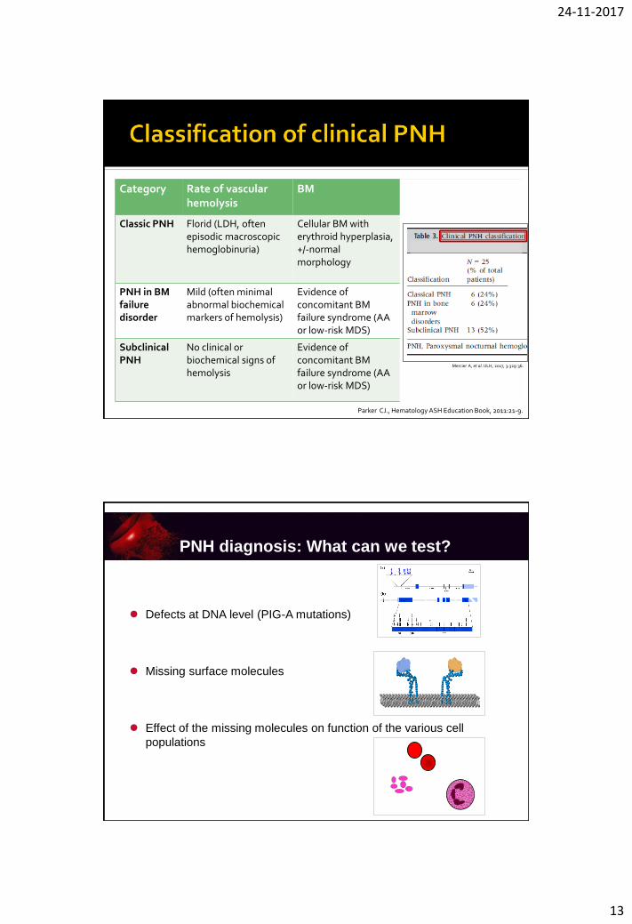
Category Rate of vascular hemolysis
BM Flow cytometry
Classic PNH Florid (LDH, often episodic macroscopic hemoglobinuria)
Cellular BM with erythroid hyperplasia, +/-normal morphology
Large population (>50%) GPI-anchor deficient PMN
PNH in BM failure disorder
Mild (often minimal abnormal biochemical markers of hemolysis)
Evidence of concomitant BM failure syndrome (AA or low-risk MDS)
% of GPI-anchor deficient PMNs is usually relatively small (<10%)
Subclinical PNH
No clinical orbiochemical signs of hemolysis
Evidence of concomitant BM failure syndrome (AA or low-risk MDS)
Small (<1%) population of GPI-anchor deficient PMN
Parker CJ., Hematology ASH Education Book, 2011:21-9.
Mercier A, et al. IJLH, 2017, 3:329-36.
PNH diagnosis: What can we test?
Defects at DNA level (PIG-A mutations)
Missing surface molecules
Effect of the missing molecules on function of the various cell
populations
24-11-2017
14
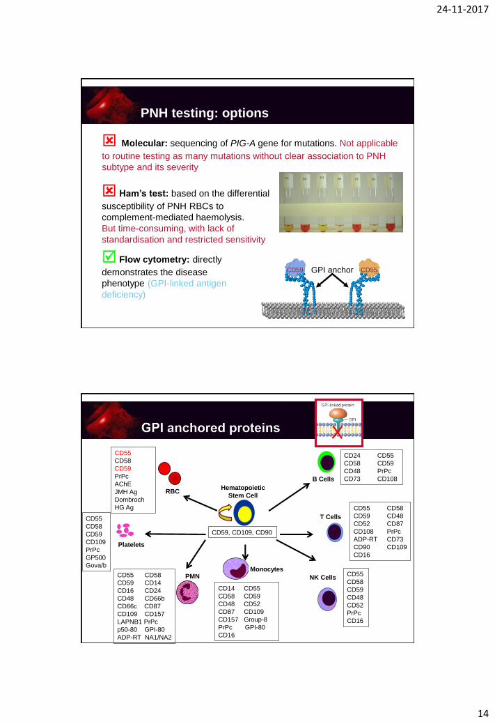
PNH testing: options
Ham’s test: based on the differential
susceptibility of PNH RBCs to
complement-mediated haemolysis.
But time-consuming, with lack of
standardisation and restricted sensitivity
Flow cytometry: directly
demonstrates the disease
phenotype (GPI-linked antigen
deficiency)
CD55CD59 GPI anchor
Molecular: sequencing of PIG-A gene for mutations. Not applicable
to routine testing as many mutations without clear association to PNH
subtype and its severity
Hematopoietic
Stem Cell
CD59, CD109, CD90
RBC
CD5555
CD58
CD59
PrPc
AChE
JMH Ag
Dombroch
HG Ag
CD55
CD58
CD59
CD109
PrPc
GP500
Gova/b
Platelets
CD55 CD58
CD59 CD14
CD16 CD24
CD48 CD66b
CD66c CD87
CD109 CD157
LAPNB1 PrPc
p50-80 GPI-80
ADP-RT NA1/NA2
PMN
CD14 CD55
CD58 CD59
CD48 CD52
CD87 CD109
CD157 Group-8
PrPc GPI-80
CD16
Monocytes
B Cells
CD24 CD55
CD58 CD59
CD48 PrPc
CD73 CD108
T Cells
CD55 CD58
CD59 CD48
CD52 CD87
CD108 PrPc
ADP-RT CD73
CD90 CD109
CD16
NK CellsCD55
CD58
CD59
CD48
CD52
PrPc
CD16

24-11-2017
15
“Guidelines for diagnosis and monitoring of PNH and related disorders by flow cytometry”. M. Borowitz et al., Cytometry Part B (clinical cytometry), 2010
Sample source PB (BM is not optimal)
Anticoagulant EDTA (preferred), heparine or ACD
Sample volume Minimum 1 ml; 3 ml is adequate for most testing, though more might be needed if WBC is very low
Maximum sample age Up to 7 days for RBC; <48h for WBC
High-sensitivity analysis 0,01%; at least 250,000 events of specific cell type collected
“Guidelines for diagnosis and monitoring of PNH and related disorders by flow cytometry”. M. Borowitz et al., Cytometry Part B (clinical cytometry), 2010
24-11-2017
16
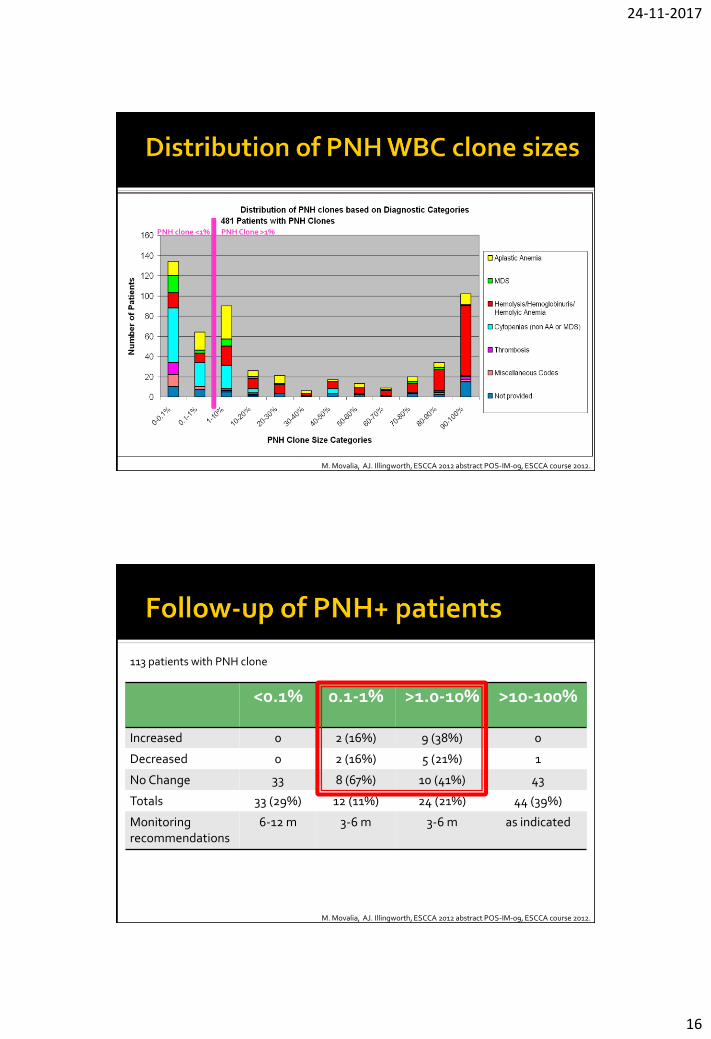
PNH clone <1% PNH Clone >1%
M. Movalia, AJ. Illingworth, ESCCA 2012 abstract POS-IM-09, ESCCA course 2012.
<0.1% 0.1-1% >1.0-10% >10-100%
Increased 0 2 (16%) 9 (38%) 0
Decreased 0 2 (16%) 5 (21%) 1
No Change 33 8 (67%) 10 (41%) 43
Totals 33 (29%) 12 (11%) 24 (21%) 44 (39%)
Monitoring recommendations
6-12 m 3-6 m 3-6 m as indicated
M. Movalia, AJ. Illingworth, ESCCA 2012 abstract POS-IM-09, ESCCA course 2012.
113 patients with PNH clone
24-11-2017
17
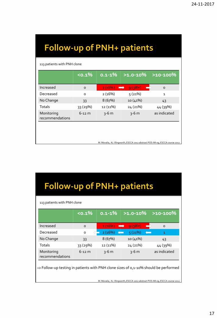
<0.1% 0.1-1% >1.0-10% >10-100%
Increased 0 2 (16%) 9 (38%) 0
Decreased 0 2 (16%) 5 (21%) 1
No Change 33 8 (67%) 10 (41%) 43
Totals 33 (29%) 12 (11%) 24 (21%) 44 (39%)
Monitoring recommendations
6-12 m 3-6 m 3-6 m as indicated
M. Movalia, AJ. Illingworth, ESCCA 2012 abstract POS-IM-09, ESCCA course 2012.
113 patients with PNH clone
Follow-up testing in patients with PNH clone sizes of 0,1-10% should be performed
<0.1% 0.1-1% >1.0-10% >10-100%
Increased 0 2 (16%) 9 (38%) 0
Decreased 0 2 (16%) 5 (21%) 1
No Change 33 8 (67%) 10 (41%) 43
Totals 33 (29%) 12 (11%) 24 (21%) 44 (39%)
Monitoring recommendations
6-12 m 3-6 m 3-6 m as indicated
M. Movalia, AJ. Illingworth, ESCCA 2012 abstract POS-IM-09, ESCCA course 2012.
113 patients with PNH clone

24-11-2017
18
Sample source PB (BM is not optimal)
Anticoagulant EDTA (preferred), heparine or ACD
Sample volume Minimum 1 ml; 3 ml is adequate for most testing, though more might be needed if WBC is very low
Maximum sample age Up to 7 days for RBC; <48h for WBC
Routine analysis 1%; at least 5,000 events of specific cell type collected
High-sensitivity analysis 0,01%; at least 250,000 events of specific cell type collected
Cell populations analyzed
Hematopoietic
Stem Cell
CD59, CD109, CD90
RBC
CD5555
CD58
CD59
PrPc
AChE
JMH Ag
Dombroch
HG Ag
CD55
CD58
CD59
CD109
PrPc
GP500
Gova/b
Platelets
CD55 CD58
CD59 CD14
CD16 CD24
CD48 CD66b
CD66c CD87
CD109 CD157
LAPNB1 PrPc
p50-80 GPI-80
ADP-RT NA1/NA2
PMN
CD14 CD55
CD58 CD59
CD48 CD52
CD87 CD109
CD157 Group-8
PrPc GPI-80
CD16
Monocytes
B Cells
CD24 CD55
CD58 CD59
CD48 PrPc
CD73 CD108
T Cells
CD55 CD58
CD59 CD48
CD52 CD87
CD108 PrPc
ADP-RT CD73
CD90 CD109
CD16
NK CellsCD55
CD58
CD59
CD48
CD52
PrPc
CD16

24-11-2017
19
Sample source PB (BM is not optimal)
Anticoagulant EDTA (preferred), heparine or ACD
Sample volume Minimum 1 ml; 3 ml is adequate for most testing, though more might be needed if WBC is very low
Maximum sample age Up to 7 days for RBC; <48h for WBC
Routine analysis 1%; at least 5,000 events of specific cell type collected
High-sensitivity analysis 0,01%; at least 250,000 events of specific cell type collected
Cell populations analyzed Granulocytes in all cases.
Monocytes provide confirmatory information.
No role for analysis of lymphocytes due to long life span.
RBC in at least those cases with a WBC PNH clone detected
by WBC analysis (or in all cases).
RBC’s alone is not recommended.
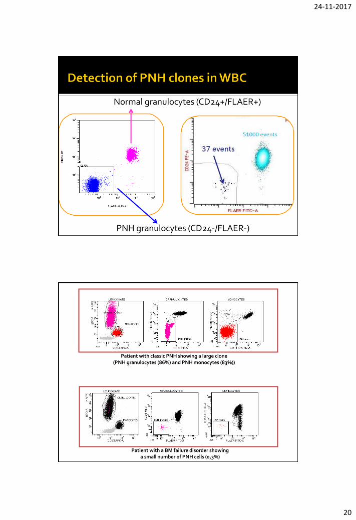
GPI-AP CD24 (granulocytes)
CD14 (monocytes)
FLAER = fluorescent aerolysin Fluorochrome-conjugated inactive
variant of the bacterial derived protein aerolysin (52-kDA protein secreted by Aeromonas Hydrophilia)
Aerolysin is a molecule that directly binds to the GPI anchors on leucocytes (not erythrocytes)
24-11-2017
20
Normal granulocytes (CD24+/FLAER+)
PNH granulocytes (CD24-/FLAER-)
Patient with classic PNH showing a large clone (PNH granulocytes (86%) and PNH monocytes (83%))
Patient with a BM failure disorder showing a small number of PNH cells (0,3%)
24-11-2017
21
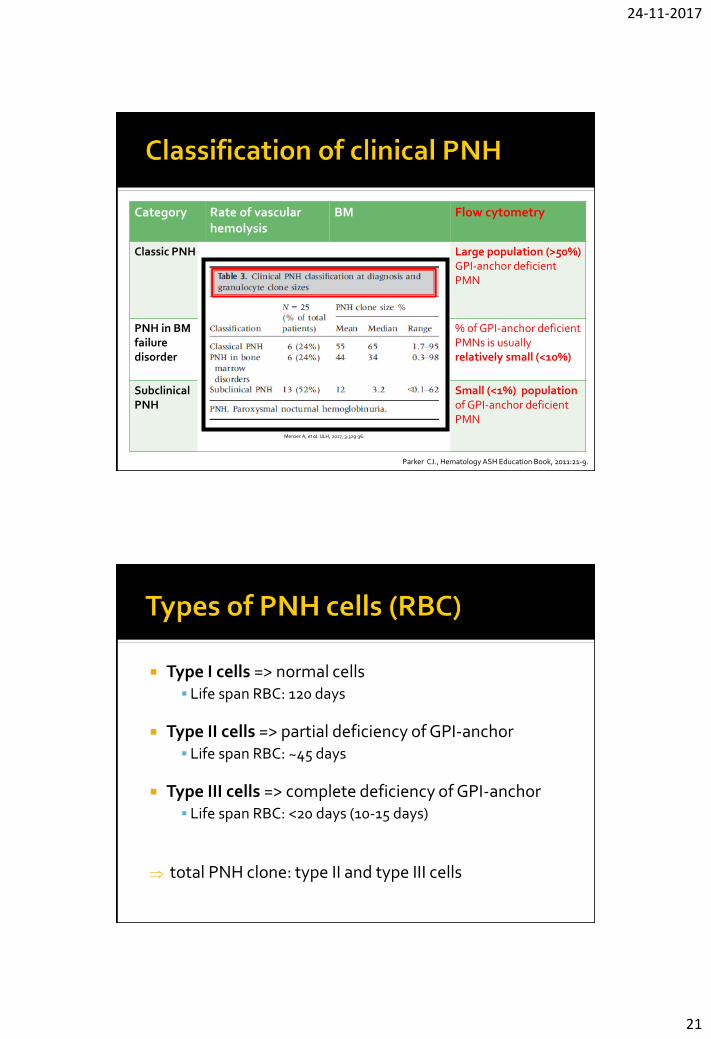
Category Rate of vascular hemolysis
BM Flow cytometry
Classic PNH Florid (LDH, often episodic macroscopic hemoglobinuria)
Cellular BM with erythroid hyperplasia, +/-normal morphology
Large population (>50%) GPI-anchor deficient PMN
PNH in BM failure disorder
Mild (often minimal abnormal biochemical markers of hemolysis)
Evidence of concomitant BM failure syndrome (AA or low-risk MDS)
% of GPI-anchor deficient PMNs is usually relatively small (<10%)
Subclinical PNH
No clinical orbiochemical signs of hemolysis
Evidence of concomitant BM failure syndrome (AA or low-risk MDS)
Small (<1%) population of GPI-anchor deficient PMN
Parker CJ., Hematology ASH Education Book, 2011:21-9.
Mercier A, et al. IJLH, 2017, 3:329-36.
Type I cells => normal cells Life span RBC: 120 days
Type II cells => partial deficiency of GPI-anchor Life span RBC: ~45 days
Type III cells => complete deficiency of GPI-anchor Life span RBC: <20 days (10-15 days)
total PNH clone: type II and type III cells
24-11-2017
22
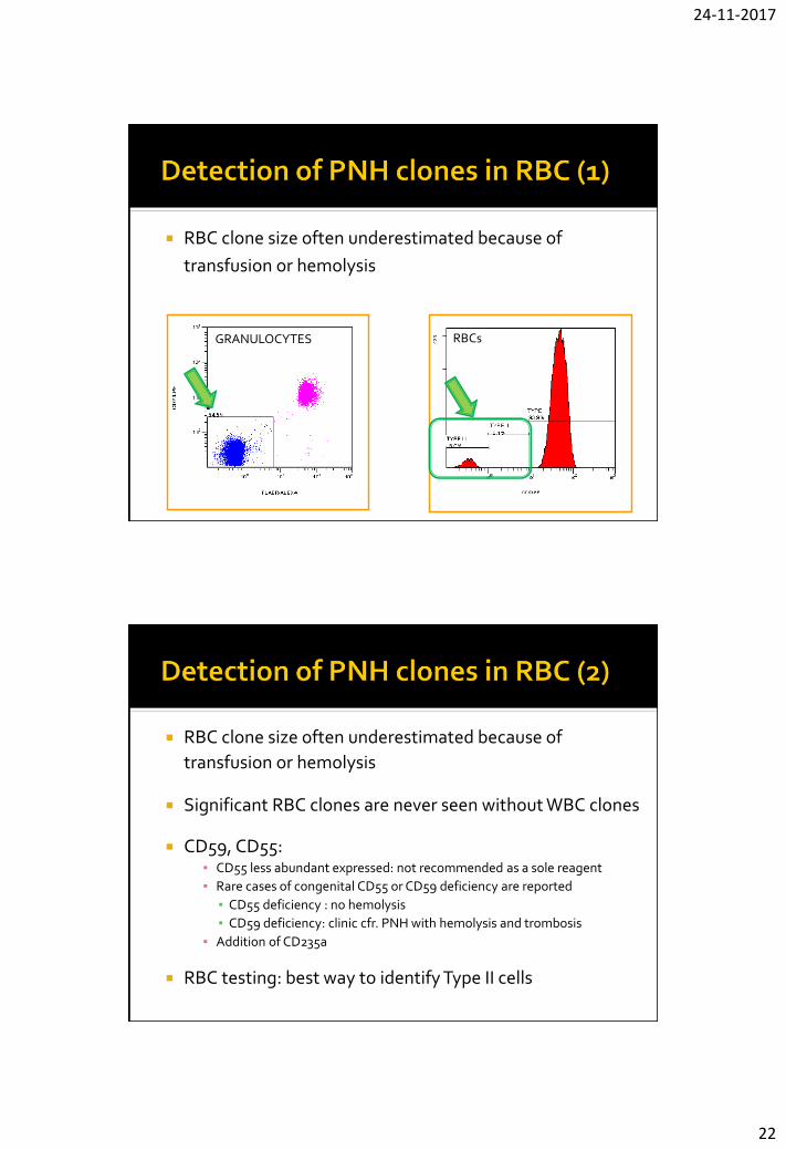
RBC clone size often underestimated because of
transfusion or hemolysis
Significant RBC clones are never seen without WBC clones
CD59, CD55: Identification and quantification of type I, II and III RBC
CD55 less abundant expressed: not recommended as a sole reagent
Rare cases of congenital CD55 or CD59 deficiency are reported
CD55 deficiency : no hemolysis
CD59 deficiency: clinic cfr. PNH with hemolysis and trombosis
Addition of CD235a
GRANULOCYTES RBCs
RBC clone size often underestimated because of
transfusion or hemolysis
Significant RBC clones are never seen without WBC clones
CD59, CD55: ▪ CD55 less abundant expressed: not recommended as a sole reagent
▪ Rare cases of congenital CD55 or CD59 deficiency are reported
▪ CD55 deficiency : no hemolysis
▪ CD59 deficiency: clinic cfr. PNH with hemolysis and trombosis
▪ Addition of CD235a
RBC testing: best way to identify Type II cells
24-11-2017
23
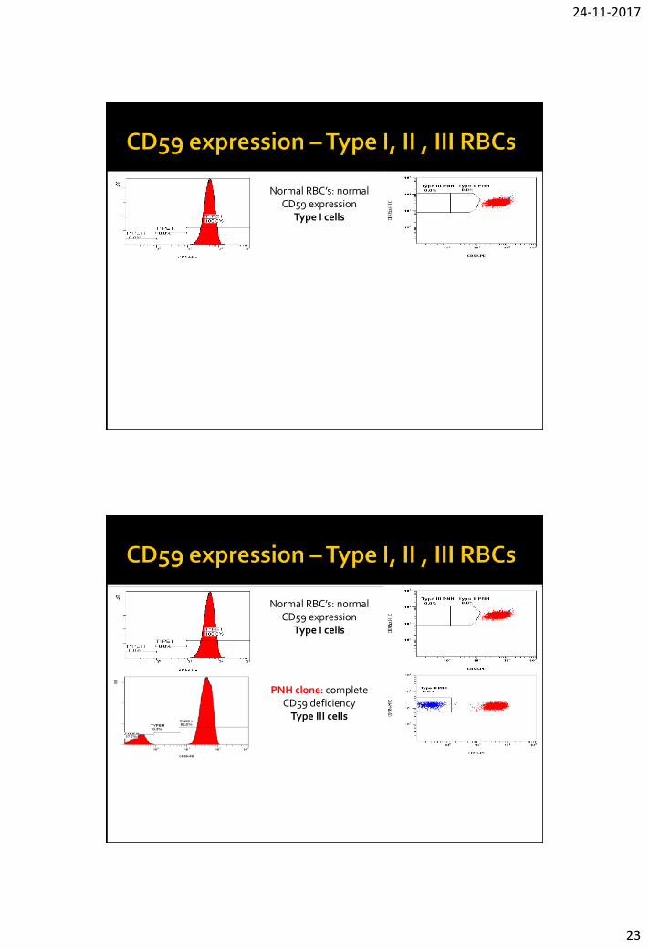
Normal RBC’s: normal CD59 expression
Type I cells
Normal RBC’s: normal CD59 expression
Type I cells
PNH clone: complete CD59 deficiency
Type III cells

24-11-2017
24
PNH clone: complete CD59 deficiency (Type III
cells) and partial CD59 deficiency (Type II cells)
Normal RBC’s: normal CD59 expression
Type I cells
PNH clone: complete CD59 deficiency
Type III cells
RBC testing has clinical significance and should be done in patients with a PNH WBC clone symptomatology is associated with size + type of RBC clone
proportion + type of cells: varies greatly among PNH patients ▪ Type I and type III: most common phenotype
▪ Type I, type II and type III: 2° most common phenotype
▪ Type I and type II: least common phenotype
low % of type III cells: only biochemical evidence of hemolysis may be observed
high % of type III RBCs: clinically apparent hemolysis
high % of type II RBCs but low % of type III cells: hemolysis may be modest
Parker CJ,Hematology Educ Book ASH, 2011; 21-29
24-11-2017
25
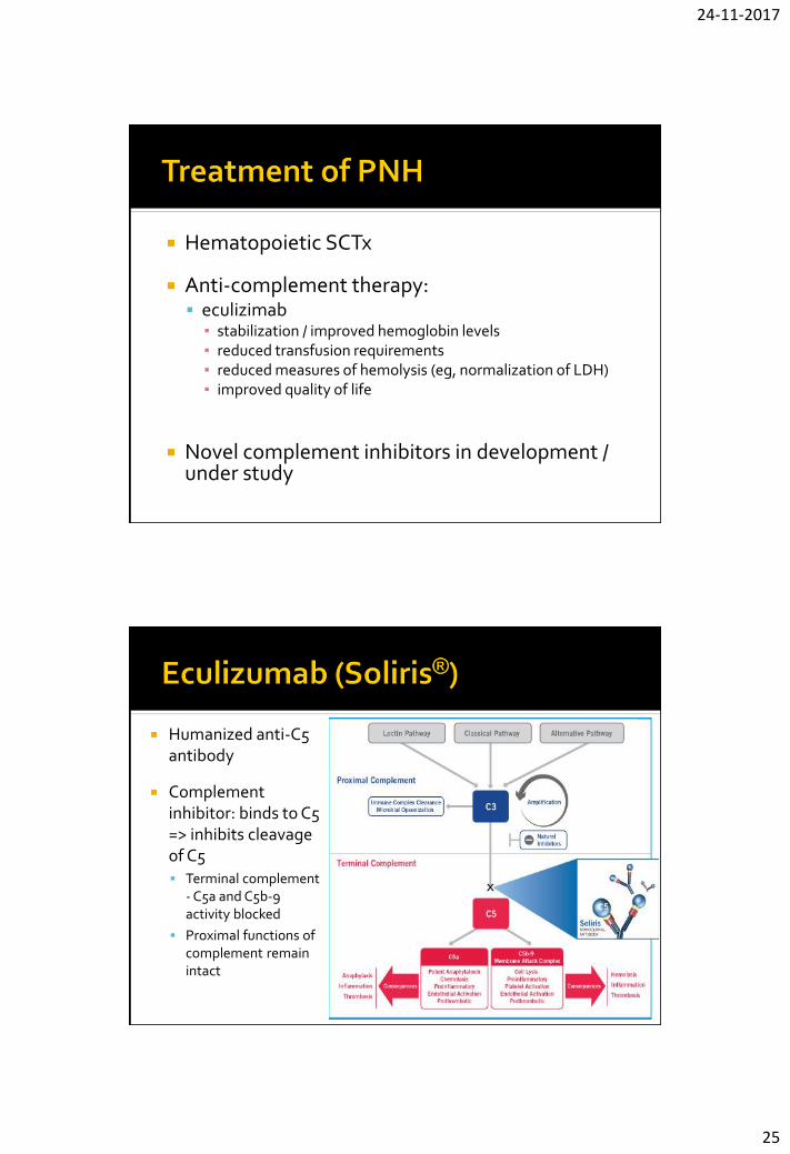
Hematopoietic SCTx
Anti-complement therapy: eculizimab
▪ stabilization / improved hemoglobin levels▪ reduced transfusion requirements ▪ reduced measures of hemolysis (eg, normalization of LDH)▪ improved quality of life
Novel complement inhibitors in development / under study
Humanized anti-C5 antibody
Complement inhibitor: binds to C5 => inhibits cleavage of C5 Terminal complement
- C5a and C5b-9 activity blocked
Proximal functions of complement remain intact

24-11-2017
26
5% PNH RBCs 19% PNH RBCs
48% PNH RBCs
Dahl-Chase Diagnostic Services; Hillmen P et al. N Engl J Med 2006;355:1233-43
92% PNH RBCs
Protection of PNH RBCs from complement-mediated lysis
Category Rate of vascular hemolysis
BM Flow cytometry
Benefit from eculizumab
Classic PNH Florid (LDH, often episodic macroscopic hemoglobinuria)
Cellular BM with erythroidhyperplasia, +/-normal morphology
Large population(>50%) GPI-anchor deficient PMN
Yes
PNH in BM failure disorder
Mild (often minimal abnormal biochemical markers of hemolysis)
Evidence of concomitant BM failure syndrome (AA or low-risk MDS)
% of GPI-anchor deficient PMNs is usually relatively small (<10%)
Typically no, but some patients (<10%) who have relatively large clones and clinically significant hemolysis, may benefit
Subclinical PNH
No clinical orbiochemical signs of hemolysis
Evidence of concomitant BM failure syndrome (AA or low-risk MDS)
Small (<1%) population of GPI-anchor deficient PMN
No
Parker CJ., Hematology ASH Education Book, 2011:21-9.
24-11-2017
27
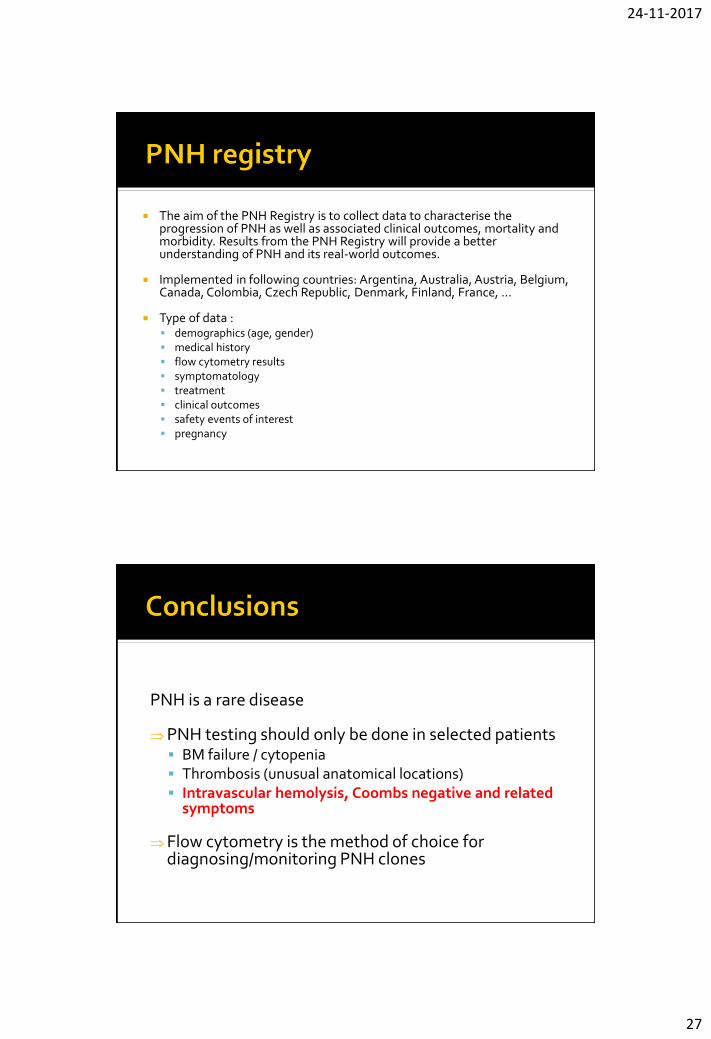
The aim of the PNH Registry is to collect data to characterise the progression of PNH as well as associated clinical outcomes, mortality and morbidity. Results from the PNH Registry will provide a better understanding of PNH and its real-world outcomes.
Implemented in following countries: Argentina, Australia, Austria, Belgium, Canada, Colombia, Czech Republic, Denmark, Finland, France, ...
Type of data : demographics (age, gender) medical history flow cytometry results symptomatology treatment clinical outcomes safety events of interest pregnancy
PNH is a rare disease
PNH testing should only be done in selected patients BM failure / cytopenia Thrombosis (unusual anatomical locations) Intravascular hemolysis, Coombs negative and related
symptoms
Flow cytometry is the method of choice fordiagnosing/monitoring PNH clones